dissection of long-range heart rate variability
TRANSCRIPT
DCMDC
MidpflScmma
pmda
CCDt
a
Journal of the American College of Cardiology Vol. 43, No. 12, 2004© 2004 by the American College of Cardiology Foundation ISSN 0735-1097/04/$30.00Published by Elsevier Inc. doi:10.1016/j.jacc.2004.01.050
Electrophysiology
issection of Long-Range Heart Rate Variabilityontrolled Induction of Prognosticeasures by Activity in the Laboratory
aniel Roach, PHD, Wendy Wilson, AART, Debbie Ritchie, MN, Robert Sheldon, MD, PHDalgary, Alberta, Canada
OBJECTIVES We sought to determine whether the long-range measures of heart rate variability (HRV)—the standard deviation of sequential 5-min heart period mean values (SDANN) and the heartperiod spectral amplitude in the ultra-low frequency band �0.0033 Hz (ULF)—had theirorigins partly in physical activity.
BACKGROUND The SDANN and ULF are prognostic HRV factors whose physiologic origins are obscure.Their discontinuous presence throughout the day suggested that they arise from changes inheart period due to activity.
METHODS Heart period sequences were recorded from 14 patients with left ventricular dysfunction and14 control subjects during an unrestricted 24-h day, 4-h supine rest, and 4-h epoch withscripted activities.
RESULTS The SDANN was higher during activity than during rest (74 � 23 ms vs. 43 � 17 ms, p �0.0001), as were ULF magnitudes (p � 0.0001). The increase in SDANN was due to specificactivities that contributed heavily (p � 0.0001 by analysis of variance); for example, a 10-minwalk and 90-min rest each contributed 22% of total SDANN. Patients with heart disease hada lower SDANN and ULF and a higher mean heart rate than control subjects during allrecordings. The proportional ranges in heart period were the same in the two groups duringcontrolled, scripted activities but were wider in control subjects than in patients duringambulatory recordings, suggesting decreased activity by patients.
CONCLUSIONS Activity increases SDANN by increasing the range of heart periods. Patients with diminishedventricular function have a reduced SDANN on ambulatory electrocardiograms, possibly andpartly because of a higher mean heart rate and reduced variations in physical activity. (J AmColl Cardiol 2004;43:2271–7) © 2004 by the American College of Cardiology Foundation
bwfSavafdap
M
EciHtdHswos
easures of long-range heart rate variability (HRV) aremportant prognostic predictors. They include the standardeviation of the mean values of successive 5-min hearteriod epochs (SDANN) and the power in the ultra-lowrequency band �0.0033 Hz (ULF). The SDANN corre-ates well with the log of the ULF power (1). Reductions inDANN and ULF predict poor survival for patients withhronic, severe mitral regurgitation (2), acute or recentyocardial infarction (3–5), and idiopathic dilated cardio-yopathy (6), as well as for 6,693 outpatients assessed for
rrhythmias (7).
See page 2278
Despite this, their physiologic origins are obscure. Hy-othesized causes include oscillatory and aperiodic, deter-inistic changes (8,9), as well as heart period fluctuations
ue to peripheral vasomotor, thermoregulatory, or renin-ngiotensin systems (10). We have shown in healthy am-
From the Cardiovascular Research Group, University of Calgary, Calgary, Alberta,anada. This study was supported by grants GR13914 from the Medical Researchouncil, Ottawa, Canada, and from the Calgary General Hospital Research andevelopment Committee, Calgary, Canada. Dr. Roach was a Postdoctoral Fellow of
he Heart and Stroke Foundation of Canada, Ottawa, Canada.Manuscript received July 17, 2003; revised manuscript received January 7, 2004,
sccepted January 12, 2004.
ulatory subjects that SDANN arises from epochs of timehen patients have local mean heart period values that differ
rom the 24-h mean heart period (i.e., mainly at night) (11).imilarly, ULF power occurs predominantly when patientsre changing their behavior from passive to active, and viceersa. We hypothesized that both SDANN and ULF do notrise from systemic oscillatory processes; rather, they ariserom changes in activity levels. Our goals were: 1) to directlyetermine the effect of changes in activity level on SDANNnd ULF; and 2) to determine the cause of reduced HRV inatients with left ventricular (LV) dysfunction.
ETHODS
xperimental and analytic approach. The subjects in-luded patients with poor LV function and healthy peoplen the same age range in order to increase the likely range of
RV and to allow a comparison between the two popula-ions. They underwent three recording sessions on separateays. The first was an unrestricted 24-h session to assessRV during usual daily activities. The second was a 4-h
ession of enforced bed rest to obtain recordings underakefulness but with a range of restricted activities. Anbserver was present to monitor activities and preventleeping. The third was a 4-h session during which the
ubjects followed a script with a range of normal daily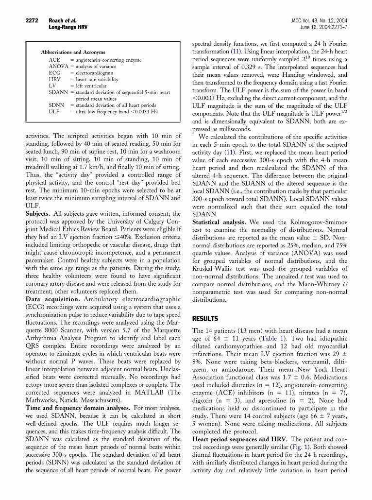
assvtTprlUSpjtimpwtctD(sflqAQowlsecMTwwqSsspt
stpsttt�Ucap
iavhaSl3wSStdnqfKncnd
R
Tadi8aAuedms5cHtdw
2272 Roach et al. JACC Vol. 43, No. 12, 2004Long-Range HRV June 16, 2004:2271–7
ctivities. The scripted activities began with 10 min oftanding, followed by 40 min of seated reading, 50 min foreated lunch, 90 min of supine rest, 10 min for a washroomisit, 10 min of sitting, 10 min of standing, 10 min ofreadmill walking at 1.7 km/h, and finally 10 min of sitting.hus, the “activity day” provided a controlled range ofhysical activity, and the control “rest day” provided bedest. The minimum 10-min epochs were selected to be ateast twice the minimum sampling interval of SDANN andLF.ubjects. All subjects gave written, informed consent; therotocol was approved by the University of Calgary Con-
oint Medical Ethics Review Board. Patients were eligible ifhey had an LV ejection fraction �40%. Exclusion criteriancluded limiting orthopedic or vascular disease, drugs that
ight cause chronotropic incompetence, and a permanentacemaker. Control healthy subjects were in a populationith the same age range as the patients. During the study,
hree healthy volunteers were found to have significantoronary artery disease and were released from the study forreatment; other volunteers replaced them.
ata acquisition. Ambulatory electrocardiographicECG) recordings were acquired using a system that uses aynchronization pulse to reduce variability due to tape speeductuations. The recordings were analyzed using the Mar-uette 8000 Scanner, with version 5.7 of the Marquetterrhythmia Analysis Program to identify and label eachRS complex. Entire recordings were analyzed by an
perator to eliminate cycles in which ventricular beats wereithout normal P waves. These beats were replaced by
inear interpolation between adjacent normal beats. Unclas-ified beats were corrected manually. No recordings hadctopy more severe than isolated complexes or couplets. Theorrected sequences were analyzed in MATLAB (The
athworks, Natick, Massachusetts).ime and frequency domain analyses. For most analyses,e used SDANN, because it can be calculated in shortell-defined epochs. The ULF requires much longer se-uences, and this makes time-frequency analysis difficult. TheDANN was calculated as the standard deviation of theequence of the mean heart periods of normal beats withinuccessive 300-s epochs. The standard deviation of all hearteriods (SDNN) was calculated as the standard deviation ofhe sequence of all heart periods of normal beats. For power
Abbreviations and AcronymsACE � angiotensin-converting enzymeANOVA � analysis of varianceECG � electrocardiogramHRV � heart rate variabilityLV � left ventricularSDANN � standard deviation of sequential 5-min heart
period mean valuesSDNN � standard deviation of all heart periodsULF � ultra-low frequency band �0.0033 Hz
a
pectral density functions, we first computed a 24-h Fourierransformation (11). Using linear interpolation, the 24-h hearteriod sequences were uniformly sampled 218 times using aample interval of 0.329 s. The interpolated sequences hadheir mean values removed, were Hanning windowed, andhen transformed to the frequency domain using a fast Fourierransform. The ULF power is the sum of the power in band
0.0033 Hz, excluding the direct current component, and theLF magnitude is the sum of the magnitude of the ULF
omponents. Note that the ULF magnitude is ULF power1/2
nd is dimensionally equivalent to SDANN; both are ex-ressed as milliseconds.We calculated the contributions of the specific activities
n each 5-min epoch to the total SDANN of the scriptedctivity day (11). First, we replaced the mean heart periodalue of each successive 300-s epoch with the 4-h meaneart period and then recalculated the SDANN of thisltered 4-h sequence. The difference between the originalDANN and the SDANN of the altered sequence is the
ocal SDANN (i.e., the contribution made by that particular00-s epoch toward total SDANN). Local SDANN valuesere normalized such that their sum equaled the totalDANN.tatistical analysis. We used the Kolmogorov-Smirnov
est to examine the normality of distributions. Normalistributions are reported as the mean value � SD. Non-ormal distributions are reported as 25%, median, and 75%uartile values. Analysis of variance (ANOVA) was usedor grouped variables of normal distributions, and theruskal-Wallis test was used for grouped variables ofon-normal distributions. The unpaired t test was used toompare normal distributions, and the Mann-Whitney Uonparametric test was used for comparing non-normalistributions.
ESULTS
he 14 patients (13 men) with heart disease had a meange of 64 � 11 years (Table 1). Two had idiopathicilated cardiomyopathies and 12 had old myocardial
nfarctions. Their mean LV ejection fraction was 29 �%. None were taking beta-blockers, verapamil, dilti-zem, or amiodarone. Their mean New York Heartssociation functional class was 1.7 � 0.6. Medicationssed included diuretics (n � 12), angiotensin-convertingnzyme (ACE) inhibitors (n � 11), nitrates (n � 7),igoxin (n � 3), and apresoline (n � 2). None hadedications held or discontinued to participate in the
tudy. There were 14 control subjects (age 66 � 7 years,women). None were taking medications. All subjects
ompleted the protocol.eart period sequences and HRV. The patient and con-
rol recordings were generally similar (Fig. 1). Both showediurnal fluctuations in heart period for the 24-h recordings,ith similarly distributed changes in heart period during the
ctivity day and relatively little variation in heart period
dt(hc
h0
s4
T
*
f
FRs
2273JACC Vol. 43, No. 12, 2004 Roach et al.June 16, 2004:2271–7 Long-Range HRV
uring the rest day. Patients had a lower mean heart periodhan did controls (Table 2) during each of the three sessionsp � 0.0002). The SDNN, the measure of total variation ineart period sequences, was lower in patients than inontrols in all three sessions (p � 0.0009 to 0.01) and was
able 1. Summary Characteristics of Control Subjects and Patien
Subject No.
Controls
Age (yrs) Gender Age (yrs)
1 51 F 392 60 F 493 60 M 504 61 F 575 63 M 626 64 M 677 64 M 698 66 M 709 66 M 70
10 67 M 7211 71 F 7312 72 F 7313 74 M 7314 79 M 78
Summary 66 � 7* 5 F/9 M 64 � 11*
Expressed as mean value � SD.CAD � coronary artery disease with old myocardial infarction; F � female; ID
raction; M � male; NYHA � New York Heart Association.
igure 1. Heart period recordings. (Top panels) Recordings from a 24-h decordings from a 4-h scripted activity day from the same subjects. (Botto
tandard deviation of sequential 5-min heart period mean values. ULFmag � U
igher on scripted activity days than scripted rest days (p �. 0013 to 0.0036).In the total population, the SDANN was higher during
cripted activity days than during rest days (74 � 23 ms vs.3 � 17 ms, p � 0.0001). This effect was noted separately
Patients
Gender Etiology LVEF (%) NYHA Class
M CAD 19 IM IDCM 12 IM CAD 35 IM CAD 36 IIF CAD 25 IIM CAD 24 IIM CAD 28 IIM CAD 37 IIM IDCM 37 IIIM CAD 38 IM CAD 27 IIM CAD 20 IIM CAD 28 IM CAD 30 II
1 F/13 M 29 � 8* 1.7 � 0.6*
idiopathic nonischemic dilated cardiomyopathy; LVEF � left ventricular ejection
m a control subject (left) and heart failure patient (right). (Middle panels)nels) Recordings from a 4-h rest day from the same subjects. SDANN �
ts
CM �
ay from pa
LF magnitude.
fa0h�s�pcissaeCdaltd(A1Sec01mcS
SWSdi�p5
and �
Fsap
2274 Roach et al. JACC Vol. 43, No. 12, 2004Long-Range HRV June 16, 2004:2271–7
or both patients (63 � 25 ms vs. 37 � 19 ms, p � 0.005)nd control subjects (84 � 14 ms vs. 50 � 12 ms, p �.0001). In the total population, the ULF magnitude wasigher during scripted activity days than during rest days (71
29 ms vs. 38 � 14 ms, p � 0.0001). This effect was notedeparately for both patients (63 � 34 ms vs. 34 � 15 ms, p
0.0067) and control subjects (80 � 22 ms vs. 42 � 13 ms,� 0.0001). Finally, the SDANN and ULF magnitude
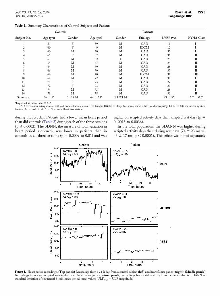
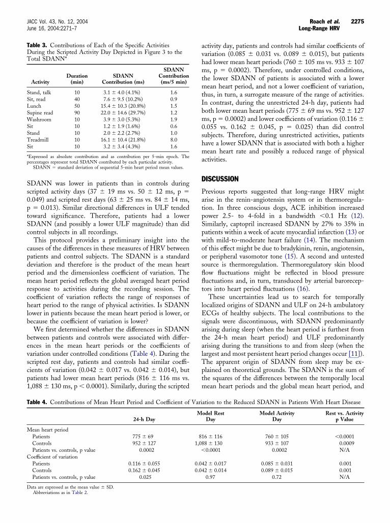
orrelated well with each other within each type of record-ng session (24-h day, R2 � 0.92; scripted rest, R2 � 0.60;cripted activity, R2 � 0.89; p � 0.0001 for each). Inummary, scripted physical activities increase the SDANNnd ULF magnitude more than that which occurs duringnforced rest.ontribution of specific activities to SDANN. We pre-icted that the induction of specific activities would beccompanied temporally by changes in SDANN. Numerousocal contributions to SDANN were scattered throughouthe recording period, particularly at times of maximal localeviation of the heart period from the global mean valueFig. 2). Specific events contributed heavily (p � 0.0001 byNOVA) to total SDANN (Table 3). For example, a0-min walk and 90-min rest each contributed 22% of totalDANN. Although the mean contribution per 5-minpoch was 1.61 ms, the intensity of the localized SDANNontribution per 5-min epoch was highly localized (p �.0001 by ANOVA). For example, a 90-min supine rest and0-min treadmill walk contributed 1.2 ms/5 min and 8.0s/5 min, respectively. Thus, specific physical activities
ontribute localized and idiosyncratic proportions of overall
Table 2. Heart Period Sequences and Heart RRecording Periods*
24-h Day
Mean heart period (ms)Total subjects 867 � 135Patients‡ 775 � 69Controls 952 � 127 1Patients vs. controls, p value 0.0002
SDNN (ms)Total subjects 140 � 61Patients‡ 102 � 50Controls 175 � 51Patients vs. controls, p value 0.0009
SDANN (ms)Total subjects 125 � 56Patients‡ 92 � 47Controls 154 � 47Patients vs. controls, p value 0.002
ULF magnitude (ms)Total subjects 123 � 59Patients 93 � 51Controls 154 � 52Patients vs. controls, p value 0.0047
*As depicted in Figure 1. ‡Patients with reduced ventricularversus controls, and paired t tests were used to compare rest
N/A � not applicable; SDANN � standard deviation odeviation of all heart periods; ULF � ultra-low-frequency b
DANN. 5
ources of reduced heart period variability in patients.e wished to determine whether (and why) induced
DANN and ULF were lower in the patients with heartisease (Table 2). The SDANN was lower in patients thann controls during their daily activities (92 � 47 ms vs. 154
47 ms, p � 0.002), and the ULF magnitude was lower inatients than in controls during their daily activities (93 �1 ms vs. 154 � 52 ms, p � 0.0047). Similarly, the
ariability During the Three
ptedDay
Scripted ActivityDay
Rest vs. Activityp Value
� 184 847 � 137 �0.0001� 116 760 � 105 �0.0001� 130 933 � 107 0.0009001 0.0002 N/A
� 28 93 � 34 �0.0001� 28 81 � 43 0.0036� 20 105 � 17 0.001310 0.0065 N/A
� 17 74 � 23 �0.0001� 19 63 � 25 �0.005� 12 84 � 14 �0.000149 0.013 N/A
� 14 71 � 29 �0.0001� 15 63 � 34 0.0067� 13 80 � 22 �0.000113 0.066 N/A
ic function. Unpaired t tests were used to compare patientsactivity. Data are presented as the mean value � SD.
ential 5-min heart period mean values; SDNN � standard0.0033.
igure 2. Temporally localized contributions to standard deviation ofequential 5-min heart period mean values (SDANN) due to scriptedctivities. (Top panel) Heart period recordings from a control subject andatient during a scripted activity day. (Bottom panel) Contribution per
ate V
ScriRest
952816,088�0.0
6855810.0
4337500.0
383442
0.
systolversus
f sequ
-min epoch of each activity in each subject to overall SDANN.
Ss0ptSc
cpdpmrchlb
bevscp1
avhmtmtIbm0shma
D
PatpSpwoosflflt
lEsatalTptm
TDT
SSLSWSSTS
*p
T
M
C
D
2275JACC Vol. 43, No. 12, 2004 Roach et al.June 16, 2004:2271–7 Long-Range HRV
DANN was lower in patients than in controls duringcripted activity days (37 � 19 ms vs. 50 � 12 ms, p �.049) and scripted rest days (63 � 25 ms vs. 84 � 14 ms,� 0.013). Similar directional differences in ULF tended
oward significance. Therefore, patients had a lowerDANN (and possibly a lower ULF magnitude) than didontrol subjects in all recordings.
This protocol provides a preliminary insight into theauses of the differences in these measures of HRV betweenatients and control subjects. The SDANN is a standardeviation and therefore is the product of the mean hearteriod and the dimensionless coefficient of variation. Theean heart period reflects the global averaged heart period
esponse to activities during the recording session. Theoefficient of variation reflects the range of responses ofeart period to the range of physical activities. Is SDANN
ower in patients because the mean heart period is lower, orecause the coefficient of variation is lower?We first determined whether the differences in SDANN
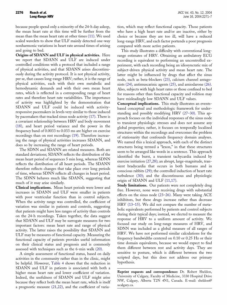
etween patients and controls were associated with differ-nces in the mean heart periods or the coefficients ofariation under controlled conditions (Table 4). During thecripted rest day, patients and controls had similar coeffi-ients of variation (0.042 � 0.017 vs. 0.042 � 0.014), butatients had lower mean heart periods (816 � 116 ms vs.,088 � 130 ms, p � 0.0001). Similarly, during the scripted
able 3. Contributions of Each of the Specific Activitiesuring the Scripted Activity Day Depicted in Figure 3 to theotal SDANN*
ActivityDuration
(min)SDANN
Contribution (ms)
SDANNContribution
(ms/5 min)
tand, talk 10 3.1 � 4.0 (4.1%) 1.6it, read 40 7.6 � 9.5 (10.2%) 0.9unch 50 15.4 � 10.3 (20.8%) 1.5upine read 90 22.0 � 14.6 (29.7%) 1.2ashroom 10 3.9 � 3.0 (5.3%) 1.9
it 10 1.2 � 1.9 (1.6%) 0.6tand 10 2.0 � 2.2 (2.7%) 1.0readmill 10 16.1 � 10.4 (21.8%) 8.0it 10 3.2 � 3.4 (4.3%) 1.6
Expressed as absolute contribution and as contribution per 5-min epoch. Theercentages represent total SDANN contributed by each particular activity.
SDANN � standard deviation of sequential 5-min heart period mean values.
able 4. Contributions of Mean Heart Period and Coefficient of
24-h Day
ean heart periodPatients 775 � 69Controls 952 � 127Patients vs. controls, p value 0.0002
oefficient of variationPatients 0.116 � 0.055Controls 0.162 � 0.045Patients vs. controls, p value 0.025
ata are expressed as the mean value � SD.
Abbreviations as in Table 2.ctivity day, patients and controls had similar coefficients ofariation (0.085 � 0.031 vs. 0.089 � 0.015), but patientsad lower mean heart periods (760 � 105 ms vs. 933 � 107s, p � 0.0002). Therefore, under controlled conditions,
he lower SDANN of patients is associated with a lowerean heart period, and not a lower coefficient of variation,
hus, in turn, a surrogate measure of the range of activities.n contrast, during the unrestricted 24-h day, patients hadoth lower mean heart periods (775 � 69 ms vs. 952 � 127s, p � 0.0002) and lower coefficients of variation (0.116 �
.055 vs. 0.162 � 0.045, p � 0.025) than did controlubjects. Therefore, during unrestricted activities, patientsave a lower SDANN that is associated with both a higherean heart rate and possibly a reduced range of physical
ctivities.
ISCUSSION
revious reports suggested that long-range HRV mightrise in the renin-angiotensin system or in thermoregula-ion. In three conscious dogs, ACE inhibition increasedower 2.5- to 4-fold in a bandwidth �0.1 Hz (12).imilarly, captopril increased SDANN by 27% to 35% inatients within a week of acute myocardial infarction (13) orith mild-to-moderate heart failure (14). The mechanismf this effect might be due to bradykinin, renin, angiotensin,r peripheral vasomotor tone (15). A second and untestedource is thermoregulation. Thermoregulatory skin bloodow fluctuations might be reflected in blood pressureuctuations and, in turn, transduced by arterial barorecep-ors into heart period fluctuations (16).
These uncertainties lead us to search for temporallyocalized origins of SDANN and ULF on 24-h ambulatoryCGs of healthy subjects. The local contributions to the
ignals were discontinuous, with SDANN predominantlyrising during sleep (when the heart period is furthest fromhe 24-h mean heart period) and ULF predominantlyrising during the transitions to and from sleep (when theargest and most persistent heart period changes occur [11]).he apparent origin of SDANN from sleep may be ex-lained on theoretical grounds. The SDANN is the sum ofhe squares of the differences between the temporally localean heart periods and the global mean heart period, and
ation to the Reduced SDANN in Patients With Heart Disease
odel RestDay
Model ActivityDay
Rest vs. Activityp Value
6 � 116 760 � 105 �0.00018 � 130 933 � 107 0.00090.0001 0.0002 N/A
2 � 0.017 0.085 � 0.031 0.0012 � 0.014 0.089 � 0.015 0.0010.97 0.72 N/A
Vari
M
811,08
�
0.040.04
btmsnaOwcoopphrroSrba(frid
smrtoTmCiwWvtotiaUfoa
abShIba
twclc
rrpslnnAfhCbsptgsoWssiesctoSfeiHbdrfSHfttssh
RUNu
2276 Roach et al. JACC Vol. 43, No. 12, 2004Long-Range HRV June 16, 2004:2271–7
ecause people spend only a minority of the 24-h day asleep,he mean heart rate at this time will be further from theean than the mean heart rate at other times (11). We used
caled wavelets to show that ULF mainly detected one-wayonharmonic variations in heart rate around times of arisingnd going to bed.
rigins of SDANN and ULF in physical activities. Heree report that SDANN and ULF are induced under
ontrolled conditions with a protocol that included a rangef physical activities, and that SDANN arises discontinu-usly during the activity protocol. It is not physical activity,er se, that causes long-range HRV; rather, it is the range ofhysical activities, each with their own metabolic andemodynamic demands and with their own mean heartates, which is reflected in a corresponding range of heartates and therefore heart rate variabilities. The importancef activity was highlighted by the demonstration thatDANN and ULF could be induced with activity-esponsive pacemakers in levels very similar to those inducedy pacemakers that tracked sinus node activity (17). There iscovariant relationship between HRV and body movement
18), and heart period variance and the power in therequency band of 0.0033 to 0.033 ms are higher on exerciseecordings than on rest recordings (19). Therefore increas-ng the range of physical activities increases SDANN, andoes so by increasing the range of heart periods.The SDNN and SDANN are related measures. Both are
tandard deviations; SDANN reflects the distribution of theean heart period of sequences 5 min long, whereas SDNN
eflects the distribution of all heart periods. The SDANNherefore reflects changes that take place over long periodsf time, whereas SDNN reflects all changes in heart period.he SDNN behaves much like SDANN, suggesting thatuch of it may arise similarly.linical implications. Mean heart periods were lower and
ncreases in SDANN and ULF were smaller in patientsith poor ventricular function than in control subjects.hen the activity range was controlled, the coefficient of
ariation was similar in patients and controls, suggestinghat patients might have less ranges of activity than controlsn the 24-h recordings. Taken together, the data suggesthat SDANN and ULF may be surrogate measures for twomportant factors: mean heart rate and range of physicalctivity. The latter raises the possibility that SDANN andLF may be measures of functional capacity. Measuring the
unctional capacity of patients provides useful informationn their clinical status and prognosis and is commonlyssessed with techniques such as the 6-min walk (20).
A simple assessment of functional status, based on dailyctivities in the community rather than in the clinic, mighte helpful. However, Table 4 shows that the reduction inDANN and ULF in patients is associated with both aigher mean heart rate and lower coefficient of variation.ndeed, the usefulness of SDANN and ULF might ariseecause they reflect both the mean heart rate, which is itself
prognostic measure (21,22), and the coefficient of varia-ion, which may reflect functional capacity. Those patientsho have a high heart rate and/or are inactive, either by
hoice or because they are too ill, will have a reducedong-range HRV, and each factor portends a poor prognosisompared with more active patients.
This study illustrates a difficulty with conventional long-ange estimates of HRV. Obtaining an ambulatory ECGecording is equivalent to performing an uncontrolled ex-eriment, with each recording being an idiosyncratic mix ofubject-driven physical activity and mean heart rate. Theatter might be influenced by drugs that affect the sinusode, such as beta-blockers (23), calcium channel antago-ists (24), antimuscarinic agents (25), and amiodarone (26).lso, subjects with high heart rates or those confined to bed
or reasons other than functional capacity and volition mayave misleadingly low SDANN and ULF values.onceptual implications. This study illustrates an events-ased conceptual and methodologic framework for under-tanding and possibly modifying HRV (27–30). This ap-roach focuses on the individual responses of the sinus nodeo transient physiologic stresses and does not assume anylobal properties; rather, it focuses on temporally localizedtructures within the recordings and overcomes the problemf stationarity that confounds frequency domain analyses.
e named this a lexical approach, with each of the distincttructures being termed a “lexon,” in that these structureseem to be arranged like words in a sentence. This approachdentified the burst, a transient tachycardia induced byxercise initiation (27,28); an abrupt, large-magnitude, tran-ient bradycardia that occurs in both humans and inonscious rabbits (29); the controlled induction of heart rateurbulence (30); and the discontinuous and physiologicrigin of SDANN and ULF (11,17).tudy limitations. Our patients were not completely drug
ree. However, none were receiving drugs with substantialffects on the sinus node (23–26). Many were taking ACEnhibitors, but these drugs increase rather than decrease
RV (13–15). We did not compare the number of meta-olic equivalents performed by patients and control subjectsuring their typical days; instead, we elected to measure theesponse of HRV to a uniform amount of activity. Weocused our study on long-range measures of HRV, andDNN was included as a global measure of all ranges ofRV. We have not performed similar calculations for the
requency bandwidths centered on 0.10 or 0.25 Hz or theirime domain equivalents, because we would expect to findhem different between rest and activity days. They areensitive to posture, which is different between the twocripted days, but this does not address our primaryypothesis.
eprint requests and correspondence: Dr. Robert Sheldon,niversity of Calgary, Faculty of Medicine, 3330 Hospital DriveW, Calgary, Alberta T2N 4N1, Canada. E-mail: sheldon@
calgary.ca.
R
1
1
1
1
1
1
1
1
1
1
2
2
2
2
2
2
2
2
2
2
3
2277JACC Vol. 43, No. 12, 2004 Roach et al.June 16, 2004:2271–7 Long-Range HRV
EFERENCES
1. Bigger JT, Fleiss JL, Steinman RC, Rolnitzky LM, Schneider WJ,Stein PK. RR variability in healthy, middle-aged persons comparedwith patients with chronic coronary heart disease or recent acutemyocardial infarction. Circulation 1995;91:1936–43.
2. Stein KM, Borer JS, Hochreiter C, et al. Prognostic value andphysiologic correlates of heart rate variability in chronic severe mitralregurgitation. Circulation 1993;88:127–35.
3. Casolo GC, Stroder P, Signori C, et al. Heart rate variability duringthe acute phase of acute myocardial infarction. Circulation 1992;85:2073–9.
4. Vaishnav S, Stevenson R, Marchant B, Lagi K, Ranjadayalan K,Timmis A. Relation between heart rate variability early after acutemyocardial infarction and long-term mortality. Am J Cardiol 1994;73:653–7.
5. Bigger JT, Fleiss JL, Steinman RC, Rolnitzky LM, Kleiger RE,Rottman JN. Frequency domain measures of heart period variabilityand mortality after myocardial infarction. Circulation 1992;85:164 –71.
6. Fauchier L, Babuty D, Cosnay P, Autret ML, Fauchier JP. Heart ratevariability in idiopathic dilated cardiomyopathy: characteristics andprognostic value. J Am Coll Cardiol 1997;30:1009–14.
7. Algra A, Tijssen JGP, Roelandt JRTC, Pool J, Lubsen J. Heart ratevariability from 24-hour electrocardiography and the 2-year risk forsudden death. Circulation 1993;88:180–5.
8. Persson PB, Baumann JE, Ehmke H, Nafz B, Wittman U, KirchheimHR. Phasic and 24-h blood pressure control by endothelium-derivedrelaxing factor in conscious dogs. Am J Physiol 1992;262:H1395–400.
9. Broten TP, Zehr JE. Autonomic modulation of ultradian bloodpressure and heart rate oscillations in dogs. Am J Physiol 1989;256:R1127–37.
0. The Task Force of the European Society of Cardiology and the NorthAmerican Society of Pacing and Electrophysiology. Heart rate vari-ability: standards of measurement, physiological interpretation, andclinical use. Circulation 1996;93:1043–65.
1. Roach D, Sheldon A, Wilson W, Sheldon R. Temporally localizedcontributions to measures of large-scale heart period variability. Am JPhysiol 1998;274:H1465–71.
2. Akselrod S, Gordon D, Ubel FA, Shannon DC, Barger AC, CohenRJ. Power spectrum analysis of heart rate fluctuation: a quantitativeprobe of beat-to-beat cardiovascular control. Science 1981;213:220–2.
3. Bonaduce D, Marciano F, Petretta M, et al. Effects of convertingenzyme inhibition of heart period variability in patients with acutemyocardial infarction. Circulation 1994;90:108–13.
4. Zhang YH, Song YC, Zhu J, Hu TH, Wan LL. Effects of enalapril onheart rate variability in patients with congestive heart failure. Am J
Cardiol 1995;76:1045–8.5. Taylor JA, Carr DL, Myers CW, Eckberg DL. Mechanisms under-lying very-low-frequency RR-interval oscillations in humans. Circula-tion 1998;98:547–55.
6. Kitney R. An analysis of the thermoregulatory influences on heart-ratevariability. In: Kitney RI, Rompelman O, editors. The Study ofHeart-Rate Variability. Oxford, England: Clarendon Press, 1980.
7. Raj SR, Roach D, Koshman ML, Sheldon RS. Activity-responsivepacing produces long-term heart rate variability. J Cardiovasc Electro-physiol 2004;15:179–83.
8. Aoyagi N, Ohashi K, Tomono S, Yamamoto Y. Temporal contribu-tion of body movement to very long-term heart rate variability inhumans. Am J Physiol 2000;278:H1035–41.
9. Bernardi L, Valle F, Coco M, Calciati A, Sleight P. Physical activityinfluences heart rate variability and very low frequency components inHolter electrocardiograms. Cardiovasc Res 1996;32:234–7.
0. Guyatt GH, Sullivan MJ, Thompson PJ, et al. The six minute walk—anew measure of exercise capacity in patients with chronic heart failure.Can Med Assoc J 1985;132:919–23.
1. Kristal-Boneh E, Silber H, Harari G, Froom P. The association ofresting heart rate with cardiovascular, cancer and all-cause mortality.Eur Heart J 2000;21:116–24.
2. Nolan J, Bastin PD, Andrews R, et al. Prospective study of the UnitedKingdom Heart Failure Evaluation and Assessment of Risk Trial(UK-Heart). Circulation 1998;98:1510–6.
3. Niemela MJ, Airaksinen KEJ, Huikuri HV. Effect of beta-blockade onheart rate variability in patients with coronary artery disease. J Am CollCardiol 1994;23:1370–1.
4. Pinar E, Garca-Alberola A, Llamas C, et al. Effects of verapamil onindexes of heart rate variability after acute myocardial infarction. Am JCardiol 1998;81:1085–9.
5. Pedretti RFE, Colombo E, Braga SS, Ballardini L, Caru B. Effects oforal pirenzepine on heart rate variability and baroreceptor reflex afteracute myocardial infarction. J Am Coll Cardiol 1995;25:915–21.
6. Rohde LEP, Polanczyk CA, Moraes R, Ferlin E, Ribeiro JP. Effect ofpartial arrhythmia suppression with amiodarone on heart ratevariability of patient with congestive heart failure. Am Heart J1998;136:31–6.
7. Roach D, Malik P, Koshman ML, Sheldon RS. Origins of heart ratevariability: inducibility and prevalence of a discrete, tachycardic event.Circulation 1999;99:3279–85.
8. Roach D, Haennel R, Koshman ML, Sheldon RS. Origins of heartrate variability: relationship of heart rate burst morphology to workduration and load. Am J Physiol 1999;277:H1491–7.
9. Roach D, Thakore E, Sheldon RS. Large magnitude, transient,bradycardic events in rabbits. Am J Physiol 1999;277:R243–9.
0. Roach D, Koshman ML, Duff HD, Sheldon RS. Induction of heartrate and blood pressure turbulence in the electrophysiologic laboratory.
Am J Cardiol 2002;90:1098–102.