genetic dissection of marfan syndrome and related
TRANSCRIPT
Fax +41 61 306 12 34E-Mail [email protected]
Review Article
Mol Syndromol 2012;3:47–58 DOI: 10.1159/000339441
Genetic Dissection of Marfan Syndrome and Related Connective Tissue Disorders: An Update 2012
S. Hoffjan
Department of Human Genetics, Ruhr-University, Bochum , Germany
Marfan syndrome (MFS), first described by Antoine Marfan in 1896, is an autosomal dominant disorder of the connective tissue with a prevalence of approximately 1: 5,000. Characteristic manifestations of MFS include involvement of the cardiovascular, ocular and skeletal systems, while lung, skin and dura are less frequently af-fected [Cañadas et al., 2010]. A key cardiovascular symp-tom of MFS is the development of an aneurysm or dissection of the thoracic aorta, especially in younger pa-tients ( ! 50 years of age). Additional to this life-threaten-ing cardiovascular feature, ectopia lentis and a so-called marfanoid habitus with long and thin extremities, arach-nodactyly, thorax deformities, scoliosis, joint hypermo-bility, and flat feet are considered to be cardinal symp-toms of MFS [Cañadas et al., 2010]. Therapeutic options for MFS generally include regular (e.g. annual) echocar-diographic evaluations, surgical repair of the aorta when the maximal measurement approaches certain thresh-olds as well as regular orthopaedic and ophthalmologic surveillance. So far, the only established pharmacological therapy is the application of � -receptor antagonists which has proven to reduce aneurysm progression by lowering the blood pressure and thus, reducing physical stress to the aortic wall [Hartog et al., 2012]. However, new thera-peutic strategies based on recent pathogenetic findings are currently under development (see below).
Key Words
Connective tissue disorders � Ehlers-Danlos syndrome � Loeys-Dietz syndrome � Marfan syndrome
Abstract
Marfan syndrome (MFS) is an autosomal dominant disorder of the connective tissue characterized by early development of thoracic aortic aneurysms/dissections together with symptoms of the ocular and skeletal systems. While most pa-tients/families with a classic phenotypic expression of MFS harbour mutations in the gene encoding fibrillin-1 (FBN1) , genetic studies of the recent years revealed that the clinical features, as well as the mutated genes, show a high degree of overlap between MFS and other connective tissue diseas-es (e.g. Loeys-Dietz syndrome, Ehlers-Danlos syndrome, fa-milial thoracic aneurysms and dissections and others). We summarize herein the current knowledge about the wide spectrum of differential diagnoses and their genetic back-ground as well as novel therapeutic approaches in order to provide appropriate counselling and clinical follow-up for the patients. Copyright © 2012 S. Karger AG, Basel
Accepted: May 7, 2012 by M. Schmid Published online: June 12, 2012
Sabine Hoffjan Department of Human Genetics, Ruhr-University Universitätsstrasse 150 DE–44801 Bochum (Germany) Tel. +49 234 322 3823, E-Mail sabine.hoffjan @ ruhr-uni-bochum.de
© 2012 S. Karger AG, Basel1661–8769/12/0032–0047$38.00/0
Accessible online at:www.karger.com/msy
Hoffjan Mol Syndromol 2012;3:47–5848
In 1991, mutations in the gene encoding fibrillin 1 (FBN1) on chromosome 15q were identified as causative for MFS [Dietz et al., 1991]. While it still holds true that most patients/families with a complete classical pheno-typic expression of MFS harbour FBN1 mutations, genet-ic studies of the recent years clearly emphasized that the clinical features, as well as the mutated genes, show a high degree of overlap between MFS and other connective tis-sue diseases (e.g. Loeys-Dietz syndrome (LDS), Ehlers-
Danlos syndrome (EDS), familial thoracic aneurysms and dissections (fTAAD), and others) [von Kodolitsch et al., 2010]. Thus, correctly establishing the diagnosis of MFS or a related connective tissue disease may be chal-lenging in clinical practice and should incorporate knowledge about the wide spectrum of differential diag-noses and their genetic background in order to provide appropriate counselling and clinical follow-up for the pa-tients.
Marfan Syndrome
Diagnostic Criteria Additional to the cardinal cardiovascular, ocular and
skeletal symptoms of MFS presented above, Marfan pa-tients often show high myopia, retinal detachment, mi-tral valve prolapse, distinct facial features (including dol-ichocephaly, high-arched palate, down-slanting palpe-bral fissures, and micrognathia), striae in the skin that do not result from extensive weight loss or pregnancy, and spontaneous pneumothorax [Cañadas et al., 2010]. Typ-ical radiologic signs further include dural ectasia and protrusio acetabuli. Yet, the phenotypic expression can be very variable between patients, even within the same family. To facilitate the clinical diagnosis of MFS, the Ghent diagnostic criteria were established in 1991, distin-guishing between major and minor symptoms in the in-volved organ systems [De Paepe et al., 1996]. These crite-ria were recently revised in order to better incorporate the variable clinical expression and the extended differential diagnosis [Loeys et al., 2010a]. In the revised Ghent nosol-ogy, summarized in table 1 , more weight is laid on the cardinal clinical features aortic root dilation/dissection and ectopia lentis. Further, it highlights the impact of ge-netic analysis since a pathogenic FBN1 mutation consti-tutes an important criterion for the diagnosis of MFS: for example, the combination of an aortic aneurysm or dis-section and a pathogenic FBN1 mutation is now sufficient to establish the diagnosis of MFS. On the other hand, all other cardiovascular and ocular manifestations, as well as symptoms in other organ systems (skeleton, dura, skin, and lung), were included in a systemic score ( table 1 ). A systemic score 6 7 in combination with an aortic aneu-rysm/dissection or a positive family history also consti-tutes the diagnosis of MFS. Some of the less specific symptoms for MFS were either removed (e.g. joint hyper-mobility) or are less emphasized in the diagnostic evalu-ation (e.g. dural ectasia). Finally, the new nosology also allows the discrimination of several phenotypes closely
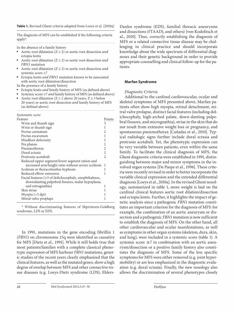
Table 1. Revised Ghent criteria adapted from Loeys et al. [2010a]
The diagnosis of MFS can be established if the following criteria applya:
In the absence of a family historyU Aortic root dilatation (Z ≥ 2) or aortic root dissection and
ectopia lentisU Aortic root dilatation (Z ≥ 2) or aortic root dissection and
FBN1 mutationU Aortic root dilatation (Z ≥ 2) or aortic root dissection and
systemic score ≥7U Ectopia lentis and FBN1 mutation known to be associated
with aortic root dilatation/dissectionIn the presence of a family historyU Ectopia lentis and family history of MFS (as defined above)U Systemic score ≥7 and family history of MFS (as defined above)U Aortic root dilatation (Z ≥ 2 above 20 years, Z ≥ 3 below
20 years) or aortic root dissection and family history of MFS (as defined above)
Systematic scoreFeature Points
Wrist and thumb sign 3Wrist or thumb sign 1Pectus carinatum 2Pectus excavatum 1Hindfoot deformity 2Pes planus 1Pneumothorax 2Dural ectasia 2Protrusio acetabuli 2Reduced upper segment/lower segment ration and
increased arm/height ratio without severe scoliosis 1Scoliosis or thoracolumbar kyphosis 1Reduced elbow extension 1Facial features (≥3 of dolichocephaly, enophthalmos,
downslanting palpebral fissures, malar hypoplasia,and retrognathia) 1
Skin striae 1Myopia (>3 dpt) 1Mitral valve proplaps 1
a Without discriminating features of Shprintzen-Goldberg syndrome, LDS or EDS.
Genetics of Marfan Syndrome Mol Syndromol 2012;3:47–58 49
related to MFS [Loeys et al., 2010a]. Ectopia lentis syn-drome is proposed, irrespective of the systemic score, for patients with ectopia lentis but no aortic aneurysm/dis-section that do not harbour an FBN1 mutation or carry an FBN1 mutation not known to be associated with aortic involvement. MASS syndrome (myopia, mitral valve pro-lapse, borderline and nonprogressive aortic root dilata-tion, skeletal findings, and striae) is defined by a mild aortic dilation (Z score ! 2) and a systemic score 6 5 with-out ectopia lentis, and mitral valve prolapse syndrome includes patients with mitral valve prolapse, mild aortic dilation and a systemic score 1 5 without ectopia lentis. As with the former Ghent criteria, establishing the cor-rect diagnosis in children and adolescents may be diffi-cult since some symptoms, especially aortic dilation, may evolve over time. For children with a systemic score ! 7 and borderline aortic root measurements, the term ‘non-specific connective tissue disorder’ is proposed, whereas for children with a known FBN1 mutation but only with borderline aortic root dilation, ‘potential MFS’ is suggest-ed [Loeys et al., 2010a]. Taken together, the authors con-clude that the revised nosology may delay a definitive di-agnosis of MFS in some patients but overall reduce the risk for premature diagnosis or misdiagnosis [Loeys et al., 2010a].
Several studies recently aimed at comparing the old and revised Ghent criteria in patient groups with suspect-ed MFS and a known FBN1 mutation. Overall, the level of agreement between both nosologies was high: 89% ver-sus 83% of patients met the old and new criteria, respec-tively, in 1,009 probands from the Marfan database (see below) [Faivre et al., 2012] and 81% versus 79% in 106 Ko-rean patients [Yang et al., 2011]. However, up to 15% of cases received a different diagnosis such as Ectopia lentis syndrome or MASS according to the revised criteria [Faivre et al., 2012]. It has to be considered, though, that these patients may develop classic MFS over time. Fur-ther, one group criticized that the definition of the cardi-nal criterion aortic dilatation is based exclusively on Z-scores which may, in their opinion, underestimate aortic involvement [Radonic et al., 2011]. Another analysis re-vealed that for the revised criteria, genotype information is essential for diagnosis or exclusion of MFS [Sheikhza-deh et al., 2011], emphasizing the importance of molecu-lar genetic analysis in patients with suspected MFS.
Mutational Analysis The FBN1 gene on chromosome 15q21 comprises 65
coding and 3 alternatively spliced exons and encodes fi-brillin-1, a cysteine-rich glycoprotein with a molecular
weight of 350 kD. Fibrillin-1 and the related fibrillin-2 constitute major components of the extracellular micro-fibrils that interact with other extracellular matrix pro-teins and form sheaths around elastic fibers, thus, playing an important role for stability and elasticity of connective tissues [Bonetti, 2009]. Fibrillin-1 is composed of 3 char-acteristic modules: an epidermal growth factor (EGF)-like motif, a latent transforming growth factor beta (TGF � )-binding protein motif and a fusion module. Of the 47 EGF-like modules, 43 include a highly conserved calcium-binding sequence (cbEGF) [Turner et al., 2009]. More than 600 mutations in the FBN1 gene have been de-scribed so far (Universal Marfan database UMD-FBN1, http://www.umd.be), two-thirds of which are missense mutations that mainly affect the cbEGF modules [Turner et al., 2009]. Other mutations include frameshift, splice site and nonsense mutations. With the implementation of MLPA analysis, partial and whole-gene deletions have been described in a few patients [Furtado et al., 2011; Hil-horst-Hofstee et al., 2011]. So far, analyses of genotype-phenotype correlations revealed only few consolidated findings. It was shown that patients presenting with a se-vere phenotype in early childhood, also referred to as ‘neonatal Marfan syndrome’, mostly harbour mutations in exons 24–32 of the FBN1 gene; however, not all patients with a severe phenotype carry mutations in this region, and some patients with mutations in exons 24–32 show a mild or classic disease [Faivre et al., 2009]. While dele-tions of one or few exons within the FBN1 gene mostly showed a more severe phenotype, whole-gene deletions were associated with mild to classic disease expression [Hilhorst-Hofstee et al., 2011]. Overall, however, the phe-notypic expression is highly variable, and thus, knowl-edge about the specific FBN1 mutation has little prognos-tic value for an individual patient and cannot reliably guide individual management. In order to facilitate the interpretation in genetic analyses, the revised Ghent no-sology includes criteria under which an FBN1 mutation should be considered as causative for MFS [Loeys et al., 2010a]. Disease-causing mutations comprise mutations that either segregate in an MFS family or occur de novo and fall into the groups of nonsense mutations, deletions, splice site mutations, and missense mutations affecting cysteine residues or residues of the conserved EGF con-sensus sequence; other missense mutations must be fur-ther evaluated by segregation analyses or investigation in at least 400 controls [Loeys et al., 2010a].
In 2004, it was first described that some patients/fam-ilies with a Marfanoid phenotype in which no FBN1 mu-tation or no linkage to FBN1 was recognized, harbour
Hoffjan Mol Syndromol 2012;3:47–5850
mutations in the gene encoding transforming growth factor beta receptor 2 ( TGFBR2 ) on chromosome 3p24 [Mizuguchi et al., 2004]. Shortly thereafter, Loeys andDietz presented a new aortic aneurysm syndrome char-acterized by hypertelorism, bifid uvula or cleft palateand generalized arterial tortuosity, subsequently calledLoeys-Dietz syndrome (LDS), that was caused by muta-tions in either TGFBR1 or TGFBR2 [Loeys et al., 2005]. Thus, first evidence emerged that there may be substan-tial overlap between phenotypes including aortic aneu-rism.
Loeys-Dietz Syndrome
LDS patients share several features with MFS, especial-ly the tendency towards early-onset dilatation of the aortic root, leading to increased risk for aortic dissection at younger age. Arachnodactyly, joint hypermobility, pectus deformity, and scoliosis are also frequently seen in both disorders. However, there are some differences in clinical appearance. First of all, LDS patients tend to show gener-alized arterial tortuosity with aneurysm formation throughout the arterial tree [Kalra et al., 2011]. Thus, echocardiographic screening is not sufficient for LDS pa-tients who should instead have regular magnetic reso-nance angiography and/or CT scans [Van Hemelrijk et al., 2010]. Further, dissection appears to occur at smaller aor-tic diameters than in MFS, requiring earlier and more ag-gressive surgical intervention [Williams et al., 2007]. Preg-nancy-related complications such as uterine rupture or arterial dissection during pregnancy have also been re-ported at higher frequency in LDS, and overall life expec-tancy appears to be lower in LDS compared to MFS [Kalra et al., 2011]. Approximately three-quarters of LDS patients show facial dysmorphologic features that are not typical for MFS, including hypertelorism, cleft palate/ bifid uvula, craniosynostosis, blue sclerae or strabismus [Van Hemel-rijk et al., 2010] (sometimes called LDS type 1). Further-more, contractures of feet and fingers are common fea-tures of LDS, but not MFS. On the other hand, ectopia lentis, as one of the cardinal symptoms of MFS, has not been reported in LDS patients [Van Hemelrijk et al., 2010]. Some LDS patients also show features suggestive of EDS (see below), such as translucent skin with visible veins, a tendency to bruising and disturbed wound healing.
LDS is caused by heterozygous mutations in the TGF-BR1 or TGFBR2 genes, located on chromosomes 9q33–34 and 3p22, respectively, and inherited in an autosomal dominant manner. Approximately two-thirds of LDS pa-
tients harbour a mutation in TGFBR2 and one-third in TGFBR1 [Stheneur et al., 2008]. Most mutations are mis-sense mutations affecting the highly conserved serine-threonine kinase domains of each receptor [Stheneur et al., 2008]. So far, no phenotypic differences have been re-ported between patients with mutations in TGFBR1 or TGFBR2. Recently, a 20-month-old female with micro-cephaly and developmental delay, but no typical features of LDS, was found to carry a microdeletion including TGFBR2 [Campbell et al., 2011]. The authors concluded that TGFBR2 haploinsufficiency may cause a phenotype that is different from LDS. Duplication of TGFBR1 on the other hand was found in a patient with symptoms sugges-tive of LDS [Campbell et al., 2011]. These findings are in line with the hypothesis that an increase in TGF � signal-ling, rather than haploinsufficiency, is the main pathoge-netic mechanism underlying LDS, and in part also MFS. Additionally, mutations in the SMAD3 gene on chromo-some 15q22, encoding a member of the TGF � signalling pathway (see below), were recently identified in families with thoracic aortic aneurysms plus craniofacial, skeletal and cutaneous features reminiscent of LDS as well as ear-ly-onset osteoarthritis [van de Laar et al., 2011]. On the other hand, a recent study did not find evidence that TGFBR3 variation constitutes a common cause of Mar-fan- or LDS-like syndromes [Singh et al., 2012].
TGF � Signalling in Aneurysm Formation: Proposed
Pathogenetic Background for MFS and LDS and New
Therapeutic Options
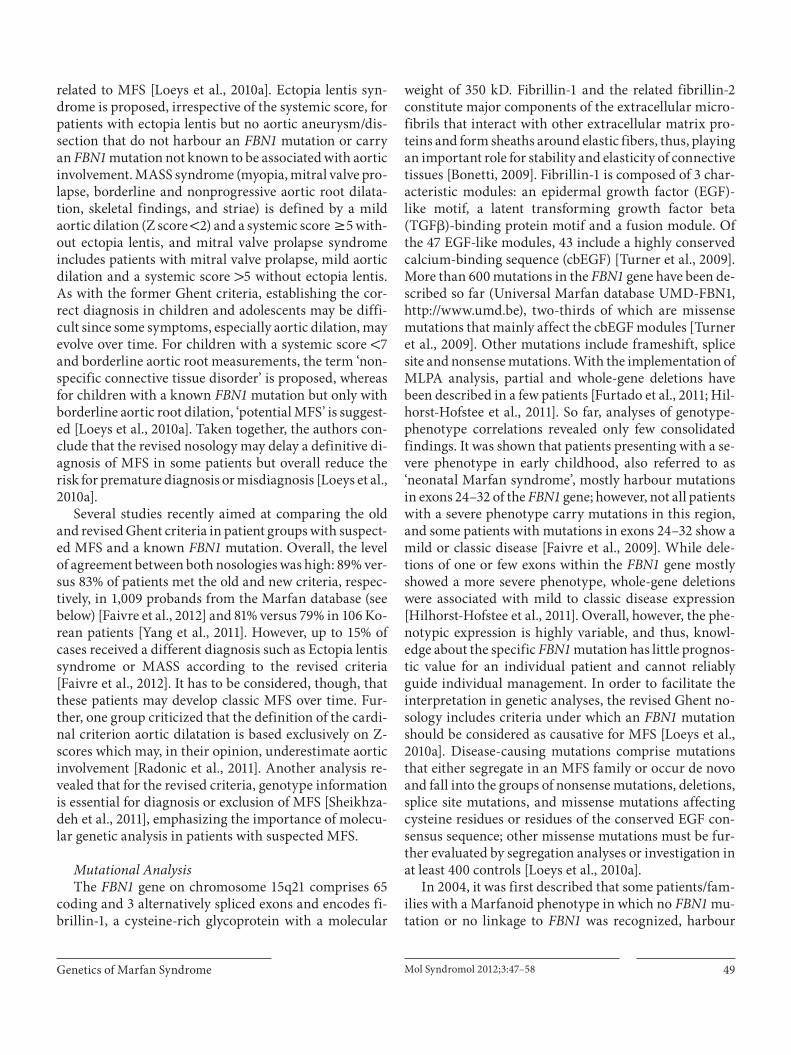
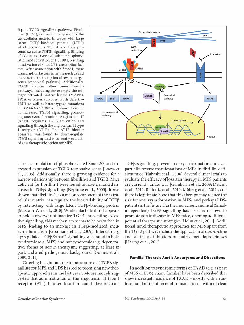
The TGF � signalling pathway plays an important role in numerous cellular processes, and dysregulation of this pathway has been implicated in several human disorders including cancer, autoimmune and cardiovascular dis-eases [Lin and Yang, 2010]. Binding of TGF � 1 to theTGFBR2 leads to phosphorylation and activation ofTGFBR1, resulting in activation of Smad2/3 transcrip-tion factors. After association with Smad4, these tran-scription factors enter the nucleus and increase the tran-scription of several target genes (canonical pathway, see fig. 1 ). Additionally, recent work has shown that TGF � also induces other (noncanonical) pathways, including for example the mitogen-activated protein kinase or RhoA cascades [Lin and Yang, 2010].
Although the exact pathogenetic mechanism remains unclear, heterozygous mutations in the TGFBR1 and TGFBR2 genes were shown to be associated with in-creased TGF � signalling, as indicated by enhanced nu-
Genetics of Marfan Syndrome Mol Syndromol 2012;3:47–58 51
clear accumulation of phosphorylated Smad2/3 and in-creased expression of TGF � -responsive genes [Loeys et al., 2005]. Additionally, there is growing evidence for a narrow relationship between fibrillin-1 and TGF � . Mice deficient for fibrillin-1 were found to have a marked in-crease in TGF � signalling [Neptune et al., 2003]. It was shown that fibrillin-1, as a major component of the extra-cellular matrix, can regulate the bioavailability of TGF � by interacting with large latent TGF � -binding protein [Massam-Wu et al., 2010]. While intact fibrillin-1 appears to hold a reservoir of inactive TGF � 1 preventing exces-sive signalling, this mechanism seems to be perturbed in MFS, leading to an increase in TGF � -mediated aneu-rysm formation [Goumans et al., 2009]. Interestingly, dysregulated TGF � /Smad2 signalling was found in both syndromic (e.g. MFS) and nonsyndromic (e.g. degenera-tive) forms of aortic aneurysm, suggesting, at least in part, a shared pathogenetic background [Gomez et al., 2009, 2011].
Growing insight into the important role of TGF � sig-nalling for MFS and LDS has led to promising new ther-apeutic approaches in the last years. Mouse models sug-gested that administration of the angiotensin II type 1 receptor (AT1) blocker losartan could downregulate
TGF � signalling, prevent aneurysm formation and even partially reverse manifestations of MFS in fibrillin-defi-cient mice [Habashi et al., 2006]. Several clinical trials to evaluate the efficacy of losartan therapy in MFS patients are currently under way [Gambarin et al., 2009; Detaint et al., 2010; Radonic et al., 2010; Möberg et al., 2011], and there is legitimate hope that this therapy may reduce the risk for aneurysm formation in MFS- and perhaps LDS- patients in the future. Furthermore, noncanonical (Smad-independent) TGF � signalling has also been shown to promote aortic disease in MFS mice, opening additional potential therapeutic strategies [Holm et al., 2011]. Addi-tional novel therapeutic approaches for MFS apart from the TGF � pathway include the application of doxycycline and statins as inhibitors of matrix metalloproteinases [Hartog et al., 2012].
Familial Thoracic Aortic Aneurysms and Dissections
In addition to syndromic forms of TAAD (e.g. as part of MFS or LDS), many families have been described that show increased incidence of TAAD – mostly with an au-tosomal dominant form of transmission – without clear
TGFβ1
TGFβ1
LTBP
Smad2/3P
PP
TGFBR1TGFBR2
TGFβac va on
Extracellular matrixFBN1
Smad4
Transcrip on
CanonicalpathwayMAPKRhoAPP2A
Non-canonicalpathway
AT1R
Losartan
MAPK
Smad2/3Smad4
Nucleus
Fig. 1. TGF � signalling pathway. Fibril-lin-1 (FBN1), as a major component of the extracellular matrix, interacts with large latent TGF � -binding protein (LTBP) which sequesters TGF � 1 and thus pre-vents excessive TGF � 1 signalling. Binding of TGF � 1 to TGFBR2 leads to phosphory-lation and activation of TGFBR1, resulting in activation of Smad2/3 transcription fac-tors. After association with Smad4, these transcription factors enter the nucleus and increase the transcription of several target genes (canonical pathway). Additionally, TGF � 1 induces other (noncanonical) pathways, including for example the mi-togen-activated protein kinase (MAPK), PP2A or RhoA cascades. Both defective FBN1 as well as heterozygous mutationsin TGFBR1/TGFBR2 were shown to result in increased TGF � 1 signalling, promot-ing aneurysm formation. Angiotensin II (AngII) regulates TGF � activation and signalling through the angiotensin II type 1 receptor (AT1R). The AT1R blocker Losartan was found to down-regulate TGF � signalling and is currently evaluat-ed as a therapeutic option for MFS.
Hoffjan Mol Syndromol 2012;3:47–5852
syndromic features (fTAAD). Occasionally, mutations in FBN1 , TGFBR1 or TGFBR2 are found in these families [Milewicz et al., 2008] further underlining the wide phe-notypic spectrum of the syndromic forms of disease. Apart from this, several other loci have been identified in linkage studies, and recently 3 new genes for fTAAD have been reported. In 2007, mutations in the gene encoding myosin heavy chain protein 11 (MYH11) on chromosome 16p13 were identified in 2 large families with autosomal dominant inheritance of TAAD and patent ductus arte-riosus [Zhu et al., 2006]. Subsequently, 2 additional fam-ilies with MYH11 mutations were described that showed substantial smooth muscle cell (SMC) disarray and focal hyperplasia of SMCs in the aortic media [Pannu et al., 2007]. Shortly thereafter, the gene encoding SMC � -actin (ACTA2) on chromosome 10q22–24 was identified in TAAD families that also showed a variety of additional symptoms such as livedo reticularis, patent ductus arte-riosus and iris flocculi [Guo et al., 2007]. Interestingly, ACTA2 mutations seem to increase risk not only for an-eurysms/dissections, but also for occlusive arterial dis-ease including premature stroke, premature coronary ar-tery disease and Moya-Moya disease [Guo et al., 2009]. While MYH11 mutations appear to be rare (only 5 muta-tions reported so far) and have been exclusively described in association with patent ductus arteriosus, ACTA2 mu-tations were found to be responsible for approximately 14–21% of fTAAD and are believed to interfere with the normal assembly of actin filaments [Hoffjan et al., 2011]. Recently, mutations in the gene encoding the kinase that controls SMC contractile function (myosin light chain kinase (MYLK) on chromosome 3q21) were also shown to cause familial aortic dissections [Wang et al., 2010].
Taken together, these findings strongly indicate an im-portant role of the SMC contractile apparatus for stabil-ity and integrity of the aortic wall, although the exact pathogenetic mechanisms remain unclear to date [Mile-wicz et al., 2008]. Interestingly, upregulated TGF � signal-ling in the aortic wall – similar to the findings in MFS and LDS –was recently demonstrated also in patients with MYH11 and ACTA2 mutations [Renard et al., 2011], pointing again towards the TGF � pathway as key regula-tor for aneurysm formation in different disorders.
Ehlers-Danlos Syndrome
EDS comprises a heterogeneous group of connective tissue disorders caused by defective synthesis of collagen. While the ‘classical’ EDS (type 1 and 2) is mainly charac-
terized by joint hypermobility, hyperelasticity of the skin, easy bruising, abnormal wound healing, and scar forma-tion (without vascular complications), EDS type IV goes along with increased risk of arterial aneurysms/dissections and thus, constitutes another important differential diag-nosis for MFS [Beridze and Frishman, 2012]. Like MFS and LDS, EDS type IV (also called the vascular type of EDS) is inherited in an autosomal dominant manner. It is caused by mutations in the gene encoding the alpha1-chain of col-lagen III (COL3A1) on chromosome 2q31 [Beridze andFrishman, 2012]. Cardinal clinical criteria for EDS IV comprise easy bruising, thin skin with visible veins, char-acteristic facial features (including thin lips and philtrum, small chin, thin nose, and large eyes), and rupture of arter-ies, uterus or intestines [Beighton et al., 1998]. Joint hyper-mobility and hyperextensibility of the skin are rather un-usual in the vascular type. In contrast to MFS, only about half of arterial complications in EDS type IV affect the tho-racic or abdominal aorta, while the rest occurs in arteries in the head, neck and limbs [Pepin and Byers, 1993]. Fur-ther, bowel rupture is a common feature of EDS type IV, and pregnancy for affected women was found to have a 12% risk for death from peripartum arterial rupture or uterine rupture [Pepin and Byers, 1993]. In order to sustain the clinical suspicion for EDS type IV, biochemical testing in cultured dermal fibroblasts is available. Further, elec-tron microscopy from a small skin biopsy can be used to confirm or reject the diagnosis of EDS [Morais et al., 2011].
More than 100 mutations in the COL3A1 gene have been reported to date, two-thirds of which are missense mutations affecting glycine residues in the triple helical domain [Pepin and Byers, 1993]. Null mutations leading to haploinsufficiency were found to be associated with a milder form of disease: compared to missense and splice site mutations, the occurrence of the first complication was delayed by almost 15 years, and almost exclusively vascular complications were observed [Leistritz et al., 2011]. Apart from this finding, no other genotype-pheno-type correlations were demonstrated for COL3A1 muta-tions [Pepin and Byers, 1993]. Rarely, vascular complica-tions can also be observed in the kyphoscoliotic type of EDS (EDS VIA), characterized by a deficiency of colla-gen lysyl hydroxylase 1 and caused by mutations in the PLOD1 gene [Rohrbach et al., 2011]. Unlike the vascular type, this form of EDS is inherited automosal recessively and characterized by progressive scoliosis, muscular hy-potonia, tissue fragility with easy bruising, and some-times cognitive delay [Rohrbach et al., 2011].
Surveillance and therapy of EDS type IV (including regular cardiovascular screening, thorough monitoring
Genetics of Marfan Syndrome Mol Syndromol 2012;3:47–58 53
of pregnancies and operative procedures) have proven challenging, and no preventive therapy has been estab-lished yet. Recently, first evidence emerged form a pro-spective trial that application of celiprolol, a long-acting � 2 receptor antagonist, might reduce the incidence of ar-terial rupture or dissection in EDS IV patients [Ong et al., 2010]. Whether increased TGF � signalling plays a patho-genetic role also for this connective tissue disorder has not been evaluated yet, but will certainly be addressed in the near future.
Arterial Tortuosity Syndrome
Arterial tortuosity syndrome (ATS) is another rare connective tissue disorder that shows substantial clinical overlap with LDS but is inherited autosomal recessively [Coucke et al., 2006]. It is characterized by tortuosity of the large and medium-sized arteries as well as aneuryms of large arteries and pulmonary stenosis. Further, the pa-tients often show joint hypermobility, arachnodactyly and distinct facial features including microretrognathia, downslanting palpebral fissures, hypertelorism and cleft palate, and/or bifid uvula. In 2007, mutations in the SLC2A10 gene on chromosome 20q13, encoding the glu-cose transporter GLUT10, were identified as causative for ATS in large families [Coucke et al., 2006]. Analysis of additional patients also revealed increased risk for isch-emic events [Callewaert et al., 2008]. While previous clin-ical reports indicated a severe clinical course with a 40% mortality rate before the age of 5 years, recent investiga-tions revealed a far more variable phenotype [Callewaert et al., 2008]. Since the SLC2A10 gene is located in a region linked to type 2 diabetes and presumably plays a role in glucose metabolism, it was considered a potential candi-date gene for diabetes [Dawson et al., 2001]; however, an association with type 2 diabetes could not be demonstrat-ed [Bento et al., 2005; Mohlke et al., 2005]. In ATS fami-lies, neither heterozygous nor homozygous mutation car-riers showed increased risk for diabetes [Callewaert et al., 2008], arguing against a major role of this gene in diabe-tes pathogenesis. Interestingly, an increase in TGF � sig-nalling, which has proven to be a cardinal pathogenetic mechanism in both MFS and LDS (see above), has also been found in ATS. It was hypothesized that GLUT10 acts as a glucose transporter into the nucleus where it modu-lates the expression of glucose-responsive genes [Coucke et al., 2006]. Deficiency of GLUT10 in ATS would then down-regulate inhibitors of the TGF � cascade, leading to increased TGF � signalling that stimulates vessel wall
cell formation. As an alternative hypothesis, however, GLUT10 has also been implicated in ascorbic acid me-tabolism [Segade, 2010].
Cutis Laxa Syndrome
Cutis laxa is a rare connective tissue disorder charac-terized by redundant, loose and hypoelastic skin due to abnormal elastic fibers [Berk et al., 2012]. The disease may be acquired or inherited with various modes of transmission (autosomal dominant, autosomal recessive or X-linked). Recently, mutations in the fibulin-4 gene (FBLN4) on chromosome 11q13 were identified as caus-ative for a rare autosomal recessive form of Cutis laxa that also led to altered TGF � signalling [Renard et al., 2010]. In contrast to other types of Cutis laxa, the patients car-rying homozygous FBLN4 mutations showed major car-diovascular events, including aortic aneurysms and arte-rial tortuosity [Renard et al., 2010], with only minor skin involvement, suggesting that this rare disorder of elastic tissue constitutes another differential diagnosis for the cardiovascular features of MFS.
Bicuspid Aortic Valve
The bicuspid aortic valve (BAV) is a relatively com-mon congenital heart disease (prevalence approximately 1–2%) that is characterized by the presence of only 2 in-stead of 3 semilunar valves, often followed by calcifica-tion [Siu and Silversides, 2010]. Affected individuals have an increased risk for aortic stenosis and/or regurgitation and also for aortic dissection. Both isolated valve defects and familial clustering have been documented. In fami-lies with BAV and severe valve calcification, causative mutations in the NOTCH1 gene were identified [Garg et al., 2005], and overrepresentation of NOTCH1 missense mutations was subsequently confirmed in additional nonrelated patients with BAV and thoracic aortic aneu-rysms [Mohamed et al., 2006; McKellar et al., 2007]. NOTCH1 is a signalling and transcriptional regulator that is involved in cell differentiation and determination during organogenesis. It was suggested that NOTCH1 mutations may cause an early developmental defect in the aortic valve and later affect calcium deposition. However, mutations in this gene explain only a small fraction of isolated as well as familial BAV, and locus heterogeneity [Ellison et al., 2007] as well as multifactorial pathogenesis have been suggested.
Hoffjan Mol Syndromol 2012;3:47–5854
Differential Diagnosis of the Ophthalmologic and
Skeletal Features of Marfan Syndrome
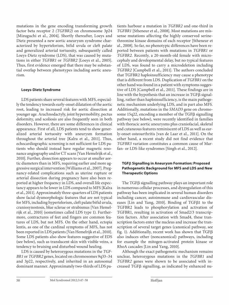
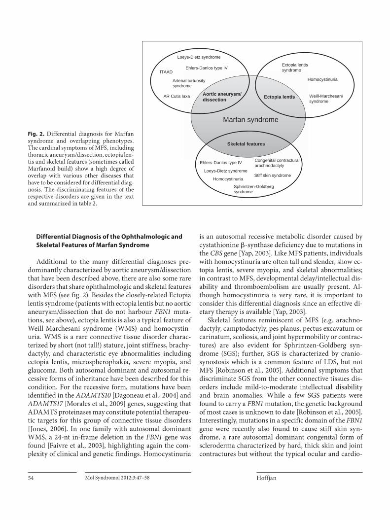
Additional to the many differential diagnoses pre-dominantly characterized by aortic aneurysm/dissection that have been described above, there are also some rare disorders that share ophthalmologic and skeletal features with MFS (see fig. 2 ). Besides the closely-related Ectopia lentis syndrome (patients with ectopia lentis but no aortic aneurysm/dissection that do not harbour FBN1 muta-tions, see above), ectopia lentis is also a typical feature of Weill-Marchesani syndrome (WMS) and homocystin-uria. WMS is a rare connective tissue disorder charac-terized by short (not tall!) stature, joint stiffness, brachy-dactyly, and characteristic eye abnormalities including ectopia lentis, microspherophakia, severe myopia, and glaucoma. Both autosomal dominant and autosomal re-cessive forms of inheritance have been described for this condition. For the recessive form, mutations have been identified in the ADAMTS10 [Dagoneau et al., 2004] and ADAMTS17 [Morales et al., 2009] genes, suggesting that ADAMTS proteinases may constitute potential therapeu-tic targets for this group of connective tissue disorders [Jones, 2006]. In one family with autosomal dominant WMS, a 24-nt in-frame deletion in the FBN1 gene was found [Faivre et al., 2003], highlighting again the com-plexity of clinical and genetic findings. Homocystinuria
is an autosomal recessive metabolic disorder caused by cystathionine � -synthase deficiency due to mutations in the CBS gene [Yap, 2003]. Like MFS patients, individuals with homocystinuria are often tall and slender, show ec-topia lentis, severe myopia, and skeletal abnormalities; in contrast to MFS, developmental delay/intellectual dis-ability and thromboembolism are usually present. Al-though homocystinuria is very rare, it is important to consider this differential diagnosis since an effective di-etary therapy is available [Yap, 2003].
Skeletal features reminiscent of MFS (e.g. arachno-dactyly, camptodactyly, pes planus, pectus excavatum or carinatum, scoliosis, and joint hypermobility or contrac-tures) are also evident for Sphrintzen-Goldberg syn-drome (SGS); further, SGS is characterized by cranio-synostosis which is a common feature of LDS, but not MFS [Robinson et al., 2005]. Additional symptoms that discriminate SGS from the other connective tissues dis-orders include mild-to-moderate intellectual disability and brain anomalies. While a few SGS patients were found to carry a FBN1 mutation, the genetic background of most cases is unknown to date [Robinson et al., 2005]. Interestingly, mutations in a specific domain of the FBN1 gene were recently also found to cause stiff skin syn-drome, a rare autosomal dominant congenital form of scleroderma characterized by hard, thick skin and joint contractures but without the typical ocular and cardio-
Marfan syndrome
Ectopia lentisAortic aneurysm/ dissection
Ehlers-Danlos type IV
Loeys-Dietz syndrome
fTAAD
Arterial tortuositysyndrome
Ectopia lentissyndrome
Homocystinuria
Weill-Marchesanisyndrome
Skeletal features
Loeys-Dietz syndrome
Ehlers-Danlos type IV
Sphrintzen-Goldberg syndrome
Congenital contracturalarachnodactyly
HomocystinuriaStiff skin syndrome
AR Cutis laxa
Fig. 2. Differential diagnosis for Marfan syndrome and overlapping phenotypes. The cardinal symptoms of MFS, including thoracic aneurysm/dissection, ectopia len-tis and skeletal features (sometimes called Marfanoid build) show a high degree of overlap with various other diseases that have to be considered for differential diag-nosis. The discriminating features of the respective disorders are given in the text and summarized in table 2.
Genetics of Marfan Syndrome Mol Syndromol 2012;3:47–58 55
vascular findings of MFS [Loeys et al., 2010b]. Addition-ally, mutations in the TGF � -binding protein-like domain 5 of the FBN1 gene were held responsible for geleophysic and acromimic dysplasia, characterized by short stature, short extremities and stiff joints [Le Goff et al., 2011]. An-
other rare differential diagnosis, especially for neonatal MFS, is autosomal dominantly inherited congenital con-tractural arachnodactyly (CCA, also called Beals syn-drome), caused by mutations in the gene encoding fibril-lin-2 (FBN2) [Callewaert et al., 2009]. Arachnodactyly,
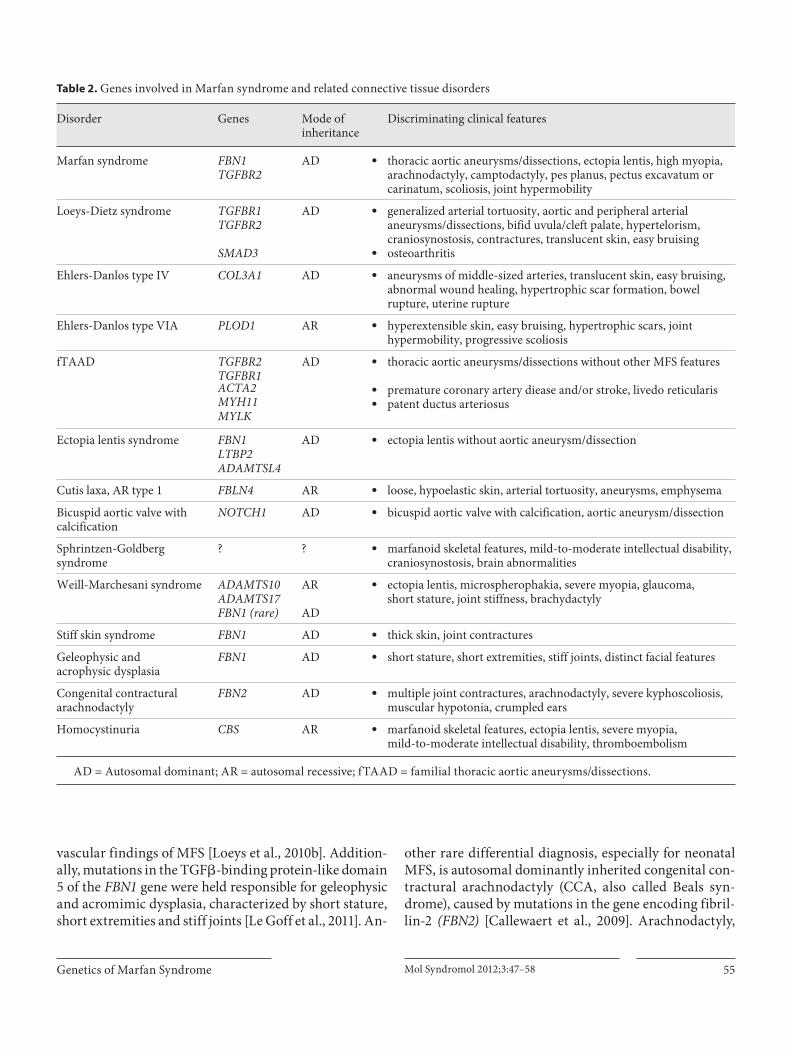
Table 2. Genes involved in Marfan syndrome and related connective tissue disorders
Disorder Genes Mode ofinheritance
Discriminating clinical features
Marfan syndrome FBN1TGFBR2
AD U thoracic aortic aneurysms/dissections, ectopia lentis, high myopia, arachnodactyly, camptodactyly, pes planus, pectus excavatum or carinatum, scoliosis, joint hypermobility
Loeys-Dietz syndrome TGFBR1TGFBR2
AD U generalized arterial tortuosity, aortic and peripheral arterial aneurysms/dissections, bifid uvula/cleft palate, hypertelorism, craniosynostosis, contractures, translucent skin, easy bruising
SMAD3 U osteoarthritis
Ehlers-Danlos type IV COL3A1 AD U aneurysms of middle-sized arteries, translucent skin, easy bruising, abnormal wound healing, hypertrophic scar formation, bowel rupture, uterine rupture
Ehlers-Danlos type VIA PLOD1 AR U hyperextensible skin, easy bruising, hypertrophic scars, joint hypermobility, progressive scoliosis
fTAAD TGFBR2TGFBR1
AD U
U
U
thoracic aortic aneurysms/dissections without other MFS features
premature coronary artery diease and/or stroke, livedo reticularispatent ductus arteriosus
ACTA2MYH11MYLK
Ectopia lentis syndrome FBN1LTBP2ADAMTSL4
AD U ectopia lentis without aortic aneurysm/dissection
Cutis laxa, AR type 1 FBLN4 AR U loose, hypoelastic skin, arterial tortuosity, aneurysms, emphysema
Bicuspid aortic valve withcalcification
NOTCH1 AD U bicuspid aortic valve with calcification, aortic aneurysm/dissection
Sphrintzen-Goldbergsyndrome
? ? U marfanoid skeletal features, mild-to-moderate intellectual disability, craniosynostosis, brain abnormalities
Weill-Marchesani syndrome ADAMTS10ADAMTS17FBN1 (rare)
AR
AD
U ectopia lentis, microspherophakia, severe myopia, glaucoma,short stature, joint stiffness, brachydactyly
Stiff skin syndrome FBN1 AD U thick skin, joint contractures
Geleophysic andacrophysic dysplasia
FBN1 AD U short stature, short extremities, stiff joints, distinct facial features
Congenital contracturalarachnodactyly
FBN2 AD U multiple joint contractures, arachnodactyly, severe kyphoscoliosis, muscular hypotonia, crumpled ears
Homocystinuria CBS AR U marfanoid skeletal features, ectopia lentis, severe myopia,mild-to-moderate intellectual disability, thromboembolism
A D = Autosomal dominant; AR = autosomal recessive; fTAAD = familial thoracic aortic aneurysms/dissections.
Hoffjan Mol Syndromol 2012;3:47–5856
severe kyphoscoliosis and muscular hypotonia are found in both MFS and CCA; features suggestive of CCA that are not commonly seen in MFS include multiple joint contractures and crumpled ears [Tuncbilek and Alanay, 2006]. However, while the first studies did not find evi-dence for aortic root dilatation in CCA, newer reports indicated that aortic dilatation may also occur in some CCA patients [Callewaert et al., 2009].
Conclusion and Future Prospects
In conclusion, due to substantial clinical and genetic overlap, correctly establishing the diagnosis of MFS or a related connective tissue disease is often challenging in clinical practice. Yet, the genetic findings of recent years have considerably contributed to unraveling the genetic background of connective tissue disorders, and molecu-lar genetic testing is more and more integrated into the diagnostic evaluations, since different disease entities go along with different therapeutic and surveillance re-quirements [von Kodolitsch et al., 2010]. Furthermore, increased insight into the pathogenetic background has already opened the way for novel therapeutic approaches [Hartog et al., 2012]. However, analysis of the many genes involved in the varying tissue disorders is time-consum-ing and expensive (especially for the FBN1 gene compris-ing 1 60 exons), and at the time being, testing for a whole panel of genes involved in connective tissue disorders in patients with overlapping phenotypes is not yet available on a routine basis. Therefore, at the moment, the decision which genes to evaluate in a patient/family is still based on the clinical observation of discriminating phenotypes ( table 2 ). For example, in a patient with thoracic aneu-rysm/dissection and additional symptoms suggestive of MFS, analysis of the FBN1 and TGFR2 genes appears sensible, while the existence of peripheral arterial an-
eurysms, especially in combination with generalized ar-terial tortuosity, should rather point to LDS (autosomal dominant, TGFBR1 and TGFBR2 genes) or ATS (autoso-mal recessive, SLC2A10 gene). If a patient shows features reminiscent of EDS such as translucent skin and bruis-ing, an electron microscopy can be performed before COL3A1 analysis in order to confirm or exclude EDS. Comprehensive analysis of TAAD genes in affected fam-ilies has not been incorporated into routine diagnostic procedures yet.
However, new technologies are already in develop-ment that will lead to more time- and cost-effective strat-egies in the near future, allowing analysis of several genes simultaneously. For example, a method using multiplex PCR followed by next-generation sequencing (massive parallel sequencing) was shown to reliably detect muta-tions in the FBN1 , TGFBR1 and TGFBR2 genes in MFS patients fulfilling the Ghent criteria [Baetens et al., 2011]. Further, a custom-based duplicate resequencing assay (Affymetrix) and a long-range PCR protocol were recent-ly presented to cover 8 genes previously associated with MFS and related disorders (FBN1, TGFBR1, TGFBR2, COL3A1, MYH11, ACTA2, SLC2A10 , and NOTCH1) [Kathiravel et al., 2012]. Finally, exome (and whole-ge-nome) sequencing strategies become more and more em-ployed for both targeted diagnostic testing and genome-wide searches for the mutations that underlie unex-plained disorders [Thorogood et al., 2012]. With rapidly decreasing costs, these strategies may also be applied to the complex field of inheritable connective tissue disor-ders in the near future and potentially identify addition-al genetic variation underlying these complex pheno-types. Yet, there is still considerable dispute about how to address the many ethical issues that go along with whole-genome sequencing strategies [Thorogood et al., 2012]. Therefore, it is not yet clear when these approaches will be used on a routine basis for MFS and related disorders.
References
Baetens M, Van Laer L, De Leeneer K, Hellemans J, De Schrijver J, et al: Applying massive par-allel sequencing to molecular diagnosis of Marfan and Loeys-Dietz syndromes. Hum Mutat 32: 1–10 (2011).
Beighton P, De Paepe A, Steinmann B, Tsipouras P, Wenstrup RJ: Ehlers-Danlos syndromes: revised nosology, Villefranche, 1997. Ehlers-Danlos National Foundation (USA) and Ehlers-Danlos Support Group (UK). Am J Med Genet 77: 31–37 (1998).
Bento JL, Bowden DW, Mychaleckyj JC, Hiraka-wa S, Rich SS, et al: Genetic analysis of the GLUT10 glucose transporter (SLC2A10) polymorphisms in Caucasian American type 2 diabetes. BMC Med Genet 6: 42 (2005).
Beridze N, Frishman WH: Vascular Ehlers-Dan-los syndrome: pathophysiology, diagnosis, and prevention and treatment of its compli-cations. Cardiol Rev 20: 4–7 (2012).
Berk DR, Bentley DD, Bayliss SJ, Lind A, Urban Z: Cutis laxa: a review. J Am Acad Dermatol 66: 842.e1–e17 (2012).
Bonetti MI: Microfibrils: a cornerstone of extra-cellular matrix and a key to understand Mar-fan syndrome. Ital J Anat Embryol 114: 201–224 (2009).
Callewaert BL, Willaert A, Kerstjens-Frederikse WS, De Backer J, Devriendt K, et al: Arterial tortuosity syndrome: clinical and molecular findings in 12 newly identified families. Hum Mutat 29: 150–158 (2008).
Genetics of Marfan Syndrome Mol Syndromol 2012;3:47–58 57
Callewaert BL, Loeys BL, Ficcadenti A, Vermeer S, Landgren M, et al: Comprehensive clinical and molecular assessment of 32 probands with congenital contractural arachnodacty-ly: report of 14 novel mutations and review of the literature. Hum Mutat 30: 334–341 (2009).
Campbell IM, Kolodziejska KE, Quach MM, Wolf VL, Cheung SW, et al: TGFBR2 deletion in a 20-month-old female with developmen-tal delay and microcephaly. Am J Med Genet A 155A:1442–1447 (2011).
Cañadas V, Vilacosta I, Bruna I, Fuster V: Mar-fan syndrome. Part 1: pathophysiology and diagnosis. Nat Rev Cardiol 7: 256–265 (2010).
Coucke PJ, Willaert A, Wessels MW, Callewaert B, Zoppi N, et al: Mutations in the facilitative glucose transporter GLUT10 alter angiogen-esis and cause arterial tortuosity syndrome. Nat Genet 38: 452–457 (2006).
Dagoneau N, Benoist-Lasselin C, Huber C, Faivre L, Mégarbané A, et al: ADAMTS10 mutations in autosomal recessive Weill-Marchesani syndrome. Am J Hum Genet 75: 801–806 (2004).
Dawson PA, Mychaleckyj JC, Fossey SC, Mihic SJ, Craddock AL, Bowden DW: Sequence and functional analysis of GLUT10: a glucose transporter in the Type 2 diabetes-linked re-gion of chromosome 20q12–13.1. Mol Genet Metab 74: 186–199 (2001).
De Paepe A, Devereux RB, Dietz HC, Hennekam RC, Pyeritz RE: Revised diagnostic criteria for the Marfan syndrome. Am J Med Genet 62: 417–426 (1996).
Detaint D, Aegerter P, Tubach F, Hoffman I, Plauchu H, et al: Rationale and design of a randomized clinical trial (Marfan Sartan) of angiotensin II receptor blocker therapy ver-sus placebo in individuals with Marfan syn-drome. Arch Cardiovasc Dis 103: 317–325 (2010).
Dietz HC, Cutting GR, Pyeritz RE, Maslen CL, Sakai LY, et al: Marfan syndrome caused by a recurrent de novo missense mutation in the fibrillin gene. Nature 352: 337–339 (1991).
Ellison JW, Yagubyan M, Majumdar R, Sarkar G, Bolander ME, et al: Evidence of genetic locus heterogeneity for familial bicuspid aortic valve. J Surg Res 142: 28–31 (2007).
Faivre L, Gorlin RJ, Wirtz MK, Godfrey M, Dagoneau N, et al: In frame fibrillin-1 gene deletion in autosomal dominant Weill-Marchesani syndrome. J Med Genet 40: 34–36 (2003).
Faivre L, Collod-Beroud G, Callewaert B, Child A, Binquet C, et al: Clinical and mutation-type analysis from an international series of 198 probands with a pathogenic FBN1 exons 24–32 mutation. Eur J Hum Genet 17: 491–501 (2009).
Faivre L, Collod-Beroud G, Adès L, Arbustini E, Child A, et al: The new Ghent criteria for Marfan syndrome: what do they change? Clin Genet 81: 433–442 (2012).
Furtado LV, Wooderchak-Donahue W, Rope AF, Yetman AT, Lewis T, et al: Characterization of large genomic deletions in the FBN1 gene using multiplex ligation-dependent probe amplification. BMC Med Genet 12: 119 (2011).
Gambarin FI, Favalli V, Serio A, Regazzi M, Pa-sotti M, et al: Rationale and design of a trial evaluating the effects of losartan vs. nebivo-lol vs. the association of both on the progres-sion of aortic root dilation in Marfan syn-drome with FBN1 gene mutations. J Cardio-vasc Med (Hagerstown) 10: 354–362 (2009).
Garg V, Muth AN, Ransom JF, Schluterman MK, Barnes R, et al: Mutations in NOTCH1 cause aortic valve disease. Nature 437: 270–274 (2005).
Gomez D, Al Haj Zen A, Borges LF, Philippe M, Gutierrez PS, et al: Syndromic and non-syn-dromic aneurysms of the human ascending aorta share activation of the Smad2 pathway. J Pathol 218: 131–142 (2009).
Gomez D, Coyet A, Ollivier V, Jeunemaitre X, Jondeau G, et al: Epigenetic control of vascu-lar smooth muscle cells in Marfan and non-Marfan thoracic aortic aneurysms. Cardio-vasc Res 89: 446–456 (2011).
Goumans MJ, Liu Z, ten Dijke P: TGF-beta sig-naling in vascular biology and dysfunction. Cell Res 19: 116–127 (2009).
Guo DC, Pannu H, Tran-Fadulu V, Papke CL, Yu RK, et al: Mutations in smooth muscle � -ac-tin (ACTA2) lead to thoracic aortic aneu-rysms and dissections. Nat Genet 39: 1488–1493 (2007).
Guo DC, Papke CL, Tran-Fadulu V, Regalado ES, Avidan N, et al: Mutations in smooth muscle alpha-actin (ACTA2) cause coronary artery disease, stroke, and Moyamoya disease, along with thoracic aortic disease. Am J Hum Genet 84: 617–627 (2009).
Habashi JP, Judge DP, Holm TM, Cohn RD, Loeys BL, et al: Losartan, an AT1 antago-nist, prevents aortic aneurysm in a mouse model of Marfan syndrome. Science 312: 117–121 (2006).
Hartog AW, Franken R, Zwinderman AH, Groe-nink M, Mulder BJ: Current and future phar-macological treatment strategies with regard to aortic disease in Marfan syndrome. Ex-pert Opin Pharmacother 13: 647–662 (2012).
Hilhorst-Hofstee Y, Hamel BC, Verheij JB, Rij-laarsdam ME, Mancini GM, et al: The clini-cal spectrum of complete FBN1 allele dele-tions. Eur J Hum Genet 19: 247–252 (2011).
Hoffjan S, Waldmüller S, Blankenfeldt W, Köt-ting J, Gehle P, et al: Three novel mutations in the ACTA2 gene in German patients with thoracic aortic aneurysms and dissections. Eur J Hum Genet 19: 520–524 (2011).
Holm TM, Habashi JP, Doyle JJ, Bedja D, Chen Y, et al: Noncanonical TGFbeta signaling contributes to aortic aneurysm progression in Marfan syndrome mice. Science 332: 358–361 (2011).
Jones GC: ADAMTS proteinases: potential ther-apeutic targets? Curr Pharm Biotechnol 7: 25–31 (2006).
Kalra VB, Gilbert JW, Malhotra A: Loeys-Dietz syndrome: cardiovascular, neuroradiologi-cal and musculoskeletal imaging findings. Pediatr Radiol 41: 1495–1504 (2011).
Kathiravel U, Keyser B, Hoffjan S, Koetting J, Müller M, et al: High-density oligonucle-otide-based resequencing assay for muta-tions causing syndromic and non-syndrom-ic forms of thoracic aortic aneurysms and dissections. (submitted 2012).
Le Goff C, Mahaut C, Wang LW, Allali S, Ab-hyankar A, et al: Mutations in the TGFbeta binding-protein-like domain 5 of FBN1 are responsible for acromicric and geleophysic dysplasias. Am J Hum Genet 89: 7–14 (2011).
Leistritz DF, Pepin MG, Schwarze U, Byers PH: COL3A1 haploinsufficiency results in a vari-ety of Ehlers-Danlos syndrome type IV with delayed onset of complications and longer life expectancy. Genet Med 13: 717–722 (2011).
Lin F, Yang X: TGF-beta signaling in aortic an-eurysm: another round of controversy. J Genet Genomics 37: 583–591 (2010).
Loeys BL, Chen J, Neptune ER, Judge DP, Podowski M, et al: A syndrome of altered cardiovascular, craniofacial, neurocognitive and skeletal development caused by muta-tions in TGFBR1 or TGFBR2 . Nat Genet 37: 275–281 (2005).
Loeys BL, Dietz HC, Braverman AC, Callewaert BL, De Backer J, et al: The revised Ghent no-sology for the Marfan syndrome. J Med Ge-net 47: 476–485 (2010a).
Loeys BL, Gerber EE, Riegert-Johnson D, Iqbal S, Whiteman P, et al: Mutations in fibrillin-1 cause congenital scleroderma: stiff skin syn-drome. Sci Transl Med 2: 23ra20 (2010b).
Massam-Wu T, Chiu M, Choudhury R, Chaudhry SS, Baldwin AK, et al: Assembly of fibrillin microfibrils governs extracellular deposi-tion of latent TGF beta. J Cell Sci 123: 3006–3018 (2010).
McKellar SH, Tester DJ, Yagubyan M, Majumdar R, Ackerman MJ, Sundt TM 3rd: Novel NOTCH1 mutations in patients with bicus-pid aortic valve disease and thoracic aortic aneurysms. J Thorac Cardiovasc Surg 134: 290–296 (2007).
Milewicz DM, Guo DC, Tran-Fadulu V, Lafont AL, Papke CL, et al: Genetic basis of thoracic aortic aneurysms and dissections: focus on smooth muscle cell contractile dysfunction. Annu Rev Genomics Hum Genet 9: 283–302 (2008).
Mizuguchi T, Collod-Beroud G, Akiyama T, Abifadel M, Harada N, et al: Heterozygous TGFBR2 mutations in Marfan syndrome. Nat Genet 36: 855–860 (2004).
Möberg K, De Nobele S, Devos D, Goetghebeur E, Segers P, et al: The Ghent Marfan Trial – a randomized, double-blind placebo con-trolled trial with losartan in Marfan patients treated with � -blockers. Int J Cardiol 2011, E-pub ahead of print.
Hoffjan Mol Syndromol 2012;3:47–5858
Mohamed SA, Aherrahrou Z, Liptau H, Erasmi AW, Hagemann C, et al: Novel missense mu-tations (p.T596M and p.P1797H) in NOTCH1 in patients with bicuspid aortic valve. Bio-chem Biophys Res Commun 345: 1460–1465 (2006).
Mohlke KL, Skol AD, Scott LJ, Valle TT, Berg-man RN, et al: Evaluation of SLC2A10 (GLUT10) as a candidate gene for type 2 dia-betes and related traits in Finns. Mol Genet Metab 85: 323–327 (2005).
Morais P, Mota A, Eloy C, Lopes JM, Torres F, et al: Vascular Ehlers-Danlos syndrome: a case with fatal outcome. Dermatol Online J 17: 1 (2011).
Morales J, Al-Sharif L, Khalil DS, Shinwari JM, Bavi P, et al: Homozygous mutations in ADAMTS10 and ADAMTS17 cause len-ticular myopia, ectopia lentis, glaucoma, spherophakia, and short stature. Am J Hum Genet 85: 558–568 (2009).
Neptune ER, Frischmeyer PA, Arking DE, Myers L, Bunton TE, et al: Dysregulation of TGF-beta activation contributes to pathogenesis in Marfan syndrome. Nat Genet 33: 407–411 (2003).
Ong KT, Perdu J, De Backer J, Bozec E, Collignon P, et al: Effect of celiprolol on prevention of cardiovascular events in vascular Ehlers-Danlos syndrome: a prospective ran-domised, open, blinded-endpoints trial. Lancet 376: 1476–1484 (2010).
Pannu H, Tran-Fadulu V, Papke CL, Scherer S, Liu Y, et al: MYH11 mutations result in a dis-tinct vascular pathology driven by insulin-like growth factor 1 and angiotensin II. Hum Mol Genet 16: 2453–2462 (2007).
Pepin MG, Byers PH: Ehlers-Danlos Syndrome Type IV, in Pagon RA, Bird TD, Dolan CR, Stephens K, Adam MP (eds): Gene Review TM [Internet] (University of Washington, Seattle 1993).
Radonic T, de Witte P, Baars MJ, Zwinderman AH, Mulder BJ, et al: Losartan therapy in adults with Marfan syndrome: study proto-col of the multi-center randomized con-trolled COMPARE trial. Trials 11: 3 (2010).
Radonic T, de Witte P, Groenink M, de Bruin-Bon R, Timmermans J, et al: Critical apprais-al of the revised Ghent criteria for diagnosis of Marfan syndrome. Clin Genet 2011, E-pub ahead of print.
Renard M, Holm T, Veith R, Callewaert BL, Adès LC, et al: Altered TGFbeta signaling and car-diovascular manifestations in patients with autosomal recessive cutis laxa type I caused by fibulin-4 deficiency. Eur J Hum Genet 18: 895–901 (2010).
Renard M, Callewaert B, Baetens M, Campens L, Macdermot K, et al: Novel MYH11 and ACTA2 mutations reveal a role for enhanced TGFbeta signaling in FTAAD. Int J Cardiol 2011, E-pub ahead of print.
Robinson PN, Neumann LM, Demuth S, Enders H, Jung U, et al: Shprintzen-Goldberg syn-drome: fourteen new patients and a clinical analysis. Am J Med Genet A 135: 251–262 (2005).
Rohrbach M, Vandersteen A, Yiş U, Serdaroglu G, Ataman E, et al: Phenotypic variability of the kyphoscoliotic type of Ehlers-Danlos syndrome (EDS VIA): clinical, molecular and biochemical delineation. Orphanet J Rare Dis 6: 46 (2011).
Segade F: Glucose transporter 10 and arterial tortuosity syndrome: the vitamin C connec-tion. FEBS Lett 584: 2990–2994 (2010).
Sheikhzadeh S, Kade C, Keyser B, Stuhrmann M, Arslan-Kirchner M, et al: Analysis of pheno-type and genotype information for the diag-nosis of Marfan syndrome. Clin Genet 2011, E-pub ahead of print.
Singh KK, Schmidtke J, Keyser B, Arslan-Kirch-ner M: TGFBR3 variation is not a common cause of Marfan-like syndrome and Loeys-Dietz-like syndrome. J Negat Results Biomed 11: 9 (2012).
Siu SC, Silversides CK: Bicuspid aortic valve dis-ease. J Am Coll Cardiol 55: 2789–2800 (2010).
Stheneur C, Collod-Béroud G, Faivre L, Gouya L, Sultan G, et al: Identification of 23 TGFBR2 and 6 TGFBR1 gene mutations and geno-type-phenotype investigations in 457 pa-tients with Marfan syndrome type I and II, Loeys-Dietz syndrome and related disor-ders. Hum Mutat 29:E284–295 (2008).
Thorogood A, Knoppers BM, Dondorp WJ, de Wert GM: Whole-genome sequencing and the physician. Clin Genet 81: 511–513 (2012).
Tuncbilek E, Alanay Y: Congenital contractural arachnodactyly (Beals syndrome). Orphanet J Rare Dis 1: 20 (2006).
Turner CL, Emery H, Collins AL, Howarth RJ, Yearwood CM, et al: Detection of 53 FBN1 mutations (41 novel and 12 recurrent) and genotype-phenotype correlations in 113 un-related probands referred with Marfan syn-drome, or a related fibrillinopathy. Am J Med Genet A 149A:161–170 (2009).
van de Laar IM, Oldenburg RA, Pals G, Roos-Hesselink JW, de Graaf BM, et al: Mutations in SMAD3 cause a syndromic form of aortic aneurysms and dissections with early-onset osteoarthritis. Nat Genet 43: 121–126 (2011).
Van Hemelrijk C, Renard M, Loeys B: The Loeys-Dietz syndrome: an update for the clinician. Curr Opin Cardiol 25: 546–551 (2010).
von Kodolitsch Y, Rybczynski M, Bernhardt A, Mir TS, Treede H, et al: Marfan syndrome and the evolving spectrum of heritable tho-racic aortic disease: do we need genetics for clinical decisions? Vasa 39: 17–32 (2010).
Wang L, Guo DC, Cao J, Gong L, Kamm KE, et al: Mutations in myosin light chain kinase cause familial aortic dissections. Am J Hum Genet 87: 701–707 (2010).
Williams JA, Loeys BL, Nwakanma LU, Dietz HC, Spevak PJ, et al: Early surgical experi-ence with Loeys-Dietz: a new syndrome of aggressive thoracic aortic aneurysm disease. Ann Thorac Surg 83:S757–763 (2007).
Yang JH, Han H, Jang SY, Moon JR, Sung K, et al: A comparison of the Ghent and revised Ghent nosologies for the diagnosis of Marfan syndrome in an adult Korean population. Am J Med Genet A 158A:989–995 (2011).
Yap S: Classical homocystinuria: vascular risk and its prevention. J Inherit Metab Dis 26: 259–265 (2003).
Zhu L, Vranckx R, Khau Van Kien P, Lalande A, Boisset N, et al: Mutations in myosin heavy chain 11 cause a syndrome associating tho-racic aortic aneurysm/aortic dissection and patent ductus arteriosus. Nat Genet 38: 343–349 (2006).