utilization of dissection videos in graduate-level gross
TRANSCRIPT
University of Nebraska Medical Center University of Nebraska Medical Center
DigitalCommons@UNMC DigitalCommons@UNMC
Theses & Dissertations Graduate Studies
Spring 5-7-2016
Utilization of Dissection Videos in Graduate-Level Gross Anatomy Utilization of Dissection Videos in Graduate-Level Gross Anatomy
Education: An Analysis of Student Confidence, Utilization, and Education: An Analysis of Student Confidence, Utilization, and
Scoring Outcomes Scoring Outcomes
Jessica Gamerl University of Nebraska Medical Center
Follow this and additional works at: https://digitalcommons.unmc.edu/etd
Part of the Anatomy Commons
Recommended Citation Recommended Citation Gamerl, Jessica, "Utilization of Dissection Videos in Graduate-Level Gross Anatomy Education: An Analysis of Student Confidence, Utilization, and Scoring Outcomes" (2016). Theses & Dissertations. 109. https://digitalcommons.unmc.edu/etd/109
This Thesis is brought to you for free and open access by the Graduate Studies at DigitalCommons@UNMC. It has been accepted for inclusion in Theses & Dissertations by an authorized administrator of DigitalCommons@UNMC. For more information, please contact [email protected].
brought to you by COREView metadata, citation and similar papers at core.ac.uk
provided by University of Nebraska Medical Center Research: DigitalCommons@UNMC
Utilization of Dissection Videos in Graduate-Level Gross Anatomy Education: An Analysis of Student Confidence,
Utilization, and Scoring Outcomes
by co-authors
Jess Gamerl and Kevin Selting
A THESIS
Presented to the Faculty of
the University of Nebraska Graduate College
in Partial Fulfillment of the Requirements
for the Degree of Master of Science
Genetics, Cell Biology, and Anatomy Graduate Program
Under the Supervision of Professors Ryan Splittgerber and Karen Gould
University of Nebraska Medical Center
Omaha, Nebraska
April, 2016
Advisory Committee:
Karen Gould, PhD.
Ryan Splittgerber, PhD
Shantaram Joshi, PhD
ii
Acknowledgements
The author would like to thank her co-author, Kevin Selting, for helping create and
produce the videos made for this study. She would also like to thank her advisor, Dr. Ryan
Splittgerber, for his guidance and help, as well as Dr. Karen Gould for all of her supervision and
assistance throughout this project. The author would also like to thank and acknowledge all of
the anatomy teaching faculty. Additionally, she would like to thank the anatomical donors for
donating their bodies to further her education. Finally, the author would like to thank her
parents and cats for all their support.

iii
Utilization of Dissection Videos in Graduate-Level Gross
Anatomy Education: An Analysis of Student Confidence,
Utilization, and Scoring Outcomes
Jessica Gamerl M.S
University of Nebraska, 2016
Advisor: Ryan Splittgerber, PhD.
The use of videos in anatomy education has become a potential tool for learning by students. It
is currently unclear if using videos that show how to perform a dissection can have an impact on
student practical scores. This thesis attempted to address that question as well as address how
videos can impact student confidence levels and how students might implement videos into
their study strategies. A set of high definition videos was made following the specific steps of a
dissector guide and provided to students prior to their third and fourth lab practicals. A series of
three surveys was conducted to gauge student experience, learning resource utilization, and
feedback for the videos. Student experience had a significant impact on grade outcomes so
students were split into groups: students with prior cadaver lab experience and students
without. Students used the videos primarily before lab to help prepare for lab and exams. The
videos may have had an effect on the final lab practical score for the students who used the
videos and may have increased confidence levels in students that used the videos compared to
those who did not. This may suggest that dissection video use could have a positive impact on
lab practical test scores.
iv
Table of Contents
Acknowledgements ii
Abstract iii
List of Figures vi
List of Tables viii
Chapter 1: Introduction 1
Chapter 2: Methods 11
Study Design 11
Video Production 12
Study Participants 13
Chapter 3: Results 14
Videos 14
Student Performance 15
Student Confidence 18
Student Survey Responses 20
Chapter 4: Discussion 24
Student Perceptions 24
Student Confidence 24
v
Student Utilization of the Videos 25
Student Performance 25
Research Limitations 26
Conclusions 27
Bibliography 29
Appendix A: Student Consent Form 31
Appendix B: Student Surveys 34
Appendix C: Blackboard Announcement for International Review Board 37
Appendix D: Educational Services Office Grant Proposal 38
vi
List of Figures
Figure 3.1 16
Caption: Figure 3.1-Difference in performance based on previous cadaver lab
experience. Students with previous experience significantly outperformed students
without it (P=0.016). This is on a 4.0 grading scale (A=4.0, B=3.0, etc.).
Figure 3.2 17
Caption: Figure 3.2- Difference in mean practical exam scores (N=48). Students with
previous cadaver lab experience significantly outperformed students without previous
experience on the first two practical exams (P=0.043) with average scores of 80.3% vs.
75.1%. On the third practical exam, students who did not watch the videos had an
average decline in scores by 1.6%, students who watched the videos increased their
average scores by 4.3%. This was statistically significant (P=0.013 by paired t test).
Figure 3.3 18
Caption: Figure 3.3- Mean Confidence Scores. Students self-reported their confidence
levels over the series of three surveys throughout the course. This figure represents only
the students who had previous cadaver lab experiences (N=48) and the two groups they
comprise: students who watched the videos (N=28, indicated by circles) and those who
did not (N=20, indicated by triangles). The students who watched the videos reported
higher confidence scores right before the final exam, this was statistically significant
(P=0.035).
Figure 3.4 19
vii
Caption: Figure 3.4- Correlation of dissection skills confidence with course grade. R2=
0.9679, the data fits a linear relationship.
viii
List of Tables
Table 3.1 15
Caption: Table 3.1- Breakdown of videos made available for each lab unit
Table 3.2 20
Caption: Table 3.2- Student usage of resources used to prepare for gross anatomy lab
Table 3.3 21
Caption: Table 3.3- Student usage of resources during gross anatomy lab
Table 3.4 22
Caption: Table 3.4- How and when students used the dissection videos
Table 3.5 23
Caption: Table 3.5- Student assessment of utility of how to dissection videos
1
Chapter 1: Introduction
Gross anatomy is a staple of education for medical students, allied health professionals,
and graduate students. The most valuable part of gross anatomy education is the use of
cadavers. In the cadaver lab, students dissect their donors and intimately learn about the human
body and its tissues. Instructors and students agree that cadaver dissection is vital to learning
anatomy but in many programs it is hard to obtain donors. This can lead to a high ratio of
students per donor which limits the amount of dissection that students are able to do. While
cadaver dissection is the best way to learn and teach gross anatomy, many programs try to use
other resources to supplement the laboratory work.
Conventional teaching resources inside and outside of the laboratory tend to be
preferred by students, according to Nageswari et. al (2004). These resources usually include the
recommended anatomy atlases and textbooks, traditional lectures supplemented with
PowerPoint presentations, and dissector guides. The quality of such resources varies by program
but Mayfield et. al (2012) looked at the use of online dissector guides during laboratory time
versus more traditional paper guides and hardcover atlases. The online dissector guides
combined the paper guide and relevant atlas images together. Students that had access to the
online dissector guides via iPads were more engaged and active in lab and reported feeling like
their time was better managed. It was observed in this study that the students who had the
more traditional guides spent more time waiting for instructor assistance and searching through
the atlases than dissecting and the students with the iPads were more efficient with their time.
While there were concerns that giving students access to iPads would lead to more distractions
and inefficiency (using inappropriate apps or using the iPad for irrelevant activities instead of
laboratory work), the study concluded that such concerns were likely unfounded, however,
2
students in this study did not have access to the internet while in the laboratory. There are no
studies showing whether internet access increases the amount of distractions during cadaver-
based anatomy laboratory courses.
The state of anatomy education is changing, however. More visual resources are being
used by students with the rise of social media and huge pools of online resources. Acland’s
Anatomy Video Atlas and the free Human Anatomy Education channel on YouTube are two very
popular video resources that many students have available to them and because anatomy
requires a large amount of visual and spatial skills, using videos or other audiovisual resources
may help anatomy students learn the material better. With such a large breadth of study
options, it is important to look at how students use such resources, if using audiovisual learning
resources affects testing outcomes, and what types of limitations or concerns such resources
may bring up.
Mahmud et. al (2011) reported that showing videos depicting the dissection of the
upper and lower limbs did not have a significant impact on class test scores (the tests consisted
of a written multiple choice exam, an oral exam, and a pinned practical of laboratory
specimens/donors) but that students did report that they liked the resources and felt they
helped them learn the material better. However, Collins et. al (2015) found the opposite of
Mahmud’s study. Collins showed a group of students at various stages in their medical training
an upper limb dissection video before a dissection course. The students that saw the video
outperformed their peers at the same level of training who did not see the video. It is possible
that these two studies made opposite conclusions because of differences in their methods.
In Mahmud et. al’s study, the sample size was significantly larger, but participation for
the lower limb dissection videos was voluntary. Additionally, based on timing within the
3
semester, there is no real control group. In this study the students in group A were shown the
videos on the upper limb after they had already been tested over the material and watching the
lower limb videos was optional. The students in group B were shown the upper limb videos but
had already been tested over the lower limb so it is unlikely that a large portion of the group B
students chose to watch the lower limb videos. Test scores from the previous two years were
used for comparison to see if the students in Mahmud’s study did better, but because of which
semester the videos were shown and the fact that only half of them were actually required to
be seen (by being shown in lecture twice), the study has some serious flaws.
In the Collins et. al study the students in the variable group were shown a video
demonstrating dissection of the upper limb prior to beginning a dissection course. The students
in the control group were outperformed by their peers with the same experience in the variable
group, which was measured in standardized multiple choice questionnaires with questions
specific to upper limb anatomy. While the Collins study had a much smaller sample size, it also
had more controls in place: the instructor to student ratio was kept constant (1:2) and the
questionnaires the students took were evenly spaced with regard to timing. The students were
also tested over the same material at the same time. None of these factors exist or are
described in the Mahmud study and do not appear to be taken into account regarding their
conclusions. The biggest problem with the Mahmud study, which they acknowledge, is the lack
of control over viewing the videos and the minimal monitoring over the pattern of use by
students.
Choi-Lundberg et. al (2015) analyzed the type of learning resources students preferred
to use while studying anatomy and found that while videos did not outrank textbooks, atlases,
or traditional lectures students still rated them very highly. The Choi-Lundberg study did not
take into account how these resources affected grading outcomes but they did conclude that a
4
variety of study resources available for students to use was a “good practice to help students
gain a variety of perspectives […] and synthesize information from multiple resources”. This
study was not the only one to find that students enjoyed videos relevant to studying anatomy
(Mahmud et. al 2011, Wang et. al 2010, Topping 2013, and Mukhopadhyay et. al 2014).
Students will use tools they like over others, so if students enjoy the videos and feel they help
them learn anatomy or make studying anatomy easier then test outcomes may not be the best
way to evaluate whether or not videos should be used as a learning resource.
Video use in learning is not a new concept. In 2010, Wang et. al looked at the use of
video-conferencing in medical school lecturing. Concerns that students would not attend
lectures if they were available online were shown to be unfounded, however, the students
selected for Wang’s study were invited to participate in the study via email and then
interviewed to determine their inclusion. Students who attended lectures were probably more
likely to volunteer for the study leading to a significant selection bias, a limitation that the
authors do acknowledge. At this point, many medical schools record lectures for students to
peruse outside of class and use to review contents for exams. The biggest advantage, according
to Wang’s study, to video-recorded lectures was the ability to rewind or fast-forward as needed.
Denning’s study also highlighted the ability to change a video’s speed as an advantage to videos
in his evaluation of video use in the classroom. Dong et. al (2016) agree with Denning that being
able to adjust the speed of the video helps learning and should be a major consideration for
choosing appropriate videos. Because students can view a video as many times as they need to
and can skip over or fast-forward through any redundant or unnecessary lessons, time
adjustments can allow students to better focus their study time on more relevant and
challenging materials with minimal time inefficiency. Additionally, Denning suggested that video
5
use may help strong visual learners understand concepts better. Nageswari et. al (2004) also
support this concept in their study.
The Nageswari study looked at different learning opportunities and resources for first
year medical students and then asked them to rank the resources numerically based on the
overall impact they had on the students’ learning. The VARK survey was used to classify the
different resources the students were presented with and it was found that many of the
resources overlapped over multiple types of learning. For example, cadaver dissection would
incorporate kinesthetic and visual learning. The Nageswari study showed that resources that
had strong visual learning and overlapped with other types of learning were rated more highly
by students. This may be due to the fact that by the time students reach medical school, the vast
majority of their education has consisted of visual types of learning (PowerPoint presentations,
lectures, diagrams, etc.) and it is what they are most familiar and comfortable with and
therefore more likely to use or prefer over other options. This may also be why students ranked
more traditional learning resources (lectures, PowerPoint presentations, textbooks, etc.) higher
than other less traditional resources (software, websites, videos, etc.) in the Choi-Lundberg
study. The fact that many of the less traditional resources in the Choi-Lundberg study required
students to pay for them is another possible explanation.
Although students seem to prefer more traditional teaching resources (PowerPoint
presentations, lectures, textbooks, atlases), the influence the internet has had on higher
education is clear. The majority of studies done to analyze videos in learning anatomy have
looked specifically at YouTube, a video sharing website combined with social media networking,
and how it can be used to teach anatomy. Mukhopadhyay et. al. (2014) determined YouTube to
be used primarily by students in developed countries but highlighted its potential for teaching
students in developing countries. The ease of use and accessibility of YouTube makes it an ideal
6
study resource for students but many studies analyzed for this review (Mukhopadhyay et. al
2014, Azer 2012, Barry et. al 2015, Raikos et. al 2013, and Jaffar 2012) brought up questions
about the accuracy of the information within videos students would have access to. Azer’s study
provided the starkest example of this where 73% of the videos containing relevant information
regarding surface anatomy did not meet the criteria to be considered educationally useful. Azer
determined whether a video was educationally useful or not based on the major and minor
criteria outlined in his study; educationally useful videos had to meet all major criteria
(scientifically accurate content, clear images, credit given to creator or organizer of video, topic
is presented clearly, and the video uses living bodies, models, or drawings for difficult concepts)
and at least three minor criteria (Covers topics identified in search query, designed for
undergraduates or medical/health students, sounds are clear with minimal background noise,
downloading time is reasonable, creator’s information is up to date, and educational objectives
are stated in the video). It is important to note that in Azer’s study, a video containing
inaccurate information could not be deemed educationally useful even if it met all other criteria.
The study also demonstrated that of the total videos screened for the study, less than 25% of
the videos even met the criteria to be considered relevant to teaching surface anatomy even
though they appeared in specific search results regarding the subject. Raikos et. al had similar
results for the videos that passed their criteria for usefulness (determined by a numerical score
based on criteria defined in their study). The Raikos study determined videos to be useful using
three separate scoring criteria categories (Anatomical content score, General quality score, and
general data score) with each criteria requirement counting as one point. A score of 13/20 was
considered a “passing” score for the videos. Azer suggests that there is not enough of a
contribution from medical education institutions and that if more medical institutions or
anatomists created content for YouTube it may address the issue of inaccurate information
7
found within relevant videos and help lower, or at least counter, the high percentage of videos
containing inaccurate information.
Jaffar’s study (2012) tried to overcome this by having students use a selection of links
verified by tutors and chosen because the content of the videos closely matched the learning
objectives for their anatomy courses. According to the study, 98% of the students surveyed used
YouTube to study. However, it was not possible to prevent students from accessing additional
videos outside of those suggested within their curriculum. In fact, the students reported using
the Human Anatomy Education YouTube channel which is not explained in the study. It is
unclear if the Human Anatomy Education channel was where students were being directed by
tutors or via a syllabus to use it, or if students were simply stumbling upon it in their internet
searches. It is unclear how the author of the Jaffar study is connected to that channel or why its
use was included in the study. Because of this confusion, but also the nature of the internet and
the lack of control over the resources used, the students could still have had access to materials
with inaccurate information. Jaffar tried to address this by suggesting that faculty members
could create their own videos or aggregate videos they approve of onto their own YouTube
channels and distribute access to their students. This particular issue should be of some concern
because Barry et. al (2015) concluded that a significant percentage of students would prefer to
use online resources (which is in direct conflict with the conclusions of the Choi-Lundberg and
Nageswari studies), which may be inaccurate or out of scope to their learning objectives, rather
than contact their instructors, who are seen as the best source of information and can provide
accurate information specific to the students’ learning objectives or course goals, for
clarification.
The Barry study contained significantly more female participants than male, this could
represent a bias in their conclusions, particularly regarding the lack of willingness students
8
reported for contacting instructors for clarification. Female students may be less confident in
approaching authority figures due to socialization or overall lower confidence levels compared
to their male peers. Female students may also have a higher interest in visually-stimulating
learning resources compared to their male counterparts, which may explain why the conclusions
the study draws seem in contention with the Choi-Lundberg and Nageswari studies, both of
which have larger sample sizes and more even distribution of genders.
Aside from the issue with inaccuracies in the educational content on YouTube, Barry et.
al and Raikos et. al point out that there are ethical questions regarding the use of cadavers or
human tissue within YouTube videos. Raikos et. al highlight that medical schools can have social
media policies in place that prevent their students from recording or photographing any donors
in the laboratory unless permission is granted for research, but there is no way to be sure if
videos on the internet were uploaded with the appropriate permissions or protocols. Because of
this, using cadaver-based videos may not be ethical, at best, and at worst it could put the
schools these videos are connected to in jeopardy of losing their anatomical gift programs. In
addition, YouTube’s use for teaching relies on the ability students have to search for videos. It is
possible to preserve the privacy of the donor in the videos by making the video private or only
providing link access (meaning that only people with a specific link invitation can access the
video) but doing so will not allow as many students to have access to the material. Making a
video private or restricting its availability to linked access limits the availability of any teaching
resource to be disseminated to a larger audience but making it widely available brings up ethical
issues regarding the privacy of anatomical donors that could affect donation rates worldwide.
Aside from the concerns about ethics and information accuracy, one important factor to
consider regarding video use is the quality of the videos being used. Azer et al, Raikos et al, and
Jaffar all mentioned some aspect of video quality within their judgment criteria or in their
9
general discussion of results. This is a simple aspect of videos that can easily be forgotten but is
probably one of the more important considerations when choosing a video; after all, if a student
can’t clearly see or hear what is being taught, the video is pretty much useless in terms of
learning from it. Dong et. al (2016) comment on this at length while discussing their “tips” on
what makes an effective video. According to their summary videos need high quality images and
sound (if applicable) in order for students to utilize such videos effectively. Denning supports
this when he points out that illegible or difficult-to-read text or poor design of transitions and
graphics are red flags to look for when deciding whether to use a video for educational
purposes. Because anatomy is such a visual medium and because many structures are small or
difficult to discern, using high quality video equipment, lighting, and editing software,
particularly of cadaveric dissections, is a necessity.
Dong et. al and Denning also bring up video length. Videos that are too long may not
hold students’ interests (Dong et. al) or may bore students if the pace is too slow (Denning). By
allowing students to control the speed of a video, the pacing issue that Denning brings up can be
resolved since the student can control the pacing of a video themselves, but Dong et. al suggest
videos be no more than 10 minutes and longer videos should be broken up into smaller
segments in order to “reduce students’ cognitive load”. They also suggest that the videos be
interactive to hold students’ interest by incorporating quizzes or annotations within the video
that highlight key instructional points or learning objectives.
While the idea that a video’s quality must be good in order for a video to be considered
as a resource is fairly straightforward and easy to accept, there is a lot of inconclusive data
regarding the use of videos in anatomy education. While it appears that students like video
resources to help them learn or study for anatomy, there are inconsistencies between studies
showing if such videos actually have any influence on institutional test scores. Additionally,
10
there are no studies that show if watching the videos more often can affect testing outcomes, it
is also unclear if when students watch the videos (as a preview, during their laboratory work, or
as review material) makes a difference in test outcomes or on information recall once the
course is complete. These inconsistencies and gaps within the literature indicate a need for
more research.
In an attempt to fill some of these gaps, this study had several aims. A major goal of this
study was to generate a series of “how-to” dissection videos and to determine if these videos,
which explicitly show students how to perform a dissection, can affect scores on lab practicals.
The videos were specifically designed to follow the dissector guide provided for students online
and as a paper manual in lab so students had a way to visualize the steps they would need to
complete during each lab. These videos were available before, during, and after lab so students
could choose when and how they wanted to use them. The final goal of the project was to
gather information regarding how students used the dissection videos and to collect feedback
that would enhance the utility of the videos for future students.
11
Chapter 2: Methods
Study Design
The study utilized videos for thirteen labs (six labs for the thorax and abdomen unit and
five labs for the lower limb unit) and a series of three surveys to evaluate student usage of the
videos as learning resources for gross anatomy. The videos were made by two second-year
students in the Medical Anatomy Graduate Program and were designed specifically to show
dissection steps and techniques outlined in the dissector guide used in the gross anatomy
laboratory curriculum. All students in the course had access to the videos and participation for
the surveys was voluntary. The videos had controlled access and were never available to
students not currently enrolled in the course or members of the public in a commitment to
respect the privacy of our anatomical donors. This study was approved by the International
Review Board (IRB# 733-15 EX), an announcement on Blackboard explaining the study was
posted for students to read and is provided in Appendix C. The surveys given to students are
provided in Appendix B, the second and third surveys were the same while the first survey was
slightly different due to the videos not having been introduced at the time it was provided to
students.
The students had a total of four lab units throughout the semester with each unit
ending in a practical exam. The videos were available for the last two units of the course and
surveys were provided after the second practical, after the third practical, and shortly before the
fourth practical. The surveys were designed to gauge student interest in the videos, assess when
and how the students chose to use the videos, evaluate student confidence levels in their
dissection skills, and experience regarding their lab work. The surveys also collected student
feedback about the videos. Students filled out a consent form (provided in Appendix A) for every
12
survey and were assigned an anonymized code so their grades and demographic information
could be analyzed in connection with their use of the videos and their survey responses.
Video Production
The videos were made with a Canon Vixia HF R62 HD Camcorder and an HD Steris block
camera mounted on the ceiling with a Steris Harmony© LED Surgical Lighting and Visualization
System. They were edited with Adobe Premiere Elements 13 Video Editor software and the
videos made with the Steris camera were captured using Viva Station capturing software. The
videos were recorded in an .mp4 format and produced/finalized in an AVCHD format before
being uploaded to a media library for students to use via links on Blackboard. Because of file size
upload limits, the videos were produced in segments exceeding no more than 1000MB and then
uploaded to a media library owner by The University of Nebraska Medical Center.
The videos had no sound but did have subtitles describing the actions in the video and
used arrows to point out specified structures as needed. Students had access to these videos
before, during, and after lab for their perusal via links on Blackboard to a media library. This
project was funded with a grant through the University of Nebraska Medical Center’s
Educational Service Office (grant proposal provided in Appendix D). Two second-year Medical
Anatomy graduate students made the videos over a course of several months using a total of
three anatomical donors (two females and one male). The students were responsible for all
recording set-up and video editing in order to maintain a sense of consistency for the videos.
The videos used for this study were essentially beta-test videos. A finalized, complete
series of videos for all dissections and demonstrations in the course will be produced by the
same students using the feedback students provided in the surveys for this project in order to
enhance the quality and usefulness of the videos they had access to. This finalized and complete
13
set of videos will be incorporated to the online dissector guide for students to use as a resource
in the future. They will have 24/7 access to the videos via the online dissector.
Survey Participants
The students invited to participate in the three surveys included a total of 125 students
enrolled in the PAPT CBA 571 gross anatomy course for the 2016 fall semester at the University
of Nebraska Medical Center. The group of students consisted of a mix of students in the
Physician Assistant Program (N=57), Physical Therapy Program (N=53), and first-year students in
the Medical Anatomy Graduate Program (N=15). The students were given a series of three
surveys; a total of 87 students filled out the first survey. The second survey obtained responses
from 84 students, of those students 55 reported using the videos. The third survey had a total of
65 student responses and of those students 39 reported using the videos. Students were divided
into two groups: those with previous cadaver lab experience and those without. A total of 48
students with previous cadaver lab experience filled out all three surveys, of those students 28
reported using the videos. Due to a low number of students without previous cadaver lab
experience filling out all three surveys, a significant analysis could not be done for that set of
data. The student responses for each survey were entered into a spreadsheet using Microsoft
Excel to facilitate use with statistical software.
14
Chapter 3: Results
Videos
The videos made for this project consisted of 22 total videos. The playing time for all
videos combined was 113 minutes and 44 seconds. The videos were made for each lab in the
third and fourth units of the course. The third unit of the course included labs on the thorax and
abdomen while the fourth unit covered labs for the pelvic cavity and lower limb. A total of
twelve videos were created for the third unit with an average running time of five minutes and
18 seconds and a total run time of 63 minutes and 40 seconds for all of the videos combined. A
total of ten videos were created for the fourth unit with an average running time of five minutes
and six seconds and a total run time of 50 minutes and four seconds. In order to upload the
videos to the media library, the file size for each video had to be under 1000MB. This was a
challenge as the videos were shot with high-definition camera equipment, which produces very
large media files. In order to work around this constraint, many of the videos had to be split into
multiple videos per lab. The number of videos, their length, and the lab each video was made for
are summarized in Table 3.1.
After the videos were uploaded to the media library for students to use, there were
some quality issues that led to graininess, odd or distorted title backgrounds, and some issues
with the readability of subtitles on some of the videos. This was due to the files being
compressed during the upload procedure for the media library and nothing could be done to
prevent it. This issue was reflected in student feedback provided on the surveys and will be
resolved once the video files are embedded in the online dissector guide without need for a
media library. A media library was used due to the file size limitations on the Blackboard site for
the course; web links to the media library were provided on Blackboard for student access.
15
Student Performance
The primary goal of this study was to analyze whether or not dissection videos influence
student performance in gross anatomy. We specifically looked at the overall course grade
students received and their practical exam scores. Previous experience with a cadaver-based
anatomy lab, even if the students did not personally dissect the cadavers themselves, was a
significant determinant of the students’ performance in the gross anatomy course. The students
Table 3.1- Breakdown of videos made available for each lab unit
Unit Three Labs
Number of videos per lab
Video Length (min : sec)
Unit Four labs Number of
videos per lab Video Length
(min : sec)
Thoracic Wall, Pleura, and Lungs
2 10:01 2:15
Pelvic Cavity 2 5:56 4:42
Heart 5
3:54 5:47 4:46 5:15 5:46
Gluteal Region and Posterior Thigh
2 6:05 2:36
Mediastinum 1 4:46 Anterior and Medial Thigh
2 4:54 5:11
Abdominal Wall
2 6:22 3:30
Leg 2 5:58 5:20
Peritoneum 1 5:51 Sole of Foot 2 5:16 4:07
Abdominal Viscera
1 5:28
Total time of videos: 113 minutes, 44 seconds; Average time of videos: 5 minutes, 12 seconds.
16
that had previous
experience performed
significantly better in
the course than those
who did not (P=0.016).
These results are
reflected in Figure 3.1.
Therefore, for all
subsequent analyses of
student performance,
the participants of this
study were separated
into two groups: those
that had previous
cadaver lab experience
and those who did not.
While students who had previous cadaver lab experience did outperform students who
did not in the overall course grades (as reflected in Figure 3.1), the dissection videos the
students had access to more similarly reflected what the students would see on their lab
practicals, therefore, we analyzed the lab practical scores students received before and after the
videos were introduced to see if there was an effect. All 125 students in the PAPT CBA 571 gross
anatomy course had access to the videos, we analyzed the information from students who
completed all three surveys. The amount of students without prior cadaver lab experience who
also completed all three surveys was not large enough for a meaningful analysis. The number of
Figure 3.1- Difference in performance based on previous cadaver lab
experience. Students with previous experience significantly outperformed
students without it (P=0.016). This is on a 4.0 grading scale (A=4.0, B=3.0,
etc.).
17
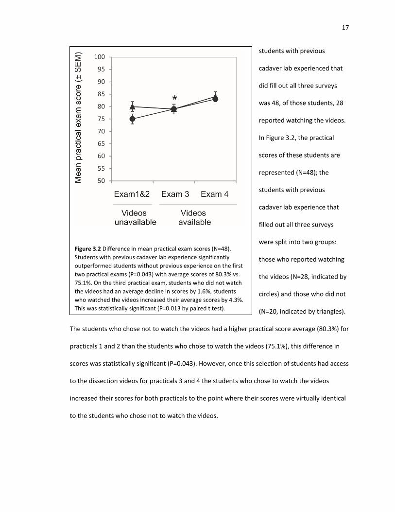
students with previous
cadaver lab experienced that
did fill out all three surveys
was 48, of those students, 28
reported watching the videos.
In Figure 3.2, the practical
scores of these students are
represented (N=48); the
students with previous
cadaver lab experience that
filled out all three surveys
were split into two groups:
those who reported watching
the videos (N=28, indicated by
circles) and those who did not
(N=20, indicated by triangles).
The students who chose not to watch the videos had a higher practical score average (80.3%) for
practicals 1 and 2 than the students who chose to watch the videos (75.1%), this difference in
scores was statistically significant (P=0.043). However, once this selection of students had access
to the dissection videos for practicals 3 and 4 the students who chose to watch the videos
increased their scores for both practicals to the point where their scores were virtually identical
to the students who chose not to watch the videos.
Figure 3.2 Difference in mean practical exam scores (N=48).
Students with previous cadaver lab experience significantly
outperformed students without previous experience on the first
two practical exams (P=0.043) with average scores of 80.3% vs.
75.1%. On the third practical exam, students who did not watch
the videos had an average decline in scores by 1.6%, students
who watched the videos increased their average scores by 4.3%.
This was statistically significant (P=0.013 by paired t test).
18
Figure 3.2 depicts the mean differences of the lab practical scores. On exam 3, the
students who watched the videos improved their scores by 4.3%, a statistically significant
improvement (P=0.013 by
paired t test). The students
who did not watch the videos
had an average decline in
scores for the third lab
practical of 1.6%. This
information suggests that
watching dissection videos
may be correlated with better
lab practical scores. However,
many other factors could
explain these outcomes such
as the presence of teaching
assistants in the lab, student
motivation, the familiarity of
the material in the third and
fourth unit, and student comfort levels approaching faculty for help.
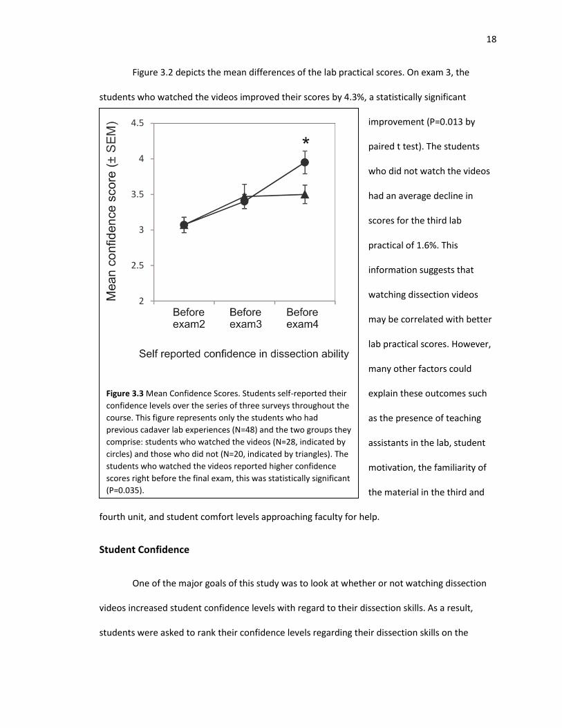
Student Confidence
One of the major goals of this study was to look at whether or not watching dissection
videos increased student confidence levels with regard to their dissection skills. As a result,
students were asked to rank their confidence levels regarding their dissection skills on the
Figure 3.3 Mean Confidence Scores. Students self-reported their
confidence levels over the series of three surveys throughout the
course. This figure represents only the students who had
previous cadaver lab experiences (N=48) and the two groups they
comprise: students who watched the videos (N=28, indicated by
circles) and those who did not (N=20, indicated by triangles). The
students who watched the videos reported higher confidence
scores right before the final exam, this was statistically significant
(P=0.035).
19
surveys so any changes could be tracked over time. For this analysis, we compared the
confidence level ratings of students who had previous cadaver lab experience and completed all
three surveys (N=48), the number of students without previous experience and who filled out all
three surveys was too small for a meaningful analysis. These students are represented in Figure
3.3; they were divided into two groups: those who reported watching the videos (N=28,
indicated by circles) and those who did not (N=20, indicated by triangles). The videos were made
available after the second lab practical. The analysis shows that confidence grew with both
groups over time. This is expected, the more time and practice students have in the lab, the
more confident they will be in their dissection skills. However, the students who reported
watching the videos reported higher confidence levels than those who did not just before the
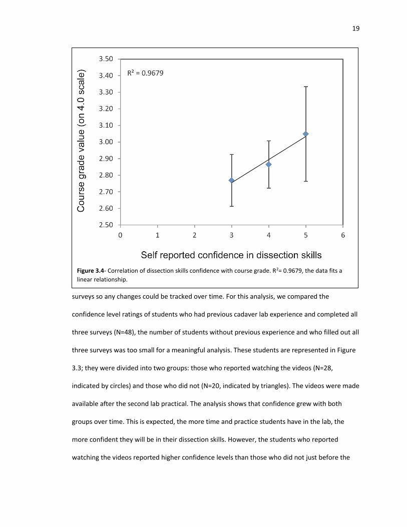
Figure 3.4- Correlation of dissection skills confidence with course grade. R2= 0.9679, the data fits a
linear relationship.
20
final lab exam (P=0.035), the students who did not watch the videos had a plateau in their self-
reported confidence levels. This result supports the hypothesis that the dissection videos helped
to increase student confidence but because the students in this analysis self-reported their
confidence levels, the data is fairly subjective. To address this, an analysis was done to see if
there was a correlation between confidence levels and overall course grade, the results are
shown in Figure 3.4. These results reflect a linear relationship between confidence and overall
course grade which adds additional support to our hypothesis that dissection videos may impact
student confidence levels. Of course, student confidence levels could have been affected by
outside factors like comfort levels in the lab, confidence gained through time and dissection
practice, course performance, and student motivation as the course went on.
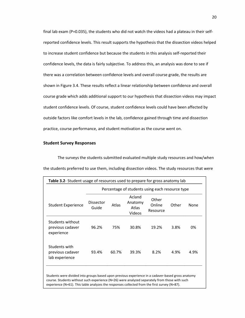
Student Survey Responses
The surveys the students submitted evaluated multiple study resources and how/when
the students preferred to use them, including dissection videos. The study resources that were
Table 3.2- Student usage of resources used to prepare for gross anatomy lab
Percentage of students using each resource type
Student Experience Dissector
Guide Atlas
Acland Anatomy
Atlas Videos
Other Online
Resource Other None
Students without previous cadaver experience
96.2% 75% 30.8% 19.2% 3.8% 0%
Students with previous cadaver lab experience
93.4% 60.7% 39.3% 8.2% 4.9% 4.9%
Students were divided into groups based upon previous experience in a cadaver-based gross anatomy course. Students without such experience (N=26) were analyzed separately from those with such experience (N=61). This table analyzes the responses collected from the first survey (N=87).
21
evaluated to help students prepare for lab were the use of the course dissector guide, use of an
anatomy atlas, use of Acland’s Anatomy Video Atlas, use of other online resources, and an
option of no resources used. Table 3.2 reflects the student preferences for lab preparation.
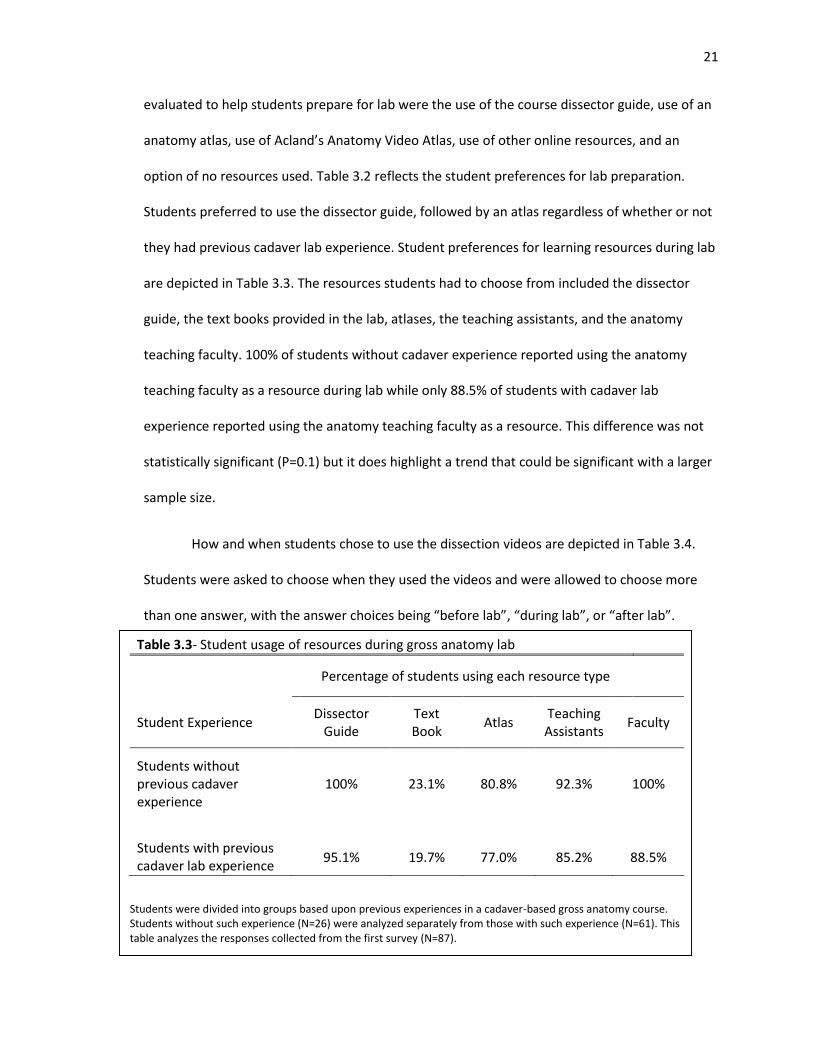
Students preferred to use the dissector guide, followed by an atlas regardless of whether or not
they had previous cadaver lab experience. Student preferences for learning resources during lab
are depicted in Table 3.3. The resources students had to choose from included the dissector
guide, the text books provided in the lab, atlases, the teaching assistants, and the anatomy
teaching faculty. 100% of students without cadaver experience reported using the anatomy
teaching faculty as a resource during lab while only 88.5% of students with cadaver lab
experience reported using the anatomy teaching faculty as a resource. This difference was not
statistically significant (P=0.1) but it does highlight a trend that could be significant with a larger
sample size.
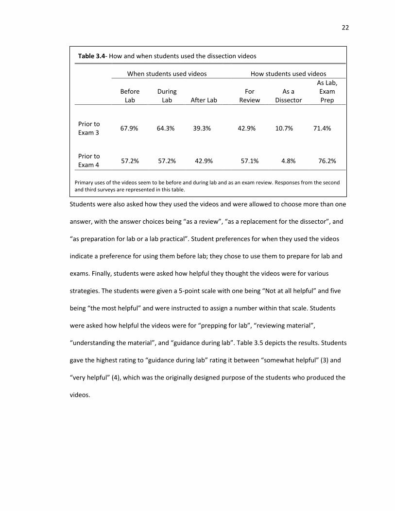
How and when students chose to use the dissection videos are depicted in Table 3.4.
Students were asked to choose when they used the videos and were allowed to choose more
than one answer, with the answer choices being “before lab”, “during lab”, or “after lab”.
Table 3.3- Student usage of resources during gross anatomy lab
Percentage of students using each resource type
Student Experience Dissector
Guide Text Book
Atlas Teaching Assistants
Faculty
Students without previous cadaver experience
100% 23.1% 80.8% 92.3% 100%
Students with previous cadaver lab experience
95.1% 19.7% 77.0% 85.2% 88.5%
Students were divided into groups based upon previous experiences in a cadaver-based gross anatomy course. Students without such experience (N=26) were analyzed separately from those with such experience (N=61). This table analyzes the responses collected from the first survey (N=87).
22
Students were also asked how they used the videos and were allowed to choose more than one
answer, with the answer choices being “as a review”, “as a replacement for the dissector”, and
“as preparation for lab or a lab practical”. Student preferences for when they used the videos
indicate a preference for using them before lab; they chose to use them to prepare for lab and
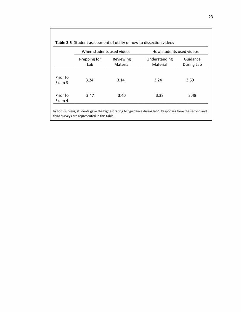
exams. Finally, students were asked how helpful they thought the videos were for various
strategies. The students were given a 5-point scale with one being “Not at all helpful” and five
being “the most helpful” and were instructed to assign a number within that scale. Students
were asked how helpful the videos were for “prepping for lab”, “reviewing material”,
“understanding the material”, and “guidance during lab”. Table 3.5 depicts the results. Students
gave the highest rating to “guidance during lab” rating it between “somewhat helpful” (3) and
“very helpful” (4), which was the originally designed purpose of the students who produced the
videos.
Table 3.4- How and when students used the dissection videos
When students used videos How students used videos
Before
Lab During
Lab After Lab For
Review As a
Dissector
As Lab, Exam Prep
Prior to Exam 3
67.9% 64.3% 39.3% 42.9% 10.7% 71.4%
Prior to Exam 4
57.2% 57.2% 42.9% 57.1% 4.8% 76.2%
Primary uses of the videos seem to be before and during lab and as an exam review. Responses from the second and third surveys are represented in this table.
23
Table 3.5- Student assessment of utility of how to dissection videos
When students used videos How students used videos
Prepping for Lab
Reviewing Material
Understanding Material
Guidance During Lab
Prior to Exam 3
3.24 3.14 3.24 3.69
Prior to Exam 4
3.47 3.40 3.38 3.48
In both surveys, students gave the highest rating to “guidance during lab”. Responses from the second and
third surveys are represented in this table.
24
Chapter 4: Discussion
Student Perceptions
Overall, the students rated the videos between “somewhat helpful” and “very helpful”
across all study strategy choices (as depicted in Table 3.5), they provided helpful feedback for
future video creation. The students were able to write in their own feedback on the second and
third survey and it was constructive and positive. Student comments such as, “Great videos!
Very helpful” and “Loved this resource” were among the many requests for audio narration and
videos of the demonstrations done by faculty during lab. The feedback collected through the
surveys will be implemented in the finalized set of videos created for future classes.
Student Confidence
With regard to student confidence levels, all students gained confidence in their
dissection skills as the course went on. This result was expected because, as the course
continued, students became more comfortable working with cadavers and ultimately logged
more practice time dissecting on cadaveric tissue and working on various dissection techniques.
However, the students who used the videos had a statistically significant higher confidence
rating before the final lab practical than those who did not. This seems to indicate that
dissection videos may increase student confidence when it comes to their dissection skills.
However, there are many other factors that could have affected this result. One such factor
could be the use of teaching assistants available during lab. There were four teaching assistants
in the lab for this particular class and part of their responsibilities included providing assistance
during dissections specifically by providing help with dissection techniques. Their presence in
the lab could have affected the results of this study. It is possible that the students who watched
the videos were more receptive to help from the teaching assistants because they had lower
25
score averages prior to the introduction of the videos. However, the teaching assistants were
present for the entire semester and the increase in confidence we recorded was specifically
after the videos were introduced prior to the third exam. It is hard to gauge how much of an
effect the teaching assistants may have had on student confidence levels.
Student Utilization of the Videos
According to the feedback collected by the surveys, students used the videos primarily
to help prepare for lab or lab practicals. The videos were specifically created to follow along with
the dissector guide the students have access to for the course. The students reported using the
dissector guide as the most common resource they used to help prepare for lab. Because the
videos were made to reflect the steps in the dissector guide, the reported student preferences
may indicate that future students might use the videos in tandem with the dissector guide to
help prepare for lab. Students also reported that they found the videos the most helpful when
using them for guidance during lab, although very high percentages of students reported using
the dissector during lab. This may indicate that the videos have a large amount of value in
helping students navigate dissections when written instructions in the dissector guide are vague,
unclear, or difficult to understand spatially.
Student Performance
Quite possibly the most important finding of this study is the impact that watching the
videos had on student practical scores. The students that watched the videos had a lower
average of scores on the first two lab practicals. As a result, these students may have been more
open to incorporating a new study tool (the videos) than students that had a higher average of
scores. The students who watched the videos increased their scores for both the third and
fourth lab practicals resulting in their scores after introduction of the videos being nearly the
26
same as the students that did not watch the videos. However, many factors could have
influenced this result. Because the students who chose to watch the videos had a lower average
of scores, they may have sought out more help. For example, the teaching assistants in the lab
provided private tutoring to students outside of class, students with lower scores may have
been more likely to seek private tutoring, which could have influenced their practical scores.
Additionally, students with lower average exam scores may have been more motivated to do
better on the last two practical exams of the course and increased their study time and learning
strategies. Finally, it is possible that the students with lower scores just took longer to feel
comfortable or become familiar with the testing format of the lab practicals or that it took them
longer to understand what was expected of them at the graduate level since all but a select few
were first-year students in their specific programs.
Research Limitations
Unfortunately, a major limitation for this study was the sample size, particularly for the
students with no prior cadaver lab experience. The sample size for that group was too small to
have any statistical significance. One way to address this in the future would be to continue to
survey students in future classes. Asking the students to fill out a pre- and post-survey at the
beginning and end of the course instead of three surveys throughout the course may also
increase the response rate. Of course, providing some incentive (extra credit, not releasing
grades until the survey is submitted, etc.) to students to fill out the surveys will increase the
response rate but that could also affect the responses obtained.
Another limitation of our research is that the surveys did not ask students how often
they used the videos. More research needs to be done to see if watching the videos multiple
times a week impacts scoring outcomes. Finally, our study was not able to control for outside
27
factors on student grading outcomes. Student motivation throughout the course was not a
variable we took into consideration; this could be addressed in future studies by having students
rank their motivation levels throughout the course on surveys given at regular intervals. We also
did not control for any influence having the teaching assistants available to students during class
and outside of class (for students who chose to use them for private tutors) may have had on
exam scores.
In the future, more studies should look at levels of experience between students and
how these levels can affect the use of learning resources. In our study, previous cadaver lab
experience was a major determinant of student performance. Future studies could explore how
having previous cadaver lab experience may affect grading outcomes compared to the grading
outcomes of students with previous human anatomy courses that were not cadaver-based as
well as students with no human anatomy experience at all. The results of those studies could
help guide future approaches to teaching anatomy particularly when teaching to students with a
wide variation of anatomy experience.
Conclusions
The videos used by the students in this study were part of a beta-testing series of videos
created by the co-authors. The feedback and comments provided by students on the surveys
will be used to create a finalized set of videos for use by students in the gross anatomy lab in
future classes. The videos will be available on the intranet used by students and embedded
within the online dissecting guide the students have access to. Student feedback from the
surveys included in this study will be used to create a set of videos that are interactive and can
be used across a variety of study strategies in addition to being a visual companion for the
dissector guide.
28
The results from this study, particularly the influence the videos may have had on
practical exam scores, add to the breadth of literature on this subject, however, many of the
results cannot provide definitive conclusions regarding the effect the videos may or may not
have had. More work will need to be done in the future, but hopefully this study has highlighted
the direction that anatomy education has taken and how incorporating new and innovative
learning resources for students can improve learning experiences and outcomes.
29
Bibliography
Azer, S. A. (2012). Can “YouTube” help students in learning surface anatomy? Surgical and
Radiologic Anatomy, 34, 465-468.
Barry, D.S., Marzouk, F., Chulak-Oglu, K., Bennett, D., Tieerney, P., and O’Keeffe, G. W. (2016).
Anatomy Education for the YouTube generation. Anatomical Sciences Education, 96, 90-96.
Choi-Lundberg, D. L., Low, T. F., Patman, P., Turner, P., & Sinha, S. N. (2015). Medical student
preferences for self-directed study resources in gross anatomy. Anatomical Sciences
Education, Vol. 9, 150–160.
Collins, A. M., Quinlan, C. S., Dolan, R. T., O’Neill, S. P., Tierney, P., Cronin, K. J., & Ridgway, P. F.
(2015). Audiovisual preconditioning enhances the efficacy of an anatomical dissection
course: A randomized study. Journal of Plastic, Reconstructive and Aesthetic Surgery, Vol.
68, 1010–1015.
Denning, D. (no date). Video in Theory and Practice: Issues for Classroom Use and Teacher Video
Evaluation. Available: https://www.ebiomedia.com/downloads/VidPM.pdf
Dong, C., & Goh, P. S. (2014). Twelve tips for the effective use of videos in medical education.
Medical Teacher, Vol. 37, 140–145.
Jaffar, A. A. (2012). YouTube: An emerging tool in anatomy education. Anatomical Sciences
Education, Vol. 5, 158–164.
Mahmud, W., Hyder, O., Butt, J., & Aftab, A. (2011). Dissection videos do not improve anatomy
examination scores. Anatomical Sciences Education, Vol. 4, 16–21.
30
Mayfield, C. H., Ohara, P. T., & O’Sullivan, P. S. (2013). Perceptions of a mobile technology on
learning strategies in the anatomy laboratory. Anatomical Sciences Education, Vol. 6, 81–
89.
Mukhopadhyay, S., Kruger, E., & Tennant, M. (2014). YouTube: a new way of supplementing
traditional methods in dental education. Journal of Dental Education, Vol. 78, 1568–71.
Nageswari, K. (2004). Pedagogical effectiveness of innovative teaching methods initiated at the
Department of Physiology, Government Medical College, Chandigarh. Advances in
Physiology Education, Vol. 28, 51–58.
Raikos, A., & Waidyasekara, P. (2014). How useful is YouTube in learning heart anatomy?
Anatomical Sciences Education, Vol. 7, 12–18.
Topping, D. B. (2014). Gross anatomy videos: Student satisfaction, usage, and effect on student
performance in a condensed curriculum. Anatomical Sciences Education, Vol. 7, 273–279.
Wang, R., Mattick, K., & Dunne, E. (2010). Medical students’ perceptions of video‐linked lectures
and video‐streaming. Alt-J, Vol. 18, 19–27.
31
Appendix A: Student Consent Form
“Evaluation of High-Definition “How-To” Dissection Videos for Gross Anatomy”.
The goal of this research is evaluate the benefit of a series of high definition “How To” dissection
videos that will be available to students 24/7 and will provide the students with visual guidance,
showing students how to perform the dissections. These videos will complement the written
instructions and static images in the interactive dissection guide. We hypothesize that these
videos will enhance students’ preparation for gross anatomy lab sessions, promote efficient and
effective use of laboratory time, and improve students’ confidence in their dissection skills.
You were selected as a possible participant in this study because you are enrolled in either CBA
571 or GCBA 908/909 Gross Anatomy courses in the fall of 2015. Your participation in this
research study is voluntary.
If you volunteer to participate in this study, you will be asked to do the following:
Complete 3 brief surveys (before Lab practical 2, 3 and 4 in fall 2015). Survey questions will
focus on if/when/how you used the videos and your perceptions regarding the value of the
videos
Participate in a focus group in which student experiences with the videos will be discussed in
more detail and ideas for possible improvements to the videos will be explored.
Completing any portion of one or more of the surveys and/or attending a focus group meeting
constitutes implied consent to participate.
32
Participation to complete the surveys will take a total of about 30 minutes spread out over
approximately 8 weeks. Each of the three surveys will require about10 minutes to complete.
Surveys will be administered before Lab practical 2, 3 and 4 in fall 2015.
Focus group meetings will require an additional 30-45 minutes. Focus groups will be held early
in 2016 in interaction rooms in MSC.
There are no anticipated risks.
You will not directly benefit from your participation in the research. However, the results of the
research may benefit future students by providing a rationale to generate and a comprehensive
set of instructional “how to” videos for a larger audience of gross anatomy students.
Any information that is obtained in connection with this study and that can identify you will
remain confidential. Confidentiality in the surveys will be maintained by means of a coding
system. Participants will write their name on the cover sheet only. After participants complete
the survey, Dr. Gould will remove the cover sheets and store these in a locked file cabinet to
which only she and Dr. Splittgerber have access. Number coded survey pages (no participant
names) will be analyzed by graduate students Jessica Gamerl and Kevin Selting as part of their
thesis research.
33
A subset of students who complete all of the surveys will be asked to participate in a focus
group. The list of focus group participants will only be known to Dr. Gould, Dr. Splittgerber, Miss
Gamerl, and Mr. Selting (and other focus group participants). A list of focus group participants
will be stored in a locked file cabinet to which only Dr. Gould and Dr. Splittgerber have access
Comments and suggestions regarding the videos that are expressed during the focus group
discussions will be written down but will not be attributed to any specific participant.
You can choose whether you want to be in this study, and you may withdraw your consent
and discontinue participation at any time by simply electing not to complete the surveys.
Whatever decision you make, there will be no penalty to you.
You may refuse to answer any questions on a survey and still remain in the study.
If you have any questions, comments or concerns about the research, you can talk to the one of
the researchers. Please contact: Karen Gould at 402-559-2456 or [email protected] or Ryan
Splittgerber at 402-559-2712 or [email protected].
34
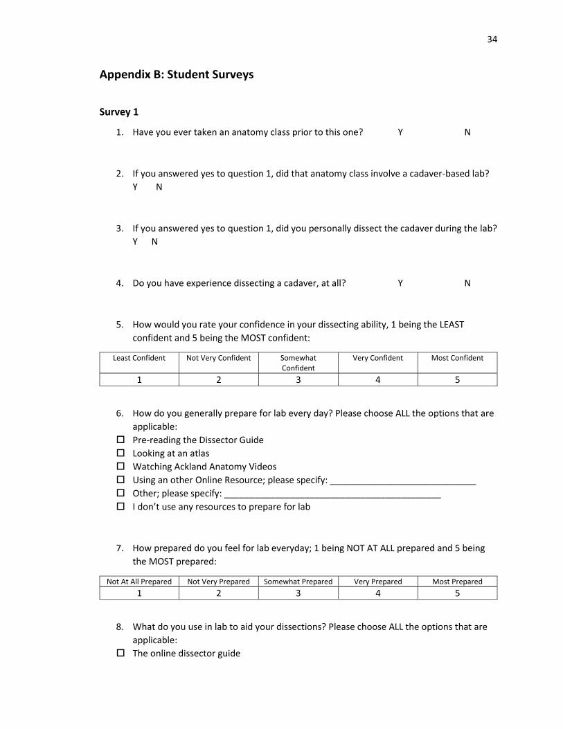
Appendix B: Student Surveys
Survey 1
1. Have you ever taken an anatomy class prior to this one? Y N
2. If you answered yes to question 1, did that anatomy class involve a cadaver-based lab?
Y N
3. If you answered yes to question 1, did you personally dissect the cadaver during the lab?
Y N
4. Do you have experience dissecting a cadaver, at all? Y N
5. How would you rate your confidence in your dissecting ability, 1 being the LEAST
confident and 5 being the MOST confident:
Least Confident Not Very Confident Somewhat Confident
Very Confident Most Confident
1 2 3 4 5
6. How do you generally prepare for lab every day? Please choose ALL the options that are
applicable:
Pre-reading the Dissector Guide
Looking at an atlas
Watching Ackland Anatomy Videos
Using an other Online Resource; please specify: _____________________________
Other; please specify: ___________________________________________
I don’t use any resources to prepare for lab
7. How prepared do you feel for lab everyday; 1 being NOT AT ALL prepared and 5 being
the MOST prepared:
Not At All Prepared Not Very Prepared Somewhat Prepared Very Prepared Most Prepared 1 2 3 4 5
8. What do you use in lab to aid your dissections? Please choose ALL the options that are
applicable:
The online dissector guide
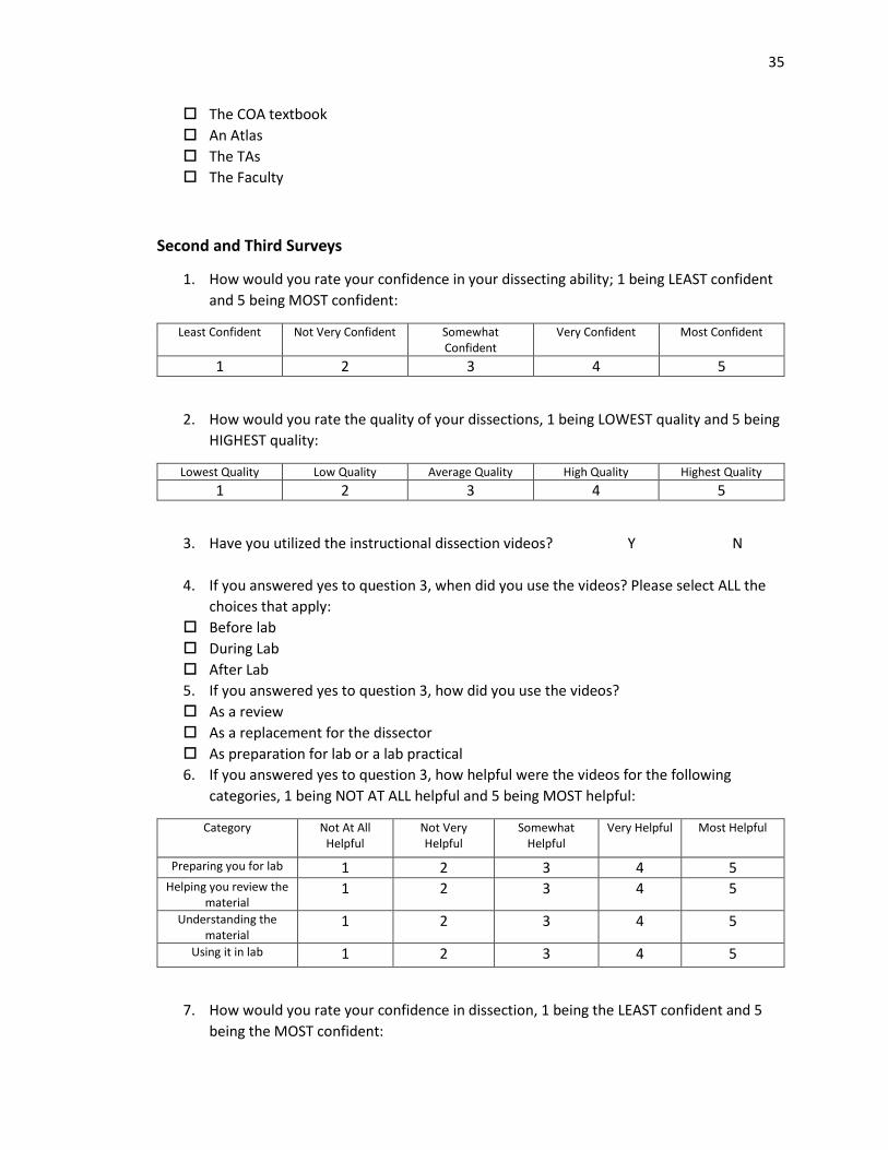
35
The COA textbook
An Atlas
The TAs
The Faculty
Second and Third Surveys
1. How would you rate your confidence in your dissecting ability; 1 being LEAST confident
and 5 being MOST confident:
Least Confident Not Very Confident Somewhat Confident
Very Confident Most Confident
1 2 3 4 5
2. How would you rate the quality of your dissections, 1 being LOWEST quality and 5 being
HIGHEST quality:
Lowest Quality Low Quality Average Quality High Quality Highest Quality 1 2 3 4 5
3. Have you utilized the instructional dissection videos? Y N
4. If you answered yes to question 3, when did you use the videos? Please select ALL the
choices that apply:
Before lab
During Lab
After Lab
5. If you answered yes to question 3, how did you use the videos?
As a review
As a replacement for the dissector
As preparation for lab or a lab practical
6. If you answered yes to question 3, how helpful were the videos for the following
categories, 1 being NOT AT ALL helpful and 5 being MOST helpful:
Category Not At All Helpful
Not Very Helpful
Somewhat Helpful
Very Helpful Most Helpful
Preparing you for lab 1 2 3 4 5 Helping you review the
material 1 2 3 4 5
Understanding the material
1 2 3 4 5
Using it in lab 1 2 3 4 5
7. How would you rate your confidence in dissection, 1 being the LEAST confident and 5
being the MOST confident:
36
Least Confident Not Very Confident Somewhat Confident
Very Confident Most Confident
1 2 3 4 5
8. Do you have any feedback or comments for the videos and how to make them better?
37
Appendix C: Blackboard Announcement
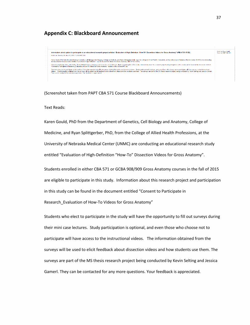
(Screenshot taken from PAPT CBA 571 Course Blackboard Announcements)
Text Reads:
Karen Gould, PhD from the Department of Genetics, Cell Biology and Anatomy, College of
Medicine, and Ryan Splittgerber, PhD, from the College of Allied Health Professions, at the
University of Nebraska Medical Center (UNMC) are conducting an educational research study
entitled “Evaluation of High-Definition “How-To” Dissection Videos for Gross Anatomy”.
Students enrolled in either CBA 571 or GCBA 908/909 Gross Anatomy courses in the fall of 2015
are eligible to participate in this study. Information about this research project and participation
in this study can be found in the document entitled “Consent to Participate in
Research_Evaluation of How-To Videos for Gross Anatomy”
Students who elect to participate in the study will have the opportunity to fill out surveys during
their mini case lectures. Study participation is optional, and even those who choose not to
participate will have access to the instructional videos. The information obtained from the
surveys will be used to elicit feedback about dissection videos and how students use them. The
surveys are part of the MS thesis research project being conducted by Kevin Selting and Jessica
Gamerl. They can be contacted for any more questions. Your feedback is appreciated.
38
Appendix D: Educational Service Office Grant Proposal
Development of High-Definition “How-To” Dissection Videos for Gross Anatomy
Applicants:
Ryan Splittgerber, PhD – Assistant Professor, School of Allied Health Professions.
Dr. Splittgerber currently serves as the M1 Neurosciences director and contributes to gross and
neuroanatomical instruction to SAHP and graduate students. Dr. Splittgerber will oversee the
new gross anatomy lab located in the UNMC Health Sciences Education Building on the
University of Nebraska-Kearney campus in June, 2015. Dr. Splittgerber will train students to use
the high definition surgical camera, oversee all dissections, and evaluate/provide video and
voice-over content.
Karen Gould, PhD – Associate Professor & Vice-Chair for Graduate Education, Department of
Genetics, Cell Biology & Anatomy. Dr. Gould oversees all graduate programs in the department.
Furthermore, she trains graduate students in her laboratory, lectures in multiple graduate and
medical school courses, and directs several graduate courses. Dr. Gould has also developed
multiple graduate courses. Dr. Gould has extensive experience mentoring students
(undergraduates & medical students) participating in summer research opportunities at UNMC.
Students will perform video editing and assembly at UNMC, and Dr. Gould will oversee this
portion of the project.
Project Description:
39
While learning human gross anatomy, medical, physician assistant (PA), physical therapy (PT),
and graduate students spend a considerable amount of time in the anatomy lab dissecting
cadavers under the guidance of multiple anatomy faculty. To provide the students with
consistent, clear instructions for these dissections, students are provided with an interactive
dissection guide (IDG). The IDG includes instructions and images for the various structures and
terms used in the dissection guide. Written instructions in the IDG can only describe what
dissections steps are required; unfortunately, such written instruction cannot demonstrate how
to perform the required steps. For instance, “dissect the facial artery from angle of the mandible
where it is located deep to the platysma…” This step, taken from the IDG, dictates what to do,
but not how to do it. Although faculty members are available to provide guidance during class
time, faculty must divide their time between 4-6 tables, which means that only one table of
students receives initial guidance and that the other tables must proceed on their own.
Frequently, students must wait 50-60 minutes before initial guidance arrives at their table. Also,
students are expected to spend a considerable amount of time outside of class in lab to study
the cadavers and complete their dissections. During this unsupervised time, students have no
guidance other than the written instructions in the IDG. Consequently, students cannot use this
time very efficiently or effectively. To address this problem, we propose to develop a series of
high definition “How To” dissection videos that will be available to students 24/7 and will
provide the students with visual guidance, showing students how to perform the dissections.
Currently, the video resources available to students for gross anatomy are limited to videos
highlighting structures on previously-dissected cadavers. Such videos allow students to visualize
what the structures will look like in the cadaver. However, these videos show the illustrated
structures after the dissection has been completed off-screen and do not show students how to
get to that endpoint. Thus, our “How To” dissection videos, which will illustrate all steps in a
40
comprehensive anatomical dissection, will be a unique and valuable resource for students at
UNMC.
Dissection Videos: This project will result in a series of short videos that correspond to the
individual labs compiled in the existing IDG. The dissections videos will be recorded using a high
definition camera system integrated into a ceiling-mounted surgical lighting boom. The camera
design will provide superior clarity, adjustable zoom, and adjustable perspective of the
dissection field.
Part I: Cadaveric Dissection
a. Students will perform all steps, for each of the 35 IDG dissections, under continuous
video recording.
b. The continuous recording will allow the capture of all procedural steps, including steps
not explicitly stated in the IDG, such as limb positioning and fat and fascia removal in the
dissection field.
c. Sound will not be recorded during the dissection process. Voiceover instructions will be
added during the video editing process.
d. After dissection instructions are complete, dissectors will manually identify anatomical
structures, facilitated by the camera’s moveable arm and zoom functions.
Part II: Video Editing
a. Using the available video editing software, raw footage will be processed to clean up
recording flaws such as unintended camera movement and extraneous footage.
b. This video series is intended to include the assumed and tedious steps between explicit
IDG instructions. To optimize viewing of this information in a timely manner, video
editing software will be used to accelerate playback of the procedural steps.
41
c. During manual identification of structures, the playback will return to real time.
d. The overall duration of each video will range from 5-10 minutes, depending on the
magnitude of the dissection.
e. Voice over instructions will be included after the video is cut and re-assembled. Special
care will be given to coordinate verbal instructions with performance of the explicit
written instructions from the IDG. Additionally, descriptive verbal instructions will
accompany the non-stated procedural steps, such as fat and fascia removal.
f. Voice over will be real time speed even during the accelerated playback of fat and fascia
removal.
Budget: total requested: $5000
1) Male cadaver for the video dissections = $1200. A video will be prepared using a female
cadaver at a later date.
2) Student stipend support= $3800. Stipend support will be provided to two students to
conduct and record the dissections and edit the videos.
Measures of Success: All students taking gross anatomy in the fall of 2015 (first year medical
students, PA students, PT students, masters in medical anatomy students) will have access to
dissection videos via blackboard (videos will be produced in the summer of 2015). We will track
the number of views of each video by students in each gross anatomy course (Med, PA/PT and
graduate). We will also track when students access the videos. Since the videos are optional,
sustained (or increased) usage of the videos throughout the semester would provide an
objective indication that students find the videos helpful. We will also compare laboratory
practical grades of students in fall 2016 to those from the previous year attempt to assess the
42
impact of the videos of student learning and performance. We will also conduct student and
faculty evaluations of the videos at the end of the semester to assess more subjective measures
of success of the videos. Also, focus groups will be convened throughout the fall to gather
student feedback on the videos.
Benefits: The development of a comprehensive series of “How-To” Dissection Videos for Gross
Anatomy has the capacity to impact medical students (N=~135), PA/PT students (N=~100), and
MS Medical Anatomy students (N=~16). These videos will allow students to come to lab fully
prepared---knowing not just what to do, but also how to do it. Such preparation will allow
students to use their time in the lab more effectively. Furthermore, these videos will allow
students to more effectively use unsupervised time in the lab as well. Finally, we are optimistic
that by enhancing the students’ ability to perform the dissections independently and properly,
the students will more effectively master the material in the gross anatomy course.