the molecular genetics of marfan syndrome and related microfibrillopathies
TRANSCRIPT

REVIEW
The molecular genetics of Marfan syndrome and relateddisordersP N Robinson, E Arteaga-Solis, C Baldock, G Collod-Beroud, P Booms, A De Paepe,H C Dietz, G Guo, P A Handford, D P Judge, C M Kielty, B Loeys, D M Milewicz,A Ney, F Ramirez, D P Reinhardt, K Tiedemann, P Whiteman, M Godfrey. . . . . . . . . . . . . . . . . . . . . . . . . . . . . . . . . . . . . . . . . . . . . . . . . . . . . . . . . . . . . . . . . . . . . . . . . . . . . . . . . . . . . . . . . . . . . . . . . . . . . . . . . . . . . . . . . . . . . . . . . . . . . . .
J Med Genet 2006;43:769–787. doi: 10.1136/jmg.2005.039669
Marfan syndrome (MFS), a relatively common autosomaldominant hereditary disorder of connective tissue withprominent manifestations in the skeletal, ocular, andcardiovascular systems, is caused by mutations in the genefor fibrillin-1 (FBN1). The leading cause of prematuredeath in untreated individuals with MFS is acute aorticdissection, which often follows a period of progressivedilatation of the ascending aorta. Recent research on themolecular physiology of fibrillin and the pathophysiologyof MFS and related disorders has changed ourunderstanding of this disorder by demonstrating changesin growth factor signalling and in matrix-cell interactions.The purpose of this review is to provide a comprehensiveoverview of recent advances in the molecular biology offibrillin and fibrillin-rich microfibrils. Mutations in FBN1and other genes found in MFS and related disorders will bediscussed, and novel concepts concerning the complex andmultiple mechanisms of the pathogenesis of MFS will beexplained.. . . . . . . . . . . . . . . . . . . . . . . . . . . . . . . . . . . . . . . . . . . . . . . . . . . . . . . . . . . . . . . . . . . . . . . . . . .
See end of article forauthors’ affiliations. . . . . . . . . . . . . . . . . . . . . . .
Correspondence to:Dr Peter N Robinson,Institute of MedicalGenetics, ChariteUniversity Hospital,Humboldt University,Augustenburger Platz 1,13353 Berlin, Germany;[email protected]
Revised 7 March 2006Accepted 9 March 2006Published Online First29 March 2006. . . . . . . . . . . . . . . . . . . . . . .
Marfan syndrome (MFS; MIM 154700) is arelatively common autosomal dominanthereditary disorder of connective tissue
with prominent manifestations in the skeletal,ocular, and cardiovascular systems. MFS iscaused by mutations in the gene for fibrillin-1(FBN1). Many affected individuals have a char-acteristic habitus with tall stature, long slenderlimbs (dolichostenomelia), arachnodactyly, sco-liosis, and pectus excavatum or carinatum.Ectopia lentis affects up to 80% of individualswith MFS and is almost always bilateral. Theleading cause of premature death in untreatedindividuals with MFS is acute aortic dissection,which follows a period of progressive dilatationof the ascending aorta. Recent comprehensivetreatments of the clinical aspects of MFS havebeen published.1 2
A review of the molecular genetics of MFSappeared in these pages in the year 2000.3 In thehalf decade since then, remarkable progress hasbeen made in elucidating the molecular anatomyof both fibrillin and the fibrillin-rich microfibrilsas well as in understanding the molecularpathogenesis of MFS. Initial ideas about thepathogenesis of MFS concentrated on a staticdominant negative model based on the concept
of fibrillin-rich microfibrils as purely architec-tural elements in the extracellular matrix. Recentfindings greatly enhanced our understanding ofthe pathogenesis of MFS by demonstratingchanges in growth factor signalling and otherchanges in matrix-cell interactions, which hasset the stage for attempts to develop novel formsof treatment.
The current review will focus on the fibrillin-LTBP gene family and tissue organisation ofmicrofibrils, mutations in genes associated withMFS and related phenotypes, the structure offibrillin-1 domains, FBN1 mutations and proteo-lysis, and the genetics of MFS in mouse models.The current review will highlight advancespublished after the first review; interested read-ers are referred to the previous article for moreinformation on other topics.3
THE FIBRILLIN-LTBP GENE FAMILYThe fibrillins and the latent-TGFb-binding pro-teins (LTBPs) form two closely related proteinfamilies with structural and non-structural func-tions in the extracellular matrix. Both familiesare characterised by a modular domain structurewith repeated cysteine-rich modules.
Members of the fibrillin-LTBP family haveboth structural and non-structural functions.Three closely related fibrillins have beendescribed. Fibrillin-24 and the recently discoveredfibrillin-35 have a domain organisation identicalto that of fibrillin-1 and an overall level of aminoacid identity of between 61% and 69%. Severallines of evidence suggest that the fibrillins haveboth overlapping and unique functions.5 6 Allthree fibrillins are structural components of
Abbreviations: BMP, bone morphogenetic protein;cbEGF, calcium binding epidermal growth factor; CCA,congenital contractural arachnodactyly; CMN, cysticmedial necrosis; CSGE, conformation sensitive gelelectrophoresis; DHPLC, denaturing high performanceliquid chromatography; EBP, elastin-binding protein;ECM, extracellular matrix; HNPCC, hereditary non-polyposis colorectal cancer; LAP, latency-associatedpeptide; LDS, Loeys-Dietz aortic aneurysm syndrome; LLC,large latent complexes; LTBP, latent-TGFb-binding protein;MAGP-1, microfibril-associated glycoprotein-1; MFS,Marfan syndrome; MFS2, type 2 Marfan syndrome;MMP, matrix metalloproteinase; MMR, mismatch repair;MSI, microsatellite instability; NMR, nuclear magneticresonance; PTC, premature termination codon; SSCP,single stranded conformation polymorphism; TAAD,thoracic ascending aortic aneurysms and dissections;TbRII, type II TGFb receptor; TGFb, transforming growthfactor-b; UMD, Universal Mutation Database; WMS,Weill-Marchesani syndrome
769
www.jmedgenet.com

microfibrils4 5 7; however, fibrillin-2 and fibrillin-3 are pre-ferentially expressed in embryonic developmental stages,4 5 8
whereas fibrillin-1 is expressed from the gastrula tothroughout adult life.9 In addition, distinct phenotypes areobserved in fbn1 and fbn2 gene targeting experiments in mice;since microfibrils are assembled in both mice, some overlapof architectural functions appears likely.5 10–12
The LTBPs associate with transforming growth factor-b(TGFb), thereby regulating its secretion and spatial andtemporal activation; in humans, four members of the LTBPfamily are known, three of which undergo alternativesplicing.13 TGFb is synthesised as a homodimeric proprotein,whereby the dimeric propeptide is cleaved intracellularlyfrom the growth factor; the propeptide is called the latency-associated peptide (LAP) because TGFb cannot bind to itssurface receptors when it is bound to the LAP. The LAP inturn is usually disulfide-bonded to an LTBP; this aggregate isreferred to as the large latent complex (LLC). The LTBPs thushave a dual function: as structural components of theextracellular matrix and as modulators of TGFb availability(the reader is referred to Rifkin14 and Todorovic et al15 forrecent reviews on this topic).
The fibrillins and LTBPs display a remarkably similardomain structure made up of repeated cysteine-rich struc-tural modules including multiple copies of an epidermalgrowth factor-like module (EGF) and an 8-cysteine (8-Cys)module found only in the fibrillins and LTBPs (fig 1). EGFmodules are approximately 45 residues in length and arecharacterised by six conserved cysteine residues that formthree intramodule disulfide bonds. Forty three of the 47 EGFmodules in fibrillin-1 and many of those in the various LTBPsadditionally conform to a consensus sequence, (D/N)-X-(D/N)(E/Q)Xm(D/N)*Xn(Y/F), that mediates calcium binding inthe N-terminal region of the module (m and n are variable,and an asterisk denotes b-hydroxylation).16
The 8-Cys module (also variously referred to in theliterature as the 8-cysteine module, the TB module, or theLTBP module) occurs only in the fibrillins and LTBPs. Anexperiment using a recombinant construct of one of theseven 8-Cys modules of fibrillin-1 showed that the structureis stabilised by four intradomain disulfide bonds.17 Some8-Cys domains can mediate binding to the LAP-TGFbcomplex.18 19 Not all domains have this function; a recentstudy showed that LTBP1 and LTBP3 bound efficiently toTGFb, LTBP4 bound weakly, and LTBP2 and fibrillin-1 andfibrillin-2 did not bind.20 An additional cysteine-rich modulewith similarities to both the cbEGF module and the 8-Cysmodule, termed the 8-Cys hybrid module,21 can mediateintermolecular disulfide bonding between fibrillin-1 mono-mers, which may be an important step in the assembly ofmicrofibrils.22 The fibrillins share a globular C-terminaldomain of about 120 residues with the fibulins23; infibrillin-1, this module may be involved in homotypicinteractions.24
TISSUE ORGANISATION OF MICROFIBRILSFibrillin microfibrils are widely distributed extracellularmatrix multimolecular assemblies comprised of fibrillin andother proteins. The microfibrils endow elastic and non-elasticconnective tissues with long range elasticity. They directtropoelastin deposition during elastic fibrillogenesis and forman outer mantle for mature elastic fibres. Microfibril arraysare also abundant in dynamic tissues that do not expresselastin, such as the ciliary zonules of the eye. Analysis offibrillin-rich microfibrils by transmission electron microscopyhas revealed them to have a diameter of 8–12 nm, a tubularappearance, and beaded periodicity, and to contain glyco-proteins. Isolated fibrillin-rich microfibrils have a complex‘‘beads on a string’’ appearance, and are extensible.
Fibrillin–1 P
Fibrillin–2 G
Fibrillin–3 PG
LTBP–1
LTBP–2
LTBP–3
LTBP–4
G
PG
P
N-terminal
4-cysP
EGF
cbEGF
LTBP
Pro-rich
Gly-rich
Pro/Gly-rich
Hybrid
C-terminal
Figure 1 The fibrillin-1 gene FBN1 spans about 235 kb of genomic DNA on chromosome 15q21.1, and has a transcript size of 9749 nucleotides; thecoding sequence of FBN1 is spread over 65 exons,21 and three alternatively spliced non-coding 59 exons have been described.249 With severalexceptions, single exons code for the domains in fibrillin-1 as shown here.
770 Robinson, Arteaga-Solis, Baldock, et al
www.jmedgenet.com

Microfibrils form loosely packed bundles in roughlyparallel alignment. These bundles adopt tissue-specificarchitectures that are dictated by cells, and by the strengthand direction of forces put upon the tissue. They are found inlocations that are subject to repeated mechanical stresses,and in the proximity of basement membranes, and they servea critical biomechanical anchoring role in dynamic connec-tive tissues.25
Elastic fibre formation is a developmentally regulatedprocess in which tropoelastin (the soluble precursor ofmature elastin) is deposited on a preformed template offibrillin microfibrils.26 Mature elastic fibres are a compositebiomaterial with an outer microfibrillar mantle and an innercore of amorphous cross-linked elastin with some embeddedmicrofibrils. The proportion of microfibrils to elastin appearsto decline with age, with adult elastic fibres often having onlysparse peripheral mantles of microfibrils.
A recent investigation by mass spectrometry of thecomposition of purified fibrillin-rich microfibrils from non-elastic and elastic tissues showed that in all microfibrilpreparations, fibrillin-1 was abundant and the only fibrillinisoform detected. Isolated microfibrils, extracted from tissuesby enzyme digestions or homogenisation, have a ‘‘beads on astring’’ appearance with untensioned periodicity of,56 nm.27 28 Unextracted hydrated zonular microfibrilsappeared, by quick freeze deep etch microscopy, to be moretubular, suggesting that molecular components are lost orthat there is a major molecular rearrangement on extrac-tion.29 The molecular basis of the ‘‘beads’’, and fibrillin-1alignment in microfibrils remain unclear.
Models of fibril l in-1 alignmentThe details of the precise molecular architecture of fibrillin-1alignment within microfibrils are not entirely clear, andseveral models have been proposed. An intermolecularfibrillin-1 transglutaminase cross-link30 provides molecularconstraints, although mass spectrometry has shown that notall fibrillin-1 molecules within tissue microfibrils are cross-linked.31
The ‘‘hinge’’ model, based on detailed scanning transmis-sion electron microscopy, mass mapping, automated electrontomography, and atomic force microscopy data,28 32–34 predictsmaturation from an initial parallel head to tail alignment toan approximately one third stagger (,100 nm) that wouldallow transglutaminase cross-link formation, and furtherpacking into a more energetically favourable ,56 nmuntensioned form.
The one third staggered model was suggested on the basisof extrapolation of molecular dimensions,35 the crystalstructure of fibrillin-1 cbEGF/TB/cbEGF domain arrays,36
and calcium binding studies of TB/cbEGF flexibility.37
Interested readers can find further discussion of issuessurrounding fibrillin-1 alignment and microfibrillar architec-ture in Baldock et al,28 Davis et al,29, Kielty et al,34 and Lee et al.36
Early studies of isolated microfibrils revealed a number ofhighly stretched microfibrils with periodicities up to,150 nm, which suggested that microfibrils may have elasticproperties.27 Stretching of isolated intact microfibrils has beenachieved using surface tension forces and molecular comb-ing.33 These studies showed that microfibrils behave asrelatively stiff elastic filaments which can perform anchoringroles in ciliary zonules and other basement membraneinterfaces. It was confirmed by x ray diffraction that hydratedmicrofibril bundles are elastic, and suggested that elasticityin microfibril-rich tissues may arise, in part, from reversiblealterations in supra-microfibrillar arrangements. Proteolyticdamage to microfibrils that may occur in MFS or ageing, maygenerate stretched microfibrils that may have lost their elasticproperties.25 38
ASSEMBLY OF MICROFIBRILSToday, a fragmented picture has emerged of the events andmolecules involved in the assembly process from profibrillinsto mature tissue microfibrils. The mechanisms includeprofibrillin processing, self-assembly, regulatory events,cross-link formation, and maturation of microfibrils.
Microfibril assembly in individuals with MFSEarly immunofluorescence studies with dermal fibroblasts orskin biopsies obtained from individuals with MFS showedreduced or qualitatively altered fibrillin networks as com-pared to controls.39–41 However, such altered patterns were notobserved in all samples.41 42 One possible interpretation ofthese data is that a subset of mutations in fibrillin-1compromises the assembly process and thus the formationof microfibril networks. Further evidence for this interpreta-tion comes from pulse chase experiments using dermalfibroblasts from individuals with MFS. These analysesshowed deficiencies at different levels such as fibrillinsynthesis and secretion, as well as deposition into theextracellular matrix.43 44 Despite the differences in thesecreted amount of fibrillin, a large portion of the fibroblastsstudied showed impaired incorporation of the mutantfibrillin into the extracellular matrix, suggesting functionaldisturbances in early stages of the assembly mechanism.45 46
In cases where higher order assembly into beaded micro-fibrils was observed, the ultrastructural appearance showedseveral types of abnormalities including diffuse, frayed, orpoorly defined interbead domains, or variable interbeadperiodicities.47–49 These consequences potentially could alsoemerge from functional problems in the assembly mechan-ism. In summary, although molecular evidence is stilllacking, it seems clear that a certain subset of mutations infibrillin-1 leading to MFS and other microfibrillopathiesdirectly affects microfibril assembly mechanisms.
Role of propeptide processing in fibrill in assemblyFibrillins are phylogenetically old proteins occurring inspecies from jellyfish to human. All known fibrillins possesshighly conserved basic recognition sites (RX(K/R)R) forprocessing by endoproteinases of the furin/PACE type withinthe unique N- and C-terminal domains. It has been shown byseveral groups and methodologies that fibrillin-1 is indeedprocessed at these recognition sites.22 50–54 Processing pro-duces a 17 or 20 residue N-terminal propeptide depending onthe actual cleavage site for the signal peptide, and a 140residue C-terminal propeptide. Due to the size of thepropeptide, C-terminal processing is accessed much betterexperimentally and thus has received more attention. Forfibrillin-1, it has been demonstrated that processing of the C-terminal propeptide is required for deposition into theextracellular matrix, suggesting that profibrillin-1 conversionto mature fibrillin-1 plays a regulatory role in assembly intohigher order aggregates.43 50 53 On the molecular level, it is notclear how the presence of a C-terminal propeptide preventsmatrix deposition and assembly. The propeptide may interactand mask self-assembly sites in fibrillin-1.24 A mutation infibrillin-1 (R2726W) associated with isolated skeletal fea-tures of MFS was shown to interfere with normal processingat the C-terminal end and thus disturbed the incorporation ofthe mutated protein into the extracellular matrix.50 Othermutations close to processing sites may have similarconsequences. Virtually no information is available as regardsthe functional role of the N-terminal processing. However, byanalogy, it is predicted that the N-terminal propeptide alsoregulates assembly of fibrillins.
Self-assembly of fibrill insAs described in the ‘‘Tissue organisation of microfibrils’’section, the molecular organisation of fibrillin-1 in microfibrils
Marfan syndrome and related disorders 771
www.jmedgenet.com

has been analysed by various groups resulting in a numberof different models for the alignment of fibrillin inmicrofibrils.7 28 30 35 36 51 Despite the differences in thesemodels in terms of stagger and molecular condensation ofindividual molecules, common to all models is a head to tailorientation of fibrillin-1 molecules as originally proposed bySakai and coworkers in 1991.7 Another commonly acceptedproperty of microfibrils is the involvement of six to eightfibrillin molecules per cross-section of the interbeadregion.28 55 56
Correlation of antibody epitopes in the fibrillin-1 moleculewith the location of the corresponding epitopes in micro-fibrils clearly revealed that the terminal ends of the fibrillinmolecules are situated in or close to the beads.7 28 51 Dataobtained with recombinant fibrillin-1 fragments have estab-lished direct interaction in a homotypic N- to C-terminalfashion.57 These results established a linear head to tail self-assembly mechanism for fibrillin-1. Heterotypic interactionsbetween fibrillin-1 and fibrillin-2 in an N- to C-terminalfashion suggested that both fibrillin isoforms can beorganised within the same microfibril,57 and colocalisationof both isoforms has indeed been demonstrated by doubleimmunogold labelling in tissue microfibrils.8 Electron micro-scopy after rotary shadowing of full length recombinantfibrillin-1 suggested that the interaction epitopes are rela-tively close to the terminal ends.57 These results were furthersubstantiated by analyses of smaller overlapping fibrillin-1fragments in various ligand interaction assays, positioningthe interaction sites in the N-terminal region encoded byexons 1–8 and the C-terminal region encoded by exons 57–65.24
In addition to a linear head to tail self-interaction, there isevidence that lateral homotypic interactions also play a rolein fibrillin assembly. Reducible homodimer formation earlyduring biosynthesis was observed for recombinant fragmentsof fibrillin-1 and -2 spanning from the proline and glycine-rich domains, respectively, to the second 8-Cys/TB domain,58
as well as for smaller recombinant fragments of the proline-rich region of fibrillin-1 and the glycine-rich region offibrillin-2 including flanking domains.59 Additionally, homo-typic lateral interactions have been observed with an N-terminal recombinant fibrillin-1 fragment encoded by exons1–8, as well as with an C-terminal fragment encoded byexons 57–65.24 The lateral homotypic interactions between N-terminally and C-terminally located fragments as well as thehead to tail linear interactions between the N- and C-terminal ends are of high affinity with dissociation constantsin the low nanomolar range, indicating that both types ofmechanisms are highly relevant for initial assemblystages.24 57
Intermolecular cross-link formation in fibril l inassemblyTwo types of intermolecular cross-links important for thestability of microfibrils have been identified: reducibledisulfide bonds and non-reducible e(c-glutamyl)lysinecross-links. Intermolecular disulfide bond formation isapparent early in the assembly of microfibrils since highermolecular weight disulfide-bonded aggregates containingfibrillin can be observed after a few hours in cell or organcultures.22 60 Most of the highly conserved cysteine residues infibrillins are predicted to stabilise individual domainsthrough intramolecular disulfide bonds.17 35 36 One cysteineresidue in the first hybrid domain of human fibrillin-1 and -2has been shown to be available for intermolecular cross-linkson the surface of the molecule.22
Additional data suggest that other cysteine residues, whichare normally involved in intramolecular domain stabilisation,may be reshuffled to participate in intermolecular cross-links.
Cysteine residues in the first and second 8-Cys/TB domainhave been suggested for such a role based on the propensityof various recombinant fibrillin-1 and -2 fragments to formreducible homodimers.58 59 Such a mechanism would likelyrequire the enzymatic activity of one or more proteindisulfide isomerases on the cell surface or in the extracellularmatrix.
Non-reducible e(c-glutamyl)lysine cross-links, catalysed bytransglutaminases, have been identified in microfibrilsextracted from various tissues.30 61 62 Detailed analyses ofmicrofibrils from human tissues have identified transgluta-minase cross-links in the N- and C-terminal regions offibrillin-1, as well as a high overall content of these cross-links.30 In addition to fibrillin-1, another prominent proteinin microfibrils, microfibril-associated glycoprotein-1 (MAGP-1), was also characterised as a substrate for transglutami-nase.63 It is possible that besides homotypic fibrillin-1transglutaminase cross-links, heterotypic fibrillin-1-MAGP-1 cross-links may be present in microfibrils. Zonular fibres inthe eye have been demonstrated to be a target fortransglutaminase 2.64 Biomechanical analyses of microfibrilssuggested that transglutaminase cross-links play an impor-tant role in strengthening the microfibrils.65 Other potentialroles of the transglutaminase cross-links may include correctlateral alignment of fibrillin or other molecules as aprerequisite for downstream assembly events. Mutationsdisrupting transglutaminase cross-link sites likely result inserious consequences for microfibril assembly and stability.To gain insight into such potential mechanisms in MFS andother microfibrillopathies, it will be important to identify theexact amino acid residues involved and the time course oftransglutaminase cross-link formation.
Accessory molecules important for fibril l in assemblyIn addition to self-assembly and cross-linking mechanisms,other molecules may be involved in the assembly process ofmicrofibrils. Several regions in fibrillin-1 have been identifiedas interacting with heparin/heparan sulfate with highaffinity.66–68 In cell culture assembly assays, these glycosami-noglycans inhibit the formation of microfibrillar net-works,66 67 leading to the hypothesis that heparan sulfate orheparan sulfate containing proteoglycans may have regula-tory functions in the assembly of microfibrils. Recently, theheparan sulfate containing proteoglycan perlecan was iden-tified as a molecule which interacts with fibrillin-1 and withmicrofibrils close to basement membrane zones.69 Reducedamounts of microfibrils in basement membrane zones ofperlecan null mice may reflect a potential role for perlecan inmicrofibril assembly.69
MICROFIBRIL-ASSOCIATED PROTEINSIn addition to fibrillin, several other proteins are integralcomponents of the microfibrils or associated with them. Thesecan be grouped into small non-fibrillin proteins that areintegral parts of fibrillin-rich microfibrils (table 1) and proteinsthat can associate with fibrillin-rich microfibrils but do notserve an integral structural function (table 2). We will notattempt a comprehensive review of the non-fibrillin proteinsbut rather will summarise the most important functions thathave been attributed to these proteins and their interactionwith fibrillin. Readers are referred to Gibson70 and thereferences in the tables 1 and 2 for further information. Itappears likely that more interacting proteins remain to bediscovered in light of a recent proteomics study of fibrillin-richmicrofibrils showing copurification of a number of proteins inaddition to fibrillin-1. For instance, c-crystallin copurified withzonular microfibrils, suggesting an interaction that couldcontribute to zonule anchorage to the lens.31
772 Robinson, Arteaga-Solis, Baldock, et al
www.jmedgenet.com

Structure and assembly of microfibrilsOne role of non-fibrillin microfibrillar proteins is as structuralconstituents of the microfibrils. For instance, microfibril-associated glycoprotein-1 (MAGP-1) is a small glycoproteinwith an apparent molecular weight of 31 kDa that is covalentlybound to microfibrils by disulfide linkages71 and is specificallylocated on the beads of the beaded-filament structure of themicrofibrils.72 MAGP-2 has a more restricted tissue distributionthan MAGP-1,73 suggesting it may have tissue- or develop-mental stage-specific functions. MAGP-1 and MAGP-2 bind todistinct regions of fibrillin-1 and it has been suggested that thismay help to regulate microfibrillar assembly.74
Interaction with other matrix componentsInteractions between fibrillin-rich microfibrils and tropoelas-tin are important for the formation of elastic fibres. Inaddition to interactions between fibrillin-1 and tropoelastin,75
MAGP-1 interacts with tropoelastin76 77 in a way that isimportant for tropoelastin deposition.78 Microfibrils interactwith a variety of other extracellular matrix structures andsome of these interactions are mediated by non-fibrillinproteins. For instance, MAGP-1 binds to the a3 chain of type
VI collagen. Since type VI collagen microfibrils and fibrillin-rich microfibrils are often found near to one another in someextracellular matrices, it is plausible that MAGP-1 maymediate a molecular interaction between type VI collagenmicrofibrils and fibrillin-containing microfibrils.79
Interaction with cellsIt has become increasingly clear that fibrillin-rich microfibrilshave functions that are not directly related to structuralintegrity but rather have to do with growth factor metabolismand triggering cellular signals. As will be discussed in moredetail below, the latent transforming growth factor-b bindingproteins (LTBP) are a family of secreted glycoproteins, threeof which play an important role in the regulation of TGFbregulation.15 In addition to TGFb, microfibrils could con-ceivably be involved in the regulation of other growth factors,although little experimental evidence is available at thispoint.14 MAGP-2 can interact with Jagged1 and induce itsshedding; it is thus conceivable that MAGP-2 may be able tomodulate the Notch signalling pathway.80 There is evidence ofinteraction between fibrillin-1 and at least one bonemorphogenetic protein (BMP),81 82 and fibrillin-2-rich micro-fibrils and BMP-7 have been shown to functionally interactin the regulation of limb patterning in a mouse model.12
Finally, an interesting area of research is the questionwhether novel cell signalling pathways are triggered inMFS or other diseases of the microfibrils, and whetherfragments of the proteins of the microfibrils can acquirenovel signalling properties through exposure of otherwisecryptic binding sites, a phenomenon that has been welldescribed in several other disorders such as osteoarthritis.83
As will be discussed below, there is evidence that fibrillinfragments can induce matrix metalloproteinase (MMP)expression, and it is well known that elastin fragments caninduce MMP expression.84 85
Enzymatic activityThe enzyme lysyl oxidase, which is involved in the cross-linking of tropoelastin monomers, has been localised to theinterface between extracellular bundles of amorphous elastinand the microfibrils.86 Although there is no evidence of adirect interaction between fibrillin and lysyl oxidase to date,it is plausible that interactions with fibrillin or othermicrofibrillar components might be important for elastogen-esis.70 Interestingly, ADAMTS10,87 an extracellular matrixprotease, is mutated in the autosomal recessive form of Weill-Marchesani syndrome (WMS).88 The dominant form of WMSis caused by mutations in fibrillin-1, suggesting, perhaps, apotential interaction between ADAMTS10 and fibrillin-1.
Table 1 Small non-fibrillin proteins that are integralparts of fibrillin-rich microfibrils in at least some tissuesand developmental stages
Protein Chromosome Potential functions
MAGP-1 1p36.1–p35 Tropoelastin deposition78
Tropoelastin binding76 89
Binding to fibrillin-176 90
Binding to fibrillin-291
Ternary complex withtropoelastin and biglycan77
Posttranslational modifications92
Ternary complex withtropoelastin and decorin93
Binding to type VI collagen79
Substrate for transglutaminase63
MAGP-2 12p12.3–p13.1 Binding to fibrillin-174 90
Binding to fibrillin-274
RGD-mediated cell attachment94
Interaction with Jagged180
AAAP-40 5q32–q33.2 40 kDa protein95 96
(MAGP-3)MFAP1 (AMP) 15q15–q21 54 kDa protein that is processed
to a 36 kDa protein97 98
MFAP3 5q32–q33.1 41 kDa serine-rich protein99
MFAP4 17p11.2 Colocalisation to elastic fibres100
(MAGP-36) Role in elastogenesis101
Table 2 Proteins that can associate with fibrillin-rich microfibrils but do not serve an integral structural function in them
Protein Class of protein Potential biological role of interaction
Elastin – Tropoelastin deposition78
Regulation of microfibril formation75
LTBP-1 Fibrillin-LTBP Sequestering of latent TGFb102
LTBP-2 Fibrillin-LTBP Structural role?103
LTBP-4 Fibrillin-LTBP Sequestering of latent TGFb102
Versican Proteoglycan (hyalectin) Link fibrillin-microfibrils to versican/hyaluronan network104
Perlecan Proteoglycan (hyalectin) Anchoring microfibrils to basement membranes and in thebiogenesis of microfibrils69
Heparin/heparan sulfate Proteoglycan Binding of related heparan sulfate chains may regulate cell-surfaceassembly of fibrillin66
Decorin Small dermatan sulfate proteoglycan Induction of fibrillin-1 expression in renal fibroblasts and mesangial cells105
Biglycan Small dermatan sulfate proteoglycan Role in elastogenesis77
Fibulin-2 Fibulin Mediate/modulate attachment of fibrillin to tropoelastin106
Fibulin-5 Fibulin Regulation of the initial deposition of tropoelastin on to microfibrils107
BMP-7 Bone morphogenetic protein Regulation of limb patterning12
EMILIN-1 Elastin-microfibril interface located proteins Role in elastinogenesis108
Marfan syndrome and related disorders 773
www.jmedgenet.com

However, the substrates of ADAMTS10 or indeed otherpotential functions of this molecule remain to be elucidated.
Mutations in FBN1, FBN2, and other genes associatedwith MFS and related phenotypesMutations in FBN1 were discovered in individuals with MFSin 1991,109 and subsequently mutations in FBN2 werediscovered in individuals with a phenotypically relateddisorder, congenital contractural arachnodactyly (CCA).110
More recently, mutations in the genes for TGFBR1 andTGFBR2 were found in several disorders with varying degreesof overlap with classic MFS.111–113 Genetic loci for other formsof isolated aortic dilatation and dissection have beenidentified. The identification of mutations in these geneshas provided significant insight into the pathogenetic path-ways involved in MFS and related disorders, and furtherinsight is to be expected from characterising the fullspectrum of mutations associated with these disorders andfrom identifying the full set of genes in which mutationscause related disorders of connective tissue. The followingsections present the current state of knowledge on FBN1,FBN2, and TGFBR2 mutations and on loci involved in isolatedaortic aneurysm and dissection.
MUTATION ANALYSIS OF THE FBN1 GENE ININDIVIDUALS WITH MFS: SENSITIVITY, METHODS,AND CLINICAL INDICATIONSIn most instances, the diagnosis of MFS can be made onclinical grounds. A set of clinical diagnostic criteria, assummarised in the Ghent nosology,114 define major criteriawith high diagnostic specificity and minor criteria with lessspecificity. In order to make a diagnosis of MFS, the Ghentnosology requires a combination of major criteria in at leasttwo organ systems and involvement of a third organ system.In the majority of cases, these criteria allow the establish-ment or exclusion of the diagnosis. However, the interpreta-tion of these criteria is not always obvious for a number ofreasons. Firstly, MFS is known for its extensive phenotypicvariability both within and between families, which maycause underdiagnosis of the condition.115 Secondly, establish-ing a diagnosis of MFS in children can be difficult becauseseveral manifestations of MFS are age-dependent and maynot yet be present in childhood. Thirdly, clinical overlapexists between MFS and other, so called Marfan-likeconditions which share some of the features of MFS but donot necessarily have the same outcome.
Therefore, if the Ghent criteria are fulfilled, then thediagnosis of MFS is certain. In adult patients presenting witha small number of non-specific skeletal manifestations oftenseen in MFS, the diagnosis is unlikely, but a full clinicalevaluation including echocardiography and ophthalmologicexamination is indicated.
In situations of clinical uncertainty, molecular analysis ofthe FBN1 gene therefore seems a logical aid to the clinicaldiagnosis of MFS. In practice, however, the large andcomplex structure of the FBN1 gene and the wide scope ofFBN1 mutations have hampered clinical implementation ofFBN1 testing. Moreover, literature data show great variationin detection rates of FBN1 mutations and methodologiesused. This can be accounted for by a variety of factors such asthe type of mutational analysis method, the substrate(genomic versus cDNA) and, most importantly, the accuracyof the clinical diagnosis. The studies that have looked at thesensitivity and specificity of FBN1 mutation analysis showsubstantial differences in one or several of these factors.
The first mutational studies, performed on cDNA, showedlow efficiency of FBN1 mutation detection using SSCP (singlestranded conformation polymorphism) and yielded mutationrates of 9% to 23%.116 117 Subsequent studies, using genomic
DNA as template for analysis of the 65 individual exons of theFBN1 gene, obtained higher detection rates but variedaccording to the type of screening method used.118–120
Initially the best results were obtained with CSGE (con-formation sensitive gel electrophoresis), with detection ratesranging from 57% to 90%.42 121 122 Subsequent studies haveshown that mutation detection by DHPLC (denaturing highperformance liquid chromatography) is highly efficient,123
although a relatively high false-positive rate may be anissue.124 It now appears that DHPLC is the most efficientapproach for mutation detection also because of its potentialfor automation when combined with robotic PCR.
The most important factor influencing the mutationdetection rate appears to be the clinical homogeneity orheterogeneity of the patient population. Indeed, severalstudies have shown that the incidence of FBN1 mutationsis significantly higher in patients who fulfil the MFSdiagnostic criteria than in patients who do not. In a largestudy including a cohort of 94 MFS patients and 77 patientswith MFS related phenotypes, Loeys et al125 found FBN1detection rates of 66% versus 5%, respectively. They demon-strated that fulfilling the clinical diagnosis of MFS in itself isa good predictor of the outcome of FBN1 mutation analysis.Katzke et al120 supported these findings by their study whichshowed a much higher incidence of mutations in a group ofMFS patients versus those with an MFS related condition,and concluded that clinical overdiagnosis is the mostimportant explanation for low FBN1 mutation detectionrates. Two other studies123 126 also showed that the majority ofpatients in whom an FBN1 mutation was found, met theclinical diagnosis of MFS, supporting the opinion that therobustness of selection criteria is the most importantdeterminant of the outcome of mutational studies.
Several clinical situations can occur in which molecularstudies of the FBN1 gene may be helpful. In patients whopresent with skeletal, cardiovascular, and/or possibly othermanifestations of MFS but have no involvement of the ocularsystem, it can be difficult to establish the diagnosis strictly onclinical grounds, particularly in the absence of a positivefamily history. Here, however, MRI studies can reveal thepresence of dural ectasia in which case the diagnostic criteriamay still be met. Several mutational studies which reportFBN1 mutations in Marfan-like patients have not verified thepresence or absence of dural ectasia, so that the possibilityremains that the diagnostic criteria are in fact met. In caseswhere no definitive conclusion can be reached with theclinical data, molecular analysis of the FBN1 gene is analternative option.
Because of the evolving nature of the phenotype, particu-larly so for the cardiovascular and skeletal manifestations,children with suspected MFS may not yet fulfil the diagnosticcriteria. In those instances, it is better to postpone a finaldiagnosis until later. The identification of an FBN1 mutationin children or young adults not (yet) fulfilling the diagnosticcriteria can help to identify those who need to be clinicallyfollowed with particular attention.
FBN1 mutations have been identified in a range ofphenotypes, the type 1 fibrillinopathies, with greater or lesserdegrees of clinical overlap with MFS (table 3). A decision as towhether mutation analysis is indicated when such phenotypesare suspected needs to be made on an individual basis.
In addition to the more or less well delineated disorderslisted in table 3, FBN1 mutations can be found in individualswith Marfan-like disorders who do not fulfil the criteria ofthe Ghent nosology.127 It is recommend that accepted clinicalguidelines for the care and management of MFS are appliedin these cases even if the criteria of the Ghent nosology arenot fulfilled, because it is possible that complications such asaortic dilatation can emerge at any age.
774 Robinson, Arteaga-Solis, Baldock, et al
www.jmedgenet.com

Finally, the availability of a molecular test also allowsprenatal or preimplantation diagnosis for prospective parents.This is one option for which requests appear to be steadilyincreasing.
In summary, after stringent clinical selection, a detectionrate of up to 90% is currently achievable in FBN1 moleculartesting of patients with classic MFS.128 This allows theimplementation of mutational studies in clinical practice.
THE UMD FBN1 DATABASE: A DATABASE FOR FBN1MUTATIONS IMPLICATED IN MFS AND RELATEDPHENOTYPESThe UMD FBN1 database (http://www.umd.be) was createdin 1995 in an effort to standardise the information regardingFBN1 mutations using UMD (Universal Mutation Database)software.143–149 The database follows the guidelines onmutation databases of the Hugo Mutation DatabaseInitiative including nomenclature of mutations.150
The mutation records of the database include pointmutations, large and small deletions, insertions, and splicemutations in the FBN1 gene. Each record contains themolecular and clinical data for a given mutation in astandardised, easily accessible, and summary form; ifavailable, data on fibrillin protein biosynthesis classificationgroups45 are included.
To date, 601 FBN1 mutations are available online. Themutations are spread throughout almost the entire genewithout obvious predilection for any given region.Approximately 12% of mutations are recurrent.149
The mutation studies performed to date generally haveconcentrated on screening the 65 coding exons of FBN1. Forthe most part, methods capable of detecting larger deletionswere not applied and it is not clear how much flankingsequence or other regions of the gene were investigated. It isalso unclear whether, once a mutation was identified, theremaining gene regions were regularly and fully evaluated.
Many different kinds of mutation have been identified inFBN1. Point mutations are the most common mutationalevent, with nonsense and missense mutations comprisingabout 10% and 60% of all reported mutations. The mostcommon missense mutations substitute cysteine residuesthat form disulfide bonds within one of the cbEGF or 8-Cysdomains, but missense mutations creating novel cysteineresidues in these modules are also common. The majority ofthe remaining mutations in these modules affect residues ofthe calcium consensus sequence. About a quarter of missensemutations affect modules other than cbEGF, and for the most
part, the pathophysiological mechanisms of these mutationsremain unclear.
Small insertions, deletions, or duplications represent about13% of all reported mutations. The majority of thesemutations create a premature termination codon (PTC).Another 13% of reported mutations consist of various classesof splicing errors, most commonly affecting canonical splicesequences at exon/intron boundaries. Many splice sitemutations in FBN1 result in in-frame exon skipping, suchthat the mutant fibrillin-1 lacks an entire cbEGF domain.Such mutations can be associated with a particularly severephenotype.151 Some exon-skipping mutations in FBN1 resultin a frameshift152 with reduced mutant RNA levels throughnonsense-mediated decay of the mutant transcript.153 Anonsense mutation136 and a silent exonic mutation154 in exon51 have been reported as inducing in-frame skipping of theentire exon 51 and demonstrate the existence of an exonicsplicing enhancer.155 156
Global analysis of FBN1 mutations reveals two classes ofmutations. The first type, which represents more than onethird of the mutations, contains mutations predicted to resultin shortened fibrillin-1 molecules, including nonsense muta-tions, splicing errors, insertions, and duplications, as well asin-frame or out-of-frame deletions. These mutations arelikely to result in nonsense-mediated decay resulting inreduction in the level of the mutant allele. The second typerepresents slightly less than two thirds of the mutations andcontains missense mutations, mostly located in cbEGF-likemodules. They can be subclassified into: (a) mutationscreating or substituting cysteine residues potentially impli-cated in disulfide bonding and consequently in the correctfolding of the monomer; (b) amino acids implicated incalcium binding and subsequently in interdomain linkage,structural integrity of affected domains, and increasedprotease susceptibility; and (c) other mutations that mightaffect the conformation of affected modules, interdomainpacking, or other functions such as protein-protein interac-tions.
Elucidating the molecular basis of MFS and relatedfibrillinopathies is the major goal of the teams working onthis subject.149 157 The extreme clinical variability, the diffi-culties associated with clinical diagnosis, and the lowdetection rate of mutations in this large gene all conspire tonegatively impact on progress. At present it is not possible topredict the phenotype for a given FBN1 mutation. On the onehand, mutations affecting different positions within a givenmodule may be associated with quite different phenotypes.
Table 3 Type 1 fibrillinopathies
Syndrome Clinical features Reference
MFS See text See textNeonatal MFS Severe end of clinical spectrum Kainulainen et al,117 Booms et al129
Atypically severe MFS Severe and early onset cardiovascular complications Putnam et al,130 Tiecke et al,131
Ectopia lentis Mainly ocular findings Lonnqvist et al,132 Ades et al,133
Kyphoscoliosis Progressive kyphoscoliosis of variable severity Ades et al134
Familial arachnodactyly Dolichostenomelia and arachnodactyly Hayward et al135
Familial thoracic ascending aortic See textaneurysms and dissectionsMASS phenotype Mitral valve prolapse, aortic dilatation without dissection, Dietz et al136
skeletal and skin abnormalitiesShprintzen-Goldberg syndrome Craniosynostosis, a marfanoid habitus, and skeletal, Sood et al,137 Kosaki et al138 Robinson et al139
neurological, cardiovascular, and connective tissue anomaliesIsolated skeletal features Tall stature, scoliosis, pectus excavatum, arachnodactyly Milewicz et al50
New variant of MFS Skeletal features of MFS, joint contractures, ectopia lentis, Stahl-Hallengren et al,140 Black et al,141
no cardiovascular manifestationsWeill-Marchesani syndrome Short stature, brachydactyly, joint stiffness, and Faivre et al142
(autosomal dominant) characteristic eye abnormalities
Although classic MFS is by far the most common disorder associated with FBN1 mutations, several other disorders with overlapping clinical findings have beendescribed due to mutations in FBN1.
Marfan syndrome and related disorders 775
www.jmedgenet.com

On the other hand, mutations affecting an analogous residuewithin two different modules may also be associated withdiffering phenotypes. Therefore, it is apparent that neitherthe location of the affected structural module in the proteinnor the position of the altered residue is, in itself, sufficient topredict potential genotype-phenotype correlations.158 Thehigh degree of intrafamilial variability suggests that environ-mental and perhaps stochastic factors or modifying genes areimportant for the phenotypic expression of disease. The levelof the expression of the normal fibrillin-1 allele159 andhyperhomocysteinaemia related to the C677T methylenete-trahydrofolate reductase polymorphism160 have been pro-posed as factors that modify the clinical severity of MFS. Theelucidation of the full range of modifying factors in MFSrepresents an interesting area for further research.
MUTATIONS IN TGFBR1 AND TGFBR2Signalling by TGFb family cytokines controls a variety ofcellular processes including proliferation, differentiation, andapoptosis; propagation of signalling into the cell is mediatedby a family of type 1 and type 2 receptors including the type 1and type 2 TGFb receptor. TGFb binds first to type IIreceptors, allowing subsequent incorporation of type Ireceptors into a ligand-receptor complex involving a TGFbdimer and four receptor molecules. The signal is thenpropagated into the cell by means of phosphorylation of theSmad proteins.161
Linkage to chromosome 3p24.2–p25 was demonstrated fora large family with a Marfan-like phenotype for whomlinkage to FBN1 and FBN2 had previously been excluded.162 163
This disorder has been termed MFS type II (MIM 154705)and shares some of the cardiovascular and skeletal featuresof classic MFS.
Identification of a chromosomal breakpoint disrupting thegene encoding the TGFb receptor 2 (TGFBR2) in a boy withshort stature, dural ectasia, and several skeletal andcardiovascular manifestations of MFS led to the identifica-tion of three further missense mutations in four families orindividuals with manifestations of MFS in whom FBN1mutations had been ruled out (one mutation was found intwo unrelated families).111 All the mutations were found inthe serine-threonine kinase domain of the TGFb receptor 2.
More recently, a new aortic aneurysm syndrome withhypertelorism, bifid uvula or cleft palate, and generalisedarterial tortuosity with ascending aortic aneurysm togetherwith other findings such as craniosynostosis, mental retarda-tion, and congenital heart disease was described; thisdisorder, Loeys-Dietz aortic aneurysm syndrome (LDS;
MIM 609192), was shown to be associated with mutationsin the genes for either TGFb receptor type 1 or TGFb receptortype 2 resulting in perturbations of TGFb signalling.112 As willbe noted below, mutations of the arginine at position 460 ofTGFBR2 have been identified in individuals with thoracicascending aortic aneurysms and dissections (TAAD),although there appears to be some degree of phenotypicoverlap with LDS in many affected individuals.
Microsatellite instability (MSI) is a prominent feature inhereditary non-polyposis colorectal cancer (HNPCC) andsome forms of acquired colon cancer. Defects in mismatchrepair (MMR) genes and associated coding region MSI cancause frameshift mutations with functional inactivation ofaffected genes, thereby providing a growth advantage toMMR deficient cells. TGFBR2 is one of the most commonlyaffected genes with mutations in a polyadenine tract in exon3 being found in up to 90% of cases of microsatellite-instablesporadic and HNPCC associated colon cancer.164 Germlinemutations in TGFBR2, however, are a rare cause of HNPCC.165
Interestingly, one mutation (R528H) was found both as asomatic mutation in colon cancer166 167 and as a germlinemutation in LDS.112 At present, there is no evidence thatindividuals with LDS are at increased risk for colon cancer.
It should be noted that there has been some controversy asto whether certain TGFBR2 mutations lead to a phenotypethat is identical or at least very similar to that of classic MFS,thus justifying the diagnosis of type 2 MFS (MFS2). Thequestion about locus heterogeneity for MFS has beenaddressed by several studies. Historical linkage data in MFSfamilies showed a cumulative LOD score for the FBN1 locusin excess of 100, which provides evidence for a singlepredominant locus for MFS. On the other hand, none of theindividuals with LDS112 fulfilled the clinical diagnostic criteriafor MFS. In a previous study, Loeys et al128 identified 86 FBN1mutations in a cohort of 93 patients with classic MFS. Noneof the remaining patients had a TGFBR2 or TGFBR1 mutation.Overall, this suggests strongly that FBN1 is the predominantif not sole locus for MFS.128 However, there have been reportsof individuals with TGFBR2 mutations diagnosed with MFS2without features characterising LDS.168 Given that theaneurysms in LDS appear to be more aggressive than thosein MFS and the fact that the cardiovascular involvement isalso characterised by arterial aneurysms throughout thearterial tree and marked arterial tortuosity, it is clinically veryimportant to be aware of the potential differences betweenthese two syndromes. Detailed clinical characterisation ofindividuals with TGFBR2 mutations will be required todetermine if a subset of these mutations is associated withMFS2 or whether the diagnosis of LDS is more appropriate.
R522N
Cancer
LDS
Extracellular Protein kinase
del32–37
A(10): frameshift
L308P
T315M
Y336NA355PG357W
V387M V447A
N435SL452M
R460skipEx6S449F
E526GR528H
R528CR528H
R537C
Figure 2 Mutations in TGFBR2 found in Loeys-Dietz aortic aneurysm syndrome (LDS)112 and related hereditary disorders111 113 and representativemissense mutations found in colon carcinoma166 250 and breast carcinoma.251 The TGFBR2 protein is drawn according to UniProt entry P37173 andcomprises an N-terminal signal sequence, an extracellular domain, a transmembrane domain (stippled rectangle), a cytoplasmatic region of unknownsignificance (the white box), the serine-threonine protein kinase domain, and a C-terminal domain (black rectangle).
776 Robinson, Arteaga-Solis, Baldock, et al
www.jmedgenet.com

MUTATIONS IN FBN2 AND CONGENITALCONTRACTURAL ARACHNODACTYLYThe discovery of a second fibrillin gene led to the geneticassociation of fibrillin-2 encoded by FBN2 with congenitalcontractural arachnodactyly (CCA). CCA or Beals syndromeis characterised by a marfanoid habitus. In addition to thetall, slender asthenic appearance, most individuals with CCAhave crumpled ears, flexion contractures, severe kyphosco-liosis, and muscular hypoplasia.169–172 The ear abnormalitiesare characterised as a folded upper helix of the external ear.In most patients, contractions of major joints (knees, elbows,ankles) are present at birth. The proximal interphalangealjoints display flexion contractures (that is, camptodactyly).Contractures of the hip, adducted thumbs, and clubfoot mayalso occur. Bowed long bones and muscular hypoplasia areadditional musculoskeletal findings in CCA. Contracturesusually resolve with time. Arachnodactyly (long slenderfingers and toes) is present in most individuals with CCA.The greatest morbidity in CCA is caused by progressivekypho/scoliosis that can begin in early infancy. It is present inabout half of all affected individuals. The spinal abnormalitiesare progressive. Severe thoracic cage abnormalities withassociated scoliosis may cause restrictive pulmonary disease.173
While CCA shares some clinical characteristics with MFS(table 4), it does not share the usually shortened life expectancy.
Putnam et al110 were the first to identify mutations in twounrelated individuals with CCA; both were cysteine substitu-tions. It is important to note that in contrast to thedistribution of FBN1 mutations causing MFS throughoutthe coding region, the FBN2 mutations so far identified inCCA appear to cluster between exons 23 and 34. Thehomologous region of FBN1, the so called neonatal region,contains the greatest percentage of mutations from MFSpatients at the most severe end of that disorder’s clinicalspectrum.117 Virtually all of the known FBN2 mutations are ofthe calcium binding epidermal growth factor-like (cbEGF)domains.174–178
Molecular studies of only one individual with severe/lethalCCA have been performed.179 This individual had an exonsplicing mutation that caused the skipping of exon 34, acbEGF-like domain. Significantly, this individual’s motherwas a somatic mosaic with one third of her fibroblasts alsoharbouring the same exon 34 mis-splicing mutation.Therefore, one can speculate that there is a threshold forcertain mutations causing skeletal perturbations versussevere developmental abnormalities in the cardiovascularand gastrointestinal systems.
Although individuals with CCA usually do not have aorticinvolvement, aortic root dilatation does occur in some casesand screening for aortic involvement should be performed inindividuals with this disorder.180
Fibrill in-2 in development and animal modelsThe temporal and spatial expression of fibrillin-2 has beenexamined in several species. In human fetal aorta, antibodiesto fibrillin-2 were found to stain most intensely in the media,where elastic fibres are most abundant. In human elasticcartilage, fibrillin-2 localised to the cartilaginous core while
fibrillin-1 localised primarily to the surrounding connectivetissue.4 Fibrillin-1 and fibrillin-2 demonstrate a similarspatial and temporal distribution in most tissues duringearly human embryonic development. Exceptions includedthe kidney, liver, rib anlagen, and notochord.181 Similarstudies in the developing mouse showed that in most tissuesfibrillin-2 was expressed earlier than fibrillin-1.182
Studies in the chick have shown that fibrillin-2 (called JB-3 in the early literature) is expressed very early in develop-ment and is found in the regions of heart development.183 Theearly expression of the fibrillins has led to speculation thatthey may mediate the tensile forces that shape the earlyembryo.184 A possible role for fibrillin-2 in lung developmenthas been shown in a rat model. Studies of fetal lung explantsdemonstrated abnormal branch morphogenesis when theexplants were incubated with antisense oligonucleotides tofibrillin-2.185
Browning et al186 described a mouse with syndactyly (sne)that was derived from chemical mutagenesis of murineembryonic stem cells. They showed that sne (now renamedsyfp-3J) was an allele of the sy locus. sy is the shaker-with-syndactylism mouse, a radiation mutant with a chromosome18 (syntenic to human chromosome 5) contiguous genedeletion syndrome.187 The deleted region contains the geneencoding fibrillin-2. Some spontaneously occurring mousemodels with syndactyly also mapped to the sy locus (syfp andsyfp-2J). All three syfp mutations are FBN2 mutations.186 188
Additional evidence that absence of FBN2 leads to syndactylycame from gene targeting studies.12 The FBN22/2 knockoutmouse displayed the same type of syndactyly observed in thesy mice. Interestingly, two of the fibrillin-2 mutations in thesyfp mice were outside the neonatal region, that is, the area inwhich all of the human CCA mutations have been found.188
These findings have led to the obvious speculation thatfibrillin-2 mutations outside the neonatal region, for exam-ple, may lead to other human phenotypes.188
FAMILIAL THORACIC ASCENDING AORTICANEURYSMS AND DISSECTIONSCystic medial necrosis (CMN) is known to be associated withsyndromes such as MFS, but is more frequently found in theabsence of an associated phenotypic syndrome. Reports offamilies with autosomal dominant inheritance of thoracicaortic aneurysms leading to type A dissections (TAAD) withmedial necrosis on pathologic examination indicate thatsingle gene mutations can cause medial necrosis in theabsence of an associated syndrome.189 190 Additionally, medialnecrosis of the proximal aorta with aneurysms/dissections isassociated with other heritable diseases such as Turnersyndrome,191 Noonan syndrome,192 Ehlers-Danlos syn-drome,193 patent ductus arteriosus,194 195 and bicuspid aorticvalve.196 197
Initial studies showed that first degree relatives ofprobands with non-syndromic TAAD have a higher risk ofthoracic aortic aneurysms and sudden death compared with acontrol group.198 199 In addition, these studies support thehypothesis that genetic factors play a role in the aetiology ofTAADs in patients who do not have an identified syndromecausing aortic disease. Milewicz and colleagues described sixfamilies with aortic aneurysms and dissections, all of whomdemonstrated autosomal dominant inheritance associatedwith decreased penetrance and variable age of onset of theaortic disease.189 Various studies indicate that the aorticdisease in the majority of these families is not due to amutation in the FBN1 gene or other genes encoding vascularproteins, such as COL3A1.189 200 201
More recently, several loci for non-syndromic TAAD havebeen mapped. The first locus, termed TAAD1, was mapped tochromosome 5q13–14 with a maximum LOD score of 4.74
Table 4 Clinical features of congenital contracturalarachnodactyly
Marfanoid habitusFlexion contractures of multiple joints including elbows, knees, hips,fingersKyphoscoliosis (sometimes severe)Muscular hypoplasiaAbnormal pinnae (presenting as crumpled outer helices)
Marfan syndrome and related disorders 777
www.jmedgenet.com

with the marker D5S2029.202 This locus was confirmed by anindependent study in a Finnish population where approxi-mately one half of the families studied show evidence oflinkage to TAAD1.203 The critical interval containing thedefective gene maps to a 7.8 cM region. Another locus forfamilial aortic aneurysms and dissections has been mappedto the long arm of chromosome 11 (11q23–24) using a singlelarge family.204 In contrast to the families linked to TAAD1,the clinical phenotype of the family linked to the FAA1 locusindicated a diffuse vascular aetiology. Apart from dilatationin the sinuses of Valsalva, involvement of other aorticsegments and arteries was also observed, such as dilatationin the abdominal aorta and left subclavian artery. In addition,the disease was fully penetrant with aortic imaging in thefamily described. The FAA1 locus is a rare cause of thevascular condition as indicated by the fact that no otherfamilies demonstrate linkage of the phenotype to markers atthis locus.
Another locus for TAAD was mapped to a 25 cM region onchromosome 3p24–25 using another large family withmultiple members with aneurysms and dissections of thethoracic aorta. The disease in the family was characterised asautosomal dominant with decreased penetrance and variableage of onset.205 Eighteen TAAD families described previouslyfailed to show linkage to 3p24–25, indicating that TAAD2 is aminor locus for TAAD.202 204 It was recently determined thatmutations in the transforming growth factor beta receptortype II gene (TGFBR2) is the cause of disease at the TAAD2locus.113 The TGFBR2 gene was screened for missense,nonsense, and exon splicing errors and mutations werefound in four out of 80 unrelated families with familialTAAD, indicating that TGFBR2 mutations are a relatively rarecause of familial TAAD. Although most vascular disease inthese families involved ascending aortic aneurysms leading totype A dissections, affected family members also haddescending aortic disease and aneurysms of other arteries,including cerebral, carotid, and popliteal aneurysms.Strikingly, all four families carried mutations that affectedarginine at amino acid 460 in the intracellular domain,suggesting a mutation hot spot for familial TAAD andestablishing a strong genotype-phenotype correlationbetween familial TAAD and mutations at this location.Structural analysis of the TGFBR2 serine/threonine kinasedomain reveals that R460 is strategically located within ahighly conserved region of this domain and that the aminoacid substitutions resulting from these mutations willinterfere with the receptor’s ability to transduce signals. Asurprising observation in these families was that there is noevidence of an increased susceptibility to cancer in familieswith germline TGFBR2 mutations, despite evidence in theliterature that somatic TGFBR2 mutations occur in a varietyof cancers.
In summary, TAAD is a genetically heterogeneous diseasethat may be inherited in conjunction with a syndrome or as anon-syndromic predisposition for TAAD. Studies of familialTAAD due to TGFBR2 mutations have highlighted thedysregulation of the TGFb pathway as a mechanism leadingto aneurysm formation. Clinically, it is important to performcardiovascular evaluation on first degree relatives of indivi-duals with suspected non-syndromic familial aortic dissec-tion, because of the possibility of potentially life-threateningvascular disease in an individual with obvious phenotypicsigns of the disorder.
THE EFFECTS OF FIBRILLIN MUTATIONS ON THESTRUCTURE OF FIBRILLIN-1 DOMAINSIn order to understand the effects of fibrillin mutations onthe structure and function of the tissues affected by MFS, itwill ultimately be necessary to understand the effects of
mutations on protein structure. It has not been possible todetermine the structure of the entire fibrillin-1 protein oreven of larger fragments thereof. However, studies onrecombinant polypeptides comprising up to several cbEGFor 8-Cys modules have provided significant insight.
The two predominant structural modules in fibrillin-1 arethe calcium-binding epidermal growth factor-like domain(cbEGF) and the transforming growth factor b bindingprotein-like (TB or 8-Cys) domain. High resolution structuresof the fibrillin-1 domain fragments cbEGF32–33, TB6, andcbEGF12–13 have been solved previously using nuclearmagnetic resonance (NMR) which identifies the solutionstructure of the molecule.17 35 206 More recently, x ray crystal-lography has been used to solve the structure of the tripledomain fragment, cbEGF22-TB4-cbEGF2336 and demon-strated a calcium-stabilised tetragonal pyramidal conforma-tion. An RGD integrin binding site localises to the tip of a b-sheet within TB4 and is thus accessible to cell-surfaceintegrins. Comparative sequence alignments of the linkerregions from cbEGF-TB domains within fibrillin-1 suggestthat the relative orientation of cbEGF22-TB4 is likely to bepreserved at homologous sites within fibrillin-1. In contrast,the variation in amino acid number and composition of TB-cbEGF linker sequences suggests that these pairs will adoptdifferent orientations with respect to one another withinfibrillin-1, and may contribute to the biomechanical propertiesof microfibrils. This is supported by the variability of Kd valuesof TB-cbEGF pairs from fibrillin-1 (see below). A model of alarge region of fibrillin-1 (cbEGF11-TB5) has been generatedfrom this combined structural information, which suggeststhat although the protein is in an extended conformation, it isnot simply linear. A significant bend is introduced by thepacking of cbEGF22 against TB4. Based on this structure, astaggered model for assembly of fibrillin-1 into the microfibrilhas been suggested as an alternative to the proposedorganisation based on electron microscopic studies.36
Single cbEGF domains expressed from fibrillin-1 and otherproteins usually display low affinity binding in the mMrange. However, in fibrillin-1, and in many other proteins,the cbEGF domains are often arranged as repeating tandemarrays. On covalent linkage of an N-terminal cbEGF, theaffinity of the C-terminal cbEGF increases.207 The boundcalcium, together with the hydrophobic packing interaction,performs a key structural role in restricting interdomainflexibility35 208 and therefore protects the modules againstproteolytic cleavage.209 210 Dynamics studies show that themost stable region of a cbEGF pair is in the vicinity of theinterdomain calcium-binding site.208 Analysis of differentcbEGF domain pairs has, however, identified a range ofaffinities from 350 mM to 300 nM, suggesting that primarysequence variation, in addition to the pairwise domaininteraction, must also influence affinity.211 212 A study ofheterologous TB-cbEGF domain pairs37 has shown that mostof these domain pairs bind Ca2+ considerably more tightlythan previously observed, with Kd values as low as 9 nM.These data suggest that under physiological conditions, manyfibrillin-1 cbEGF domains will be fully saturated and mayimpart rigidity to the native protein. However, the TB6-cbEGF32 domain pair, with a Kd of 1.6 mM,213 appears themost likely of the TB-cbEGF domain pairs to be flexible andmay contribute to the extensibility and elasticity of themicrofibrils.
Insights into the role of a number of disease-causing FBN1mutations in the pathogenesis of MFS have been gained fromNMR, calcium chelation, and limited proteolysis studies ofrecombinantly expressed fragments of fibrillin-1. Reductionof calcium binding caused by substitution of a calcium ligandor destabilisation of the interdomain interface would bepredicted to produce a less extended, more flexible structure
778 Robinson, Arteaga-Solis, Baldock, et al
www.jmedgenet.com

within a region of fibrillin-1. This may result in increasedproteolytic susceptibility due to exposure of enzyme-specificcryptic cleavage sites. The effect of a missense mutation onprotease susceptibility of a cbEGF domain can, however, beinfluenced by a number of factors such as the particularresidue mutated and the position of the mutant domainwithin the fibrillin-1 peptide.
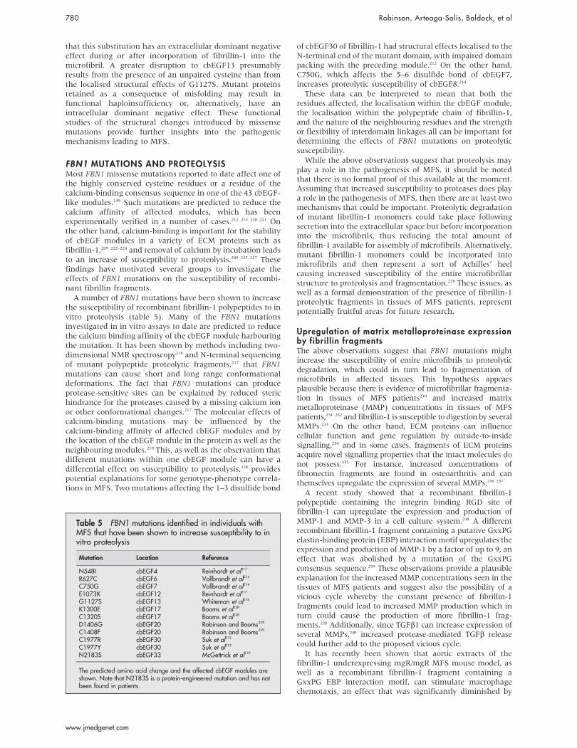
The structural effects of the pathogenic mutations C1977Yand C1977R which disrupt the 1–3 disulphide bond ofcbEGF30 and are therefore predicted to cause misfolding,have been studied in a cbEGF29–31 triple construct using thecombined methods of NMR, chelation, and limited proteo-lysis.212 The substitutions caused loss of Ca2+ binding tocbEGF30, consistent with intradomain misfolding anddisrupted cbEGF29–30 domain-domain packing.Surprisingly, the calcium binding properties of cbEGF29and cbEGF31 were unaffected, suggesting these cysteinesubstitutions have relatively localised effects confined to theN-terminal end of the mutant domain (fig 3A). However, adisruption of the 5–6 disulphide bond by a C750G substitu-tion which affects the C-5 residue of cbEGF7 in an EGF4-TB3fragment caused increased proteolytic susceptibility ofcbEGF8.214 This is presumably due to disruption of domainpacking between cbEGF7 and 8 and hence reduction of thecalcium binding affinity of cbEGF8. Cysteine substitutionsare therefore likely to have different structural effects, whichdepend on the particular disulphide bond affected, and henceresult in a variety of pathogenic mechanisms.
These studies, together with earlier reports, emphasise thestructural heterogeneity that can be introduced into fibrillin-1 by different FBN1 mutations. In the case of the foldingsubstitution, G1127S, in cbEGF13, it was shown215 216 that themutant domain retained the ability to bind calcium (fig 3B).Studies210 213 of the effects of the calcium binding substitu-tion, N2144S, in domain pairs demonstrated that, while thestructure of the mutant domain was unaffected, its ability tobind Ca2+ was reduced (fig 3C). Calcium binding substitu-tions which occur in the context of a cbEGF domain pair can,however, result in more significant structural changes.210 217
For example, the protein engineered N2183S substitution incbEGF33 (fig 3D) resulted in an increased proteolyticsusceptibility of the cbEGF32–33 domain pair and the lackof calcium dependent protection indicated the absence ofCa2+ binding to the mutant domain.210 Thus, as with cysteinesubstitutions, calcium-binding substitutions may cause vari-able intramolecular effects dependent upon domain context.
It is evident that the structural effects of different FBN1missense mutations are complex. In the neonatal region offibrillin-1, for example, missense mutations which affectstructurally analogous calcium ligands in different cbEGFdomains or cause substitution of different ligands coordinat-ing the same Ca2+ (D1113G and N1131Y) produce varyingphenotypes. Three missense mutations, K1043R, I1048T, andV1128I, which have no clear structural effect (although theI1048T substitution does introduce a glycosylation consensussequence) are found to cluster on one face of a modelconstructed for the cbEGF11–15 region of fibrillin-1. Anunstructured, extended loop, present in cbEGF12 betweencysteines 5 and 6, may also localise to this face of the modeland be involved in intra- or intermolecular contacts.206
Analysis of the model shows that substitutions that mayaffect the calcium-binding properties of cbEGF12 give rise tosevere phenotypes. An increase in the intrinsic flexibility ofthis region resulting from defective calcium binding coulddistort a potential binding interface, which may be importantfor the microfibril assembly process and/or interactions withother microfibril components.
A correlation between the in vitro structural effects ofamino acid substitutions with their cellular behaviour and
consequences for intracellular trafficking and secretion isimportant for understanding the pathogenesis of MFS.Fibrillin-1 biosynthesis, processing, and matrix depositionhave been studied by pulse-chase analyses of patientfibroblast cell cultures.44 45 218 The interpretation of suchpulse-chase studies, however, is complicated by the presenceof normal fibrillin-1 produced from the wild type allele,which cannot be distinguished from the mutant product. Inorder to study the fate of mutant fibrillin-1, a recombinantsystem has been developed using a fibroblast host cell.219
In this system, fibrillin-1 fragments containing twocysteine substitutions associated with classic MFS, C1117Yand C1129Y in cbEGF13, were retained intracellularly in theendoplasmic reticulum when expressed as a shortened form(100 kDa) of fibrillin-1. This suggests that the delay insecretion observed in the patient cells is due to selectiveretention of mutant protein in the cell. In contrast, theG1127S folding substitution in the same domain wassecreted into conditioned medium. This, together with thepulse-chase studies of patient fibroblasts containing G1127S,which showed normal synthesis and secretion of fibrillin-1but reduced deposition in the extracellular matrix, suggests
cbEGF31cbEGF30
C1977R/Y
cbEGF29A
cbEGF14cbEGF13
G1127S
cbEGF12B
cbEGF32
N2144S
TB6C
cbEGF33cbEGF32
N2144S
cbEGF33cbEGF32
N2183S
D
Figure 3 Schematic illustration of the variable effects of missensemutations on calcium binding in multi-domain fragments from fibrillin-1.Calcium binding properties were assessed by NMR, limited proteolysis,and (for A) calcium chelation. Bound calcium is depicted as a grey circleand the absence of detectable binding as an open circle. The greydiagonal striped circle indicates reduced binding.
Marfan syndrome and related disorders 779
www.jmedgenet.com
that this substitution has an extracellular dominant negativeeffect during or after incorporation of fibrillin-1 into themicrofibril. A greater disruption to cbEGF13 presumablyresults from the presence of an unpaired cysteine than fromthe localised structural effects of G1127S. Mutant proteinsretained as a consequence of misfolding may result infunctional haploinsufficiency or, alternatively, have anintracellular dominant negative effect. These functionalstudies of the structural changes introduced by missensemutations provide further insights into the pathogenicmechanisms leading to MFS.
FBN1 MUTATIONS AND PROTEOLYSISMost FBN1 missense mutations reported to date affect one ofthe highly conserved cysteine residues or a residue of thecalcium-binding consensus sequence in one of the 43 cbEGF-like modules.149 Such mutations are predicted to reduce thecalcium affinity of affected modules, which has beenexperimentally verified in a number of cases.212 213 220 221 Onthe other hand, calcium-binding is important for the stabilityof cbEGF modules in a variety of ECM proteins such asfibrillin-1,209 222–224 and removal of calcium by incubation leadsto an increase of susceptibility to proteolysis.209 225–227 Thesefindings have motivated several groups to investigate theeffects of FBN1 mutations on the susceptibility of recombi-nant fibrillin fragments.
A number of FBN1 mutations have been shown to increasethe susceptibility of recombinant fibrillin-1 polypeptides to invitro proteolysis (table 5). Many of the FBN1 mutationsinvestigated in in vitro assays to date are predicted to reducethe calcium binding affinity of the cbEGF module harbouringthe mutation. It has been shown by methods including two-dimensional NMR spectroscopy216 and N-terminal sequencingof mutant polypeptide proteolytic fragments,217 that FBN1mutations can cause short and long range conformationaldeformations. The fact that FBN1 mutations can produceprotease-sensitive sites can be explained by reduced sterichindrance for the proteases caused by a missing calcium ionor other conformational changes.217 The molecular effects ofcalcium-binding mutations may be influenced by thecalcium-binding affinity of affected cbEGF modules and bythe location of the cbEGF module in the protein as well as theneighbouring modules.210 This, as well as the observation thatdifferent mutations within one cbEGF module can have adifferential effect on susceptibility to proteolysis,228 providespotential explanations for some genotype-phenotype correla-tions in MFS. Two mutations affecting the 1–3 disulfide bond
of cbEGF30 of fibrillin-1 had structural effects localised to theN-terminal end of the mutant domain, with impaired domainpacking with the preceding module.212 On the other hand,C750G, which affects the 5–6 disulfide bond of cbEGF7,increases proteolytic susceptibility of cbEGF8.214
These data can be interpreted to mean that both theresidues affected, the localisation within the cbEGF module,the localisation within the polypeptide chain of fibrillin-1,and the nature of the neighbouring residues and the strengthor flexibility of interdomain linkages all can be important fordetermining the effects of FBN1 mutations on proteolyticsusceptibility.
While the above observations suggest that proteolysis mayplay a role in the pathogenesis of MFS, it should be notedthat there is no formal proof of this available at the moment.Assuming that increased susceptibility to proteases does playa role in the pathogenesis of MFS, then there are at least twomechanisms that could be important. Proteolytic degradationof mutant fibrillin-1 monomers could take place followingsecretion into the extracellular space but before incorporationinto the microfibrils, thus reducing the total amount offibrillin-1 available for assembly of microfibrils. Alternatively,mutant fibrillin-1 monomers could be incorporated intomicrofibrils and then represent a sort of Achilles’ heelcausing increased susceptibility of the entire microfibrillarstructure to proteolysis and fragmentation.229 These issues, aswell as a formal demonstration of the presence of fibrillin-1proteolytic fragments in tissues of MFS patients, representpotentially fruitful areas for future research.
Upregulation of matrix metalloproteinase expressionby fibril l in fragmentsThe above observations suggest that FBN1 mutations mightincrease the susceptibility of entire microfibrils to proteolyticdegradation, which could in turn lead to fragmentation ofmicrofibrils in affected tissues. This hypothesis appearsplausible because there is evidence of microfibrillar fragmenta-tion in tissues of MFS patients230 and increased matrixmetalloproteinase (MMP) concentrations in tissues of MFSpatients,231 232 and fibrillin-1 is susceptible to digestion by severalMMPs.233 On the other hand, ECM proteins can influencecellular function and gene regulation by outside-to-insidesignalling,234 and in some cases, fragments of ECM proteinsacquire novel signalling properties that the intact molecules donot possess.235 For instance, increased concentrations offibronectin fragments are found in osteoarthritis and canthemselves upregulate the expression of several MMPs.236 237
A recent study showed that a recombinant fibrillin-1polypeptide containing the integrin binding RGD site offibrillin-1 can upregulate the expression and production ofMMP-1 and MMP-3 in a cell culture system.238 A differentrecombinant fibrillin-1 fragment containing a putative GxxPGelastin-binding protein (EBP) interaction motif upregulates theexpression and production of MMP-1 by a factor of up to 9, aneffect that was abolished by a mutation of the GxxPGconsensus sequence.239 These observations provide a plausibleexplanation for the increased MMP concentrations seen in thetissues of MFS patients and suggest also the possibility of avicious cycle whereby the constant presence of fibrillin-1fragments could lead to increased MMP production which inturn could cause the production of more fibrillin-1 frag-ments.228 Additionally, since TGFb1 can increase expression ofseveral MMPs,240 increased protease-mediated TGFb releasecould further add to the proposed vicious cycle.
It has recently been shown that aortic extracts of thefibrillin-1 underexpressing mgR/mgR MFS mouse model, aswell as a recombinant fibrillin-1 fragment containing aGxxPG EBP interaction motif, can stimulate macrophagechemotaxis, an effect that was significantly diminished by
Table 5 FBN1 mutations identified in individuals withMFS that have been shown to increase susceptibility to invitro proteolysis
Mutation Location Reference
N548I cbEGF4 Reinhardt et al217
R627C cbEGF6 Vollbrandt et al214
C750G cbEGF7 Vollbrandt et al214
E1073K cbEGF12 Reinhardt et al217
G1127S cbEGF13 Whiteman et al216
K1300E cbEGF17 Booms et al228
C1320S cbEGF17 Booms et al228
D1406G cbEGF20 Robinson and Booms229
C1408F cbEGF20 Robinson and Booms229
C1977R cbEGF30 Suk et al212
C1977Y cbEGF30 Suk et al212
N2183S cbEGF33 McGettrick et al210
The predicted amino acid change and the affected cbEGF modules areshown. Note that N2183S is a protein-engineered mutation and has notbeen found in patients.
780 Robinson, Arteaga-Solis, Baldock, et al
www.jmedgenet.com

pretreatment of macrophages with lactose or with theelastin-derived peptide VGVAPG, and by a mutation of theEBP recognition sequence in the fibrillin-1 fragment,indicating involvement of EBP in mediating the effects(Gao and Robinson, under review). Additionally, investiga-tion of macrophages in aortic specimens of Marfan patientsdemonstrated macrophage infiltration in the tunica media.These findings provide a plausible molecular mechanism forthe inflammatory infiltrates observed in the mgR mousemodel and suggest that inflammation may represent acomponent of the complex pathogenesis of MFS. Furtherwork will be necessary to determine if inflammation andalterations of MMP regulation contribute in a significant wayto the initiation or progression of disease.
GENETICS OF MFS IN MOUSE MODELSCreation of genetically engineered mouse models of MFS hasbeen instrumental in modifying our long-held belief thatMFS is a classic dominant-negative disorder, in which theabnormal protein derived from the mutant allele functionallyneutralises the normal protein produced from the wild typeallele. These animal studies have revealed unsuspectedfunctions of extracellular microfibrils in orchestrating cellularactivities and morphogenetic programs through the modula-tion of critical signalling events. Most importantly, they haveopened exciting new opportunities for the development ofproductive treatment strategies in MFS, in addition toimplicating dysregulated TGFb signalling in the genesis ofother developmentally and postnatally acquired elastin-deficient states.
The first new insight regarding the pathogenesis ofcardiovascular disease derived from the study of mousestrains homozygous for hypomorphic (low expressing)fibrillin-1 (Fbn1) alleles. They included the mgD/mgD mice,which express a centrally deleted protein at about 5% of thenormal level and die shortly after birth, and mgR/mgR mice,which express full length protein at about 15% of the normallevel and die at 3–6 months of age.10 11 241 That mgD/mgDmice exhibit only focal disruptions in elastic fibre organisa-tion demonstrated that minimal (if any) residual microfi-brillar function is sufficient to support the deposition ofextended elastic structures.10 On the other hand, the longerlife span and normal aortic wall architecture of mgR/mgRmice allowed demonstration of the pathogenic sequence thatculminates in aneurysm in these animals.11 The secondaryevents include regional elastic fibre calcification, intimalhyperplasia, and adventitial inflammation, findings that weregenerally spatially coincident and which were subsequentlyconfirmed in MFS patients as well.241 Loss of physicalconnections between vascular smooth muscle cells andneighbouring elastic fibres that are normally mediated byfibrillin-1 are believed to contribute to the initiation of thisdestructive process.241 As a result, the morphology andsynthetic program of the cells are altered, perhaps in anabortive attempt to reconstitute a normal extracellularenvironment, and the ensuing remodelling process ischaracterised by overproduction of multiple structural matrixcomponents and mediators of elastolysis, including MMPs 2and 9.241 Subsequent breach of the internal or external elasticlaminae allows infiltration of inflammatory cells into themedia, resulting in intense elastolysis that correlates tempo-rally with aneurysm formation and dissection. The criticalinsights derived from this work are that a normal comple-ment of fibrillin-1 is needed for elastic fibre homeostasisrather than formation, and that there may be importantopportunities to alter the natural history of disease throughmodification of secondary events including inflammationand matrix destruction.
Haploinsufficiency contributes to disease progressionThe above data suggested a threshold hypothesis, wherebythe relative abundance of functionally competent fibrillin-1microfibrils determines the clinical severity of the MFSphenotype. Consistent with this postulate, recent evidenceindicated that half-normal production of wild type fibrillin-1(haploinsufficiency) can contribute to reaching the thresholdloss of microfibril function needed for clinical expression ofMFS.242 First, transgenic overexpression of mutant protein, inthe context of two normal Fbn1 alleles, was insufficient tocause the vascular changes of MFS seen in mice heterozygousfor a comparable missense mutation (C1039G). Second, miceheterozygous for a null Fbn1 allele showed similar alterationsin aortic wall architecture as Fbn1C1039G/+
mice. Finally, thetransgenic addition of an additional wild type allele toC1039G mice rescued the aortic phenotype. Thus, a normalcomplement of fibrillin-1 may be needed to initiate produc-tive microfibrillar assembly.242 While the haploinsufficiency-imposed reduction in microfibril abundance may or may notbe sufficient, in isolation, to lead to classic MFS, it appearscritical to the context within which a dominant negativeeffect can achieve clinical significance. These data raised thepossibility that boosting fibrillin-1 expression may be aproductive therapeutic strategy and thus offered attractivehypotheses regarding the role of genetic modifiers in themodulation of disease severity.
Microfibril lar modulation of signalling moleculesMore recent observations have indicated that microfibril-associated disorders are actually developmental abnormal-ities that affect multiple morphogenetic programs andbecome clinically manifest later in life.12 243 244
Multifunctional molecules that are secreted into the extra-cellular space and which signal through membrane-boundreceptors are the primary triggers and modulators of cellularactivities underlying development and growth, and tissuehomeostasis and repair.245 Findings in fibrillin deficient micehave implicated the participation of extracellular microfibrilsin TGF/BMP signalling events that control patterning andmorphogenesis. In turn, these results have suggested func-tional coupling between tissue-specific organisation of elasticfibre macroaggregates, and their ability to perform instructiveas well as structural functions.
The first evidence implicating fibrillin-rich microfibrils indevelopmental process derived from the finding that fibrillin-2(Fbn2) mice display bilateral syndactyly, as a result of failedBMP-induced apoptosis of the prospective interdigitalmesenchyme.12 The patterning defect was associated withaltered microfibril assembly only and specifically in thedeveloping autopod, and with loss of interdigital tissuecompetence to respond to apoptotic cues from exogenouslyadministered BMP signals. Importantly, the combination ofFbn2 and Bmp7 haploinsufficiency, which are by themselvesphenotypically silent, resulted in impaired digit formation inthe absence of additional manifestation. These data thereforesuggested that the tissue-specific architecture of fibrillin-richmicrofibrils may positively regulate BMP signalling by con-centrating effector molecules at the site of intended function.
The other evidence for a direct role of the fibrillins in theregulation of signalling molecules derived from the study oflung disease in mgD and mgR mice.243 A distinct subset ofindividuals with MFS have clinically manifest lung disease,generally presenting as spontaneous pneumothorax, andeven more of them show radiographic evidence of obstructivelung disease, including upper lobe bullae and/or more diffuseairspace widening.246 247 Such findings have been traditionallyreconciled with models that invoke structural failure of theconnective tissue with subsequent inflammation anddestructive emphysematous changes. However, mgD/mgD
Marfan syndrome and related disorders 781
www.jmedgenet.com

mice displayed homogeneous widening of the distal alveolarsaccules without any evidence of inflammation or tissuedestruction.243 Instead, a dramatic paucity of primordialalveolar septa was the predominant finding, suggesting aprimary developmental failure of distal alveolar septationrather than classic emphysema, as previously inferred.Failure of lung septation in the mutant mice was correlatedtemporally and spatially with excess immunoreactive freeTGFb resulting from increased local activation rather thanproduction and secretion of the cytokine.243 More impor-tantly, TGFb neutralising antibodies rescued lung morpho-genesis in mgD/mgD mice. These results raised the possibilitythat a similar mechanism may underlie other manifestationsof MFS, including bone overgrowth, valve changes, andaortic dilatation.243 Indeed, myxomatous changes of themitral valve in Fbn1C1039G/+
mice were recently correlatedwith excess TGFb signalling and were prevented by TGFbantagonism in vivo.244
Dysregulated TGFb signalling in MFS-like conditionsA recent report suggested heterozygous loss of functionmutations in the type II TGFb receptor (TbRII) phenocopyMFS.111 Selected mutant receptors were shown to beincapable of propagating TGFb signal when expressed incells naive for TbRII. A subsequent report suggested that thephenotype associated with heterozygous mutations in eitherTbRI or TbRII is distinct from MFS, but shows some overlapincluding aortic aneurysm, arachnodactyly, pectus defor-mity, and scoliosis.112 Highly prevalent distinguishingfeatures included cleft palate/bifid uvula, hypertelorism,and arterial tortuosity; more variable features includedcraniosynostosis, neurocognitive abnormalities, and conge-nital heart disease. Importantly, heterozygous patient cellsshowed full preservation of the acute phase response toTGFb while patient-derived tissues showed evidence ofincreased (rather than decreased) TGFb signalling.112 Whileadding to the complexity, these data support the conten-tions that many features of microfibril disorders likelymanifest failure of proper regulation of cytokine function,and that consideration of both primary and secondaryevents will be required to attain full mechanistic insight.Efforts are underway to comprehensively evaluate the utilityof TGFb antagonism in the treatment of the multisystemmanifestations of MFS, an initiative that will require bothanimal studies and multicentre collaborative human clinicaltrials. There is particular attractiveness for consideration ofthe use of angiotensin II type I receptor blockers, such aslosartan, that both lower blood pressure (a known positiveeffect in MFS) and lead to a clinically relevant attenuationof TGFb signalling.
CONCLUSIONSThe past 5 years have witnessed remarkable progress inelucidating the anatomy and physiology of the microfibrilsand in understanding the pathogenesis of MFS on amolecular level. In particular, our understanding of thepathophysiology of MFS has evolved away from previousmore or less static concepts involving a dominant negativemechanism resulting in structural weakness of connectivetissue.
A full understanding of the pathogenesis of MFS willrequire further advances in many different areas. We arebeginning to understand a range of pathophysiologicalprocesses involving protein folding and stability, microfibril-lar assembly, proteolysis of mutant fibrillin monomers,secondary effects of fragmentation, loss of smooth musclecell attachments, and alterations in TGFb signalling. Thechallenge for the coming years will be to sort out the relativecontributions of these defects to the pathogenesis of the
various manifestations of MFS. There is no reason to believethat the molecular pathogenesis of ectopia lentis must be thesame as that of long bone overgrowth or aortic dissection,and indeed the fact that the various manifestations of MFSdo not always occur together in the same patient suggeststhat there may be important differences in detailed patho-genetic mechanisms.
We still do not know enough about the molecularmechanisms in MFS to develop a model that would explainthe sequence of events leading from a mutation in FBN1 toclinical disease. Clinical observations and experimentalstudies using in vitro systems, cell culture models, oranimal models have demonstrated multiple abnormalities,but it is not entirely clear how the sequence of pathogeneticevents seen in MFS occurs. One interpretation of theavailable evidence is that FBN1 mutations lead to either amicrofibrillar assembly defect with reduced levels offibrillin-rich microfibrils in tissues, or that mutant fibrillinmonomers are incorporated into microfibrils and cause aslow but progressive loss of microfibrils. By whatevermechanism this occurs, the loss of fibrillin-rich microfibrilsthen has a series of other secondary effects, including adisturbance of the ability to sequester TGFb and modulateits activity appropriately. Disturbances in the ability ofmicrofibrils to adequately sequester other growth factorsmay also be important. Increased levels of MMPs may be aresult of increased TGFb activity, of phenotypic alteration ofvascular smooth muscle cells owing to loss of cell attach-ments that are normally mediated by fibrillin-1, or of effectsof the fibrillin fragments themselves. Other secondaryeffects of fibrillin or elastin fragments could includerecruitment of macrophages into the aortic wall bychemotactic stimulation, which could contribute to furtherprogression of aneurysm formation.
The study of mutations and genotype-phenotype correla-tions in MFS and related disorders may provide clues thatwill help elucidate the above issues. At present, there are noplausible molecular explanations for most of the genotype-phenotype correlations that have been observed with FBN1mutations. For instance, we are not able to explain why amutation in exon 59 is associated with isolated ectopialentis132 and why other mutations are associated with thesevere neonatal form of MFS.129
Although our knowledge of the molecular pathogenesis ofMFS is still incomplete, the new paradigms emerging fromrecent studies have the potential to improve treatment forindividuals with MFS. The most promising new direction isthe pharmacological manipulation of the TGFb signallingpathway with angiotensin II type 1 receptor blockers such aslosartan, which both lowers blood pressure and attenuatesTGFb signalling.248
Although judgments concerning the usefulness andindications of losartan will have to await the results ofclinical trials, it does appear certain that our currentunderstanding of MFS as a no-longer purely structuraldisorder of the matrix will reveal new molecular targets thatcan be exploited pharmacologically to improve the lives ofindividuals with this disorder.
ACKNOWLEDGEMENTSThe authors would like to thank the anonymous reviewers for veryuseful suggestions.
ELECTRONIC-DATABASE INFORMATION
The UMD FBN1 database is at http://www.umd.be.
782 Robinson, Arteaga-Solis, Baldock, et al
www.jmedgenet.com

Authors’ affiliations. . . . . . . . . . . . . . . . . . . . .
P N Robinson, G Guo, A Ney, Institute of Medical Genetics, ChariteUniversity Hospital, Humboldt University, Berlin, GermanyE Arteaga-Solis, Department of Pediatrics, Mount Sinai School ofMedicine-New York University, One Gustave L. Levy Place, New York,NY 10029, USAC Baldock, C M Kielty, Wellcome Trust Centre for Cell-Matrix Research,Faculty of Life Sciences, University of Manchester, Manchester, UKG Collod-Beroud, Laboratoire de Genetique Moleculaire, InstitutUniversitaire de Recherche Clinique, Montpellier, FranceP Booms, Institute of Integrative and Comparative Biology, Faculty ofBiological Sciences, University of Leeds, Leeds, UKA D Paepe, B Loeys, Centre for Medical Genetics, Ghent UniversityHospital, Ghent, BelgiumH C Dietz, Departments of Pediatrics, Medicine and Molecular Biologyand Genetics, and Howard Hughes Medical Institute, John HopkinsUniversity School of Medicine, Baltimore, MD, USAP A Handford, P Whiteman, Division of Molecular and CellularBiochemistry, Department of Biochemistry, University of Oxford, Oxford, UKD P Judge, Department of Medicine, Division of Cardiology, Johns HopkinsUniversity School of Medicine, Baltimore, MD, USAD M Milewicz, Department of Internal Medicine, University of Texas MedicalSchool at Houston, Houston, TX, USAF Ramirez, Laboratory of Genetics and Organogenesis, Research Division ofthe Hospital for Special Surgery and Department of Physiology andBiophysics at the Weill Medical College of Cornell University, NewYork, NY,USAD P Reinhardt, K Tiedemann, McGill University, Department of Anatomyand Cell Biology and Faculty of Dentistry, Montreal, CanadaM Godfrey, Center for Human Molecular Genetics, University of NebraskaMedical Center, Omaha, NE, USA
The authors of this work would like to acknowledge support from MarfanHilfe (Deutschland) e.V., the Canadian Marfan Association, the NationalMarfan Foundation (USA), the Deutsche Forschungsgemeinschaft (GrantSFB367-A1), the Canadian Institutes of Health Research (Grant MOP-68836), the Fund for Scientific Research Flanders, the Howard HughesMedical Institute, National Institutes of Health (AR-41135, AR-049698and AR-42044), the St Giles Foundation, the James D. Farley Family, theSmilow Center for Marfan Syndrome Research, and the MedicalResearch Council (G000164)
Competing interests: none declared
REFERENCES1 Robinson PN, Godfrey M. Marfan syndrome: a primer for clinicians and
scientists. New York: Kluwer Academic/Plenum, 2004.2 Pyeritz RE. The Marfan syndrome. Annu Rev Med 2000;51:481–510.3 Robinson PN, Godfrey M. The molecular genetics of Marfan syndrome and
related microfibrillopathies. J Med Genet 2000;37(1):9–25.4 Zhang H, Apfelroth SD, Hu W, Davis EC, Sanguineti C, Bonadio J,
Mecham RP, Ramirez F. Structure and expression of fibrillin-2, a novelmicrofibrillar component preferentially located in elastic matrices. J Cell Biol1994;124(5):855–63.
5 Corson GM, Charbonneau NL, Keene DR, Sakai LY. Differential expressionof fibrillin-3 adds to microfibril variety in human and avian, but not rodent,connective tissues. Genomics 2004;83(3):461–72.
6 Carta L, Pereira L, Arteaga-Solis E, Lee-Arteaga SY, Lenart B, Starcher B,Merkel CA, Sukoyan M, Kerkis A, Hazeki N, Keene DR, Sakai LY, Ramirez F.Fibrillins 1 and 2 perform partially overlapping functions during aorticdevelopment. J Biol Chem 2006;281:8016–23.
7 Sakai LY, Keene DR, Glanville RW, Bachinger HP. Purification and partialcharacterization of fibrillin, a cysteine-rich structural component ofconnective tissue microfibrils. J Biol Chem 1991;266(22):14763–70.
8 Charbonneau NL, Dzamba BJ, Ono RN, Keene DR, Corson GM, Reinhardt DP,Sakai LY. Fibrillins can co-assemble in fibrils, but fibrillin fibril compositiondisplays cell-specific differences. J Biol Chem 2003;278(4):2740–9.
9 Gallagher BC, Sakai LY, Little CD. Fibrillin delineates the primary axis of theearly avian embryo. Dev Dyn 1993;196(1):70–8.
10 Pereira L, Andrikopoulos K, Tian J, Lee SY, Keene DR, Ono R, Reinhardt DP,Sakai LY, Biery NJ, Bunton T, Dietz HC, Ramirez F. Targetting of the geneencoding fibrillin-1 recapitulates the vascular aspect of Marfan syndrome.Nat Genet 1997;17(2):218–22.
11 Pereira L, Lee SY, Gayraud B, Andrikopoulos K, Shapiro SD, Bunton T,Biery NJ, Dietz HC, Sakai LY, Ramirez F. Pathogenetic sequence foraneurysm revealed in mice underexpressing fibrillin-1. Proc Natl AcadSci U S A 1999;96(7):3819–23.
12 Arteaga-Solis E, Gayraud B, Lee SY, Shum L, Sakai L, Ramirez F. Regulation oflimb patterning by extracellular microfibrils. J Cell Biol 2001;154(2):275–81.
13 Oklu R, Hesketh R. The latent transforming growth factor beta bindingprotein (LTBP) family. Biochem J 2000;352(Pt 3):601–10.
14 Rifkin DB. Latent transforming growth factor-beta (TGF-beta) binding proteins:orchestrators of TGF-beta availability. J Biol Chem 2005;280(9):7409–12.
15 Todorovic V, Jurukovski V, Chen Y, Fontana L, Dabovic B, Rifkin DB. LatentTGF-beta binding proteins. Int J Biochem Cell Biol 2005;37(1):38–41.
16 Handford PA, Mayhew M, Baron M, Winship PR, Campbell ID,Brownlee GG. Key residues involved in calcium-binding motifs in EGF-likedomains. Nature 1991;351(6322):164–7.
17 Yuan X, Downing AK, Knott V, Handford PA. Solution structure of thetransforming growth factor beta-binding protein-like module, a domainassociated with matrix fibrils. EMBO J 1997;16(22):6659–66.
18 Gleizes PE, Beavis RC, Mazzieri R, Shen B, Rifkin DB. Identification andcharacterization of an eight-cysteine repeat of the latent transforming growthfactor-beta binding protein-1 that mediates bonding to the latenttransforming growth factor-beta1. J Biol Chem 1996;271(47):29891–6.
19 Saharinen J, Taipale J, Keski-Oja J. Association of the small latenttransforming growth factor-beta with an eight cysteine repeat of its bindingprotein LTBP-1. EMBO J 1996;15(2):245–53.
20 Saharinen J, Keski-Oja J. Specific sequence motif of 8-Cys repeats of TGF-beta binding proteins, LTBPs, creates a hydrophobic interaction surface forbinding of small latent TGF-beta. Mol Biol Cell 2000;11(8):2691–704.
21 Pereira L, D’Alessio M, Ramirez F, Lynch JR, Sykes B, Pangilinan T, Bonadio J.Genomic organization of the sequence coding for fibrillin, the defective geneproduct in Marfan syndrome. Hum Mol Genet 1993;2(10):1762.
22 Reinhardt DP, Gambee JE, Ono RN, Bachinger HP, Sakai LY. Initial steps inassembly of microfibrils. Formation of disulfide-cross-linked multimerscontaining fibrillin-1. J Biol Chem 2000;275(3):2205–10.
23 Giltay R, Timpl R, Kostka G. Sequence, recombinant expression and tissuelocalization of two novel extracellular matrix proteins, fibulin-3 and fibulin-4.Matrix Biol 1999;18(5):469–80.
24 Marson A, Rock MJ, Cain SA, Freeman LJ, Morgan A, Mellody K,Shuttleworth CA, Baldock C, Kielty CM. Homotypic fibrillin-1 interactions inmicrofibril assembly. J Biol Chem 2005;280(6):5013–21.
25 Kielty CM, Wess TJ, Haston JL, Sherratt MJ, Baldock C, Shuttleworth CA.Organisation and biomechanical properties of fibrillin microfibrils. In:Robinson PN, Godfrey M, eds. Marfan syndrome: a primer for clinicians andscientists. New York: Kluwer Academic/Plenum, 2004:130–42.
26 Kielty CM, Sherratt MJ, Shuttleworth CA. Elastic fibres. J Cell Sci2002;115(Pt 14):2817–28.
27 Keene DR, Maddox BK, Kuo HJ, Sakai LY, Glanville RW. Extraction ofextendable beaded structures and their identification as fibrillin-containingextracellular matrix microfibrils. J Histochem Cytochem 1991;39(4):441–9.
28 Baldock C, Koster AJ, Ziese U, Rock MJ, Sherratt MJ, Kadler KE,Shuttleworth CA, Kielty CM. The supramolecular organization of fibrillin-richmicrofibrils. J Cell Biol 2001;152(5):1045–56.
29 Davis EC, Roth RA, Heuser JE, Mecham RP. Ultrastructural properties ofciliary zonule microfibrils. J Struct Biol 2002;139(2):65–75.
30 Qian RQ, Glanville RW. Alignment of fibrillin molecules in elastic microfibrilsis defined by transglutaminase-derived cross-links. Biochemistry1997;36(50):15841–7.
31 Cain SA, Morgan A, Sherratt MJ, Ball SG, Shuttleworth CA, Kielty CM.Proteomic analysis of fibrillin-rich microfibrils. Proteomics2006;6(1):111–22.
32 Baldock C, Gilpin CJ, Koster AJ, Ziese U, Kadler KE, Kielty CM, Holmes DF.Three-dimensional reconstructions of extracellular matrix polymers usingautomated electron tomography. J Struct Biol 2002;138(1–2):130–6.
33 Sherratt MJ, Baldock C, Haston JL, Holmes DF, Jones CJ, Shuttleworth CA,Wess TJ, Kielty CM. Fibrillin microfibrils are stiff reinforcing fibres incompliant tissues. J Mol Biol 2003;332(1):183–93.
34 Kielty CM, Sherratt MJ, Marson A, Baldock C. Fibrillin microfibrils. In:Parry D, Squire J, eds. Advances in protein chemistry.Vol 70. San Diego:Academic Press, 2005:405–36.
35 Downing AK, Knott V, Werner JM, Cardy CM, Campbell ID, Handford PA.Solution structure of a pair of calcium-binding epidermal growth factor-likedomains: implications for the Marfan syndrome and other genetic disorders.Cell 1996;85(4):597–605.
36 Lee SS, Knott V, Jovanovic J, Harlos K, Grimes JM, Choulier L, Mardon HJ,Stuart DI, Handford PA. Structure of the integrin binding fragment fromfibrillin-1 gives new insights into microfibril organization. Structure (Camb)2004;12(4):717–29.
37 Jensen SA, Corbett AR, Knott V, Redfield C, Handford PA. Ca2+-dependentinterface formation in fibrillin-1. J Biol Chem 2005;280(14):14076–84.
38 Kielty CM, Wess TJ, Haston L, Ashworth JL, Sherratt MJ, Shuttleworth CA.Fibrillin-rich microfibrils: elastic biopolymers of the extracellular matrix.J Muscle Res Cell Motil 2002;23(5–6):581–96.
39 Godfrey M, Olson S, Burgio RG, Martini A, Valli M, Cetta G, Hori H,Hollister DW. Unilateral microfibrillar abnormalities in a case of asymmetricMarfan syndrome. Am J Hum Genet 1990;46(4):661–71.
40 Godfrey M, Menashe V, Weleber RG, Koler RD, Bigley RH, Lovrien E,Zonana J, Hollister DW. Cosegregation of elastin-associated microfibrillarabnormalities with the Marfan phenotype in families. Am J Hum Genet1990;46(4):652–60.
41 Hollister DW, Godfrey M, Sakai LY, Pyeritz RE. Immunohistologicabnormalities of the microfibrillar-fiber system in the Marfan syndrome.N Engl J Med 1990;323(3):152–9.
42 Halliday D, Hutchinson S, Kettle S, Firth H, Wordsworth P, Handford PA.Molecular analysis of eight mutations in FBN1. Hum Genet1999;105(6):587–97.
43 Milewicz DM, Pyeritz RE, Crawford ES, Byers PH. Marfan syndrome:defective synthesis, secretion, and extracellular matrix formation of fibrillinby cultured dermal fibroblasts. J Clin Invest 1992;89(1):79–86.
Marfan syndrome and related disorders 783
www.jmedgenet.com

44 Aoyama T, Tynan K, Dietz HC, Francke U, Furthmayr H. Missense mutationsimpair intracellular processing of fibrillin and microfibril assembly in Marfansyndrome. Hum Mol Genet 1993;2(12):2135–40.
45 Aoyama T, Francke U, Dietz HC, Furthmayr H. Quantitative differences inbiosynthesis and extracellular deposition of fibrillin in cultured fibroblastsdistinguish five groups of Marfan syndrome patients and suggest distinctpathogenetic mechanisms. J Clin Invest 1994;94(1):130–7.
46 Brenn T, Aoyama T, Francke U, Furthmayr H. Dermal fibroblast culture as amodel system for studies of fibrillin assembly and pathogenetic mechanisms:defects in distinct groups of individuals with Marfan’s syndrome. Lab Invest1996;75(3):389–402.
47 Kielty CM, Shuttleworth CA. Abnormal fibrillin assembly by dermalfibroblasts from two patients with Marfan syndrome. J Cell Biol1994;124(6):997–1004.
48 Kielty CM, Phillips JE, Child AH, Pope FM, Shuttleworth CA. Fibrillinsecretion and microfibril assembly by Marfan dermal fibroblasts. Matrix Biol1994;14(2):191–9.
49 Kielty CM, Davies SJ, Phillips JE, Jones CJ, Shuttleworth CA, Charles SJ.Marfan syndrome: fibrillin expression and microfibrillar abnormalities in afamily with predominant ocular defects. J Med Genet 1995;32(1):1–6.
50 Milewicz DM, Grossfield J, Cao SN, Kielty C, Covitz W, Jewett T. A mutationin FBN1 disrupts profibrillin processing and results in isolated skeletalfeatures of the Marfan syndrome. J Clin Invest 1995;95(5):2373–8.
51 Reinhardt DP, Keene DR, Corson GM, Poschl E, Bachinger HP, Gambee JE,Sakai LY. Fibrillin-1: organization in microfibrils and structural properties.J Mol Biol 1996;258(1):104–16.
52 Lonnqvist L, Reinhardt D, Sakai L, Peltonen L. Evidence for furin-type activity-mediated C-terminal processing of profibrillin-1 and interference in theprocessing by certain mutations. Hum Mol Genet 1998;7(13):2039–44.
53 Raghunath M, Putnam EA, Ritty T, Hamstra D, Park ES, Tschodrich-Rotter M,Peters R, Rehemtulla A, Milewicz DM. Carboxy-terminal conversion ofprofibrillin to fibrillin at a basic site by PACE/furin-like activity required forincorporation in the matrix. J Cell Sci 1999;112(Pt 7):1093–100.
54 Ritty TM, Broekelmann T, Tisdale C, Milewicz DM, Mecham RP. Processing ofthe fibrillin-1 carboxyl-terminal domain. J Biol Chem1999;274(13):8933–40.
55 Wright DW, Mayne R. Vitreous humor of chicken contains two fibrillarsystems: an analysis of their structure. J Ultrastruct Mol Struct Res1988;100(3):224–34.
56 Wallace RN, Streeten BW, Hanna RB. Rotary shadowing of elastic systemmicrofibrils in the ocular zonule, vitreous, and ligamentum nuchae. Curr EyeRes 1991;10(1):99–109.
57 Lin G, Tiedemann K, Vollbrandt T, Peters H, Batge B, Brinckmann J,Reinhardt DP. Homo- and heterotypic fibrillin-1 and -2 interactions constitutethe basis for the assembly of microfibrils. J Biol Chem2002;277(52):50795–804.
58 Trask TM, Ritty TM, Broekelmann T, Tisdale C, Mecham RP. N-terminaldomains of fibrillin 1 and fibrillin 2 direct the formation of homodimers: apossible first step in microfibril assembly. Biochem J 1999;340(Pt3):693–701.
59 Ashworth JL, Kelly V, Wilson R, Shuttleworth CA, Kielty CM. Fibrillinassembly: dimer formation mediated by amino-terminal sequences. J Cell Sci1999;112(Pt 20):3549–58.
60 Kielty CM, Shuttleworth CA. Synthesis and assembly of fibrillin by fibroblastsand smooth muscle cells. J Cell Sci 1993;106(Pt 1):167–73.
61 Bowness JM, Tarr AH. epsilon(gamma-Glutamyl)lysine crosslinks areconcentrated in a non-collagenous microfibrillar fraction of cartilage.Biochem Cell Biol 1997;75(1):89–91.
62 Thurmond FA, Koob TJ, Bowness JM, Trotter JA. Partial biochemical andimmunologic characterization of fibrillin microfibrils from sea cucumberdermis. Connect Tissue Res 1997;36(3):211–22.
63 Brown-Augsburger P, Broekelmann T, Mecham L, Mercer R, Gibson MA,Cleary EG, Abrams WR, Rosenbloom J, Mecham RP. Microfibril-associatedglycoprotein binds to the carboxyl-terminal domain of tropoelastin and is asubstrate for transglutaminase. J Biol Chem 1994;269(45):28443–9.
64 Raghunath M, Cankay R, Kubitscheck U, Fauteck JD, Mayne R,Aeschlimann D, Schlotzer-Schrehardt U. Transglutaminase activity in the eye:cross-linking in epithelia and connective tissue structures. Invest OphthalmolVis Sci 1999;40(12):2780–7.
65 Thurmond F, Trotter J. Morphology and biomechanics of the microfibrillarnetwork of sea cucumber dermis. J Exp Biol 1996;199(Pt 8):1817–28.
66 Tiedemann K, Batge B, Muller PK, Reinhardt DP. Interactions of fibrillin-1with heparin/heparan sulfate, implications for microfibrillar assembly. J BiolChem 2001;276(38):36035–42.
67 Ritty TM, Broekelmann TJ, Werneck CC, Mecham RP. Fibrillin-1 and -2contain heparin-binding sites important for matrix deposition and thatsupport cell attachment. Biochem J 2003;375(Pt 2):425–32.
68 Cain SA, Baldock C, Gallagher J, Morgan A, Bax DV, Weiss AS,Shuttleworth CA, Kielty CM. Fibrillin-1 interactions with heparin: implicationsfor microfibril and elastic fiber assembly. J Biol Chem2005;280(34):30526–37.
69 Tiedemann K, Sasaki T, Gustafsson E, Gohring W, Batge B, Notbohm H,Timpl R, Wedel T, Schlotzer-Schrehardt U, Reinhardt DP. Microfibrils atbasement membrane zones interact with perlecan via fibrillin-1. J Biol Chem2005;280(12):11404–12.
70 Gibson MA. Microfibril-associated glycoprotein-1 (MAGP-1) and other non-fibrillin macromolecules which may possess a functional association with the10 nm microfibrils. In: Robinson PN, Godfrey M, eds. Marfansyndrome: a primer for clinicians and scientists. New York: KluwerAcademic/Plenum, 2005:161–78.
71 Gibson MA, Hughes JL, Fanning JC, Cleary EG. The major antigen of elastin-associated microfibrils is a 31-kDa glycoprotein. J Biol Chem1986;261(24):11429–36.
72 Henderson M, Polewski R, Fanning JC, Gibson MA. Microfibril-associatedglycoprotein-1 (MAGP-1) is specifically located on the beads of the beaded-filament structure for fibrillin-containing microfibrils as visualized by therotary shadowing technique. J Histochem Cytochem 1996;44(12):1389–97.
73 Gibson MA, Finnis ML, Kumaratilake JS, Cleary EG. Microfibril-associatedglycoprotein-2 (MAGP-2) is specifically associated with fibrillin-containingmicrofibrils but exhibits more restricted patterns of tissue localization anddevelopmental expression than its structural relative MAGP-1. J HistochemCytochem 1998;46(8):871–86.
74 Penner AS, Rock MJ, Kielty CM, Shipley JM. Microfibril-associatedglycoprotein-2 interacts with fibrillin-1 and fibrillin-2 suggesting a role forMAGP-2 in elastic fiber assembly. J Biol Chem 2002;277(38):35044–9.
75 Rock MJ, Cain SA, Freeman LJ, Morgan A, Mellody K, Marson A,Shuttleworth CA, Weiss AS, Kielty CM. Molecular basis of elastic fiberformation. Critical interactions and a tropoelastin-fibrillin-1 cross-link. J BiolChem 2004;279(22):23748–58.
76 Jensen SA, Reinhardt DP, Gibson MA, Weiss AS. Protein interaction studiesof MAGP-1 with tropoelastin and fibrillin-1. J Biol Chem2001;276(43):39661–6.
77 Reinboth B, Hanssen E, Cleary EG, Gibson MA. Molecular interactions ofbiglycan and decorin with elastic fiber components: biglycan forms a ternarycomplex with tropoelastin and microfibril-associated glycoprotein 1. J BiolChem 2002;277(6):3950–7.
78 Tsuruga E, Yajima T, Irie K. Microfibril-associated glycoprotein-1 andfibrillin-2 are associated with tropoelastin deposition in vitro. Int J BiochemCell Biol 2005;37(1):120–9.
79 Finnis ML, Gibson MA. Microfibril-associated glycoprotein-1 (MAGP-1)binds to the pepsin-resistant domain of the alpha3(VI) chain of type VIcollagen. J Biol Chem 1997;272(36):22817–23.
80 Nehring LC, Miyamoto A, Hein PW, Weinmaster G, Shipley JM. Theextracellular matrix protein MAGP-2 interacts with Jagged1 and induces itsshedding from the cell surface. J Biol Chem 2005;280(21):20349–55.
81 Charbonneau NL, Ono RN, Corson GM, Keene DR, Sakai LY. Fine tuning ofgrowth factor signals depends on fibrillin microfibril networks. Birth DefectsRes C Embryo Today 2004;72(1):37–50.
82 Gregory KE, Ono RN, Charbonneau NL, Kuo CL, Keene DR, Bachinger HP,Sakai LY. The prodomain of BMP-7 targets the BMP-7 complex to theextracellular matrix. J Biol Chem 2005;280(30):27970–80.
83 Homandberg GA, Hui F, Wen C, Purple C, Bewsey K, Koepp H, Huch K,Harris A. Fibronectin-fragment-induced cartilage chondrolysis is associatedwith release of catabolic cytokines. Biochem J 1997;321(Pt 3):751–7.
84 Brassart B, Randoux A, Hornebeck W, Emonard H. Regulation of matrixmetalloproteinase-2 (gelatinase A, MMP-2), membrane-type matrixmetalloproteinase-1 (MT1-MMP) and tissue inhibitor of metalloproteinases-2(TIMP-2) expression by elastin-derived peptides in human HT-1080fibrosarcoma cell line. Clin Exp Metastasis 1998;16(6):489–500.
85 Brassart B, Fuchs P, Huet E, Alix AJ, Wallach J, Tamburro AM, Delacoux F,Haye B, Emonard H, Hornebeck W, Debelle L. Conformational dependenceof collagenase (matrix metalloproteinase-1) up-regulation by elastin peptidesin cultured fibroblasts. J Biol Chem 2001;276(7):5222–7.
86 Kagan HM, Vaccaro CA, Bronson RE, Tang SS, Brody JS. Ultrastructuralimmunolocalization of lysyl oxidase in vascular connective tissue. J Cell Biol1986;103(3):1121–8.
87 Somerville RP, Jungers KA, Apte SS. Discovery and characterization of anovel, widely expressed metalloprotease, ADAMTS10, and its proteolyticactivation. J Biol Chem 2004;279(49):51208–17.
88 Dagoneau N, Benoist-Lasselin C, Huber C, Faivre L, Megarbane A,Alswaid A, Dollfus H, Alembik Y, Munnich A, Legeai-Mallet L, Cormier-Daire V. ADAMTS10 mutations in autosomal recessive Weill-Marchesanisyndrome. Am J Hum Genet 2004;75(5):801–6.
89 Clarke AW, Weiss AS. Microfibril-associated glycoprotein-1 binding totropoelastin: multiple binding sites and the role of divalent cations.Eur J Biochem 2004;271(14):3085–90.
90 Hanssen E, Hew FH, Moore E, Gibson MA. MAGP-2 has multiple bindingregions on fibrillins and has covalent periodic association with fibrillin-containing microfibrils. J Biol Chem 2004;279(28):29185–94.
91 Werneck CC, Trask BC, Broekelmann TJ, Trask TM, Ritty TM, Segade F,Mecham RP. Identification of a major microfibril-associated glycoprotein-1-binding domain in fibrillin-2. J Biol Chem 2004;279(22):23045–51.
92 Trask BC, Broekelmann T, Ritty TM, Trask TM, Tisdale C, Mecham RP.Posttranslational modifications of microfibril associated glycoprotein-1(MAGP-1). Biochemistry 2001;40(14):4372–80.
93 Trask BC, Trask TM, Broekelmann T, Mecham RP. The microfibrillar proteinsMAGP-1 and fibrillin-1 form a ternary complex with the chondroitin sulfateproteoglycan decorin. Mol Biol Cell 2000;11(5):1499–507.
94 Gibson MA, Leavesley DI, Ashman LK. Microfibril-associated glycoprotein-2specifically interacts with a range of bovine and human cell types viaalphaVbeta3 integrin. J Biol Chem 1999;274(19):13060–5.
95 Hirose H, Ozsvath KJ, Xia S, Tilson MD. Molecular cloning of thecomplementary DNA for an additional member of the family of aorticaneurysm antigenic proteins. J Vasc Surg 1997;26(2):313–8.
96 Xia S, Ozsvath K, Hirose H, Tilson MD. Partial amino acid sequence of anovel 40-kDa human aortic protein, with vitronectin-like, fibrinogen-like, andcalcium binding domains: aortic aneurysm-associated protein-40 (AAAP-40) [human MAGP-3, proposed]. Biochem Biophys Res Commun1996;219(1):36–9.
97 Yeh H, Chow M, Abrams WR, Fan J, Foster J, Mitchell H, Muenke M,Rosenbloom J. Structure of the human gene encoding the associated
784 Robinson, Arteaga-Solis, Baldock, et al
www.jmedgenet.com

microfibrillar protein (MFAP1) and localization to chromosome 15q15–q21.Genomics 1994;23(2):443–9.
98 Horrigan SK, Rich CB, Streeten BW, Li ZY, Foster JA. Characterization of anassociated microfibril protein through recombinant DNA techniques. J BiolChem 1992;267(14):10087–95.
99 Abrams WR, Ma RI, Kucich U, Bashir MM, Decker S, Tsipouras P,McPherson JD, Wasmuth JJ, Rosenbloom J. Molecular cloning of themicrofibrillar protein MFAP3 and assignment of the gene to humanchromosome 5q32–q33.2. Genomics 1995;26(1):47–54.
100 Hirano E, Fujimoto N, Tajima S, Akiyama M, Ishibashi A, Kobayashi R,Okamoto K. Expression of 36-kDa microfibril-associated glycoprotein(MAGP-36) in human keratinocytes and its localization in skin. J DermatolSci 2002;28(1):60–7.
101 Toyoshima T, Yamashita K, Furuichi H, Shishibori T, Itano T, Kobayashi R.Ultrastructural distribution of 36-kD microfibril-associated glycoprotein(MAGP-36) in human and bovine tissues. J Histochem Cytochem1999;47(8):1049–56.
102 Isogai Z, Ono RN, Ushiro S, Keene DR, Chen Y, Mazzieri R,Charbonneau NL, Reinhardt DP, Rifkin DB, Sakai LY. Latent transforminggrowth factor beta-binding protein 1 interacts with fibrillin and is amicrofibril-associated protein. J Biol Chem 2003;278(4):2750–7.
103 Gibson MA, Hatzinikolas G, Davis EC, Baker E, Sutherland GR, Mecham RP.Bovine latent transforming growth factor beta 1-binding protein 2: molecularcloning, identification of tissue isoforms, and immunolocalization to elastin-associated microfibrils. Mol Cell Biol 1995;15(12):6932–42.
104 Isogai Z, Aspberg A, Keene DR, Ono RN, Reinhardt DP, Sakai LY. Versicaninteracts with fibrillin-1 and links extracellular microfibrils to other connectivetissue networks. J Biol Chem 2002;277(6):4565–72.
105 Schaefer L, Mihalik D, Babelova A, Krzyzankova M, Grone HJ, Iozzo RV,Young MF, Seidler DG, Lin G, Reinhardt DP, Schaefer RM. Regulation offibrillin-1 by biglycan and decorin is important for tissue preservation in thekidney during pressure-induced injury. Am J Pathol 2004;165(2):383–96.
106 Reinhardt DP, Sasaki T, Dzamba BJ, Keene DR, Chu ML, Gohring W,Timpl R, Sakai LY. Fibrillin-1 and fibulin-2 interact and are colocalized insome tissues. J Biol Chem 1996;271(32):19489–96.
107 Freeman LJ, Lomas A, Hodson N, Sherratt MJ, Mellody KT, Weiss AS,Shuttleworth A, Kielty CM. Fibulin-5 interacts with fibrillin-1 molecules andmicrofibrils. Biochem J 2005;388:1–5.
108 Bressan GM, Daga-Gordini D, Colombatti A, Castellani I, Marigo V,Volpin D. Emilin, a component of elastic fibers preferentially located at theelastin-microfibrils interface. J Cell Biol 1993;121(1):201–12.
109 Dietz HC, Cutting CR, Pyeritz RE, Maslen CL, Sakai LY, Corson GM,Puffenberger EG, Hamosh A, Nanthakumar EJ, Curristin SM, et al. Marfansyndrome caused by a recurrent de novo missense mutation in the fibrillingene. Nature 1991;352(6333):337–9.
110 Putnam EA, Zhang H, Ramirez F, Milewicz DM. Fibrillin-2 (FBN2) mutationsresult in the Marfan-like disorder, congenital contractural arachnodactyly.Nat Genet 1995;11(4):456–8.
111 Mizuguchi T, Collod-Beroud G, Akiyama T, Abifadel M, Harada N,Morisaki T, Allard D, Varret M, Claustres M, Morisaki H, Ihara M,Kinoshita A, Yoshiura K, Junien C, Kajii T, Jondeau G, Ohta T, Kishino T,Furukawa Y, Nakamura Y, Niikawa N, Boileau C, Matsumoto N.Heterozygous TGFBR2 mutations in Marfan syndrome. Nat Genet2004;36(8):855–60.
112 Loeys BL, Chen J, Neptune ER, Judge DP, Podowski M, Holm T, Meyers J,Leitch CC, Katsanis N, Sharifi N, Xu FL, Myers LA, Spevak PJ, Cameron DE,De Backer J, Hellemans J, Chen Y, Davis EC, Webb CL, Kress W, Coucke P,Rifkin DB, De Paepe AM, Dietz HC. A syndrome of altered cardiovascular,craniofacial, neurocognitive and skeletal development caused by mutationsin TGFBR1 or TGFBR2. Nat Genet 2005;37(3):275–81.
113 Pannu H, Fadulu VT, Chang J, Lafont A, Hasham SN, Sparks E,Giampietro PF, Zaleski C, Estrera AL, Safi HJ, Shete S, Willing MC,Raman CS, Milewicz DM. Mutations in transforming growth factor-betareceptor type II cause familial thoracic aortic aneurysms and dissections.Circulation 2005;112(4):513–20.
114 De Paepe A, Devereux RB, Dietz HC, Hennekam RC, Pyeritz RE. Reviseddiagnostic criteria for the Marfan syndrome. Am J Med Genet1996;62(4):417–26.
115 Pereira L, Levran O, Ramirez F, Lynch JR, Sykes B, Pyeritz RE, Dietz HC. Amolecular approach to the stratification of cardiovascular risk in families withMarfan’s syndrome. N Engl J Med 1994;331(3):148–53.
116 Tynan K, Comeau K, Pearson M, Wilgenbus P, Levitt D, Gasner C, Berg MA,Miller DC, Francke U. Mutation screening of complete fibrillin-1 codingsequence: report of five new mutations, including two in 8-cysteine domains.Hum Mol Genet 1993;2(11):1813–21.
117 Kainulainen K, Karttunen L, Puhakka L, Sakai L, Peltonen L. Mutations in thefibrillin gene responsible for dominant ectopia lentis and neonatal Marfansyndrome. Nat Genet 1994;6(1):64–9.
118 Hayward C, Porteous ME, Brock DJ. Mutation screening of all 65 exons ofthe fibrillin-1 gene in 60 patients with Marfan syndrome: report of 12 novelmutations. Hum Mutat 1997;10(4):280–9.
119 Nijbroek G, Sood S, McIntosh I, Francomano CA, Bull E, Pereira L,Ramirez F, Pyeritz RE, Dietz HC. Fifteen novel FBN1 mutations causingMarfan syndrome detected by heteroduplex analysis of genomic amplicons.Am J Hum Genet 1995;57(1):8–21.
120 Katzke S, Booms P, Tiecke F, Palz M, Pletschacher A, Turkmen S,Neumann LM, Pregla R, Leitner C, Schramm C, Lorenz P, Hagemeier C,Fuchs J, Skovby F, Rosenberg T, Robinson PN. TGGE screening of the entireFBN1 coding sequence in 126 individuals with marfan syndrome and relatedfibrillinopathies. Hum Mutat 2002;20(3):197–208.
121 Pepe G, Giusti B, Evangelisti L, Porciani MC, Brunelli T, Giurlani L,Attanasio M, Fattori R, Bagni C, Comeglio P, Abbate R, Gensini GF. Fibrillin-1 (FBN1) gene frameshift mutations in Marfan patients: genotype-phenotypecorrelation. Clin Genet 2001;59(6):444–50.
122 Korkko J, Kaitila I, Lonnqvist L, Peltonen L, Ala-Kokko L. Sensitivity ofconformation sensitive gel electrophoresis in detecting mutations in Marfansyndrome and related conditions. J Med Genet 2002;39(1):34–41.
123 Halliday DJ, Hutchinson S, Lonie L, Hurst JA, Firth H, Handford PA,Wordsworth P. Twelve novel FBN1 mutations in Marfan syndrome andMarfan related phenotypes test the feasibility of FBN1 mutation testing inclinical practice. J Med Genet 2002;39(8):589–93.
124 Matyas G, De Paepe A, Halliday D, Boileau C, Pals G, Steinmann B.Evaluation and application of denaturing HPLC for mutation detection inMarfan syndrome: identification of 20 novel mutations and two novelpolymorphisms in the FBN1 gene. Hum Mutat 2002;19(4):443–56.
125 Loeys B, Nuytinck L, Delvaux I, De Bie S, De Paepe A. Genotype andphenotype analysis of 171 patients referred for molecular study of thefibrillin-1 gene FBN1 because of suspected Marfan syndrome. Arch InternMed 2001;161(20):2447–54.
126 Rommel K, Karck M, Haverich A, Schmidtke J, Arslan-Kirchner M. Mutationscreening of the fibrillin-1 (FBN1) gene in 76 unrelated patients with Marfansyndrome or Marfanoid features leads to the identification of 11 novel andthree previously reported mutations. Hum Mutat 2002;20(5):406–7.
127 Schrijver I, Liu W, Odom R, Brenn T, Oefner P, Furthmayr H, Francke U.Premature termination mutations in FBN1: distinct effects on differentialallelic expression and on protein and clinical phenotypes. Am J Hum Genet2002;71(2):223–37.
128 Loeys B, De Backer J, Van Acker P, Wettinck K, Pals G, Nuytinck L, Coucke P,De Paepe A. Comprehensive molecular screening of the FBN1 gene favorslocus homogeneity of classical Marfan syndrome. Hum Mutat2004;24(2):140–6.
129 Booms P, Cisler J, Mathews KR, Godfrey M, Tiecke F, Kaufmann UC,Vetter U, Hagemeier C, Robinson PN. Novel exon skipping mutation in thefibrillin-1 gene: two ‘hot spots’ for the neonatal Marfan syndrome. ClinGenet 1999;55(2):110–7.
130 Putnam EA, Cho M, Zinn AB, Towbin JA, Byers PH, Milewicz DM.Delineation of the Marfan phenotype associated with mutations in exons 23–32 of the FBN1 gene. Am J Med Genet 1996;62(3):233–42.
131 Tiecke F, Katzke S, Booms P, Robinson PN, Neumann L, Godfrey M,Mathews KR, Scheuner M, Hinkel GK, Brenner RE, Hovels-Gurich HH,Hagemeier C, Fuchs J, Skovby F, Rosenberg T. Classic, atypically severe andneonatal Marfan syndrome: twelve mutations and genotype-phenotypecorrelations in FBN1 exons 24–40. Eur J Hum Genet 2001;9(1):13–21.
132 Lonnqvist L, Child A, Kainulainen K, Davidson R, Puhakka L, Peltonen L. Anovel mutation of the fibrillin gene causing ectopia lentis. Genomics1994;19(3):573–6.
133 Ades LC, Holman KJ, Brett MS, Edwards MJ, Bennetts B. Ectopia lentisphenotypes and the FBN1 gene. Am J Med Genet A 2004;126(3):284–9.
134 Ades LC, Sreetharan D, Onikul E, Stockton V, Watson KC, Holman KJ.Segregation of a novel FBN1 gene mutation, G1796E, with kyphoscoliosisand radiographic evidence of vertebral dysplasia in three generations.Am J Med Genet 2002;109(4):261–70.
135 Hayward C, Porteous ME, Brock DJ. A novel mutation in the fibrillin gene(FBN1) in familial arachnodactyly. Mol Cell Probes 1994;8(4):325–7.
136 Dietz HC, McIntosh I, Sakai LY, Corson GM, Chalberg SC, Pyeritz RE,Francomano CA. Four novel FBN1 mutations: significance for mutanttranscript level and EGF-like domain calcium binding in the pathogenesis ofMarfan syndrome. Genomics 1993;17(2):468–75.
137 Sood S, Eldadah ZA, Krause WL, McIntosh I, Dietz HC. Mutation in fibrillin-1and the Marfanoid-craniosynostosis (Shprintzen-Goldberg) syndrome. NatGenet 1996;12(2):209–11.
138 Kosaki K, Takahashi D, Udaka T, Kosaki R, Matsumoto M, Ibe S, Isobe T,Tanaka Y, Takahashi T. Molecular pathology of Shprintzen-Goldbergsyndrome. Am J Med Genet A 2006;140A(1):104–8.
139 Robinson PN, Neumann LM, Tinschert S. Response to Kosaki et al.‘‘Molecular pathology of Shprintzen-Goldberg syndrome’’. Am J MedGenet A 2006;140A(1):109–10.
140 Stahl-Hallengren C, Ukkonen T, Kainulainen K, Kristofersson U, Saxne T,Tornqvist K, Peltonen L. An extra cysteine in one of the non-calcium-bindingepidermal growth factor-like motifs of the FBN1 polypeptide is connected toa novel variant of Marfan syndrome. J Clin Invest 1994;94(2):709–13.
141 Black C, Withers AP, Gray JR, Bridges AB, Craig A, Baty DU, Boxer M.Correlation of a recurrent FBN1 mutation (R122C) with an atypical familialMarfan syndrome phenotype. Hum Mutat 1998;Suppl 1:S198–200.
142 Faivre L, Gorlin RJ, Wirtz MK, Godfrey M, Dagoneau N, Samples JR, LeMerrer M, Collod-Beroud G, Boileau C, Munnich A, Cormier-Daire V. Inframe fibrillin-1 gene deletion in autosomal dominant Weill-Marchesanisyndrome. J Med Genet 2003;40(1):34–6.
143 Cariello NF, Cui L, Beroud C, Soussi T. Database and software for theanalysis of mutations in the human p53 gene. Cancer Res1994;54(16):4454–60.
144 Beroud C, Collod-Beroud G, Boileau C, Soussi T, Junien C. UMD (universalmutation database): a generic software to build and analyze locus-specificdatabases. Hum Mutat 2000;15(1):86–94.
145 Beroud C, Hamroun D, Collod-Beroud G, Boileau C, Soussi T, Claustres M.UMD (Universal Mutation Database): 2005 update. Hum Mutat2005;26(3):184–91.
146 Collod G, Beroud C, Soussi T, Junien C, Boileau C. Software and databasefor the analysis of mutations in the human FBN1 gene. Nucleic Acids Res1996;24(1):137–40.
Marfan syndrome and related disorders 785
www.jmedgenet.com

147 Collod-Beroud G, Beroud C, Ades L, Black C, Boxer M, Brock DJ, Godfrey M,Hayward C, Karttunen L, Milewicz D, Peltonen L, Richards RI, Wang M,Junien C, Boileau C. Marfan database (second edition): software anddatabase for the analysis of mutations in the human FBN1 gene. NucleicAcids Res 1997;25(1):147–50.
148 Collod-Beroud G, Beroud C, Ades L, Black C, Boxer M, Brock DJ, Holman KJ,de Paepe A, Francke U, Grau U, Hayward C, Klein HG, Liu W, Nuytinck L,Peltonen L, Alvarez Perez AB, Rantamaki T, Junien C, Boileau C. Marfandatabase (third edition): new mutations and new routines for the software.Nucleic Acids Res 1998;26(1):229–3.
149 Collod-Beroud G, Le Bourdelles S, Ades L, Ala-Kokko L, Booms P, Boxer M,Child A, Comeglio P, De Paepe A, Hyland JC, Holman K, Kaitila I, Loeys B,Matyas G, Nuytinck L, Peltonen L, Rantamaki T, Robinson P, Steinmann B,Junien C, Beroud C, Boileau C. Update of the UMD-FBN1 mutation databaseand creation of an FBN1 polymorphism database. Hum Mutat2003;22(3):199–208.
150 den Dunnen JT, Antonarakis SE. Nomenclature for the description of humansequence variations. Hum Genet 2001;109(1):121–4.
151 Liu W, Qian C, Comeau K, Brenn T, Furthmayr H, Francke U. Mutant fibrillin-1 monomers lacking EGF-like domains disrupt microfibril assembly andcause severe marfan syndrome. Hum Mol Genet 1996;5(10):1581–7.
152 Guo D, Tan FK, Cantu A, Plon SE, Milewicz DM. FBN1 exon 2 splicing errorin a patient with Marfan syndrome. Am J Med Genet 2001;101(2):130–4.
153 Frischmeyer PA, Dietz HC. Nonsense-mediated mRNA decay in health anddisease. Hum Mol Genet 1999;8(10):1893–900.
154 Liu W, Qian C, Francke U. Silent mutation induces exon skipping of fibrillin-1gene in Marfan syndrome. Nat Genet 1997;16(4):328–9.
155 Dietz HC, Kendzior RJ Jr. Maintenance of an open reading frame as anadditional level of scrutiny during splice site selection. Nat Genet1994;8(2):183–8.
156 Caputi M, Kendzior RJ Jr, Beemon KL. A nonsense mutation in the fibrillin-1gene of a Marfan syndrome patient induces NMD and disrupts an exonicsplicing enhancer. Genes Dev 2002;16(14):1754–9.
157 Collod-Beroud G, Boileau C. Marfan syndrome in the third millennium.Eur J Hum Genet 2002;10(11):673–81.
158 Palz M, Tiecke F, Booms P, Goldner B, Rosenberg T, Fuchs J, Skovby F,Schumacher H, Kaufmann UC, von Kodolitsch Y, Nienaber CA, Leitner C,Katzke S, Vetter B, Hagemeier C, Robinson PN. Clustering of mutationsassociated with mild Marfan-like phenotypes in the 39 region of FBN1suggests a potential genotype-phenotype correlation. Am J Med Genet2000;91(3):212–21.
159 Hutchinson S, Furger A, Halliday D, Judge DP, Jefferson A, Dietz HC, Firth H,Handford PA. Allelic variation in normal human FBN1 expression in a familywith Marfan syndrome: a potential modifier of phenotype? Hum Mol Genet2003;12(18):2269–76.
160 Giusti B, Marcucci R, Lapini I, Sestini I, Lenti M, Yacoub M, Pepe G. Role ofhyperhomocysteinemia in aortic disease. Cell Mol Biol (Noisy-le-grand)2004;50(8):945–52.
161 Shi Y, Massague J. Mechanisms of TGF-beta signaling from cell membraneto the nucleus. Cell 2003;113(6):685–700.
162 Boileau C, Jondeau G, Babron MC, Coulon M, Alexandre JA, Sakai L,Melki J, Delorme G, Dubourg O, Bonaiti-Pellie C, et al. Autosomal dominantMarfan-like connective-tissue disorder with aortic dilation and skeletalanomalies not linked to the fibrillin genes. Am J Hum Genet1993;53(1):46–54.
163 Collod G, Babron MC, Jondeau G, Coulon M, Weissenbach J, Dubourg O,Bourdarias JP, Bonaiti-Pellie C, Junien C, Boileau C. A second locus forMarfan syndrome maps to chromosome 3p24.2–p25. Nat Genet1994;8(3):264–8.
164 Parsons R, Myeroff LL, Liu B, Willson JK, Markowitz SD, Kinzler KW,Vogelstein B. Microsatellite instability and mutations of the transforminggrowth factor beta type II receptor gene in colorectal cancer. Cancer Res1995;55(23):5548–50.
165 Shin KH, Park YJ, Park JG. Mutational analysis of the transforming growthfactor beta receptor type II gene in hereditary nonpolyposis colorectal cancerand early-onset colorectal cancer patients. Clin Cancer Res2000;6(2):536–40.
166 Markowitz S. TGF-beta receptors and DNA repair genes, coupled targets ina pathway of human colon carcinogenesis. Biochim Biophys Acta2000;1470(1):M13–20.
167 Markowitz S, Wang J, Myeroff L, Parsons R, Sun L, Lutterbaugh J, Fan RS,Zborowska E, Kinzler KW, Vogelstein B, et al. Inactivation of the type II TGF-beta receptor in colon cancer cells with microsatellite instability. Science1995;268(5215):1336–8.
168 Disabella E, Grasso M, Marziliano N, Ansaldi S, Lucchelli C, Porcu E,Tagliani M, Pilotto A, Diegoli M, Lanzarini L, Malattia C, Pelliccia A,Ficcadenti A, Gabrielli O, Arbustini E. Two novel and one known mutation ofthe TGFBR2 gene in Marfan syndrome not associated with FBN1 genedefects. Eur J Hum Genet 2006;14(1):34–8.
169 Hecht F, Beals RK. ‘‘New’’ syndrome of congenital contracturalarachnodactyly originally described by Marfan in 1896. Pediatrics1972;49(4):574–9.
170 Epstein CJ, Graham CB, Hodgkin WE, Hecht F, Motulsky AG. Hereditarydysplasia of bone with kyphoscoliosis, contractures, and abnormally shapedears. J Pediatr 1968;73(3):379–86.
171 Ramos Arroyo MA, Weaver DD, Beals RK. Congenital contracturalarachnodactyly. Report of four additional families and review of literature.Clin Genet 1985;27(6):570–81.
172 Viljoen D. Congenital contractural arachnodactyly (Beals syndrome). J MedGenet 1994;31(8):640–3.
173 Jones JL, Lane JE, Logan JJ, Vanegas ME. Beals-Hecht syndrome. SouthMed J 2002;95(7):753–5.
174 Gupta PA, Putnam EA, Carmical SG, Kaitila I, Steinmann B, Child A,Danesino C, Metcalfe K, Berry SA, Chen E, Delorme CV, Thong MK, Ades LC,Milewicz DM. Ten novel FBN2 mutations in congenital contracturalarachnodactyly: delineation of the molecular pathogenesis and clinicalphenotype. Hum Mutat 2002;19(1):39–48.
175 Park ES, Putnam EA, Chitayat D, Child A, Milewicz DM. Clustering of FBN2mutations in patients with congenital contractural arachnodactyly indicatesan important role of the domains encoded by exons 24 through 34 duringhuman development. Am J Med Genet 1998;78(4):350–5.
176 Belleh S, Zhou G, Wang M, Der Kaloustian VM, Pagon RA, Godfrey M. Twonovel fibrillin-2 mutations in congenital contractural arachnodactyly.Am J Med Genet 2000;92(1):7–12.
177 Maslen C, Babcock D, Raghunath M, Steinmann B. A rare branch-pointmutation is associated with missplicing of fibrillin-2 in a large family withcongenital contractural arachnodactyly. Am J Hum Genet1997;60(6):1389–98.
178 Babcock D, Gasner C, Francke U, Maslen C. A single mutation that results inan Asp to His substitution and partial exon skipping in a family withcongenital contractural arachnodactyly. Hum Genet 1998;103(1):22–8.
179 Wang M, Clericuzio CL, Godfrey M. Familial occurrence of typical andsevere lethal congenital contractural arachnodactyly caused by missplicingof exon 34 of fibrillin-2. Am J Hum Genet 1996;59(5):1027–34.
180 Gupta PA, Wallis DD, Chin TO, Northrup H, Tran-Fadulu VT, Towbin JA,Milewicz DM. FBN2 mutation associated with manifestations of Marfansyndrome and congenital contractural arachnodactyly. J Med Genet2004;41(5):e56.
181 Quondamatteo F, Reinhardt DP, Charbonneau NL, Pophal G, Sakai LY,Herken R. Fibrillin-1 and fibrillin-2 in human embryonic and early fetaldevelopment. Matrix Biol 2002;21(8):637–46.
182 Mariencheck MC, Davis EC, Zhang H, Ramirez F, Rosenbloom J,Gibson MA, Parks WC, Mecham RP. Fibrillin-1 and fibrillin-2 show temporaland tissue-specific regulation of expression in developing elastic tissues.Connect Tissue Res 1995;31(2):87–97.
183 Wunsch AM, Little CD, Markwald RR. Cardiac endothelial heterogeneitydefines valvular development as demonstrated by the diverse expression ofJB3, an antigen of the endocardial cushion tissue. Dev Biol1994;165(2):585–601.
184 Rongish BJ, Drake CJ, Argraves WS, Little CD. Identification of thedevelopmental marker, JB3-antigen, as fibrillin-2 and its de novoorganization into embryonic microfibrous arrays. Dev Dyn1998;212(3):461–71.
185 Yang Q, Ota K, Tian Y, Kumar A, Wada J, Kashihara N, Wallner E,Kanwar YS. Cloning of rat fibrillin-2 cDNA and its role in branchingmorphogenesis of embryonic lung. Dev Biol 1999;212(1):229–42.
186 Browning VL, Chaudhry SS, Planchart A, Dixon MJ, Schimenti JC. Mutationsof the mouse Twist and sy (fibrillin 2) genes induced by chemical mutagenesisof ES cells. Genomics 2001;73(3):291–8.
187 Johnson KR, Cook SA, Zheng QY. The original shaker-with-syndactylismmutation (sy) is a contiguous gene deletion syndrome. Mamm Genome1998;9(11):889–92.
188 Chaudhry SS, Gazzard J, Baldock C, Dixon J, Rock MJ, Skinner GC,Steel KP, Kielty CM, Dixon MJ. Mutation of the gene encoding fibrillin-2results in syndactyly in mice. Hum Mol Genet 2001;10(8):835–43.
189 Milewicz DM, Chen H, Park ES, Petty EM, Zaghi H, Shashidhar G, Willing M,Patel V. Reduced penetrance and variable expressivity of familial thoracicaortic aneurysms/dissections. Am J Cardiol 1998;82(4):474–9.
190 Nicod P, Bloor C, Godfrey M, Hollister D, Pyeritz RE, Dittrich H, Polikar R,Peterson KL. Familial aortic dissecting aneurysm. J Am Coll Cardiol1989;13(4):811–9.
191 Lie JT. Aortic dissection in Turner’s syndrome. Am Heart J1982;103(6):1077–80.
192 Shachter N, Perloff JK, Mulder DG. Aortic dissection in Noonan’s syndrome(46 XY turner). Am J Cardiol 1984;54(3):464–5.
193 Leier CV, Call TD, Fulkerson PK, Wooley CF. The spectrum of cardiac defectsin the Ehlers-Danlos syndrome, types I and III. Ann Intern Med 1980;92(2 Pt1):171–8.
194 Lund JT, Jensen MB, Hjelms E. Aneurysm of the ductus arteriosus. A review ofthe literature and the surgical implications. Eur J Cardiothorac Surg1991;5(11):566–70.
195 Cusick DA, Frederiksen JW, Mehlman DJ. Acute aortic dissection:association with patent ductus arteriosus. Am J Card Imaging1996;10(3):200–3.
196 McKusick VA, Logue RB, Bahson HT. Association of aortic valvular diseaseand cystic medial necrosis of the ascending aorta. Circulation1957;16:188–90.
197 McKusick VA. Association of congenital bicuspid aortic valve and Erdheim’scystic medial necrosis. Lancet 1972;1(7758):1026–7.
198 Biddinger A, Rocklin M, Coselli J, Milewicz DM. Familial thoracic aorticdilatations and dissections: a case control study. J Vasc Surg1997;25(3):506–11.
199 Coady MA, Rizzo JA, Goldstein LJ, Elefteriades JA. Natural history,pathogenesis, and etiology of thoracic aortic aneurysms and dissections.Cardiol Clin 1999;17(4):615–35; vii.
200 Francke U, Berg MA, Tynan K, Brenn T, Liu W, Aoyama T, Gasner C,Miller DC, Furthmayr H. A Gly1127Ser mutation in an EGF-like domain ofthe fibrillin-1 gene is a risk factor for ascending aortic aneurysm anddissection. Am J Hum Genet 1995;56(6):1287–96.
201 Tromp G, Wu Y, Prockop DJ, Madhatheri SL, Kleinert C, Earley JJ, Zhuang J,Norrgard O, Darling RC, Abbott WM, et al. Sequencing of cDNA from 50
786 Robinson, Arteaga-Solis, Baldock, et al
www.jmedgenet.com

unrelated patients reveals that mutations in the triple-helical domain of typeIII procollagen are an infrequent cause of aortic aneurysms. J Clin Invest1993;91(6):2539–45.
202 Guo D, Hasham S, Kuang SQ, Vaughan CJ, Boerwinkle E, Chen H,Abuelo D, Dietz HC, Basson CT, Shete SS, Milewicz DM. Familial thoracicaortic aneurysms and dissections: genetic heterogeneity with a major locusmapping to 5q13–14. Circulation 2001;103(20):2461–8.
203 Kakko S, Raisanen T, Tamminen M, Airaksinen J, Groundstroem K,Juvonen T, Ylitalo A, Uusimaa P, Savolainen MJ. Candidate locus analysis offamilial ascending aortic aneurysms and dissections confirms the linkage tothe chromosome 5q13–14 in Finnish families. J Thorac Cardiovasc Surg2003;126(1):106–13.
204 Vaughan CJ, Casey M, He J, Veugelers M, Henderson K, Guo D,Campagna R, Roman MJ, Milewicz DM, Devereux RB, Basson CT.Identification of a chromosome 11q23.2–q24 locus for familial aorticaneurysm disease, a genetically heterogeneous disorder. Circulation2001;103(20):2469–75.
205 Hasham SN, Willing MC, Guo DC, Muilenburg A, He R, Tran VT,Scherer SE, Shete SS, Milewicz DM. Mapping a locus for familial thoracicaortic aneurysms and dissections (TAAD2) to 3p24–25. Circulation2003;107(25):3184–90.
206 Smallridge RS, Whiteman P, Werner JM, Campbell ID, Handford PA,Downing AK. Solution structure and dynamics of a calcium bindingepidermal growth factor-like domain pair from the neonatal region of humanfibrillin-1. J Biol Chem 2003;278(14):12199–206.
207 Smallridge RS, Whiteman P, Doering K, Handford PA, Downing AK. EGF-like domain calcium affinity modulated by N-terminal domain linkage inhuman fibrillin-1. J Mol Biol 1999;286(3):661–8.
208 Werner JM, Knott V, Handford PA, Campbell ID, Downing AK. Backbonedynamics of a cbEGF domain pair in the presence of calcium. J Mol Biol2000;296(4):1065–78.
209 Reinhardt DP, Ono RN, Sakai LY. Calcium stabilizes fibrillin-1 againstproteolytic degradation. J Biol Chem 1997;272(2):1231–6.
210 McGettrick AJ, Knott V, Willis A, Handford PA. Molecular effects of calciumbinding mutations in Marfan syndrome depend on domain context. Hum MolGenet 2000;9(13):1987–94.
211 Knott V, Downing AK, Cardy CM, Handford P. Calcium binding propertiesof an epidermal growth factor-like domain pair from human fibrillin-1. J MolBiol 1996;255(1):22–7.
212 Suk JY, Jensen S, McGettrick A, Willis AC, Whiteman P, Redfield C,Handford PA. Structural consequences of cysteine substitutions C1977Y andC1977R in calcium-binding epidermal growth factor-like domain 30 ofhuman fibrillin-1. J Biol Chem 2004;279(49):51258–65.
213 Kettle S, Yuan X, Grundy G, Knott V, Downing AK, Handford PA. Defectivecalcium binding to fibrillin-1: consequence of an N2144S change forfibrillin-1 structure and function. J Mol Biol 1999;285(3):1277–87.
214 Vollbrandt T, Tiedemann K, El-Hallous E, Lin G, Brinckmann J, John H,Batge B, Notbohm H, Reinhardt DP. Consequences of cysteine mutations incalcium-binding epidermal growth factor modules of fibrillin-1. J Biol Chem2004;279(31):32924–31.
215 Whiteman P, Downing AK, Smallridge R, Winship PR, Handford PA. A Gly—.Ser change causes defective folding in vitro of calcium-bindingepidermal growth factor-like domains from factor IX and fibrillin-1. J BiolChem 1998;273(14):7807–13.
216 Whiteman P, Smallridge RS, Knott V, Cordle JJ, Downing AK, Handford PA.A G1127S change in calcium-binding epidermal growth factor-like domain13 of human fibrillin-1 causes short range conformational effects. J BiolChem 2001;276(20):17156–62.
217 Reinhardt DP, Ono RN, Notbohm H, Muller PK, Bachinger HP, Sakai LY.Mutations in calcium-binding epidermal growth factor modules renderfibrillin-1 susceptible to proteolysis. A potential disease-causing mechanismin Marfan syndrome. J Biol Chem 2000;275(16):12339–45.
218 Schrijver I, Liu W, Brenn T, Furthmayr H, Francke U. Cysteine substitutions inepidermal growth factor-like domains of fibrillin-1: distinct effects onbiochemical and clinical phenotypes. Am J Hum Genet1999;65(4):1007–20.
219 Whiteman P, Handford PA. Defective secretion of recombinant fragments offibrillin-1: implications of protein misfolding for the pathogenesis of Marfansyndrome and related disorders. Hum Mol Genet 2003;12(7):727–37.
220 Handford P, Downing AK, Rao Z, Hewett DR, Sykes BC, Kielty CM. Thecalcium binding properties and molecular organization of epidermal growthfactor-like domains in human fibrillin-1. J Biol Chem 1995;270(12):6751–6.
221 Whiteman P, Downing AK, Handford PA. NMR analysis of cbEGF domainsgives new insights into the structural consequences of a P1148A substitutionin fibrillin-1. Protein Eng 1998;11(11):957–9.
222 Kielty CM, Shuttleworth CA. The role of calcium in the organization offibrillin microfibrils. FEBS Lett 1993;336(2):323–6.
223 Cardy CM, Handford PA. Metal ion dependency of microfibrils supports arod-like conformation for fibrillin-1 calcium-binding epidermal growthfactor-like domains. J Mol Biol 1998;276(5):855–60.
224 Wess TJ, Purslow PP, Sherratt MJ, Ashworth J, Shuttleworth CA, Kielty CM.Calcium determines the supramolecular organization of fibrillin-richmicrofibrils. J Cell Biol 1998;141(3):829–37.
225 Sasaki T, Mann K, Murphy G, Chu ML, Timpl R. Different susceptibilities offibulin-1 and fibulin-2 to cleavage by matrix metalloproteinases and othertissue proteases. Eur J Biochem 1996;240(2):427–34.
226 Colosetti P, Hellman U, Heldin CH, Miyazono K. Ca2+ binding of latenttransforming growth factor-beta 1 binding protein. FEBS Lett1993;320(2):140–4.
227 Hyytiainen M, Taipale J, Heldin CH, Keski-Oja J. Recombinant latenttransforming growth factor beta-binding protein 2 assembles to fibroblastextracellular matrix and is susceptible to proteolytic processing and release.J Biol Chem 1998;273(32):20669–76.
228 Booms P, Tiecke F, Rosenberg T, Hagemeier C, Robinson PN. Differentialeffect of FBN1 mutations on in vitro proteolysis of recombinant fibrillin-1fragments. Hum Genet 2000;107(3):216–24.
229 Robinson PN, Booms P. The molecular pathogenesis of the Marfansyndrome. Cell Mol Life Sci 2001;58(11):1698–707.
230 Fleischer KJ, Nousari HC, Anhalt GJ, Stone CD, Laschinger JC.Immunohistochemical abnormalities of fibrillin in cardiovascular tissues inMarfan’s syndrome. Ann Thorac Surg 1997;63(4):1012–7.
231 Segura AM, Luna RE, Horiba K, Stetler-Stevenson WG, McAllister HA Jr,Willerson JT, Ferrans VJ. Immunohistochemistry of matrix metalloproteinasesand their inhibitors in thoracic aortic aneurysms and aortic valves of patientswith Marfan’s syndrome. Circulation 1998;98(19 Suppl):II331–7, discussionII337–8.
232 Sachdev NH, Di Girolamo N, McCluskey PJ, Jennings AV, McGuinness R,Wakefield D, Coroneo MT. Lens dislocation in Marfan syndrome: potentialrole of matrix metalloproteinases in fibrillin degradation. Arch Ophthalmol2002;120(6):833–5.
233 Ashworth JL, Murphy G, Rock MJ, Sherratt MJ, Shapiro SD, Shuttleworth CA,Kielty CM. Fibrillin degradation by matrix metalloproteinases: implicationsfor connective tissue remodelling. Biochem J 1999;340(Pt 1):171–81.
234 Hering TM. Regulation of chondrocyte gene expression. Front Biosci1999;4:D743–61.
235 Homandberg GA. Potential regulation of cartilage metabolism inosteoarthritis by fibronectin fragments. Front Biosci 1999;4:D713–30.
236 Hu B, Kapila YL, Buddhikot M, Shiga M, Kapila S. Coordinate induction ofcollagenase-1, stromelysin-1 and urokinase plasminogen activator (uPA) bythe 120-kDa cell-binding fibronectin fragment in fibrocartilaginous cells: uPAcontributes to activation of procollagenase-1. Matrix Biol2000;19(7):657–69.
237 Loeser RF, Forsyth CB, Samarel AM, Im HJ. Fibronectin-fragment activationof proline-rich tyrosine kinase PYK2 mediates integrin signals regulatingcollagenase-3 expression by human chondrocytes through a protein kinaseC-dependent pathway. J Biol Chem 2003;278(27):24577–85.
238 Booms P, Pregla R, Ney A, Barthel F, Reinhardt DP, Pletschacher A,Mundlos S, Robinson PN. RGD-containing fibrillin-1 fragments upregulatematrix metalloproteinase expression in cell culture: a potential factor in thepathogenesis of the Marfan syndrome. Hum Genet 2005;116(1–2):51–61.
239 Booms P, Ney A, Barthel F, Moroy G, Counsell D, Gille C, Guo G, Pregla R,Mundlos S, Alix AJ, Robinson PN. A fibrillin-1 fragment containing theelastin-binding protein GxxPG consensus sequence upregulates matrixmetalloproteinase-1: biochemical and computational analysis. J Mol CellCardiol 2006;40(2):234–46.
240 Munshi HG, Wu YI, Mukhopadhyay S, Ottaviano AJ, Sassano A,Koblinski JE, Platanias LC, Stack MS. Differential regulation of membranetype 1-matrix metalloproteinase activity by ERK 1/2- and p38 MAPK-modulated tissue inhibitor of metalloproteinases 2 expression controlstransforming growth factor-beta1-induced pericellular collagenolysis. J BiolChem 2004;279(37):39042–50.
241 Bunton TE, Biery NJ, Myers L, Gayraud B, Ramirez F, Dietz HC. Phenotypicalteration of vascular smooth muscle cells precedes elastolysis in a mousemodel of Marfan syndrome. Circ Res 2001;88(1):37–43.
242 Judge DP, Biery NJ, Keene DR, Geubtner J, Myers L, Huso DL, Sakai LY,Dietz HC. Evidence for a critical contribution of haploinsufficiency in thecomplex pathogenesis of Marfan syndrome. J Clin Invest2004;114(2):172–81.
243 Neptune ER, Frischmeyer PA, Arking DE, Myers L, Bunton TE, Gayraud B,Ramirez F, Sakai LY, Dietz HC. Dysregulation of TGF-beta activationcontributes to pathogenesis in Marfan syndrome. Nat Genet2003;33(3):407–11.
244 Ng CM, Cheng A, Myers LA, Martinez-Murillo F, Jie C, Bedja D,Gabrielson KL, Hausladen JM, Mecham RP, Judge DP, Dietz HC. TGF-b-dependent pathogenesis of mitral valve prolapse in a mouse model ofMarfan syndrome. J Clin Invest 2004;114(11):1586–92.
245 Ramirez F, Rifkin DB. Cell signaling events: a view from the matrix. MatrixBiol 2003;22(2):101–7.
246 Wood JR, Bellamy D, Child AH, Citron KM. Pulmonary disease in patientswith Marfan syndrome. Thorax 1984;39(10):780–4.
247 Rigante D, Segni G, Bush A. Persistent spontaneous pneumothorax in anadolescent with Marfan’s syndrome and pulmonary bullous dysplasia.Respiration 2001;68(6):621–4.
248 Dietz HC, Loeys B, Carta L, Ramirez F. Recent progress towards a molecularunderstanding of Marfan syndrome. Am J Med Genet C Semin Med Genet2005;139(1):4–9.
249 Corson GM, Chalberg SC, Dietz HC, Charbonneau NL, Sakai LY. Fibrillinbinds calcium and is coded by cDNAs that reveal a multidomain structureand alternatively spliced exons at the 59 end. Genomics1993;17(2):476–84.
250 Grady WM, Rajput A, Myeroff L, Liu DF, Kwon K, Willis J, Markowitz S.Mutation of the type II transforming growth factor-beta receptor is coincidentwith the transformation of human colon adenomas to malignant carcinomas.Cancer Res 1998;58(14):3101–4.
251 Lucke CD, Philpott A, Metcalfe JC, Thompson AM, Hughes-Davies L,Kemp PR, Hesketh R. Inhibiting mutations in the transforming growth factorbeta type 2 receptor in recurrent human breast cancer. Cancer Res2001;61(2):482–5.
Marfan syndrome and related disorders 787
www.jmedgenet.com