optogenetic brain interfaces
TRANSCRIPT

IEEE REVIEWS IN BIOMEDICAL ENGINEERING, VOL. 7, 2014 3
Optogenetic Brain InterfacesRamin Pashaie, Polina Anikeeva, Jin Hyung Lee, Rohit Prakash, Ofer Yizhar, Matthias Prigge,
Divya Chander, Thomas J. Richner, and Justin Williams
Methodological Review
Abstract—The brain is a large network of interconnected neu-rons where each cell functions as a nonlinear processing element.Unraveling the mysteries of information processing in the com-plex networks of the brain requires versatile neurostimulation andimaging techniques. Optogenetics is a new stimulation methodwhich allows the activity of neurons to be modulated by light.For this purpose, the cell-types of interest are genetically targetedto produce light-sensitive proteins. Once these proteins are ex-pressed, neural activity can be controlled by exposing the cells tolight of appropriate wavelengths. Optogenetics provides a uniquecombination of features, including multimodal control over neuralfunction and genetic targeting of specific cell-types. Together, theseversatile features combine to a powerful experimental approach,suitable for the study of the circuitry of psychiatric and neurologi-cal disorders. The advent of optogenetics was followed by extensiveresearch aimed to produce new lines of light-sensitive proteins andto develop new technologies: for example, to control the distribu-tion of light inside the brain tissue or to combine optogenetics withother modalities including electrophysiology, electrocorticography,nonlinear microscopy, and functional magnetic resonance imaging.In this paper, the authors review some of the recent advances in
Manuscript received September 13, 2013; revised December 2, 2013; ac-cepted December 5, 2013. Date of publication December 9 2013; date of currentversion April 28, 2014. The work of J. Williams, R. Pashaie, and T. Richner’sresearch was supported in part by the Defense Advanced Research ProjectsAgency (DARPA) MTO under the auspices of Dr. J. Judy through the Space andNaval Warfare Systems Center, Pacific Grant/Contract No. N66001-12-C-4025.The work of R. Pashaie was supported in part by the University of Wisconsin re-search growth initiative; grants 101X172, 101X213, and 101X254. The work ofP. Anikeeva was supported by the National Science Foundation (NSF, MRSECDMR-0819762, and NSF CAREER CBET-1253890) and by the Defense Ad-vanced Research Projects Agency (DARPA YFA D13AP00045). The work ofJ. H. Lee was supported by the NIH/NIBIB R00 Award (4R00EB008738),Okawa Foundation Research Grant Award, NIH Director’s New InnovatorAward (1DP2OD007265), the NSF CAREER Award (1056008), and theAlfred P. Sloan Research Fellowship. The work of O. Yizhar was supportedby the Human Frontier Science Program under Grant No. 1351/12, and fromthe Israeli Science Foundation and the Israeli Center of Research Excellencein Cognition grant I-CORE, Program 51/11. The work of Matthias Prigge wassupported by the Postdoctoral fellowship from the Minerva Foundation.
R. Pashaie is with the Electrical Engineering Department, University of Wis-consin, Milwaukee, WI 53211, USA (e-mail: [email protected]).
P. Anikeeva is with the Material Sciences and Engineering Department, Mas-sachusetts Institute of Technology, Cambridge, MA 02139, USA (e-mail: [email protected]).
J. H. Lee is with the Bioengineering, Neurology and Neurological Sciences,and Neurosurgery Departments, Stanford University, Stanford, CA 94305, USA(e-mail: [email protected]).
R. Prakash is with the Bioengineering Department and Medical school ofStanford University, Stanford, CA 94305, USA (e-mail: [email protected]).
O. Yizhar and M. Prigge are with the Department of Neurobiology, WeizmannInstitute of Science, Rehovot, 76100, Israel (e-mail: [email protected]; [email protected]).
D. Chander is with the medical school of Stanford University, Stanford, CA94305, USA (e-mail: [email protected]).
T. Richner and J. Williams are with the Biomedical Engineering Depart-ment, University of Wisconsin–Madison, WI 53706, USA (e-mail: [email protected]; [email protected]).
Digital Object Identifier 10.1109/RBME.2013.2294796
the field of optogenetics and related technologies and provide theirvision for the future of the field.
Index Terms—Brain interface, micro-ECoG, optogenetic fMRI,optogenetics, optrode, two-photon neurostimulation.
I. INTRODUCTION
OVER THE last few decades, the dominant hypothesis intreating neurological and psychiatric diseases has been
the chemical imbalance paradigm which assumes any mentaldisorder is the result of imbalance in the concentration of chem-icals in the central or peripheral nervous system. Based on thishypothesis, such diseases are curable if we invent mechanismsto control and monitor the concentration of the correspondingchemicals by using appropriate pharmacological substances.This approach has been somewhat successful in treating sev-eral mental disorders such as depression or anhedonia that arecaused by the reduction in the concentration of the monoamineneurotransmitter serotonin [1].
Recent advances in the development of brain interface in-strumentation and prosthetic devices has opened a new line ofresearch to treat mental disease by speaking the electrical lan-guage of neurons, usually known as interventional psychiatry.Deep-brain stimulation (DBS) [2], [3] is an example of this ap-proach which has been reasonably successful in treating someneurological diseases including Parkinson’s. Nonetheless, mostinterventional therapeutic procedures lack specific-cell-type tar-geting capabilities and potentially cause serious side effects. Forinstance, implanted electrodes in Parkinsonian patients stimu-late most cells in their reach with no preference to target anyspecific cell-type, and as a result, cause several side effectsincluding depression, mood alteration, or sensory and motorcontrol problems which are all suppressed by turning off thestimulations pulses.
To address this challenge, a new neurostimulation technique,known as optogenetics, was invented by combining the tools ofmolecular genetics with recent advances in the fields of opticsand photonics. In this technique, a family of light-gated micro-bial opsin proteins that function as light-activated proteins, areexpressed in genetically targeted neurons [4]–[9], [11], [12].Once these light sensitive proteins are expressed in a neuron,the activity of the cell can be increased or suppressed, withmillisecond temporal accuracy, by exposing the cell to lightwith appropriate wavelengths, even without the addition of theexogeneous cofactor in mammalian cells.
Some major advantages of optogenetic stimulation are.1) Specific cell-type targeting can be achieved in optogenet-
ics by controlling the gene delivery process to expresslight sensitive proteins predominantly in the cell-type of
1937-3333 © 2013 IEEE. Personal use is permitted, but republication/redistribution requires IEEE permission.See http://www.ieee.org/publications standards/publications/rights/index.html for more information.

4 IEEE REVIEWS IN BIOMEDICAL ENGINEERING, VOL. 7, 2014
interest. This goal can be achieved, for example, by usingappropriate promoters [13].
2) Bidirectional control of cellular activities is feasible in op-togenetics. It is possible to simultaneously express cationchannels and anion pumps that are sensitive to differentwavelengths in one cell-type of interest. Thus, by exposingthe cell to appropriate wavelengths, researchers can depo-larize or hyperpolarize the neurons to manipulate theiractivities [5], [14].
3) The inherent parallelism of optics can help to manipulateneural activities in large-scale neural networks of the brain,particularly in the cortex.
Some major applications of optogenetic neuromodulation are.1) Cracking neural codes: Neurons communicate by gener-
ating sequences of action potentials while information isembedded in the mean firing rate or the precise timing ofaction potentials. As described earlier, optogenetic toolsprovide a bidirectional mechanism to control neural activ-ities with millisecond temporal resolution. Consequently,by engineering a sequence of light pulses, any firing pat-tern is producible which, in principle, represents a specificneural code. Then, with a suitable quantitative readout, theeffect of the generated codeword on the postsynaptic neu-rons or the corresponding behavioral phenotypes can beinvestigated.
2) Interrogating neural circuits: By providing a discrimina-tive mechanism for controlling the activity of cell-typesin a neural network, optogenetics enables the dissectionof mental disease circuitries (e.g., Parkinsonian neural cir-cuits [15]) or interrogation of the role of circuit elementsin the overall dynamics of the network (e.g., functional-ity of fast-spiking Parvalbumin inhibitory interneurons incortical microcircuits of prefrontal cortex [16]).
3) Generating reversible models of neurological diseases:The mechanism for bidirectional control of geneticallydesignated neural populations gives the opportunity to em-ulate and study new models of neurological diseases (e.g.,optogenetics can be used to reversibly generate patternsof epileptogenic activity [4]).
4) Developing new and efficient therapeutic treatments: Op-tical modulation of activity in targeted cell-types can helpin the discovery of efficient treatments for mental dis-eases with minimum side effects, since we can selectivelymodulate the activity of excitatory or inhibitory neuronsseparately (e.g., hyperpolarization of glutamatergic neu-rons in subthalamic nucleus with expression of NpHR andsuitable light exposure has shown to be effective in treatingParkinson’s [15] in animal models of the disease).
The advent of optogenetics has opened new lines of researchto develop other light sensitive ion channels with faster or slowerkinetics or different spectral sensitivities. It also opens newfields of investigation to control the distribution of light in-side the three-dimensional structure of the brain tissue or com-bine optogenetics with other stimulation or imaging modali-ties including electrophysiology, electrocorticography (ECoG),nonlinear microscopy, and functional magnetic resonanceimaging.
In this paper, the authors review some of the recent advancesin the field of optogenetics and related technologies and providetheir vision for the future of the field. In Section II, the progressin development of new tools of optogenetics via bioprospectingsearch or genetic engineering is discussed. Mechanisms of lightdelivery are explained in Section III, while Section IV is devotedto probes that are designed and microfabricated for optogenticstimulation. The recently developed methodologies to combineoptogenetics with fMRI are discussed in Section V. Multiphotonstimulation is another new development in the field which allowsresearchers to reach deeper within the cortex and simultaneouslystimulate and image neural activities. Multiphoton stimulationis covered in Section VI. An example of a hybrid brain interfaceplatform where optogenetic stimulation is combined with ECoGis detailed in Section VII and applications of optogenetics inthe study and treatment of neurological diseases are covered inSection VIII. Finally, concluding remarks are summarized inSection IX.
II. TOOLS OF OPTOGENETICS AND MECHANISMS
OF GENE DELIVERY
A. Light-Activated Proteins
Optogenetics applies light-sensitive proteins which have beenisolated from various microorganisms and plants, to manipulateexcitable cells in heterologous systems. Initial work in the fieldused naturally occurring photosensitive proteins such as chan-nelrhodopsin (ChR) [6] and halorhodopsin (HR) [5] to induceactivation or inhibition of neural activity in mammalian neuronsand this has opened up a rapidly expanding field utilizing addi-tional genetically encoded actuators in a wide range of applica-tions [7]–[9]. Beyond the utilization of such naturally-occurringphotoreceptors, protein engineering over the last decade hasgenerated an expanded optogenetic toolbox for more preciseand effective manipulation, allowing refined modes of controlthat are tailored to individual systems.
The most widely used optogenetic tools are proteins of themicrobial rhodopsin family [10]. These proteins were first dis-covered in the 1970s and were already then thought to serveas a “scientific goldmine” for renewable energy [17]. Micro-bial rhodopsins are single-component units, which can trans-form light energy into current with efficiency higher than anyman-made mechanisms so far. Microbial rhodopsins such asbacteriorhodopsin (BR) and HR, were found in the archaeaHalobacterium salinarium which inhabit extreme environmen-tal surroundings such as the Dead Sea in Israel [18], [19]. Inthese ancient halobacteria, rhodopsins are believed to use lightenergy for generating the electrochemical gradient required forATP synthesis in the absence of oxygen and for withstandingthe high osmotic pressure in these extreme surroundings [20].In 2002, a new member of the microbial rhodposin familywas isolated from the fresh water algae Chlamydomonas rhein-hardtii [21]. Unlike previously discovered microbial rhodopsins,which typically serve as ion pumps and transport ions againstelectrochemical gradients, ChRs possess an aqueous ion chan-nel pore, allowing passive conduction of cations across the cellmembrane, according to the electrochemical gradient [22].

PASHAIE et al.: OPTOGENETIC BRAIN INTERFACES 5
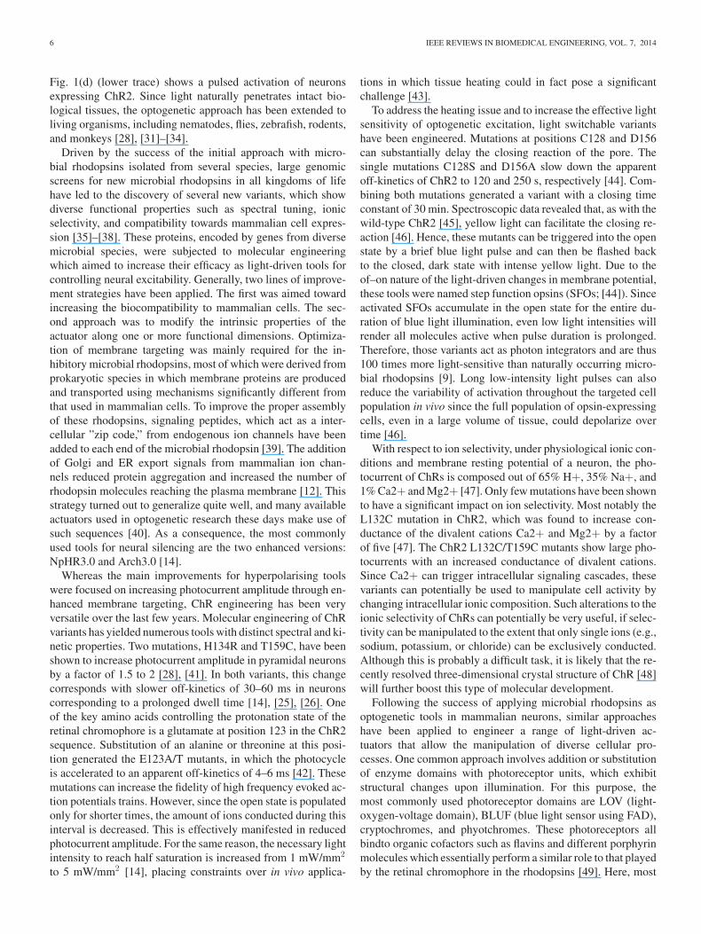
Fig. 1. Photocycle of the wild-type ChR2 and light-induced photocurrents. (a) Schematic drawing of channel opening upon light illumination. In the closeddarkstate, the light antenna, all-trans retinal, can isomerize after absorption of a photon to 13-cis retinal, thereby triggering opening of the channel. (b) The light-inducedchanges in the electrical properties of the channels upon opening can be modeled as a four-state photocycle consisting of two dark states (D1 and D2) and twolight-induced open states O1 and O2. (c) Illustrates the electrical response of a single cell expressing ChR2 to two successive light pulses. Upon illumination,a transient peak inward current is triggered, which decays to the stationary photocurrent. This drop in photocurrent is commonly referred as inactivation. Uponlight-off, the photocurrent returns to baseline with an apparent τ off 21 ms. The seconds light pulse only evokes a smaller peak photocurrent while reaching thesame stationary current. The time it takes to return to full peak current size is known as recovery time. (d) Shows current-clamp recordings of neurons expressingNpHR (top) and ChR2 (bottom). Action potentials can be triggered with pulses of blue light in ChR2-expressing cells (473 nm), whereas spontaneous activity canbe suppressed by activating eNpHR3.0 with yellow light (589 nm).
Therefore, microbial rhodopsins can be grouped into pumps,acting as photodiodes, and light-triggered ion channels whichcan be considered as light-dependent resistors. Regardless ofthe mechanism of action or the ion species conducted, all mi-crobial rhodopsins rely on the same fundamental photoreactioncomprising the isomerization of a covalently bound retinal co-factor [see Fig. 1(a)]. Upon photon absorption, the cofactorisomerizes from so called all-trans to 13-cis conformation. Thisconformational shift changes the dipole moment of the retinalmolecule and initiates pronounced structural rearrangements ofthe protein, which finally lead to the transport of ions [23]. Allthese complex rearrangements occur within less than a millisec-ond whereby the activated rhodopsin cycles through variousphotochemically distinct states. This series of states can be col-lectively described as a photocycle in various levels of detail.Fig. 1(b) shows a four-state electrophysiological photocycle,which explains the major electrical features of channel func-tion, including inactivation, photocurrent kinetics, and adaptionphenomena as observed in electrical recordings from cells ex-pressing ChR [see Fig. 1(c)] [24]. For example, a fully darkadapted ChR2 molecule activated with a short blue light pulsewill first proceed through an open state of high conductance(O1), causing a transient peak photocurrent and then entering alower conductive state (O2) which leads to lower photocurrent.
This drop in photocurrent is commonly referred as inactivation[see Fig. 1(c)], and can be as large as 72% of the initial currentfor the wild-type ChR2 [25]. The dwell time for the conduc-tive states is estimated to be 10 ms [26]. Molecules in the openstate can transition to two different dark states with an apparentτ1,2 off off of 21 ms [25], [26]. Upon repeated illumination atthis stage, molecules reexcited from the second dark state willthen only pass through the low conductance state, giving rise toa light-adapted steady-state photocurrent which is smaller thanthe initial peak current [see Fig. 1(c)]. Consequently, a seriesof 2-ms light pulses will cause attenuation of the photocurrentto a lower equilibrium level [25]. Only after a recovery periodof >6 s, all molecules will have returned to their initial dark-adapted state [see Fig. 1(c)].
To achieve light-based control of neural function, these bio-electric units have been selectively targeted to different excitablecells such as neurons, heart, or muscle cells [6], [27]–[29]. Inneurons, microbial pumps such as HR or BR induce a square-like hyperpolarization response of the membrane potential uponillumination [5], [30], which can rapidly and effectively silenceneuronal activity [see Fig. 1(d) (upper trace)]. ChRs performthe opposite function of depolarizing neurons in response to il-lumination by allowing positively charged cations to passivelyenter the cell. ChRs therefore serve as light-induced activators.
6 IEEE REVIEWS IN BIOMEDICAL ENGINEERING, VOL. 7, 2014
Fig. 1(d) (lower trace) shows a pulsed activation of neuronsexpressing ChR2. Since light naturally penetrates intact bio-logical tissues, the optogenetic approach has been extended toliving organisms, including nematodes, flies, zebrafish, rodents,and monkeys [28], [31]–[34].
Driven by the success of the initial approach with micro-bial rhodopsins isolated from several species, large genomicscreens for new microbial rhodopsins in all kingdoms of lifehave led to the discovery of several new variants, which showdiverse functional properties such as spectral tuning, ionicselectivity, and compatibility towards mammalian cell expres-sion [35]–[38]. These proteins, encoded by genes from diversemicrobial species, were subjected to molecular engineeringwhich aimed to increase their efficacy as light-driven tools forcontrolling neural excitability. Generally, two lines of improve-ment strategies have been applied. The first was aimed towardincreasing the biocompatibility to mammalian cells. The sec-ond approach was to modify the intrinsic properties of theactuator along one or more functional dimensions. Optimiza-tion of membrane targeting was mainly required for the in-hibitory microbial rhodopsins, most of which were derived fromprokaryotic species in which membrane proteins are producedand transported using mechanisms significantly different fromthat used in mammalian cells. To improve the proper assemblyof these rhodopsins, signaling peptides, which act as a inter-cellular ”zip code,” from endogenous ion channels have beenadded to each end of the microbial rhodopsin [39]. The additionof Golgi and ER export signals from mammalian ion chan-nels reduced protein aggregation and increased the number ofrhodopsin molecules reaching the plasma membrane [12]. Thisstrategy turned out to generalize quite well, and many availableactuators used in optogenetic research these days make use ofsuch sequences [40]. As a consequence, the most commonlyused tools for neural silencing are the two enhanced versions:NpHR3.0 and Arch3.0 [14].
Whereas the main improvements for hyperpolarising toolswere focused on increasing photocurrent amplitude through en-hanced membrane targeting, ChR engineering has been veryversatile over the last few years. Molecular engineering of ChRvariants has yielded numerous tools with distinct spectral and ki-netic properties. Two mutations, H134R and T159C, have beenshown to increase photocurrent amplitude in pyramidal neuronsby a factor of 1.5 to 2 [28], [41]. In both variants, this changecorresponds with slower off-kinetics of 30–60 ms in neuronscorresponding to a prolonged dwell time [14], [25], [26]. Oneof the key amino acids controlling the protonation state of theretinal chromophore is a glutamate at position 123 in the ChR2sequence. Substitution of an alanine or threonine at this posi-tion generated the E123A/T mutants, in which the photocycleis accelerated to an apparent off-kinetics of 4–6 ms [42]. Thesemutations can increase the fidelity of high frequency evoked ac-tion potentials trains. However, since the open state is populatedonly for shorter times, the amount of ions conducted during thisinterval is decreased. This is effectively manifested in reducedphotocurrent amplitude. For the same reason, the necessary lightintensity to reach half saturation is increased from 1 mW/mm2
to 5 mW/mm2 [14], placing constraints over in vivo applica-
tions in which tissue heating could in fact pose a significantchallenge [43].
To address the heating issue and to increase the effective lightsensitivity of optogenetic excitation, light switchable variantshave been engineered. Mutations at positions C128 and D156can substantially delay the closing reaction of the pore. Thesingle mutations C128S and D156A slow down the apparentoff-kinetics of ChR2 to 120 and 250 s, respectively [44]. Com-bining both mutations generated a variant with a closing timeconstant of 30 min. Spectroscopic data revealed that, as with thewild-type ChR2 [45], yellow light can facilitate the closing re-action [46]. Hence, these mutants can be triggered into the openstate by a brief blue light pulse and can then be flashed backto the closed, dark state with intense yellow light. Due to theof–on nature of the light-driven changes in membrane potential,these tools were named step function opsins (SFOs; [44]). Sinceactivated SFOs accumulate in the open state for the entire du-ration of blue light illumination, even low light intensities willrender all molecules active when pulse duration is prolonged.Therefore, those variants act as photon integrators and are thus100 times more light-sensitive than naturally occurring micro-bial rhodopsins [9]. Long low-intensity light pulses can alsoreduce the variability of activation throughout the targeted cellpopulation in vivo since the full population of opsin-expressingcells, even in a large volume of tissue, could depolarize overtime [46].
With respect to ion selectivity, under physiological ionic con-ditions and membrane resting potential of a neuron, the pho-tocurrent of ChRs is composed out of 65% H+, 35% Na+, and1% Ca2+ and Mg2+ [47]. Only few mutations have been shownto have a significant impact on ion selectivity. Most notably theL132C mutation in ChR2, which was found to increase con-ductance of the divalent cations Ca2+ and Mg2+ by a factorof five [47]. The ChR2 L132C/T159C mutants show large pho-tocurrents with an increased conductance of divalent cations.Since Ca2+ can trigger intracellular signaling cascades, thesevariants can potentially be used to manipulate cell activity bychanging intracellular ionic composition. Such alterations to theionic selectivity of ChRs can potentially be very useful, if selec-tivity can be manipulated to the extent that only single ions (e.g.,sodium, potassium, or chloride) can be exclusively conducted.Although this is probably a difficult task, it is likely that the re-cently resolved three-dimensional crystal structure of ChR [48]will further boost this type of molecular development.
Following the success of applying microbial rhodopsins asoptogenetic tools in mammalian neurons, similar approacheshave been applied to engineer a range of light-driven ac-tuators that allow the manipulation of diverse cellular pro-cesses. One common approach involves addition or substitutionof enzyme domains with photoreceptor units, which exhibitstructural changes upon illumination. For this purpose, themost commonly used photoreceptor domains are LOV (light-oxygen-voltage domain), BLUF (blue light sensor using FAD),cryptochromes, and phyotchromes. These photoreceptors allbindto organic cofactors such as flavins and different porphyrinmolecules which essentially perform a similar role to that playedby the retinal chromophore in the rhodopsins [49]. Here, most

PASHAIE et al.: OPTOGENETIC BRAIN INTERFACES 7
notable is a photoactivatable adenylate cyclase bPac, which wasfound in the bacteria Beggiatoa sp. Habitas hydrogen sulfiderich environments. This cyclase is light-sensitive via a BLUFdomain. In Drosophila, bPac expression increases the level ofsignaling molecule cAMP by a factor of ten upon blue lightillumination [50]. Recently, the toolbox was extended by a verypromising member—a light-inducible transcriptional effectorfor endogenous gene expression. Here, a light-inducible dimer-ization of a cryptochrome called Cry2 with its native truncatedbinding partner CIBN [51], was fused to a transcription effectordomain and a DNA recognition domain, respectively. Upon bluelight illumination, the DNA bound CIBN recruits the Cry2-fusedactivator domain and can thereby increase the transcription oftarget genes [52]. These and other tools have extended the rangeof optogenetic manipulation beyond the simple control of neu-ronal excitability and are likely to contribute significantly to arange of experimental applications.
Despite the increased number of applications, all present op-togenetic tools utilize light in the visible range. New tools, sen-sitive at the longer range of the electromagnetic spectrum (e.g.,infrared or radio frequency), are desirable since they wouldallow deeper tissue penetration and perhaps even facilitate non-invasive approaches. Promising candidates for this approach,for example, are the infrared thermal sensitive ion channelsfrom bats and phythochrome-based tools [53], [54]. Tools whichcan be specifically switched ON and OFF with minimal cross-excitation would allow consecutive control of different cellularprocesses without optically cross-activating each other in thesame organism.
General speaking, ChRs impose another activation pathwayon the top of naturally occurring ones. Upon light stimulation,action potentials can be evoked in an all-or-none fashion. Froman analytical point of view, genetically-encoded tools renderingspecific endogenous light-activatable proteins would be morevaluable, since they would enable researchers to modulate theendogenous cellular mechanisms which drive actions poten-tials. But the greatest need for improvements in the field ofoptogenetics are the development of cellular activity reporters.Particularly, fast, noninvasive reporters for membrane poten-tial at the far end of the electromagnetic spectrum would bevery valuable. In addition, effective optogenetic reporters atdifferent wavelength for Ca2+, cAMP, cGCMP, or differentneuromodulators such as dopamine or acetylcholine are stillneeded or have room for improvement. Together with the grow-ing range of optogenetic actuators, these tools will allow thecharacterization of brain dynamics on levels other than electricalactivity.
Manipulating cellular processes with light at high spatial-temporal resolution has already been proven as an indispensablemethod in neuroscience research. Therefore, the preparadig-matic stage, where optogenetic tools are developed just in aproof-of-concept fashion has passed. With the increasing knowl-edge of the mechanisms of action of these proteins and the com-plexities of their application in neural systems, new tools shouldbe engineered such that they maximally fulfill their intendedfunction and allow predictable, well-controlled manipulation ofneural activity.
B. Gene Delivery Considerations in Optogenetics
One of the most important strengths of optogenetic method-ology is the fact that the actuators, microbial rhodopsins orother proteins discussed above, are genetically-encoded. Thisallows scientists to target specific cell populations for light-based manipulation. These populations can be defined based ongenetic signatures called promoters, with DNA sequences thatare selectively activated only in particular circuits or cell types.By coupling the microbial opsin gene to a promoter of choiceand inserting this DNA into neurons, expression of the opsinwill only occur in cells that selectively express the chosen pro-moter. Therefore, application of optogenetics in vivo requiresthe genetic modification of neurons to induce expression of thelight-gated tools. Although many methods exist for such genedelivery [55], genetically engineered viruses [56] are by far themost popular means of delivering optogenetic tools. Lentivi-ral vectors (LV) [57], [58] and adeno-associated viral vectors(AAV) [59] have been widely used to introduce opsin genesinto mouse, rat, and primate neural tissues [8]. These vectorsallow high expression levels over long periods of time with lit-tle adverse effects [59]. AAV-based expression vectors are lessimmunogenic, and some types of AAV-based vectors allow thetransduction of larger tissue volumes compared with LV due totheir small particle size and increased viral titers. They are there-fore increasingly used both in basic research and in gene therapytrials [60]. Additionally, AAV is considered safer than LV as thecurrently available strains do not broadly integrate into the hostgenome and are thus rated as biosafety level (BSL) one or twoagents. Both LV and AAV vectors can be used in conjunctionwith cell type-specific promoters [58], [61], [62], facilitatingtheir application in cell-type specific optogenetic manipulation.
III. MECHANISMS OF LIGHT DELIVERY
To study brain microcircuits via optogenetics, we need lightdelivery mechanisms to control the distribution of light not onlyon the surface but also inside the brain tissue. Distribution oflight on the surface can be controlled precisely by spatial lightmodulators (SLMs). To deliver light inside the tissue, light-guides, e.g., optical fibers, are implanted in the brain to guidelaser pulses with appropriate wavelengths to regions of interest.Recently, more advanced mechanisms such as micro-fabricatedchannel waveguide arrays or holographic microscopy systemsare being developed and tested to control the distribution of lighteven in the three-dimensional structure of the brain.
A. Waveguiding Systems
Optical fibers are the most popular lightguides that are used tostimulate deep brain objects. In most applications, an asphericlens couples the beam of a solid-state laser to a multimodestep index fiber which guides and deliver the optical power tothe region of interest. Superluminescence light emitting diodes(LEDs), which are more stable light sources, are also becomingpopular for optogenetic stimulation. Electronically driven LEDsare particularly suitable for integrated systems such as prostheticdevices. However, coupling efficiency dramatically drops when

8 IEEE REVIEWS IN BIOMEDICAL ENGINEERING, VOL. 7, 2014
Fig. 2. (a) Optical fiber implanted in the brain tissue to deliver light and stimulate deep brain objects in transfected areas. (b) A rat receiving two implants in themotor cortex on both hemispheres. (c) Structure of a multichannel probe [64], (d) and multichannel waveguide arrays (Reprinted with permission from [65]). (e)A side-firing fiber that radiates power almost orthogonal to the optical axis of the fiber [66]. (f), (g) Microactuator assembly that moves a side-firing fiber insidethe brain; mechanical design and a prototype [66].
incoherent sources are used and as a result, researchers usuallyincrease the fiber diameter to couple enough light into the fiber.The stereotaxic surgery protocols for implanting fibers are cur-rently well established [11], [63] [see Fig. 2(a), (b)]. Obviously,optical fibers only deliver light to the area close to the fibertip. To control the distribution of light in the three-dimensionalstructure of the brain, more complex light delivery mechanismsare needed.
It is possible to make an array of channel waveguides whereeach waveguide in the array terminates at a different depth [64].An example of this approach is displayed in Fig. 2(c). Here,a 1.45-cm-long probe is microfabricated in the form of a 360micron-wide array of 12 parallel silicon oxynitride (SiON) mul-timode waveguides clad with SiO2 and coated with aluminum.Each waveguide in the array accepts light from a light source atits input terminal and guides the coupled light down to an alu-minum corner mirror which deflects light away from the probeaxis. Based on the published data, light losses are relativelysmall so that each waveguide can almost deliver about 30% ofthe injected light at the output terminal. The main benefits of thisapproach are the simplicity of the design and efficiency of lightdelivery. Moreover, light pulses can be delivered independentlyat multiple sites following any arbitrary temporal sequence ofinterest. A new generation of such probes have been recentlydeveloped and tested where a matrix of waveguide arrays are in-tegrated on a single substrate to build a prosthetic device whichcan potentially control the light distribution in different layersof cortex [65] [see Fig. 2(d)]. The major drawback of this ap-proach is the overall size of the probe, which is relatively large,and the complexity of the optics required to couple light intothe channel waveguides in large arrays.
Another more simple approach for developing a distributedlight delivery mechanism is to physically move an optical fiberinside the tissue [66]. In this design, a microactuator assembly,which sits on the skull, moves a side-firing fiber inside a glassmade capillary that is implanted inside the brain prior to theexperiment [see Fig. 2(e)–(g)]. Side firing fibers are opticalfibers where the tip is precisely angled and polished so that theradiation pattern of the fiber is almost orthogonal to the opticalaxis of the fiber. It is possible to optimize the parameters of the
side-firing fiber, including the tip angle and numerical aperture,to minimize the divergence of the radiating beam and improvethe spatial resolution of the device for light delivery. This systemcan deliver different wavelengths at numerous positions insidethe tissue, and the size of the capillary can be less than 50micron. The device has spatial resolution of a few microns overa dynamic range of about 4 mm and the total weight of thedevice is less than 2g. The major drawbacks of this design arethe limited speed of the system to change the position of the fiber.Also, it is practically difficult, if not impossible, to integrate anarray of such side-firing fibers and microactuators into a singleprosthetic device.
Another approach remained to be explored in the future isthe use of tilted fiber gratings that couple guided modes of anoptical fiber to radiation modes [67].
B. Spatial Light Modulator Based Systems
Two types of spatial light modulators are used to producedistributed patterns of light in the brain: microelectromechanical(MEMS) based SLMs and liquid crystals.
The most well-known MEMS light modulator is the TexasInstrument’s digital micromirror device (DMD). The DMD sys-tem is a light processor consisting of a large array of bistablemicromirrors. The position of each mirror in the array is un-der independent computer control. Exposure is controlled byprogramming the duration each mirror reflects light toward asample [68]. An example of the optical design of a DMD sys-tem which is specifically designed for optogenetic applicationsis shown in Fig. 3(a). In most DMD designs, a high-powerincoherent light source, with a wide spectrum, produces the op-tical power which is filtered to select the appropriate range ofwavelengths. The filtered beam is then homogenized by an in-tegrating rod and collimated by telecentric optics to expose theactive area of the DMD through a total internal reflection (TIR)prism. The TIR prism, which is placed in front of the DMD,guides the beam of light to illuminate the DMD surface withthe right angle considering the specific design of micromirrorsin DMD chips. This prism also separates the illuminating beamfrom the modulated reflected beam. Next, the DMD modulates

PASHAIE et al.: OPTOGENETIC BRAIN INTERFACES 9
Fig. 3. Spatial light modulator systems for optogenetic stimulation, (a) Basicoptical design of a DMD based system for optogenetic stimulation and imaging.(b) Liquid crystal based light delivery mechanism and holographic microscopy.
the beam and a telecentric lens projects the pattern on the en-trance pupil of an image combiner. The image combiner is anassembly of mirrors and epi-fluorescence optics which makes itpossible to simultaneously project a pattern on the sample andperform bright field or fluorescence imaging.
An interesting example of optogenetic stimulation by a DMDsystem is the study of the motor circuit in freely movingCaenorhabditis elegans [69]. Since the DMD can producerapidly changing high-resolution illumination patterns, this sys-tem has been used combined with a real-time image processingsoftware to track a freely moving worm and optically target itsmotor system with single cell resolution. Another applicationof DMD based system for optogenetic stimulation is discussedlater in the discussion of optogenetic ECoG recording.
Liquid crystal SLMs are specifically designed to be used withlasers and coherent light sources. A typical design of such sys-tems is shown in Fig. 3(b). In this design, a laser source producesa collimated beam of light which is phase modulated by a liquidcrystal SLM and the modulated beam in launched into the opticalpath of a fluorescence microscope [70], [71]. The SLM generatesa dynamic mask which reshapes the beam of a laser to controlthe distribution of light on the sample. From Fourier optics, weknow that a lens is nothing but a simple phase mask. Therefore,it is possible to emulate the function of any arbitrary lens bya phase modulating liquid crystal to focus the beam of laser atdifferent depths, even in parallel, to produce three-dimensionalpatterns of light distribution, such as holograms, inside the tis-sue [71]. Recently, liquid crystals have been integrated intothe optical path of multiphoton microscopes, and femto-secondpulsed lasers are used to produce three-dimensional patterns ofoptogenetic stimulation over the entire depth of the cortex insmall rodents. In other words, dynamically changing hologramsare produced in the cortex to generate complex spatial-temporal
patterns of activities in the cortical microcircuits. This topic isdiscussed further in Section IV.
IV. ENGINEERING NEURAL PROBES FOR
OPTOGENETIC EXPERIMENTS
From the origins of modern neuroscience, direct electronicrecording of neural activity has been proven essential to de-tailed understanding of the dynamics and structure of neuralcircuits underlying observed behaviors. Consequently, neuralprobe engineering has evolved into a mature discipline coveringa variety of aspects ranging from the electronic design and ma-terials development to tissue characterization and computerizedneural data analysis, to name a few.
Since optogenetic approaches to neural control have spreadthrough the neuroscience community, the designers of neuralrecording devices found themselves confronted with a demandfor integration of optical elements into the neural probes. Asthe majority of currently available technologies for simultane-ous electrophysiology and optogenetics are based on previouslydeveloped recording-only devices, we will review the progressand the challenges in neural probe design and consider the im-plications of optoelectronic integration at the leading edge ofneural engineering.
Over the course of the past several decades a number of neu-ral probe designs have been proposed, fabricated, and appliedin basic as well as clinical neuroscience experiments [72]. Inthis paper, we will focus on invasive tissue-penetrating probesthat allow for recordings of individual action potentials (spikes)from the neighboring neurons (single units) based on the localchanges in ionic concentrations in the vicinity of the electrodes.While these devices are primarily employed in research, therecent applications in brain–machine interfaces (BMI) demon-strate the utility of single-unit recordings from motor cortex inneuroprosthetics for paralyzed patients ( [73], [74]).
In addition to single-unit recording, these devices can pro-vide the temporal profiles of local field potentials (LFPs) in theneighboring networks by simply changing the signal filter set-tings (from �300–8000 Hz for single units to �1–1000 Hz forLFPs). LFP data can, for example, be subject to Fourier analysisto reveal predominant oscillation frequencies within the moni-tored neuronal network. Changes in LFP frequency spectra areoften characteristic to a particular neurological disorder (e.g.,Parkinson’s disease or epilepsy).
While patch-clamp electrophysiology offers the most preciserecording of voltage changes across neuronal membranes, thiscell-penetrating technique suffers from relatively low through-put and is not compatible with chronic implantation. The vastmajority of neuroscience experiments, require recordings of ac-tivity of large numbers of individual neurons during a specificbehavioral paradigm and hence single-neuron resolution andrecording stability over a course of an experiment (days to years)are often the key requirements for the neural probe design [80].
Currently, in addition to the anatomy of the specific region ofthe nervous system and the desired resolution of the acquireddata, the limitations of the available fabrication approaches dic-tate the neural probe geometry. Individual metallic microwires

10 IEEE REVIEWS IN BIOMEDICAL ENGINEERING, VOL. 7, 2014
Fig. 4. Examples of commonly used single unit and LFP recording devices and their optogenetics-compatible modifications. (a) Silica-encapsulated tungstenelectrodes (Reprinted with permission from Thomas Recording GmbH). (b) Tungsten microwire array (Reprinted with permission from Tucker-Davis TechnologiesInc.). (c) Tetrode microwire bundle (Reprinted with permission from Dr. T. Davidson, Stanford University). (d) Silicon ”Utah” multielectrode array (BlackrockInc.). (e) Silicon multitrode probes (Reprinted with permission from Neuronexus Inc.). (f) An optrode consisting of a silica fiber and a tungsten electrode [13]. (g)Metal microwire array equipped with a silica fiber (Reprinted with permission from [92]). (h) Optetrode combining four tetrode bundles and an optical fiber [93].The inset shows the cross-section schematic (Reprinted with permission from Dr. A. Andalman, Standford University). (i) Utah array incorporating a tapered silicafiber (Reprinted with permission from [94]), [101]. (j) Silicon multitrode probes integrated with tapered silica fibers (Reprinted with permission from [102]).
�50–100 micron in diameter are the simplest forms of neuralprobes [see Fig. 4(a)]. These electrodes are primarily based ontungsten, steel, gold, platinum, and iridium and can be assem-bled into arrays of up to several tens to increase the numberof recorded single units [see Fig. 4(b)]. The ability to distin-guish between action potential shapes originating from differ-ent neurons was shown to be improved by assembling indi-vidual microwires into tightly wound stereotrodes (two inter-twined microwires) and tetrodes (four microwires) [75], [76][see Fig. 4(c)]. These neural probes usually consist of polymer-coated nickel-chromium alloy electrodes �12 micron in diame-ter, since larger and stiffer electrodes do not lend themselves tothe assembly process and produce bulky and tissue-damagingdevices. Stereotrodes and tetrodes were originally developed forhigh-throughput recordings from dense hippocampal layers inrodents, which remains the main application of these devices todate. These wire assemblies are often used in combination withmicrodrives that allow individual propagation of each assemblyindependently thus enabling active search of firing neurons.
Due to the maturity of the processing methods enabled byelectronics industry, silicon has become a popular material forthe fabrication of neural probes. While numerous silicon-baseddesigns have been produced over the past two decades, theirbasic structure can be approximately divided into multielec-trode ”Utah” arrays [77], [78], MEA [see Fig. 4(d)], and multi-trodes [80], [82], which are sometimes referred to as ”Michiganprobes” [see Fig. 4(e)]. The former structures consist of arrays ofhighly-doped silicon ”peaks” created by a combination of directmicromachining and wet etching and insulated from each otherby regions of glass. The bodies of the electrodes are coatedwith a chemical-vapor deposited (CVD) polymer parylene Cand the conductive tips are exposed. The size of the individualelectrodes within the array is on the order of several tens of mi-
crons at the recording tip but 300–400 micron at the base, whichyields a comparatively large footprint for arrays comprised ofmore than sixteen electrodes (e.g., a 10 × 10 MEA has dimen-sions of approximately 4 × 4 mm2 , not including connectorparts). One of the main advantages of these devices lies in theirfloating design. The MEAs are deposited on the cortical surfaceattached to the connector mounted on the skull with long flexibleloosely bundled wires. As a result, these devices require a sensi-tive implantation procedure during which the MEA is deployedpneumatically into the cortex. Since rodents lack sufficient in-tracranial space, floating MEAs are limited to applications inprimates. Furthermore, the length of the electrodes is restrictedto �1–1.5 mm by the fabrication and implantation proceduresand hence these devices are used primarily for cortical record-ings in primates, facilitating an entire field of BMI and, recently,neural control of robotic aids by paralyzed human patients[73].
Multitrode probes employ a thin silicon substrate with litho-graphically patterned metal electrodes distributed along theshaft. This streamlined geometry and deterministic positioningof the recording sites makes these probes attractive for record-ings in deep brain structures with high cell density such as therodent hippocampus. These probes have recently been extendedto devices incorporating logical elements thus enabling on-sitesignal amplification, which holds the potential to improve theyield of recorded single units. However, the lengths of the siliconshaft is still limited to a few millimeters by the micromachiningprocess restricting the applications to experiments in rodentsand cortical recordings in primates. Similarly to stereotrodesand tetrodes, these devices can be combined with microdrives.However, due to the device geometry, the entire shaft is prop-agated through the brain thus advancing all of the patternedrecording channels.

PASHAIE et al.: OPTOGENETIC BRAIN INTERFACES 11
Neurotrophic electrodes comprised of a silica pipette withseveral internal electrodes incorporate neural tissue, which pro-motes neurite ingrowth into the cone allow for high signal-to-noise (SNR) recordings. However, to our knowledge, they areyet to be combined with optogenetics and hence will not bediscussed in detail in this review. Interested readers may referto [81] for further details.
The majority of opsins, with the exception of SFOs, at averageexpression levels (2–8 weeks of incubation time following viraldelivery into the neural tissue) require light power densities ofapproximately 1 mW/mm2 in order to robustly evoke or inhibitaction potentials in vivo. This poses a requirement of directlight delivery into the area of interest, as the neural tissues arehighly scattering and absorptive within the visible range, whichcurrently encompasses the activation/inactivation spectra of allretinal-containing opsins. Consequently, the vast majority ofoptogenetic experiments are performed with implanted opticalfibers and the light is supplied by an external source such as alaser or a high-power LED. The direct implantation of miniatureLEDs into the brain is an alternative strategy for light delivery,which will be discussed in the following paragraphs.
While the initial proof-of-concept optogenetic experimentsperformed in vitro employed intracellular patch-clamp record-ings [6], the need for combined light delivery and extracellularelectrophysiological readout was recognized as this technol-ogy transitioned in vivo. Furthermore, as the state of the awakebrain of behaving rodents differs dramatically from activity pat-terns of the unconscious brain of anesthetized animals, not tomention a dissociated neuronal culture, it became essential toobserve the electrophysiological outcomes of optogenetic stim-ulation and correlate them to an observed behavior. As opticalfibers are routinely employed in optogenetic experiments, theirintegration with electrophysiological probes provides the moststraightforward engineering strategy. Thus, to date, the majorityof commonly used neural probes have been modified to carryexternally attached conventional commercially available silicafibers.
The simplest devices ”optrodes” comprise of individual metalelectrodes and optical fibers held together by an adhesive [seeFig. 4(f)]. These devices are primarily employed for recordingof single-unit and population (combined signals from multipleunits) activity in anesthetized animals [13]. Similarly, an opticalfiber can be attached to a metal electrode array and the resultingdevices are employed during behavioral experiments primarilyfor population activity and LFP recordings [see Fig. 4(g)] [92].
Single-unit recording during optical stimulation can be read-ily achieved with ”optetrodes”, which comprise of an opticalfiber surrounded by several tetrode bundles [see Fig. 4(h)] [93].These devices allow for recording during free behavior andprovide access to multiple recording sites due to a miniatureconcentric mechanical drive. While these devices ensure illumi-nation of all the neurons that can be recorded with the tetrodesthey do not allow for independent manipulation of individualtetrodes.
In order to introduce a fiber into a Utah-style MEA, one ofthe electrode positions can be replaced with an opening [seeFig. 4(i)] [94]. In this case, the scattering within the tissue only
allows for illumination of cells within the direct vicinity of thefiber. Consequently, the utility of this approach is mainly inexploring the effects of localized stimulation or inhibition ona broader cell population that can be accessed by electrodesdistributed in a several millimeter area, as only a few (�4–9)electrodes will be in contact with the illuminated tissue. Theintegration of a fiber with a Utah MEA compromises the oth-erwise floating design of the device, as the conventional silicafibers are too rigid and, hence, anchor the device to the skull.
Silicon multitrode probes can be outfitted with fibers similarlyto optrodes by simply attaching the latter to the former withan adhesive following the fabrication process [see Fig. 4(j)][102]. These devices often employ tapered fibers, which allowfor reduction of the overall device dimensions but significantlyincrease optical losses and reduce the numerical aperture leadingto reduced illuminated tissue volume. The relative position ofthe fiber with respect to the electrode pads determines, whichof the channels will record activity of the illuminated neurons.Positioning the fiber at the top of the array may require very highlight powers to reach the electrodes at the tip of the array, whilepositioning of the fiber tip within the array pads compromisesthe recording on the upper pads as the tissue may be damagedor pushed away by a relatively large fiber.
Connector design remains one of the main challenges forsimultaneous neural recording during optogenetic experimentsas optical coupling is required in addition to an electrical con-nector. This imposes the design constrains, which often lead todevices too large or heavy to be employed during free behaviorin smaller mammals (mice, bats) or birds. High fidelity opticalcoupling (approaching 100% transmission) can now be achievedwith knife-edge polish of pigtailed ferrules (stainless steel orzirconia) held together with ceramic sleeves. While ferrule toferrule connection is not considered stable by photonics stan-dards, its performance is sufficient for behavioral experimentson timescales of minutes to hours. Ferrule coupling, however,restricts the position of the fiber with respect to the recordingdevice, which generally results in a fiber illuminating only afraction of the recording electrodes.
Note, that all the devices discussed so far are based on rigidmaterials (metals, silicon, silica) with Young’s moduli (e.g.,E(Si) = 130–170 GPa, E(SiO2) = 73 GPa, E(W) = 411 GPa,E(Pt) = 168 GPa) that are many orders of magnitude higher thanthat of neural tissues (E � kPa–MPa) [79], [83]. It is thoughtthat this mismatch in stiffness contributes to tissue damage andsubsequent decay of the recording quality [84], [85]. It is rea-sonable to assume that the insertion of the implant into the tissuecauses neuronal death and displacement, however, the initial in-flammatory response usually ceases within approximately twoweeks following the implantation, which yields improved SNRand the overall number of single units recorded. However, theSNR deteriorates and the number of monitored units decaysover the course of following weeks to months [97]. While theorigins of neural probe failure are being actively investigated,several complimentary hypotheses have been proposed. Duringnormal movement, the brain undergoes several tens of micronsshifts (micromotion) with respect to the skull [98], [99]. Conse-quently, the tissue surrounding the hard fixed implant is subject

12 IEEE REVIEWS IN BIOMEDICAL ENGINEERING, VOL. 7, 2014
to chronic damage, which is manifested in glial and astrocyticactivity resulting in device encapsulation. If the latter were adominant mechanism of device failure, the floating Utah arrayswould be immune to failure, which is not the case. The disrup-tion of glial networks by the implants significantly exceedingthe dimensions of individual cells has also been suggested asa potential trigger of glial and astrocytic response. This is sup-ported by the reduced glial scarring around miniature implantswith cross sections approaching single cell size. Finally, re-cent work suggests that the disruption of blood–brain barrier(BBB) is yet another factor associated with the tissue responseto the implanted devices [82]. The latter findings imply that thesharper devices such as Utah MEA or multitrode probes pro-duce a greater BBB damage and yield more severe gliosis ascompared to the metal electrode arrays consisting of relativelyblunt cylindrical wires [95].
As the majority of optogentic neural probes incorporate rigidsilica fibers of 50–400 micron in diameter, it is reasonable toexpect that the reliability of the neural probes will be furthercompromised by such optoelectronic integration. Furthermore,so far this review has focused on the devices designed for appli-cations within the brain, while the recent interest in the periph-eral nervous system as a potential early therapeutic target foroptogenetics emphasizes the need for devices tailored to highlymobile and flexible nerves. Consequently, flexible, biocompat-ible platforms are needed for the advancement of combinedlong-term optogenetic and electrophysiological studies.
Over the past several years, a number of technologies havespurred in attempt to address the elastic modulus mismatchbetween neural probes and neural tissues. Silicone resins [pre-dominantly poly(dimethyl sulfoxane) (PDMS)] have been suc-cessfully employed as substrates for metal electrode arrays byLacour and Fawcett among others [96], [100]. This allowed forthe development of chronically implanted peripheral nerve cuffscapable of single-unit recordings from the nerve surface. Thistechnology however, has not yet been integrated with light de-livery, as silica fibers are rigid, and hence an alternative solutionsuch as an LED array is required. Furthermore, this technologyis not yet compatible with applications beyond the nerve or cor-tical surface (micro-ECoG) as it has limited resolution due tothe fabrication constraints and the implantation surgery into thedeep tissue is yet to be developed.
Similarly, parylene C has been employed as a substrate forthermal molded soft cone-electrodes incorporating recordingsites within the cone. These devices are potentially intendedto operate akin to neurotrophic silica-cone electrodes and mayyield improved SNR and stability. However, this technology isnot currently scalable to high-density recordings, nor does itlend itself to the incorporation of optical fibers [86] thus facingthe challenges of the silicone-backed devices.
Microcontact printing process developed by Rogers and col-leagues has been recently employed in fabrication of sophis-ticated integrated structures on highly flexible silicone, poly-imide, and silk fibroin substrates [see Fig. 5(a)], [87]–[89]. Thisinnovative approach takes advantage of the mature chip-scalesemiconductor processing and combines it with designer pre-strained interconnects, which enable the release and transfer of
Fig. 5. (a) Contact printed AlInGaP LED on a PDMS substrate (Reprintedwith permission from [87]). (b) Releasable GaN micro-LED for deep-structureoptogenetics (Reprinted with permission from [90]).
Fig. 6. (a) Photograph and (b) a tip close-up of a MEMS-processed polyimide-backed device equipped with a SU-8 waveguide and drug delivery channel(Reprinted with permission from [91]).
the devices fabricated on wafers onto flexible substrates. Con-sequently, these devices can be designed to integrate a variety ofelectronic components enabling on-site data processing as wellas micro LEDs based on proven group III–V semiconductors.The utility of these devices lies primarily in the cortical LFPrecordings as the resolution of the printing process limits thechannel size. The devices, integrating a single recording chan-nel with LEDs, photodetectors, and a thermal sensor (resistor)have been recently tested in deep brain structures of mice [seeFig. 5(b)]. The insertion into the depth of the tissue in this casewas enabled by silicon microneedles adhered to the devices byresorbable silk fibroin layers [90]. However, the mechanics ofthe printing process would yield comparatively large structures(several millimeters in area) if the number of recording siteswere to increase to several tens or hundreds, hence these struc-tures are currently limited to a small number of electrodes.
A more traditional approach to flexible neural probes bor-rows fabrication strategies developed by MEMS industry andproduces devices resembling multitrode probes, in which sili-con substrates have been replaced with polyimide [91]. In thiscase, optogenetics can be enabled by integrated polymer waveg-uides based on photoresists poly(methyl methacrylate) (PMMA)or SU-8 [see Fig. 6]. The resulting structures are very flexibleand require temporally stiffening encapsulation in order to pen-etrate deep into the tissue. A number of encapsulation strategiessuch as coatings with sugars, poly(ethylene glycol) (PEG) and,more recently, tyrosine-based polymers allow to tune the timeof the implant softening into the neural tissue. One of the mainchallenges of this approach is relatively low transmittance ofthe polymer waveguides (>1 dB/cm), which translates into theneed for high input light powers (>100-mW sources) in order

PASHAIE et al.: OPTOGENETIC BRAIN INTERFACES 13
to enable opsin-facilitated optical neural interrogation at the tipof fairly short (length <5 mm) polymer waveguides.
Currently, MEMS-style fabrication and the microcontactprinting processes suffer from the relatively low yield, whichdrives the high cost of these devices reducing the number ofanimals that can be employed in a given optogentic behav-ioral study and hereby increasing the variability of the results.Furthermore, the wafer-scale processing limits the device di-mensions to the applications in rodents and the primate cortex.Applications such as deep brain areas or the peripheral nervesin primates or humans currently remain inaccessible with thesestate-of-the-art technologies for combined optical stimulationand recording.
A robust fabrication approach is needed to enable integrationof optical components (LEDs and/or waveguides) and high-density single-unit electrophysiology. The development of suchintegrated devices may require exploration of new materialssystems such as conductive polymers and polymer composites,which have been shown to improve the recording SNR andstability. Electrospinning and CVD enable processing of poly-mer probes with diameters as low as 10 micron [103], [105].However, the application of these methods to integrated multi-channel devices will require further development as the numberof materials investigated to date remains limited.
Fabrication methods allowing for processing polymers be-yond photoresists may allow to reduce the losses in flexiblewaveguides. The photonics industry may provide insights intohigh-throughput fabrication of high-quality optical waveguides.For example, thermal drawing process, routinely used to pro-duce conventional silica fibers, can actually be applied to pro-cessing of multiple materials such as polymers, metals, andsemiconductors [104]. Initial attempts to employ thermal draw-ing in the design of combined optical and electrical neural probesdemonstrate the promise of this approach, however, the scalingto high-density electrophysiology as well as combined opticaland electronic connectorization would need to be addressed be-fore this fabrication method can be adopted at large.
Nanowires of III–IV semiconductors provide highly intimateinterfaces with individual neurons and can also be designed to in-corporate p-n junctions necessary for LEDs and field-effect tran-sistors enabling high SNR recordings [106], [107]. The majorityof nanowire devices are designed on flat rigid substrates and areprimarily used for studies of individual cultured cells. However,flexible substrates have recently enabled integration of nanowireelectronics with hydrogel scaffolds creating three-dimensionalhybrids of neural tissues with embedded electrophysiologicalprobes [108]. This approach provides a particularly promisingstrategy for optogentic neural regeneration devices, granted thatchallenges associated with scalability and connectorization areovercome.
The design and implementation of future intimate and inte-grated optogenetic electrophysiological probes now depends onthe collaboration between materials scientists, chemists, elec-tronics, photonics, and packaging engineers as the solutions tothe optoelectronic integration challenges would also need to ad-dress the biocompatibility and reliability issues stemming fromthe fragile and inhomogeneous structure of the neural tissue.
V. OPTOGENETIC STIMULATION COMBINED WITH MAGNETIC
RESONANCE IMAGING
The optogenetic functional magnetic resonance imaging(ofMRI) [109]–[111] technology combines optogenetic controlwith high-field fMRI readout. This enables cellular level pre-cision in control while whole brain network function can bereadout in vivo. Systematic control of the brain with specificityin the cellular level combined with whole brain dynamic imag-ing has been a fundamental tool missing in the neuroimaging andneuroscience fields; and arguably has been a key missing tool tolink findings from the more precise cellular level investigationto the overall brain function. While standard fMRI providedimportant clues and insights into brain function including map-ping of visual, sensory, motor areas, and association of brainactivation changes to neurological diseases, they were limitedin fidelity due to the brain’s densely interconnected network ofheterogeneous cellular populations. OfMRI addresses this prob-lem by combining genetically targeted control of neurons witha method capable of whole brain functional readout.
In the first study, it was shown that cell types specified bygenetic identity, cell body location, and axonal projection targetcan be specifically excited while the whole brain function associ-ated with the stimulation can be monitored with spatiotemporalprecision. In this study, it was first shown that ofMRI couldreliably detect local activities resulting from cell type-specificcontrol of neurons, see Fig. 7. For example, upon stimulationof the ChR2-expressing excitatory neurons in the motor cortex,a positive blood oxygen level-dependent (BOLD) signal wasobserved at the site of stimulation. In addition, the temporalshape of the optogenetically induced BOLD responses matchedthe conventional BOLD signals that are typically evoked duringsensory stimulation, suggesting that excitation of a single pop-ulation is sufficient to drive the conventional BOLD response.
Then, to demonstrate the global imaging capabilities of fMRI,the authors measured the BOLD response at the thalamus, whichhas direct axonal projections from the motor cortex. At the tha-lamus, robust BOLD signal was measured, demonstrating theglobal imaging capability. However, an unexpected differencein the BOLD response shape was also observed. While largelysimilar in shape as the motor cortex hemodynamic responsefunction (HRF), there was a marked reduction in HRF signalrise slope. To identify the source of such difference, electrophys-iology recordings were made at the motor cortex stimulation siteand the location within the thalamus where the ofMRI signal wasobserved. The electrophysiological recording revealed a delayin spike-rate increase in thalamic neurons compared to corticalneurons, matching the smaller HRF slope observed with ofMRI.This opened up a possibility for the ofMRI technology to enablenot only spatially resolved neural activity mapping but also totemporally resolve underlying activity patterns.
Next, to further expand the ofMRI technology’s capability tomap responses associated with specific modulations, the authorsvisualized the brain’s response to light stimulation delivered tothe thalamus with anterograde viral injection in M1 cortex. Withanterograde vital injection in M1 cortex, thalamic regions thatreceive direct axonal projection will have opsin expression in

14 IEEE REVIEWS IN BIOMEDICAL ENGINEERING, VOL. 7, 2014
Fig. 7. OfMRI with stimulation of excitatory neurons in the primary motor cortex. (a) During the ofMRI scan, the subject is intubated and lightly anesthetized.The respiration rate, body temperature, and expiratory CO2 are monitored and controlled. A surface coil (not shown) is mounted and centered over the regionof interest on the skull to maximize the SNR ratio. The subject’s eyes are also covered (not shown) to avoid unintended visual stimulus. (b) Six cycles of20 s-on-40 s-off optical stimulation are delivered to the primary motor cortex of the animal during the scan. The gradient-recalled-echo (GRE) BOLD sequenceis used and 23 slices are collected for full brain coverage with 3 s temporal resolution. (c) BOLD signals are detected at both the stimulation site (primary motorcortex, image 1,2) and downstream areas (thalamus, image 3,4). The bottom tip of the white triangle indicates the optical simulation site.
the axons of neurons projecting from M1. With the light stimu-lation of these axons, robust activities were observation at boththe thalamus and motor cortex, demonstrating the feasibility ofmapping the causal influence of cells defined not only by geneticidentity and cell body location, but also by connection topology.Finally, the authors showed that ofMRI could be used to tracethe different global responses to activation of two distinct thala-mic nuclei that matched predictions from the literature, demon-strating the feasibility of the in vivo circuit function mappingcapability of ofMRI. These findings laid the basic foundationfor how the ofMRI technology can be a potent tool that canenable systematic in vivo circuit analysis.
Since the original publication, several studies have built uponthese results, further demonstrating the utility of the ofMRI tech-nology. For example, ofMRI implementation was demonstratedin awake mice [112] that showed that a larger BOLD responsewas evoked in awake animals compared to anesthetized condi-tions during optical stimulation of the primary somatosensorycortex. The study also showed that the degree of functional con-nectivity between active regions was strengthened. Therefore,under the following conditions, ofMRI studies could preferablybe performed without anesthesia: animal does not experiencesignificant levels of stress due to restraint and noise and impor-tantly, large motion is that is not compatible with the imagingsetup is not evoked by the stimulation.
The ofMRI technology have also been demonstrated at anumber of different stimulation locations including motor andsensory cortex [109], [112], [113], ventral tegmental area [114],and hippocampus [115], further demonstrating its utility. Look-ing forward, demonstrating the ofMRI technology’s capabilityto identify dynamic network function across multiple synapsesalso be a key next step.
Advances of the optogenetics technology [8] will also en-able diverse ofMRI experiments to be performed. For exam-ple, with optogenetics now becoming feasible in nonhumanprimates [61], [116], future ofMRI experiments may turn tomonkeys as an animal model for studying more complex braincircuits that is difficult to study with rodents. Improvementsin optogenetics may also enable precise control of subcellular
compartments. With such a capability, ofMRI could provide aunique bridge between subcellular perturbation and the result-ing global brain function. This could provide a new platformto study the specific role of axons and dendrites in the contextof overall brain function, which have been implicated in manyneurological diseases and disorders including Alzheimer’s dis-ease [117], multiple sclerosis [118], and autism [119].
In addition to advances in ofMRI’s capability that is expectedto come with the development of new optogenetic tools, thedevelopment of advanced imaging technologies is of criticalimportance to accelerate scientific discovery. For example, whilefMRI uniquely provides the readout of the global brain function,the image quality is often a concern. To accurately register neuralactivity to anatomical locations, it is necessary to have highquality, distortion-free images. Solutions to this problem includethe passband SSFP fMRI [120] method that was demonstrated inthe original ofMRI publication. Distortion-free passband SSFPfMRI combined with optogenetics was shown to result in morerobust, and spatially accurate registration of the activities.
Another important technical development that needs to takeplace is to increase spatiotemporal resolution of ofMRI images.While fMRI is unlikely to deliver single cell level, millisecondtemporal resolution, it is quite possible that advance imagingtechnology development would enable cortical layer and sub-nuclei specific imaging. Furthermore, the temporal resolution ofthe imaging could be improved to a subsecond range. Note how-ever that, for the improvement of the spatiotemporal resolution,it is important to maintain the whole brain volume coverage.In MRI, temporal and spatial resolution can be easily tradedoff with smaller spatial volume coverage. Therefore, withoutthe large volume coverage, high spatiotemporal resolution is oflimited value. For example, taking several slices or even sin-gle slice images at high spatiotemporal resolution, might be ofinterest for a dedicated study looking at that particular spatiallocation, it does not allow the key advantage of ofMRI, whichis the global imaging capability, to be fully utilized. A methodthat can fundamentally improve spatial and temporal resolutionwithout sacrificing the volume coverage therefore is of criticalimportance.

PASHAIE et al.: OPTOGENETIC BRAIN INTERFACES 15
Fig. 8. Schematic of a high-throughput, closed-loop ofMRI system. fMRI images are acquired using ultrafast acquisition techniques during optogeneticstimulation in awake or anesthetized animals. High-throughput, real-time image reconstruction, motion correction, and activation detection are performed in series.The resulting patterns of activity can by analyzed with automated algorithms or inspected manually to decide how to update stimulation parameters. Degrees offreedom available include the location and type of cell targeted, the temporal pattern of stimulation, and the type of modulation (e.g., excitation versus inhibition).(Modified figure modified from [121]).
With the large degree of freedom in neuronal cell populationcontrol including the type of cell targeted, the temporal patternof stimulation, and the type of response elicited (e.g., excitationor inhibition), methods for robust, fast, and high-throughputdata acquisition in a closed-loop system will greatly acceler-ate scientific discovery utilizing the of MRI technology, seeFig. 8. As a first step in achieving a real-time ofMRI system,Fang et al. [121] developed a highly parallelized method for per-forming image reconstruction, motion correction, and activationdetection in a fraction of the typical MRI acquisition repetitiontime. Combined with methods such as compressed sensing [122]and multiple coil arrays [123], cryo-cooled coils will for SNRimprovement, real-time image acquisition, and activation de-tection can be achieved. With such a system is in place, it willbe possible to interactively perturb specific brain circuits andimmediately visualize the network-level activity that results.The simplest form of closed-loop ofMRI would rely on manualinspection of the evoked patterns of brain activity and corre-sponding change of stimulation parameters. To enable a morerobust and high-throughput system, automated analysis tech-niques that can identify activations and quantify their spatialand temporal characteristics could also be combined. Integra-tion of such automated analysis methods with real-time imageacquisition will considerably accelerate the discoveries madewith ofMRI technology.
Optogenetic fMRI enables the study the causal global effectof perturbing specific brain circuit elements in live animals.This has great potential to providing insights into the mech-anisms that underlie normal and diseased brain function, anddriving the development of new therapies. For example, ofMRImay be used to track how the functional interactions within thebrain progress over time. Specifically, ofMRI may be used to
identify which brain regions fail to communicate with anotherregion properly, how they fail, and when. Studies have shownthat global structural and functional changes can predate clinicalsymptoms of neurological diseases [124], [125] and ofMRI willprovide an important new opportunity to systematically assesschanges during a disease’s progression. Unlike standard neu-roimaging techniques, ofMRI can attribute these changes to themodulation of selective neuronal elements and temporal param-eters of stimulation (i.e., temporal encoding). This will illustratethe highly dynamic nature of brain circuits [110], and provideinformation about the manifestation of a disease not obtainablewith other techniques.
Just as ofMRI can be used to visualize how neurological dis-eases affect the brain network function, it can also be used todetermine how a diseased brain will respond to therapy. Forexample, optogenetic control may be used to selectively sim-ulate separate elements of neuromodulation therapeutic targetscurrently in use in humans, such as in DBS and spinal cordstimulation therapies. Monitoring how the brain responds todifferent parameters of stimulation with ofMRI could then beused to predict which parameters optimize therapeutic benefit.
Given the extensive library of the literature that has used fMRIto study neurological diseases, a subtle yet significant contri-bution of ofMRI experiments to the clinical domain will alsocome from an improved understanding of the blood oxygena-tion level dependent (BOLD) responses. Despite two decadesof fMRI research in humans, the nature of the BOLD signalremains poorly understood, due to an incomplete understandingof the relationship between neural activity and haemodynamicprocesses in the brain [126]–[128]. Optogenetic fMRI providesa unique opportunity to probe these systems and to determinethe role that different neuronal populations play in generating

16 IEEE REVIEWS IN BIOMEDICAL ENGINEERING, VOL. 7, 2014
these responses. The original ofMRI paper [109] was the firstto demonstrate this principle, showing that causal activation ofa specific single population of neurons could lead to BOLD re-sponses with classical kinetics. Since then, a number of otherofMRI experiments have begun to disentangle the convolutedrelationship between neural activity and the BOLD signal. Forexample, one study compared the temporal characteristics ofthe ofMRI BOLD response with different electrophysiologicalmeasurements, finding that it correlated best with neuronal spik-ing activity [129]. Another study showed that the BOLD signalsummates in response to closely spaced trains of stimulation,confirming that the BOLD signal provides a linear approxima-tion of neural activity levels [113].
To further link the BOLD response evoked by optical stimu-lation with the fMRI responses typically measured during sen-sory or cognitive tasks, quantitative comparison of the two willalso be important. It has already been shown that optical acti-vation of ChR2-expressing neurons can lead to local changesin blood flow while the normal activation of neurons throughglutamatergic receptors would be blocked under the same con-dition [130]. Future integration utilizing combinatorial optoge-netics with ofMRI will allow the contributions of distinct cellpopulations to the BOLD signal to be even further dissected.In current ofMRI experiments, the optically driven populationlikely recruits or inhibits other neuronal types and glial cellsin generating the measured fMRI signal. With multiple wave-lengths of light control used to prevent additional populationsfrom being excited or inhibited, resulting signal would reflectthe change solely caused by the initially targeted population.This information could be used to interpret fMRI results byidentifying the types of neuronal elements involved. A simpleexample of this includes understanding the effect of suppressingneural activity compared to activating inhibitory neurons. Sincethe BOLD response only provides an indirect measure of neuralactivity, without a proper tool to control the neural elements withprecision while measuring fMRI signal, how these two distinctprocesses which both reduce the level of neural activity affectthe measured signal has remained elusive.
The significance of ofMRI lies in its ability to map globalpatterns of brain activity that result from the selective controlof precise neuronal populations. Currently, no other existingmethod can bridge this gap between whole-brain dynamics andthe activity of genetically, spatially, and topologically definedneurons. While a number of studies have already started toexploit the unique capabilities of this technology, ofMRI willlikely experience significant growth and development over thenext decade. Both the advances in the molecular toolbox ofoptogenetics, and improvements in imaging technology holdimportant roles in brining ofMRI closer to its full potential.In particular, the integration of ultra-fast, high-resolution dataacquisition and analysis, and combinatorial optogenetics willenable powerful closed-loop ofMRI that can real-time visualizebrain activity with real-time feedback control. Further researchuncovering the nature of the BOLD signal will also make itpossible to extract more detailed information about the neu-ral activity measured in fMRI studies. Finally, the applicationof ofMRI to translational research has the potential to funda-
mentally transform therapeutics design for neurological disor-ders. New targets of therapeutic neuromodulation will likelybe discovered, and improved models of neurological diseasewill be introduced. Taken together, ofMRI is anticipated to playan important role in the future understanding of both normaland diseased brain functions and enable alternative treatmentdevelopments.
VI. COMBINING OPTOGENETICS WITH
TWO-PHOTON MICROSCOPY
As discussed earlier, methods for guiding spatial delivery ofsingle-photon light excitation have been used to improve theprecision of optogenetic modulation [69], [131]–[138]. Lightlocalization with higher precision can be achieved using mul-tiphoton excitation microscopy. In neuroscience, two-photonexcitation microscopy has allowed for the direct investigationof the nervous system at different length scales (from synapsesto neural circuits) and on different time scales (millisecondsto months) [139], [140]. The physiological importance of suchwork is highlighted in studies investigating the role of the hip-pocampus in spatial navigation tasks in rodents. Dombeck andcolleagues used two-photon microscopy in combination withgenetically encoded calcium indicators in the mouse hippocam-pus to map the spatial representation of place cells and foundthat the anatomical proximity of neurons had no correlation tothe spatial place field they represent [141]. Such observationsnecessitate the formation of tools that enable researchers to takeforward control of the activity of single cells in order that causalinferences can be made on the role of these neural circuit ele-ments to behavior.
An array of techniques have been developed to activate spe-cific cells in a spatially restricted manner but the use of caged-chemical compounds such as MNI-glutamate [142] or CDNI–GABA [143] in combination with two-photon laser scanningmicroscopy (TPLSM) has been the most popular. Nevertheless,these techniques have inherent limitations including the lack ofspatial restriction for single cell activation due to diffusion of theun-caged compounds, temporal imprecision in controlling neu-ronal firing rates, certainly not suitable for in vivo applications,and the inability to be generalized for stimulation of differentcell-types in the nervous system [144], [145].
Recent research endeavors were aimed at overcoming theselimitations by combining optogenetics and two-photon excita-tion to manipulate activities in single or multiple geneticallyand spatially-targeted cells with high temporal resolution oversustained intervals and within intact tissue volumes. The mainchallenge in such efforts has been the need for the simultaneousopening of multiple opsins located throughout the cell to over-come the modest conductance of individual light-gated channels(even the more light-sensitive opsin ChR2-H134R). Rickagauerand colleagues were able to overcome this obstacle and produceaction potentials in cultured neurons by introducing a complexspiral scan pattern to open sufficient number of channels onindividual neurons [146]. In two other studies, two-photon op-togenetic stimulation were performed in slice preparations butrelied on development of innovative hardware and using larger

PASHAIE et al.: OPTOGENETIC BRAIN INTERFACES 17
Fig. 9. Two-photon optogenetic control of spike firing in vivo in adult mammals. (a) Schematic of the experimental setup for in vivo two-photon control of layer2/3 somatosensory neurons transduced with C1V1T-p2A-YFP. (b) Left, two-photon image of layer 2/3 pyramidal neurons transduced with C1V1T-p2A-YFP insomatosensory cortex (150–250 micron below pia). Right, two-photon image of layer 1 pyramidal neurons transduced with C1V1T-p2A-YFP in somatosensorycortex (50–150 micron below pia). (c) Two-photon image of dendritic spines on pyramidal cells transduced with C1V1T-p2A-YFP in layer 2/3 of somatosensorycortex, d. Upper left, in vivo two-photon image of layer 2/3 pyramidal cells transduced with C1V1T-p2A-YFP (imaged under loose patch). Lower left, traceshowing precise spike train control with 5-Hz, 1040-nm raster-scanning illumination. Upper right, axial resolution of two-photon optogenetic control of spiking invivo. Blue triangles indicate pyramidal neurons and red boxes illustrate ROI positioning. Spiking of layer 2/3 cells as a function of raster scan position is shown.Lower left, lateral resolution of two-photon optogenetic control of spiking in vivo.
focal spots of laser illumination [147], [148]. While the latterstudy yielded effective stimulation of single cells, the amountof laser power required to generate action potentials was quitehigh (>100 mW). Consequently, this technique is vulnerable tolight scattering events which potentially turns to a problem inapplications where deep tissue activation is necessary. A recentstudy has shown activation of neurons more than 200 micronbelow the surface using temporal focusing in combination withgeneralized-phase contrast [149]. While promising, the high op-tical power per cell requirement still remains as the main limita-tion specifically in applications where the activities of multiplecells are controlled simultaneously.
The advent of new opsins, particularly the chimeric red-shifted opsin C1V1 family, has allowed alternative methods tobe investigated. Prakash and colleagues took advantage of theincreased off-kinetics and the high maximal photocurrents ofC1V1 (compared to ChR2-H134R) to generate robust sequenceof action potentials with TPLSM in cultured neurons, brain slice,and in vivo preparations [see Fig. 9]. The total power requiredto stimulate an individual cell in this technique is about 20 mWwhere the single action potentials were produced 10–15 ms af-ter the initiation of a scan [150]. Moreover, they observed thatsubcellular compartments of a neuron in slice preparation couldbe activated using the same raster scanning paradigm over thebody of a single dendritic spine. Using the same strategy, Packer
and colleagues demonstrated that using a SLM in combinationwith TPLSM C1V1 activation allowed multiple single cells tobe activated simultaneously [151]. Two-photon optogenetic in-hibition protocols were also developed for the first time byusing inhibitory opsins with increased photocurrent and longeroff-kinetics [150].
In order to effectively dissect the circuit elements that aresignificant for neural computation and behavior, it will be im-portant to identify activity patterns within in-tact tissue. Read-outs for neural activity have been developed including activ-ity dependent calcium and voltage indicators. To extend uponthe power of two-photon optogenetic applications, a noninva-sive all optical method to monitor induced or intrinsic neuralactivity in parallel is necessary. Two important challenges forthese experiments are: First, wavelengths should be chosen pre-cisely to avoid activating optogenetic tools while reading thesignal of activity-dependent probes (e.g., voltage sensitive dyesor calcium indicators) and vice versa. This challenge can be ad-dressed via development of spectral variants of optical readoutprobes and two-photon opsins. Second, spatiotemporal pattern-ing of readout and manipulation must be done simultaneouslyin the same intact volumes with arbitrary patterns and at highspeeds.
Arbitrary readout and manipulation requires multiple lasersto be coupled to patterning modalities such as SLMs while

18 IEEE REVIEWS IN BIOMEDICAL ENGINEERING, VOL. 7, 2014
maintaining the galvanometer based scanning systems for ac-tivating, inhibiting, and imaging neural populations. In termsof speed, multiple strategies can be taken to improve the aver-age time to spike seen with two-photon optogenetic activationof neurons. These include improvement in the scan trajecto-ries and developing more sensitive two-photon opsins. As notedin [150], the typical raster scanning paradigm has an extensive”fly-back” time where the galvanometers are repositioning tothe first pixel of the next line (�4 ms on and �10 ms off perscan). By decreasing the unnecessary fly-back time, the averagetime to spike can be decreased significantly. Moreover, by engi-neering more sensitive two-photon opsins, scan times, and thustime to spike initiation, can be decreased.
The noninvasiveness of two-photon optogenetic activationof neurons allows for the development of algorithms for high-throughput mapping techniques in ex vivo and in vivo prepa-rations. It is known that the local connectivity maps of a neu-ron depends not only on the type of cell, but also on where itprojects [152], [153]. There is evidence that local connectivitybetween neurons in different regions of the brain are drasticallychanged in some neurological diseases including autism [154].Therefore, the high throughput mapping of neural connectivityvia two-photon optogenetics in intact tissue potentially opens anew horizon in the study of such diseases.
Two-photon optogenetic stimulation combined with othertechnologies will help to causally understand the fundamentalunderpinnings of neural computation and behavior. By recreat-ing neural activity patterns and using both cellular activity andbehavioral readouts, the sufficiency can be shown. Meanwhile,by using the same readouts, and inhibiting the same patterns ofactivity, the necessity can be shown.
Understanding details of the brain’s complex patterns of ac-tivities that occur within milliseconds and across centimeters ofspatial distribution is a formidable task. The traditional mech-anisms for optogenetic stimulation, for example by implantingoptical fibers within the brain tissue, do not have the capabilityto elucidate how these micro circuits with a diversity of ele-ments and intricate topologies lead to a myriad of behaviorssince several hundreds to thousands of neurons are controlled ina synchronous manner with each light pulse. To causally dissectthe spatiotemporal patterns of activity necessary to study the be-havior, each circuit element must be controlled independently onthe same time and length scales as the brain functions. With theadvent of simple and scalable methods for two-photon optoge-netic control over individual neurons, we are approaching suchgoals as we are now able to modulate multiple neurons at theresolution of single cells with millisecond temporal precision.
VII. OPTOGENETIC MICRO-ECOG: EXAMPLE OF A HYBRID
BRAIN INTERFACE PLATFORM
A versatile brain interface should be able to control and mon-itor brain activity across the large-scale networks of the brain.Neural interfaces incorporating both electrode arrays and opto-genetic stimulation that reside on the surface of the brain havethe potential to fill a niche that exists between intracortical (e.g.,optrodes) and minimally invasive approaches (e.g., ofMRI). Op-
togenetics and electrode array recordings offer millisecond tem-poral resolution and the ability to spatially modulate and mapneural sources.
Over the last few decades, several enabling technologies havebeen developed to manipulate and record the brain’s sponta-neous and stimulus evoked activities, and each technique hasboth strengths and limitations. For instance, fMRI has becomea method of choice for in vivo neurophysiological analysis ofbrain function and has been translated into a powerful clinicaltool for diagnosis and treatment planning for patients with braintumors and other focal pathologies. However, the ambiguity ofinterpreting recorded BOLD signals plus its low temporal res-olution are the main limitations of fMRI. For stimulation, tran-scranial magnetic stimulation (TMS) is noninvasive but offersvery low spatial resolution and electrical stimulation systemsprovide no strategy to target specific cell types or to selectivtyinhibit and excite cell activity. Given the spatial, temporal, andcell type advantages afforded, hybrid interfaces incorporatingoptogenetics should be explored. An example of such a system isthe recently introduced optogenetic micro-electrocorticography(micro-ECoG) platform.
ECoG arrays are implanted on the surface of the cortex with-out penetrating into the brain. Compared to electroencephalog-raphy (EEG), they offer better spatial resolution and SNR [155].ECoG was first implemented with a set of discrete electrodesto map cortical function and to localize aberrant activity in pa-tients with epilepsy [156]. Over the next half century, largeelectrode arrays became common clinical technology. In par-allel, during the most recent decade, ECoG based BCIs wereimplemented [158]–[160], and photolithography was leveragedto create high-density microfabricated ECoG (micro-ECoG) ar-rays [89], [91], [157]. Micro-ECoG arrays are generally usedfor electrophysiological recordings, but towards implementingbidirectional neural interfaces, the arrays could be used for elec-trical microstimulation as well. However, suppressing electricalstimulation artifacts in recorded signals remains significant chal-lenge [161], [162]. Considering all benefits of optogenetics assummarized earlier, optical stimulation is potentially an idealtechnique to be combined with ECoG recording to create a newgeneration of optoelectronic BCI instrumentation.
To create a bidirectional chronic neural interface, micro-ECoG arrays were fabricated on a transparent biocompatiblepolymer substrate and implanted over the dura, but under a cra-nial window, in Thy1::ChR2-H134R mice [163] [see Fig. 10].Next, a spatial light modulator and fluorescence imaging sys-tem were employed to precisely pattern light onto the cortexthrough the window and electrode array to stimulate or inhibitneural activities. The micro-ECoG arrays were fabricated bypatterning platinum electrode sites on a transparent insulativesubstrate polymer, Parylene C [163] which remains transpar-ent in the visible and infrared range of spectrum (400 nm–1000nm) [164] in contrast to some other commonly used materials inflexible electrode array fabrication such as polyimide. ParyleneC, because of its biocompatibility [165], [166], is a well-knownand widely used polymer in neuroengineering particularly infabrication of flexible implants [167]–[169]. The platinum elec-trode sites and connecting traces occluded a small fraction of

PASHAIE et al.: OPTOGENETIC BRAIN INTERFACES 19
Fig. 10. Microfabricated electricorticography array fabricated on a transparent substrate and implanted under a cranial window for simultaneous electrophysio-logical recordings and optogenetic stimulation. (a) A mouse sized array with connector. A grid of 16 platinum sites (150-micron diameter) were fabricated with 500micron site-to-site spacing and a nominal 1-kHz impedance of 50 kΩ. The arrays are implanted onto the dura, under a glass cranial window. (b) A cross-sectionalillustration of an implanted array with a photostimulus pattern created with a spatial light modulator is depicted. (c) A brightfield image through a window showsthe array resting atop the cortex in a chronically implanted mouse.
the device’s area, so optical access to the cortex was maintainedthrough the array [163].
Arrays composed entirely of transparent conductive polymersare potentially the next step in optogenetic micro-ECoG tech-nology. In vivo imaging and photostimulus patterning directlyunder electrode sites would be possible in completely transpar-ent arrays, and fully polymeric fabrication could increase theflexibility and biocompatibility of the devices while reducingproduction costs. Indium tin oxide (ITO), a transparent metallayer commonly used for LEDs and photovoltaic cells, wereused with PEDOT, a mostly transparent conductive polymer, tomake early transparent arrays [170]. However, ITO is brittle.Laminating with PEDOT could bridge potential cracks in theITO, but forgoing metal all together may be just as good. Con-ductive polymers can be patterned in multiple ways includingspin coating and lift-off, inkjet printing [171], and many can beelectropolymerized from their base monomer. Electropolymer-ization is generally the most durable, but the process requiresan existing electrode. Maintaining mechanical durability andelectrophysiological recording quality similar to that obtainedwith noble metals is an ongoing challenge as a wider pallet oftransparent materials are used to push a hybrid of optogeneticand electrode array interfaces forward.
A. Source Localization and Intracortical Stimulation
One major application of optogenetic micro-ECoG is neuralsource localization. Several algorithms exist to solve the inverseproblem of localizing neural activity from electrode recordings,but testing and comparing the accuracy of these algorithms ischallenging using the existing methods [172]–[174]. In mostalgorithms, the number of neural sources and their spatial ex-tent is set and ideally such assumptions are testable. However,current electrical stimulation methods (e.g., microstimulationor TMS) cannot easily create multiple sources at known loca-tions or replicate diffuse activity. Optogenetics, together withmicro-ECoG and spatial light modulation, creates a platformto emperically test source localization methods over a range ofspatially and temporally defined neural sources in experimental
models. Using this platform, it is possible to evoke and recordlocalized neural activity by projecting patterns of light onto thecortical surface while simultaneously mapping cortical poten-tials with a micro-ECoG array [see Fig. 11(a)]. With the addedprecision of a DMD projection system, one can aim around theelectrode sites to avoid any potential photoelectric artifact. Itis also possible to combine the optogenetic micro-ECoG tech-nology with spatial light modulators to produce a sequence ofpredesigned illumination patterns where the location, area, anddistance separating sources are systematically varied to gener-ate a large dataset of micro-ECoG recording and neural activitycaused at known locations via optogenetic stimulation. Thisdatabase could be used to analyze complex signals and identifythe corresponding sources that are producing the recorded localfiled potentials.
Causing and recording neural activity at defined depths be-low the cortical surface would further improve understandingof the ECoG signal and source localization methods. The DMDprojection system enabled precise photostimulation at the cor-tical system. Holography [71], two-photon methods [150], andwaveguides provide potential methods to photostimulate withinthe cortex, under an implanted array. Fig. 11(b) shows corticalpotentials evoked by photostimulating at three depths within thecortex with a stereotaxically positioned fiber. The relative ampli-tude of potential peaks following the stimulus varried with fiberdepth, but the largest potentials were still colocalized with thefiber location. Similar experiments with other opsins expressedunder different promotoers could fill out a cell type-specificstudy of ECoG.
B. In Vivo Imaging with Optogenetic Micro-ECoG
Several in vivo optical microscopy methods could be appliedthrough the cranial window and transparent electrode to imageneurovascular, metabolic, and neural activity signals. Neurovas-cular coupling, the causal relationship between neural activity,and blood flow, is of great interest given the importance offMRI and the prevalence of neurovascular diseases. Consider-able progress has been made using sensory stimulation [175],

20 IEEE REVIEWS IN BIOMEDICAL ENGINEERING, VOL. 7, 2014
Fig. 11. Mapping of optogenetically induced cortical potentials. (a) Optogenetically evoked cortical potentials driven with a spatial light modulator (SLM) andrecorded by a micro-electrocorticography (micro-ECoG) array (4 x 4 grid of platinum sites, 500 micron site-to-site spacing, 150 micron site diameter, 50 kΩ @1 kHz) implanted under a cranial window in a Thy1::ChR2-H134R mouse. Spatial and temporal patterns of light were defined with digital micromirror device(DMD) and projected onto the brain, through the cranial window and the array’s transparent substrate. In this example, two areas were illuminated simultaneously(8 ms, 4 mW/mm2 , 445 nm, 60 trials averaged). Potentials nearest each photostimulus region had the greatest amplitude. Potentials were recorded at 3 kHZusing a high-impedance amplifier (Tucker-Davis Technologies). The photostimulus was imaged through 400–500 nm emission filter with an EMCCD camera.(b) Potentials recorded on the cortical surface with micro-ECoG in response to photostimulation at three depths within the cortex with a fiber-coupled laser. Amicro-ECoG array was implanted epidurally in a Thy1::ChR2-H134R mouse. A laser-coupled fiber (200 micron, 0.2 NA, 2.4 mW, 473 nm) was first used tophotostimulate at the cortical surface and then stereotactically inserted into the cortex 0.30 and then 0.60-mm deep (50 trials at each location).
Fig. 12. Diameter of the MCA increased by 20 um following optogeneticstimulation. Images were taken in sequence (a) before, (b) during, and (c) afterapplication of a train of blue pulses (133 pulses/2 s, 5 ms, 9 mW/mm2).
and more spatially defined cell type-specific studies could beachieved with direct activation of neurons through optogenetics.Brightfield images of a branch of the middle cerebral artery(MCA) showed a large increase in diameter following pho-tostimulation [see Fig. 12]. Simultaneous potential recordingswould help quantify the amplitude of optogenetically evoked ac-tivity and complete the neurovascular picture. Further, intrinsicmetabolic markers such as reduced nicotinamide adenine dinu-
cleotide (NADH) and flavin adenine dinucleotide (FAD) couldbe imaged following optogenetic stimulation, similar to studiesusing electrical microstimulation and caged glutamate [176].Miniaturized microscopes [177], optical coherence tomogra-phy, and two-photon microscopy are among the methods thatcould be used in conjunction with optogenetics and microarraysto elucidate the neurovascular system.
VIII. OPTOGENETIC APPLICATIONS TO NEUROLOGICAL
AND PSYCHIATRIC DISEASE
A. Overview
In the preceding pages, the protein engineering and techni-cal advancements that have made optogenetics such a promis-ing tool have been developed. Ultimately, our goal is to utilizeopto-technology to map the nervous system and relieve humansuffering. To that end, optogenetics heralds a new era of diagnos-tic and therapeutic approaches to neurological and psychiatricdisorders. Its power to unwrap disease states is multifold, andbegins first by the ability to selectively perturb and interrogatebrain nuclei and circuits in order to determine elements criticalto normal function. The second is to determine points at whichintervention is possible. Finally, new therapeutic avenues areunlocked through the unique power of optogenetics as a toolto selectively control cell types and circuits. A comprehensivesummary of the current state of the field is beyond the scope ofthis review, and the reader is directed to other reviews on thistopic [8], [9], [63], [178]–[180]. We focus here on the approach

PASHAIE et al.: OPTOGENETIC BRAIN INTERFACES 21
to considering diseases of the nervous system as dysfunction ofneural circuits, and then novel ways to combine optogeneticswith temporal circuit dynamics to achieve improved knowledgeof pathophysiological mechanisms and opportunities for ther-apeutic interventions. Finally, we conclude by imagining the(near) future in which optogenetic techniques are combinedwith other powerful tools for disease analysis and therapeutics,to realize its full potential within this realm.
Neurological and psychiatric diseases of the brain were tra-ditionally understood as loss or perturbation of function basedon changes in cell mass (neurodegenerative diseases), cell func-tion (loss of memory), neurotransmitter systems (Parkinson’s),or disrupted connections (addiction). Unfortunately, most dis-orders result from a combination of such derangements, andconventional therapies are, by definition, either too limited intheir approach or too broad in their side-effect profile. Further-more, the finer structure of the cortical and subcortical circuitsthat serve these input–output loops are incompletely understood,defined by genetically and chemically heterogenous populationsof neurons and their projections. Finally, different disease statesmay share similar pathological features, such as altered beta orgamma oscillations, but produce distinct phenotypes, such asschizophrenia, autism, and Parkinson’s disease.
To understand the normal and disrupted function of circuits,optogenetics has been used to great effect to dissect them atdifferent levels in the brain. At the level of the synapse, thefunctional communication point between neurons, ChRs havebeen used to explore both synaptic potentiation and depres-sion [182]–[184], providing a deeper understanding of circuitdevelopment and pathologies. By stimulating both cell bodiesand axonal projections, optical circuit mapping has been initi-ated to understand local connectivity amongst various neuronalcell types [185], [186] and long-range connectivity betweenbrain regions [185], [187]. Activation and inhibition of theseconnections locally and at a distance may alter the balance ofneuromodulation. For instance, an imbalance in dopamine ac-tivity within the indirect and direct pathways of the basal gangliais thought to underlie Parkinson’s disease. By targeting expres-sion of ChR2 to two different classes of dopaminergic neurons(D1, D2), Kravitz and colleagues [188] were able to demon-strate prokinetic and antikinetic pathways in the basal ganglia,by direct photostimulation of each dopamine subtype in normalanimals. In an animal model of Parkinson’s (6-OHDA), theywere then able to rescue the disease phenotype by selectivelyphotostimulating D1-ChR2-expressing medium spiny neurons,providing a potential therapeutic intervention point for DBS.The mix and distribution of local neuronal connections alsoaffects regional inhibition versus excitation. Using targeted ex-pression of the super step-function opsin (SSFO) in both excita-tory and inhibitory cells of the medial prefrontal cortex (mPFC),a region that exerts significant top-down control over decision-making, impulse control, and many social behaviors, the balanceof excitation and inhibition in this region of the brain could beselectively altered [9]. Mice were tested on a variety of socialinteraction tests and in a fear-conditioning paradigm. Increasingthe balance of excitation over inhibition caused disturbed socialinteractions and an inability to form appropriate memories for
conditioned responses, whereas increasing inhibition did not.Furthermore, these behavioral pathologies were associated withan increase in gamma oscillations in the local field potentialsof mPFC neurons. These results are striking because a noveloptogenetic tool, SSFO, was utilized to define a mechanismthat might link disparate observations of changes in corticalexcitatory/inhibitory balance, relative loss or hypofunction ofmPFC inhibitory interneurons, altered gamma oscillations, andsocial and cognitive dysfunction in diseases such as autism andschizophrenia. Two complementary optogenetic studies haveprovided a more precise link between the loss of inhibitory cor-tical neurons and the aberrant generation of gamma oscillationsin these diseases, potentially linking these to the alterations inobserved cognitive flexibility. In an in vivo study, Cardin andcolleagues [189] genetically targeted ChR2 to either inhibitoryinterneurons or excitatory pyramidal cells of the somatosen-sory barrel cortex. These populations were then selectivelyphotostimulated over a range of frequencies. Only stimulationof inhibitory (but not excitatory) neurons at gamma frequen-cies generated local field potentials in the gamma frequencyrange. Further, they demonstrated that inhibitory interneuron-induced gamma rhythms temporally sharpened the transmis-sion of sensory information from the periphery. In an in vitrostudy utilizing bidirectional optogenetic control in a brain slicepreparation from PFC, suppression of interneurons was foundto dampen local gamma band power while feedback inhibi-tion of interneurons on downstream excitatory cells generatedemergent gamma oscillations, demonstrating a necessary andsufficient role for these inhibitory cells in the generation of cor-tical gamma rhythms [16]. Evoked gamma oscillations also in-creased the gain of the neuronal response to realistic spike trainsby increasing mutual information and decreasing noise entropy.Both studies have provided the first support for a causal rolefor interneurons in the generation of gamma, and their role insharpening cortical sensory transmission and enhancing mutualinformation in connected cells. These results imply that loss oraberrant expression of inhibitory cortical cells, often observedin psychiatric disease, may lead to the cognitive derangementsseen in patients with schizophrenia and autism.
Optogenetics combining circuit manipulations and behavioralphenotypes has also been used to great success to determineprojection-specific targets within normal function and disease.One very successful example is in defining the cortico-brainstemcircuitry underlying depressive phenotypes. Most interventions(pharmacological, electroconvulsive therapy, light therapy, tran-scranial magnetic stimulation) have had incomplete success andmultiple side-effects, partly due to the lack of specificity of thesetreatments and an incomplete understanding of the appropriatecircuit sites to intervene. Pharmacological approaches have beenparticularly nonspecific, often globally antagonizing serotogen-ergic signaling systems in the brain, producing unwanted effects.Just as with some of the aforementioned disorders of social dys-function, the medial prefrontal cortex (mPFC) sends multipleprojections to subcortical nuclei implicated in depression. Inan elegant demonstration of the power of optogenetics to queryprojection specificity, Warden and colleagues [190] optically de-fined two heterogenous circuits that alternately lead to impulse

22 IEEE REVIEWS IN BIOMEDICAL ENGINEERING, VOL. 7, 2014
control disorders and amotivational states, initiated in mPFC.Typically, depressed patients express a degree of hopelessnesswhen challenged with difficult situations. Using a test of learnedhelplessness in rats challenged with a water environment,selective photostimulation of one set of mPFC projections,terminating in the brainstem dorsal raphe nucleus (DRN), re-versibly rescued the depressive phenotype. However, photo-stimulation of an alternate projection from mPFC to the lat-eral habenula increased immobility, mimicking more stronglythe depressive phenotype. Surprisingly, global stimulation ofmPFC primary cortical projection neurons had no effect on be-havior. These studies provided a means to dissect depressivecircuitry, and identify a point of intervention for future applica-tions of DBS, taking advantage of this cell-type and projectionspecificity to limit some of the side effects associated with non-selective electrical stimulation. A similar optogenetic strategywas used to define opposing roles for distinct subregions of thebed nucleus of the stria terminalis (BNST) in anxiety behaviors,both groups of neurons having multiple, distinct projectionswhich independently control individual features of the anxio-genic and anxiolytic response [191].
Optogenetics has been used in this projection specific mannerto disentangle the role of brain regions that participate in var-ied activities. One example is the amygdala, a heterogeneouslydense collection of cell types that participates in diverse up- anddownstream connections. The amygdala is a deep brain structureimplicated in processing and forming memories for emotion-ally salient or fear-laden experiences. In one set of experiments,by combining projection specificity of opsin expression withdirectionally-restricted illumination techniques, a causal rolefor the basolateral to central amygdalar circuit in promotinganxiety was defined [181]. Alternately, a role for the basolat-eral amygdalar projection to the nucleus accumbens, anotherdeep brain nucleus, was demonstrated for reward seeking andself-stimulation [180]. Though both behaviors can be linked tothe development of substance abuse, they are quite distinct, andoriginate from within the same nucleus, differentiated by theirdiscrete projection patterns.
Combining optogenetics with behavioral read-out methodscan also be used to determine up- and downstream effectors ofnuclei within a circuit, and the relationship between patternsof activity within these nuclei and behavioral phenotype. Oneof the first such demonstrations was in the arousal circuit, aseries of interconnected subcortical nuclei. Understanding thearousal system is important because disorders of sleep (e.g.,narcolepsy) and consciousness (e.g., delayed emergence fromanesthesia, coma) arise from derangements in these pathways.Within this pathway, a group of neurons of the lateral hypothala-mus (LH) releases peptides known as hypocretins/orexins [192],[193]. Gene deletions in the hypocretins lead to disorders suchas narcolepsy [194], and activity and downstream signaling inhypocretin neurons had been correlated but not causually linkedto wakefulness. Adamantidis and colleagues [195] achieved thisby leveraging a lentivirus strategy to express ChR2 in LH. Di-rect stimulation of hypocretin-producing neurons within thisnucleus increased the probability of sleep to wake transitions,and shortened the latency to wakefulness in a rate-dependent
fashion. Until this experiment was performed, it was not knownthat release of these neuropeptides was sufficient to promotethe transition to wakefulness. Stimulation rates of <5 Hz pro-duced no effect while those between 5–20 Hz shifted latency,demonstrating that different patterns of activity within the samecircuit could have completely different behavioral effects. Asimilar causal role for norepinephrine in arousal was demon-strated by using a Cre-driver line to express ChR2 in the locuscoeruleus (LC) [196]. Precise optical activation of LC neuronscaused sleep to wake transitions with a shorter time constant thanfor hypocretin neurons. Using combined photo-modulation ofLH and LC in sequence, they then showed that LC is both anecessary and sufficient downstream effector of the wakeful-promoting effects of the hypocretins. These studies should en-able new circuit-specific and rate-dependent targets for the treat-ment of narcolepsy, other disorders of sleep and arousal, anddelayed emergence from general anesthesia.
B. The Optogenetic Revolution in Disease
The range of circuit-specific derangements in neurologicaldisease is vast and complex, and optogenetic tools have providednew methods to break open these circuit codes. However, thereach of optogenetics in diagnostics and therapeutics requiresmore imagination. Utilizing animal models of disease, optoge-netic techniques can be applied to treating these circuits. Onesuch mechanism is by applying modulated patterns of activity.For instance, the selective photoinhibition of cortical excita-tory cells [197] or ventrobasilar thalamic cells [198] by HR canabort seizure-like bursting activity in vivo and in vitro modelsof epilepsy. Studies using optogenetics have also been used todefine specific nuclei, projections, and rate codes for the ther-apeutic effect of DBS in Parkinson’s disease [15], [188] anddepression [199], [201], minimizing side-effects or the cancela-tion of opposing effects that can be caused by electrically stim-ulating brain regions with a mixture of diverse cell types. Moregenerally, there is great hope that optogenetics will provide theability to treat brain diseases that in the past, were amelioratedby surgical removal or lesioning, through a more precise func-tional resection. Optomodulation of these circuits is certainlya less invasive means of achieving therapeutic effects. Pattern-specific optomodulation could also be used to correct aberrantoscillatory patterns that are the basis of so many diseases [16],[189].
Another possible role for optogenetic therapeutic interven-tions is to restore or probe newly functional connections. Mul-tiple techniques might exist to achieve this end. As the adage”neurons that fire together, wire together” (the Hebbian rule ofplasticity [200]) states, driving presynaptic terminals, either torestore transmitter release, or in a temporally precise fashion tostrengthen synaptic connections, could reestablish synapses thathave been lost (e.g., in neurodegenerative diseases), or engineersynapses that do not currently exist (e.g., to enhance memory orother function). The restoration of functional connections mightalso be achieved by combining light-sensitive technologies withBMIs, to create closed loop systems in which either opticalor electrical sensors are used to read circuit activity and then

PASHAIE et al.: OPTOGENETIC BRAIN INTERFACES 23
respond with either optical or electrical actuators [197], [198].Finally, in therapeutic models in which stem cells have beengrafted into a host system, optogenetics can be used to assesssynaptic to circuit level wiring. Tonnesen and colleagues [202]describe utilizing optogenetic techniques to test synaptic con-nectivity following dopamine stem-cell replacement in an ani-mal model of Parkinson’s disease.
Optogenetics might not only be used to test therapies, butto generate reversible animal models for disease [203]. Suchmodels might be created by introducing opsins (through tar-geted viral delivery or the generation of transgenic lines) into anumber of potential locations within a circuit, and then activat-ing or inhibiting that circuit with precise temporal dynamics. Inthis paradigm, dysfunction is induced with light, while multiplepharmacological, electrical, or surgical interventions to amelio-rate symptoms might then be tried with high throughput in orderto determine the most effective therapy.
The optogenetic toolkit has now expanded from the tradi-tional light-gated ion channels, which activate or silence neu-ronal activity, to opsins that activate intracellular events. This isimportant as many of the targets of disease are intracellular, andtranscriptional and epigenetic events govern the expression ofmany neuropsychiatric disorders. A new array of photoswitch-able molecules that activate G protein-coupled signaling, nu-cleotidyl cyclases, glutamate receptor signaling, and intracellu-lar trafficking have created the opportunity to use the precise de-livery of light to control downstream events [50], [204]–[208]. Insome cases, light-gated enzymes and transcription factors can becombined to achieve selective gene expression or control inputsand behavior in intact animal [51], [209]–[217]. Ultimately, suchmanipulations affect can affect transcription and epigenetic ex-pression, leading to permanent cell, circuit, and heritable behav-ioral changes. Immediate early genes such as c-fos, Arc, Npas4,and Zif268 [195], [218], and their upstream transcriptional reg-ulatory elements [219], have been used in combination withphotostimulation to identify cells that have been differentiallyactivated or silenced during optomodulation. Together, opticalstimulation combined with mapping gene expression changesin up- or downstream elements opens new doors to linkingbehavior to genetic changes. Novel approaches in which high-throughput techniques to characterize the mRNA transcribedin response to optogenetically-controlled behaviors or circuitactivation (”integrated optogenetic–transcriptomics approach”)have been described in detail elsewhere [180].
C. Final Challenges: Human Therapeutic Applications
While optogenetics has shown great promise in circuit-breaking neuropsychiatric diseases and finding points of inter-vention within animal models, human therapeutics has provenmore elusive. For one, there is considerable light-scattering inbrain tissue, with blue wavelengths (473 nm) used to activateChR2 diminishing to �1% of their initial power 1 mm awayfrom the illumination source [9]. As brain volume scales fromrodent to nonhuman primate to human by a factor of at least×103 , the potential stimulated volume in the human brain iscut drastically [220]. Indeed, although there has been some
success achieving opsin expression and activation in primatecortex, optogenetically-controlled behaviors are more difficultto achieve than they have been in rodents [34], [61]. This may berelated to sparse volumetric photo-excitation of primate cortex.Only a few primate studies, thus far, have achieved optogeneticcontrol of behavior, and these have all been in the visual sys-tem [116], [221], [222]. In general, volumetric recruitment ofexcitable brain tissue has been achieved by using tricks suchas pan-cellular promoters (CAG) for expression, task-basedfMRI navigation to direct viral injections, and dual electrodes toachieve greater volumetric excitation. Thus, more precise brain-behavior targeting strategies, more widespread infection, largervolumetric stimulation, or transduction with opsins with higherphoton sensitivity or longer wavelength absorption for deeperlight penetration will be required to achieve tissue activation inprimates.
It may be that the typical optical stimulation will have tobe complemented by even newer non-photostimulating tech-niques [223], or methods that utilize an optical window (e.g.,transparent silicone artificial dura) to guide injections and trackexpression in such a way that the underlying cortex is not ex-cessively damaged or deformed [225]. Such an optical windowmay also permit bulk volume illumination of superficial layers ofcortex. The second is genetic specificity and targeting strategiesunique to non-human primates and humans. Tropism, vectorpackaging, and cross-species variations in promoter specificityare limitations requiring new testing.
Further, the efficiency of gene expression in nonhuman pri-mates and humans is currently a poorly defined variable. EarlyCNS gene therapy reveals successes with both lentiviral andAAV transduction, though AAV may be the more promisingvector secondary to decreased immunogenicity [226]. Unfortu-nately, different serotypes of AAV do not uniformly transduceheterologous brain regions, and this suggests that expressionefficiency will have to be tested on a serotype and region-by-region basis. The volumetric light sensitivity of a given volumeof brain will be related to expression efficiency of the opsin,potentially in a nonlinear fashion, but both acute and chronicoverexpression might also be neurotoxic. In some of the firststudies in which opsins were expressed in macaque cortex, in-jections of opsins packaged within AAV5 [61] or lentivirus [34]vectors under control of hSyn- and hThy-1 or α-CaMKII led toexpression efficiencies of 60–80% within 1 mm of the site ofinjection (viral titer of 109–1010 particles/ml) after 4–5 months.Longitudinal studies in nonhuman primates are crucial to de-termining the duration of stable opsin expression, the durationof maximal recruitment and light sensitivity, and the cross-overpoint into neurotoxicity.
In order to enable the evolution of delivery technologies,new optogenetic toolkits are being developed in nonhuman pri-mates [34], [61], [224], [225]. In the case of viral gene delivery,human safety is also an issue. The AAV family is less pathogenicand immunogenic than lentiviruses, decreasing the probabilityof host cell mutagenesis after infection. Invasive techniquesfor modulation are also less desirable than noninvasive tech-niques for human photomodulation. Delivery of drugs and/orviruses across the BBB is typically difficult. One option for gene

24 IEEE REVIEWS IN BIOMEDICAL ENGINEERING, VOL. 7, 2014
delivery might be direct injection into the brain parenchyma orventricles/cisterna to circumvent such difficulties [227]. Thereis recent evidence that some serotypes of AAV may cross theBBB, and could therefore be delivered via intravenous injection[228], [229]. Newer, less invasive technologies to deliver smalldesigner drugs or molecules have been proposed using conju-gated nanoparticles or specific enzyme-substrate pairs, cleavedafter crossing the BBB [180], [230]–[232]. In addition, tool-boxes of cellular and regional mini-promoters in the brainare being defined [233] that can be utilized for targeting ap-plications. Ultimately, optogenetics may exploit genetics, notsimply for targeting cell types for expression, but to createpersonalized toolkits to treat patients with a particular DNAmakeup, utilizing opsin therapeutics aimed at their genotype, notphenotype.
IX. CONCLUSION
Optogenetics, as a new methodology for neurostimulation,has become a popular technique that is widely used in the studyof the nervous systems in different animal models. The specificcell-type targeting capability of optogenetics has opened newhorizons in dissecting the circuitry of neurological disordersand the parallelism of optics has made it possible to study thedynamics of large-scale neural networks particularly in the cor-tex. The advent of optogenetics has opened new lines of researchincluding the development of new light-sensitive proteins, therealization of technologies for light delivery and detection inbrain tissue, the study of diseases, and the combination of op-togenetics with other established brain stimulation and record-ing systems such as electrode arrays and magnetic resonanceimaging.
Optogenetics has also opened a new line of thinking amongneuroscience and bioengineering communities. For instance,one question is whether it is possible to use other forms ofenergy, instead of light, to manipulate neural activities. Usingacoustic waves instead of light can potentially help to noninva-sively reach deeper inside the tissue with the cost of reducingthe spatial resolution. The tools of optogenetics were originallyadopted from nature. Further searching in the nature to findnew forms of energy-gated ion channels is potentially a newresearch endeavor for next few years which can benefit from re-cent advances in biotechnology and bioinformatics. Extendingthe scope of optogenetics may be possible though the devel-opment of other light sensitive proteins (e.g., enzymes that aredysfunctional until exposed to light).
The success story of optogenetics particularly highlights theimportance and value of basic-science research. Perhaps, thescientists who were studying the metabolism of algae and bac-teria few decades ago could not predict that their discoverieswill eventually help to develop one of the most powerful toolsfor the study of the brain at least in early the 21st century.
REFERENCES
[1] D. F. Levinson, “The genetics of depression: a review,” Biol. Psychiatry,vol. 60, no. 2, pp. 84–92, 2006.
[2] J. Talan, “Deep Brain Stimulation,” in A New Treatment Shows Promisein the Most Difficult Cases, 1st ed. New York, NY, USA: DANA Press,Feb. 2009.
[3] K. L. Chou, S. Grube, and P. Patil, “Deep Brain Stimulation,” in A NewLife for People with Parkinson‘s, Dystonia and Essential Tremor. NewYork, NY, USA: Demos Health, Jan. 2012.
[4] F. Zhang, A. M. Aravanis, A. Adamantidis, L. de Lecea, andK. Deisseroth, “Circuit-breakers: Optical technologies for probing neuralsignals and systems,” Nat. Rev. Neurosci., vol. 8, pp. 577–581, 2007.
[5] F. Zhang et al., “Multimodal fast optical interrogation of neural circuitry,”Nature, vol. 446, pp. 633–639, 2007.
[6] E. S. Boyden, F. Zhang, E. Bamberg, G. Nagel, and K. Deisseroth,“Millisecond-timescale, genetically targeted optical control of neuralactivity,” Nat. Neurosci., vol. 8, pp. 1263–1268, 2005.
[7] K. Deisseroth, “Optogenetics,” Nat. Meth., vol. 8, no. 1, pp. 26–29, 2011.[8] L. Fenno, O. Yizhar, and K. Deisseroth, “The development and ap-
plication of optogenetics,” Ann. Rev. Neurosci., vol. 34, pp. 389–412,2011.
[9] O. Yizhar, L. E. Fenno, T. J. Davidson, M. Mogri, and K. Deisseroth,“Optogenetics in neural systems,” Neuron, vol. 71, no. 1, pp. 9–34,2011.
[10] J. C. Williams and T. Denison, “From optogenetic technologies to neuro-modulation therapies,” Sci. Translat. Med., vol. 5, no. 177, pp. 177ps6–177ps6, 2013.
[11] A. Aravanis et al., “An optical neural interface: In vivo control of rodentmotor cortex with integrated fiberoptic and optogenetic technology,” J.Neural Eng., vol. 4, pp. S143–S156, 2007.
[12] V. Gradinaru, K. R. Thompson, and K. Deisseroth, “eNpHR: A Na-tronomonas halorhodopsin enhanced for optogenetic applications,”Brain Cell Biol., vol. 36, no. 1–4, pp. 129–139, Aug. 2008.
[13] V. Gradinaru et al., “Targeting and readout strategies for fast optical neu-ral control in vitro and in vivo,” J. Neurosci., vol. 27, no. 52, pp. 14231–14238, 2007.
[14] J. Mattis et al., “Principles for applying optogenetic tools derived fromdirect comparative analysis of microbial opsins,” Nat. Methods., vol. 9,no. 2, pp. 159–172, Dec. 18, 2011.
[15] V. Gradinaru, M. Mogri, K. R. Thompson, J. M. Henderson, and K.Deisseroth, “Optical deconstruction of Parkinsonian neural circuitry,”Science, 324, no. 5925, pp. 354–359, 2009.
[16] V. Sohal, F. Zhang, O. Yizhar, and K. Deisseroth, “Parvalbumin neu-rons and gamma rhythms enhance cortical circuit performance,” Nature,vol. 459, pp. 698–702, 2009.
[17] J. E. Brody, “A Strange Bacteria’s Purple Pigment, Which Uses Light toGenerate Energy, may Yield Scientific Goldmine,” New York Times, 5Jul. 1977, p. 17.
[18] D. Oesterhelt and W. Stoeckenius, “Rhodopsin-like protein from thepurple membrane of Halobacterium halobium,” Nat. New Biol., vol. 233,no. 39, pp. 149–152, 1971.
[19] I. Bodaker, M. T. Suzuki, A. Oren, and O. Beja, “Dead Sea rhodopsinsrevisited,” Environ. Microbiol. Rep., vol. 4, no. 6, pp. 617–621, 2012.
[20] M. Kolbe, H. Besir, L. O. Essen, and D. Oesterhelt, “Structure of thelight-driven chloride pump halorhodopsin at 1.8 A resolution,” Science,vol. 288, no. 5470, pp. 1390–1396, 2000.
[21] G. Nagel et al., “Channelrhodopsin-1: A light-gated proton channel ingreen algae,” Science, vol. 296, no. 5577, pp. 2395–2398, 2002.
[22] G. Nagel et al., “Channelrhodopsin-2, a directly light-gated cation-selective membrane channel,” Proc. Natl. Acad. Sci. USA, vol. 100,no. 24, pp. 13940–13945, 2003.
[23] F. Zhang et al., “The microbial opsin family of optogenetic tools,” Cell,vol. 147, no. 7, pp. 1446–1457, 2011.
[24] K. Stehfest and P. Hegemann, “Evolution of the channelrhodopsin pho-tocycle model,” Chemphyschem, vol. 11, no. 6, pp. 1120–1126, 2010.
[25] M. Prigge et al., “Color-tuned channelrhodopsins for multiwavelengthoptogenetics,” J. Biol. Chem., vol. 287, no. 38, pp. 31804–31812, 2012.
[26] K. Feldbauer, D. Zimmermann, V. Pintschovius, J. Spitz, C. Bamann,and E. Bamberg, “Channelrhodopsin-2 is a leaky proton pump,” Proc.Natl. Acad. Sci. USA, vol. 106, no. 30, pp. 12317–12322, 2009.
[27] T. Bruegmann et al., “Optogenetic control of heart muscle in vitro andin vivo,” Nat. Methods, vol. 7, no. 11, pp. 897–900, 2010.
[28] G. Nagel, M. Brauner, J. F. Liewald, N. Adeishvili, E. Bamberg, and A.Gottschalk, “Light activation of channelrhodopsin-2 in excitable cells ofCaenorhabditis elegans triggers rapid behavioral responses,” Curr. Biol.,vol. 15, no. 24, pp. 2279–2284, 2005.
[29] A. Bi et al., “Ectopic expression of a microbial-type rhodopsin restoresvisual responses in mice with photoreceptor degeneration,” Neuron,vol. 50, no. 1, pp. 23–33, 2006.

PASHAIE et al.: OPTOGENETIC BRAIN INTERFACES 25
[30] X. Han and E. S. Boyden, “Multiple-color optical activation, silencing,and desynchronization of neural activity, with single-spike temporal res-olution,” PLoS ONE, vol. 2, no. 3, 2007.
[31] W. Zhang, W. Ge, and Z. Wang, “A toolbox for light control of Drosophilabehaviors through Channelrhodopsin 2-mediated photoactivation of tar-geted neurons,” Eur. J. Neurosci., vol. 26, pp. 2405–2416, 2007.
[32] F. Zhang, L.-P. Wang, E. S. Boyden, and K. Deisseroth,“Channelrhodopsin-2 and optical control of excitable cells,” Nat. Meth-ods, vol. 3, pp. 785–792, 2006.
[33] A. D. Douglass, S. Kraves, K. Deisseroth, A. F. Schier, and F. Engert,“Escape behavior elicited by single, channelrhodopsin-2-evoked spikesin zebrafish somatosensory neurons,” Curr. Biol., vol. 18, pp. 1133–1137,2008.
[34] X. Han et al., “Millisecond-timescale optical control of neural dynam-ics in the nonhuman primate brain,” Neuron, vol. 62, pp. 191–198,2009.
[35] B. Y. Chow et al., “High-performance genetically targetable optical neu-ral silencing by light-driven proton pumps,” Nature, vol. 463, no. 7277,pp. 98–102, 2010.
[36] E. G. Govorunova, O. A. Sineshchekov, H. Li, R. Janz, and J. L. Spudich,“Characterization of a highly efficient blue-shifted channelrhodopsinfrom the marine alga platymonas subcordiformis,” J. Biol. Chem., vol.288, no. 41, pp 29911–29922, 2013.
[37] E. G. Govorunova, E. N. Spudich, C. E. Lane, O. A. Sineshchekov, andJ. L. Spudich, “New channelrhodopsin with a red-shifted spectrum andrapid kinetics from Mesostigma viride,” MBio, vol. 2, no. 3, pp. e00115–11, 2011.
[38] F. Zhang et al., “Red-shifted optogenetic excitation: A tool for fast neu-ral control derived from Volvox carteri,” Nat. Neurosci, vol. 11, no. 6,pp. 631–633, 2008.
[39] S. Zhao et al., “Improved expression of halorhodopsin for light-inducedsilencing of neuronal activity,” Brain Cell Biol., vol. 36, no. 1–4, pp. 141–154, 2008.
[40] V. Gradinaru et al., “Molecular and cellular approaches for diversifyingand extending optogenetics,” Cell, vol. 141, no. 1, pp. 154–165, 2010.
[41] A. Berndt et al., “High-efficiency channelrhodopsins for fast neuronalstimulation at low light levels,” Proc. Natl. Acad. Sci. USA, vol. 108,no. 18, pp. 7595–7600, 2011.
[42] L. A. Gunaydin, O. Yizhar, A. Berndt, V. S. Sohal, K. Deisseroth, andP. Hegemann, “Ultrafast optogenetic control,” Nat. Neurosci., vol. 13,no. 3, pp. 387–392, 2010.
[43] J. A. Cardin et al., “Targeted optogenetic stimulation and recording ofneurons in vivo using cell-type-specific expression of Channelrhodopsin-2,” Nat. Protocol, vol. 5, no. 2, pp. 247–254, 2010.
[44] A. Berndt, O. Yizhar, L. A. Gunaydin, P. Hegemann, and K. Deisseroth,“Bi-stable neural state switches,” Nat. Neurosci., vol. 12, no. 2, pp. 229–234, 2009.
[45] C. Bamann, T. Kirsch, G. Nagel, and E. Bamberg, “Spectral character-istics of the photocycle of channelrhodopsin-2 and its implication forchannel function,” J. Mol. Biol., vol. 375, no. 3, pp. 686–694, 2008.
[46] O. Yizhar et al., “Neocortical excitation/inhibition balance in informationprocessing and social dysfunction,” Nature, vol. 477, no. 7363, pp. 171–178, 2011.
[47] F. Schneider, D. Gradmann, and P. Hegemann, “Ion selectivity and com-petition in channelrhodopsins,” Biophys. J., vol. 105, no. 1, pp. 91–100,2013.
[48] H. E. Kato et al., “Crystal structure of the channelrhodopsin light-gatedcation channel,” Nature, vol. 482, no. 7385, pp. 369–374, 2012.
[49] A. Moglich, X. Yang, R. A. Ayers, and K. Moffat, “Structure and functionof plant photoreceptors,” Annu. Rev. Plant Biol., vol. 61, pp. 21–47, 2010.
[50] M. Stierl et al., “Light modulation of cellular cAMP by a small bacterialphotoactivated adenylyl cyclase, bPAC, of the soil bacterium Beggiatoa,”J. Biol. Chem., vol. 286, pp. 1181–1188, 2011.
[51] M. J. Kennedy, R. M. Hughes, L. A. Peteya, J. W. Schwartz, M. D.Ehlers, and C. L. Tucker, “Rapid blue-light-mediated induction of proteininteractions in living cells,” Nat. Methods, vol. 7, no. 12, pp. 973–975,2010.
[52] S. Konermann et al., “Optical control of mammalian endogenous tran-scription and epigenetic states,” Nature, vol. 500, no. 7463, pp. 472–476,2013.
[53] E. O. Gracheva et al., “Ganglion-specific splicing of TRPV1 underliesinfrared sensation in vampire bats,” Nature, vol. 476, no. 7358, pp. 88–91, 2011.
[54] E. O. Gracheva et al., “Molecular basis of infrared detection by snakes,”Nature, vol. 464, no. 7291, pp. 1006–1011, 2010.
[55] D. Karra and R. Dahm, “Transfection techniques for neuronal cells,” J.Neurosci., vol. 30, no. 18, pp. 6171–6177, 2010.
[56] I. M. Verma and M. D. Weitzman, “Gene therapy: Twenty-first centurymedicine,” Annu. Rev. Biochem., vol. 74, pp. 711–738, 2005.
[57] L. Naldini et al., “In vivo gene delivery and stable transduction of nondi-viding cells by a lentiviral vector,” Science, vol. 272, no. 5259, pp. 263–267, 1996.
[58] T. Dittgen et al., “Lentivirus-based genetic manipulations of corticalneurons and their optical and electrophysiological monitoring in vivo,”Proc. Natl. Acad. Sci. USA, 101, no. 52, pp. 18206–18211, 2004.
[59] P. E. Monahan and R. J. Samulski, “AAV vectors: Is clinical success onthe horizon?,” Gene Ther., vol. 7, no. 1, pp. 24–30, 2000.
[60] G. J. Logan and I. E. Alexander, “Adeno-associated virus vectors: Im-munobiology and potential use for immune modulation,” Curr. GeneTher., vol. 12, no. 4, pp. 333–343, 2012.
[61] I. Diester et al., “An optogenetic toolbox designed for primates,” Nat.Neurosci., vol. 14, no. 3, pp. 387–397, 2011.
[62] J. L. Nathanson et al., “Short promoters in viral vectors drive selectiveexpression in mammalian inhibitory neurons, but do not restrict activityto specific inhibitory cell-types,” Front Neural Circuits, vol. 3, p. 19,2009.
[63] F. Zhang et al., “Optogenetic interrogation of neural circuits: Technologyfor probing mammalian brain structures,” Nat. Protocols, vol. 5, no. 3,pp. 439–456. 439, 2010.
[64] A. Zorzos, E. Boyden, and C. Fonstad, “Multiwaveguide implantableprobe for light delivery to sets of distributed brain targets,” Opt. Lett.,vol. 35, no. 24, pp. 4133–4135, 2010.
[65] A. Zorzos, J. Scholvin, E. Boyden, and C. Fonstad, “Three-dimensionalmultiwaveguide probe array for light delivery to distributed brain cir-cuits,” Opt. Lett., vol. 37, no. 23, pp. 4841–4843, 2012.
[66] R. Pashaie and R. R. Falk, “Single optical fiber probe for fluorescence de-tection and optogenetic stimulation,” IEEE Trans. Biomed. Eng., vol. 60,no. 2, pp. 268–280, Feb. 2013.
[67] R. Pashaie, “Distributed light delivery and detection via single opticalfiber and tilted grating,” J. Modern Opt., 2014, to be published.
[68] D. Dudley, W. Duncan, and J. Slaughter, “Emerging digital micromirrordevice (DMD) applications,” Proc. SPIE, vol. 4985, pp. 14–25, 2003.
[69] A. M. Leifer, C. Fang-Yen, M. Gershow, M. J. Alkema, andA. D. T. Samuel, “Optogenetic manipulation of neural activity in freelymoving Caenorhabditis elegans,” Nat. Methods, vol. 8, pp. 147–152,2011.
[70] C. Lutz, T. S. Otis, V. DeSars, S. Charpak, D. A. DiGregorio, andV. Emiliani, “Holographic photolysis of caged neurotransmitters,” Nat.Methods, vol. 5, no. 9, pp. 821–827, 2008.
[71] V. Nikolenko, B. O. Watson, R. Araya, A. Woodruff, D. S. Peterka, andR. Yuste, “SLM microscopy: Scanless two-photon imaging and photo-stimulation with spatial light modulators,” Front Neural Circuits, vol. 2,p. 5, 2008.
[72] G. Buzsaki, C. A. Anastassiou, and C. Koch, “The origin of extracellularfields and currents—EEG, ECoG, LFP and spikes,” Nat. Rev. Neurosci.,vol. 13, no. 6, pp. 407–420, 2012.
[73] N. G. Hatsopoulos and J. P. Donoghue, “The science of neural interfacesystems,” Annu. Rev. Neurosci., vol. 32, no. 1, pp. 249–266, 2009.
[74] L. R. Hochberg et al., “Reach and grasp by people with tetraplegia usinga neurally controlled robotic arm,” Nature, vol. 485, no. 7398, pp. 372–375, 2012.
[75] B. L. McNaughton, J. O’Keefe, and C. A. Barnes, “The stereotrode: Anew technique for simultaneous isolation of several single units in thecentral nervous system from multiple unit records,” J. Neurosci. Methods,vol. 8, pp. 391–397, 1983.
[76] C. M. Gray, P. E. Maldonado, M. Wilson, and B. McNaughton, “Tetrodesmarkedly improve the reliability and yield of multiple single-unit isola-tion from multi-unit recordings in cat striate cortex,” J. Neurosci. Meth-ods, vol. 63, pp. 43–54, 1995.
[77] R. Bhandari, S. Negi, L. Rieth, R. A. Normann, and F. Solzbacher, “Anovel method of fabricating convoluted shaped electrode arrays for neuraland retinal prostheses,” Sens. Actuat. A: Phys., vol. 145–146, pp. 123–130, 2008.
[78] P. K. Campbell, K. E. Jones, R. J. Huber, K. W. Horch, andR. A. Normann, “A silicon-based, three-dimensional neural interface:Manufacturing processes for an intracortical electrode array,” IEEETrans. Biomed. Eng., vol. 38, no. 8, pp. 758–768, Aug. 1991.
[79] G. H. Borschel, K. F. Kia, W. M. Kuzon, and Jr, R. G. Dennis, “Mechan-ical properties of acellular peripheral nerve,” J. Surg. Res., vol. 114, no. 2,pp. 133–139, 2003.

26 IEEE REVIEWS IN BIOMEDICAL ENGINEERING, VOL. 7, 2014
[80] D. R. Kipke, R. J. Vetter, J. C. Williams, and J. F. Hetke, “Silicon-substrate intracortical microelectrode arrays for long-term recording ofneuronal spike activity in cerebral cortex,” IEEE Trans. Neural Syst.Rehabil. Eng., vol. 11, no. 2, pp. 151–155, Jun. 2003.
[81] J. Bartels et al., “Neurotrophic electrode: Method of assembly andimplantation into human motor speech cortex,” J. Neurosci. Methods,vol. 174, no. 2, pp. 168–176, 2008.
[82] J. Seymour, N. Langhals, D. Anderson, and D. Kipke, “Novel multi-sided, microelectrode arrays for implantable neural applications,”Biomed. Microdev., vol. 13, no. 3, pp. 441–451, 2011.
[83] M. A. Green, L. E. Bilston, and R. Sinkus, “In vivo brain viscoelas-tic properties measured by magnetic resonance elastography,” NMRBiomed., vol. 21, no. 7, pp. 755–764, 2008.
[84] H. Lee, R. V. Bellamkonda, W. Sun, and M. E. Levenston, “Biomechan-ical analysis of silicon microelectrode-induced strain in the brain,” J.Neural Eng., vol. 2, pp. 81–89, 2005.
[85] V. S. Polikov, P. A. Tresco, and W. M. Reichert, “Response of braintissue to chronically implanted neural electrodes,” J. Neurosci. Methods,vol. 148, no. 1, pp. 1–18, 2005.
[86] S. A. Hara et al., “Pre-implantation electrochemical characterization of aParylene C sheath microelectrode array probe,” in Proc. Annu. Int. Conf.IEEE Eng. Med. Biol. Soc., 2012, pp. 5126–5129.
[87] R. H. Kim et al., “Waterproof AlInGaP optoelectronics on stretchablesubstrates with applications in biomedicine and robotics,” Nat. Mater.,vol. 9, no. 11, pp. 929–937, 2010.
[88] D. H. Kim et al., “Dissolvable films of silk fibroin for ultrathin conformalbio-integrated electronics,” Nat. Mater., vol. 9, pp. 511–517, 2010.
[89] J. Viventi et al., “Flexible, foldable, actively multiplexed, high-densityelectrode array for mapping brain activity in vivo,” Nat. Neurosci.,vol. 14, pp. 1599–1605, 2011.
[90] T. I. Kim et al., “Injectable, cellular-scale optoelectronics with applica-tions for wireless optogenetics,” Science, vol. 340, no. 6129, pp. 211–216,2013.
[91] B. Rubehn, S. B. Wolff, P. Tovote, and A. Luthi, T. Stieglitz, “A polymer-based neural microimplant for optogenetic applications: Design and firstin vivo study,” Lab Chip, vol. 13, no. 4, pp. 579–588, 2013.
[92] A. V. Kravitz, S. F. Owen, and A. C. Kreitzer, “Optogenetic identificationof striatal projection neuron subtypes during in vivo recordings,” BrainRes., vol. 1511, no. 0, pp. 21–32, 2013.
[93] P. Anikeeva et al., “Optetrode: A multichannel readout for optogeneticcontrol in freely moving mice,” Nat. Neurosci., vol. 15, pp. 163–170,2011.
[94] J. Zhang et al., “Integrated device for optical stimulation and spatiotem-poral electrical recording of neural activity in light-sensitized brain tis-sue,” J. Neural Eng., vol. 6, no. 5, p. 055007, 2009.
[95] T. Saxena et al., “The impact of chronic blood-brain barrier breach onintracortical electrode function,” Biomaterials, vol. 34, no. 20, pp. 4703–4713, 2013.
[96] S. P. Lacour et al., “Flexible and stretchable micro-electrodes for in vitroand in vivo neural interfaces,” Med. Biol. Eng. Comput., vol. 48, no. 10,pp. 945–954, 2010.
[97] A. Prasad et al., “Comprehensive characterization and failure modes oftungsten microwire arrays in chronic neural implants,” J. Neural Eng.,vol. 9, no. 5, p. 056015, 2012.
[98] R. H. Britt and G. T. Rossi, “Quantitative analysis of methods for reduc-ing physiological brain pulsations,” J. Neurosci. Methods, vol. 6, no. 3,pp. 219–229, 1982.
[99] J. Muthuswamy, A. Gilletti, T. Jain, and M. Okandan, “MicroactuatedNeural Probes to Compensate for Brain Micromotion,” presented at the25th Annu. Int. Conf. IEEE Eng. Med. Biol. Soc., Cancun, Mexico, Sep.2003.
[100] I. R. Minev, D. J. Chew, E. Delivopoulos, J. W. Fawcett, and S. P. Lacour,“High sensitivity recording of afferent nerve activity using ultra-compliant microchannel electrodes: An acute in vivo validation,” J. Neu-ral Eng., vol. 9, no. 2, p. 026005, 2012.
[101] J Wang et al., “Integrated device for combined optical neuromodulationand electrical recording for chronic in vivo applications,” J. Neural Eng.,vol. 9, no. 1, p. 016001, 2012.
[102] S. Royer, B. V. Zemelman, M. Barbic, A. Losonczy, G. Buzsaki, andJ. C. Magee, “Multi-array silicon probes with integrated optical fibers:light-assisted perturbation and recording of local neural circuits in thebehaving animal,” Eur. J. Neurosci., vol. 31, no. 12, pp. 2279–2291,2010.
[103] M. R. Abidian, J. M. Corey, D. R. Kipke, and D. C. Martin, “Conducting-polymer nanotubes improve electrical properties, mechanical adhesion,
neural attachment, and neurite outgrowth of neural electrodes,” Small,vol. 6, no. 3, pp. 421–429, 2010.
[104] A. F. Abouraddy et al., “Towards multimaterial multifunctional fibresthat see, hear, sense and communicate,” Nat. Mater., vol. 6, pp. 336–347,2007.
[105] T. D. Kozai et al., “Ultrasmall implantable composite microelectrodeswith bioactive surfaces for chronic neural interfaces,” Nat. Mater., vol. 11,no. 12, pp. 1065–1073, 2012.
[106] F. Patolsky, B. P. Timko et al., “Detection, stimulation, and inhibition ofneuronal signals with high-density nanowire transistor arrays,” Science,vol. 313, no. 5790, pp. 1100–1104, 2006.
[107] B. Tian, T. Cohen Karni et al., “Three-dimensional, flexible nanoscalefield-effect transistors as localized bioprobes,” Science, vol. 329,no. 5993, pp. 830–834, 2010.
[108] B. Tian et al., “Macroporous nanowire nanoelectronic scaffoldsfor synthetic tissues,” Nat. Mater., vol. 11, no. 11, pp. 986–994,2012.
[109] J. H. Lee et al., “Global and local fMRI signals driven by neurons definedoptogenetically by type and wiring,” Nature, vol. 465, no. 7299, pp. 788–792, 2010.
[110] J. H. Lee, “Tracing activity across the whole brain neural network withoptogenetic functional magnetic resonance imaging,” Front Neuroinf.,vol. 5, p. 21, 2011.
[111] J. H. Lee, “Informing brain connectivity with optogenetic functionalmagnetic resonance imaging,” Neuroimage, vol. 62, no. 4, pp. 2244–2249, 2012.
[112] M. Desai et al., “Mapping brain networks in awake mice using com-bined optical neural control and fMRI,” J. Neurophysiol., vol. 105, no. 3,pp. 1393–1405, 2011.
[113] I. Kahn et al., “Characterization of the functional MRI response tem-poral linearity via optical control of neocortical pyramidal neurons,” J.Neurosci., vol. 31, no. 42, pp. 15086–15091, 2011.
[114] A. I. Domingos et al., “Leptin regulates the reward value of nutrient,”Nat Neurosci., vol. 14, no. 12, pp. 1562–1568, 2011.
[115] Y. Abe et al., “Opto-fMRI analysis for exploring the neuronal connec-tivity of the hippocampal formation in rats,” Neurosci. Res., vol. 74,no. 3–4, pp. 248–255, 2012.
[116] A. Gerits, R. Farivar, B. R. Rosen, L. L. Wald, E. S. Boyden, andW. Vanduffel, “Optogenetically induced behavioral and functional net-work changes in primates,” Curr. Biol., vol. 22, no. 18, pp. 1722–1726,2012.
[117] T. Bittner et al., “Multiple events lead to dendritic spine loss intriple transgenic Alzheimer’s disease mice,” PLoS One, vol. 5, no. 11,p. e15477, 2010.
[118] B. D. Trapp and P. K. Stys, “Virtual hypoxia and chronic necrosis ofdemyelinated axons in multiple sclerosis,” Lancet Neurol., vol. 8, no. 3,pp. 280–291, 2009.
[119] J. J. Hutsler and H. Zhang, “Increased dendritic spine densities on corticalprojection neurons in autism spectrum disorders,” Brain Res., vol. 1309,pp. 83–94, 2010.
[120] J. H. Lee et al., “Full-brain coverage and high-resolution imaging capa-bilities of passband b-SSFP fMRI at 3T,” Magn. Reson. Med., vol. 59,no. 5, pp. 1099–1110, 2008.
[121] Z. Fang and J. H. Lee, “High-throughput optogenetic functional magneticresonance imaging with parallel computations,” J. Neurosci. Method.,vol. 218, no. 2, pp. 184–195, 2013.
[122] N. Van Le, T. H. Nguyen, X. Yu, Z. Fang, and J. H. Lee, “Compressedsensing enabled ultra-high resolution optogenetic functional magneticresonance imaging (ofMRI),” in Proc. Intl. Soc. Mag. Reson. Med., LosAngeles, CA, USA, 2012, p. 2051.
[123] M. A. Ohliger and D. K. Sodickson, “An introduction to coil array de-sign for parallel MRI,” NMR Biomed., vol. 19, no. 3, pp. 300–315,2006.
[124] B. H. Ridha et al., “Tracking atrophy progression in familial Alzheimer’sdisease: A serial MRI study,” Lancet Neurol., vol. 5, no. 10, pp. 828–834,2006.
[125] J. S. Paulsen et al., “fMRI biomarker of early neuronal dysfunction inpresymptomatic Huntington’s Disease,” Am. J. Neuroradiol., vol. 25,no. 10, pp. 1715–1721, 2004.
[126] N. K. Logothetis J. Pauls, M.Augath, T. Trinath, and A. Oeltermann,“Neurophysiological investigation of the basis of the fMRI signal,” Na-ture, vol. 412, no. 6843, pp. 150–157, 2001.
[127] N. K. Logothetis and J. Pfeuffer, “On the nature of the BOLD fMRIcontrast mechanism,” Magn. Reson. Imag., vol. 22, no. 10, pp. 1517–1531, 2004.

PASHAIE et al.: OPTOGENETIC BRAIN INTERFACES 27
[128] N. K. Logothetis, “What we can do and what we cannot do with fMRI,”Nature, vol. 453, no. 7197, pp. 869–878, 2008.
[129] I. Kahn et al., “Optogenetic drive of neocortical pyramidal neurons gen-erates fMRI signals that are correlated with spiking activity,” Brain Res.,vol. 1511, pp. 33–45, 2013.
[130] N. A. Scott and T. H. Murphy, “Hemodynamic responses evoked byneuronal stimulation via channelrhodopsin-2 can be independent of in-tracortical glutamatergic synaptic transmission,” PLoS One, vol. 7, no. 1,p. e29859, 2012.
[131] J. Jerome, R. C. Foehring, W. E. Armstrong, W. Spain, and D. H. Heck,“Parallel optical control of spatiotemporal neuronal spike activity usinghigh-frequency digital light processing technology,” Front. Syst. Neu-rosci., vol. 5, pp. 1–13, 2011.
[132] F. Blumhagen et al., “Neuronal filtering of multiplexed odour represen-tations,” Nature, vol. 479, pp. 493–498, 2011.
[133] Z. V. Guo, A. C. Hart, and S. Ramanathan, “Optical interrogation ofneural circuits in Caenorhabditis elegans,” Nat. Methods, vol. 6, pp. 891–896, 2009.
[134] C. Wyart et al., “Optogenetic dissection of a behavioural mod-ule in the vertebrate spinal cord,” Nature, vol. 461, pp. 407–410,2009.
[135] S. J. Husson et al., “Optogenetic analysis of a nociceptor neuron andnetwork reveals ion channels acting downstream of primary sensors,”Curr. Biol., vol. 22, no. 9, pp. 743–752, 2012.
[136] J. N. Stirman et al., “Real-time multimodal optical control of neuronsand muscles in freely behaving Caenorhabditis elegans,” Nat. Methods,vol. 8, pp. 153–158, 2011.
[137] S. Sakai, K. Ueno, T. Ishizuka, and H. Yawo, “Parallel and pat-terned optogenetic manipulation of neurons in the brain slice usinga DMD-based projector,” Neurosci. Res., vol. 75, no. 1, pp. 59–64,2013.
[138] H. Janovjak, S. Szobota, C. Wyart, D. Trauner, and E. Y. Isacoff, “Alight-gated, potassium-selective glutamate receptor for the optical in-hibition of neuronal firing,” Nat. Neurosci., vol. 13, pp. 1027–1032,2010.
[139] W. Denk, J. H. Strickler, and W. W. Webb, “Two-photon laser scanningfluorescence microscopy,” Science, vol. 248, pp. 73–76, 1990.
[140] K. Svoboda and R. Yasuda, “Principles of two-photon excitation mi-croscopy and its applications to neuroscience,” Neuron, vol. 50, pp. 823–839, 2006.
[141] D. A. Dombeck, C. D. Harvey, L. Tian, L. L. Looger, and D. W. Tank,“Functional imaging of hippocampal place cells at cellular resolutionduring virtual navigation,” Nat. Neurosci., vol. 13, pp. 1433–1440,2010.
[142] D. L. Pettit, S. S. H. Wang, K. R. Gee, and G. J. Augustine, “Chemicaltwo-photon uncaging: A novel approach to mapping glutamate recep-tors,” Neuron, vol. 19, pp. 465–471, 1997.
[143] M. Matsuzaki, T. Hayama, H. Kasai, and G. C. R. Ellis-Davies, “Two-photon uncaging of Y-aminobutyric acid in intact brain tissue,” Nat.Chem. Biol., vol. 6, pp. 255–257, 2010.
[144] A. M. Thomson and C. Lamy, “Functional maps of neocortical localcircuitry,” Front Neurosci., vol. 1, no. 1, pp. 19–42, 2007.
[145] D. Warther et al., “Two-photon uncaging: New prospects in neuroscienceand cellular biology,” Bioorgan. Medicinal Chem., vol. 18, pp. 7753–7758, 2010.
[146] J. P. Rickgauer and D. W. Tank, “Two-photon excitation ofchannelrhodopsin-2 at saturation,” Proc. Nat. Acad. Sci., vol. 106,pp. 15025–15030, 2009.
[147] E. Papagiakoumou et al., “Scanless two-photon excitation ofchannelrhodopsin-2,” Nat. Methods, vol. 7, pp. 848–854, 2010.
[148] B. K. Andrasfalvy, B. V. Zemelman, J. Tang, and A. Vaziri, “Two-photonsingle-cell optogenetic control of neuronal activity by sculpted light,”Proc. Nat. Acad. Sci., vol. 107, no. 26, pp. 11981–11986, 2010.
[149] E. Papagiakoumou et al., “Functional patterned multiphoton excitationdeep inside scattering tissue,” Nat. Photon., vol. 7, pp. 274–278, 2013.
[150] R. Prakash et al., “Two-photon optogenetic toolbox for fast inhibition,excitation and bistable modulation,” Nat. Methods, vol. 9, pp. 1171–1179, 2012.
[151] A. M. Packer, D. S. Peterka, J. J. Hirtz, R. Prakash, K. Deisseroth, andR. Yuste, “Two-photon optogenetics of dendritic spines and neural cir-cuits,” Nat. Methods, vol. 9, pp. 1202–1205, 2012.
[152] S. P. Brown and S. Hestrin, “Intracortical circuits of pyramidal neuronsreflect their long-range axonal targets,” Nature, vol. 457, pp. 1133–1136,2009.
[153] S. P. Brown and S. Hestrin, “Cell-type identity: A key to unlockingthe function of neocortical circuits,” Curr. Opin. Neurobiol., vol. 19,pp. 415–421, 2009.
[154] T. Rinaldi, C. Perrodin, and H. Markram, “Hyper-connectivity andhyper-plasticity in the medial prefrontal cortex in the valproic acidanimal model of autism,” Front. Neural Circuits, vol. 4, no. 12,2008.
[155] G. Schalk and E. C. Leuthardt, “Brain–Computer Interfaces Using Elec-trocorticographic Signals,” IEEE Rev. Biomed. Eng., vol. 4, pp. 140–154,2011, doi: 10.1109/RBME.2011.2172408.
[156] W. Penfield and H. Jasper, Epilepsy and the Functional Anatomy of theHuman Brain. New York, NY, USA: Little, Brown and Company, Jun.1954.
[157] S. Thongpang et al., “A micro-electrocorticography platform and de-ployment strategies for chronic BCI applications,” Clin. EEG Neurosci.,vol. 42, no. 4, p. 259, 2011.
[158] J. Wilson, E. Felton, P. Garell, G. Schalk, and J. Williams, “ECoG factorsunderlying multimodal control of a brain–computer interface,” IEEETrans. Neural Syst. Rehabil. Eng., vol. 14, no. 2, pp. 246–250, Jun.2006.
[159] E. Leuthardt, G. Schalk, J. Wolpaw, J. Ojemann, and D. Moran, “A brain-computer interface using electrocorticographic signals in humans,” J.Neural Eng., vol. 1, pp. 63–71, 2004.
[160] A. Rouse, J. Williams, J. Wheeler, and D. Moran, “Cortical adaptationto a chronic micro-electrocorticographic brain computer interface,” J.Neurosci., vol. 33, pp. 1326–1330, 2013.
[161] K. Otto, P. Rousche, and D. Kipke, “Microstimulation in auditory cortexprovides a substrate for detailed behaviors,” Hear. Res., vol. 210, pp. 112–117, 2005.
[162] S. Venkatraman, K. Elkabany, J. Long, Y. Yao, and J. Carmena, “A systemfor neural recording and closed-loop intracortical microstimulation inawake rodents,” IEEE Trans. Biomed. Eng., vol. 56, no. 1, pp. 15–22,Jan. 2009.
[163] T. Richner et al.,“Optogenetic micro-electrocorticography for modulat-ing and localizing cerebral cortex activity,” J. Neural Eng.vol. 11, no. 1,016010, 2014, doi: 10.1088/1741-2560/11/1/016010.
[164] Y. Jeonga, B. Ratiera, A. Molitona, and L. Guyard, “UV-visible and in-frared characterization of poly(p-xylylene) films for waveguide applica-tions and OLED encapsulation,” Synthet. Metals, vol. 127, pp. 189–193,2002.
[165] T. Yuen, W. Agnew, and L. Bullara, “Tissue response to potential neuro-prosthetic materials implanted subdurally,” Biomaterials, vol. 8, pp. 138–141, 1987.
[166] T. Stieglitz, S. Kammer, K. Koch, S. Wien, and A. Robitzki, “Encapsu-lation of flexible biomedical microimplants with parylene C,” in Proc.Int. Funct. Elect. Stimul. Soc., 2002, vol. 5, pp. 231–233.
[167] G. Loeb, M. Bak, M. Salcman, and E. Schmidt, “Parylene as a chronicallystable, reproducible microelectrode insulator,” IEEE Trans. Biomed.Eng., vol. 24, no. 2, pp. 121–128, Mar. 1977.
[168] E. Schmidt, J. Mcintosh, and M. Bak, “Long-term implants of Parylene-Ccoated microelectrodes,” Med. Biol. Eng. Comput., vol. 26, pp. 96–101,1988.
[169] D. Rodger et al., “Flexible parylene-based multielectrode array technol-ogy for high-density neural stimulation and recording,” Sens. Actuat.B-Chem, vol. 132, pp. 449–460, 2008.
[170] P. Ledochowitsch, E. Olivero, T. Blanche, and M. M. Maharbiz,“A transparent micro-ECoG array for simultaneous recording andoptogenetic stimulation,” in Proc. Annu. Int. Conf. IEEE Eng. Med.Biol. Soc., 2011, pp. 2937–2940.
[171] H. Sirringhaus et al., “High-resolution inkjet printing of all-polymertransistor circuits,” Science, vol. 290, no. 5499, pp. 2123–2126, Dec.2000.
[172] B. Van Veen, W. Van Drogelen, and S. A. Yuchtman M, “Localizationof brain electrical activity via linearly constrained minimum variancespatial filtering,” IEEE Trans. Biomed. Eng., vol. 44, no. 9, pp. 867–880,Sep. 1997.
[173] G. Lantz et al., “Extracranial localization of intracranial interictal epilep-tiform activity using LORETA (low resolution electromagnetic tomogra-phy),” Electroencephalogr. Clin. Neurophysiol., vol. 102, no. 5, pp. 414–422, May 1997.
[174] I. F. Gorodnitsky and B. D. Rao, “Sparse signal reconstruction fromlimited data using FOCUSS: A re-weighted minimum norm algo-rithm,” IEEE Trans. Signal Process., vol. 45, no. 3, pp. 600–616, Mar.1997.

28 IEEE REVIEWS IN BIOMEDICAL ENGINEERING, VOL. 7, 2014
[175] P. J. Drew, A. Y. Shih, and D. Kleinfeld, “Fluctuating and sensory-induced vasodynamics in rodent cortex extend arteriole capacity,” PNAS,vol. 108, no. 20, pp. 8473–8478, May 2011.
[176] D. A. Llano, B. B. Theyel, A. K. Mallik, S. M. Sherman, and N. P. Issa,“Rapid and sensitive mapping of long-range connections In vitro us-ing flavoprotein autofluorescence imaging combined with laser pho-tostimulation,” J. Neurophysiol., vol. 101, no. 6, pp. 3325–3340, Jun.2009.
[177] K. K. Ghosh et al., “Miniaturized integration of a fluorescence micro-scope,” Nat. Methods, vol. 8, no. 10, pp. 871–878, 2011.
[178] J. G. Bernstein and E. S. Boyden, “Optogenetic tools for analyzing theneural circuits of behavior,” Trends Cogn. Sci., vol. 15, no. 12, pp. 592–600, 2011.
[179] K. M. Tye and K. Deisseroth, “Optogenetic investigation of neural cir-cuits underlying brain disease in animal models,” Nat. Rev. Neurosci.,vol. 13, no. 4, pp. 251–266, 2012.
[180] G. D. Stuber and A. O. Mason, “Integrating optogenetic and pharmaco-logical approaches to study neural circuit function: Current applicationsand future directions,” Pharmacol. Rev., vol. 65, no. 1, pp. 156–170,2013.
[181] K. M. Tye et al., “Amygdala circuitry mediating reversible and bidirec-tional control of anxiety,” Nature, vol. 471, pp. 358–362, 2011.
[182] Y. Zhang, N. Holbro, and T. G. Oertner, “Optical induction of plasticityat single synapses reveals input-specific accumulation of alphaCaMKII,”Proc. Natl. Acad. Sci. USA, vol. 105, pp. 12039–12044, 2008.
[183] C. P. Goold and R. A. Nicoll, “Single-cell optogenetic excitation driveshomeostatic synaptic depression,” Neuron, vol. 68, pp. 512–528, 2010.
[184] P. Schoenenberger, Y. P. Scharer, and T. G. Oertner, “Channelrhodopsinas a tool to investigate synaptic transmission and plasticity,” Exp. Phys-iol., vol. 96, pp. 34–39, 2011.
[185] L. Petreanu, T. Mao, S. M. Sternson, and K. Svoboda, “The subcellularorganization of neocortical excitatory connections,” Nature, vol. 457,pp. 1142–1145, 2009.
[186] R. Hira et al., “Transcranial optogenetic stimulation for functional map-ping of the motor cortex,” J. Neurosci. Methods, vol. 179, pp. 258–263,2009.
[187] S. J. Cruikshank, H. Urabe, A. V. Nurmikko, and B. W. Connors, “Path-way specific feedforward circuits between thalamus and neocortex re-vealed by selective optical stimulation of axons,” Neuron, vol. 65,pp. 230–245, 2010.
[188] A. V. Kravitz et al., “Regulation of parkinsonian motor behaviours byoptogenetic control of basal ganglia circuitry,” Nature, vol. 466, pp. 622–626, 2010.
[189] J. A. Cardin et al., “Driving fast-spiking cells induces gamma rhythmand controls sensory responses,” Nature, vol. 459, pp. 663–667, 2009.
[190] M. R. Warden et al., “A prefrontal cortex-brainstem neuronal projec-tion that controls response to behavioural challenge,” Nature, vol. 492,no. 7429, pp. 428–432, Dec. 20, 2012.
[191] S. Y. Kim et al., “Diverging neural pathways assemble a behaviouralstate from separable features in anxiety,” Nature, vol. 496, no. 7444,pp. 219–223, 2013.
[192] T. Sakurai et al., “Orexins and orexin receptors: A family ofhypothalamic neuropeptides and G protein-coupled receptors thatregulate feeding behavior,” Cell, vol. 92, no. 4, pp. 573–585,Feb. 20 1998.
[193] J. G. Sutcliffe and L. de Lecea, “The hypocretins: Setting the arousalthreshold,” Nat. Rev. Neurosci., vol. 3, no. 5, pp. 339–349, May 2002.
[194] M. S. Aldrich and P. R. Reynolds, “Narcolepsy and the hypocretin re-ceptor 2 gene,” Neuron, vol. 23, no. 4, pp. 625–626, 1999.
[195] A. R. Adamantidis, F. Zhang, A. M. Aravanis, K. Deisseroth, and L. deLecea, “Neural substrates of awakening probed with optogenetic con-trol of hypocretin neurons,” Nature, vol. 450, no. 7168, pp. 420–424,2007.
[196] M. E. Carter, J. Brill, P. Bonnavion, J. R. Huguenard, R. Huerta, and L. deLecea, “Mechanism for Hypocretin-mediated sleep-to-wake transitions,”Proc. Natl. Acad. Sci. USA., vol. 109, no. 39, pp. E2635–E2644, Sep. 252012.
[197] J. Tonnesen, A. T. Sorensen, K. Deisseroth, C. Lundberg, and M. Kokaia,“Optogenetic control of epileptiform activity,” PNAS, 2009.
[198] J. T. Paz et al., “Closed-loop optogenetic control of thalamus as a toolfor interrupting seizures after cortical injury,” Nat. Neurosci., vol. 16,no. 1, pp. 64–70, 2013.
[199] C. Hamani et al., “Antidepressant-like effects of medial prefrontal cortexdeep brain stimulation in rats,” Biol. Psychiatry, vol. 67, pp. 117–124,2010.
[200] D. O. Hebb, The Organization of Behavior. New York, NY, USA:Wiley, 1949.
[201] H. E. Covington, 3rd., M. K. Lobo, I. Maze, V. Vialou, J. M. Hyman,S. Zaman et al., “Antidepressant effect of optogenetic stimulation ofthe medial prefrontal cortex,” J. Neurosci., vol. 30, pp. 16082–16090,2010.
[202] J. Tonnesen et al., “Functional integration of grafted neural stem cell-derived dopaminergic neurons monitored by optogenetics in an In vitroParkinson model,” PLoS One, vol. 6, no. 3, p. e17560, 2011.
[203] S. E. Ahmari et al., “Repeated cortico-striatal stimulation generates per-sistent OCD-like behavior,” Science, vol. 7, 340, no. 6137, pp. 1234–1239, Jun. 2013.
[204] M. Volgraf, P. Gorostiza, R. Numano, R. H. Kramer, E. Y. Isacoff, andD. Trauner, “Allosteric control of an ionotropic glutamate receptor withan optical switch,” Nat. Chem. Biol., vol. 2, pp. 47–52, 2006.
[205] R. D. Airan, K. R. Thompson, L. E. Fenno, H. Bernstein, andK. Deisseroth, “Temporally precise in vivo control of intracellular sig-nalling,” Nature, vol. 458, pp. 1025–1029, 2009.
[206] R. Numano et al., “Nanosculpting reversed wavelength sensitivity into aphotoswitchable iGluR,” Proc. Natl. Acad. Sci. USA, vol. 106, pp. 6814–6819, 2009.
[207] E. Oh, T. Maejima, C. Liu, E. Deneris, and S. Herlitze, “Substi-tution of 5-HT1A receptor signaling by a light-activated G protein-coupled receptor,” J. Biol. Chem., vol. 285, pp. 30825–30836,2010.
[208] M. H. Ryu, O. V. Moskvin, J. Siltberg-Liberles, and M. Gomelsky,“Natural and engineered photoactivated nucleotidyl cyclases for op-togenetic applications,” J. Biol. Chem, vol. 285, pp. 41501–41508,2010.
[209] D. Strickland, K. Moffat, and T. R. Sosnick, “Light-activated DNA bind-ing in a designed allosteric protein,” Proc. Natl. Acad. Sci. USA, vol. 105,pp. 10709–10714, 2008.
[210] Y. I. Wu et al., “A genetically encoded photoactivatable Rac controls themotility of living cells,” Nature, vol. 461, pp. 104–108, 2009.
[211] M. Yazawa et al., “Induction of protein–protein interactions in live cellsusing light,” Nat. Biotechnol., vol. 27, pp. 941–945, 2009.
[212] J. D. Sander et al., “Selection-free zinc-finger-nuclease engineering bycontext-dependent assembly (CoDA),” Nat. Methods, vol. 8, pp. 67–69,2011.
[213] J. Boch et al., “Breaking the code of DNA binding specificity of TAL-type III effectors,” Science, vol. 326, pp. 1509–1512, 2009.
[214] F. Zhang, L. Cong, S. Lodato, S. Kosuri, G. M. Church, and P. Arlotta,“Efficient construction of sequence-specific TAL effectors for modulat-ing mammalian transcription,” Nat. Biotechnol., vol. 29, pp. 149–153,2011.
[215] M. J. Moscou and A. J. Bogdanove, “A simple cipher governs DNArecognitionby TAL effectors,” Science, vol. 326, p. 1501, 2009.
[216] H. Scholze and J. Boch, “TAL effectors are remote controls for geneactivation,” Curr. Opin. Microbiol., vol. 14, pp. 47–53, 2011.
[217] F. Zhang and Y. Mei, “Molecular Tools and Approahces for Optogenet-ics,” Biol. Psychiatry, vol. 71, no. 12, pp. 1033–1038, 2012.
[218] T. K. Kim et al., “Widespread transcription at neuronal activity-regulatedenhancers,” Nature, vol. 465, pp. 182–187, 2010.
[219] L. G. Reijmers, B. L. Perkins, N. Matsuo, and M. Mayford, “Localizationof a stable neural correlate of associative memory,” Science, vol. 317,pp. 1230–1233, 2007.
[220] P. S. Kalanithi and J. M. Henderson, “Optogenetic neuromodulation,”Int. Rev. Neurobiol., vol. 107, pp. 185–205, 2012.
[221] M. Jazayeri, Z. Lindbloom-Brown, and G. D. Horwitz, “Saccadic eyemovements evoked by optogenetic activation of primate V1,” Nat. Neu-rosci., vol. 15, no. 10, pp. 1368–1370, 2012.
[222] J. Cavanaugh et al., “Optogenetic inactivation modifies monkey visuo-motor behavior,” Neuron, vol. 76, no. 5, pp. 901–907, 2012.
[223] J. G. Bernstein, P. A. Garrity, and E. S. Boyden, “Optogenetics and ther-mogenetics: technologies for controlling the activity of targeted cellswithin intact neural circuits,” Curr. Opin. Neurobiol., vol. 22, no. 1,pp. 61–71, Feb. 2012.
[224] X. Han, “Optogenetics in the nonhuman primate,” Prog. Brain Res.,vol. 196, pp. 215–233, 2012.
[225] O. Ruiz, B. R. Lustig, J. J. Nassi, A. H. Cetin, J. H. Reynolds,T. D. Albright, E. M. Callaway, G. R. Stoner, and A. W. Roe, “Op-togenetics through windows on the brain in the nonhuman primate,”J. Neurophysiol., Jun. 12, 2013.
[226] (2013). Gene therapy clinical trials worldwide. J. Gene Med. [Online].Available: http://www.abedia.com/wiley/index.html

PASHAIE et al.: OPTOGENETIC BRAIN INTERFACES 29
[227] T. Bucher et al., “scAAV9 intracisternal delivery results in efficient genetransfer to the central nervous system of a feline model of motor neurondisease,” Hum. Gene Ther., vol. 24, no. 7, pp. 670–682, 2013.
[228] S. Duque et al., “Intravenous administration of self-complementaryAAV9 enables transgene delivery to adult motor neurons,” Mol. Ther.,vol. 17, no. 7, pp. 1187–1196, 2009.
[229] H. Zhang et al., “Several rAAV vectors efficiently cross the blood-brainbarrier and transduce neurons and astrocytes in the neonatal mouse cen-tral nervous system,” Mol. Ther., vol. 19, no. 8, pp. 1440–1448, 2011.
[230] F. Tang, K. Lao, and M. A. Surani, “Development and applications ofsingle-cell transcriptome analysis,” Nat. Methods, vol. 8, no. 4 Suppl.,pp. S6–S11, 2011.
[231] C. S. Paulo, R. Pires das Neves, and L. S. Ferreira, “Nanoparticles forintracellular-targeted drug delivery,” Nanotechnology, vol. 22, no. 49,p. 494002, 2011.
[232] L. Tian, Y. Yang, L. M. Wysocki, A. C. Arnold, A. Hu, B. Ravichandran,S. M. Sternson, L. L. Looger, and L. D. Lavis, “Selective esterase-esterpair for targeting small molecules with cellular specificity,” Proc. Natl.Acad. Sci. USA, vol. 109, pp. 4756–4761, 2012.
[233] E. Portales-Casamar et al., “A regulatory toolbox of MiniPromoters todrive selective expression in the brain,” Proc. Natl. Acad. Sci. USA,vol. 107, pp. 16589–16594, 2010.
Ramin Pashaie received the B.S. degree in elec-trical engineering (with a major in microelectron-ics) from Shahid Beheshti University (SBU), Tehran,Iran, in 1998; the M.S. degree in fields and wavesfrom Khaje Nasir Tousi University of Technology(KNTU), Tehran, in 2001; and the Ph.D. degree inelectrical and systems engineering from Universityof Pennsylvania, Philadelphia, PA, USA, in Decem-ber 2007, under supervision of Professor Nabil H.Farhat.
After earning the Ph.D. degree, he joined the KarlA. Deisseroth lab as a Postdoctoral Scholar in the bioengineering departmentat Stanford University, Stanford, CA, USA. During his Postdoctoral training,he focused on technology development for optical modulation of neural activi-ties using the tools of photonics and molecular genetics. In September 2009, hejoined the Department of Electrical Engineering at the University of Wisconsin–Milwaukee, Milwaukee, USA, as an Assistant Professor and the Director ofthe Bio-Inspired Sciences and Technology Laboratory (BIST-LAB), where theresearch is about optical interrogation of the dynamics of large scale neuralnetworks mostly in the brain cortical regions. In particular, his current researchinterests include implementation of neuroprosthetic devices to extract detailsof information processing in cortical networks and the nonlinear dynamics ofcortical columns.
Polina Anikeeva received the B.S. degree in physicsfrom St. Petersburg State Polytechnic University,St. Petersburg, Russia, in 2003. She received thePh.D. degree under the Materials Science Ph.D. pro-gram from the Massachussetts Institute of Technol-ogy (MIT), Cambridge, MA, USA, for her thesis ti-tled ”Physical Properties and Design of Light Emit-ting Devices and Nanoparticles.”
After graduation, she spent a year at Los AlamosNational Lab, Los Alamos, NM, USA, where sheworked on developing novel types of photovoltaic
cells based on semiconductor quantum dots. She completed her Postdoctoraltraining at Stanford University, Stanford, CA, USA, where under supervisionof Karl Deisseroth she developed implantable devices for simultaneous opticalstimulation and high-throughput electronic recording from neural circuits dur-ing free behavior. Since July 2011, she has been with the faculty of the MITDepartment of Materials Science and Engineering as an AMAX career develop-ment Assistant Professor. Her lab at MIT focuses on the development of flexibleand minimally invasive materials and devices for neural recording, stimulation,and repair.
Dr. Anikeeva is a recipient of the Sanofi Biomedical Innovation Award, NSFCAREER Award, DARPA Young Faculty Award, and the Dresselhaus FundAward.
Jin Hyung Lee received the Bachelor’s degree fromSeoul National University, Seoul, Korea, in 1998, andthe Masters and the Doctoral degrees, in 2000 and2004, respectively, from Stanford University, Stan-ford, CA, USA, all in electrical engineering.
She is currently working as an Assistant Profes-sor of Bioengineering, Neurology and NeurologicalSciences, and Neurosurgery at Stanford University.She pioneered the optogenetic functional magneticresonance imaging technology and as an ElectricalEngineer by training with Neuroscience research in-
terest, her goal is to analyze, debug, and engineer the brain circuit throughinnovative technology.
Dr. Lee is a recipient of the 2008 NIH/NIBIB K99/R00 Pathway to Indepen-dence Award, the 2010 NIH Director’s New Innovator Award, the 2010 OkawaFoundation Research Grant Award, the 2011 NSF CAREER Award, the 2012Alfred P Sloan Research Fellowship, the 2012 Epilepsy Therapy Project Award,and the 2013 Alzheimer’s Association New Investigator Award.
Rohit Prakash received the Bachelor of Sciences de-gree in physics with a minor in philosophy from theUniversity of North Carolina, Chapel Hill, USA, in2005. He received the Ph.D degree in neurosciencefrom Stanford University, Stanford, CA, USA, un-der the direction of Karl Deisseroth, in 2012. He iscurrently working toward the M.D./Ph.D degree. Hisresearch interests include combining nanotechnologyapplications, optical engineering techniques, and op-togenetics in order to build tools to probe the causalunderpinnings of human behavior.
Ofer Yizhar received the B.S. degree in biology fromHebrew University, Jerusalem, Israel, and the Ph.D.degree from Tel Aviv University, Tel Aviv.
During his Ph.D. studies at Tel Aviv University,he studied the presynaptic mechanisms of neurotrans-mitter release using electrophysiological and imagingtechniques, and developed software for analysis ofsingle-vesicle motion in time-lapse movies. Workingwith the Deisseroth Lab, Stanford University, Stan-ford, CA, USA, he developed and applied new op-togenetic tools to the study of the neural circuits
involved in the pathophysiology of neuropsychiatric disease. In 2011, he es-tablished his lab at the Department of Neurobiology in Weizmann Institute ofScience, Rehovot, Israel, where his research is focused on combining molecularbiology, electrophysiology, imaging, and behavioral techniques to study neuralcircuits.
Matthias Prigge received the Diploma degree inbiochemistry from the Free University in Berlin,Germany, in 2005, and the Ph.D. degree in bio-physics, in 2012.
He became a Research Assistant with the mem-brane biophysics group at Johannes Kepler Univer-sity, Linz, Austria. Finding his way back to Berlin,he joined the group of Peter Hegemann at Hum-boldt University, Berlin, Germany, where he devel-oped new channelrhodopsin variants for optogeneticresearch. He then joined the neuroscience group of
Ofer Yizhar at the Weizmann Institute of Science, Rehovot, Israel. Here, hisresearch interest focuses on engineering new light activatable biological toolsto dissect neuronal circuits in behaving animals.

30 IEEE REVIEWS IN BIOMEDICAL ENGINEERING, VOL. 7, 2014
Divya Chander received the M.D. and the Ph.D. de-grees from the University of California, San Diego,CA, USA, and the Salk Institute in the Systems Neu-robiology Lab, La Jolla, USA, studying adaptive gaincontrol in the retina.
She did a residency in Anesthesia at the Univer-sity of California, San Francisco, USA, and a Post-doctoral fellowship in the Deisseroth lab at StanfordUniversity, Stanford, CA. She is currently on the fac-ulty with the Department of Anesthesia at StanfordUniversity School of Medicine. Her research inter-
ests focus on transitions between levels of arousal, using natural sleep-wake oranesthesia-induced unconsciousness as her model systems. She uses optoge-netic approaches to dissect these circuits in rodents and translates these insightsto intraoperative EEG-based studies in humans undergoing changes in levels ofconsciousness
Thomas J. Richner received the B.S. degree in math-ematics from Northland College, Ashland, WI, USA,in 2006; the B.S. degree in biomedical engineeringfrom Washington University, St. Louis, MO, in 2008;and the M.S. degree in biomedical engineering fromthe University of Wisconsin–Madison, Madison, WI,in 2010, where he is currentlyworking toward thePh.D. degree.
His research interests include electrocorticogra-phy, optogenetics, and neural interfacing.
Justin Williams received the B.Sc. degrees in me-chanical engineering and in engineering physics fromSouth Dakota State University, Brookings, USA, in1995 and 1996, respectively; and the M.Sc. and thePh.D. degrees in bioengineering from Arizona StateUniversity, Tempe, in 2001.
From 2002 to 2003, he was a jointly appointedResearch Fellow in the Department of BiomedicalEngineering, and as a Research Scientist in the De-partment of Neurological Surgery, University of Wis-consin, Madison, where his work focused on neuro-
surgical applications of neural engineering devices. He joined the Departmentof Biomedical Engineering at the University of Wisconsin in 2003, where heis currently the Vilas Distinguished Achievement Professor in Biomedical En-gineering and Neurological Surgery. His research interests include BioMEMS,neuroprostheses, and functional neurosurgery.