the right thalamic ventral posterolateral nucleus seems to be
TRANSCRIPT
CASE REPORT Open Access
The right thalamic ventral posterolateralnucleus seems to be determinant formacrosomatognosia: a case reportAmir H. ElTarhouni1, Laura Beer1, Michael Mouthon2, Britt Erni1, Jerome Aellen3, Jean-Marie Annoni2,Ettore Accolla2, Sebastian Dieguez2 and Joelle N. Chabwine1,2*
Abstract
Background: Macrosomatognosiais the illusory sensation of a substantially enlarged body part. This disorder of thebody schema, also called “Alice in wonderland syndrome” is still poorly understood and requires carefuldocumentation and analysis of cases. The patient presented here is unique owing to his unusualmacrosomatognosia phenomenology, but also given the unreported localization of his most significant lesion inthe right thalamus that allowed consistent anatomo-clinical analysis.
Case presentation: This 45-years old man presented mainly with long-lasting and quasi-delusionalmacrosomatognosia associated to sensory deficits, both involving the left upper-body, in the context of a rightthalamic ischemic lesion most presumably located in the ventral posterolateral nucleus. Fine-grained probabilisticand deterministic tractography revealed the most eloquent targets of the lesion projections to be the ipsilateralprecuneus, superior parietal lobule,but also the right primary somatosensory cortex and, to a lesser extent, the rightprimary motor cortex. Under stationary neurorehabilitation, the patient slowly improved his symptoms and couldbe discharged back home and, later on, partially return to work.
Conclusion: We discuss deficient neural processing and integration of sensory inputs within the right ventralposterolateral nucleus lesion as possible mechanisms underlying macrosomatognosia in light of observed anatomo-clinical correlations. On the other hand, difficulty to classify this unique constellation of Alice in wonderlandsyndrome calls for an alternative taxonomy of cognitive and psychic aspects of illusory body-size perceptions.
Keywords: Macrosomatognosia, Alice in wonderland syndrome, Thalamus, Stroke, Ventral posterolateral nucleus,Diffusion tractography
© The Author(s). 2020 Open Access This article is licensed under a Creative Commons Attribution 4.0 International License,which permits use, sharing, adaptation, distribution and reproduction in any medium or format, as long as you giveappropriate credit to the original author(s) and the source, provide a link to the Creative Commons licence, and indicate ifchanges were made. The images or other third party material in this article are included in the article's Creative Commonslicence, unless indicated otherwise in a credit line to the material. If material is not included in the article's Creative Commonslicence and your intended use is not permitted by statutory regulation or exceeds the permitted use, you will need to obtainpermission directly from the copyright holder. To view a copy of this licence, visit http://creativecommons.org/licenses/by/4.0/.The Creative Commons Public Domain Dedication waiver (http://creativecommons.org/publicdomain/zero/1.0/) applies to thedata made available in this article, unless otherwise stated in a credit line to the data.
* Correspondence: [email protected] of Neurorehabilitation, Fribourg Hospital, Meyriez, Switzerland2Neurology Unit, Department of Neuroscience and Movement Science,Medicine Section, Faculty of Science and Medicine, University of Fribourg,Chemin du Musée, 5, CH-1700 Fribourg, SwitzerlandFull list of author information is available at the end of the article
ElTarhouni et al. BMC Neurology (2020) 20:393 https://doi.org/10.1186/s12883-020-01970-3
BackgroundBody schema disorders (BSD) constitute a broad groupof neuropsychological dysfunctions altering perceptionsor beliefs regarding the bodily self [1]. They include dis-ownership for specific body parts (somatoparaphrenia),feelings of duplicated limbs (supernumerary phantomlimb), increased/decreased size of body parts (macro−/microsomatognosia) and illusory self-perceptions re-garding the whole body (autoscopic phenomena). Focalbrain lesions associated with such disorders can belocated in an extended network predominantly involvingthe right hemisphere, including parietal, frontal andtemporal areas. Patients presenting with BSD are oftenquite unique in their clinical presentation, hence theneed for careful documentation of each case and neuro-anatomical correlates.Macrosomatognosia (MSG) is the proprioceptive, tact-
ile and sometimes visual illusory experience that part ofone’s body has substantially grown in size [2], also classi-fied as “Alice in wonderland syndrome” (AIWS) in theliterature [3, 4]. Illusory body size distortions have beenhistorically related to vestibular disorders and referred toas “hyperschematia” [5]. They are widespread in literaryaccounts and can be studied experimentally in healthysubjects [6]. In neurological patients, the sensation ofbody parts enlargement can be quite striking and dis-turbing, but is still usually recognized as illusory [7].Here we present a new case of left-sided MSG with
unusual features: a quasi-delusional conviction that theleft arm’s circumference really increased (despite object-ive evidence to the contrary), a chronic duration of MSGdespite regression of most other associated symptoms,and a non-cortical-causing lesion (ischemic stroke in theright thalamic ventral posterolateral (VPL) nucleus). Wealso offer, for the first time, fine-grained tractographicevidence for a thalamo-cortical mechanism in MSG.Because this case could not be properly classified, wepropose AIWS as a spectrum of symptoms spanningfrom one cognitive (somesthetic) extreme to a morepsychic (delusional) one.The present study was conducted in compliance of all
local and international ethical rules, after the patientgave his informed and written consent.
Case presentationFirst symptoms and managementThis 45-year old right-handed man was admitted in theemergency department (ED) for sudden left visual fielddisturbances and left-sided paresthesia associated withright occipital headache that occurred ~ 7 h ago, after a2–3-min transient episode of general discomfort includ-ing vertigo, black veil and left facial tingling. Thepatient’s medical history was otherwise remarkable forsmoking, obesity, arterial hypertension, dyslipidemia,
gout and childhood psychological trauma from which hetotally recovered several years ago under long-termpsychotherapy. Neurological evaluation on arrival (day0) showed only moderate left homonymous lateral hemi-anopia (NIHSS 2). Head CT-scan (General Electrics®scanner) performed with the stroke protocol on arrival(day 0) noticed no ischemic core lesion, but a righttemporo-occipital penumbra (Fig. 1a) and a 4.5 cm-longocclusion of the right vertebral artery in the V1 portion(still visible at day 2, see Fig. 1, panel B2). The patientbenefited from intravenous thrombolysis with alteplase.About 12 h after occurrence of the left hemianopia
and accompanying symptoms (day 1), the patient deteri-orated with new left-sided sensory deficits, left hemipar-esis and cerebellar ataxia in addition to left hemianopia(NIHSS 10) and was, for this reason, referred to the clos-est tertiary hospital. A new perfusion Head CT-scanshowed penumbra in the right posterior cerebral artery(PCA) territory (Fig. 1, panel B1), due to occlusion ofthe PCA (Fig. 1, panel B2). Brain MRI (Siemens®, 3 Teslascanner) including Diffusion Weighted Imaging (DWI)sequences (5 mm slice thickness) showed restrictions inthe right lateral thalamus, the right hippocampus, theright lingual and parahippocampal gyri, and the rightcerebellum (not shown). About 24 h after neurologicalworsening (day 2), arterial thrombectomy and stentingof the proximal portion of the right vertebral artery wereperformed. A new brain MRI obtained during thrombec-tomy showed new diffusion restrictions compatible withacute ischemic insults in right occipital regions (withdiscrete haemorrhagic transformation), punctiformischemia in left and right cerebellar hemispheres, inaddition to the previously noticed lesions (Fig. 1, panelsB4–7). A minor involvement of the right internal capsulewas also suspected (Fig. 1, panel B7). Although thromb-ectomy was successful, leaving only a residual distalocclusion of the right PCA, no clinical improvement wasnoticed.When discharged back to the peripheral hospital (day
5), the first cognitive assessment showed mild executivedysfunction, non-lateralized slowing down of visualstimuli processing, left hemianopia without evidence forhemineglect, and isolated difficult lexical access forproper names (no language deficit was noticed). TheMoCA test score was 26/30. Stroke workup revealedsleep apnea, increased LDL (3.86 mmol/l), a patent for-amen ovale that was finally analyzed as non-contributiveto the occurrence of stroke. There was no diabetes, nosystemic inflammatory disease, infection, tumor orthrombophilia. Cardiac rhythmic monitoring showed nosignificant abnormality and the cardiac function wasnormal. Thus, the etiology of stroke remained undeter-mined. A double anti-platelet treatment (Aspirin 100mg/d and Clopidogrel 75 mg/d) was initiated for
ElTarhouni et al. BMC Neurology (2020) 20:393 Page 2 of 9
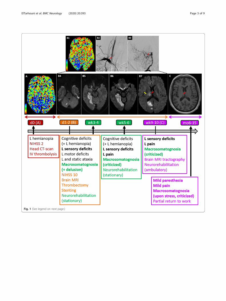
Fig. 1 (See legend on next page.)
ElTarhouni et al. BMC Neurology (2020) 20:393 Page 3 of 9
3 months (due to stenting) and then a monotherapy(Aspirin) was planned for long term. The patient startedAtorvastatin 40 mg/d, stopped smoking, and hadcontinuous positive airway pressure (CPAP) therapy forsleep apnea.
Symptom description (including MSG) and clinicalfindings in NeurorehabilitationOn admission in Neurorehabilitation division (20 dayspost-stroke), the patient had left arm heaviness withparesthesia corresponding, on neurological examination,to left-sided hypoesthesia in all modalities (touch, pain,temperature), hypo-pallesthesia, severely disturbed senseof position and mild left upper limb hemiparesis andcerebellar ataxia were still present. Neuropsychologicaldeficits remained unchanged, but did not interfere dailyactivities.Besides, the patient complained about a strange feeling
of increased size (i.e. MSG) mainly in his left upper limband left flank (see the illustration in the supplementary“figure macrosomatognosia” panel A). Although nodifference in arm size was evidenced on examinationand notified to him, the patient persistently declared thathis feeling of increased limb size was real, showing forexample, as a proof, that his shirt leaves were tighter onthe left compared to the right side (no objective differ-ence was observed by the medical team). He furtherdescribed his sensation, saying: “there is a space on theleft side and it feels precisely like a balloon beneath myarm when I press on it. … If I drop my arms at rest, here[showing the left arm] I have the impression that myarm hangs like this [he laterally raises his right arm upto horizonal position about 90 °], as if I am walking with
a friend in the street arm in arm; it feels the same[because] the entire left side is inflated like a balloon [heshows with the right hand, the entire area along thelateral left trunk starting from the armpit]” . Closing eyesor looking in the mirror did not modify MSG, whiletouching or moving the affected limb (see the supple-mentary “figure macrosomatognosia” panel A) enhancedit. The patient did not report (even upon proactive ques-tioning) any feeling of illusory, supernumerary or dis-owned limb, and did not show particular emotiontowards inflated body parts. There was no sensation ofsplitting of the self either. Rather, he reported misloca-tion of sensory stimulation during neurological assess-ments (e.g. he perceived a light left forearm tactilestimulation on the left back side of his neck).One to 2 weeks upon admission in stationary neuror-
ehabilitation (i.e. 4–5 weeks post-stroke), he could stillprecisely describe MSG in the neck, around the left eye,the left ear and the whole left side of the neck, the leftarm and trunk (see the supplementary “figure macroso-matognosia” panel B). Otherwise he specified experien-cing it in particular when he touched or mobilized thoseareas. This last statement has to be taken with cautionas the patient still reported spontaneous MSG in his leftarm and left flank. He possibly meant that MSG in add-itional areas did not exist at rest). Sensory disorderssimilarly worsened with movement. As the examinerstarted passively moving the patient’s left upper limbfrom a 90° external abduction towards the left flank, thepatient felt the arm heavier (he had his eyes closed dur-ing this evaluation). He started feeling that his left armpushed against the trunk “balloon” at ~ 80° abductionand this sensation increased with greater adduction,
(See figure on previous page.)Fig. 1 Standard brain imaging and symptom timeline. a Perfusion head CT-scan performed on arrival in the emergency department (day 0). TheTime-To-Maximum (TMax, color code at bottom left) was prolonged predominantly in the right internal occipital region and the right thalamuswhile the cerebral blood volume remained normal overall (not shown), thereby defining a penumbra. b Brain images performed upon neurologicalaggravation (day 1 and day 2). Perfusion head CT-scan (B1) (day 1) showing increased TMax (i.e. penumbra) in the right posterior cerebral artery(PCA) vascular territory (color code at bottom left) due to new occlusion of the right PCA. A conventional arteriography performed at day 2 inorder to repermealize the previously occluded right vertebral (B2, anterior view of the right subclavian artery angiography with ostial occlusion ofthe right vertebral artery, red arrow) by thrombectomy, allowed visualization of the PCA occlusion (B3, lateral view of the right vertebral arteryand the basilar artery with the occluded P1 segment of the right posterior cerebral artery, red arrow). The Diffusion Weight Imaging sequence ofbrain Magnetic Resonance Imaging done subsequently the same day showed several restriction areas in the cerebellum (B4), right hippocampaland parahippocampal regions (B5), right lingual gyrus (B6), right occipital lobe (B7), right thalamus (B7, red arrow) and minor involvement of theright internal capsule (B7, yellow arrow), confirming acute ischemic lesions. (C) 3 T Brain Magnetic Resonance Imaging (10 weeks after stroke). Onthe T1 sequence (the section shown is approximately on the same level as on the B7 panel), the occipital lesion is not visible anymore, contraryto the right thalamic residual lesion (red arrow). Bottom panel: The timeline of the main clinical finding evolution (until the end of follow up, greyhorizontal arrow) in parallel with respective brain imaging (letters in () next to time points correspond to figures in upper panels) andmanagement strategies. Color codes relate to different treatment places, in relation with respective time points (ED at day 0 in dark red; tertiaryhospital at day 1–2 post-stroke in orange; ambulatory follow up from week 9–10 until the 15th month, in purple), symptoms (symptoms ofinterest are in bold) and brain imaging (left-right arrows below brain imaging panels relate to the same timeline as in the text boxes below),while vertical black arrows correspond to change of management place (admission, transfer or discharge). Initial symptoms that persisted throughthe follow up are written in black. Detailed description of macrosomatognosia and sensory deficits was done during stationaryneurorehabilitation. Macrosomatognosia and sensory deficits significantly decreased 6months after stroke
ElTarhouni et al. BMC Neurology (2020) 20:393 Page 4 of 9
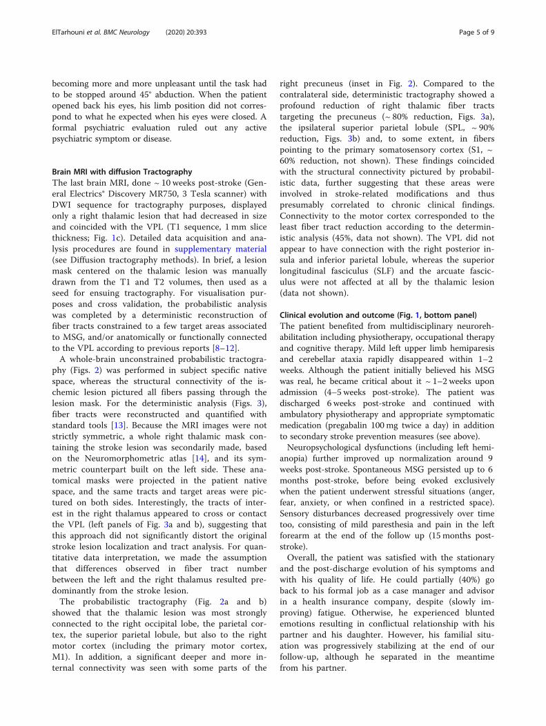
becoming more and more unpleasant until the task hadto be stopped around 45° abduction. When the patientopened back his eyes, his limb position did not corres-pond to what he expected when his eyes were closed. Aformal psychiatric evaluation ruled out any activepsychiatric symptom or disease.
Brain MRI with diffusion TractographyThe last brain MRI, done ~ 10 weeks post-stroke (Gen-eral Electrics® Discovery MR750, 3 Tesla scanner) withDWI sequence for tractography purposes, displayedonly a right thalamic lesion that had decreased in sizeand coincided with the VPL (T1 sequence, 1 mm slicethickness; Fig. 1c). Detailed data acquisition and ana-lysis procedures are found in supplementary material(see Diffusion tractography methods). In brief, a lesionmask centered on the thalamic lesion was manuallydrawn from the T1 and T2 volumes, then used as aseed for ensuing tractography. For visualisation pur-poses and cross validation, the probabilistic analysiswas completed by a deterministic reconstruction offiber tracts constrained to a few target areas associatedto MSG, and/or anatomically or functionally connectedto the VPL according to previous reports [8–12].A whole-brain unconstrained probabilistic tractogra-
phy (Figs. 2) was performed in subject specific nativespace, whereas the structural connectivity of the is-chemic lesion pictured all fibers passing through thelesion mask. For the deterministic analysis (Figs. 3),fiber tracts were reconstructed and quantified withstandard tools [13]. Because the MRI images were notstrictly symmetric, a whole right thalamic mask con-taining the stroke lesion was secondarily made, basedon the Neuromorphometric atlas [14], and its sym-metric counterpart built on the left side. These ana-tomical masks were projected in the patient nativespace, and the same tracts and target areas were pic-tured on both sides. Interestingly, the tracts of inter-est in the right thalamus appeared to cross or contactthe VPL (left panels of Fig. 3a and b), suggesting thatthis approach did not significantly distort the originalstroke lesion localization and tract analysis. For quan-titative data interpretation, we made the assumptionthat differences observed in fiber tract numberbetween the left and the right thalamus resulted pre-dominantly from the stroke lesion.The probabilistic tractography (Fig. 2a and b)
showed that the thalamic lesion was most stronglyconnected to the right occipital lobe, the parietal cor-tex, the superior parietal lobule, but also to the rightmotor cortex (including the primary motor cortex,M1). In addition, a significant deeper and more in-ternal connectivity was seen with some parts of the
right precuneus (inset in Fig. 2). Compared to thecontralateral side, deterministic tractography showed aprofound reduction of right thalamic fiber tractstargeting the precuneus (~ 80% reduction, Figs. 3a),the ipsilateral superior parietal lobule (SPL, ~ 90%reduction, Figs. 3b) and, to some extent, in fiberspointing to the primary somatosensory cortex (S1, ~60% reduction, not shown). These findings coincidedwith the structural connectivity pictured by probabil-istic data, further suggesting that these areas wereinvolved in stroke-related modifications and thuspresumably correlated to chronic clinical findings.Connectivity to the motor cortex corresponded to theleast fiber tract reduction according to the determin-istic analysis (45%, data not shown). The VPL did notappear to have connection with the right posterior in-sula and inferior parietal lobule, whereas the superiorlongitudinal fasciculus (SLF) and the arcuate fascic-ulus were not affected at all by the thalamic lesion(data not shown).
Clinical evolution and outcome (Fig. 1, bottom panel)The patient benefited from multidisciplinary neuroreh-abilitation including physiotherapy, occupational therapyand cognitive therapy. Mild left upper limb hemiparesisand cerebellar ataxia rapidly disappeared within 1–2weeks. Although the patient initially believed his MSGwas real, he became critical about it ~ 1–2 weeks uponadmission (4–5 weeks post-stroke). The patient wasdischarged 6 weeks post-stroke and continued withambulatory physiotherapy and appropriate symptomaticmedication (pregabalin 100 mg twice a day) in additionto secondary stroke prevention measures (see above).Neuropsychological dysfunctions (including left hemi-
anopia) further improved up normalization around 9weeks post-stroke. Spontaneous MSG persisted up to 6months post-stroke, before being evoked exclusivelywhen the patient underwent stressful situations (anger,fear, anxiety, or when confined in a restricted space).Sensory disturbances decreased progressively over timetoo, consisting of mild paresthesia and pain in the leftforearm at the end of the follow up (15 months post-stroke).Overall, the patient was satisfied with the stationary
and the post-discharge evolution of his symptoms andwith his quality of life. He could partially (40%) goback to his formal job as a case manager and advisorin a health insurance company, despite (slowly im-proving) fatigue. Otherwise, he experienced bluntedemotions resulting in conflictual relationship with hispartner and his daughter. However, his familial situ-ation was progressively stabilizing at the end of ourfollow-up, although he separated in the meantimefrom his partner.
ElTarhouni et al. BMC Neurology (2020) 20:393 Page 5 of 9
DiscussionGeneral considerationsWe report here a 15month-follow up of a 45-year oldman known for several cardiovascular risk factors, whopresented several neurological symptoms (includingMSG) in the context of multiple acute ischemic strokelesions of undetermined origin, sequentially treated withIV thrombolysis and arterial thrombectomy with stent-ing, out of the recommended time windows, most prob-ably owing to his particular clinical context (young age,no severe comorbidity, sequential aggravation with per-sistent penumbra, arterial occlusions). In general, som-atic and cognitive neurological deficits were compatiblewith stroke lesion localizations. Motor, cerebellar, visualfield and cognitive deficits rapidly normalized concomi-tantly with disappearance of lesions on brain imaging,while left-sided MSG and sensory disturbances persistedover months in parallel with a residual right thalamiclesion. Progressive functional improvement in stationaryNeurorehabilitation led to discharge back home and topartial return to work.
Stroke etiology of MSGSeveral pathologies can lead to MSG [4, 15]. Our patientdid not suffer from migraine, infection or any activepsychiatric disease at the time of stroke. The clinicalconstellation did not suggest an ictal origin neither, al-though no electroencephalogram was performed. Thus,the only plausible etiology of MSG appeared to be strokelesion. In support to this assumption, MSG persisted inparallel with left-sided sensory symptoms that wereanatomically compatible with the residual right thalamicischemic lesion. To our best knowledge, there is no pre-vious report of MSG due to thalamic lesions, BSD beingtraditionally associated to (right) parietal lesions [10] ormore widely to lesions of the temporoparietal-occipitalcarrefour [4]. Of notice, focal lesions constitute a rarecause of MSG (< 10%), with stroke representing less thanone third of the cases [3].
Anatomical substrate of MSGWhile the anatomo-clinical coherence between the mainneurological symptoms (left homonymous hemianopia,
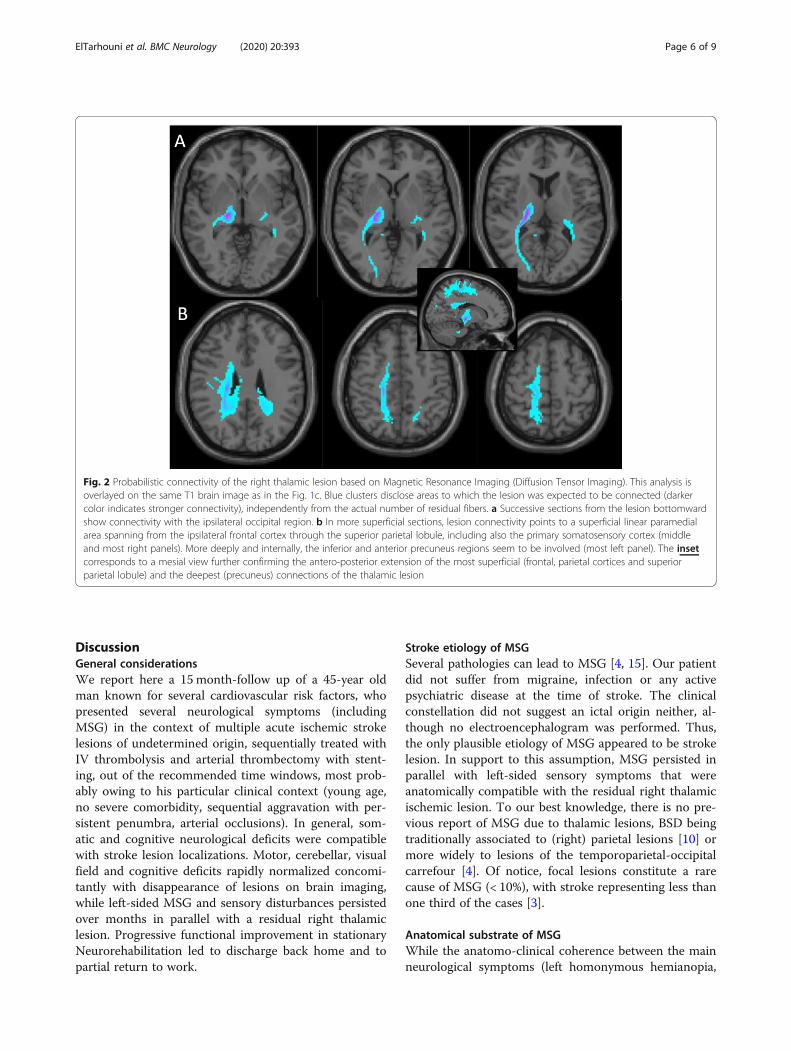
Fig. 2 Probabilistic connectivity of the right thalamic lesion based on Magnetic Resonance Imaging (Diffusion Tensor Imaging). This analysis isoverlayed on the same T1 brain image as in the Fig. 1c. Blue clusters disclose areas to which the lesion was expected to be connected (darkercolor indicates stronger connectivity), independently from the actual number of residual fibers. a Successive sections from the lesion bottomwardshow connectivity with the ipsilateral occipital region. b In more superficial sections, lesion connectivity points to a superficial linear paramedialarea spanning from the ipsilateral frontal cortex through the superior parietal lobule, including also the primary somatosensory cortex (middleand most right panels). More deeply and internally, the inferior and anterior precuneus regions seem to be involved (most left panel). The insetcorresponds to a mesial view further confirming the antero-posterior extension of the most superficial (frontal, parietal cortices and superiorparietal lobule) and the deepest (precuneus) connections of the thalamic lesion
ElTarhouni et al. BMC Neurology (2020) 20:393 Page 6 of 9
ataxia and left-sided sensory symptoms) and identifiedlesions (right occipital, cerebellar and right thalamiclesions) was clear, lesion in the (right) parietal cortex,most generally reported in MSG [10], lacked in ourpatient. Furthermore, the other incriminated brain areas(the frontal lobe, the posterior insula, the superior andinferior parietal lobules, the precuneus) [9–11], owing totheir connections with the parietal cortex, were notaffected neither.Among the patient’s lesions, tractography data showed
that only the thalamic lesion shared anatomical link withthe parietal cortex. Yet, within the thalamus, the VPL(where the lesion was most probably located) constitutesan important provider of thalamic connections targetingthe superior parietal lobule and the precuneus [8, 9, 12](both involved MSG). Interestingly, our data not onlyconfirmed this strong connectivity, but they showed, inaddition, a drastic reduction in fiber tracts numberbetween the VPL ischemic lesion and both areas incomparison to the left side. The SLF, which supposedlyunderlies the association between the frontal lobe andMSG [9, 10], was not damaged in our patient. Likewise,
no connection was seen with the posterior insula andthe inferior parietal lobule, most probably because theydo not receive projections from the VPL [16]. Thestrong connectivity observed with the motor cortexcould either be related to altered functional connectionbetween the VPL and M1 [12] or to a wallerian degener-ation explaining the modest reduction in fiber tracts tar-geting the M1 area. Overall, M1 involvement appearedto have minor clinical significance, apart from the initialtransient mild left hemiparesis and MSG enhancementinduced by the affected limb movements.In summary, brain imaging data disclose the right VPL
as an essential relay mediating appearance of MSG, evenin absence of direct lesion of the parietal lobe, therebysuggesting a “diaschisis”-like mechanism engaged.
Mechanisms underlying MSGTractography showed significant structural connectivitycoinciding with important reduction of fiber tract num-ber between the thalamic lesion and S1. The prominentleft-sided sensory dysfunction further confirmed the roleof VPL as a major thalamic relay of somatosensory
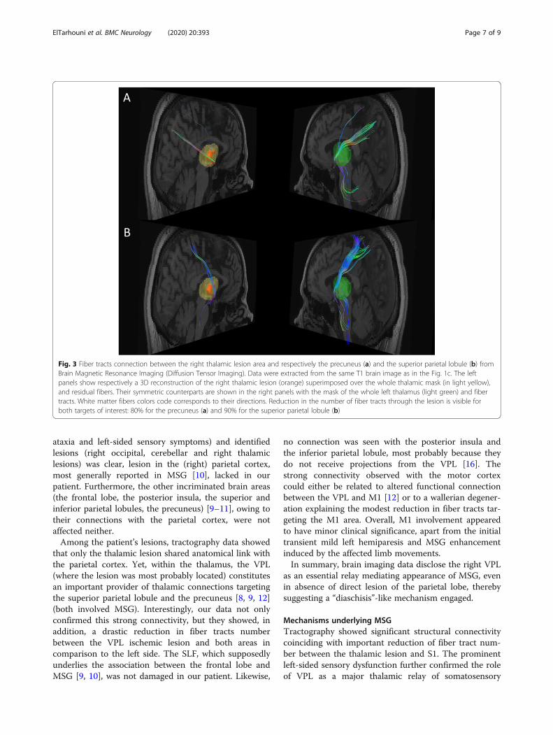
Fig. 3 Fiber tracts connection between the right thalamic lesion area and respectively the precuneus (a) and the superior parietal lobule (b) fromBrain Magnetic Resonance Imaging (Diffusion Tensor Imaging). Data were extracted from the same T1 brain image as in the Fig. 1c. The leftpanels show respectively a 3D reconstruction of the right thalamic lesion (orange) superimposed over the whole thalamic mask (in light yellow),and residual fibers. Their symmetric counterparts are shown in the right panels with the mask of the whole left thalamus (light green) and fibertracts. White matter fibers colors code corresponds to their directions. Reduction in the number of fiber tracts through the lesion is visible forboth targets of interest: 80% for the precuneus (a) and 90% for the superior parietal lobule (b)
ElTarhouni et al. BMC Neurology (2020) 20:393 Page 7 of 9
inputs unto S1 [17]. Whether sensory deficits contrib-uted to MSG constitutes an interesting question, espe-cially if we consider that both symptoms improved inparallel and affected the same body parts.Peripheral sensory deafferentation/lesion, one mechan-
ism that could mediate this association [15, 18], was notobserved in our patient. Alternatively, MSG could be aresult of disordered primary or high order neural pro-cessing of body representation [15, 18] taking place inthe VPL, as suggested by experimental animal results[19]. Defective integration of visual and somatosensoryinputs [3] could also be discussed. In previous reports,patients who criticized their MSG [10] or witnessedMSG disappearance under visual control [9] had intactSPL (a structure where integration of visual and somato-sensory information takes place), contrary to the patientreported here in whom a significant reduction of fiberstargeting the SPL was observed. The role played by theobserved mild executive dysfunction in the lack of criti-cism towards MSG could also be questioned. However,not only it outlasted the moment when the patientbecame critical regarding his MSG, but it had no otherbehavioral consequence.
Classification of MSGAccording to his description of MSG, the patient hadhyperschematia (enlarged body parts size; included amongaschematia) and paraschematia (displacement of bodyparts position; noticed when his eyes were closed) [5],thereby fulfilling criteria for the rare (< 10%) type A ofAIWS [4]. However, if we take into account thedelusional-like feature (not typical for MSG [20]), he couldbe as well considered as type C (MSG+ delusion) al-though delusions are typically much more florid andprominent in this group [4]. Thus, the phenomenology ofMSG in this patient seems to represent an intermediatetype of AIWS not individualized in existing classifications.The later tend to separate (sometimes even oppose)somesthetic (cognitive) and psychic (delusional) symp-toms [3, 4], while the wide variety of semiological descrip-tions makes this distinction challenging for an inclusiveclassification. Perhaps AIWS should be considered as acontinuous spectrum including on one extreme, predom-inantly cognitive (non-delusional) forms due to structuralbrain (focal) lesions, and on the other extreme, more delu-sional (psychic) profiles without individualized lesion onbrain imaging. This new perspective would allow integra-tion of intermediate profiles and more comprehensivepathophysiological considerations.
ConclusionWe report the case of a patient presenting mainly withlong-lasting left-sided MSG and sensory deficits presum-ably associated with a right thalamic stroke lesion
restricted to the VPL, that progressively improved overmonths. Tractographic data pointed out the essentialrole of thalamic connections to the parietal cortex (espe-cially the precuneus) via the VPL, a crucial nucleus inbody size coding and perception. Damaged SPL projec-tions of the VPL might have contributed to the lack ofcriticism towards MSG and absence of visual control ofMSG. Defective neural processing and integration ofsensory information by and/or through the VPL seemsto be essential in generation of MSG. The patient’s MSGwas classified as an intermediate (non-individualized)stage between the types A and C AIWS. Further discus-sion on MSG phenomenology is needed, consideringAIWS as a continuum flexibly associating cognitive andpsychic symptoms, and thereby possibly featuring analternative classification.The case reported here was well-documented during
and after the neurorehabilitation stay, which allowedconsistent anatomo-clinical discussion. However, de-tailed description of the patient’s initial neurologicalcomplains, including a precise time when the mainsymptoms discussed in the paper started, lacked. Inaddition, tractography data were obtained in the chronicphase of long-lasting neurological symptoms and lesions,making it difficult to extrapolate with certainty correl-ation between acute ischemic lesions and initial symp-toms. Thus, our findings have to be considered withcaution, especially when generalizing derived conclu-sions. Nevertheless, this first detailed analysis of MSG inthe context of a non-cortical brain lesion explores inter-esting hypotheses that would highly contribute, ifconfirmed, to understand mechanisms underlying thisunusual symptom, as well as the role of the VPL.
Supplementary informationSupplementary information accompanies this paper at https://doi.org/10.1186/s12883-020-01970-3.
Additional file 1.
Additional file 2.
AbbreviationsAIWS: Alice in wonderland syndrome; BSD: Body Schema Disorders;CPAP: Continuous Positive Airway Pressure; CT: Computerized Tomography;DTI: Diffusion Tensor Imaging; DWI: Diffusion Weighted Imaging;ED: Emergency Department; LDL: Low Density Lipoproteins; M1: Primarymotor cortex; MRI: Magnetic Resonance Imaging; MSG: Macrosomatognosia;MoCA: Montreal Cognitive Assessment; NIHSS: National Health InstituteStroke Scale; PCA: Posterior Cerebral Artery; S1: Primary somatosensorycortex; SLF: Superior Longitudinal Fasciculus; SPL: Superior Parietal Lobule;VPL: Ventral posterolateral nucleus
AcknowledgementsNone.
Authors’ contributionsAHET: data collection, manuscript writing, manuscript revision. LB: datacollection, manuscript revision. MM: data collection, data analysis, manuscript
ElTarhouni et al. BMC Neurology (2020) 20:393 Page 8 of 9
writing, manuscript revision. BE: data analysis, manuscript writing, manuscriptrevision. JA: data collection, data analysis, manuscript revision. JMA: dataanalysis, manuscript revision. EA: data analysis, manuscript revision. SD: studyconception, data analysis, manuscript writing, manuscript revision. JNC: studyinitiation and conception, data collection, data analysis, manuscript writing,manuscript revision. All authors have read and approved the manuscript.
FundingThis study was not funded.
Availability of data and materialsAll data and materials related to this study are ready to be made availablefrom the Neurology Unit server in a format that does not allow recognitionof the reported patient.
Ethics approval and consent to participateThe present study was conducted according to local and internationalethical standards. Local ethical rules require no formal clearance for casereports. The participant gave his written consent to participate to the studyprior to data collection.
Consent for publicationThe reported patient gave his written consent for the publication of his data.
Competing interestsAuthors have no financial and non-financial conflict of interest to disclose.
Author details1Division of Neurorehabilitation, Fribourg Hospital, Meyriez, Switzerland.2Neurology Unit, Department of Neuroscience and Movement Science,Medicine Section, Faculty of Science and Medicine, University of Fribourg,Chemin du Musée, 5, CH-1700 Fribourg, Switzerland. 3Department ofRadiology, Fribourg Hospital, Riaz, Switzerland.
Received: 26 April 2020 Accepted: 19 October 2020
References1. Dieguez S, Blanke O. Altered states of bodily consciousness. In: Cardeña E,
Winkelman, editors. Altering consciousness: a multidisciplinary perspective.Volume 2. Westport: Praeger; 2011. pp. 237–62.
2. Frederiks J. Macrosomatognosia and microsomatognosia. Psychiatr NeurolNeurochir. 1963;66:531–6.
3. Blom J. Alice in wonderland syndrome, a systematic review. Neurology.2016;6:259–70.
4. Mastria G, Mancini V, Vigano A, Di Piero V. Alice in wonderland syndrome: aclinical and pathophysiological review. Biomed Res Int. 2016. https://doi.org/10.1155/2016/8243145.
5. Vallar G, Rode G. Commentary on Bonnier P. L'aschématie Rev Neurol (Paris)1905; 13:605-9. Epilepsy Behav. 2009;16:397–400.
6. Dieguez S. Micromegas: altered body/environment scaling in literary fiction:thought-experients on embodiment, spatiality and agency. Front Psychol.2016;7:556.
7. Dieguez S, Lopez C. La représentation du corps et ses troubles: approchesde la neuropsychologie et des neurosciences cognitives; 2015. p. 63–88.
8. Grant E, Hoerder-Suabedissen A, Monàr Z. Development of thecorticothalamic projections. Front Neurosci. 2012;6:53. https://doi.org/10.3389/fnins.2012.00053.
9. Herbert G, Lemaitre A, Moritz-Gasser S, Cochereau J, Duffau H. The antero-dorsal precuneal cortex supports specific aspects of bodily awareness Brain,vol. 142; 2019. p. 2207–14.
10. Weijers R, Rietveld A, Meijer F, de Leeuw F. Macrosomatognosia in frontallobe infarct, a case report. J Neurol. 2013;260:925–6.
11. Yeterian E, Pandya D. Corticothalamic connections of the posterior parietalcortex in the rhesus monkey. J Comp Neurol. 1985;518:3725–251.
12. Zhang S, Li C. Functional connectivity parcellation of the human thalamusby independent component analysis. Brain Connectivity. 2017;7:602–16.
13. Wang R, Wedeen V. http://trackvis.org/ (2019). Accessed 2019.14. Marcus D, Fotenos A, JG C, JC M, RL B. Open access series of imaging
studies: longitudinal MRI data in nondemented and demented older adults.J Cogn Neurosci. 2010;22:2677–84.
15. Haggard P, Wolpert D. Disorders of body scheme: Freund H, M J, Hallett M,Leiguarda R, editors. Higher-order motor disorders From neuroanatomy andneurobiology to clinical neurology. Oxford: Oxford University Press; 2005. p.261–271.
16. Matsuzaki R, Kyuhou S, Matsuura-Nakao K, Gemba H. THalamo-corticalprojections to the posterior parietal cortex in the monkey. Neurosci Lett.2004;355:113–6.
17. Song W, Semework M. Tactile representation in somatosensory thalamus(VPL) and cortex (S1) of awake primate and the plasticity induced by VPLneuroprosthetic stimulation. Brain Res. 1625;2015:301–13.
18. Paqueron X, Leguen M, Rosenthal D, Coriat P, Willer J, Danziger N. Thephenomenology of body image distorsions induced by regional anesthesia.Brain. 2003;126:702–12.
19. Krubitzer L, Kaas J. Thalamic connections of three representations of thebody surface in somatosensory cortex of gray squirrels. J Comp Neurol.1987;265:549–80.
20. Dieguez S, Staub F, Bogousslavsky. Asomatognosia. In: Bogousslavsky J,Godefroy O, editors. The Behavioral and Cognitive Neurology of Storke:Cambridge University Press; 2007. p. 215–53.
Publisher’s NoteSpringer Nature remains neutral with regard to jurisdictional claims inpublished maps and institutional affiliations.
ElTarhouni et al. BMC Neurology (2020) 20:393 Page 9 of 9