prefrontal mechanisms of impaired cognition - udspace home
TRANSCRIPT
PREFRONTAL MECHANISMS OF IMPAIRED COGNITION
IN A RAT MODEL OF FASD
by
Nicholas A. Heroux
A dissertation submitted to the Faculty of the University of Delaware in partial
fulfillment of the requirements for the degree of Doctor of Philosophy in
Psychological and Brain Sciences
Winter 2020
© 2020 Nicholas A. Heroux
All Rights Reserved
PREFRONTAL MECHANISMS OF IMPAIRED COGNITION
IN A RAT MODEL OF FASD
by
Nicholas A. Heroux
Approved: __________________________________________________________
Tania Roth, Ph.D.
Chair of the Department of Psychological and Brain Sciences
Approved: __________________________________________________________
John Pelesko, Ph.D.
Dean of the College of Arts and Sciences
Approved: __________________________________________________________
Douglas J. Doren, Ph.D.
Interim Vice Provost for Graduate and Professional Education and
Dean of the Graduate College
I certify that I have read this dissertation and that in my opinion it meets
the academic and professional standard required by the University as a
dissertation for the degree of Doctor of Philosophy.
Signed: __________________________________________________________
Mark Stanton, Ph.D.
Professor in charge of dissertation
I certify that I have read this dissertation and that in my opinion it meets
the academic and professional standard required by the University as a
dissertation for the degree of Doctor of Philosophy.
Signed: __________________________________________________________
Jeffrey Rosen, Ph.D.
Member of dissertation committee
I certify that I have read this dissertation and that in my opinion it meets
the academic and professional standard required by the University as a
dissertation for the degree of Doctor of Philosophy.
Signed: __________________________________________________________
Anna Klintsova, Ph.D.
Member of dissertation committee
I certify that I have read this dissertation and that in my opinion it meets
the academic and professional standard required by the University as a
dissertation for the degree of Doctor of Philosophy.
Signed: __________________________________________________________
Amy Griffin, Ph.D.
Member of dissertation committee
I certify that I have read this dissertation and that in my opinion it meets
the academic and professional standard required by the University as a
dissertation for the degree of Doctor of Philosophy.
Signed: __________________________________________________________
Mary Dozier, Ph.D.
Member of dissertation committee
I certify that I have read this dissertation and that in my opinion it meets
the academic and professional standard required by the University as a
dissertation for the degree of Doctor of Philosophy.
Signed: __________________________________________________________
Jee Hyun Kim, Ph.D.
Member of dissertation committee
v
Dr. Mark E. Stanton, for his unwavering support of my development as a
scientist. It has been a distinct privilege of my life to have been trained by you, and for
our mentor/mentee partnership these last five and a half years.
My dissertation committee and BN faculty and students, for your scientific and
personal support during my graduate career. Thanks for being part of my journey – I
couldn’t have picked a better village.
My mother, Diane, and my sister, Molly, for your unwavering support and love
throughout my life and especially during my graduate career. I couldn’t have done this
without both of you.
ACKNOWLEDGMENTS
vi
LIST OF TABLES ........................................................................................................ xi
LIST OF FIGURES ...................................................................................................... xii
LIST OF ABBREVIATIONS ...................................................................................... xii
ABSTRACT .............................................................................................................. xviii
Chapter
1 INTRODUCTION .............................................................................................. 1
1.1 The Context Preexposure Facilitation Effect ............................................ 1
1.2 Developmental Alcohol Exposure and the CPFE ..................................... 6 1.3 Dissertation Overview ............................................................................... 8
2 NEUROBIOLOGY OF CONTEXTUAL FEAR CONDITIONING ................. 9
2.1 Neuroanatomical Substrates of Contextual Fear Conditioning in
Rodents ...................................................................................................... 9
2.1.1 Hippocampal Involvement in Acquisition and Retention of
Contextual Fear Conditioning ..................................................... 12 2.1.2 Prefrontal Involvement in Acquisition and Retention of
Contextual Fear Conditioning ..................................................... 20
2.1.3 Prefrontal and Hippocampal Dynamics of Contextual Memory
in the CPFE .................................................................................. 28
2.2 Molecular Substrates of Contextual Fear Conditioning: Immediate
Early Genes ............................................................................................. 30
2.2.1 Immediate Early Genes: c-Fos .................................................... 31 2.2.2 Immediate Early Genes: Arc ....................................................... 33
2.2.3 Immediate Early Genes: Egr-1 .................................................... 34 2.2.4 Immediate Early Genes: Npas4 ................................................... 35
2.3 Conclusion ............................................................................................... 36
3 ANIMAL MODELS OF FETAL ALCOHOL SPECTRUM DISORDER ...... 38
TABLE OF CONTENTS
vii
3.1 Neurocognitive Consequences of Gestational Alcohol Exposure in
Developing Humans ................................................................................ 38
3.2 Animal Models of Fetal Alcohol Spectrum Disorders ............................ 40 3.3 Hippocampal Insult after Neonatal Alcohol Exposure in Rats ............... 41 3.4 Prefrontal Insult after Neonatal Alcohol Exposure in Rats ..................... 43 3.5 Cholinergic Insult after Neonatal Alcohol Exposure in Rats .................. 44 3.6 Behavioral Consequences of Neonatal Alcohol Exposure in Rats .......... 46
3.7 Impairment of the CPFE by Neonatal Alcohol Exposure in Rats ........... 49 3.8 Conclusions ............................................................................................. 53
4 FOUNDATIONAL STUDIES ......................................................................... 54
4.1 NMDA-Receptor Cell Signaling and the CPFE ...................................... 54 4.2 Involvement of the mPFC across Variants of Contextual Fear
Conditioning ............................................................................................ 59 4.3 Role of mPFC and dHPC Muscarinic-Receptor Activity in the CPFE ... 64
4.4 Regional Expression of Immediate Early Genes across Phases of the
CPFE ....................................................................................................... 67
4.5 Conclusions ............................................................................................. 72
5 GENERAL METHODS ................................................................................... 73
5.1 Material and Methods .............................................................................. 73
5.1.1 Animal Subjects ........................................................................... 73
5.1.2 Stereotaxic Surgery, Drug Infusion, and Histology .................... 74
5.1.2.1 Stereotaxic Surgery ...................................................... 74 5.1.2.2 Intra-mPFC and Intra-vHPC Drug Infusions ............... 75
5.1.2.3 Histology ...................................................................... 75
5.1.3 Animal Model of Fetal Alcohol Spectrum Disorder ................... 76
5.1.3.1 Neonatal Alcohol Dosing (PD4-9, 5.25g/kg/d) ............ 76
5.1.3.2 Blood Alcohol Concentrations (BACs) ........................ 77
5.1.4 Behavioral Equipment and Procedures ....................................... 77
5.1.4.1 Apparatus, Stimuli, and Freezing Software Analyses .. 78 5.1.4.2 Context Preexposure Facilitation Effect (CPFE) ......... 79 5.1.4.3 Standard Contextual Fear Conditioning (sCFC) .......... 80
5.1.5 IEG Assays .................................................................................. 81
viii
5.1.5.1 Brain Removal and Tissue Dissection .......................... 81 5.1.5.2 Quantitative Real-time PCR (qPCR) ............................ 84
5.1.6 Statistical Analyses ...................................................................... 84
6 MEDIAL PREFRONTAL AND VENTRAL HIPPOCAMPAL
CONTRIBUTIONS TO INCIDENTAL CONTEXT LEARNING AND
MEMORY IN ADOLESCENT RATS ............................................................ 86
6.1 Introduction ............................................................................................. 86
6.2 Materials and Methods ............................................................................ 87
6.2.1 Subjects ........................................................................................ 87
6.2.2 Stereotaxic Surgery ..................................................................... 87
6.2.3 Apparatus and Stimuli ................................................................. 87 6.2.4 Behavioral Procedures and Drug Infusion .................................. 87 6.2.5 Brain Removal, Tissue Dissection, and qPCR ............................ 88 6.2.6 Histology ..................................................................................... 88
6.2.7 Data Analysis ............................................................................... 88
6.2.7.1 Analysis of Behavioral Data ......................................... 88
6.2.7.2 Analysis of IEG Expression ......................................... 89
6.3 Results ..................................................................................................... 92
6.3.1 Experiment 6.1: Prefrontal Inactivation during Context
Exposure Impairs Contextual Fear Conditioning and IEG
Expression in the mPFC, vHPC, and VMT ................................. 92 6.3.2 Experiment 6.2: Ventral Hippocampal Inactivation during
Context Exposure Impairs Contextual Fear Conditioning and
IEG Expression in the mPFC, dHPC, and vHPC ........................ 97
6.4 Discussion .............................................................................................. 101
7 NEONATAL ETHANOL EXPOSURE IMPAIRS LONT-TERM
CONTEXT MEMORY FORMATION AND PREFRONTAL
IMMEDIATE EARLY GENE EXPRESSION IN ADOLESCENT RATS .. 109
7.1 Introduction ........................................................................................... 109 7.2 Materials and Methods .......................................................................... 110
7.2.1 Subjects ...................................................................................... 110 7.2.2 Neonatal Alcohol Dosing .......................................................... 110 7.2.3 Apparatus and Stimuli ............................................................... 110
ix
7.2.4 Behavioral Procedures ............................................................... 111 7.2.5 Brain Removal, Tissue Dissection, and qPCR .......................... 111
7.2.6 Data Analysis ............................................................................. 111
7.2.6.1 Analysis of Neonatal and Adolescent Body Weight .. 111 7.2.6.2 Analysis of Behavioral Data ....................................... 114 7.2.6.3 Analysis of IEG Expression ....................................... 114
7.3 Results ................................................................................................... 117
7.3.1 Body Weight and BACs ............................................................ 117 7.3.2 Experiment 7.1A: PD4-9 Alcohol Exposure Abolishes Post-
shock and Retention Test Freezing Under 1-Shock
Reinforcement ........................................................................... 118 7.3.3 Experiment 7.1B: PD4-9 Alcohol Exposure Impairs Post-
shock and Retention Test Freezing Under 2-Shock
Reinforcement ........................................................................... 120
7.3.4 Experiment 7.2: PD4-9 Alcohol Exposure Does Not Impair
Retention Test Freezing in sCFC .............................................. 122
7.3.5 Experiment 7.3: PD4-9 Alcohol Exposure Impairs Medial-
Prefrontal but Not Dorsal-Hippocampal IEG Expression
during Context Exposure ........................................................... 124
7.4 Discussion .............................................................................................. 128
8 CHOLINERGIC RESCUE OF NEUROCOGNITIVE INSULT
FOLLOWING THIRD-TRIMESTER EQUIVALENT ALCOHOL
EXPOSURE IN RATS ................................................................................... 133
8.1 Introduction ........................................................................................... 133 8.2 Materials and Methods .......................................................................... 134
8.2.1 Subjects ...................................................................................... 134
8.2.2 Neonatal Alcohol Dosing .......................................................... 134 8.2.3 Apparatus and Stimuli ............................................................... 134 8.2.4 Behavioral Procedures and Drug Injection ................................ 134 8.2.5 Brain Removal, Tissue Dissection, and qPCR .......................... 135
8.2.6 Data Analysis ............................................................................. 135
8.2.6.1 Analysis of Neonatal and Adolescent Body Weight .. 135 8.2.6.2 Analysis of Behavioral Data ....................................... 136
8.2.6.3 Analysis of IEG Expression ....................................... 136
8.3 Results ................................................................................................... 137
x
8.3.1 Body Weight and BACs ............................................................ 137 8.3.2 Experiment 8.1: Systemic Administration of Physostigmine
Prior to Every Phase Rescues the CPFE in Alcohol-exposed
Rats ............................................................................................ 139 8.3.3 Experiment 8.2: Systemic Administration of Physostigmine
Prior to Context Preexposure Rescues the CPFE and Elevates
IEG Expression in Alcohol-exposed Rats ................................. 141
8.3.3.1 Behavioral Results ...................................................... 141 8.3.3.2 IEG Results ................................................................. 143
8.4 Discussion .............................................................................................. 147
9 SUMMARY AND CONCLUSIONS ............................................................. 152
9.1 Prefrontal and Hippocampal Mechanisms of Context and Contextual
Fear Learning and Memory in the CPFE .............................................. 152 9.2 Acute Enhancement of Cholinergic Function Rescues
Neurobehavioral Disruption after Neonatal Alcohol Exposure in Rats 161 9.3 Future Directions ................................................................................... 165
9.3.1 Limitations ................................................................................. 165 9.3.2 Experimental Predictions ........................................................... 166
9.4 Summary Statement ............................................................................... 168
REFERENCES ........................................................................................................... 169
Appendix
A PERMISSIONS AND APPROVALS ............................................................ 203
B IACUC Approval ............................................................................................ 213
xi
Table 6.1 Final group numbers (n), number of outliers removed (HC, MUSC,
PBS), and statistical results for all factorial ANOVAs (see F and p
values) for each gene (c-Fos, Arc, Egr-1, and Npas4) in each region
(mPFC, dHPC, vHPC, VMT) for Experiment 6.1 .................................. 90
Table 6.2 Final group numbers (n), number of outliers removed (HC, MUSC,
PBS), and statistical results for all factorial ANOVAs (see F and p
values) for each gene (c-Fos, Arc, Egr-1, and Npas4) in each region
(mPFC, dHPC, vHPC, VMT) for Experiment 6.2 .................................. 91
Table 7.1 Body weights and BACs for Experiments 7.1A, 7.1B, 7.2, and 7.3.
Average body weights (in grams ± SE) are given from the SI and
EtOH groups at the first and last day of the dosing period (PD4 and
PD9, respectively) and the first day of behavioral training (PD31). ..... 113
Table 7.2 Final group numbers (n), number of outliers removed (HC, Alt-Pre,
Pre), and statistical results for all one-way ANOVAs (see F and p
values) for each gene (c-Fos, Arc, Egr-1, and Npas4) in each region
(mPFC and dHPC) for Experiment 7.3. ................................................ 116
Table 8.1 Body weights and BACs for Experiment 8.1 and 8.2. Average body
weights (in grams ± SE) are given from the SI and EtOH groups at the
first and last day of the dosing period (PD4 and PD9, respectively)
and the first day of behavioral training (PD31). .................................... 138
Table 8.2 Final group numbers (n), number of outliers removed (HC, SI-SAL,
SI-PHY, EtOH-SAL, EtOH-PHY), and statistical results for all
factorial ANOVAs (see F and p values) for each gene (c-Fos, Arc,
Egr-1, and Npas4) in each region (mPFC, dHPC, and vHPC) for
Experiment 8.2. ..................................................................................... 145
LIST OF TABLES
xii
Figure 1.1 Schematic representation of standard contextual fear conditioning
(sCFC) and associated learning and memory processes during each
phase. ......................................................................................................... 4
Figure 1.2 Schematic representation of the Context Preexposure Facilitation
Effect (CPFE) and associated learning and memory processes during
each phase .................................................................................................. 5
Figure 2.1 Hypothetical circuit mediating the different aspects of conditioned
fear.. ......................................................................................................... 11
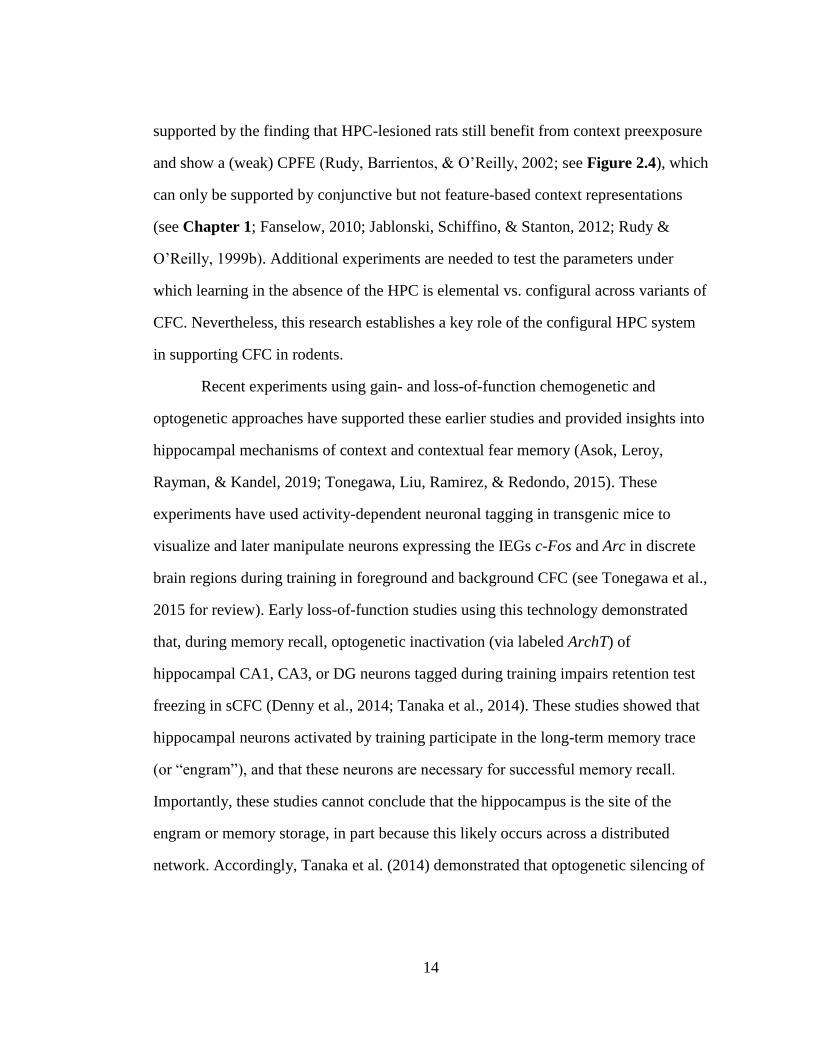
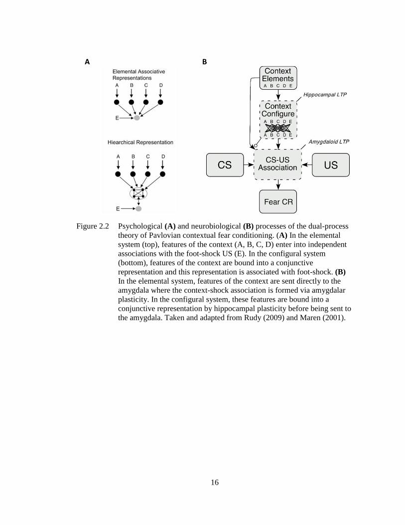
Figure 2.2 Psychological (A) and neurobiological (B) processes of the dual-
process theory of Pavlovian contextual fear conditioning ...................... 16
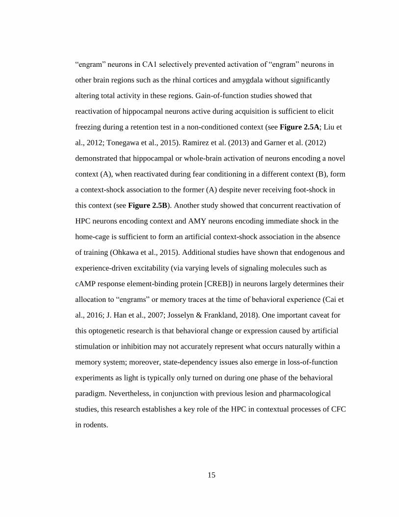
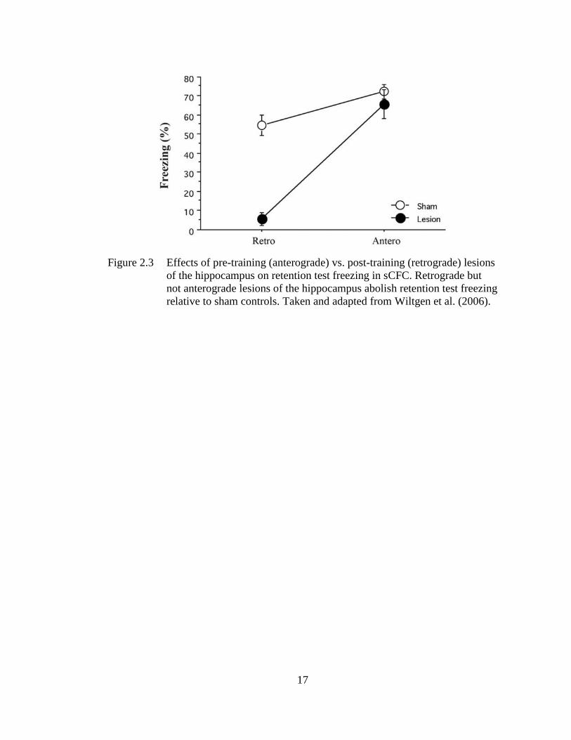
Figure 2.3 Effects of pre-training (anterograde) vs. post-training (retrograde)
lesions of the hippocampus on retention test freezing in sCFC. ............. 17
Figure 2.4 Effects of anterograde HPC lesions on retention test freezing in the
CPFE ....................................................................................................... 18
Figure 2.5 Optogenetic manipulations of memory engram cell populations. ........... 19
Figure 2.6 Effects of pre-training NMDA-receptor antagonism (via Ro25-6981,
A) or post-training protein synthesis inhibition (via anisomycin, B) in
the AC during sCFC ................................................................................ 25
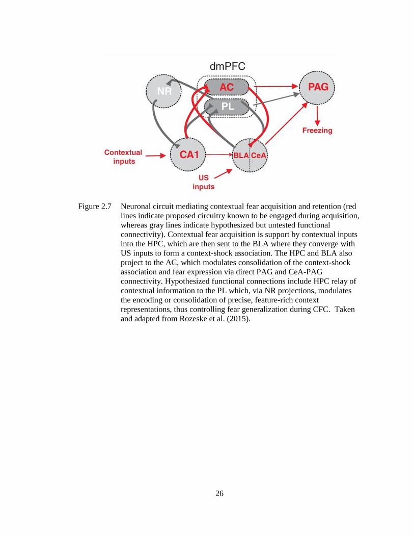
Figure 2.7 Neuronal circuit mediating contextual fear acquisition and retention
(red lines indicate proposed circuitry known to be engaged during
acquisition, whereas gray lines indicate hypothesized but untested
functional connectivity) ........................................................................... 26
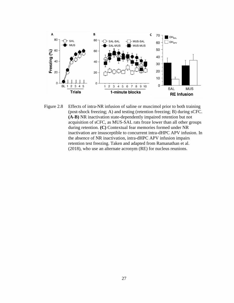
Figure 2.8 Effects of intra-NR infusion of saline or muscimol prior to both
training (post-shock freezing; A) and testing (retention freezing; B)
during sCFC ............................................................................................ 27
Figure 3.1 Unbiased stereology data for CA1 region of the hippocampus. CA1
pyramidal cell number estimates by unbiased stereology. ...................... 48
LIST OF FIGURES
xiii
Figure 3.2 Mean estimates of CA1 c-Fos+ cells (±SE) 2h following preexposure
(5 minutes of context exploration) in Groups SI, 4.00g, and 5.25g. ....... 51
Figure 3.3 Systemic administration of the acetylcholinesterase inhibitor
Physostigmine (PHY) prior to each phase of the CPFE (PD31-PD33)
rescues impaired retention freezing resulting from PD7-9 EtOH
exposure (5.25g/kg/day) .......................................................................... 52
Figure 4.1 Mean percent freezing (± SEM) depicted for Pre (black bars) and Alt-
Pre (white bars) groups across drug treatment conditions when the
drug is administered prior to context preexposure .................................. 57
Figure 4.2 Mean percent freezing (± SEM) depicted for drug conditions when the
drug is administered prior to immediate-shock training followed by a
1 minute post-shock freezing test (Panel A) and a retention freezing
test 24 hours later (Panel B) .................................................................... 58
Figure 4.3 Mean percent freezing (± SEM) on the retention test day as a function
of drug and behavioral treatment group .................................................. 61
Figure 4.4 Mean percent freezing (± SEM) in an immediate 3min post-shock and
5 min 24 hr retention test depicted for rats receiving PBS or muscimol
15 min prior to context-shock training. ................................................... 62
Figure 4.5 Mean percent freezing (± SEM) depicted for rats receiving PBS or
muscimol 15 min prior to a 5 min retention test occurring 24hr after
conditioning with no post-shock test. ...................................................... 63
Figure 4.6 Mean percent freezing (± SEM) in a between-subjects 3min post-
shock test (left panel) or 24hr retention test (right panel) depicted for
rats receiving PBS or muscimol 15 min prior to conditioning in sCFC
(note change in y-axis scale across panels) ............................................. 64
Figure 4.7 Mean (± SEM) percent test freezing as a function of group (No-Pre,
Pre-Scop, Pre-PBS and Pre-UND). ......................................................... 67
Figure 4.8 Behavioral data confirming CPFE is present in unsacrificed
littermates (A) and post-context-preexposure IEG expression in the
mPFC (B), dHPC (C), and BLA (D) for the HC, Alt-Pre, and Pre
experimental groups ................................................................................ 70
xiv
Figure 4.9 Behavioral data confirming CPFE in unsacrificed littermates (A) and
post-training (without a post-shock freezing test) IEG expression in
the mPFC (B), dHPC (C), and BLA (D) for the HC, Alt-Pre, and Pre
experimental groups ................................................................................ 71
Figure 5.1 Illustration of brain regions analyzed (A, Left: mPFC; B, Right:
dHPC), with dissected regions outlined in black and shaded in dark
gray. ......................................................................................................... 82
Figure 5.2 Illustration of brain regions analyzed (A, Left: VMT; B, Right:
vHPC), with dissected regions outlined in black and shaded in dark
gray.. ........................................................................................................ 83
Figure 6.1 Schematic representation of the majority of injection cannula tip
placements in the coronal plane for mPFC for Experiment 6.1
behavior (left panel, A) or in the vHPC for Experiment 6.2 behavior
(right panel, B). ....................................................................................... 94
Figure 6.2 Behavioral design (A) and mean percent freezing (± SEM) for the 3-
min post-shock (B) or 5-min retention (C) freezing tests in
Experiment 6.1 ........................................................................................ 95
Figure 6.3 Relative mRNA expression (± SEM) of c-Fos, Arc, Egr-1, and Npas4
in the mPFC (A), dHPC (B), vHPC (C), and VMT (D) in rats infused
with PBS, Muscimol, and behaviorally-naïve home-cage control rats
(pooled across drug), sacrificed 30 min after context preexposure. ........ 96
Figure 6.4 Behavioral design (A) and mean percent freezing (± SEM) for the
3min post-shock (B) or 5 min retention (C) freezing tests in
Experiment 6.2 ........................................................................................ 99
Figure 6.5 Relative mRNA expression (± SEM) of c-Fos, Arc, Egr-1, and Npas4
in the mPFC (A), dHPC (B), vHPC (C), and VMT (D) in rats infused
with PBS, Muscimol, and behaviorally-naïve home-cage control rats
(pooled across drug), sacrificed 30 min after context preexposure ....... 100
Figure 7.1 Behavioral design (A) and mean percent freezing (± SEM) for the
3min post-shock (B) or 5 min retention (C) freezing tests .................... 119
Figure 7.2 Behavioral design (A) and mean percent freezing (± SEM) for the
3min post-shock (B) or 5 min retention (C) freezing tests .................... 121
xv
Figure 7.3 Behavioral design (A) and mean percent freezing (± SEM) for the 5
min retention (B) freezing test occurring 24hrs after context-shock
pairing .................................................................................................... 123
Figure 7.4 Behavioral design (A) and data (B, C), and post-context-preexposure
IEG expression in the mPFC (D) and dHPC (E) for the HC, EtOH,
and SI experimental groups. .................................................................. 127
Figure 8.1 Behavioral design (A) and mean percent freezing (± SEM) for the
3min post-shock (B) or 5 min retention (C) freezing tests in
Experiment 8.1. ..................................................................................... 140
Figure 8.2 Behavioral design (A) and mean percent freezing (± SEM) for the
3min post-shock (B) or 5 min retention (C) freezing tests in
Experiment 8.2. ..................................................................................... 142
Figure 8.3 mRNA expression of c-Fos, Arc, Egr-1, and Npas4 in the mPFC (A),
dHPC (B), or vHPC (C) in SI and EtOH rats treated with SAL or PHY
sacrificed 30 min after context exposure in the CPFE .......................... 146
Figure 9.1 Hypothetical model of the neurobiology underlying discrete
processes occurring during Context Preexposure in the CPFE ...... 158
Figure 9.2 Hypothetical model of the neurobiology underlying discrete
processes occurring during Immediate-shock Training in the
CPFE ..................................................................................................... 160

xvi
LIST OF ABBREVIATIONS
AAV Adeno-associated virus
AC Anterior cingulate
Alt-Pre Rats preexposed to an alternative context
AMPA α-amino-3-hydroxy-5-methyl-4-isoxazolepropionic acid
AMY amygdala
AP anteroposterior
Arc activity-regulated cytoskeleton-associated protein
ASO antisense oligonucleotides
BAC Blood alcohol concentration
BLA Basolateral amygdala
CA1 Cornu Ammonis area 1
CA3 Cornu Ammonis area 3
CeA Central nucleus of the amygdala
CFC Contextual fear conditioning
CPFE Context Preexposure Facilitation Effect
CREB cAMP response element-binding protein
CS Conditional stimulus
DG Dentate gyrus
dHPC Dorsal hippocampus
DNA deoxyribonucleic acid
DV dorsoventral
EB Embryonic day
Egr-1 early growth response gene 1
EPSP Excitatory postsynaptic potentials
EtOH Ethanol; Alcohol-exposed
FASD Fetal Alcohol Spectrum Disorders
g gram
GABA gamma-aminobutyric acid
GD Gestational day
HC Home-cage controls (behaviorally naïve)
HPC Hippocampus
hr hour
IEG Immediate early gene
IL Infralimbic cortex
ISD Immediate-shock deficit
kg kilogram
LA Lateral amygdala
LTD Long-term depression
LTP Long-term potentiation
ML mediolateral
mm millimeter
xvii
mPFC Medial prefrontal cortex
mRNA messenger ribonucleic acid
MUSC Muscimol
NMDAR N-methyl-D-aspartate receptor
Npas4 Neuronal PAS Domain Protein 4
NR Nucleus reunions
PAG Periaqueductal gray matter
PBS Phosphate buffered saline
PD Postnatal day
PFC Prefrontal cortex
PHR Para-hippocampal region (rhinal cortices)
PHY physostigmine
PL Prelimbic cortex
Pre Rats preexposed to the training context
qPCR Quantitative real-time PCR
RNA Ribonucleic acid
s second
sCFC Standard contextual fear conditioning
SCOP Scopolamine
SI Sham intubation
TFC Trace fear conditioning
US Unconditional stimulus
vHPC Ventral hippocampus
VMT Ventral midline thalamus
μg microgram
μL microliter
xviii
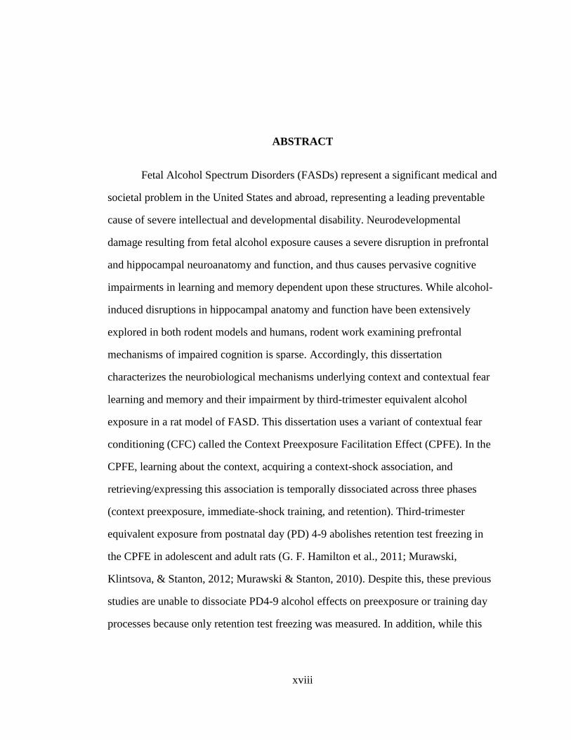
Fetal Alcohol Spectrum Disorders (FASDs) represent a significant medical and
societal problem in the United States and abroad, representing a leading preventable
cause of severe intellectual and developmental disability. Neurodevelopmental
damage resulting from fetal alcohol exposure causes a severe disruption in prefrontal
and hippocampal neuroanatomy and function, and thus causes pervasive cognitive
impairments in learning and memory dependent upon these structures. While alcohol-
induced disruptions in hippocampal anatomy and function have been extensively
explored in both rodent models and humans, rodent work examining prefrontal
mechanisms of impaired cognition is sparse. Accordingly, this dissertation
characterizes the neurobiological mechanisms underlying context and contextual fear
learning and memory and their impairment by third-trimester equivalent alcohol
exposure in a rat model of FASD. This dissertation uses a variant of contextual fear
conditioning (CFC) called the Context Preexposure Facilitation Effect (CPFE). In the
CPFE, learning about the context, acquiring a context-shock association, and
retrieving/expressing this association is temporally dissociated across three phases
(context preexposure, immediate-shock training, and retention). Third-trimester
equivalent exposure from postnatal day (PD) 4-9 abolishes retention test freezing in
the CPFE in adolescent and adult rats (G. F. Hamilton et al., 2011; Murawski,
Klintsova, & Stanton, 2012; Murawski & Stanton, 2010). Despite this, these previous
studies are unable to dissociate PD4-9 alcohol effects on preexposure or training day
processes because only retention test freezing was measured. In addition, while this
ABSTRACT
xix
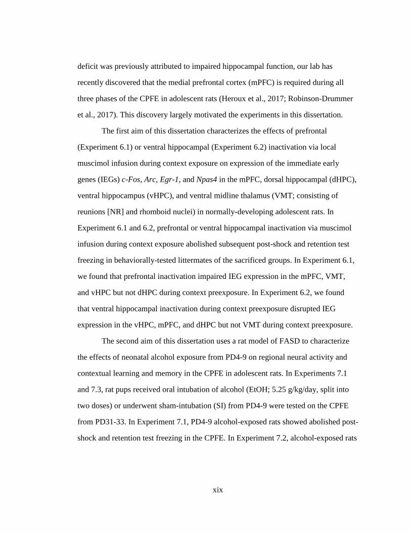
deficit was previously attributed to impaired hippocampal function, our lab has
recently discovered that the medial prefrontal cortex (mPFC) is required during all
three phases of the CPFE in adolescent rats (Heroux et al., 2017; Robinson-Drummer
et al., 2017). This discovery largely motivated the experiments in this dissertation.
The first aim of this dissertation characterizes the effects of prefrontal
(Experiment 6.1) or ventral hippocampal (Experiment 6.2) inactivation via local
muscimol infusion during context exposure on expression of the immediate early
genes (IEGs) c-Fos, Arc, Egr-1, and Npas4 in the mPFC, dorsal hippocampal (dHPC),
ventral hippocampus (vHPC), and ventral midline thalamus (VMT; consisting of
reunions [NR] and rhomboid nuclei) in normally-developing adolescent rats. In
Experiment 6.1 and 6.2, prefrontal or ventral hippocampal inactivation via muscimol
infusion during context exposure abolished subsequent post-shock and retention test
freezing in behaviorally-tested littermates of the sacrificed groups. In Experiment 6.1,
we found that prefrontal inactivation impaired IEG expression in the mPFC, VMT,
and vHPC but not dHPC during context preexposure. In Experiment 6.2, we found
that ventral hippocampal inactivation during context preexposure disrupted IEG
expression in the vHPC, mPFC, and dHPC but not VMT during context preexposure.
The second aim of this dissertation uses a rat model of FASD to characterize
the effects of neonatal alcohol exposure from PD4-9 on regional neural activity and
contextual learning and memory in the CPFE in adolescent rats. In Experiments 7.1
and 7.3, rat pups received oral intubation of alcohol (EtOH; 5.25 g/kg/day, split into
two doses) or underwent sham-intubation (SI) from PD4-9 were tested on the CPFE
from PD31-33. In Experiment 7.1, PD4-9 alcohol-exposed rats showed abolished post-
shock and retention test freezing in the CPFE. In Experiment 7.2, alcohol-exposed rats
xx
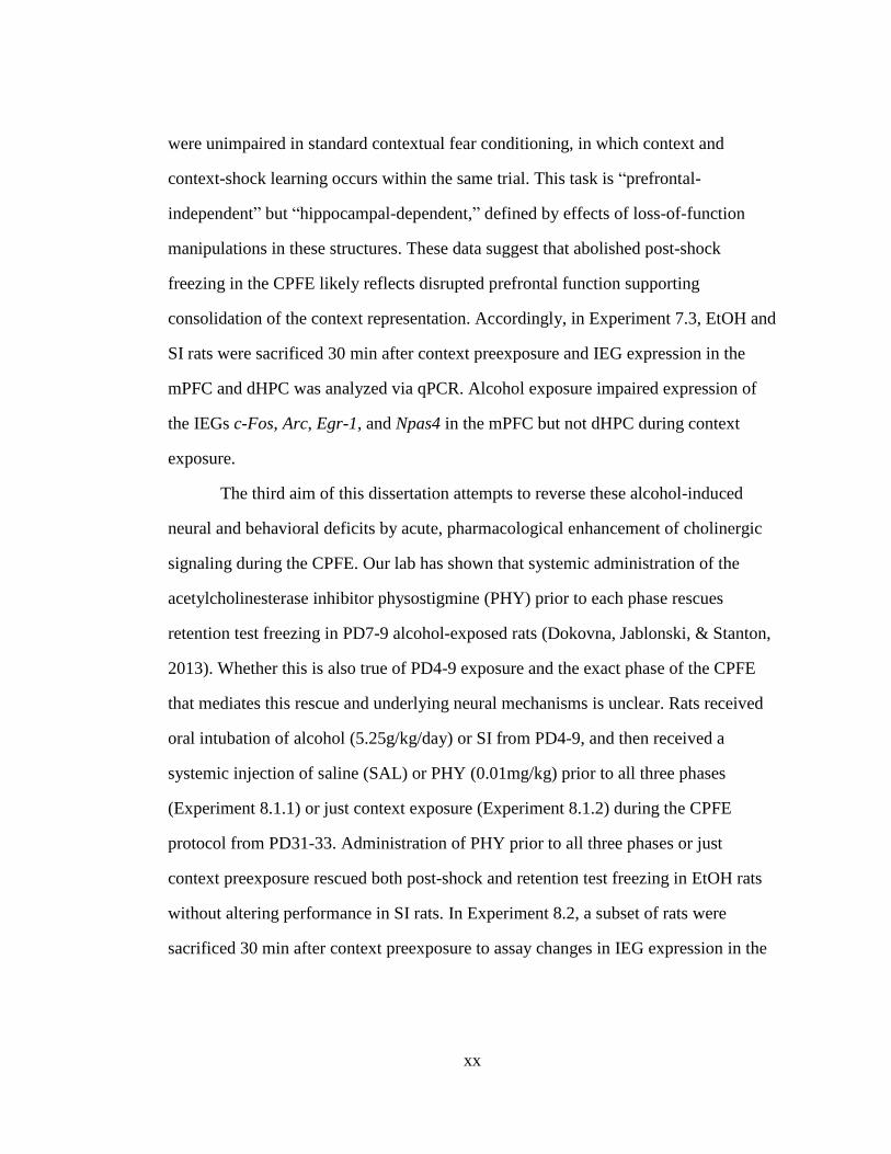
were unimpaired in standard contextual fear conditioning, in which context and
context-shock learning occurs within the same trial. This task is “prefrontal-
independent” but “hippocampal-dependent,” defined by effects of loss-of-function
manipulations in these structures. These data suggest that abolished post-shock
freezing in the CPFE likely reflects disrupted prefrontal function supporting
consolidation of the context representation. Accordingly, in Experiment 7.3, EtOH and
SI rats were sacrificed 30 min after context preexposure and IEG expression in the
mPFC and dHPC was analyzed via qPCR. Alcohol exposure impaired expression of
the IEGs c-Fos, Arc, Egr-1, and Npas4 in the mPFC but not dHPC during context
exposure.
The third aim of this dissertation attempts to reverse these alcohol-induced
neural and behavioral deficits by acute, pharmacological enhancement of cholinergic
signaling during the CPFE. Our lab has shown that systemic administration of the
acetylcholinesterase inhibitor physostigmine (PHY) prior to each phase rescues
retention test freezing in PD7-9 alcohol-exposed rats (Dokovna, Jablonski, & Stanton,
2013). Whether this is also true of PD4-9 exposure and the exact phase of the CPFE
that mediates this rescue and underlying neural mechanisms is unclear. Rats received
oral intubation of alcohol (5.25g/kg/day) or SI from PD4-9, and then received a
systemic injection of saline (SAL) or PHY (0.01mg/kg) prior to all three phases
(Experiment 8.1.1) or just context exposure (Experiment 8.1.2) during the CPFE
protocol from PD31-33. Administration of PHY prior to all three phases or just
context preexposure rescued both post-shock and retention test freezing in EtOH rats
without altering performance in SI rats. In Experiment 8.2, a subset of rats were
sacrificed 30 min after context preexposure to assay changes in IEG expression in the
xxi
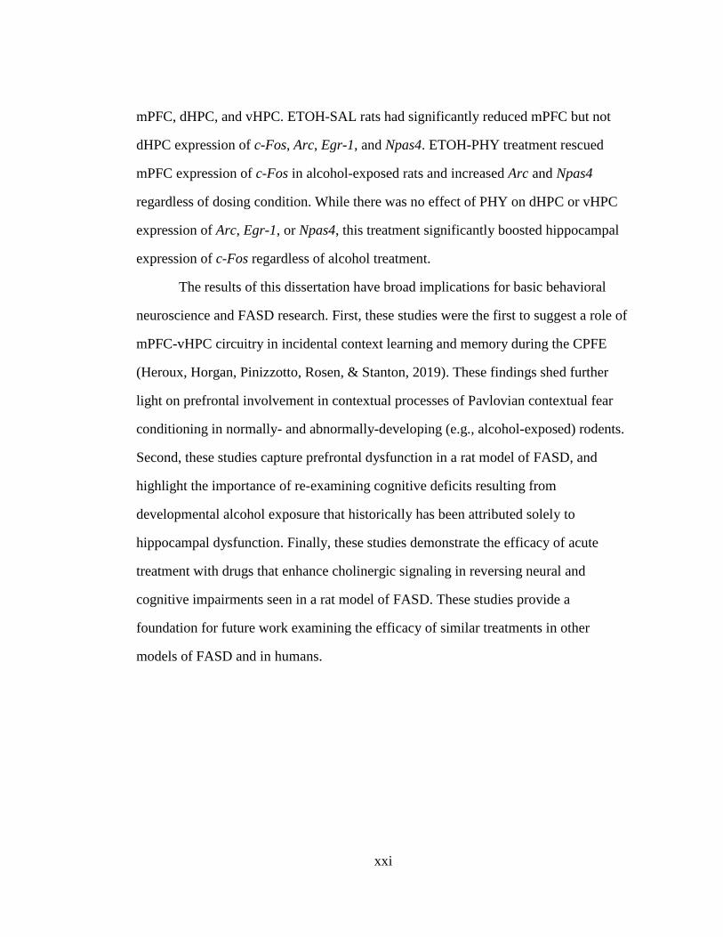
mPFC, dHPC, and vHPC. ETOH-SAL rats had significantly reduced mPFC but not
dHPC expression of c-Fos, Arc, Egr-1, and Npas4. ETOH-PHY treatment rescued
mPFC expression of c-Fos in alcohol-exposed rats and increased Arc and Npas4
regardless of dosing condition. While there was no effect of PHY on dHPC or vHPC
expression of Arc, Egr-1, or Npas4, this treatment significantly boosted hippocampal
expression of c-Fos regardless of alcohol treatment.
The results of this dissertation have broad implications for basic behavioral
neuroscience and FASD research. First, these studies were the first to suggest a role of
mPFC-vHPC circuitry in incidental context learning and memory during the CPFE
(Heroux, Horgan, Pinizzotto, Rosen, & Stanton, 2019). These findings shed further
light on prefrontal involvement in contextual processes of Pavlovian contextual fear
conditioning in normally- and abnormally-developing (e.g., alcohol-exposed) rodents.
Second, these studies capture prefrontal dysfunction in a rat model of FASD, and
highlight the importance of re-examining cognitive deficits resulting from
developmental alcohol exposure that historically has been attributed solely to
hippocampal dysfunction. Finally, these studies demonstrate the efficacy of acute
treatment with drugs that enhance cholinergic signaling in reversing neural and
cognitive impairments seen in a rat model of FASD. These studies provide a
foundation for future work examining the efficacy of similar treatments in other
models of FASD and in humans.
1
INTRODUCTION
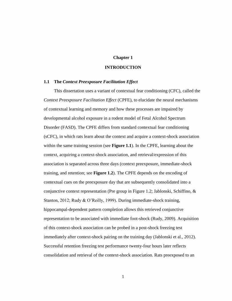
1.1 The Context Preexposure Facilitation Effect
This dissertation uses a variant of contextual fear conditioning (CFC), called the
Context Preexposure Facilitation Effect (CPFE), to elucidate the neural mechanisms
of contextual learning and memory and how these processes are impaired by
developmental alcohol exposure in a rodent model of Fetal Alcohol Spectrum
Disorder (FASD). The CPFE differs from standard contextual fear conditioning
(sCFC), in which rats learn about the context and acquire a context-shock association
within the same training session (see Figure 1.1). In the CPFE, learning about the
context, acquiring a context-shock association, and retrieval/expression of this
association is separated across three days (context preexposure, immediate-shock
training, and retention; see Figure 1.2). The CPFE depends on the encoding of
contextual cues on the preexposure day that are subsequently consolidated into a
conjunctive context representation (Pre group in Figure 1.2; Jablonski, Schiffino, &
Stanton, 2012; Rudy & O’Reilly, 1999). During immediate-shock training,
hippocampal-dependent pattern completion allows this retrieved conjunctive
representation to be associated with immediate foot-shock (Rudy, 2009). Acquisition
of this context-shock association can be probed in a post-shock freezing test
immediately after context-shock pairing on the training day (Jablonski et al., 2012).
Successful retention freezing test performance twenty-four hours later reflects
consolidation and retrieval of the context-shock association. Rats preexposed to an
Chapter 1
2
alternate context on the first day (Alt-Pre group in Figure 1.2) demonstrate the
immediate-shock deficit, which reflects an inability to form a context-shock
association with insufficient exposure to the training context (Fanselow, 1990). The
CPFE emerges between postnatal day (PD) 17 and PD24 and is dependent on activity
in dorsal (dHPC) and ventral (vHPC) hippocampus during all three phases in rats
(Cullen, Ferrara, Pullins, & Helmstetter, 2017; Jablonski et al., 2012; Matus-Amat,
Higgins, Barrientos, & Rudy, 2004; Rudy & Matus-Amat, 2005; Schiffino, Murawski,
Rosen, & Stanton, 2011). Our lab has recently discovered that inactivation of the
medial prefrontal cortex (mPFC) during any phase disrupts the CPFE in adolescent
rats (Heroux, Robinson-Drummer, Sanders, Rosen, & Stanton, 2017; Robinson-
Drummer, Heroux, & Stanton, 2017). While these data indicate a novel role of this
structure in contextual learning and memory during incidental context exposure,
prefrontal mechanisms supporting this behavior are unclear. We hypothesized that
prefrontal inactivation disrupts hippocampal activity recruited for the formation of a
long-term context representation via their reciprocal connectivity with the ventral
midline thalamus (VMT; consisting of reunions [NR] and rhomboid nuclei).
Accordingly, the first aim of this dissertation asked the question: Does prefrontal
inactivation during incidental context exposure effect molecular activity in the ventral
midline thalamus and hippocampus? This question is addressed by examining the
effects of prefrontal inactivation on immediate early gene (IEG) expression in the
mPFC, dHPC, and VMT during context preexposure. Given that this inactivation
impaired gene expression in the vHPC and VMT but not the dHPC, we performed a
second experiment that asked: Does ventral hippocampal inactivation during
incidental context exposure impair IEG expression in the prefrontal cortex?

3
Collectively, these studies further characterize circuitry underlying incidental context
learning and memory, which can then inform deficits that we observe in subsequent
aims directed at our rodent model of FASD. The results of the first aim are presented
in Chapter 6.
4
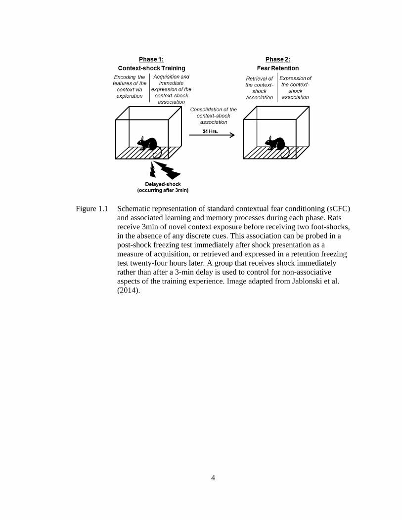
Figure 1.1 Schematic representation of standard contextual fear conditioning (sCFC)
and associated learning and memory processes during each phase. Rats
receive 3min of novel context exposure before receiving two foot-shocks,
in the absence of any discrete cues. This association can be probed in a
post-shock freezing test immediately after shock presentation as a
measure of acquisition, or retrieved and expressed in a retention freezing
test twenty-four hours later. A group that receives shock immediately
rather than after a 3-min delay is used to control for non-associative
aspects of the training experience. Image adapted from Jablonski et al.
(2014).
5
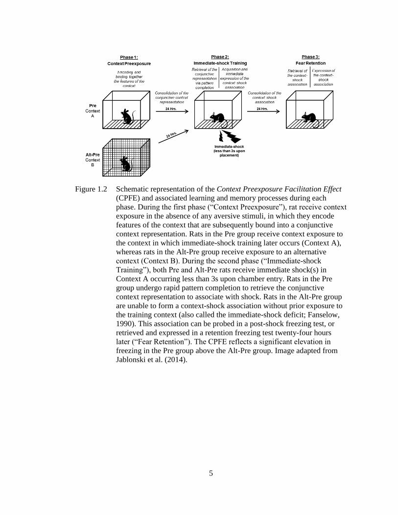
Figure 1.2 Schematic representation of the Context Preexposure Facilitation Effect
(CPFE) and associated learning and memory processes during each
phase. During the first phase (“Context Preexposure”), rat receive context
exposure in the absence of any aversive stimuli, in which they encode
features of the context that are subsequently bound into a conjunctive
context representation. Rats in the Pre group receive context exposure to
the context in which immediate-shock training later occurs (Context A),
whereas rats in the Alt-Pre group receive exposure to an alternative
context (Context B). During the second phase (“Immediate-shock
Training”), both Pre and Alt-Pre rats receive immediate shock(s) in
Context A occurring less than 3s upon chamber entry. Rats in the Pre
group undergo rapid pattern completion to retrieve the conjunctive
context representation to associate with shock. Rats in the Alt-Pre group
are unable to form a context-shock association without prior exposure to
the training context (also called the immediate-shock deficit; Fanselow,
1990). This association can be probed in a post-shock freezing test, or
retrieved and expressed in a retention freezing test twenty-four hours
later (“Fear Retention”). The CPFE reflects a significant elevation in
freezing in the Pre group above the Alt-Pre group. Image adapted from
Jablonski et al. (2014).
6
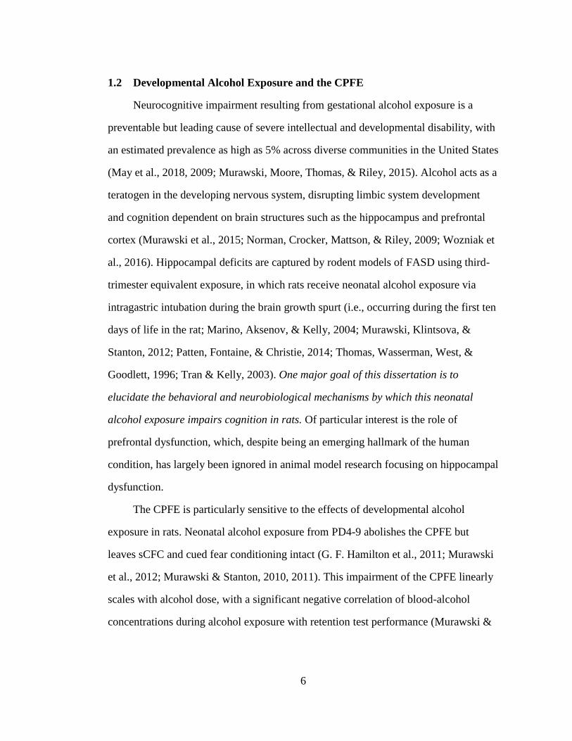
1.2 Developmental Alcohol Exposure and the CPFE
Neurocognitive impairment resulting from gestational alcohol exposure is a
preventable but leading cause of severe intellectual and developmental disability, with
an estimated prevalence as high as 5% across diverse communities in the United States
(May et al., 2018, 2009; Murawski, Moore, Thomas, & Riley, 2015). Alcohol acts as a
teratogen in the developing nervous system, disrupting limbic system development
and cognition dependent on brain structures such as the hippocampus and prefrontal
cortex (Murawski et al., 2015; Norman, Crocker, Mattson, & Riley, 2009; Wozniak et
al., 2016). Hippocampal deficits are captured by rodent models of FASD using third-
trimester equivalent exposure, in which rats receive neonatal alcohol exposure via
intragastric intubation during the brain growth spurt (i.e., occurring during the first ten
days of life in the rat; Marino, Aksenov, & Kelly, 2004; Murawski, Klintsova, &
Stanton, 2012; Patten, Fontaine, & Christie, 2014; Thomas, Wasserman, West, &
Goodlett, 1996; Tran & Kelly, 2003). One major goal of this dissertation is to
elucidate the behavioral and neurobiological mechanisms by which this neonatal
alcohol exposure impairs cognition in rats. Of particular interest is the role of
prefrontal dysfunction, which, despite being an emerging hallmark of the human
condition, has largely been ignored in animal model research focusing on hippocampal
dysfunction.
The CPFE is particularly sensitive to the effects of developmental alcohol
exposure in rats. Neonatal alcohol exposure from PD4-9 abolishes the CPFE but
leaves sCFC and cued fear conditioning intact (G. F. Hamilton et al., 2011; Murawski
et al., 2012; Murawski & Stanton, 2010, 2011). This impairment of the CPFE linearly
scales with alcohol dose, with a significant negative correlation of blood-alcohol
concentrations during alcohol exposure with retention test performance (Murawski &
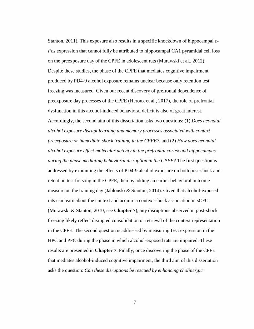
7
Stanton, 2011). This exposure also results in a specific knockdown of hippocampal c-
Fos expression that cannot fully be attributed to hippocampal CA1 pyramidal cell loss
on the preexposure day of the CPFE in adolescent rats (Murawski et al., 2012).
Despite these studies, the phase of the CPFE that mediates cognitive impairment
produced by PD4-9 alcohol exposure remains unclear because only retention test
freezing was measured. Given our recent discovery of prefrontal dependence of
preexposure day processes of the CPFE (Heroux et al., 2017), the role of prefrontal
dysfunction in this alcohol-induced behavioral deficit is also of great interest.
Accordingly, the second aim of this dissertation asks two questions: (1) Does neonatal
alcohol exposure disrupt learning and memory processes associated with context
preexposure or immediate-shock training in the CPFE?, and (2) How does neonatal
alcohol exposure effect molecular activity in the prefrontal cortex and hippocampus
during the phase mediating behavioral disruption in the CPFE? The first question is
addressed by examining the effects of PD4-9 alcohol exposure on both post-shock and
retention test freezing in the CPFE, thereby adding an earlier behavioral outcome
measure on the training day (Jablonski & Stanton, 2014). Given that alcohol-exposed
rats can learn about the context and acquire a context-shock association in sCFC
(Murawski & Stanton, 2010; see Chapter 7), any disruptions observed in post-shock
freezing likely reflect disrupted consolidation or retrieval of the context representation
in the CPFE. The second question is addressed by measuring IEG expression in the
HPC and PFC during the phase in which alcohol-exposed rats are impaired. These
results are presented in Chapter 7. Finally, once discovering the phase of the CPFE
that mediates alcohol-induced cognitive impairment, the third aim of this dissertation
asks the question: Can these disruptions be rescued by enhancing cholinergic
8
signaling via acute, pre-phase acetylcholinesterase inhibitor treatment? Emerging
evidence suggests that cholinergic dysfunction represents a substantial mechanism by
which alcohol disrupts neuronal development and cognition across the lifespan (see
Chapter 3 for detailed discussion). Our lab has shown that systemic administration of
an acetylcholinesterase inhibitor prior to all three phases recues in CPFE in rats
receiving PD7-9 alcohol exposure (Dokovna et al., 2013). The exact phase-dependent
cognitive processes and neural correlates this treatment rescues and whether this
would generalize to the PD4-9 window is unclear. The final aim of this dissertation
addresses this question and the results are presented in Chapter 8.
1.3 Dissertation Overview
This dissertation is organized into three sections. The first section consists of
Chapters 2-4. It provides background information on the neurobiology of Pavlovian
contextual fear conditioning (Chapter 2), rodent models of FASDs (Chapter 3), and
previously published work from our lab that is foundational for the experiments in this
dissertation (Chapter 4). The second section consists of Chapters 5-8. It details the
general methods and materials common across dissertation experiments (Chapter 5)
followed by an overview, analysis, and interpretation of each dissertation aim adapted
from published manuscripts (Chapters 6-8). The third section includes a summary of
experimental findings, general conclusions, and future directions relating to the aims
of this dissertation (Chapter 9).
9
NEUROBIOLOGY OF CONTEXTUAL FEAR CONDITIONING
The preceding chapter described the structure and aims of this dissertation, and
introduced sCFC and CPFE behavioral paradigms. Using the CPFE, this dissertation
examines the neurobiological and behavioral mechanisms of context and contextual
fear learning and memory in normally and abnormally (i.e., alcohol-exposed)
developed adolescent rats. The current chapter provides a brief overview of the
involvement of HPC and PFC in sCFC and background conditioning occurring during
auditory and trace fear conditioning. This chapter also introduces the study of IEGs as
molecular markers of regional neural activity and plasticity during discrete phases of
behavioral tasks in rodents.
2.1 Neuroanatomical Substrates of Contextual Fear Conditioning in Rodents
It is a widely accepted view that multiple neural systems support distinct
processes of learning and memory in Pavlovian contextual fear conditioning
(Fanselow & Poulos, 2005). CFC involves learning an association between a neutral
context a rat is exploring (or has explored previously) and aversive foot-shock(s) that
occurs in this context. The context can be defined as a set of stable, multi-modal
(spatial, olfactory, auditory, interoceptive, etc.) features that are distinct from more
transient and salient discrete cues occurring within the context (Maren, Phan, &
Liberzon, 2013; Rudy, 2009). The foot-shock(s) serves as the unconditional stimulus
(US), and the context serves as the conditional stimulus (CS) that elicits a species-
Chapter 2
10
typical freezing response that is one of many responses that reflect a central state of
conditioned fear in rodents (Davis, 1992; Fendt & Fanselow, 1999). Early models of
the neurobiology of CFC in rodents focused on a circuit involving coordinated
communication between the thalamus, para-hippocampal region (PHR; consisting of
the rhinal cortices), HPC, basolateral amygdala (BLA), and midbrain output structures
(Anagnostaras, Gale, & Fanselow, 2001; Fanselow & Poulos, 2005; Fendt &
Fanselow, 1999; Maren, 2001; Phillips & LeDoux, 1992; see Figure 2.1). In these
models, sensory information about the context CS and the foot-shock US activate
thalamic nuclei, after which US information is sent directly to the BLA, whereas CS
information is sent to the PHR and HPC. These latter regions were thought to encode
features of the context and form a context representation (Anagnostaras et al., 2001;
Rudy, 2009), and this information is propagated to the BLA, where the context-shock
association is formed (Fendt & Fanselow, 1999). The BLA then projects to the central
nucleus of the amygdala (CeA), which drives conditioned autonomic arousal and
species-typical freezing responses via efferent projections to the hypothalamus/brain
stem and periaqueductal gray (PAG), respectively (Fendt & Fanselow, 1999). While
this basic framework has received much empirical support, these early models did not
include any role of prefrontal neuronal circuitry in processes of CFC. Recent models
have suggested roles of the mPFC in attentional processing, consolidation,
reconsolidation, contextual control, and generalization of fear memories (Fanselow &
Poulos, 2005; Gilmartin, Balderston, & Helmstetter, 2014; Giustino & Maren, 2015;
Rozeske, Valerio, Chaudun, & Herry, 2015). In the following section, I include an
overview of research characterizing both hippocampal and prefrontal involvement in
acquisition and retention of CFC and related behavioral paradigms.
11
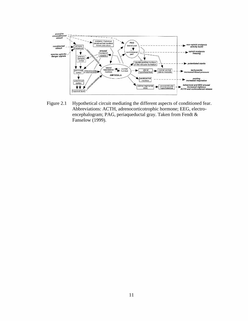
Figure 2.1 Hypothetical circuit mediating the different aspects of conditioned fear.
Abbreviations: ACTH, adrenocorticotrophic hormone; EEG, electro-
encephalogram; PAG, periaqueductal gray. Taken from Fendt &
Fanselow (1999).
12
2.1.1 Hippocampal Involvement in Acquisition and Retention of Contextual
Fear Conditioning
The hippocampus has long been thought to underlie contextual learning and
memory across variants of Pavlovian fear conditioning (Fanselow, 2010; Fanselow &
Poulos, 2005; Fendt & Fanselow, 1999; Maren et al., 2013; Rudy & O’Reilly, 1999b).
The dual-process theory posits that contextual fear learning can be supported by two
different associative systems: a neocortical elemental system or a hippocampal
configural system (Anagnostaras, Gale, & Fanselow, 2001; Maren, 2001; O’Reilly &
Rudy, 2001; Rudy, 2009; Rudy & O’Reilly, 1999a; see Figure 2.2A). In the elemental
system, individual features of the context enter into an independent association with
foot-shock and their additive strength determines conditioned fear. In the configural
system, features of the context are bound (non-additively) into a conjunctive
representation that is associated with foot-shock. Biological models of this theory
suggested that elemental conditioning occurs via direct projections from the PHR and
sensory cortex to the BLA, in which elemental CS-US associations are supported by
amygdalar plasticity in the absence of higher cortical processing (Maren, 2001; see
Figure 2.2B). In contrast, configural conditioning occurs via sensory information
about context features converging in the HPC, in which features are bound into a
conjunctive representation by hippocampal plasticity before being sent to the
amygdala (AMY; see Figure 2.2B). Evidence from lesion studies suggests that the
default system that supports contextual fear conditioning is the hippocampal system
(Wiltgen, Sanders, Anagnostaras, Sage, & Fanselow, 2006). For example, Maren et al.
(1997) and Wiltgen et al. (2006) demonstrated that retrograde (post-training) but not
anterograde (pre-training) lesions of the HPC disrupt retention test freezing during
sCFC in rats (see Figure 2.3). These data indicate that sCFC does not require the
13
HPC, but if intact, the HPC supports contextual fear learning and overshadows the
elemental feature-based system. Importantly, rats with anterograde lesions required
two additional trials and longer placement-to-shock intervals to acquire a context-
shock association, which indicates the elemental system takes additional training and
is slower than the more rapid hippocampal system (Wiltgen et al., 2006). Numerous
other studies have supported hippocampal system dominance, as pre-training lesions
or reversible inactivation of either the dHPC or vHPC leave CFC intact, while post-
training manipulations produce severe retrograde amnesia (Ballesteros, De Oliveira
Galvão, Maisonette, & Landeira-Fernandez, 2014; Paul W. Frankland, Cestari,
Filipkowski, McDonald, & Silva, 1998; Hunsaker & Kesner, 2008; J. J. Kim, Rison,
& Fanselow, 1993; J. Q. Lee, Sutherland, & McDonald, 2017; Maren, Aharonov, &
Fanselow, 1997; Maren & Holt, 2004; Matus-Amat et al., 2004; Wiltgen et al., 2006;
W.-N. Zhang, Bast, Xu, & Feldon, 2014). In contrast, pre-training NMDA receptor
(NMDAR) antagonism in the dHPC or vHPC disrupts contextual fear retention but
leaves post-shock freezing intact (Czerniawski et al., 2011; J. J. Kim, DeCola,
Landeira-Fernandez, & Fanselow, 1991; J. J. Kim, Fanselow, DeCola, & Landeira-
Fernandez, 1992; Quinn, Loya, Ma, & Fanselow, 2005; Sanders & Fanselow, 2003;
Tayler et al., 2011). These data indicate that compensation occurs after HPC lesion or
inactivation, but in an otherwise intact rat, consolidation of long-term contextual fear
memories during sCFC requires dHPC and vHPC plasticity. It’s also important to note
that these previous lesion and reversible inactivation studies do not show whether
conditioning was elemental vs. configural, so it’s possible that, in the absence of the
HPC, the neocortical system also supports configural learning but is slower than the
hippocampal system (i.e., requires additional training; Fanselow, 2010). This notion is
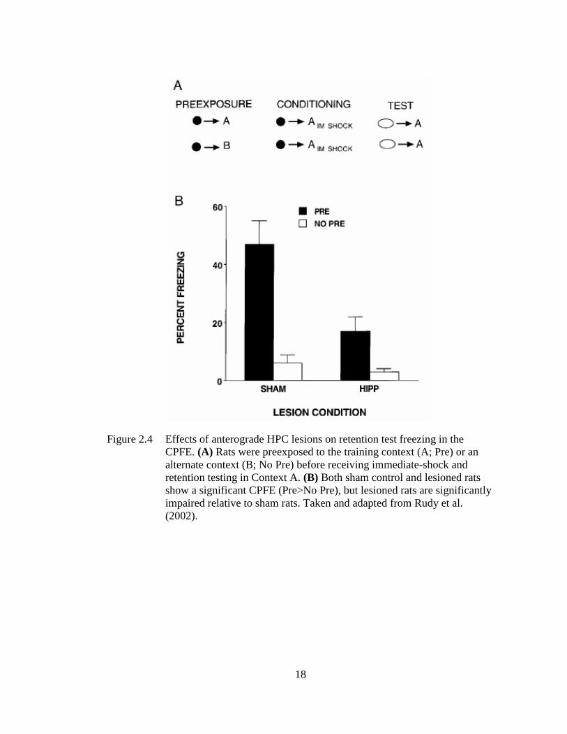
14
supported by the finding that HPC-lesioned rats still benefit from context preexposure
and show a (weak) CPFE (Rudy, Barrientos, & O’Reilly, 2002; see Figure 2.4), which
can only be supported by conjunctive but not feature-based context representations
(see Chapter 1; Fanselow, 2010; Jablonski, Schiffino, & Stanton, 2012; Rudy &
O’Reilly, 1999b). Additional experiments are needed to test the parameters under
which learning in the absence of the HPC is elemental vs. configural across variants of
CFC. Nevertheless, this research establishes a key role of the configural HPC system
in supporting CFC in rodents.
Recent experiments using gain- and loss-of-function chemogenetic and
optogenetic approaches have supported these earlier studies and provided insights into
hippocampal mechanisms of context and contextual fear memory (Asok, Leroy,
Rayman, & Kandel, 2019; Tonegawa, Liu, Ramirez, & Redondo, 2015). These
experiments have used activity-dependent neuronal tagging in transgenic mice to
visualize and later manipulate neurons expressing the IEGs c-Fos and Arc in discrete
brain regions during training in foreground and background CFC (see Tonegawa et al.,
2015 for review). Early loss-of-function studies using this technology demonstrated
that, during memory recall, optogenetic inactivation (via labeled ArchT) of
hippocampal CA1, CA3, or DG neurons tagged during training impairs retention test
freezing in sCFC (Denny et al., 2014; Tanaka et al., 2014). These studies showed that
hippocampal neurons activated by training participate in the long-term memory trace
(or “engram”), and that these neurons are necessary for successful memory recall.
Importantly, these studies cannot conclude that the hippocampus is the site of the
engram or memory storage, in part because this likely occurs across a distributed
network. Accordingly, Tanaka et al. (2014) demonstrated that optogenetic silencing of
15
“engram” neurons in CA1 selectively prevented activation of “engram” neurons in
other brain regions such as the rhinal cortices and amygdala without significantly
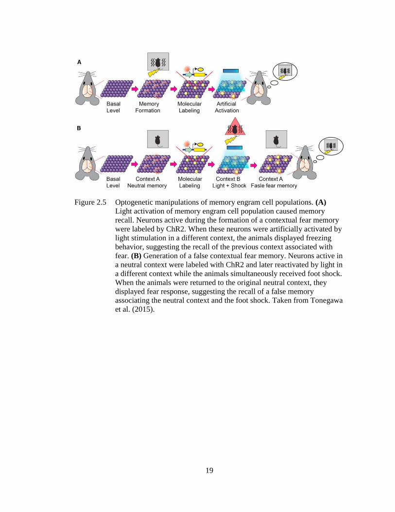
altering total activity in these regions. Gain-of-function studies showed that
reactivation of hippocampal neurons active during acquisition is sufficient to elicit
freezing during a retention test in a non-conditioned context (see Figure 2.5A; Liu et
al., 2012; Tonegawa et al., 2015). Ramirez et al. (2013) and Garner et al. (2012)
demonstrated that hippocampal or whole-brain activation of neurons encoding a novel
context (A), when reactivated during fear conditioning in a different context (B), form
a context-shock association to the former (A) despite never receiving foot-shock in
this context (see Figure 2.5B). Another study showed that concurrent reactivation of
HPC neurons encoding context and AMY neurons encoding immediate shock in the
home-cage is sufficient to form an artificial context-shock association in the absence
of training (Ohkawa et al., 2015). Additional studies have shown that endogenous and
experience-driven excitability (via varying levels of signaling molecules such as
cAMP response element-binding protein [CREB]) in neurons largely determines their
allocation to “engrams” or memory traces at the time of behavioral experience (Cai et
al., 2016; J. Han et al., 2007; Josselyn & Frankland, 2018). One important caveat for
this optogenetic research is that behavioral change or expression caused by artificial
stimulation or inhibition may not accurately represent what occurs naturally within a
memory system; moreover, state-dependency issues also emerge in loss-of-function
experiments as light is typically only turned on during one phase of the behavioral
paradigm. Nevertheless, in conjunction with previous lesion and pharmacological
studies, this research establishes a key role of the HPC in contextual processes of CFC
in rodents.
16
Figure 2.2 Psychological (A) and neurobiological (B) processes of the dual-process
theory of Pavlovian contextual fear conditioning. (A) In the elemental
system (top), features of the context (A, B, C, D) enter into independent
associations with the foot-shock US (E). In the configural system
(bottom), features of the context are bound into a conjunctive
representation and this representation is associated with foot-shock. (B)
In the elemental system, features of the context are sent directly to the
amygdala where the context-shock association is formed via amygdalar
plasticity. In the configural system, these features are bound into a
conjunctive representation by hippocampal plasticity before being sent to
the amygdala. Taken and adapted from Rudy (2009) and Maren (2001).
17
Figure 2.3 Effects of pre-training (anterograde) vs. post-training (retrograde) lesions
of the hippocampus on retention test freezing in sCFC. Retrograde but
not anterograde lesions of the hippocampus abolish retention test freezing
relative to sham controls. Taken and adapted from Wiltgen et al. (2006).
18
Figure 2.4 Effects of anterograde HPC lesions on retention test freezing in the
CPFE. (A) Rats were preexposed to the training context (A; Pre) or an
alternate context (B; No Pre) before receiving immediate-shock and
retention testing in Context A. (B) Both sham control and lesioned rats
show a significant CPFE (Pre>No Pre), but lesioned rats are significantly
impaired relative to sham rats. Taken and adapted from Rudy et al.
(2002).
19
Figure 2.5 Optogenetic manipulations of memory engram cell populations. (A)
Light activation of memory engram cell population caused memory
recall. Neurons active during the formation of a contextual fear memory
were labeled by ChR2. When these neurons were artificially activated by
light stimulation in a different context, the animals displayed freezing
behavior, suggesting the recall of the previous context associated with
fear. (B) Generation of a false contextual fear memory. Neurons active in
a neutral context were labeled with ChR2 and later reactivated by light in
a different context while the animals simultaneously received foot shock.
When the animals were returned to the original neutral context, they
displayed fear response, suggesting the recall of a false memory
associating the neutral context and the foot shock. Taken from Tonegawa
et al. (2015).
20
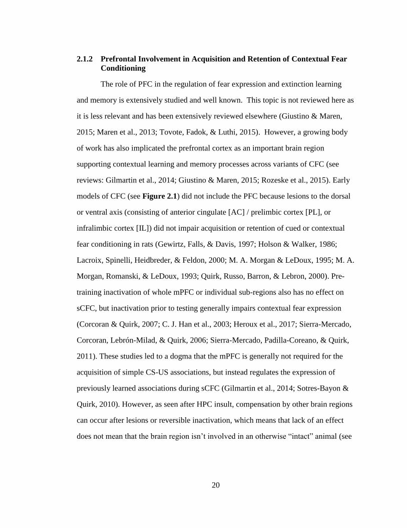
2.1.2 Prefrontal Involvement in Acquisition and Retention of Contextual Fear
Conditioning
The role of PFC in the regulation of fear expression and extinction learning
and memory is extensively studied and well known. This topic is not reviewed here as
it is less relevant and has been extensively reviewed elsewhere (Giustino & Maren,
2015; Maren et al., 2013; Tovote, Fadok, & Luthi, 2015). However, a growing body
of work has also implicated the prefrontal cortex as an important brain region
supporting contextual learning and memory processes across variants of CFC (see
reviews: Gilmartin et al., 2014; Giustino & Maren, 2015; Rozeske et al., 2015). Early
models of CFC (see Figure 2.1) did not include the PFC because lesions to the dorsal
or ventral axis (consisting of anterior cingulate [AC] / prelimbic cortex [PL], or
infralimbic cortex [IL]) did not impair acquisition or retention of cued or contextual
fear conditioning in rats (Gewirtz, Falls, & Davis, 1997; Holson & Walker, 1986;
Lacroix, Spinelli, Heidbreder, & Feldon, 2000; M. A. Morgan & LeDoux, 1995; M. A.
Morgan, Romanski, & LeDoux, 1993; Quirk, Russo, Barron, & Lebron, 2000). Pre-
training inactivation of whole mPFC or individual sub-regions also has no effect on
sCFC, but inactivation prior to testing generally impairs contextual fear expression
(Corcoran & Quirk, 2007; C. J. Han et al., 2003; Heroux et al., 2017; Sierra-Mercado,
Corcoran, Lebrón-Milad, & Quirk, 2006; Sierra-Mercado, Padilla-Coreano, & Quirk,
2011). These studies led to a dogma that the mPFC is generally not required for the
acquisition of simple CS-US associations, but instead regulates the expression of
previously learned associations during sCFC (Gilmartin et al., 2014; Sotres-Bayon &
Quirk, 2010). However, as seen after HPC insult, compensation by other brain regions
can occur after lesions or reversible inactivation, which means that lack of an effect
does not mean that the brain region isn’t involved in an otherwise “intact” animal (see
21
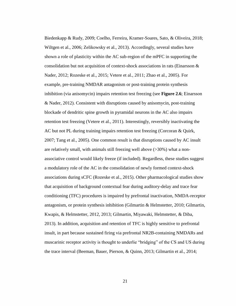
Biedenkapp & Rudy, 2009; Coelho, Ferreira, Kramer-Soares, Sato, & Oliveira, 2018;
Wiltgen et al., 2006; Zelikowsky et al., 2013). Accordingly, several studies have
shown a role of plasticity within the AC sub-region of the mPFC in supporting the
consolidation but not acquisition of context-shock associations in rats (Einarsson &
Nader, 2012; Rozeske et al., 2015; Vetere et al., 2011; Zhao et al., 2005). For
example, pre-training NMDAR antagonism or post-training protein synthesis
inhibition (via anisomycin) impairs retention test freezing (see Figure 2.6; Einarsson
& Nader, 2012). Consistent with disruptions caused by anisomycin, post-training
blockade of dendritic spine growth in pyramidal neurons in the AC also impairs
retention test freezing (Vetere et al., 2011). Interestingly, reversibly inactivating the
AC but not PL during training impairs retention test freezing (Corcoran & Quirk,
2007; Tang et al., 2005). One common result is that disruptions caused by AC insult
are relatively small, with animals still freezing well above (>30%) what a non-
associative control would likely freeze (if included). Regardless, these studies suggest
a modulatory role of the AC in the consolidation of newly formed context-shock
associations during sCFC (Rozeske et al., 2015). Other pharmacological studies show
that acquisition of background contextual fear during auditory-delay and trace fear
conditioning (TFC) procedures is impaired by prefrontal inactivation, NMDA-receptor
antagonism, or protein synthesis inhibition (Gilmartin & Helmstetter, 2010; Gilmartin,
Kwapis, & Helmstetter, 2012, 2013; Gilmartin, Miyawaki, Helmstetter, & Diba,
2013). In addition, acquisition and retention of TFC is highly sensitive to prefrontal
insult, in part because sustained firing via prefrontal NR2B-containing NMDARs and
muscarinic receptor activity is thought to underlie “bridging” of the CS and US during
the trace interval (Beeman, Bauer, Pierson, & Quinn, 2013; Gilmartin et al., 2014;
22
Gilmartin, Kwapis, et al., 2013; Zhao et al., 2005). Moreover, in the CPFE, separation
of context vs. context-shock learning necessitates activity and cholinergic function in
the mPFC (see Chapter 4; Heroux, Robinson-Drummer, Sanders, Rosen, & Stanton,
2017; Robinson-Drummer, Heroux, & Stanton, 2017). Collectively, these studies
suggest prefrontal involvement in 1) plasticity underlying consolidation of newly
formed context-shock associations (Rozeske et al., 2015), and 2) CFC procedures
where contextual learning is less salient (e.g., in background conditioning with
discrete cues present) or when there is temporal separation of component processes
(e.g., in trace fear conditioning or the CPFE; see Heroux et al., 2017; Gilmartin et al.,
2014).
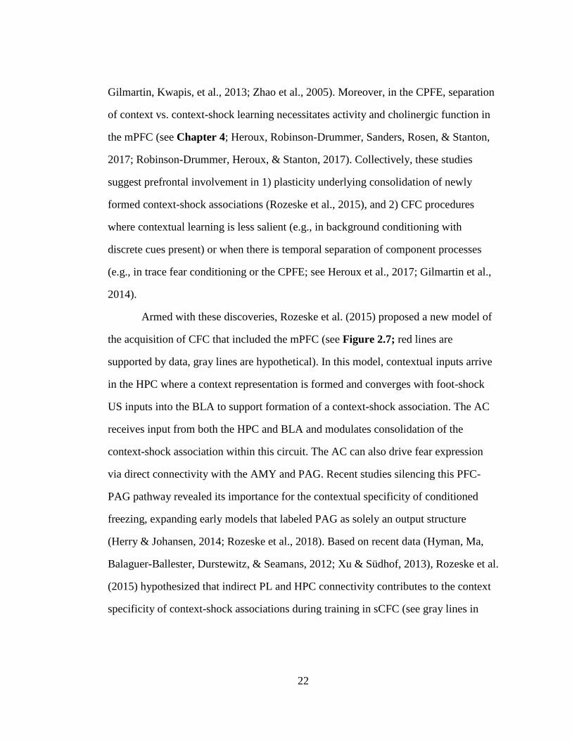
Armed with these discoveries, Rozeske et al. (2015) proposed a new model of
the acquisition of CFC that included the mPFC (see Figure 2.7; red lines are
supported by data, gray lines are hypothetical). In this model, contextual inputs arrive
in the HPC where a context representation is formed and converges with foot-shock
US inputs into the BLA to support formation of a context-shock association. The AC
receives input from both the HPC and BLA and modulates consolidation of the
context-shock association within this circuit. The AC can also drive fear expression
via direct connectivity with the AMY and PAG. Recent studies silencing this PFC-
PAG pathway revealed its importance for the contextual specificity of conditioned
freezing, expanding early models that labeled PAG as solely an output structure
(Herry & Johansen, 2014; Rozeske et al., 2018). Based on recent data (Hyman, Ma,
Balaguer-Ballester, Durstewitz, & Seamans, 2012; Xu & Südhof, 2013), Rozeske et al.
(2015) hypothesized that indirect PL and HPC connectivity contributes to the context
specificity of context-shock associations during training in sCFC (see gray lines in
23
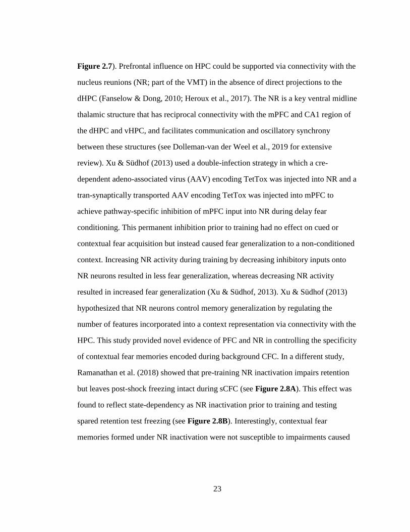
Figure 2.7). Prefrontal influence on HPC could be supported via connectivity with the
nucleus reunions (NR; part of the VMT) in the absence of direct projections to the
dHPC (Fanselow & Dong, 2010; Heroux et al., 2017). The NR is a key ventral midline
thalamic structure that has reciprocal connectivity with the mPFC and CA1 region of
the dHPC and vHPC, and facilitates communication and oscillatory synchrony
between these structures (see Dolleman-van der Weel et al., 2019 for extensive
review). Xu & Südhof (2013) used a double-infection strategy in which a cre-
dependent adeno-associated virus (AAV) encoding TetTox was injected into NR and a
tran-synaptically transported AAV encoding TetTox was injected into mPFC to
achieve pathway-specific inhibition of mPFC input into NR during delay fear
conditioning. This permanent inhibition prior to training had no effect on cued or
contextual fear acquisition but instead caused fear generalization to a non-conditioned
context. Increasing NR activity during training by decreasing inhibitory inputs onto
NR neurons resulted in less fear generalization, whereas decreasing NR activity
resulted in increased fear generalization (Xu & Südhof, 2013). Xu & Südhof (2013)
hypothesized that NR neurons control memory generalization by regulating the
number of features incorporated into a context representation via connectivity with the
HPC. This study provided novel evidence of PFC and NR in controlling the specificity
of contextual fear memories encoded during background CFC. In a different study,
Ramanathan et al. (2018) showed that pre-training NR inactivation impairs retention
but leaves post-shock freezing intact during sCFC (see Figure 2.8A). This effect was
found to reflect state-dependency as NR inactivation prior to training and testing
spared retention test freezing (see Figure 2.8B). Interestingly, contextual fear
memories formed under NR inactivation were not susceptible to impairments caused
24
by concurrent NMDAR antagonism in the HPC, which impair fear retention in an
otherwise intact rat (see Figure 2.8C; Ramanathan, Ressler, Jin, & Maren, 2018).
Taken together with Xu & Südhof (2013), these data suggest that NR activity, likely
within an mPFC-NR-HPC system, is involved in the formation of precise,
hippocampal–dependent context representations or contextual fear memories.
Collectively, these and earlier pharmacological studies support and expand Rozeske’s
(2015) proposed model to emphasize a role of prefrontal-hippocampal circuitry in
contextual processes of CFC.
25
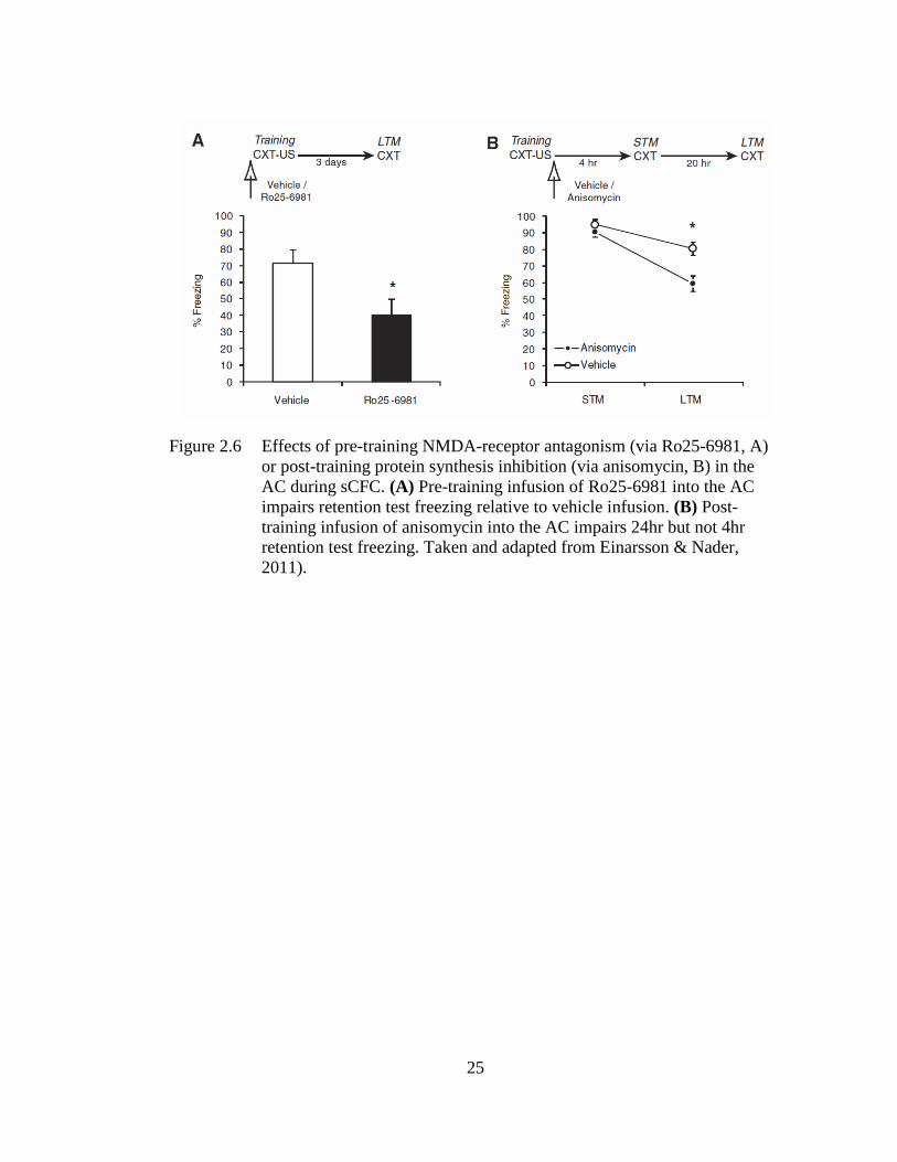
Figure 2.6 Effects of pre-training NMDA-receptor antagonism (via Ro25-6981, A)
or post-training protein synthesis inhibition (via anisomycin, B) in the
AC during sCFC. (A) Pre-training infusion of Ro25-6981 into the AC
impairs retention test freezing relative to vehicle infusion. (B) Post-
training infusion of anisomycin into the AC impairs 24hr but not 4hr
retention test freezing. Taken and adapted from Einarsson & Nader,
2011).
26
Figure 2.7 Neuronal circuit mediating contextual fear acquisition and retention (red
lines indicate proposed circuitry known to be engaged during acquisition,
whereas gray lines indicate hypothesized but untested functional
connectivity). Contextual fear acquisition is support by contextual inputs
into the HPC, which are then sent to the BLA where they converge with
US inputs to form a context-shock association. The HPC and BLA also
project to the AC, which modulates consolidation of the context-shock
association and fear expression via direct PAG and CeA-PAG
connectivity. Hypothesized functional connections include HPC relay of
contextual information to the PL which, via NR projections, modulates
the encoding or consolidation of precise, feature-rich context
representations, thus controlling fear generalization during CFC. Taken
and adapted from Rozeske et al. (2015).
27
Figure 2.8 Effects of intra-NR infusion of saline or muscimol prior to both training
(post-shock freezing; A) and testing (retention freezing; B) during sCFC.
(A-B) NR inactivation state-dependently impaired retention but not
acquisition of sCFC, as MUS-SAL rats froze lower than all other groups
during retention. (C) Contextual fear memories formed under NR
inactivation are insusceptible to concurrent intra-dHPC APV infusion. In
the absence of NR inactivation, intra-dHPC APV infusion impairs
retention test freezing. Taken and adapted from Ramanathan et al.
(2018), who use an alternate acronym (RE) for nucleus reunions.

28
2.1.3 Prefrontal and Hippocampal Dynamics of Contextual Memory in the
CPFE
As reviewed in Chapter 1, the CPFE is a variant of contextual fear
conditioning that separates learning about the context, acquiring a context-shock
association, and retrieval/expression of this association (see Figure 1.1). Acquisition
or consolidation of a context representation requires activity and NMDAR plasticity in
both the dHPC and vHPC (Cullen et al., 2017; Matus-Amat, Higgins, Sprunger,
Wright-Hardesty, & Rudy, 2007; Schiffino et al., 2011). In addition, inactivation of
the whole mPFC prior to any phase of the CPFE abolishes retention test freezing in
rats (see Section 4.2 in Chapter 4 for full review; Heroux et al., 2017). This
inactivation prior to immediate-shock training leaves post-shock freezing intact,
indicating that mPFC is not required for retrieving the context representation or
associating it with shock. These data suggest that mPFC is necessary for the
consolidation of a context representation and/or context-shock association in the
CPFE, and that other structure don’t compensate for impaired function of mPFC.
Taken together with Xu & Südhof (2013) and Ramanathan et al. (2018), these results
suggest that mPFC inactivation impairs the CPFE by interfering with mPFC-NR-HPC
circuitry recruited for the acquisition or consolidation of a precise, hippocampal-
dependent conjunctive context representation. These effects could reflect modulatory
influences of mPFC on dHPC (via NR) or vHPC activity, or a reciprocal neural
interaction between these two structures (i.e., in which activity in both structures affect
each other). These data ultimately led us to establish a hypothesis by which prefrontal-
hippocampal interaction support configural learning and memory during context
preexposure in the CPFE. Several additional lines of evidence support the formation
of this hypothesis. First, both of these structures show robust gene expression in
29
response to context exposure in CFC, suggesting that they both process contexts
(Heroux, Horgan, Rosen, & Stanton, 2019; Heroux et al., 2018; Heroux, Robinson-
Drummer, Kawan, Rosen, & Stanton, 2019; Schreiber, Asok, Jablonski, Rosen, &
Stanton, 2014; Zelikowsky, Hersman, Chawla, Barnes, & Fanselow, 2014). Rozeske’s
(2015) model would suggest that this prefrontal activation during context exposure is
driven by contextual inputs coming from the HPC, which, via projections back to HPC
via NR, would support a hypothesis that PFC and HPC interact cooperatively to
produce context learning. Although this cooperative role of HPC is not known,
prefrontal neurons respond to context exposure, and activity patterns of these neurons
can be location-specific and different across separate spatial contexts (Hyman et al.,
2012; Zelikowsky et al., 2014). Second, mPFC underlies sparing of behavioral
performance of sCFC after hippocampal damage (Wiltgen et al., 2006; Zelikowsky,
Bissiere, & Fanselow, 2012; Zelikowsky et al., 2013). While this may suggest
redundancy within the circuit involving parallel processing of contexts by the PFC and
HPC during CFC, very little sparing of performance in the CPFE occurs after HPC
disruption (Matus-Amat et al., 2004; Robinson-Drummer, Dokovna, Heroux, &
Stanton, 2016; Rudy et al., 2002). This indicates that PFC cannot compensate for HPC
damage via parallel processing during context exposure in the CPFE, likely because it
needs to receive contextual inputs from the HPC (see Figure 2.7). Third, additional
evidence for a role of this PFC-NR-HPC circuit comes from studies showing
pharmacological or optogenetic silencing of NR during sample phases of spatial
delayed-non-match-to-position disrupts task performance, neural activity and
oscillatory synchrony between the PFC and HPC (Hallock, Wang, & Griffin, 2016;
Maisson, Gemzik, & Griffin, 2018). Furthermore, mPFC or NR lesions disrupt
30
hippocampal representations of space by modulating place field stability and precision
(Cholvin, Hok, Giorgi, Chaillan, & Poucet, 2017; Hok, Chah, Save, & Poucet, 2013;
Hok, Save, Lenck-Santini, & Poucet, 2005; Kyd & Bilkey, 2003, 2005). Collectively,
this research characterizes circuitry that could underlie PFC and HPC involvement in
incidental context learning and memory. Accordingly, impairments in conditioning in
the CPFE caused by prefrontal inactivation during context exposure could reflect
downstream effects on HPC, which in turn projects back to PFC via NR (see Heroux
et al., 2017). This hypothesis is further explored in Aim 1 of this dissertation (see
Chapter 6).
2.2 Molecular Substrates of Contextual Fear Conditioning: Immediate Early
Genes
At the core of most biological models of memory lies stabilization and cellular
consolidation of various forms of experience-driven synaptic plasticity, mediated
through de novo gene expression and protein synthesis (Alberini, 2009; Kandel,
Dudai, & Mayford, 2014). Long-term potentiation (LTP) is an enhancement of
synaptic strength and efficacy resulting from strong synaptic stimulation primarily
driven by sensory and behavioral experiences (Minatohara, Akiyoshi, & Okuno,
2016). Molecular mechanisms of LTP induction include the depolarization of
postsynaptic cells via glutamatergic AMPA- and NMDA-receptor activation, resulting
in intracellular Ca2+ influx and AMPA-receptor trafficking into the postsynaptic
membrane. This Ca2+ influx underlies the stabilization of LTP via activation of
multiple intracellular signaling cascades leading to de novo transcription and
translation of plasticity-associated genes (Kandel et al., 2014). Immediate early genes
represent a subset of these genes in which transcription is rapidly and transiently
31
induced by this neural activity and do not require prior protein synthesis (Mukherjee et
al., 2018). The IEGs c-Fos, early-growth-response-gene-1 (Egr-1), and neuronal PAS
domain protein 4 (Npas4) are transcription factors that regulate the expression of other
downstream late-response effector genes that support long-term memory, whereas
activity-regulated cytoskeleton-associated protein (Arc) encodes a protein that directly
regulates dendritic synaptic plasticity (Gallo, Katche, Morici, Medina, & Weisstaub,
2018; Minatohara et al., 2016). In the current dissertation, we examine expression of
these IEGs as correlational, regional markers of neural activity and plasticity during
discrete behavioral processes of the CPFE in normally-reared and neonatal-alcohol-
exposed adolescent rats (see Chapters 6-8). Although their role in supporting memory
is likely universal, I include a basic overview of research probing the link between
these IEGs and learning and memory in CFC and similar behavioral tasks in rodents in
the following sections.
2.2.1 Immediate Early Genes: c-Fos
A component of the transcription factor AP-1, the IEG c-Fos is induced by
intracellular Ca2+ and CREB signaling and has historically been used as a general
marker of neural activity seen within minutes of behavioral experience (Alberini,
2009; J. Morgan, 1991). Early studies demonstrated a correlational link between c-Fos
expression in limbic structures and behavioral performance of sCFC, signaled fear
conditioning, spatial memory, and conditional discrimination tasks in mice and rats
(Amin, Pearce, Brown, & Aggleton, 2006; Guzowski, Setlow, Wagner, & McGaugh,
2001; Hess, Lynch, & Gall, 1995; Huff et al., 2006; Nagahara & Handa, 1995;
Radulovic, Kammermeier, & Spiess, 1998; Rosen, Fanselow, Young, Sitcoske, &
Maren, 1998; Vann, Brown, & Aggleton, 2000; Vann, Brown, Erichsen, & Aggleton,
32
2000). Mutant or transgenic c-Fos knockout mice have impaired hippocampal
NMDAR-mediated LTP induction and show deficits in retention of CFC and place
learning in Morris water maze (MWM; Fleischmann et al., 2003; Paylor, Johnson,
Papaioannou, Spiegelman, & Wehner, 1994). Pharmacological disruption of c-Fos
expression via intra-hippocampal infusion of c-Fos antisense oligonucleotides (ASO)
impairs retention of MWM, and object location recognition, and visual conditional
discrimination tasks in rats (Grimm et al., 1997; Guzowski, 2002; Guzowski &
McGaugh, 2002; Kemp, Tischmeyer, & Manahan-Vaughan, 2013). In inhibitory
avoidance tasks, post-training ASO infusion into the hippocampus or retrosplenial
cortex disrupts the retention and persistence of long-term memory (Katche et al.,
2013; Katche, Goldin, Gonzalez, Bekinschtein, & Medina, 2012; Katche & Medina,
2017; Slipczuk et al., 2009). These studies suggest that c-Fos expression induced by
neural activity during behavioral experience plays a role in subsequent cellular
consolidation of memory. This notion is strongly supported by recent research using
optogenetic excitation or inhibition of hippocampal, amygdalar, prefrontal, or
retrosplenial neurons expressing c-Fos during acquisition in cued and contextual fear
conditioning (Cai et al., 2016; Cowansage et al., 2014; DeNardo et al., 2019; Liu et al.,
2012; Matsuo, 2015; Miyashita, Kikuchi, Horiuchi, & Saitoe, 2018; Ramirez et al.,
2013; Rashid et al., 2016; Tanaka et al., 2014). For example, as noted above (Figure
2.5), optogenetic activation of hippocampal neurons expressing c-Fos during CFC
acquisition results in expression of fear regardless of testing context (Liu et al., 2012).
Taken together, these studies suggest a functional role of c-Fos-expressing neurons in
the long-term memory trace.
33
2.2.2 Immediate Early Genes: Arc
The IEG Arc codes for a protein that directly regulates dendritic synaptic
plasticity, and supports consolidation and persistence of long-term memory that
depends on limbic structures, particularly the hippocampus (Korb & Finkbeiner,
2011). Neural Arc induction serves several behaviorally-relevant functions (see
Minatohara et al., 2016). First, Arc regulates synaptic scaling by local transport into
dendritic spines where it facilitates AMPAR endocytosis in weak or silent synapses,
and augments AMPAR exocytosis in activated synapses. Second, previously translated
Arc protein can augment LTP induced by weak learning that would otherwise not be
consolidated into long-term memory. Third, untranslated Arc transcripts can be locally
transported to dendritic compartments where they can subsequently effect LTP and
long-term depression (LTD) processes induced by sensory or behavioral experience.
Finally, Arc stabilizes dendritic F-Actin expression induced by activity, which is
involved in dendritic remodeling that supports consolidation of LTP (Fukazawa et al.,
2003; Messaoudi et al., 2007). Interestingly, unlike c-Fos or Egr-1, Arc expression has
been suggested to have a selective or preferential role in spatial learning and memory
engaging the hippocampus (Guzowski et al., 2001; Lonergan, Gafford, Jarome, &
Helmstetter, 2010). Context exposure during CFC induces Arc expression in the PFC
and HPC regardless of foot-shock presentation (Heroux et al., 2018; Huff et al., 2006;
Lonergan et al., 2010; Pevzner & Guzowski, 2015; Zelikowsky et al., 2014). One
study examined the time-course of Arc expression in the HPC in response to varying
amounts of context exposure occurring before foot-shock in rats (Pevzner &
Guzowski, 2015). Rats given 30s but not 3s of context exposure showed maximal
CA1 Arc expression (identical to the 300s group), which corresponds to the amount of
time required to form a context representation in CFC (Fanselow, 1990; Pevzner &
34
Guzowski, 2015). Transgenic Arc knockout mice show deficits in retention of MWM,
CFC, conditioned taste aversion, and novel object recognition, concurrent with
disruptions in spine morphology and late-phase LTP consolidation (Peebles et al.,
2010; Plath et al., 2006). In addition, local hippocampal or amygdalar Arc ASO
infusion during training disrupts these tasks in rodents (Czerniawski et al., 2011;
Guzowski et al., 2000; Nakayama et al., 2016, 2015). Taken together, these studies
implicate a role of Arc in regulating synaptic plasticity and memory consolidation
engaging hippocampal circuitry.
2.2.3 Immediate Early Genes: Egr-1
The IEG Egr-1 codes for a transcription factor which regulates the expression
of other late-phase plasticity-related proteins supporting memory consolidation across
a wide range of behavioral tasks (Gallo et al., 2018). Egr-1 is induced in the HPC,
PFC, and AMY during un-signaled and signaled CFC, inhibitory avoidance, and
MWM tasks in rodents (Donley & Rosen, 2017; Guzowski et al., 2001; Hall, Thomas,
& Everitt, 2001; Malkani & Rosen, 2000; Nikolaev, Kaminska, Tischmeyer, Matthies,
& Kaczmarek, 1992; Rosen et al., 1998; Stern, Gazarini, Vanvossen, Hames, &
Bertoglio, 2014). Our lab has shown that both context preexposure and immediate-
shock training induces expression of Egr-1 in the mPFC, dHPC, and LA (Asok,
Schreiber, Jablonski, Rosen, & Stanton, 2013; Heroux et al., 2018; Schreiber et al.,
2014). Interestingly, the only region in which learning-related expression above non-
associative controls is seen is the mPFC, but this expression is not required for CPFE
performance (see Chapter 4 for review; Robinson-Drummer, Chakraborty, Heroux,
Rosen, & Stanton, 2018). Transgenic Egr-1 knockout or mutant mice exhibit deficits
in long-term but not short-term memory in CFC, MWM, object-in-place recognition,
35
and conditioned taste aversion, concurrent with impaired late-phase LTP consolidation
(Besnard, Caboche, & Laroche, 2013; Bozon, Davis, & Laroche, 2003; Davis et al.,
2010; Gallo et al., 2018; S. Han et al., 2014; Jones, Errington, & French, 2001).
Depletion of Egr-1 expression via local ASO or NMDAR-antagonist infusion into the
HPC, PFC, or AMY disrupts long-term memory in many of these behavioral tasks
(Katche et al., 2012; J. L. Lee, 2008, 2010; Maddox, Monsey, & Schafe, 2011;
Malkani & Rosen, 2001; Malkani, Wallace, Donley, & Rosen, 2004). One common
finding is that regional Egr-1 expression may have a functional role in memory
reconsolidation or updating (J. L. Lee, 2008, 2010; Maddox et al., 2011; Stern et al.,
2014). Taken together, these latter studies suggest a role of Egr-1 expression in late-
phase consolidation and updating of aversive memory.
2.2.4 Immediate Early Genes: Npas4
The IEG Npas4 codes for a neuron-specific transcription factor that is rapidly
and exclusively induced by intracellular Ca2+ signaling and neural activity, compared
to c-Fos, Arc, and Egr-1, which are expressed in other cells and can be induced by
stimuli such as growth factors, neurotrophins, or kinase signaling (Lin et al., 2008;
Sun & Lin, 2016). Npas4 is thought to modulate plasticity in both excitatory and
inhibitory synapses by regulating secondary waves of neurotrophic factor and IEG
expression after neuronal activation (Bloodgood, Sharma, Browne, Trepman, &
Greenberg, 2013; Sun & Lin, 2016). Unlike other IEGs, there are very few studies
measuring or manipulating Npas4 expression during behavior in rodents. Npas4 is
rapidly expressed in the hippocampus and lateral amygdala during cued and contextual
fear conditioning, respectively (Ploski, Monsey, Nguyen, DiLeone, & Schafe, 2011;
Ploski, Park, Ping, Monsey, & Schafe, 2010; Ramamoorthi et al., 2011; Weng et al.,
36
2018). In CFC, there’s no difference in hippocampal Npas4 expression between rats
receiving context exposure and context-shock pairing, with both groups showing
higher expression than an immediate-shock control group (Ramamoorthi et al., 2011).
This suggests that hippocampal Npas4 expression is specific to incidental contextual
learning processes of CFC. Transgenic Npas4 knockout mice show selective deficits
in contextual but not cued fear conditioning (Ramamoorthi et al., 2011). Interestingly,
depletion of CA3 but not CA1 Npas4 expression impairs CFC, indicating a role for
sub-region-specific expression in behavioral performance (Ramamoorthi et al., 2011;
Weng et al., 2018). Disruption of Npas4 expression via AAV-mediated gene delivery
of micro RNAs (for RNA interference) into the amygdala disrupts the consolidation of
new and reactivated cued fear memory (Ploski et al., 2011). Taken together, these
studies demonstrate a role of hippocampal and amygdalar Npas4 expression in cued
and contextual fear memory. This dissertation adds to this literature by characterizing
Npas4 expression in extended prefrontal-hippocampal circuitry (i.e., in mPFC, dHPC,
vHPC, and VMT) during context exposure in adolescent rats (see Chapter 6).
2.3 Conclusion
This chapter summarized key studies highlighting the regional neurobiological
and molecular substrates of Pavlovian CFC. These studies demonstrate involvement of
the HPC, PFC, and AMY in processes of CFC. One difficulty with interpretation of
previous CFC research is the inability to fully dissociate mechanisms of context vs.
contextual fear memory, as these two processes occur within the same training session
in these tasks. This weakness is addressed by CPFE studies because these two
processes are temporally dissociated. Despite this, the role of HPC and PFC circuitry
in supporting context learning that is incidental vs. reinforcement-driven in CFC is
37
poorly characterized. Accordingly, this dissertation examines hippocampal and
prefrontal mechanisms of incidental context learning and memory in the CPFE in
normally-reared and neonatal-alcohol-exposed adolescent rats (see Chapters 6 and
7). Taken together with recent research, these studies suggest a key role of prefrontal
circuitry in contextual processes of Pavlovian CFC (Gilmartin et al., 2014; Giustino &
Maren, 2015; Rozeske et al., 2015).
38
ANIMAL MODELS OF FETAL ALCOHOL SPECTRUM DISORDER
The preceding chapter described the neurobiology of conditioning in rats,
focusing on the involvement of the HPC and PFC in supporting context and contextual
fear memory. The present chapter examines the neurobiological and behavioral
consequences of developmental alcohol exposure in rodent models of FASDs. The
discussion focuses on hippocampal, prefrontal, and cholinergic mechanisms of
impaired cognition following third-trimester equivalent alcohol exposure in rats. This
research framework is briefly discussed in relation to previous work from our lab and
the experiments in this dissertation.
3.1 Neurocognitive Consequences of Gestational Alcohol Exposure in
Developing Humans
Neurodevelopmental and cognitive impairment resulting from gestational
alcohol exposure in humans is a leading preventable cause of severe intellectual
disability in the United States and abroad (May et al., 2009; Murawski et al., 2015;
Rasmussen, Andrew, Zwaigenbaum, & Tough, 2008). Recent conservative estimates
of FASD prevalence place it as high as 5% in diverse US communities, which
underscores it as a serious and unanswered societal problem (May et al., 2018). Fetal
alcohol spectrum disorders describe an array of somatic, neural, physiological, and
cognitive impairments resulting from the teratogenic effects of alcohol on the
developing central nervous system. Gestational exposure in humans via maternal
consumption during pregnancy decreases cortical thickness and gyrification
Chapter 3
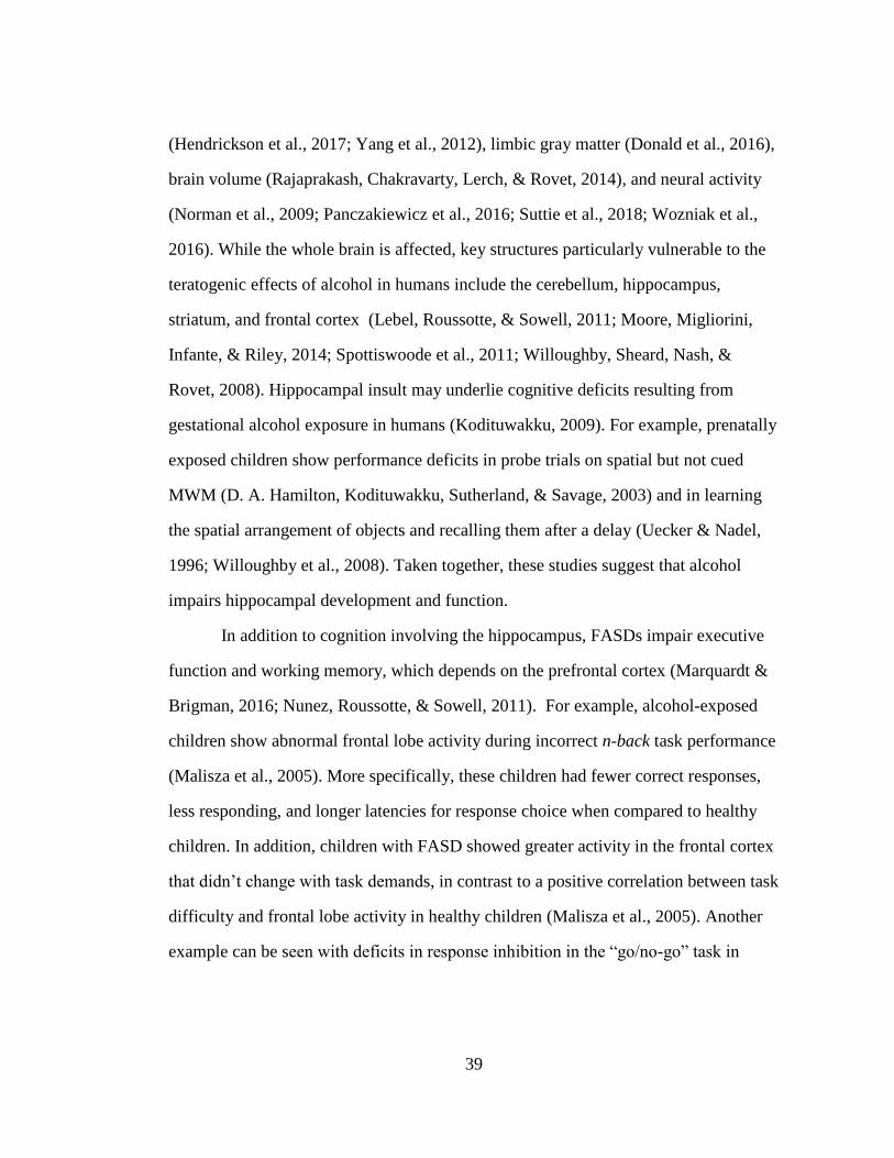
39
(Hendrickson et al., 2017; Yang et al., 2012), limbic gray matter (Donald et al., 2016),
brain volume (Rajaprakash, Chakravarty, Lerch, & Rovet, 2014), and neural activity
(Norman et al., 2009; Panczakiewicz et al., 2016; Suttie et al., 2018; Wozniak et al.,
2016). While the whole brain is affected, key structures particularly vulnerable to the
teratogenic effects of alcohol in humans include the cerebellum, hippocampus,
striatum, and frontal cortex (Lebel, Roussotte, & Sowell, 2011; Moore, Migliorini,
Infante, & Riley, 2014; Spottiswoode et al., 2011; Willoughby, Sheard, Nash, &
Rovet, 2008). Hippocampal insult may underlie cognitive deficits resulting from
gestational alcohol exposure in humans (Kodituwakku, 2009). For example, prenatally
exposed children show performance deficits in probe trials on spatial but not cued
MWM (D. A. Hamilton, Kodituwakku, Sutherland, & Savage, 2003) and in learning
the spatial arrangement of objects and recalling them after a delay (Uecker & Nadel,
1996; Willoughby et al., 2008). Taken together, these studies suggest that alcohol
impairs hippocampal development and function.
In addition to cognition involving the hippocampus, FASDs impair executive
function and working memory, which depends on the prefrontal cortex (Marquardt &
Brigman, 2016; Nunez, Roussotte, & Sowell, 2011). For example, alcohol-exposed
children show abnormal frontal lobe activity during incorrect n-back task performance
(Malisza et al., 2005). More specifically, these children had fewer correct responses,
less responding, and longer latencies for response choice when compared to healthy
children. In addition, children with FASD showed greater activity in the frontal cortex
that didn’t change with task demands, in contrast to a positive correlation between task
difficulty and frontal lobe activity in healthy children (Malisza et al., 2005). Another
example can be seen with deficits in response inhibition in the “go/no-go” task in
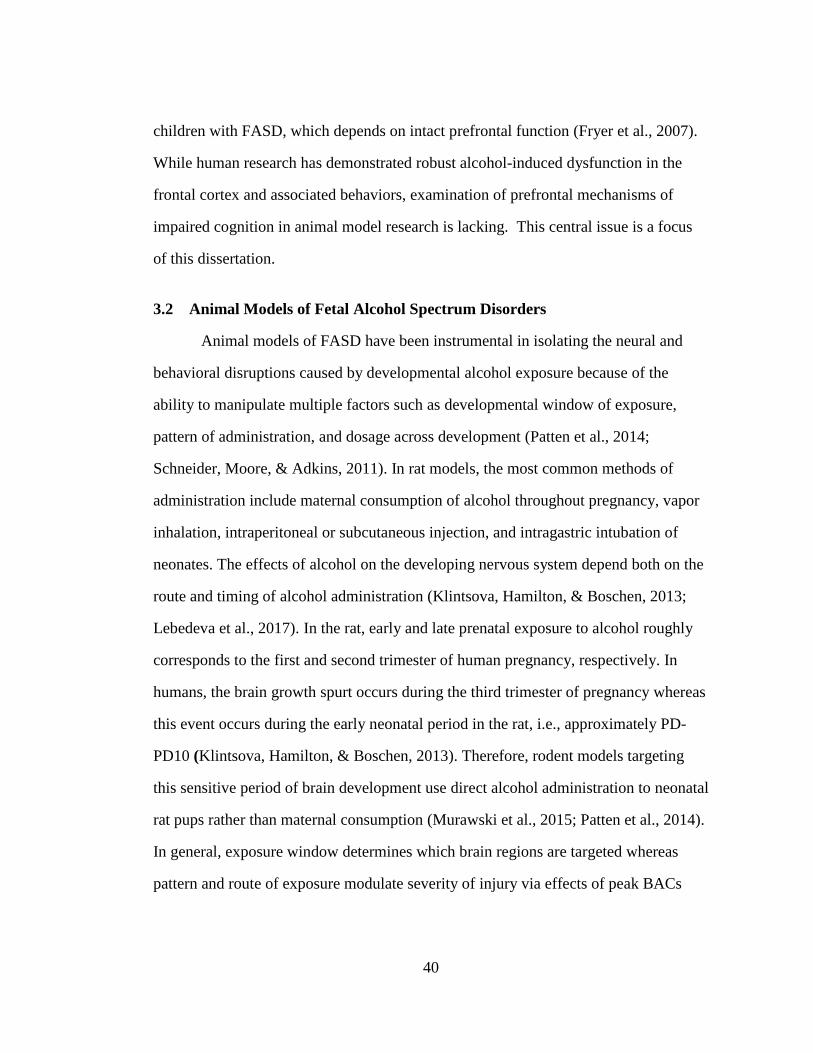
40
children with FASD, which depends on intact prefrontal function (Fryer et al., 2007).
While human research has demonstrated robust alcohol-induced dysfunction in the
frontal cortex and associated behaviors, examination of prefrontal mechanisms of
impaired cognition in animal model research is lacking. This central issue is a focus
of this dissertation.
3.2 Animal Models of Fetal Alcohol Spectrum Disorders
Animal models of FASD have been instrumental in isolating the neural and
behavioral disruptions caused by developmental alcohol exposure because of the
ability to manipulate multiple factors such as developmental window of exposure,
pattern of administration, and dosage across development (Patten et al., 2014;
Schneider, Moore, & Adkins, 2011). In rat models, the most common methods of
administration include maternal consumption of alcohol throughout pregnancy, vapor
inhalation, intraperitoneal or subcutaneous injection, and intragastric intubation of
neonates. The effects of alcohol on the developing nervous system depend both on the
route and timing of alcohol administration (Klintsova, Hamilton, & Boschen, 2013;
Lebedeva et al., 2017). In the rat, early and late prenatal exposure to alcohol roughly
corresponds to the first and second trimester of human pregnancy, respectively. In
humans, the brain growth spurt occurs during the third trimester of pregnancy whereas
this event occurs during the early neonatal period in the rat, i.e., approximately PD-
PD10 (Klintsova, Hamilton, & Boschen, 2013). Therefore, rodent models targeting
this sensitive period of brain development use direct alcohol administration to neonatal
rat pups rather than maternal consumption (Murawski et al., 2015; Patten et al., 2014).
In general, exposure window determines which brain regions are targeted whereas
pattern and route of exposure modulate severity of injury via effects of peak BACs
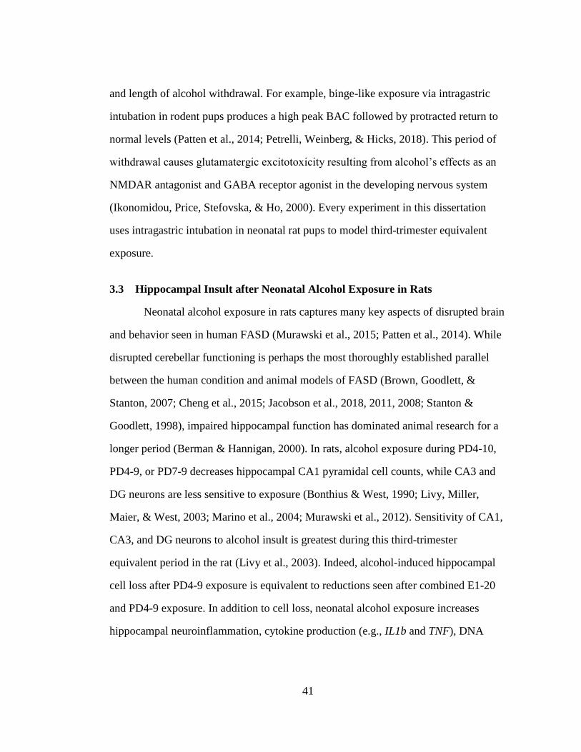
41
and length of alcohol withdrawal. For example, binge-like exposure via intragastric
intubation in rodent pups produces a high peak BAC followed by protracted return to
normal levels (Patten et al., 2014; Petrelli, Weinberg, & Hicks, 2018). This period of
withdrawal causes glutamatergic excitotoxicity resulting from alcohol’s effects as an
NMDAR antagonist and GABA receptor agonist in the developing nervous system
(Ikonomidou, Price, Stefovska, & Ho, 2000). Every experiment in this dissertation
uses intragastric intubation in neonatal rat pups to model third-trimester equivalent
exposure.
3.3 Hippocampal Insult after Neonatal Alcohol Exposure in Rats
Neonatal alcohol exposure in rats captures many key aspects of disrupted brain
and behavior seen in human FASD (Murawski et al., 2015; Patten et al., 2014). While
disrupted cerebellar functioning is perhaps the most thoroughly established parallel
between the human condition and animal models of FASD (Brown, Goodlett, &
Stanton, 2007; Cheng et al., 2015; Jacobson et al., 2018, 2011, 2008; Stanton &
Goodlett, 1998), impaired hippocampal function has dominated animal research for a
longer period (Berman & Hannigan, 2000). In rats, alcohol exposure during PD4-10,
PD4-9, or PD7-9 decreases hippocampal CA1 pyramidal cell counts, while CA3 and
DG neurons are less sensitive to exposure (Bonthius & West, 1990; Livy, Miller,
Maier, & West, 2003; Marino et al., 2004; Murawski et al., 2012). Sensitivity of CA1,
CA3, and DG neurons to alcohol insult is greatest during this third-trimester
equivalent period in the rat (Livy et al., 2003). Indeed, alcohol-induced hippocampal
cell loss after PD4-9 exposure is equivalent to reductions seen after combined E1-20
and PD4-9 exposure. In addition to cell loss, neonatal alcohol exposure increases
hippocampal neuroinflammation, cytokine production (e.g., IL1b and TNF), DNA
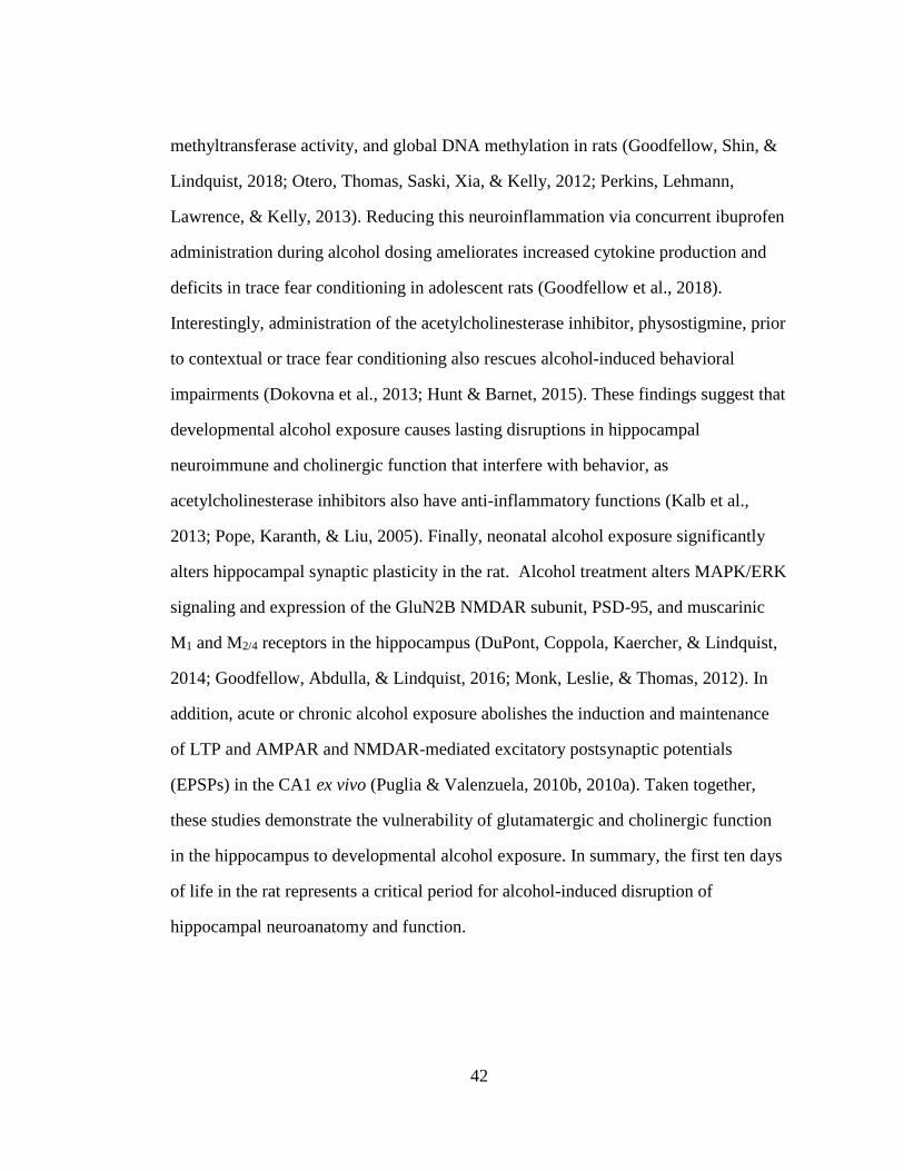
42
methyltransferase activity, and global DNA methylation in rats (Goodfellow, Shin, &
Lindquist, 2018; Otero, Thomas, Saski, Xia, & Kelly, 2012; Perkins, Lehmann,
Lawrence, & Kelly, 2013). Reducing this neuroinflammation via concurrent ibuprofen
administration during alcohol dosing ameliorates increased cytokine production and
deficits in trace fear conditioning in adolescent rats (Goodfellow et al., 2018).
Interestingly, administration of the acetylcholinesterase inhibitor, physostigmine, prior
to contextual or trace fear conditioning also rescues alcohol-induced behavioral
impairments (Dokovna et al., 2013; Hunt & Barnet, 2015). These findings suggest that
developmental alcohol exposure causes lasting disruptions in hippocampal
neuroimmune and cholinergic function that interfere with behavior, as
acetylcholinesterase inhibitors also have anti-inflammatory functions (Kalb et al.,
2013; Pope, Karanth, & Liu, 2005). Finally, neonatal alcohol exposure significantly
alters hippocampal synaptic plasticity in the rat. Alcohol treatment alters MAPK/ERK
signaling and expression of the GluN2B NMDAR subunit, PSD-95, and muscarinic
M1 and M2/4 receptors in the hippocampus (DuPont, Coppola, Kaercher, & Lindquist,
2014; Goodfellow, Abdulla, & Lindquist, 2016; Monk, Leslie, & Thomas, 2012). In
addition, acute or chronic alcohol exposure abolishes the induction and maintenance
of LTP and AMPAR and NMDAR-mediated excitatory postsynaptic potentials
(EPSPs) in the CA1 ex vivo (Puglia & Valenzuela, 2010b, 2010a). Taken together,
these studies demonstrate the vulnerability of glutamatergic and cholinergic function
in the hippocampus to developmental alcohol exposure. In summary, the first ten days
of life in the rat represents a critical period for alcohol-induced disruption of
hippocampal neuroanatomy and function.
43
3.4 Prefrontal Insult after Neonatal Alcohol Exposure in Rats
As in the human condition of FASD (Section 3.1), developmental alcohol
exposure also alters medial prefrontal neuroanatomy and function in rodents.
However, examination of this issue in rodent models is relatively limited. In contrast
to the lasting cell loss seen in the hippocampus, neonatal alcohol exposure has no
lasting effect on prefrontal pyramidal cell number but instead alters synaptic plasticity,
dendritic complexity, gene expression, and epigenetic markers (Boschen, Keller, Roth,
& Klintsova, 2018; Granato, Di Rocco, Zumbo, Toesca, & Giannetti, 2003; Granato,
Palmer, De Giorgio, Tavian, & Larkum, 2012; G. F. Hamilton, Whitcher, & Klintsova,
2010; Otero et al., 2012). In contrast to pyramidal cell counts, neonatal exposure does
decrease parvalbumin interneurons in the AC but not PL or IL in adult rats (G. F.
Hamilton, Hernandez, Krebs, Bucko, & Rhodes, 2017). Neonatal alcohol exposure
also decreases dendritic complexity and density that may alter circuit organization and
behavior in rats (G. F. Hamilton et al., 2010; Lawrence, Otero, & Kelly, 2012;
Whitcher & Klintsova, 2008). Exposure across prenatal and neonatal development
decreases apical and basilar spine density and complexity in layer II/III pyramidal
cells in the mPFC of adult rats (Lawrence et al., 2012). Exposure limited to the third-
trimester equivalent causes reduced apical dendrite density and basilar dendrite
complexity in prefrontal pyramidal neurons also in adolescent rats (Granato et al.,
2003; G. F. Hamilton et al., 2010; Whitcher & Klintsova, 2008). Neonatal exposure
also alters voltage-gated Ca2+ channel activity while decreasing the amount and
duration of dendritic spiking in layer V pyramidal neurons in the PFC (Granato et al.,
2012). Taken together with increased global DNA methylation (Boschen et al., 2018;
Otero et al., 2012), these physiological changes likely alter activity and plasticity-
associated gene expression supporting learning and memory.
44
While studies of neonatal exposure are limited, prenatal alcohol exposure in
rats alters experience-dependent gene expression in the medial prefrontal cortex. For
example, ethanol exposure throughout gestation decreases expression of the IEGs Arc
and c-Fos in the PL in adult rats during wrestling and social interaction tasks (D. A.
Hamilton, Akers, et al., 2010; D. A. Hamilton, Candelaria-Cook, et al., 2010). A
narrower second-trimester equivalent exposure disrupts the expression of the
transcription factors c-Fos and jun-B in the PL and ACC during T-maze alternation
(Nagahara & Handa, 1995). Finally, late gestational exposure also decreases c-Fos
protein expression in the IL during open field exploration in adolescent rats (Fabio et
al., 2013). More research is needed to establish a link between prefrontal targeting and
cognitive deficits resulting from neonatal ethanol exposure in rats. Chapters 7 and 8
in this dissertation examine this issue by measuring activity and plasticity-associated
gene expression in the prefrontal cortex of rats receiving neonatal alcohol exposure.
3.5 Cholinergic Insult after Neonatal Alcohol Exposure in Rats
Animal model research suggests that alcohol exposure disrupts acute and
chronic cholinergic muscarinic-receptor cell signaling involved in neuronal and
microglial proliferation, synaptic plasticity, and behavioral performance on tasks
involving hippocampal and prefrontal cholinergic activity (Costa, Giordano, &
Guizzetti, 2013; Monk et al., 2012; Wilhelm & Guizzetti, 2015). In neonatal rats,
alcohol decreases the bioavailability of the acetylcholine precursor choline and
inhibits glial muscarinic-receptor cell signaling important for neuritogenesis in
proliferating hippocampal pyramidal neurons (Costa et al., 2013; Giordano, Guizzetti,
Dao, Mattison, & Costa, 2011; Goeke, Roberts, Hashimoto, Finn, & Guizzetti, 2018;
Guizzetti, Moore, Giordano, VanDeMark, & Costa, 2010; Guizzetti, Moore,
45
VanDeMark, Giordano, & Costa, 2011). Importantly, alcohol-induced disruptions in
cholinergic signaling are not limited to the period of alcohol exposure (Monk et al.,
2012). Decrease in hippocampal M1 muscarinic receptors and a significant increase in
M2/4 receptors are seen in adolescent rats. This change in hippocampal muscarinic
receptor composition likely interferes with synaptic plasticity via increased activation
of M2/4 receptors, which downregulate cyclic AMP activity important for induction of
transcription factors and calcium channel activity (Migeon & Nathansons, 1994; Monk
et al., 2012). Whether this lasting interference with muscarinic receptor composition
and signaling is seen in other brain regions such as the prefrontal cortex is unknown
and remains a fruitful direction for future animal model research.
A growing body of work suggests that increasing bioavailability of choline and
thus cholinergic signaling across development has potential as a therapeutic
intervention for FASD in rodent models. Alcohol-induced impairment of MWM, fear
conditioning, spatial discrimination, working memory, and motor behavior are
attenuated by developmental choline supplementation in rats (Hunt & Barnet, 2015;
Idrus, Breit, & Thomas, 2017; Ryan, Williams, & Thomas, 2008; Thomas, Abou, &
Dominguez, 2009; Thomas, Biane, O’Bryan, O’Neill, & Dominguez, 2007; Thomas,
Garrison, & O’Neill, 2004; Thomas, Idrus, Monk, & Dominguez, 2010; Wagner &
Hunt, 2006). In addition to these behavioral effects, developmental choline
administration reverses increased global DNA methylation in the HPC and PFC,
which in the absence of choline causes a restrictive state for activity-driven gene
expression (Otero et al., 2012; Perkins et al., 2013). This choline administration also
attenuates alcohol-induced increases in M2/4 muscarinic receptors in the HPC, which
likely results in decreased inhibition of cyclic AMP activity (Monk et al., 2012).
46
While more research is needed, these benefits of choline supplementation during
gestation are also promising in the human condition (Jacobson et al., 2018; Wozniak et
al., 2015, 2013). Clearly, the specific long-term therapeutic mechanisms of choline
supplementation on neurobiological and behavioral outcome measures in humans and
animal models demands further investigation. Moreover, the effects of enhancing
cholinergic system function acutely later in life on cognition is not well characterized.
3.6 Behavioral Consequences of Neonatal Alcohol Exposure in Rats
Animal model research examining third-trimester equivalent alcohol exposure
(e.g., from PD7-9 or PD4-9) in rats has focused on establishing a link between altered
hippocampal development and behavioral performance on traditionally “hippocampal-
dependent” behavioral tasks. Neonatal alcohol exposure has the most disruptive
effects on “hippocampus-dependent” tasks that also depend on PFC. For example,
neonatal alcohol impairs later spatial learning and memory in place but not cued
MWM procedures, especially during probe trials with long delays (Girard, Xing,
Ward, & Wainwright, 2000; Goodlett & Johnson, 1997; Goodlett & Peterson, 1995;
Johnson & Goodlett, 2002; Marino et al., 2004; Thomas et al., 2007; Thomas, Sather,
& Whinery, 2008). However, lesions or pharmacological disruption of the mPFC in
rats also interfere with allocentric spatial learning in the MWM (Fantie & Kolb, 1990;
Kolb, Buhrmann, McDonald, & Sutherland, 1994; Kolb & Cioe, 1996; Leon, Bruno,
Allard, Nader, & Cuello, 2010; McDonald, King, Foong, Rizos, & Hong, 2008; Sang
Jo et al., 2007). Consistent with potential prefrontal mechanisms of MWM disruption,
rescuing hippocampal CA1 cell loss by developmental Vitamin E supplementation
(Marino et al., 2004; see Figure 3.1) does not rescue probe trial performance in rats
after PD7-9 exposure (Marino et al., 2004). Alcohol-exposed rats also show deficits on
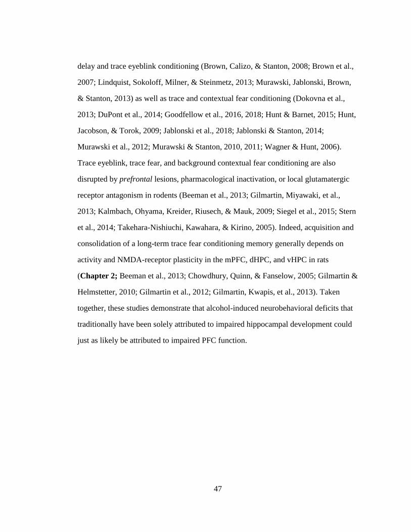
47
delay and trace eyeblink conditioning (Brown, Calizo, & Stanton, 2008; Brown et al.,
2007; Lindquist, Sokoloff, Milner, & Steinmetz, 2013; Murawski, Jablonski, Brown,
& Stanton, 2013) as well as trace and contextual fear conditioning (Dokovna et al.,
2013; DuPont et al., 2014; Goodfellow et al., 2016, 2018; Hunt & Barnet, 2015; Hunt,
Jacobson, & Torok, 2009; Jablonski et al., 2018; Jablonski & Stanton, 2014;
Murawski et al., 2012; Murawski & Stanton, 2010, 2011; Wagner & Hunt, 2006).
Trace eyeblink, trace fear, and background contextual fear conditioning are also
disrupted by prefrontal lesions, pharmacological inactivation, or local glutamatergic
receptor antagonism in rodents (Beeman et al., 2013; Gilmartin, Miyawaki, et al.,
2013; Kalmbach, Ohyama, Kreider, Riusech, & Mauk, 2009; Siegel et al., 2015; Stern
et al., 2014; Takehara-Nishiuchi, Kawahara, & Kirino, 2005). Indeed, acquisition and
consolidation of a long-term trace fear conditioning memory generally depends on
activity and NMDA-receptor plasticity in the mPFC, dHPC, and vHPC in rats
(Chapter 2; Beeman et al., 2013; Chowdhury, Quinn, & Fanselow, 2005; Gilmartin &
Helmstetter, 2010; Gilmartin et al., 2012; Gilmartin, Kwapis, et al., 2013). Taken
together, these studies demonstrate that alcohol-induced neurobehavioral deficits that
traditionally have been solely attributed to impaired hippocampal development could
just as likely be attributed to impaired PFC function.
48
Figure 3.1 Unbiased stereology data for CA1 region of the hippocampus. CA1
pyramidal cell number estimates by unbiased stereology. Unilateral cell
number estimates are reported. Groups with different letters were
significantly different. Error bars represent S.E.M. ET= ethanol-exposed
rats, E = vitamin E treatment, IC = intubated control. Developmental
vitamin E treatment eliminated differences in CA1 pyramidal cell counts
between the ET and IC groups. Taken from Marino et al., 2004.
49
3.7 Impairment of the CPFE by Neonatal Alcohol Exposure in Rats
Our lab has shown that the CPFE is particularly sensitive to disrupted HPC and
PFC function resulting from third-trimester equivalent alcohol exposure in rats. The
CPFE requires both activity and cholinergic muscarinic-receptor cell signaling in the
dHPC and mPFC during all three phases (Heroux et al., 2017; Matus-Amat et al.,
2004; Robinson-Drummer et al., 2016, 2017). Our lab has consistently reported robust
impairments in retention test freezing during the CPFE in adolescent and adult rats
after neonatal alcohol exposure from PD7-9 or PD4-9 (G. F. Hamilton et al., 2011;
Jablonski et al., 2018; Jablonski & Stanton, 2014; Murawski et al., 2012; Murawski &
Stanton, 2010, 2011). Moreover, impairment of performance scales with alcohol dose,
with a significant negative correlation of BACs during exposure and test freezing
(Murawski & Stanton, 2011). The specific effects of neonatal alcohol dosing on
learning and memory processes in the CPFE depend largely on the dosing window.
Our lab has examined behavioral performance in adolescent rats after neonatal alcohol
exposure from PD7-9, PD4-6, and PD4-9 (Murawski & Stanton, 2011). Unlike after
PD4-9 or PD7-9, PD4-6 alcohol exposure has no effect on retention test freezing in the
CPFE (Murawski & Stanton, 2011). The notion that exposure over PD7-9 window
alone is sufficient to capture alcohol-induced behavioral deficits is supported by
previous research showing that PD7-9 and PD4-9, but not PD4-6, alcohol exposure
impairs performance in the MWM (Goodlett & Johnson, 1997; Goodlett & Peterson,
1995). Further examination of the PD7-9 window has shown that exposed rats have
intact post-shock but impaired retention test freezing in the CPFE (Jablonski &
Stanton, 2014). These data indicate that PD7-9 alcohol-exposed rats are able to form a
context-shock association but consolidation of this association is impaired. This notion
is supported by the finding that alcohol-exposed rats have reduced prefrontal
50
expression of the IEG egr-1 after preexposure and training (Jablonski et al., 2018).
Interestingly, while these data suggest that PD4-6 exposure is not relevant to
behavioral disruption of the CPFE, PD4-9 alcohol exposure results in knockdown of
hippocampal c-Fos protein expression and CA1 pyramidal cell loss on the preexposure
day (Murawski et al., 2012; see Figure 3.2). In addition, reducing the interval between
context preexposure and training to 2hr instead of 24hr rescues retention test freezing
in alcohol-exposed animals, further suggesting additional impairment of preexposure
day processes after PD4-9 exposure (Goodfellow & Lindquist, 2014). The effect of
this exposure on prefrontal activity during context learning and post-shock freezing in
the CPFE is not known. Given the early timing of neonatal prefrontal development
(Ferguson & Gao, 2014; van Eden & Uylings, 1985; Z. wei Zhang, 2004), PD4-9
alcohol exposure may alter prefrontal function important for behavior across the
lifespan. Finally, our lab has shown that administration of the acetylcholinesterase
inhibitor physostigmine (PHY) prior to all three phases of the CPFE rescues retention
test freezing in PD7-9 ethanol-exposed rats, indicating a role for disrupted cholinergic
signaling in the alcohol-induced behavioral deficit (Dokovna et al., 2013; see Figure
3.3). The specific memory processes of the CPFE as well as regional neural and
cholinergic function that is disrupted by neonatal alcohol exposure are not well
characterized. These questions are explored further in Chapters 7 and 8 of this
dissertation.
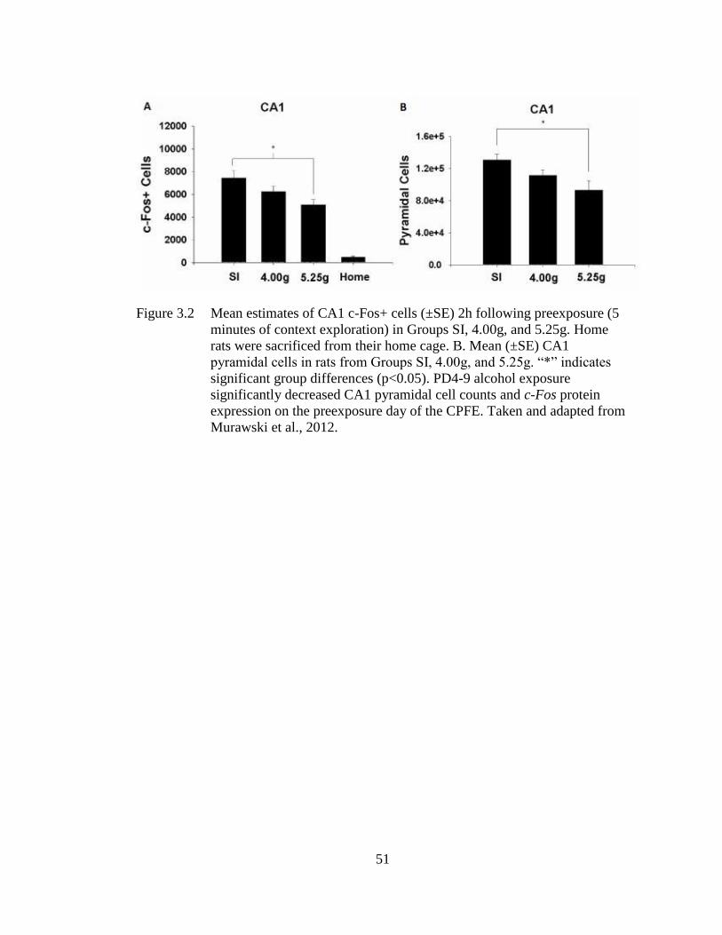
51
Figure 3.2 Mean estimates of CA1 c-Fos+ cells (±SE) 2h following preexposure (5
minutes of context exploration) in Groups SI, 4.00g, and 5.25g. Home
rats were sacrificed from their home cage. B. Mean (±SE) CA1
pyramidal cells in rats from Groups SI, 4.00g, and 5.25g. “*” indicates
significant group differences (p<0.05). PD4-9 alcohol exposure
significantly decreased CA1 pyramidal cell counts and c-Fos protein
expression on the preexposure day of the CPFE. Taken and adapted from
Murawski et al., 2012.
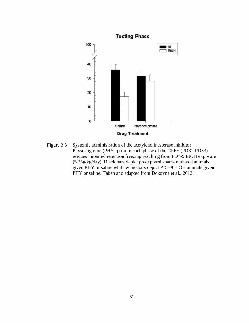
52
Figure 3.3 Systemic administration of the acetylcholinesterase inhibitor
Physostigmine (PHY) prior to each phase of the CPFE (PD31-PD33)
rescues impaired retention freezing resulting from PD7-9 EtOH exposure
(5.25g/kg/day). Black bars depict preexposed sham-intubated animals
given PHY or saline while white bars depict PD4-9 EtOH animals given
PHY or saline. Taken and adapted from Dokovna et al., 2013.
53
3.8 Conclusions
This chapter summarized key findings highlighting prefrontal, hippocampal,
cholinergic, and behavioral dysfunction in rodent models of FASDs involving third-
trimester equivalent alcohol exposure. While human FASD research has recently
started to emphasize prefrontal dysfunction as a feature of the disorder, animal model
research has largely continued its focus on hippocampus. One main hypothesis of the
experiments in this dissertation is that functional, molecular changes in the mPFC
underlie cognitive deficits arising from third-trimester equivalent alcohol exposure in
rats. Armed with recent discoveries that mPFC is critical for the CPFE (Heroux et al.,
2017; Robinson-Drummer et al., 2017), this dissertation re-examines disruptions in the
CPFE that our lab has traditionally attributed to disrupted hippocampal functioning in
alcohol-exposed rats. In the experimental chapters of this dissertation, I explore
hippocampal-prefrontal circuitry supporting contextual memory processes of the
CPFE, which informs this alcohol-induced insult (see Chapter 6). Next, I examine the
effects of neonatal alcohol exposure on activity-driven prefrontal and hippocampal
gene expression during the acquisition of context memory in the CPFE (see Chapter
7). Finally, I examine the effects of enhancing cholinergic signaling prior to
behavioral testing on CPFE performance and regional gene expression (see Chapter
8).
54
FOUNDATIONAL STUDIES
The preceding chapters reviewed the neurobehavioral consequences of
developmental alcohol exposure and the neural mechanisms of contextual fear
conditioning. The present chapter describes my published research that serves as a
foundation for the experiments performed in this dissertation. These studies
characterize 1) the role of NMDAR plasticity in the acquisition and consolidation of
contextual fear memory in the CPFE, 2) the involvement of the mPFC across variants
of contextual fear conditioning, 3) the role of mPFC and dHPC cholinergic signaling
in the CPFE, and 4) regional patterns of IEG induction during context and contextual
fear learning in the CPFE.
4.1 NMDA-Receptor Cell Signaling and the CPFE
As reviewed in Chapters 1and 2, the CPFE paradigm permits separate analysis
of learning about the context, acquiring a context-shock association, and
retrieval/expression of that association (see Figure 1.2). In adult rats, context learning
depends on NMDAR activity in dHPC whereas context-shock learning (during
immediate-shock training) depends on NMDAR activity in BLA (Matus-Amat et al.,
2007). Our lab has shown that the same is true for juvenile (PD24) and adolescent
(PD31) rats (Burman, Murawski, Schiffino, Rosen, & Stanton, 2009; Miller, Heroux,
& Stanton, 2019; Schiffino et al., 2011). Importantly, previous pharmacological
studies using the CPFE fail to dissociate between acquisition and consolidation
Chapter 4
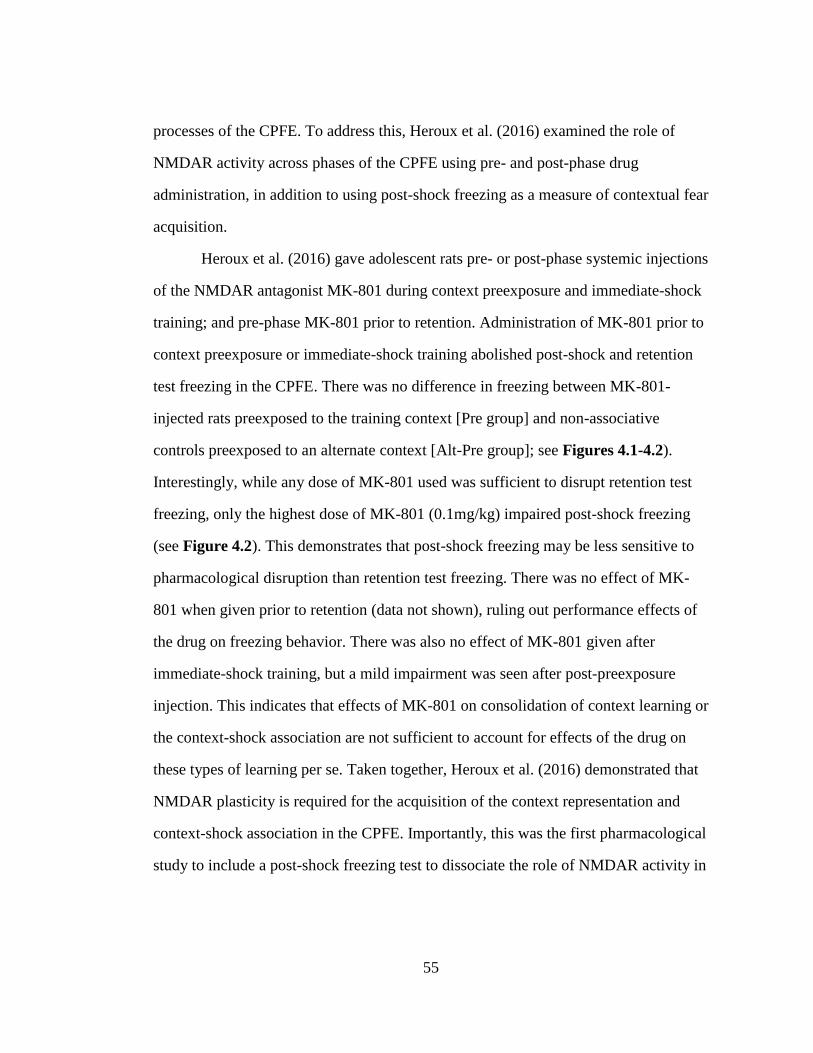
55
processes of the CPFE. To address this, Heroux et al. (2016) examined the role of
NMDAR activity across phases of the CPFE using pre- and post-phase drug
administration, in addition to using post-shock freezing as a measure of contextual fear
acquisition.
Heroux et al. (2016) gave adolescent rats pre- or post-phase systemic injections
of the NMDAR antagonist MK-801 during context preexposure and immediate-shock
training; and pre-phase MK-801 prior to retention. Administration of MK-801 prior to
context preexposure or immediate-shock training abolished post-shock and retention
test freezing in the CPFE. There was no difference in freezing between MK-801-
injected rats preexposed to the training context [Pre group] and non-associative
controls preexposed to an alternate context [Alt-Pre group]; see Figures 4.1-4.2).
Interestingly, while any dose of MK-801 used was sufficient to disrupt retention test
freezing, only the highest dose of MK-801 (0.1mg/kg) impaired post-shock freezing
(see Figure 4.2). This demonstrates that post-shock freezing may be less sensitive to
pharmacological disruption than retention test freezing. There was no effect of MK-
801 when given prior to retention (data not shown), ruling out performance effects of
the drug on freezing behavior. There was also no effect of MK-801 given after
immediate-shock training, but a mild impairment was seen after post-preexposure
injection. This indicates that effects of MK-801 on consolidation of context learning or
the context-shock association are not sufficient to account for effects of the drug on
these types of learning per se. Taken together, Heroux et al. (2016) demonstrated that
NMDAR plasticity is required for the acquisition of the context representation and
context-shock association in the CPFE. Importantly, this was the first pharmacological
study to include a post-shock freezing test to dissociate the role of NMDAR activity in

56
the acquisition vs. consolidation of contextual fear in the CPFE. These findings
provide a procedural framework both for future pharmacological studies and most of
my subsequent studies, as reviewed below and presented in this dissertation.
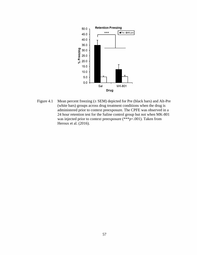
57
Figure 4.1 Mean percent freezing (± SEM) depicted for Pre (black bars) and Alt-Pre
(white bars) groups across drug treatment conditions when the drug is
administered prior to context preexposure. The CPFE was observed in a
24 hour retention test for the Saline control group but not when MK-801
was injected prior to context preexposure (***p<.001). Taken from
Heroux et al. (2016).
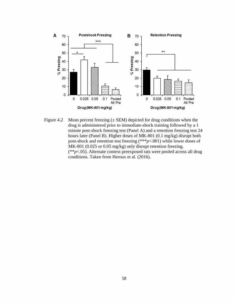
58
Figure 4.2 Mean percent freezing (± SEM) depicted for drug conditions when the
drug is administered prior to immediate-shock training followed by a 1
minute post-shock freezing test (Panel A) and a retention freezing test 24
hours later (Panel B). Higher doses of MK-801 (0.1 mg/kg) disrupt both
post-shock and retention test freezing (***p<.001) while lower doses of
MK-801 (0.025 or 0.05 mg/kg) only disrupt retention freezing.
(**p<.05). Alternate context preexposed rats were pooled across all drug
conditions. Taken from Heroux et al. (2016).
59
4.2 Involvement of the mPFC across Variants of Contextual Fear Conditioning
As reviewed in Chapter 2, the role of the mPFC in fear conditioning has been
traditionally attributed to the long-term, systems-level consolidation of memory and to
the regulation of fear memory expression via the PL and IL sub-regions (Frankland &
Bontempi, 2005; Gilmartin, Balderston, & Helmstetter, 2014; Giustino & Maren,
2015; Rozeske et al., 2015). Despite this, recent evidence suggests that the mPFC is
important for processing contextual information during trace and contextual fear
conditioning (see Section 2.1.2 in Chapter 2). The functional role of the mPFC in
supporting distinct learning and memory processes during contextual fear conditioning
is unclear.
To address this, Heroux et al. (2017) examined the causal role of the mPFC in
distinct learning and memory processes across phases of the CPFE and sCFC in
adolescent rats. Rats were given a bilateral intra-mPFC infusion of the GABAA
agonist muscimol (MUSC) or the vehicle phosphate buffered saline (PBS) prior to
each phase of the CPFE or prior to training in sCFC. Inactivation via muscimol
infusion during any phase of the CPFE (context preexposure, immediate-shock
training, or retention) disrupted retention test freezing (see Figures 4.3-4.5).
Importantly, mPFC inactivation prior to immediate-shock training did not disrupt
post-shock freezing even though it abolished 24-h retention freezing in the same rats.
This same infusion prior to training in sCFC had no effect on post-shock or retention
test freezing (see Figure 4.6). These experiments demonstrate that, in the CPFE, the
mPFC is required for the 1) acquisition and/or consolidation of the conjunctive context
representation, 2) consolidation of the context-shock association, and 3) long-term
retrieval and expression of the context-shock association. The mPFC is not required
for the retrieval of the context representation or its association with shock in the
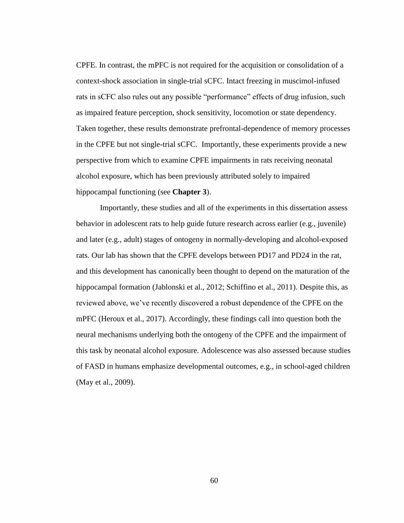
60
CPFE. In contrast, the mPFC is not required for the acquisition or consolidation of a
context-shock association in single-trial sCFC. Intact freezing in muscimol-infused
rats in sCFC also rules out any possible “performance” effects of drug infusion, such
as impaired feature perception, shock sensitivity, locomotion or state dependency.
Taken together, these results demonstrate prefrontal-dependence of memory processes
in the CPFE but not single-trial sCFC. Importantly, these experiments provide a new
perspective from which to examine CPFE impairments in rats receiving neonatal
alcohol exposure, which has been previously attributed solely to impaired
hippocampal functioning (see Chapter 3).
Importantly, these studies and all of the experiments in this dissertation assess
behavior in adolescent rats to help guide future research across earlier (e.g., juvenile)
and later (e.g., adult) stages of ontogeny in normally-developing and alcohol-exposed
rats. Our lab has shown that the CPFE develops between PD17 and PD24 in the rat,
and this development has canonically been thought to depend on the maturation of the
hippocampal formation (Jablonski et al., 2012; Schiffino et al., 2011). Despite this, as
reviewed above, we’ve recently discovered a robust dependence of the CPFE on the
mPFC (Heroux et al., 2017). Accordingly, these findings call into question both the
neural mechanisms underlying both the ontogeny of the CPFE and the impairment of
this task by neonatal alcohol exposure. Adolescence was also assessed because studies
of FASD in humans emphasize developmental outcomes, e.g., in school-aged children
(May et al., 2009).
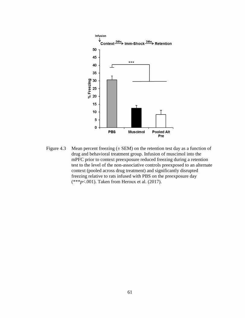
61
Figure 4.3 Mean percent freezing (± SEM) on the retention test day as a function of
drug and behavioral treatment group. Infusion of muscimol into the
mPFC prior to context preexposure reduced freezing during a retention
test to the level of the non-associative controls preexposed to an alternate
context (pooled across drug treatment) and significantly disrupted
freezing relative to rats infused with PBS on the preexposure day
(***p<.001). Taken from Heroux et al. (2017).
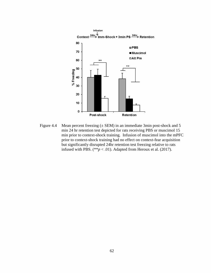
62
Figure 4.4 Mean percent freezing (± SEM) in an immediate 3min post-shock and 5
min 24 hr retention test depicted for rats receiving PBS or muscimol 15
min prior to context-shock training. Infusion of muscimol into the mPFC
prior to context-shock training had no effect on context-fear acquisition
but significantly disrupted 24hr retention test freezing relative to rats
infused with PBS. (**p < .01). Adapted from Heroux et al. (2017).
63
Figure 4.5 Mean percent freezing (± SEM) depicted for rats receiving PBS or
muscimol 15 min prior to a 5 min retention test occurring 24hr after
conditioning with no post-shock test. Infusion of muscimol into the
mPFC prior to the retention test reduced freezing to a level comparable to
non-associative controls (pooled across drug treatment) and significantly
lower than rats infused with PBS (**p<.01). Taken from Heroux et al.
(2017).
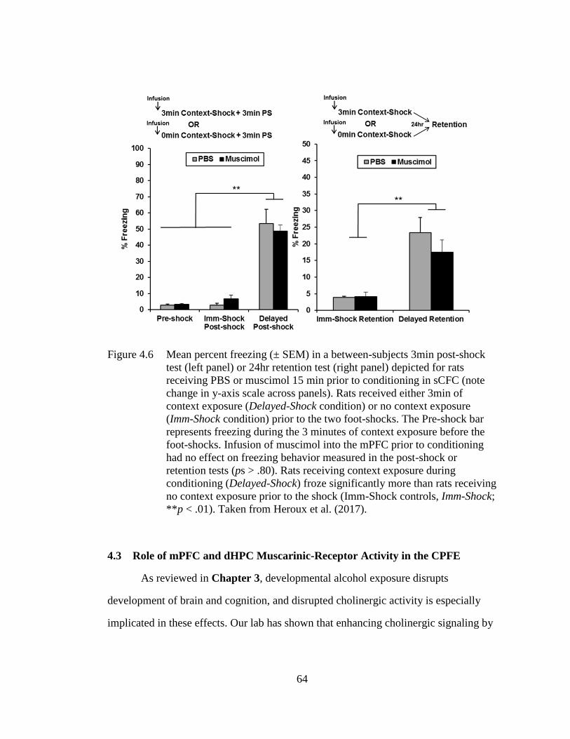
64
Figure 4.6 Mean percent freezing (± SEM) in a between-subjects 3min post-shock
test (left panel) or 24hr retention test (right panel) depicted for rats
receiving PBS or muscimol 15 min prior to conditioning in sCFC (note
change in y-axis scale across panels). Rats received either 3min of
context exposure (Delayed-Shock condition) or no context exposure
(Imm-Shock condition) prior to the two foot-shocks. The Pre-shock bar
represents freezing during the 3 minutes of context exposure before the
foot-shocks. Infusion of muscimol into the mPFC prior to conditioning
had no effect on freezing behavior measured in the post-shock or
retention tests (ps > .80). Rats receiving context exposure during
conditioning (Delayed-Shock) froze significantly more than rats receiving
no context exposure prior to the shock (Imm-Shock controls, Imm-Shock;
**p < .01). Taken from Heroux et al. (2017).
4.3 Role of mPFC and dHPC Muscarinic-Receptor Activity in the CPFE
As reviewed in Chapter 3, developmental alcohol exposure disrupts
development of brain and cognition, and disrupted cholinergic activity is especially
implicated in these effects. Our lab has shown that enhancing cholinergic signaling by
65
systemic administration of PHY rescues impaired retention test freezing in the CPFE
(Dokovna et al., 2013). The effects of PHY might be due to enhanced muscarinic-
receptor signaling, as prior work has demonstrated that systemic muscarinic-receptor
antagonism disrupts both sCFC and the CPFE (Anagnostaras, Maren, & Fanselow,
1995; Anagnostaras, Maren, Sage, Goodrich, & Fanselow, 1999; Brown, Kennard,
Sherer, Comalli, & Woodruff-Pak, 2011; Hunt & Richardson, 2007). These findings
led us to examine the functional role of prefrontal and hippocampal cholinergic
muscarinic-receptor activity across phases of the CPFE (Robinson-Drummer et al.,
2016, 2017). We administered the muscarinic-receptor antagonist scopolamine
(SCOP) systemically or into dHPC prior to all phases or each individual phase of the
CPFE in adolescent rats. All of these manipulations disrupted retention test freezing.
For example, the Pre-SCOP group froze at the same level as the Alt-Pre control group
(pooled across drug) and at lower levels than vehicle (Pre-PBS) or no-surgery controls
(Pre-UND; Robinson-Drummer et al., 2016; see Figure 4.7). Similar SCOP infusions
delivered into the mPFC disrupted retention test freezing when given prior to context
preexposure, immediate-shock training, or all three phases, but not the retention phase
(Robinson-Drummer et al., 2017). Taken together, these results demonstrate that
muscarinic-receptor signaling in both the mPFC and dHPC is crucial for both context
learning and contextual fear acquisition and/or consolidation in the CPFE. These
results inform our lab’s previous findings that physostigmine rescues retention test
freezing in rats receiving neonatal alcohol exposure from PD7-9 (Dokovna et al.,
2013), suggesting that developmental alcohol exposure may impair the CPFE by
disrupting prefrontal and/or hippocampal cholinergic signaling during behavior.

66
Collectively, this work serves as an empirical foundation for Chapter 8 of this
dissertation.
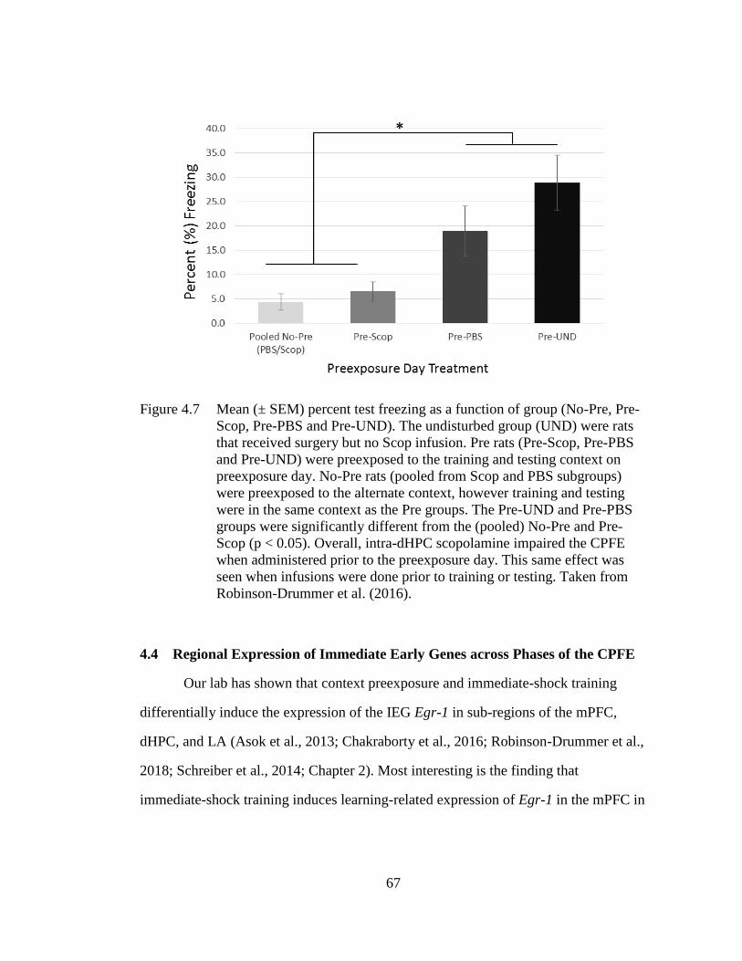
67
Figure 4.7 Mean (± SEM) percent test freezing as a function of group (No-Pre, Pre-
Scop, Pre-PBS and Pre-UND). The undisturbed group (UND) were rats
that received surgery but no Scop infusion. Pre rats (Pre-Scop, Pre-PBS
and Pre-UND) were preexposed to the training and testing context on
preexposure day. No-Pre rats (pooled from Scop and PBS subgroups)
were preexposed to the alternate context, however training and testing
were in the same context as the Pre groups. The Pre-UND and Pre-PBS
groups were significantly different from the (pooled) No-Pre and Pre-
Scop (p < 0.05). Overall, intra-dHPC scopolamine impaired the CPFE
when administered prior to the preexposure day. This same effect was
seen when infusions were done prior to training or testing. Taken from
Robinson-Drummer et al. (2016).
4.4 Regional Expression of Immediate Early Genes across Phases of the CPFE
Our lab has shown that context preexposure and immediate-shock training
differentially induce the expression of the IEG Egr-1 in sub-regions of the mPFC,
dHPC, and LA (Asok et al., 2013; Chakraborty et al., 2016; Robinson-Drummer et al.,
2018; Schreiber et al., 2014; Chapter 2). Most interesting is the finding that
immediate-shock training induces learning-related expression of Egr-1 in the mPFC in
68
adolescent and adult rats. As reviewed in Chapter 1, the CPFE emerges between PD17
and PD24 in rats (Jablonski et al., 2012; Schiffino et al., 2011). Accordingly, we
hypothesized that experience- and learning-related Egr-1 expression in the prefrontal
cortex may correlate or even cause the ontogeny of the CPFE. To test this, we
analyzed freezing performance and Egr-1 expression during context preexposure and
immediate-shock training in PD17 and PD24 rats (Robinson-Drummer et al., 2018).
PD17 rats failed to show a significant CPFE compared to PD24 rats regardless of
single or multiple exposures to the training context. Moreover, PD24 rats showed an
elevation in prefrontal, hippocampal, and amygdalar Egr-1 expression during context
preexposure that was absent in PD17 rats. Interestingly, using a single context
exposure, training-day Egr-1 expression was not different between PD17 and PD24
rats and was not related to learning. These results showed that PD24 rats show the
CPFE even in the absence of any observed learning-related Egr-1 expression in the
prefrontal cortex during training (Robinson-Drummer et al., 2018). Ultimately, this
suggests that preexposure but not training-day Egr-1 expression may underlie the
ontogeny of the CPFE.
To extend this research to other IEGs, Heroux et al. (2018) examined
expression of c-Fos, Arc, Egr-1, and Npas4 during different phases of the CPFE in
adolescent rats, for reasons described previously (see Chapter 2, Section 2.2.3). In this
study, IEG expression was analyzed in the mPFC, dHPC, and BLA during context
preexposure and immediate-shock training (with or without a post-shock freezing test)
via quantitative real-time polymerase chain reaction (qPCR) on RNA extracted from
dissected tissue. Context preexposure to the training (Pre group) or alternate context
(Alt-Pre group) identically induced the expression of every IEG in all three regions
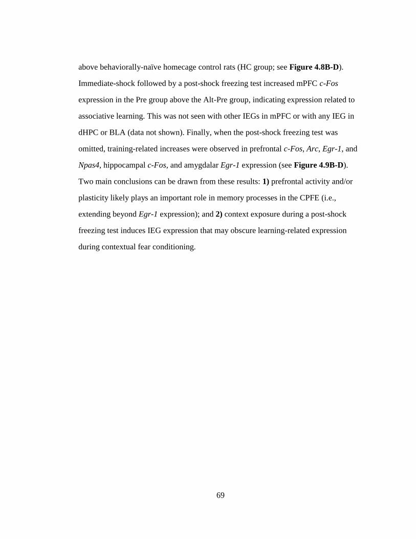
69
above behaviorally-naïve homecage control rats (HC group; see Figure 4.8B-D).
Immediate-shock followed by a post-shock freezing test increased mPFC c-Fos
expression in the Pre group above the Alt-Pre group, indicating expression related to
associative learning. This was not seen with other IEGs in mPFC or with any IEG in
dHPC or BLA (data not shown). Finally, when the post-shock freezing test was
omitted, training-related increases were observed in prefrontal c-Fos, Arc, Egr-1, and
Npas4, hippocampal c-Fos, and amygdalar Egr-1 expression (see Figure 4.9B-D).
Two main conclusions can be drawn from these results: 1) prefrontal activity and/or
plasticity likely plays an important role in memory processes in the CPFE (i.e.,
extending beyond Egr-1 expression); and 2) context exposure during a post-shock
freezing test induces IEG expression that may obscure learning-related expression
during contextual fear conditioning.
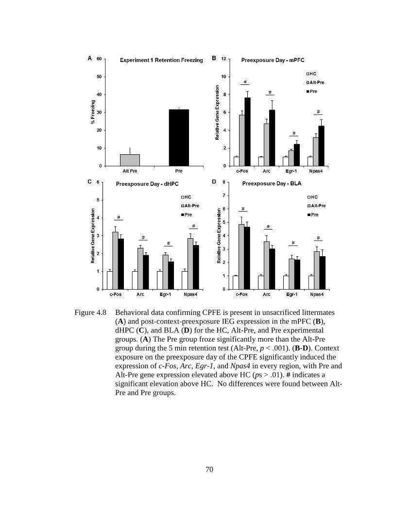
70
Figure 4.8 Behavioral data confirming CPFE is present in unsacrificed littermates
(A) and post-context-preexposure IEG expression in the mPFC (B),
dHPC (C), and BLA (D) for the HC, Alt-Pre, and Pre experimental
groups. (A) The Pre group froze significantly more than the Alt-Pre
group during the 5 min retention test (Alt-Pre, p < .001). (B-D). Context
exposure on the preexposure day of the CPFE significantly induced the
expression of c-Fos, Arc, Egr-1, and Npas4 in every region, with Pre and
Alt-Pre gene expression elevated above HC (ps > .01). # indicates a
significant elevation above HC. No differences were found between Alt-
Pre and Pre groups.
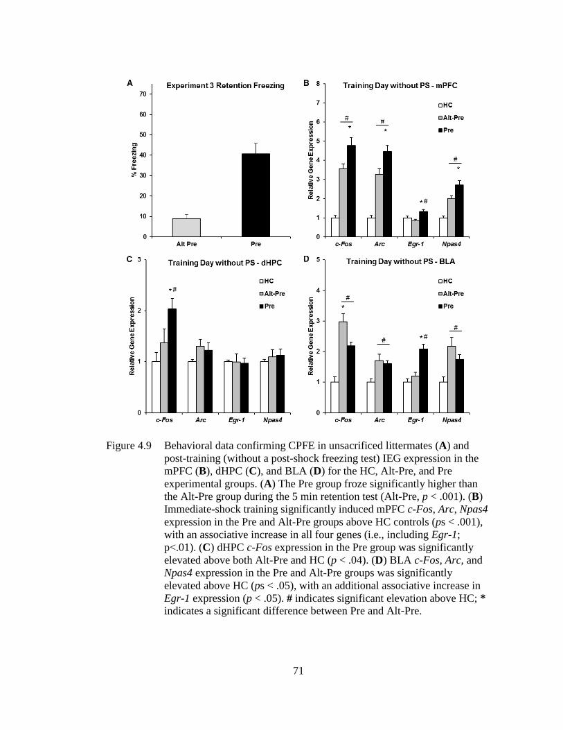
71
Figure 4.9 Behavioral data confirming CPFE in unsacrificed littermates (A) and
post-training (without a post-shock freezing test) IEG expression in the
mPFC (B), dHPC (C), and BLA (D) for the HC, Alt-Pre, and Pre
experimental groups. (A) The Pre group froze significantly higher than
the Alt-Pre group during the 5 min retention test (Alt-Pre, p < .001). (B)
Immediate-shock training significantly induced mPFC c-Fos, Arc, Npas4
expression in the Pre and Alt-Pre groups above HC controls (ps < .001),
with an associative increase in all four genes (i.e., including Egr-1;
p<.01). (C) dHPC c-Fos expression in the Pre group was significantly
elevated above both Alt-Pre and HC (p < .04). (D) BLA c-Fos, Arc, and
Npas4 expression in the Pre and Alt-Pre groups was significantly
elevated above HC (ps < .05), with an additional associative increase in
Egr-1 expression (p < .05). # indicates significant elevation above HC; *
indicates a significant difference between Pre and Alt-Pre.
72
4.5 Conclusions
The experiments reviewed above establish a foundation and framework with
which to examine prefrontal and hippocampal involvement in context memory
(Chapter 6), and the neurobehavioral mechanisms of impaired cognition in our rat
model of FASD (Chapters 7 and 8). These studies established: 1) the role of
NMDAR plasticity and use of post-shock freezing as a measure of fear acquisition in
pharmacological and molecular studies of the CPFE, 2) the prefrontal dependence of
memory processes in the CPFE but not sCFC, 3) the involvement of prefrontal and
hippocampal cholinergic signaling in supporting the CPFE, and 4) the regional
patterns of expression of multiple different IEGs across phases of the CPFE. Taken
together, these studies demonstrated prefrontal, glutamatergic, and cholinergic
dependence of the CPFE, which will provide novel insight into CPFE impairments
seen after neonatal alcohol exposure in our rat model of FASD.
73
GENERAL METHODS
The present chapter describes the general methods used throughout the
experiments in this dissertation. Experimental procedures that are unique to or altered
in particular experiments are noted in the respective method section for that
experiment. The descriptions below are based on previously published descriptions
(Dokovna et al., 2013; Heroux, Horgan, Rosen, et al., 2019; Heroux et al., 2018, 2016,
2017; Heroux, Robinson-Drummer, et al., 2019; Murawski & Stanton, 2011).
5.1 Material and Methods
5.1.1 Animal Subjects
Rats were derived from litters bred by the Office of Laboratory Animal
Medicine at the University of Delaware. Time-mated females were housed with
breeder males overnight and, if an ejaculatory plug was found the following morning
that day was designated as gestational day (GD) 0. Dams were housed in clear
polypropylene cages (45 cm × 24 cm × 21 cm) with standard bedding and access to ad
libitum water and rat chow. Rats were maintained on a 12:12 h light/dark cycle with
lights on at 7:00 am. Date of birth was designated as postnatal day (PD) 0. Litters were
culled on PD3 to eight pups (4 males and 4 females when possible) and were paw-
marked with subcutaneous injections of non-toxic black ink for later identification.
Pups were weaned from their mother on PD21 and housed with same-sex littermates
in clear cages (45 cm × 24 cm × 17 cm). On PD29, rats were individually housed in
Chapter 5
74
clear cages (30 cm × 18 cm × 17 cm) with ad libitum access to water and rat chow for
the remainder of the experiment. All procedures were approved by the Institutional
Animal Care and Use Committee at the University of Delaware following guidelines
established by the National Institute of Health.
5.1.2 Stereotaxic Surgery, Drug Infusion, and Histology
This section describes stereotaxic surgery, intra-cranial drug infusion,
histological procedures used in both experiments in Chapter 6.
5.1.2.1 Stereotaxic Surgery
Surgical implantation of intracranial cannula in the mPFC and vHPC for CPFE
behavioral experiments has been previously done in adolescent rats (Heroux et al.,
2017; Robinson-Drummer, Heroux, & Stanton, 2017) and adult rats (Rudy & Matus-
Amat, 2005), respectively. On PD29, rats were anesthetized with an i.p. injection of 1
mg/kg of an 85:15 ketamine/xylazine drug mixture prior to surgery. In Experiment
6.1, guide cannulas (Plastics One, Roanoke, VA) were bilaterally implanted to
terminate in the mPFC using the following coordinates: anteroposterior (AP) +9.0
mm, mediolateral (ML), ±0.6 mm relative to interaural midline, and dorsoventral
(DV), -2.3 mm relative to the top of the skull. In Experiment 6.2, cannulas were
bilaterally implanted in the vHPC using the following coordinates: AP +1.9 mm, ML
±4.7 mm relative to interaural midline, and DV -5.3 mm relative to the top of the
skull. Twenty-four hours following surgery (PD30), rats were infused with 0.25-0.50
μl of the vehicle PBS in both hemispheres to reduce occlusion in the guide cannula
and to acclimate the rats to being handled during infusions before the start of
behavioral procedures the following day (PD31).
75
5.1.2.2 Intra-mPFC and Intra-vHPC Drug Infusions
In Experiments 6.1 and 6.2, microinjections of the vehicle PBS or the GABAA
receptor agonist muscimol (Sigma-Aldrich, St. Louis, MO) were administered
approximately 15 minutes prior to context preexposure on PD31. In Experiment 6.1,
mPFC-cannulated rats were infused with .25 μl/side of PBS or muscimol (0.5 μg) over
1 min, and injector tips were left in the guide cannula for one minute following
infusion to allow sufficient diffusion of the drug. Previous studies from our lab have
used this dose to inactivate the mPFC during the CPFE, with additional analyses
confirming drug spread using fluorescent muscimol (Heroux et al., 2017; Section 4.2
above). In Experiment 6.2, vHPC-cannulated rats were infused with .5 μl/side of PBS
or muscimol (0.5 μg) over 1 min, with injectors left in for 1 min after infusion (Rudy
& Matus-Amat, 2005). Rats were returned to their home-cage for approximately 15
minutes until the start of behavioral testing.
5.1.2.3 Histology
Rats in the IEG group were rapidly decapitated without anesthesia 30 min after
context preexposure. In Experiments 6.1 and 6.2, half of the brain was used to verify
cannula placements, and coronal brain slabs were cut out the other half for IEG
analyses. The methods for tissue dissection and IEG analysis can be seen in Section
5.1.5. In the rats that underwent the full CPFE procedure, rats were sacrificed by rapid
decapitation after testing, with brains removed and frozen in -45°C isopentane and
then stored at -80°C before being sectioned on a microtome. Sagittal and coronal
sections (50-60 μm) were taken from the gene expression and behavior sampling
groups, respectively. Slides were photo-captured and analyzed to confirm the
placement of the cannula injector tip in the mPFC.
76
5.1.3 Animal Model of Fetal Alcohol Spectrum Disorder
This section describes neonatal alcohol dosing procedures and blood alcohol
concentration analyses that were used across experiments in Chapters 7 and 8.
5.1.3.1 Neonatal Alcohol Dosing (PD4-9, 5.25g/kg/d)
Neonatal ethanol dosing via intragastric intubation occurred over PD4-9.
Littermates were randomly assigned to receive either ethanol (EtOH group) or sham
intubations (SI group), with an equal number of males and females in each litter
whenever possible. Same-sex littermates assigned to the same dosing condition (EtOH
or SI) were assigned to different experimental groups so that no more than one same-
sex littermate was assigned to any particular condition. Briefly, on PD4, pups were
separated from their mothers and placed into weigh boats set over a heating pad that
provided warmth during the separation. Pups were weighed prior to the first intubation
session (occurring daily at 9am ± 1hr). The intubation process involved passing PE10
tubing lubricated with corn oil down the esophagus and into the stomach of the rat
pup. Rats in the SI group received intragastric intubations on the same schedule as the
EtOH group, and the tube was removed after approximately 6-8 seconds during each
scheduled intubation without the infusion of any solution. Rats in the EtOH group
were intubated and given a daily dose of 5.25 g/kg of alcohol, [11.9% v/v ethanol
(made from 95% ethanol)] in a custom milk formula previously described (Kelly &
Lawrence, 2008). This dose was divided into two feedings each day, separated by 2hr.
The formula was delivered in a volume of 0.02778 ml ⁄g body weight. A third
intubation of the milk formula (containing no ethanol) was administered two hours
after the second daily alcohol dosing. After each intubation was completed (<20
minutes per litter), pups were returned as a litter to their mothers. Importantly,
77
although intragastric intubation of alcohol using these parameters results in a transient
reduction in body weight and growth (Jablonski & Stanton, 2014; Kelly & Lawrence,
2008; Murawski & Stanton, 2011), this reduction is absent by adolescence and thus
likely does not contribute to behavioral deficits resulting from exposure. Importantly,
this neonatal exposure produces high BACs (~400 mg/dl) modeling severe binge
drinking in the rat model; while manipulation is not possible, BACs of over 200 mg/dl
are commonly associated with later development of FAS in the human condition (see
Patten et al., 2014 for full discussion).
5.1.3.2 Blood Alcohol Concentrations (BACs)
On PD4, 90 min following the second alcohol intubation, pups received a
small tail-clip and a 20μl blood sample was collected using a capillary tube. Blood
samples from Group SI were discarded and those from alcohol-exposed pups were
saved for further blood alcohol analysis. Blood samples from alcohol-exposed pups
were centrifuged, and the plasma was collected and stored at -20°C. Blood alcohol
concentrations were determined using an Analox GL5 Analyzer (Analox Instruments,
Luneburg, MA) by measuring the rate of oxidation of alcohol in each plasma sample.
BACs (expressed in mg⁄dl) were calculated based on comparisons to known values of
an alcohol standard solution.
5.1.4 Behavioral Equipment and Procedures
This section describes the apparatus, stimuli, and behavioral procedures for the
CPFE used across experiments in Chapters 6, 7, and 8 and sCFC procedures used in
Chapter 7.
78
5.1.4.1 Apparatus, Stimuli, and Freezing Software Analyses
The apparatus and stimuli used have been previously described (Heroux et al.,
2016, 2017; Murawski & Stanton, 2010; Robinson-Drummer et al., 2016). Fear
conditioning occurred in four clear Plexiglas chambers measuring 16.5 cm × 12.1 cm
× 21.6 cm, which were arranged in a 2 × 2 formation on a Plexiglas stand within a
fume hood to provide ambient light and background noise (Context A). Each chamber
had a grid floor made of nine stainless steel bars (11.5 cm from the top of the
chamber), 0.5 cm in diameter and spaced 1.25 cm apart. The alternate context
(Context B) consisted of the same Plexiglas chambers with a convex wire mesh insert
that covered the back wall and floor of the chamber and a white paper sleeve that
covered the outside walls of the chamber. Except where noted (see Chapter 7), the
unconditioned stimuli (US) were two, 1.5 mA foot-shocks, each 2s in duration, and
presented 1s apart. These were delivered using a shock scrambler (Med Associates,
Georgia, VT-ENV-414S) connected to the grid floor of the chamber. The fear
chambers were cleaned with 5% ammonium hydroxide solution prior to each load of
experimental rats.
Videos of each session (preexposure, training, testing) were recorded using
Freeze Frame 3.0 software (Actimetrics, Wilmette IL) with freezing defined as a bout
of 0.75 s or longer without a change in video pixilation. A human observer blind to the
experimental groups verified the freezing threshold setting with Freeze View 3.0
(Actimetrics, Wilmette IL). The software program computes a “motion index” that
was adjusted to set a freezing threshold separately for each animal (per software
instructions) by a blind observer who verified from the video record whether small
movements were scored as freezing. Once set, the threshold did not change during a
session. We have validated this procedure against other scoring methods (e.g., hand
79
scoring of video records by two blind observers). Freezing behavior was scored as the
total percent time spent freezing longer than .75s bins (defined as the cessation of all
movement except breathing) in each respective session bin (context exposure, post-
shock freezing, and a 24 h retention test). The data were imported into STATISTICA
64 data analysis software and freezing behavior was analyzed.
5.1.4.2 Context Preexposure Facilitation Effect (CPFE)
The CPFE procedure consisted of three phases (context preexposure,
immediate-shock training, retention testing) and took place over the course of three
days from PD31 to PD33. Rats were assigned to either preexposure condition (Pre
group), alternate preexposure condition (Alt-Pre group), or when applicable, a
behaviorally naïve home-cage condition (HC group) that serves to establish baseline
gene expression for experiments that include IEG analyses. Rats in the Pre group
received exposure to Context A, the training context, while rats in the Alt-Pre group
received exposure to Context B (see section 5.1.4.1). Alt-Pre rats serve as non-
associative behavioral controls as they demonstrate the immediate-shock deficit (ISD),
which reflects an inability to form a context-shock association without prior exposure
to Context A (Fanselow, 1990).
On PD31, rats were placed in Context A or B and underwent multiple context
preexposure, consisting of one initial 5 min exposure to the chamber, followed by five
1 min exposures, with a 1 min interval between exposures. In experiments that include
IEG analyses, a subset of rats in the Pre and HC groups were sacrificed via live
decapitation and tissue was collected for RNA extraction and qPCR 30 min after
context preexposure. Alt-Pre rats are not included in IEG designs as there is no
difference between Pre and Alt-Pre gene expression on the preexposure day of the
80
CPFE (Heroux et al., 2018, Figure 4.9, above). On PD32, rats in the behavior group
(i.e., not sacrificed after context preexposure) were carried into the testing room,
placed in their respective Context A training chamber, and within 3s, received two
1.5mA foot-shocks separated by 1s. In most cases, foot-shocks were followed by a 3-
min post-shock freezing test to assess fear acquisition. Otherwise, this test was
omitted. In either case, at the end of training, rats were returned immediately to their
transport cages and within 1-2 minutes returned to their home cages in the colony
room. On PD33, rats were returned to the same Context A chamber in which they
were trained for a 5 min retention freezing test.
5.1.4.3 Standard Contextual Fear Conditioning (sCFC)
The sCFC procedure was used in Experiment 7.3 in Chapter 7. The sCFC
procedure took place over the course of two days from PD31 to PD32. All chambers,
stimuli, and drug infusion protocols used were identical to the ones used in Context A
for the CPFE experiments (see Section 5.1.4.2). On PD31, animals were assigned to
either the Delayed-shock or Imm-Shock control condition. Animals in the Delayed-
shock condition received three minutes of context exposure in Context A, followed by
two 1.5 mA 2s foot-shocks separated by 1s. Animals in the Imm-Shock conditions
were given two foot-shocks without any context exposure. This group served as
behavioral controls for the delayed-shock conditions as the placement-to-shock
interval was under 5 sec resulting in the immediate-shock deficit (Fanselow, 1990).
Rats were removed immediately after conditioning without any post-shock freezing
test. On PD32, rats in both retention conditions were tested in Context A for 5 min in
the same chamber in which training occurred.
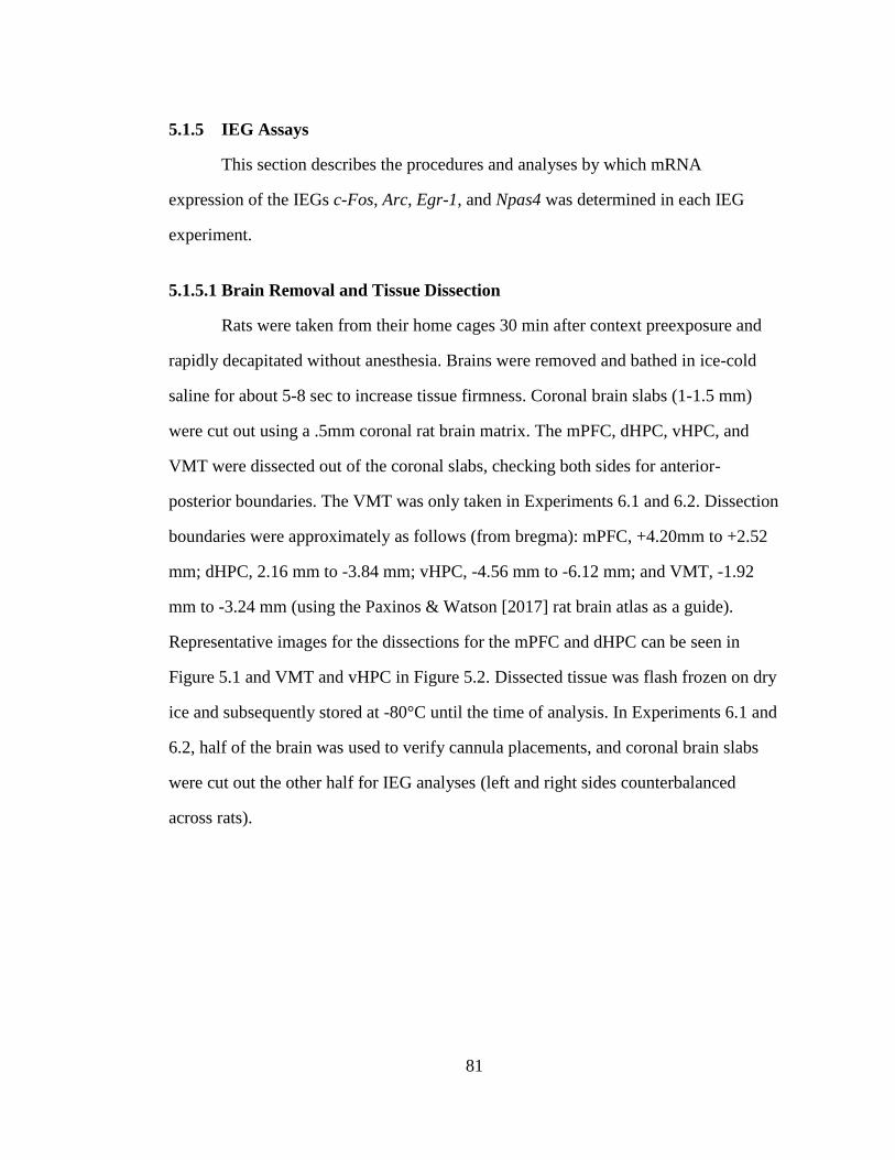
81
5.1.5 IEG Assays
This section describes the procedures and analyses by which mRNA
expression of the IEGs c-Fos, Arc, Egr-1, and Npas4 was determined in each IEG
experiment.
5.1.5.1 Brain Removal and Tissue Dissection
Rats were taken from their home cages 30 min after context preexposure and
rapidly decapitated without anesthesia. Brains were removed and bathed in ice-cold
saline for about 5-8 sec to increase tissue firmness. Coronal brain slabs (1-1.5 mm)
were cut out using a .5mm coronal rat brain matrix. The mPFC, dHPC, vHPC, and
VMT were dissected out of the coronal slabs, checking both sides for anterior-
posterior boundaries. The VMT was only taken in Experiments 6.1 and 6.2. Dissection
boundaries were approximately as follows (from bregma): mPFC, +4.20mm to +2.52
mm; dHPC, 2.16 mm to -3.84 mm; vHPC, -4.56 mm to -6.12 mm; and VMT, -1.92
mm to -3.24 mm (using the Paxinos & Watson [2017] rat brain atlas as a guide).
Representative images for the dissections for the mPFC and dHPC can be seen in
Figure 5.1 and VMT and vHPC in Figure 5.2. Dissected tissue was flash frozen on dry
ice and subsequently stored at -80°C until the time of analysis. In Experiments 6.1 and
6.2, half of the brain was used to verify cannula placements, and coronal brain slabs
were cut out the other half for IEG analyses (left and right sides counterbalanced
across rats).
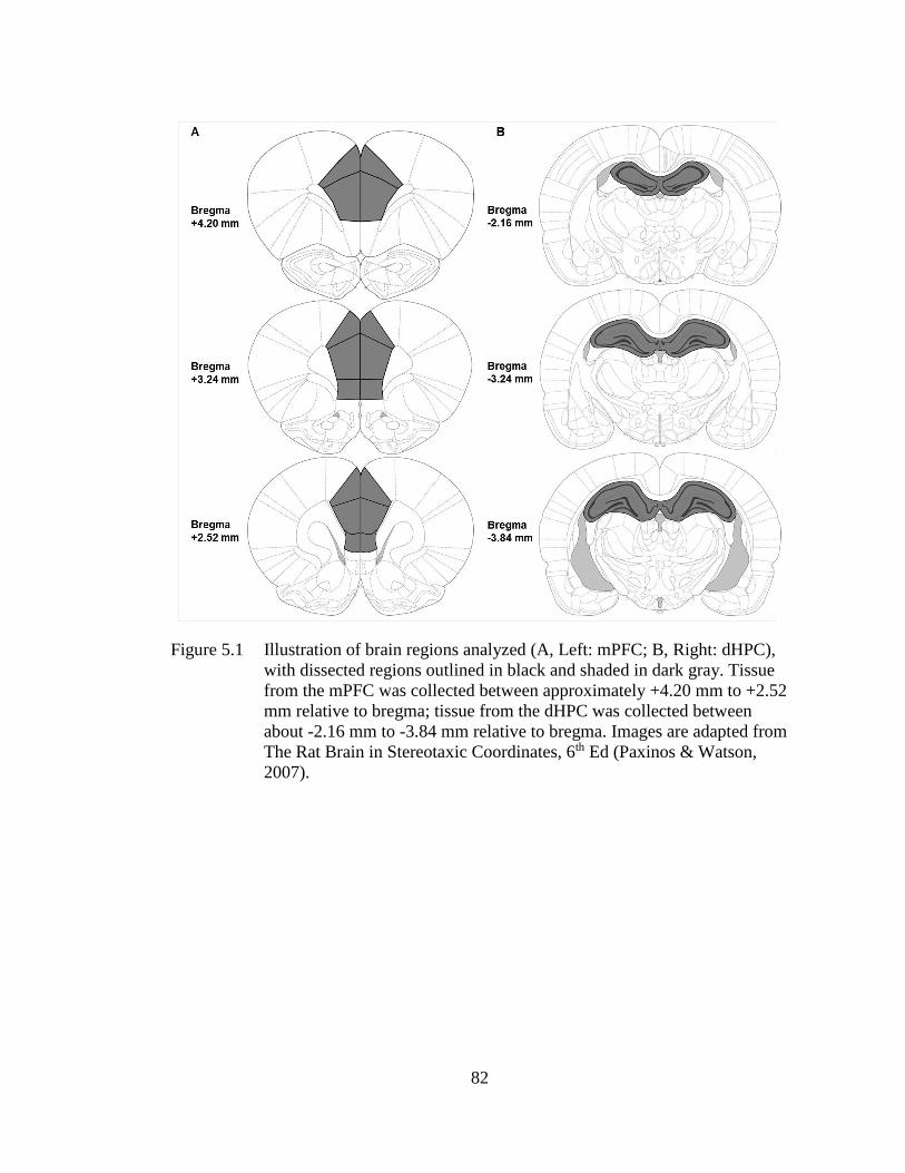
82
Figure 5.1 Illustration of brain regions analyzed (A, Left: mPFC; B, Right: dHPC),
with dissected regions outlined in black and shaded in dark gray. Tissue
from the mPFC was collected between approximately +4.20 mm to +2.52
mm relative to bregma; tissue from the dHPC was collected between
about -2.16 mm to -3.84 mm relative to bregma. Images are adapted from
The Rat Brain in Stereotaxic Coordinates, 6th Ed (Paxinos & Watson,
2007).
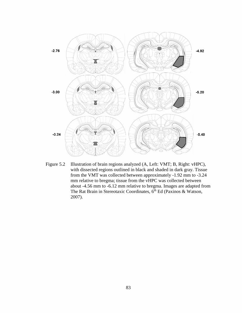
83
Figure 5.2 Illustration of brain regions analyzed (A, Left: VMT; B, Right: vHPC),
with dissected regions outlined in black and shaded in dark gray. Tissue
from the VMT was collected between approximately -1.92 mm to -3.24
mm relative to bregma; tissue from the vHPC was collected between
about -4.56 mm to -6.12 mm relative to bregma. Images are adapted from
The Rat Brain in Stereotaxic Coordinates, 6th Ed (Paxinos & Watson,
2007).
84
5.1.5.2 Quantitative Real-time PCR (qPCR)
RNA was extracted from frozen tissue samples using TRIzol Reagent (Cat. No.
15596018, Invitrogen). Genomic DNA was eliminated and cDNA was synthesized
from extracted RNA (1000ng/L) using the QuantiTect® Reverse Transcription Kit
(Cat. No. 205314, Qiagen). Relative gene expression was quantified by real-time PCR
using the GREEN FASTMIX PERFECTA-SYBR Kit (Cat. No. 101414-270,
Quantabio) in 10µL reactions on a CFX96Touch real time PCR machine. Expression
of Egr-1 was analyzed using a QuantiTect® Primer Assay (Cat. No. QT00182896,
Qiagen) and diluted according to protocol. All other primers were ordered through
Integrated DNA Technologies and diluted to a final concentration of 0.13 µM (18s,
Arc, c-Fos, and Npas-4). The gene 18s was used as a control for all experimental
groups because it is a ribosomal housekeeping gene unchanged across any groups or
manipulations. Samples were numbered, blinded to treatment group and run in
duplicate on real-time PCR plates. For each reaction, the average quantitative
threshold amplification cycle number (Cq) value was determined from each duplicate,
and the 2-ΔΔCq method was used to calculate the relative gene expression for each gene
relative to 18s. The relative gene expression value was obtained by normalizing the
data to the reference gene (18s) and to the average delta CT of the home-cage control
group for each gene.
5.1.6 Statistical Analyses
STATISTICA 13 (Statsoft, Tulsa, Oklahoma) was used for all statistical
analyses across all experiments. Analysis of behavioral and IEG data has been
previously described (Heroux et al., 2018; Heroux, Robinson-Drummer, et al., 2019).
In both IEG and behavioral data, rats were excluded from analysis as an outlier if they
85
had a score of ± 1.96 standard deviations from the group mean, but average z-scores of
removed outliers historically range much higher as reported for each experiment
(below). Specific experimental designs, analyses, and outlier removal can be found in
the method section of each experiment. When applicable, post-hoc contrasts were
performed with Newman-Keuls or Dunnett’s test to assess any significant main effect
or interactions revealed by the ANOVAs in each design.
86
MEDIAL PREFRONTAL AND VENTRAL HIPPOCAMPAL
CONTRIBUTIONS TO INCIDENTAL CONTEXT LEARNING AND
MEMORY IN ADOLESCENT RATS
6.1 Introduction
This chapter was recently published in Neurobiology of Learning and Memory
(Heroux, Horgan, Pinizzotto, et al., 2019), and the text appears here in adapted form.
As noted in Chapters 2 and 4, our lab has recently discovered a novel role of the
mPFC in the acquisition and/or consolidation of a conjunctive context representation
in the CPFE. We hypothesized that the PFC interacts with the HPC during context
learning to support these processes (see Section 2.1.3 in Chapter 2). Therefore, the
purpose of the current study was to further characterize regional neurobiological
mechanisms underlying incidental context learning during the preexposure phase of
the CPFE. We examined the effects of inactivating the mPFC (Experiment 6.1) or
vHPC (Experiment 6.2) during context preexposure on expression of the IEGs c-Fos,
Arc, Egr-1, and Npas4 in the mPFC, dHPC, vHPC, and VMT. Across both
experiments, we hypothesized that inactivation of either structure would interfere with
the acquisition and/or consolidation the context representation, resulting in abolished
post-shock and retention test freezing in the CPFE. Given that neither of these
structures have an exclusive role in context memory, we hypothesized that inactivation
of either structure would interfere with activity and thus gene expression in the other
region and additional connected regions engaged by context exposure (e.g., the dHPC
Chapter 6
87
and VMT). By using the CPFE to isolate distinct processes of CFC, these experiments
provide novel evidence for the involvement of the mPFC and vHPC in incidental
context learning and memory.
6.2 Materials and Methods
6.2.1 Subjects
Animal husbandry was as described as in Chapter 5. Across both experiments,
there was a total of 155 adolescent (PD31) Long Evans rats (78 females and 77
males), derived from 39 separate litters bred by the Office of Laboratory Animal
Medicine at the University of Delaware.
6.2.2 Stereotaxic Surgery
The methods for stereotaxic surgery and drug infusion are as described in
Chapter 5. Briefly, adolescent rats underwent bilateral surgical implantation of
intracranial cannula in the mPFC (Experiment 6.1) or vHPC (Experiment 6.2). Rats in
both the Pre and Alt-Pre groups were infused with PBS or MUSC 30 min prior to
context preexposure in the CPFE.
6.2.3 Apparatus and Stimuli
The apparatus and stimuli used for the current experiments was as described in
Chapter 5. The US was two 1.5mA foot-shocks separated by 1sec.
6.2.4 Behavioral Procedures and Drug Infusion
The CPFE procedures used in the current experiments was as described in
Chapter 5. Briefly, rats were assigned to either the Pre or Alt-Pre group, and then
88
given PBS or MUSC 30 min prior to context preexposure in the CPFE. Rats in the
behaviorally-naïve home-cage (HC) control group also received this infusion.
6.2.5 Brain Removal, Tissue Dissection, and qPCR
In Experiment 6.2, preexposed or behaviorally-naïve home-cage control rats
rats given saline or muscimol were sacrificed 30 min after context exposure in the
CPFE. Expression of the immediate early genes c-Fos, Arc, Egr-1, and Npas4 in the
mPFC, dHPC, and vHPC was analyzed via qPCR. The methods for rat sacrifice, brain
removal, tissue dissection and qPCR are described in Chapter 5.
6.2.6 Histology
Cannula placements were verified in both the behavior and sacrificed groups as
described in Chapter 5.
6.2.7 Data Analysis
6.2.7.1 Analysis of Behavioral Data
The data were imported into STATISTICA 64 data analysis software and
freezing behavior was analyzed as described in Chapter 5. Across both experiments,
there were no main effects or interactions involving sex on freezing behavior (ps >
.30), so the data were collapsed across this variable. Consistent with previous reports
(Heroux et al., 2017; Robinson-Drummer et al., 2017), in both experiments, the Alt-
Pre group was pooled across drug condition (“Pooled-Alt-Pre” group) as there was no
significant difference between the two drug groups and they froze at uniformly low
levels (p > .25). Post-shock and retention test freezing data were analyzed using 3
(Condition; Pooled-Alt-Pre, PBS, Muscimol) × 2 (within subjects; Phase of testing;
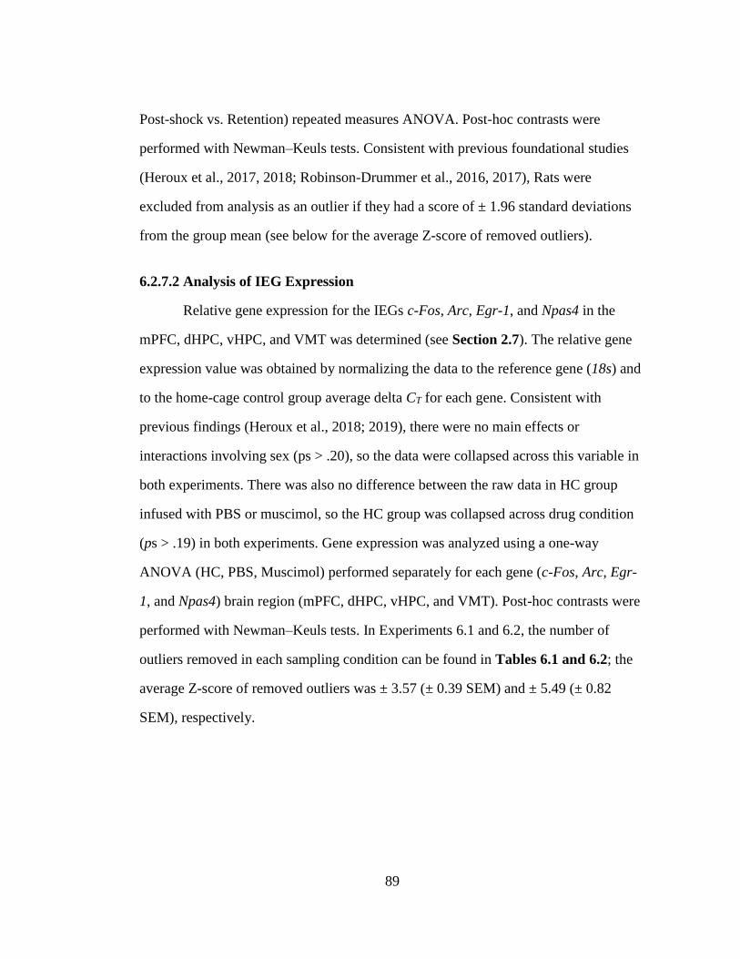
89
Post-shock vs. Retention) repeated measures ANOVA. Post-hoc contrasts were
performed with Newman–Keuls tests. Consistent with previous foundational studies
(Heroux et al., 2017, 2018; Robinson-Drummer et al., 2016, 2017), Rats were
excluded from analysis as an outlier if they had a score of ± 1.96 standard deviations
from the group mean (see below for the average Z-score of removed outliers).
6.2.7.2 Analysis of IEG Expression
Relative gene expression for the IEGs c-Fos, Arc, Egr-1, and Npas4 in the
mPFC, dHPC, vHPC, and VMT was determined (see Section 2.7). The relative gene
expression value was obtained by normalizing the data to the reference gene (18s) and
to the home-cage control group average delta CT for each gene. Consistent with
previous findings (Heroux et al., 2018; 2019), there were no main effects or
interactions involving sex (ps > .20), so the data were collapsed across this variable in
both experiments. There was also no difference between the raw data in HC group
infused with PBS or muscimol, so the HC group was collapsed across drug condition
(ps > .19) in both experiments. Gene expression was analyzed using a one-way
ANOVA (HC, PBS, Muscimol) performed separately for each gene (c-Fos, Arc, Egr-
1, and Npas4) brain region (mPFC, dHPC, vHPC, and VMT). Post-hoc contrasts were
performed with Newman–Keuls tests. In Experiments 6.1 and 6.2, the number of
outliers removed in each sampling condition can be found in Tables 6.1 and 6.2; the
average Z-score of removed outliers was ± 3.57 (± 0.39 SEM) and ± 5.49 (± 0.82
SEM), respectively.
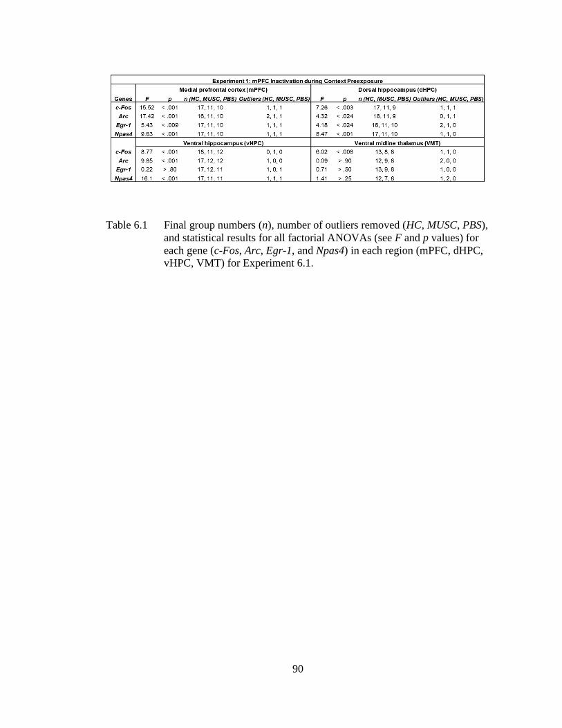
90
Table 6.1 Final group numbers (n), number of outliers removed (HC, MUSC, PBS),
and statistical results for all factorial ANOVAs (see F and p values) for
each gene (c-Fos, Arc, Egr-1, and Npas4) in each region (mPFC, dHPC,
vHPC, VMT) for Experiment 6.1.
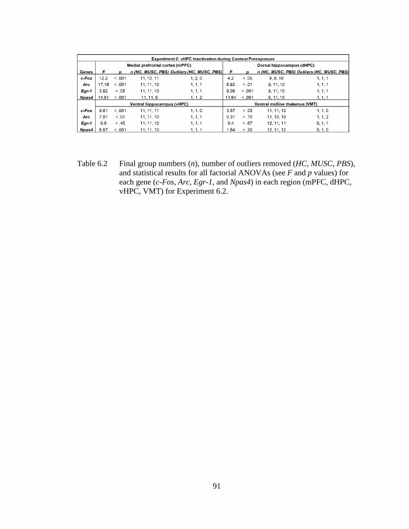
91
Table 6.2 Final group numbers (n), number of outliers removed (HC, MUSC, PBS),
and statistical results for all factorial ANOVAs (see F and p values) for
each gene (c-Fos, Arc, Egr-1, and Npas4) in each region (mPFC, dHPC,
vHPC, VMT) for Experiment 6.2.
92
6.3 Results
6.3.1 Experiment 6.1: Prefrontal Inactivation during Context Exposure Impairs
Contextual Fear Conditioning and IEG Expression in the mPFC, vHPC,
and VMT
The purpose of Experiment 6.1 was to examine the effects of prefrontal
inactivation during context learning on regional gene expression and contextual fear
conditioning. Location of cannula tips is shown in Figure 6.1 (left column). The
experimental design and behavioral results can be seen in Figure 6.2A-C. We
predicted that intra-mPFC muscimol infusion would abolish freezing and impair IEG
expression in the mPFC, dHPC, and VMT. Behavioral analyses were run on 32
animals distributed across the following groups: Pooled-Alt-Pre (n=11), PBS-Pre
(n=11), Muscimol-Pre (n=10). Repeated measures ANOVA revealed a significant
main effect of Condition [F(2, 26) = 44.53, p < .001]. There was no main effect or any
interactions involving Phase (ps > .23). The PBS-Pre group froze significantly more
than any other group during both the post-shock and retention freezing tests (ps <
.001). Additionally, there was no significant difference between Muscimol-Pre and the
Pooled-Alt-Pre control group (ps > .38). These results indicate that prefrontal
inactivation during context learning abolishes contextual fear conditioning during
subsequent post-shock and retention test freezing phases of the CPFE.
Littermates of the behavior group were sacrificed 30 min after context
exposure on the preexposure day of the CPFE. The IEG results can be seen in Figure
6.3A-D. Gene expression was analyzed using separate one-way ANOVAs (HC, PBS,
and Muscimol) for each gene (c-Fos, Arc, Egr-1, and Npas4) and region (mPFC,
dHPC, vHPC, and VMT). Specific F statistics, p values, group n, and outliers
removed for the sixteen one-way ANOVAs can be found in Table 6.1. Post hoc
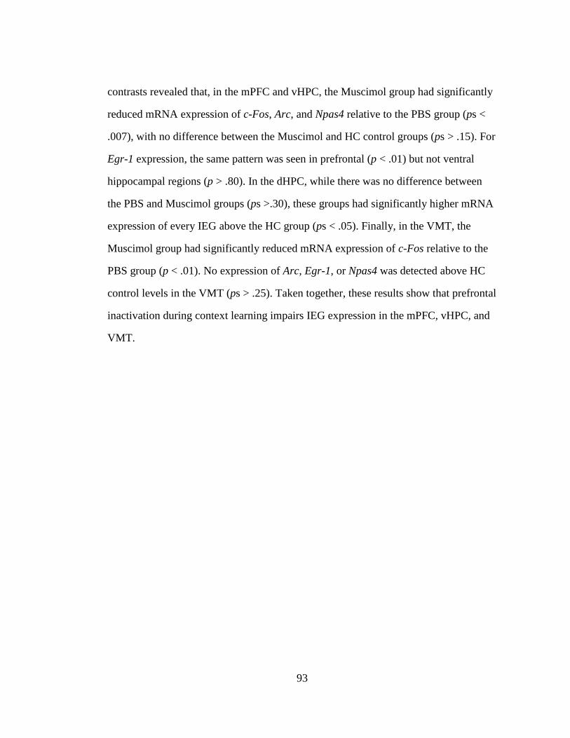
93
contrasts revealed that, in the mPFC and vHPC, the Muscimol group had significantly
reduced mRNA expression of c-Fos, Arc, and Npas4 relative to the PBS group (ps <
.007), with no difference between the Muscimol and HC control groups (ps > .15). For
Egr-1 expression, the same pattern was seen in prefrontal (p < .01) but not ventral
hippocampal regions (p > .80). In the dHPC, while there was no difference between
the PBS and Muscimol groups (ps >.30), these groups had significantly higher mRNA
expression of every IEG above the HC group (ps < .05). Finally, in the VMT, the
Muscimol group had significantly reduced mRNA expression of c-Fos relative to the
PBS group (p < .01). No expression of Arc, Egr-1, or Npas4 was detected above HC
control levels in the VMT (ps > .25). Taken together, these results show that prefrontal
inactivation during context learning impairs IEG expression in the mPFC, vHPC, and
VMT.
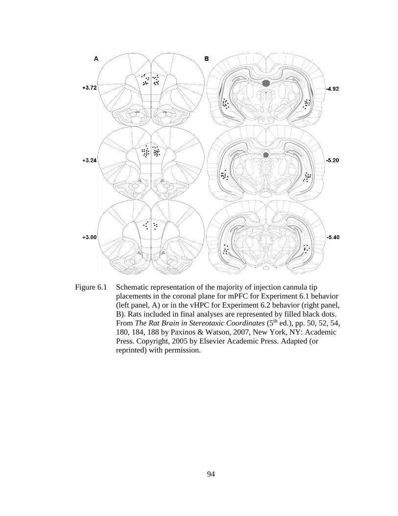
94
Figure 6.1 Schematic representation of the majority of injection cannula tip
placements in the coronal plane for mPFC for Experiment 6.1 behavior
(left panel, A) or in the vHPC for Experiment 6.2 behavior (right panel,
B). Rats included in final analyses are represented by filled black dots.
From The Rat Brain in Stereotaxic Coordinates (5th ed.), pp. 50, 52, 54,
180, 184, 188 by Paxinos & Watson, 2007, New York, NY: Academic
Press. Copyright, 2005 by Elsevier Academic Press. Adapted (or
reprinted) with permission.
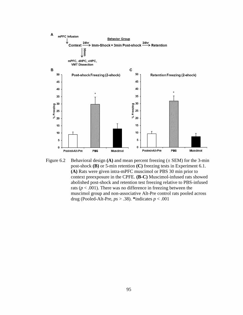
95
Figure 6.2 Behavioral design (A) and mean percent freezing (± SEM) for the 3-min
post-shock (B) or 5-min retention (C) freezing tests in Experiment 6.1.
(A) Rats were given intra-mPFC muscimol or PBS 30 min prior to
context preexposure in the CPFE. (B-C) Muscimol-infused rats showed
abolished post-shock and retention test freezing relative to PBS-infused
rats (p < .001). There was no difference in freezing between the
muscimol group and non-associative Alt-Pre control rats pooled across
drug (Pooled-Alt-Pre, ps > .38). *indicates p < .001
96
Figure 6.3 Relative mRNA expression (± SEM) of c-Fos, Arc, Egr-
1, and Npas4 in the mPFC (A), dHPC (B), vHPC (C), and VMT (D) in
rats infused with PBS, Muscimol, and behaviorally-naïve home-cage
control rats (pooled across drug), sacrificed 30 min after context
preexposure. (A) Muscimol infusion impaired expression of mPFC c-
Fos, Arc, Egr-1, and Npas4 relative to PBS-infused rats. (B) There was
no difference between PBS and Muscimol groups for dHPC IEG
expression, and both of these groups had elevated expression of all IEGs
above the HC group (C) Muscimol infusion impaired expression of
vHPC c-Fos, Arc, and Npas4 relative to PBS-infused rats. (D) Muscimol
infusion impaired expression of VMT c-Fos relative to PBS-infused rats,
with no expression of any other IEG above HC. # indicates significant
elevation above the HC group; * indicates a significant difference
between PBS and Muscimol groups.
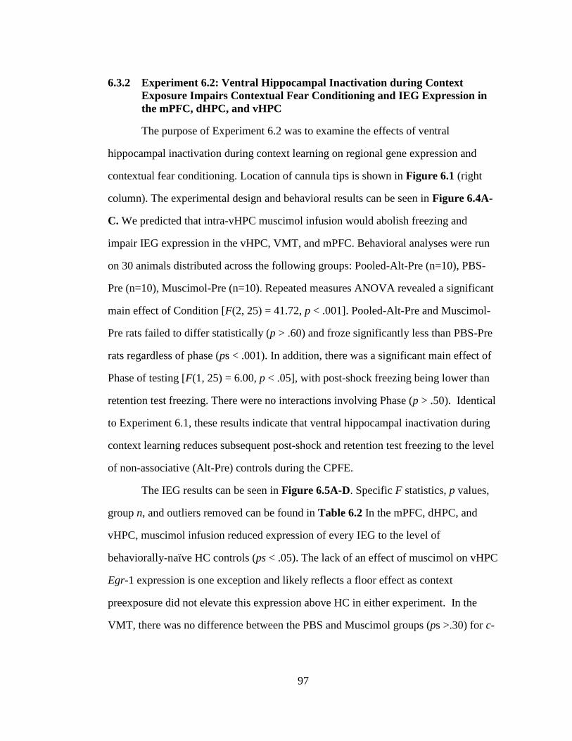
97
6.3.2 Experiment 6.2: Ventral Hippocampal Inactivation during Context
Exposure Impairs Contextual Fear Conditioning and IEG Expression in
the mPFC, dHPC, and vHPC
The purpose of Experiment 6.2 was to examine the effects of ventral
hippocampal inactivation during context learning on regional gene expression and
contextual fear conditioning. Location of cannula tips is shown in Figure 6.1 (right
column). The experimental design and behavioral results can be seen in Figure 6.4A-
C. We predicted that intra-vHPC muscimol infusion would abolish freezing and
impair IEG expression in the vHPC, VMT, and mPFC. Behavioral analyses were run
on 30 animals distributed across the following groups: Pooled-Alt-Pre (n=10), PBS-
Pre (n=10), Muscimol-Pre (n=10). Repeated measures ANOVA revealed a significant
main effect of Condition [F(2, 25) = 41.72, p < .001]. Pooled-Alt-Pre and Muscimol-
Pre rats failed to differ statistically (p > .60) and froze significantly less than PBS-Pre
rats regardless of phase (ps < .001). In addition, there was a significant main effect of
Phase of testing [F(1, 25) = 6.00, p < .05], with post-shock freezing being lower than
retention test freezing. There were no interactions involving Phase (p > .50). Identical
to Experiment 6.1, these results indicate that ventral hippocampal inactivation during
context learning reduces subsequent post-shock and retention test freezing to the level
of non-associative (Alt-Pre) controls during the CPFE.
The IEG results can be seen in Figure 6.5A-D. Specific F statistics, p values,
group n, and outliers removed can be found in Table 6.2 In the mPFC, dHPC, and
vHPC, muscimol infusion reduced expression of every IEG to the level of
behaviorally-naïve HC controls (ps < .05). The lack of an effect of muscimol on vHPC
Egr-1 expression is one exception and likely reflects a floor effect as context
preexposure did not elevate this expression above HC in either experiment. In the
VMT, there was no difference between the PBS and Muscimol groups (ps >.30) for c-
98
Fos expression, and both of these groups showed higher c-Fos expression than the HC
group (ps < .05). No expression of Arc, Egr-1, or Npas4 was detected above HC
control levels in the VMT (ps > .25). Taken together, these results show that ventral
hippocampal inactivation during context learning impairs IEG expression in the
mPFC, dHPC, and vHPC.
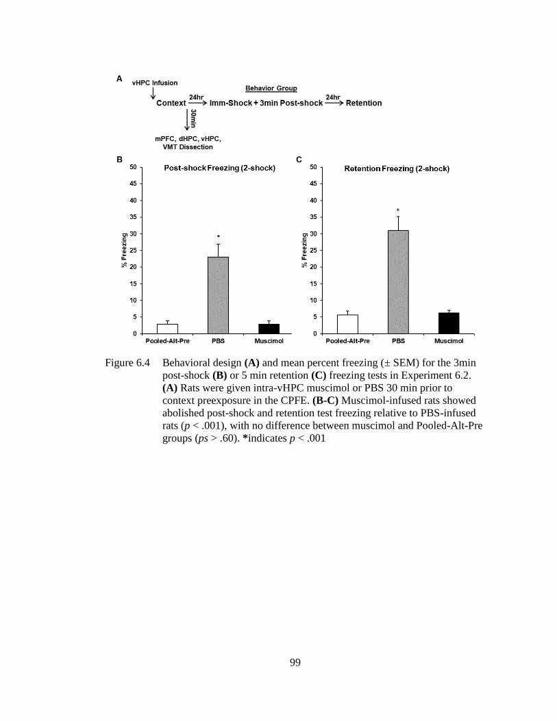
99
Figure 6.4 Behavioral design (A) and mean percent freezing (± SEM) for the 3min
post-shock (B) or 5 min retention (C) freezing tests in Experiment 6.2.
(A) Rats were given intra-vHPC muscimol or PBS 30 min prior to
context preexposure in the CPFE. (B-C) Muscimol-infused rats showed
abolished post-shock and retention test freezing relative to PBS-infused
rats (p < .001), with no difference between muscimol and Pooled-Alt-Pre
groups (ps > .60). *indicates p < .001
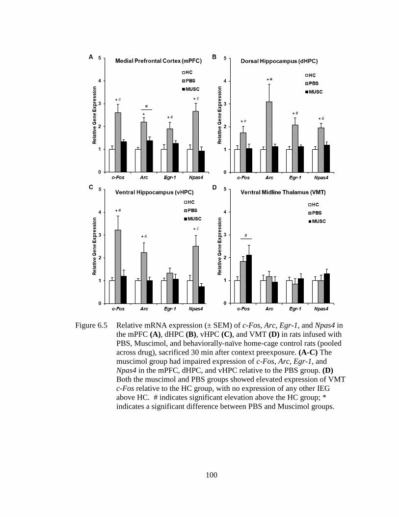
100
Figure 6.5 Relative mRNA expression (± SEM) of c-Fos, Arc, Egr-1, and Npas4 in
the mPFC (A), dHPC (B), vHPC (C), and VMT (D) in rats infused with
PBS, Muscimol, and behaviorally-naïve home-cage control rats (pooled
across drug), sacrificed 30 min after context preexposure. (A-C) The
muscimol group had impaired expression of c-Fos, Arc, Egr-1, and
Npas4 in the mPFC, dHPC, and vHPC relative to the PBS group. (D)
Both the muscimol and PBS groups showed elevated expression of VMT
c-Fos relative to the HC group, with no expression of any other IEG
above HC. # indicates significant elevation above the HC group; *
indicates a significant difference between PBS and Muscimol groups.
101
6.4 Discussion
The current study examined disruption of regional IEG expression caused by
inactivation of mPFC (Experiment 6.1) or vHPC (Experiment 6.2) during incidental
context learning. In both cases, inactivation abolished subsequent post-shock and
retention test freezing in the CPFE. These results extend prior studies (Heroux et al.,
2017; Rudy & Matus-Amat, 2005) by including a post-shock freezing test, which
confirms that MUSC-infused rats either did not acquire or retain a context
representation, or were unable to retrieve and/or associate it with foot-shock during
immediate-shock training in the CPFE. In Experiment 6.1, mPFC inactivation
attenuated expression of mPFC c-Fos, Arc, Egr-1, and Npas4, vHPC c-Fos, Arc, and
Npas4, and VMT c-Fos to the level of behaviorally-naïve home-cage controls. There
was no effect of prefrontal inactivation on dHPC IEG expression, suggesting that
interactions between these two structures likely do not underlie long-term context
memory in the CPFE. In Experiment 6.2, vHPC inactivation attenuated expression of
all four IEGs in the mPFC, dHPC, and vHPC, with no effect on VMT IEG expression.
Collectively, these results suggest that incidental context learning and/or memory
processes are likely supported by extended mPFC-vHPC circuitry.
This suggestion of prefrontal and ventral hippocampal circuit involvement in
these processes is subject to some important limitations. First, we cannot infer a
functional role of regional IEG expression in supporting the CPFE without
experiments involving intra-regional knockdown of specific IEGs or their downstream
pathways (e.g., via ASO infusion or RNAi). Second, we cannot address the role of
individual sub-regions or cell-specific expression as whole regions were dissected
(e.g., mPFC consisted of the AC, PL, and IL). Third, as all tissue was collected at one
time-point, it’s possible that we missed the effects of muscimol infusion on regional
102
IEG expression occurring during later states of consolidation (e.g., intra-mPFC
muscimol may disrupt dHPC IEG expression at later time-points). Importantly, one
caveat that we can address is that the drug and behavioral effects seen in the current
study cannot be attributed to “performance effects” of the muscimol infusion, such as
impaired sensory processing (e.g., feature perception and encoding, shock sensitivity),
motor performance (e.g., hyperactivity), or state dependency. Indeed, previous studies
showed that mPFC or vHPC inactivation prior to sCFC has no effect on acquisition,
and prior CPFE studies using cholinergic antagonists have ruled out state dependency
effects following mPFC infusion (Chapters 2 and 4; Heroux et al., 2017; Robinson-
Drummer et al., 2017; Rudy & Matus-Amat, 2005). In the following discussion, we
consider the implications of our results in relation to previous research examining the
roles of the mPFC, dHPC, vHPC, and VMT in distinct processes of CFC.
The current study further characterizes the involvement of the mPFC in
incidental context learning and related CFC paradigms. Prefrontal inactivation during
context preexposure abolished subsequent post-shock and retention test freezing, as
well as experience-driven IEG expression in the mPFC, vHPC, and VMT (see
Experiment 6.1). We’ve previously shown that prefrontal inactivation or muscarinic-
receptor antagonism during any phase of the CPFE abolishes retention test freezing
(Chapter 4; Heroux et al., 2017; Robinson-Drummer et al., 2017). This inactivation
during training leaves post-shock freezing intact, indicating that the mPFC is not
required for retrieving the context representation, associating it with shock, or the
immediate expression of this association. In addition, rats can learn about contexts and
acquire sCFC under prefrontal inactivation (Heroux et al., 2017), so the mPFC likely
facilitates long-term consolidation of the conjunctive context representation following
103
context preexposure. This is supported by other fear conditioning research reviewed in
Chapter 2 suggesting that, in an intact rodent, the mPFC processes components of
CFC in parallel with the dHPC (Corcoran & Quirk, 2007; Gilmartin et al., 2014;
Gilmartin, Miyawaki, et al., 2013; Giustino & Maren, 2015; Heroux et al., 2017;
Zelikowsky et al., 2013, 2014). Taken together, the data in this chapter support our
previous point (Chapter 2) that strong recruitment of the prefrontal cortex in CFC
occurs during procedures in which contexts are less salient (i.e., when competing
discrete cues are present) or when there is a delay between learning the distinct
components of CFC (i.e., in the CPFE; see Heroux et al., 2017).
We originally hypothesized that the mPFC interacts with dHPC via reciprocal
connectivity with the VMT (i.e., rhomboid and nucleus reunions) to support context
learning and memory (see Heroux et al., 2017). The VMT facilitates behaviorally-
relevant communication and synchrony between these structures during spatial
working memory tasks (Dolleman-van der Weel et al., 2019; Hallock et al., 2016;
Maisson et al., 2018). In CFC procedures, mPFC-VMT and dHPC-VMT connectivity
is important for encoding and contextual specificity of acquired context-shock
associations (Chapter 2; Ramanathan et al., 2018; Xu & Südhof, 2013). Other
evidence also suggests that mPFC and dHPC interact during context learning (Cholvin
et al., 2017; Heroux et al., 2018, 2017; Heroux, Robinson-Drummer, et al., 2019; Hok
et al., 2013, 2005; Kyd & Bilkey, 2003; Matus-Amat et al., 2004; Robinson-Drummer
et al., 2016; Wiltgen et al., 2006; Zelikowsky et al., 2013, 2014). Despite these studies,
the current results do not support this “mPFC-dHPC” interaction hypothesis. Indeed,
prefrontal inactivation impaired IEG expression in the vHPC and VMT but not dHPC
during context preexposure. These results suggest a possible role of mPFC-VMT-
104
vHPC connectivity in incidental context learning that demands further investigation
with causal, pathway-specific manipulations.
The current chapter demonstrates a role of the vHPC, possibly in conjunction
with the mPFC and dHPC, in incidental context learning and memory. Ventral
hippocampal inactivation during context preexposure abolished both post-shock and
retention test freezing, and disrupted IEG expression in the mPFC, dHPC, and vHPC
in adolescent rats (see Experiment 6.2). This extends prior work demonstrating a
necessary role of the vHPC during context preexposure in the CPFE in adult rats
(Cullen et al., 2017; Rudy & Matus-Amat, 2005). Plasticity-related expression of
phosphorylated ERK in the vHPC peaks around 2hr after context preexposure, and
local anisomycin infusion during this consolidation period disrupts the CPFE (Cullen
et al., 2017; Rudy & Matus-Amat, 2005). These CPFE studies challenge traditional
views that the vHPC is not important for spatial learning, as assessed in Morris water
maze and radial arm maze tasks (Bannerman et al., 1999; Fanselow & Dong, 2010;
Feldon, Yee, Pothuizen, Zhang, & Jongen-Relo, 2004; E. I. Moser, Moser, &
Andersen, 1993; M. B. Moser, Forrest, Moser, Morris, & Andersen, 1995). As
discussed previously (Chapter 2), pre-training lesions or inactivation of the dHPC or
vHPC leave CFC intact, while post-training manipulations produce severe retrograde
amnesia (Ballesteros et al., 2014; Paul W. Frankland et al., 1998; Hunsaker & Kesner,
2008; J. Q. Lee et al., 2017; Maren et al., 1997; Maren & Holt, 2004; Matus-Amat et
al., 2004; Wiltgen et al., 2006; Zelikowsky et al., 2013; W.-N. Zhang et al., 2014; Zhu
et al., 2014). In contrast, pre-training NMDAR antagonism in the dHPC or vHPC
disrupts contextual fear retention but leaves acquisition intact (Czerniawski et al.,
2011; J. J. Kim et al., 1991, 1992; Quinn et al., 2005; Sanders & Fanselow, 2003;
105
Tayler et al., 2011). These studies suggest a similar role of both the dHPC and VHPC
in contextual processes of CFC. In the current study, despite robust projections from
the CA1 layer of vHPC to the VMT (Hoover & Vertes, 2012; Varela, Kumar, Yang, &
Wilson, 2014), vHPC inactivation had no effect on VMT IEG expression. This
suggests that vHPC inactivation impairs mPFC IEG expression by disrupting direct
projections between these two structures. In contrast, the effects of vHPC inactivation
on dHPC IEG expression are likely via indirect projections via the entorhinal cortex
(Fanselow & Dong, 2010). This role of vHPC-mPFC connectivity in contextual and
spatial learning is supported by recent studies manipulating these structures during
CFC and spatial working memory tasks (Dolleman-van der Weel et al., 2019; W. Bin
Kim & Cho, 2017). One recent study demonstrated that a subset of vHPC neurons
projecting to the amygdala and mPFC preferentially respond to context exposure, and
have excitatory influences on these structures during training (W. Bin Kim & Cho,
2017). In delay non-match to place and spatial conditional discrimination tasks,
suppressing activity of vHPC terminals in the mPFC or activity of nucleus reunions
during sample phases disrupts choice accuracy, indicating a role of these circuits in
information related to spatial and/or working memory during these tasks (Hallock,
Wang, Shaw, & Griffin, 2013; Maisson et al., 2018; Spellman et al., 2015). One
mechanism of this impairment may be a disruption of theta synchrony and coupling
between the mPFC-dHPC system as a result of altered vHPC activity (Dolleman-van
der Weel et al., 2019; Spellman et al., 2015). Taken together, these studies suggest
that vHPC-mPFC connectivity may underlie context memory and the effects seen in
the current study. Furthermore, these results demonstrate a role of the vHPC in the
106
acquisition and/or consolidation of a context representation in the absence of aversive
stimuli such as foot-shock.
These results may also inform the psychological and neural mechanisms
underlying the ontogeny of distinct processes of the CPFE. Performance during post-
shock and retention freezing tests in the CPFE emerges between PD17 and PD24 in
rats (Jablonski et al., 2012; Schiffino et al., 2011). Until recently, intrinsic and
experience-dependent development of the dHPC was thought to be the main neural
substrate of the ontogeny of memory processes subserving the CPFE. This view
should now be updated for several reasons. First, the CPFE requires activity in the
mPFC, dHPC, vHPC, and BLA during context preexposure and immediate-shock
training (Cullen et al., 2017; Heroux et al., 2017; Huff & Rudy, 2004; Matus-Amat et
al., 2004; Miller et al., 2019; Robinson-Drummer et al., 2016; Rudy & Matus-Amat,
2005). Maturation of the amygdala cannot account for the ontogeny of CFC
performance, as it can support acquisition and long-term memory of fear signaled by
discrete cues such as odors or tones by PD17 (J. H. Kim, Li, Hamlin, McNally, &
Richardson, 2012; Pugh & Rudy, 1996; Rudy & Morledge, 1994). Evidence of
hippocampal and prefrontal involvement in supporting long-term memory in CFC
emerges in tandem between PD17-24, depending on the sensory system the task
requires (J. H. Kim et al., 2012; J. H. Kim & Richardson, 2007; Li, Kim, &
Richardson, 2012; Travaglia, Bisaz, Sweet, Blitzer, & Alberini, 2016). There’s little
evidence to suggest a stronger role of hippocampal vs. prefrontal maturation in
supporting the ontogeny of long-term memory in CFC. Indeed, while spatial firing
properties of hippocampal place cells and entorhinal grid cells continue to develop
over PD17-24, these cells do show location-specific firing patterns during context
107
exploration by PD17 (Langston et al., 2010; Wills, Barry, & Cacucci, 2012; Wills,
Cacucci, Burgess, & O’Keefe, 2010). One potential caveat is that maturation of
hippocampal replay in these cell ensembles during sleep may underlie development of
successful long-term memory consolidation (Ghandour et al., 2019; Muessig, Lasek,
Varsavsky, Cacucci, & Wills, 2019). In contrast, cytoarchitectonic and
neurophysiological properties of the mPFC also develop over the first three weeks of
life in the rat (van Eden & Uylings, 1985; Z. wei Zhang, 2004), the end of which
corresponds to the period in which the mPFC starts contributing to CFC (J. H. Kim &
Richardson, 2007; Li et al., 2012). Second, context preexposure induces robust
activity- and plasticity-associated IEG expression in the mPFC that is absent in PD17
rats (Asok et al., 2013; Heroux et al., 2018; Robinson-Drummer et al., 2018; Schreiber
et al., 2014). Finally, while PD17 rats fail to show 24hr retention test freezing in CFC,
they are able to acquire a context-shock association and express this association in an
immediate post-shock freezing test (Rudy & Morledge, 1994). Thus, the inability for
PD17 rats to show the CPFE is likely not due to underdeveloped visual sensory
processing or contextual feature encoding. One unique aspect of the CPFE is that it
cannot be supported by individual features of the context, instead requiring the
consolidation of these features into a conjunctive context representation to be retrieved
and associated with shock during training (Jablonski et al., 2012; Rudy, 2009; Rudy &
O’Reilly, 1999a). Therefore, the CPFE could require the mPFC and vHPC during
context preexposure because these structures may facilitate successful consolidation of
this conjunctive representation. Future experiments should use both gain-of-function
and loss-of-function manipulations (e.g., pathway-specific optogenetic excitation and
inhibition) in these structures across earlier development stages to provide important
108
insights into the neurobiology of learning and memory across the lifespan. In
summary, development of molecular activity and connectivity between the mPFC,
dHPC, and vHPC likely underlies the ontogeny the CPFE.
In summary, the current study provides novel evidence of medial prefrontal
and ventral hippocampal circuitry being recruited for incidental contextual learning
and memory in adolescent rats. Accordingly, severe disruption of activity- and
plasticity-associated IEG expression in the mPFC and vHPC during context exposure
was a common result across both experiments. While this hypothesis needs to be
further directly tested via optogenetic or pharmacogenetic tools, our results suggest
that mPFC inactivation impairs the CPFE by disrupting synchronization between the
mPFC-VMT-HPC system, whereas vHPC inactivation impairs direct vHPC-mPFC
and vHPC-dHPC communication engaged by incidental context exposure. Several
basic predictions emerging from this hypothesis are discussed at the conclusion of this
thesis (see Section 9.3, Chapter 9). Finally, this research further demonstrates the
promise of the CPFE as a behavioral paradigm to explore interactions between
prefrontal cortex and hippocampus during discrete processes of contextual fear
conditioning.
109
NEONATAL ETHANOL EXPOSURE IMPAIRS LONT-TERM CONTEXT
MEMORY FORMATION AND PREFRONTAL IMMEDIATE EARLY GENE
EXPRESSION IN ADOLESCENT RATS
7.1 Introduction
This chapter was recently published in Behavioural Brain Research (Heroux,
Robinson-Drummer, et al., 2019), and the text appears here in adapted form. As
reviewed in Chapter 3, neonatal alcohol exposure from PD4-9 results in a robust
impairment in retention test freezing in the CPFE in adolescent and adult rats (G. F.
Hamilton et al., 2011; Murawski et al., 2012; Murawski & Stanton, 2011). Despite
this, these previous studies have been unable to elucidate whether this exposure
impairs preexposure or training day processes as only retention test freezing was
measured. Moreover, the role of the PFC in this disruption is not known. The purpose
of the current study was to address these questions and to characterize the role of
altered prefrontal molecular signaling in alcohol-induced disruption of the CPFE.
Based on our recent findings demonstrating a necessary role of the mPFC in context
memory (Heroux et al., 2017; Robinson-Drummer et al., 2017), we hypothesized that
PD4-9 ethanol exposure would result in impaired context learning as well as disrupted
prefrontal and hippocampal IEG expression on the preexposure day of the CPFE.
Furthermore, we hypothesized that this disruption would result in reduced or abolished
post-shock freezing in the CPFE, as alcohol-exposed rats will have encoded or
Chapter 7
110
consolidated a weak context representation during context preexposure. While there is
some evidence that prenatal alcohol exposure impairs prefrontal IEG expression (D.
A. Hamilton, Akers, et al., 2010; Nagahara & Handa, 1995), to our knowledge, this is
the one of the first studies to examine behaviorally-driven expression of multiple
activity- and plasticity-associated IEGs in the mPFC after neonatal ethanol exposure
in a rat model of FASD.
7.2 Materials and Methods
7.2.1 Subjects
Animal husbandry was as described as in Chapter 5. Across the three
experiments, there were a total of 250 adolescent (PD31) Long Evans rats (131
females and 119 males), derived from 36 separate litters bred by the Office of
Laboratory Animal Medicine at the University of Delaware.
7.2.2 Neonatal Alcohol Dosing
Neonatal ethanol dosing occurred over PD4-9 with methods described in
Chapter 5. Briefly, rats were assigned to the EtOH or SI conditions. The EtOH group
was given 5.25g/kg/d of alcohol in dosing milk over PD4-9, with blood for BAC
analyses being collected via tail clip on PD4. The method for analyzing BACs was as
described in Chapter 5.
7.2.3 Apparatus and Stimuli
The apparatus and stimuli used for the current experiments is as described in
Chapter 5 with one exception. Briefly, in Experiment 7.1A, one 1.5mA foot-shock
111
was used whereas two 1.5mA foot-shocks separated by 1sec were used in Experiments
7.1B, 7.2, and 7.3.
7.2.4 Behavioral Procedures
The CPFE (Experiments 7.1A-B and 7.3) and sCFC (Experiment 7.2)
procedures used in the current experiments was as described in Chapter 5. Briefly, in
Experiments 7.1A, 7.1B, and 7.3, SI and EtOH rats were assigned to the Pre or Alt-Pre
conditions and underwent the CPFE using the multiple context preexposure procedure.
In Experiment 7.2, SI and EtOH rats were assigned to the Delayed or Imm-shock
conditions and underwent sCFC, with freezing being measured during a retention but
not a post-shock freezing test.
7.2.5 Brain Removal, Tissue Dissection, and qPCR
In Experiment 7.3, rats in the SI-Pre, EtOH-Pre, and behaviorally naïve home-
cage control group were sacrificed 30 min after context exposure in the CPFE.
Expression of the immediate early genes c-Fos, Arc, Egr-1, and Npas4 in the mPFC
and dHPC was analyzed via qPCR as described in Chapter 5. Boundaries for tissue
dissection can be seen in Figure 5.1.
7.2.6 Data Analysis
7.2.6.1 Analysis of Neonatal and Adolescent Body Weight
Neonatal body weight was analyzed with a repeated measures ANOVA with a
between-subjects factor of dosing condition (SI vs. EtOH) and the within-subjects
factor of age (PD4 vs. PD9). Analyses of neonatal body weight was collapsed across
both experiments, as there was no main effect or interaction as a function of
112
experiment (ps > .50). There were also no main effects or interactions involving sex
in the PD4 and PD9 weights (ps > .40) so the data were collapsed across this variable
at these ages. Body weight at PD31 was analyzed with a 2 (Sex; male vs. female) × 2
(Dosing condition; SI vs. EtOH) factorial ANOVA. Body weight averages (PD4, PD9,
PD31 males and females) and BACs for both dosing conditions appear in Table 7.1.
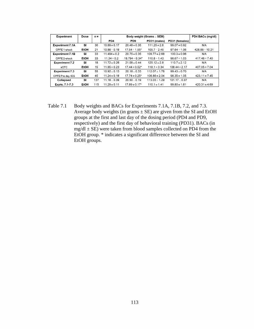
113
Table 7.1 Body weights and BACs for Experiments 7.1A, 7.1B, 7.2, and 7.3.
Average body weights (in grams ± SE) are given from the SI and EtOH
groups at the first and last day of the dosing period (PD4 and PD9,
respectively) and the first day of behavioral training (PD31). BACs (in
mg/dl ± SE) were taken from blood samples collected on PD4 from the
EtOH group. * indicates a significant difference between the SI and
EtOH groups.
114
7.2.6.2 Analysis of Behavioral Data
Scoring and behavioral data analysis is as described in Chapter 5. There were
no main effects or interactions involving sex on freezing behavior across any of the
experiments (ps > .20), so the data were collapsed across this variable. In Experiments
7.1 and 7.3, post-shock and retention test freezing data were analyzed using 2 (Dosing
condition; SI vs. EtOH) × 2 (Exposure condition; Pre vs. Alt-Pre) × 2 (within subjects;
Phase of testing; Post-shock vs. Retention) repeated measures ANOVAs. In
Experiment 7.2, the immediate shock control group was pooled across dosing
condition as there was no significant difference between the SI and EtOH groups and
they froze uniformly low (p > .50). Retention test freezing data were analyzed using a
one-way ANOVA (EtOH-Delayed vs. SI-Delayed vs. Pooled-Imm-Shock). Post-hoc
contrasts were performed with Newman–Keuls tests. Rats were excluded from
analysis as an outlier if they had a score of ± 1.96 standard deviations from the group
mean, however, the average Z-score of removed outliers averaged across all
experiments was ± 5.45 (± 1.04 SEM). One animal from each group (EtOH-Alt-Pre,
EtOH-Pre, SI-Alt-Pre, and SI-Pre in Experiments 7.1 and 7.3; SI-Delayed and EtOH-
Delayed in Experiment 7.2) was excluded as an outlier from both the post-shock and
retention freezing data in each experiment.
7.2.6.3 Analysis of IEG Expression
Relative gene expression for the IEGs c-Fos, Arc, Egr-1, and Npas4 in the
mPFC and dHPC was determined in Experiment 7.3 as described in Chapter 5.
Consistent with previous findings (Heroux et al., 2018), there were no main effects or
interactions involving sex across any of the experiments (ps > .30), so the data were
collapsed across this variable. There was also no difference between the raw data in
115
HC group dosed with alcohol or sham-intubated, so the HC group was collapsed
across dosing condition (ps > .20). Gene expression in Experiment 7.3 was analyzed
using a one-way ANOVA (HC, SI-Pre, and EtOH-Pre) for each gene (c-Fos, Arc, Egr-
1, and Npas4) in both the mPFC and dHPC. Post-hoc contrasts were performed with
Newman–Keuls tests. The number of outliers removed in each sampling condition in
Experiment 7.3 can be found in Table 7.2. Two rats were excluded from IEG analyses
in Experiment 7.3 because of insufficient RNA for cDNA synthesis. The average Z-
score of removed outliers was ± 3.22 (± 0.22 SEM).
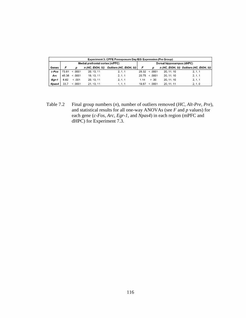
116
Table 7.2 Final group numbers (n), number of outliers removed (HC, Alt-Pre, Pre),
and statistical results for all one-way ANOVAs (see F and p values) for
each gene (c-Fos, Arc, Egr-1, and Npas4) in each region (mPFC and
dHPC) for Experiment 7.3.
117
7.3 Results
7.3.1 Body Weight and BACs
Body weight averages for sham-intubated and alcohol-exposed rats at PD4,
PD9, and PD31 appear in Table 7.1. Both the SI and EtOH groups gained substantial
weight during the dosing period (PD4-PD9) up until the age of testing (PD31). A 2
(Dosing condition; SI vs. EtOH) × 2 (Age; PD4 vs. PD9) repeated measures ANOVA
revealed significant main effects of Dosing condition [F(1, 250) = 45.10, p < .001],
Age [F(1, 250) = 5372.73, p < .001], as well as a Dosing condition × Age interaction
[F(1, 250) = 156.90, p < .001]. Newman-Keuls tests revealed no difference between
group weights on PD4 (ps > .50), but on PD9, EtOH rats weighed about 13% less than
SI rats (ps < .001). Transient growth retardation in ethanol treated rats over this dosing
period has been reported previously (Brown et al., 2007; G. F. Hamilton et al., 2011;
Murawski et al., 2012; Murawski & Stanton, 2010). Ethanol did not alter body weight
at the time of testing. A 2 (Dosing condition; SI vs. EtOH) × 2 (Sex; male vs. female)
factorial ANOVA performed on PD31 body weights revealed a significant main effect
of Sex [F(1, 248) = 111.45, p < .001] but not Dosing condition [F(1, 248) = 3.86, p >
.05], with no interaction between these two variables [F(1, 248) = 0.41, p > .50].
Females had reduced body weights compared to males at PD31 regardless of dosing
condition (see Table 7.1).
BACs taken from the blood samples of the EtOH group on PD4 are also shown
in Table 7.1 (grouped by experiment and then collapsed across all experiments). The
EtOH group showed an average BAC of 422.45 ± 4.69 mg/dl. There was no
significant effect of experiment (7.1A vs. 7.1B vs. 7.2 vs. 7.3) or sex (male vs. female)
on BACs (ps > .30).
118
7.3.2 Experiment 7.1A: PD4-9 Alcohol Exposure Abolishes Post-shock and
Retention Test Freezing Under 1-Shock Reinforcement
The purpose of Experiment 1A was to examine the effects of PD4-9 ethanol
exposure on post-shock and retention test freezing in the CPFE. The behavioral design
and results for Experiment 1A can be seen in Figure 7.1. Analyses for Experiment
7.1A were run on 47 rats distributed across the following groups: EtOH-Alt-Pre (n=7),
EtOH-Pre (n=12), SI-Alt-Pre (n=13), and SI-Pre (n=15). Repeated measures ANOVA
revealed a significant main effect of Dosing [F(1, 41) = 55.35, p < .001], Exposure
[F(1, 41) = 45.92, p < .001], and a significant Dosing × Exposure interaction [F(1, 41)
= 26.98, p < .001]. There was no main effect or any interactions involving Phase (ps >
.08). The SI-Pre group froze significantly more than all other groups during both the
post-shock and retention freezing tests (ps < .001). There was no difference between
EtOH rats preexposed to the training context (EtOH-Pre) and non-associative controls
preexposed to an alternate context (EtOH-Alt-Pre or SI-Alt-Pre; ps > .50) in either
phase. These results show that PD4-9 ethanol exposure abolishes post-shock and
retention test freezing in the CPFE.
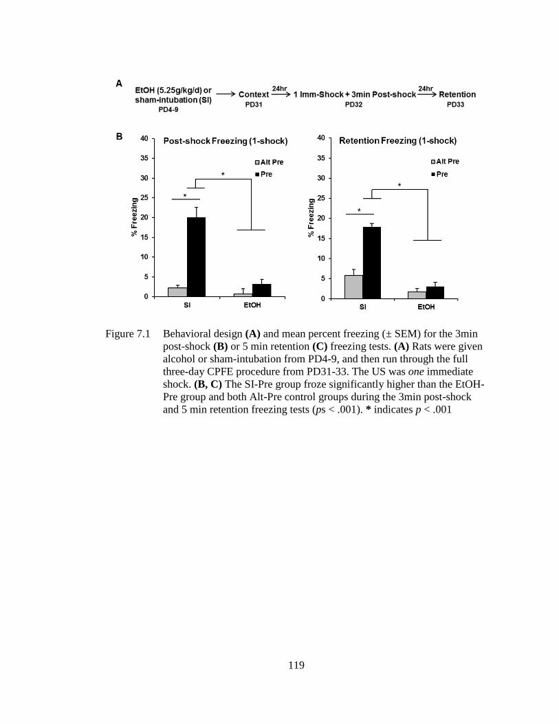
119
Figure 7.1 Behavioral design (A) and mean percent freezing (± SEM) for the 3min
post-shock (B) or 5 min retention (C) freezing tests. (A) Rats were given
alcohol or sham-intubation from PD4-9, and then run through the full
three-day CPFE procedure from PD31-33. The US was one immediate
shock. (B, C) The SI-Pre group froze significantly higher than the EtOH-
Pre group and both Alt-Pre control groups during the 3min post-shock
and 5 min retention freezing tests (ps < .001). * indicates p < .001
120
7.3.3 Experiment 7.1B: PD4-9 Alcohol Exposure Impairs Post-shock and
Retention Test Freezing Under 2-Shock Reinforcement
The purpose of Experiment 7.1B was to determine whether or not increasing
the strength of the immediate-shock reinforcement (i.e., by increasing number of
shocks to 2 instead of 1) would alter behavioral impairments seen in Experiment 7.1A.
The behavioral design and results for Experiment 7.1B can be seen in Figure 7.2.
Analyses for Experiment 7.1B were run on 61 rats distributed across the following
groups: EtOH-Alt-Pre (n=9), EtOH-Pre (n=21), SI-Alt-Pre (n=11), and SI-Pre (n=20).
Repeated measures ANOVA revealed a significant main effect of Dosing [F(1, 55) =
6.99, p < .01], Exposure [F(1, 55) = 34.04, p < .001], and a significant Dosing ×
Exposure interaction [F(1, 55) = 4.97, p < .05]. There was no main effect or any
interactions involving the repeated measure of Phase (ps > .15). SI-Pre rats froze
significantly more than EtOH-Pre rats during the post-shock (p < .05) and retention (p
< .001) freezing tests. While there was no difference between EtOH rats and both Alt-
Pre groups in retention freezing (p > .30), EtOH rats froze significantly more than the
Alt-Pre groups during the post-shock freezing test (p < .05). These results suggest that
doubling the amount of shock-reinforcement given during training improves post-
shock freezing in EtOH rats but is not fully effective in rescuing ethanol-induced
impairment of the CPFE during either test phase.
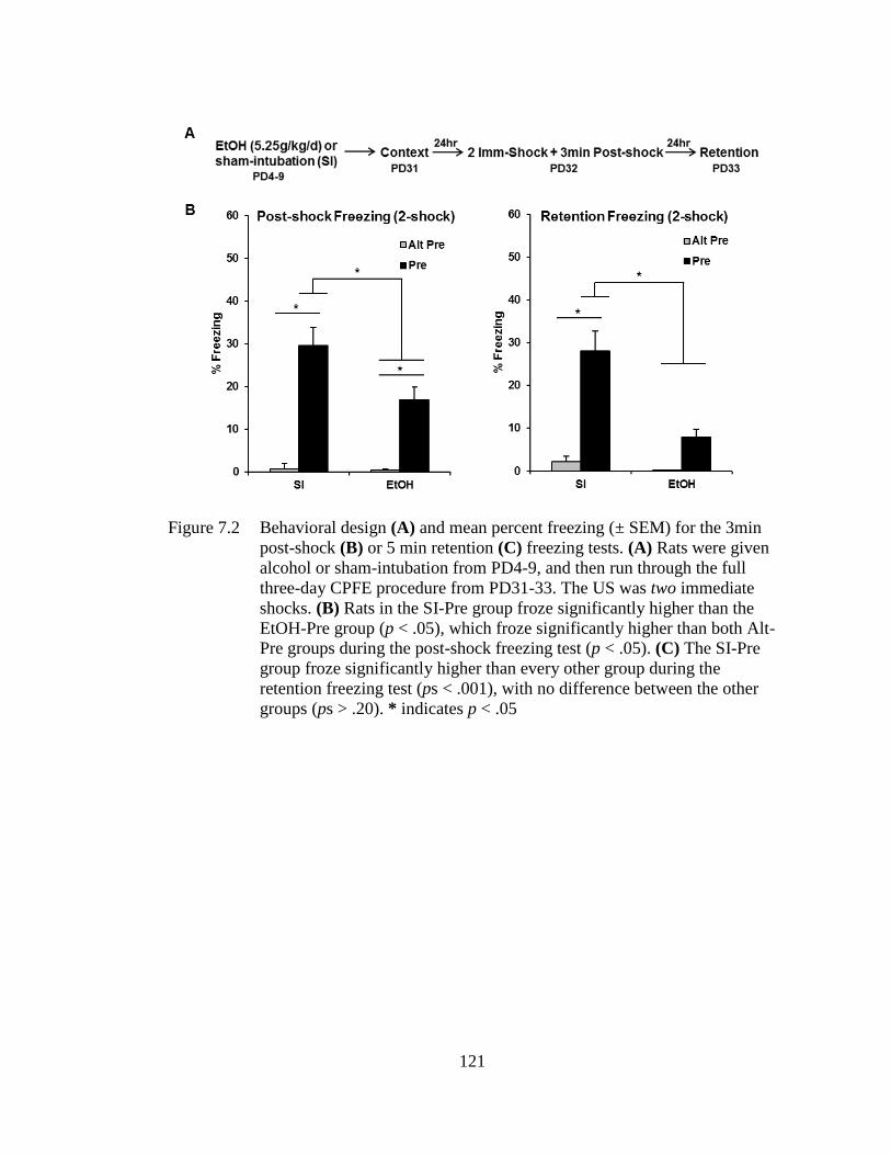
121
Figure 7.2 Behavioral design (A) and mean percent freezing (± SEM) for the 3min
post-shock (B) or 5 min retention (C) freezing tests. (A) Rats were given
alcohol or sham-intubation from PD4-9, and then run through the full
three-day CPFE procedure from PD31-33. The US was two immediate
shocks. (B) Rats in the SI-Pre group froze significantly higher than the
EtOH-Pre group (p < .05), which froze significantly higher than both Alt-
Pre groups during the post-shock freezing test (p < .05). (C) The SI-Pre
group froze significantly higher than every other group during the
retention freezing test (ps < .001), with no difference between the other
groups (ps > .20). * indicates p < .05
122
7.3.4 Experiment 7.2: PD4-9 Alcohol Exposure Does Not Impair Retention Test
Freezing in sCFC
The purpose of Experiment 7.2 was to examine whether or not ethanol-
exposed rats are impaired in standard contextual fear conditioning, in which learning
about the context and acquiring a context-shock association occurs within the same
trial. The behavioral design and results for Experiment 7.2 can be seen in Figure 7.3.
Analyses for Experiment 7.2 were run on 33 rats distributed across the following
groups: EtOH-Delayed (n=10), SI-Delayed (n=11), and Pooled-Imm-Shock (n=12;
SI=6, EtOH=6). One-way ANOVA revealed a significant main effect of Group [F(1,
30) = 25.47, p < .001]. Both SI-Delayed and EtOH-Delayed groups froze significantly
higher than Pooled-Imm-Shock control group (ps < .001), with no difference between
the two Delayed groups (p > .50). These results show that ethanol-exposed rats are
able to acquire and retain contextual fear when context exposure and foot-shock occur
within the same trial.
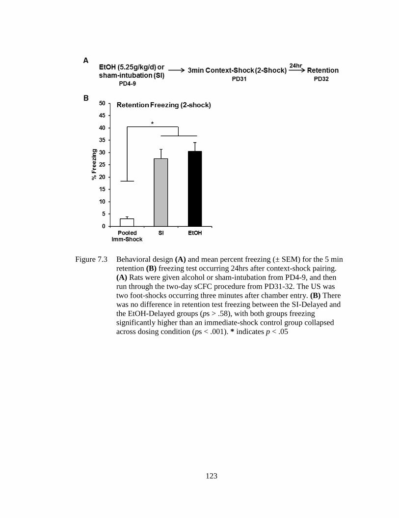
123
Figure 7.3 Behavioral design (A) and mean percent freezing (± SEM) for the 5 min
retention (B) freezing test occurring 24hrs after context-shock pairing.
(A) Rats were given alcohol or sham-intubation from PD4-9, and then
run through the two-day sCFC procedure from PD31-32. The US was
two foot-shocks occurring three minutes after chamber entry. (B) There
was no difference in retention test freezing between the SI-Delayed and
the EtOH-Delayed groups (ps > .58), with both groups freezing
significantly higher than an immediate-shock control group collapsed
across dosing condition (ps < .001). * indicates p < .05
124
7.3.5 Experiment 7.3: PD4-9 Alcohol Exposure Impairs Medial-Prefrontal but
Not Dorsal-Hippocampal IEG Expression during Context Exposure
The purpose of Experiment 7.3 was to determine whether impaired context
memory in ethanol-exposed rats is accompanied by disrupted IEG expression in the
mPFC and dHPC during context preexposure (see Figure 5.1 for dissected regions).
The behavioral design and results for Experiment 7.3 can be seen in Figure 7.4A-C.
Analyses for Experiment 7.3 were run on 48 rats distributed across the following
groups: EtOH-Alt-Pre (n=8), EtOH-Pre (n=13), SI-Alt-Pre (n=11), and SI-Pre (n=16).
Repeated measures ANOVA revealed a significant main effect of Dosing [F(1, 42) =
22.15, p < .001], Exposure [F(1, 42) = 34.98, p < .001], and a significant Dosing ×
Exposure interaction [F(1, 42) = 7.88, p < .01]. There was no main effect or any
interactions involving Phase (ps > .40). The SI-Pre group froze significantly more than
any other group during both the post-shock and retention freezing tests (ps < .001).
Additionally, there was no significant difference between EtOH-Pre and the Alt-Pre
control groups (ps > .20).
The IEG results can be seen in Figure 7.4D-E. Gene expression in Experiment
3 was analyzed using a one-way ANOVA (HC, SI-Pre, and EtOH-Pre) for each gene
(c-Fos, Arc, Egr-1, and Npas4) in both the mPFC and dHPC. Specific F statistics, p
values, group n, and outliers removed for all eight one-way ANOVAs for Experiment
3 can be found in Table 7.2. Post hoc contrasts revealed that, in the mPFC, EtOH rats
showed significantly reduced mRNA expression of every IEG (c-Fos, Arc, Egr-1, and
Npas4; see Figure 7.4D) compared to SI rats (ps < .001). However, expression of c-
Fos, Arc, and Npas4 in EtOH rats was still significantly above HC control levels (ps >
.01). This disruption of IEG expression in the EtOH group was not seen in the dHPC,
with both SI and EtOH rats having significantly higher expression of c-Fos, Arc, and
125
Npas4 above HC control levels, with no difference between the two dosing groups (ps
> .18; see Figure 7.4E). These results indicate that neonatal PD4-9 ethanol exposure
impairs prefrontal but not hippocampal IEG expression induced by context exposure.
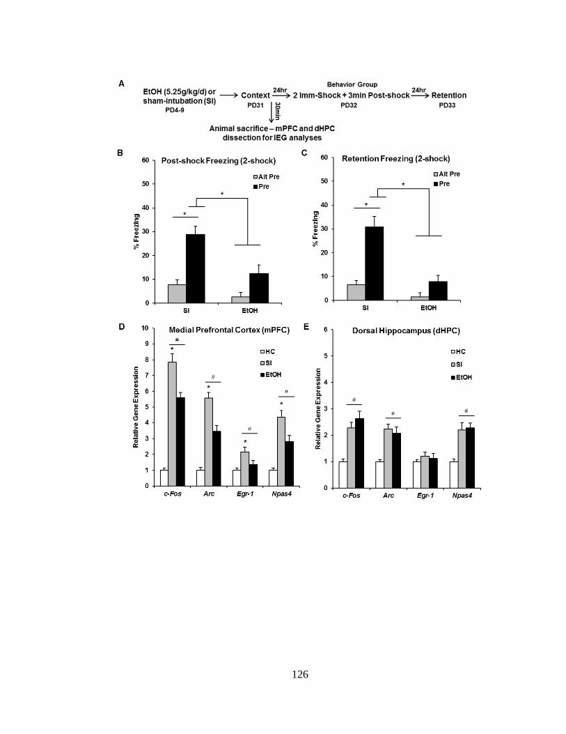
126
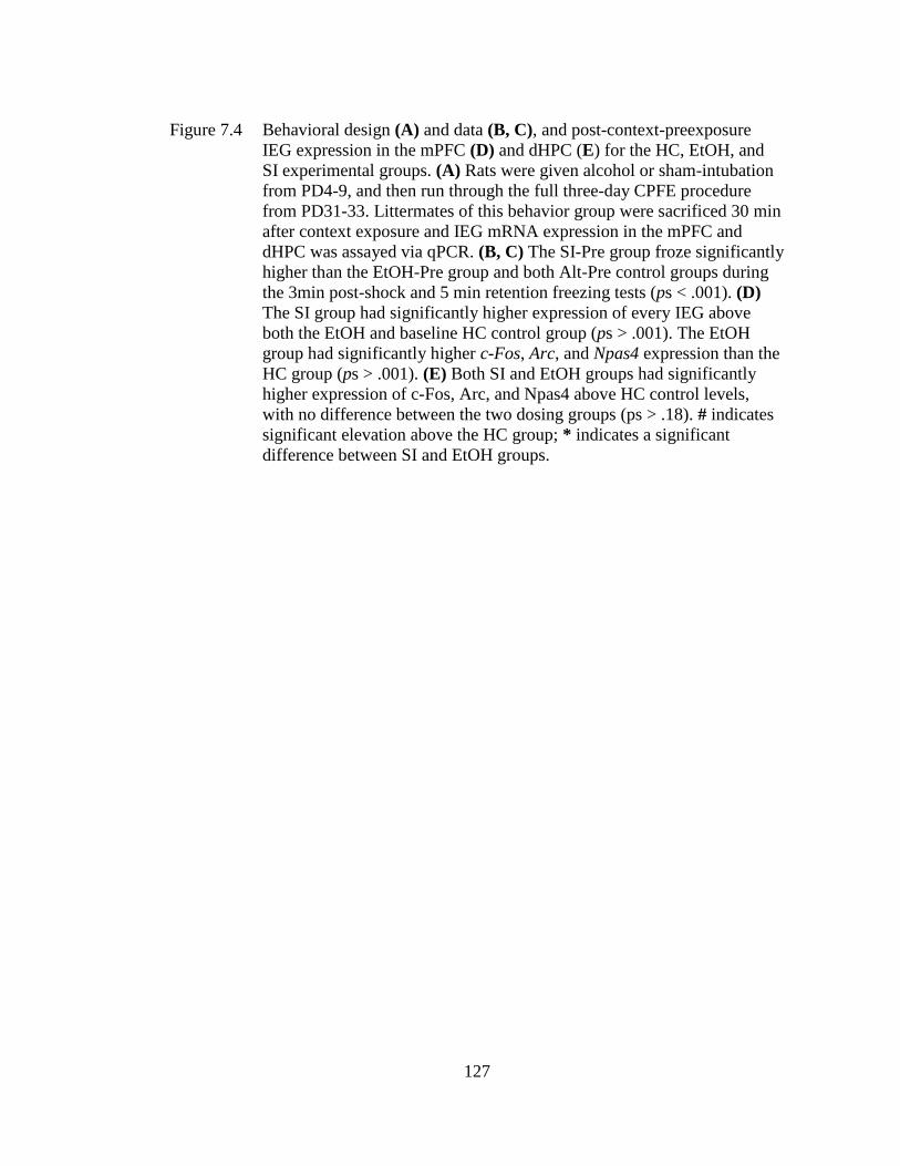
127
Figure 7.4 Behavioral design (A) and data (B, C), and post-context-preexposure
IEG expression in the mPFC (D) and dHPC (E) for the HC, EtOH, and
SI experimental groups. (A) Rats were given alcohol or sham-intubation
from PD4-9, and then run through the full three-day CPFE procedure
from PD31-33. Littermates of this behavior group were sacrificed 30 min
after context exposure and IEG mRNA expression in the mPFC and
dHPC was assayed via qPCR. (B, C) The SI-Pre group froze significantly
higher than the EtOH-Pre group and both Alt-Pre control groups during
the 3min post-shock and 5 min retention freezing tests (ps < .001). (D)
The SI group had significantly higher expression of every IEG above
both the EtOH and baseline HC control group (ps > .001). The EtOH
group had significantly higher c-Fos, Arc, and Npas4 expression than the
HC group (ps > .001). (E) Both SI and EtOH groups had significantly
higher expression of c-Fos, Arc, and Npas4 above HC control levels,
with no difference between the two dosing groups (ps > .18). # indicates
significant elevation above the HC group; * indicates a significant
difference between SI and EtOH groups.

128
7.4 Discussion
The current set of experiments examined the disruption caused by neonatal
alcohol exposure on context and contextual fear learning in the CPFE in adolescent
rats. Consistent with previous CPFE studies (Goodfellow & Lindquist, 2014; G. F.
Hamilton et al., 2011; Murawski et al., 2012; Murawski & Stanton, 2010, 2011), high
binge-like doses of ethanol given over PD4-9 abolished 24-hr retention test freezing
(Experiments 7.1A, 7.1B, and 7.3). Importantly, previous research has been unable to
elucidate whether this disruption in retention reflects an impairment in preexposure or
training day processes. In the current study, ethanol exposure left freezing in sCFC
intact (Experiment 7.2), but post-shock freezing on the training day of the CPFE was
severely impaired in ethanol-exposed rats regardless of reinforcement intensity (i.e.,
one vs. two shocks) used (Experiments 7.1A, 7.1B, and 7.3). Furthermore, ethanol-
exposed rats showed a selective disruption in medial prefrontal but not dorsal
hippocampal expression of the IEGs Arc, c-Fos, Egr-1, and Npas4 induced by context
preexposure in the CPFE (Experiment 7.3). Taken together, these results indicate that
PD4-9 ethanol exposure disrupts prefrontal but not hippocampal activity- and
plasticity-associated gene expression during incidental context learning, which may
reflect a disruption in configural memory processes of the CPFE (i.e., acquisition,
consolidation, or retrieval of a conjunctive context representation).
Extending previous work examining retention only (Goodfellow & Lindquist,
2014; G. F. Hamilton et al., 2011; Murawski et al., 2012; Murawski & Stanton, 2010,
2011), PD4-9 ethanol exposure abolished post-shock and retention test freezing in the
CPFE. In contrast, retention freezing in single-trial sCFC in ethanol-exposed rats was
spared. Accordingly, ethanol-induced disruptions in the CPFE cannot be attributed to
reduced shock sensitivity, hyperactivity, or impaired context exploration or feature
129
perception. Shortening the interval between context exposure and immediate-shock
training to 2hr rescues 24hr retention test freezing in ethanol-exposed rats, suggesting
that alcohol exposure does not impair the ability to associate a previously learned
context with a shock in the CPFE (Goodfellow & Lindquist, 2014). Therefore, the
observed ethanol-induced deficit in post-shock freezing likely reflects a disruption in
the consolidation of the conjunctive context representation after context preexposure
in the CPFE. Additionally, because the CPFE requires the mPFC and dHPC during all
three phases (Heroux et al., 2017; Matus-Amat et al., 2004; Robinson-Drummer et al.,
2016, 2017), whereas single-trial conditioning in sCFC depends on the dHPC but not
mPFC (Heroux et al., 2017; Wiltgen et al., 2006), these results implicate impaired
prefrontal mechanisms of the CPFE. This notion is consistent with the observed
ethanol-induced disruptions in prefrontal but not hippocampal IEG expression during
context learning (Experiment 7.3).
Our lab has previously characterized disruptions in brain and behavior after
different exposure windows in the rat, notably after PD4-6, PD4-9, and PD7-9. Unlike
in the PD4-9 or PD7-9 dosing scenarios, PD4-6 ethanol exposure has no effect on
retention test freezing in the CPFE (Murawski & Stanton, 2011). While this might
suggest that the disruptive effects of ethanol exposure could be solely attributed to the
PD7-9 window, this exposure leaves post-shock freezing on the training day of the
CPFE intact (Jablonski & Stanton, 2014). Moreover, impaired contextual fear
retention in these rats is associated with reduced prefrontal Egr-1 mRNA expression
on the training day of the CPFE (Jablonski et al., 2018). In contrast, we report that the
broader PD4-9 exposure results in impaired context memory and prefrontal IEG
expression on the preexposure day of the CPFE. Our lab has previously shown that
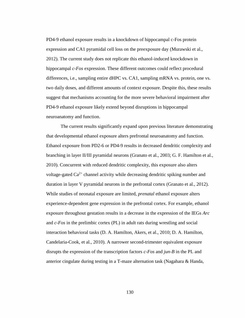
130
PD4-9 ethanol exposure results in a knockdown of hippocampal c-Fos protein
expression and CA1 pyramidal cell loss on the preexposure day (Murawski et al.,
2012). The current study does not replicate this ethanol-induced knockdown in
hippocampal c-Fos expression. These different outcomes could reflect procedural
differences, i.e., sampling entire dHPC vs. CA1, sampling mRNA vs. protein, one vs.
two daily doses, and different amounts of context exposure. Despite this, these results
suggest that mechanisms accounting for the more severe behavioral impairment after
PD4-9 ethanol exposure likely extend beyond disruptions in hippocampal
neuroanatomy and function.
The current results significantly expand upon previous literature demonstrating
that developmental ethanol exposure alters prefrontal neuroanatomy and function.
Ethanol exposure from PD2-6 or PD4-9 results in decreased dendritic complexity and
branching in layer II/III pyramidal neurons (Granato et al., 2003; G. F. Hamilton et al.,
2010). Concurrent with reduced dendritic complexity, this exposure also alters
voltage-gated Ca2+ channel activity while decreasing dendritic spiking number and
duration in layer V pyramidal neurons in the prefrontal cortex (Granato et al., 2012).
While studies of neonatal exposure are limited, prenatal ethanol exposure alters
experience-dependent gene expression in the prefrontal cortex. For example, ethanol
exposure throughout gestation results in a decrease in the expression of the IEGs Arc
and c-Fos in the prelimbic cortex (PL) in adult rats during wrestling and social
interaction behavioral tasks (D. A. Hamilton, Akers, et al., 2010; D. A. Hamilton,
Candelaria-Cook, et al., 2010). A narrower second-trimester equivalent exposure
disrupts the expression of the transcription factors c-Fos and jun-B in the PL and
anterior cingulate during testing in a T-maze alternation task (Nagahara & Handa,
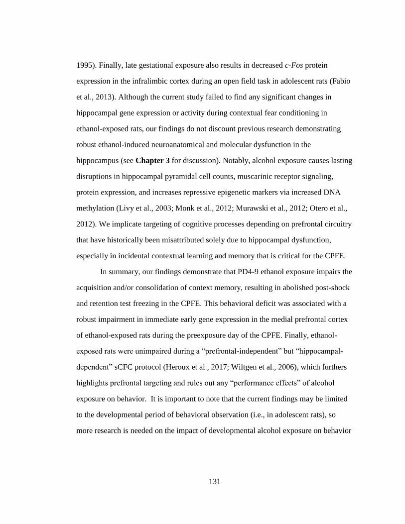
131
1995). Finally, late gestational exposure also results in decreased c-Fos protein
expression in the infralimbic cortex during an open field task in adolescent rats (Fabio
et al., 2013). Although the current study failed to find any significant changes in
hippocampal gene expression or activity during contextual fear conditioning in
ethanol-exposed rats, our findings do not discount previous research demonstrating
robust ethanol-induced neuroanatomical and molecular dysfunction in the
hippocampus (see Chapter 3 for discussion). Notably, alcohol exposure causes lasting
disruptions in hippocampal pyramidal cell counts, muscarinic receptor signaling,
protein expression, and increases repressive epigenetic markers via increased DNA
methylation (Livy et al., 2003; Monk et al., 2012; Murawski et al., 2012; Otero et al.,
2012). We implicate targeting of cognitive processes depending on prefrontal circuitry
that have historically been misattributed solely due to hippocampal dysfunction,
especially in incidental contextual learning and memory that is critical for the CPFE.
In summary, our findings demonstrate that PD4-9 ethanol exposure impairs the
acquisition and/or consolidation of context memory, resulting in abolished post-shock
and retention test freezing in the CPFE. This behavioral deficit was associated with a
robust impairment in immediate early gene expression in the medial prefrontal cortex
of ethanol-exposed rats during the preexposure day of the CPFE. Finally, ethanol-
exposed rats were unimpaired during a “prefrontal-independent” but “hippocampal-
dependent” sCFC protocol (Heroux et al., 2017; Wiltgen et al., 2006), which furthers
highlights prefrontal targeting and rules out any “performance effects” of alcohol
exposure on behavior. It is important to note that the current findings may be limited
to the developmental period of behavioral observation (i.e., in adolescent rats), so
more research is needed on the impact of developmental alcohol exposure on behavior
132
across the lifespan. Nevertheless, these findings are important because prefrontal
dysfunction is an integral hallmark of FASD in humans, but animal models have thus
far largely failed to capture prefrontal dysfunction after third-trimester equivalent
exposure. The CPFE has proven to be a promising behavioral paradigm that can
facilitate linking alterations in prefrontal and hippocampal function to discrete phases
of learning and memory that are impaired by developmental alcohol exposure in rats.
More research is needed to establish a link between disrupted brain circuitry and
cognitive dysfunction in animal models of FASD.
133
CHOLINERGIC RESCUE OF NEUROCOGNITIVE INSULT FOLLOWING
THIRD-TRIMESTER EQUIVALENT ALCOHOL EXPOSURE IN RATS
8.1 Introduction
This chapter was recently published in Neurobiology of Learning and Memory
(Heroux, Horgan, Rosen, et al., 2019), and the text appears here in adapted form. As
reviewed in Chapter 3 and 4, our lab has shown that systemic administration of the
acetylcholinesterase inhibitor PHY prior to every phase of the CPFE rescues retention
test freezing in PD7-9 alcohol-exposed rats (Dokovna et al., 2013). The specific phase
of the CPFE that mediates this rescue effect, the regional neural correlates of the
effect, and whether it would extend to the more severe PD4-9 exposure window is not
known. Therefore, the purpose of the current study was to examine the effects of
augmenting cholinergic signaling on behavioral performance and mPFC, dHPC, and
vHPC IEG expression in PD4-9 ethanol-exposed rats. In addition, the current study
analyzed vHPC IEG expression given our discovery of mPFC-vHPC circuitry being
involved during context preexposure in the CPFE (see Chapter 6). In Experiment
8.1, we extended our earlier findings with PD7-9 exposure (Dokovna et al., 2013) by
examining whether or not systemic administration of PHY prior to all three phases
would rescue freezing in rats receiving PD4-9 ethanol exposure. In Experiment 8.2,
we sought to determine if this same injection prior to just context learning would
rescue disrupted behavior, and more importantly, prefrontal IEG expression
previously observed during the CPFE (Chapter 7; Heroux, Robinson-Drummer, et al.,
Chapter 8
134
2019; Jablonski et al., 2018). To our knowledge, this is the first study to examine the
effects of boosting cholinergic function (via PHY) on neural activity during behavior
in rats receiving neonatal ethanol exposure during the brain growth spurt.
8.2 Materials and Methods
8.2.1 Subjects
Animal husbandry was as described as in Chapter 5. Across both experiments,
there were a total of 197 adolescent (PD31) Long Evans rats (97 females and 100
males), derived from 30 separate litters bred by the Office of Laboratory Animal
Medicine at the University of Delaware.
8.2.2 Neonatal Alcohol Dosing
Dosing procedures (SI and EtOH groups), window (PD4-9), dose (5.25g/kg/d),
blood collection, and BAC analyses occurred as described in Chapters 5 and 7.
8.2.3 Apparatus and Stimuli
The apparatus and stimuli used for the current experiments were as described
in Chapter 5. The US was two 1.5mA foot-shocks separated by 1sec.
8.2.4 Behavioral Procedures and Drug Injection
The multiple preexposure CPFE procedure used in the current experiments was
as described in Chapter 5, 6, and 7. Briefly, SI and EtOH rats were assigned to the
Pre condition (Experiment 8.1) or the Pre and Alt-Pre conditions (Experiment 8.2 and
underwent the CPFE. The Alt-Pre condition was omitted from Experiment 8.1 to
streamline the design and to avoid needless duplication with Experiment 8.2 where
inclusion of the Alt-Pre group to analyze freezing behavior was more useful. Rats in
135
both dosing groups received systemic intraperitoneal (i.p.) injections of 1ml/kg of
0.01mg/ml/kg physostigmine or the vehicle sterile saline. This injection occurred 30
min prior to all three phases in Experiment 8.1, and 30 min prior to only context
preexposure in Experiment 8.2. Post-shock (3 min) and retention test (5 min) freezing
was measured in both experiments. In Experiment 8.2, behavior was assessed in
littermates of rats assigned to IEG assays.
8.2.5 Brain Removal, Tissue Dissection, and qPCR
In Experiment 8.2, rats in the SI-Pre-PHY, SI-Pre-SAL, EtOH-Pre-PHY,
EtOH-Pre-SAL, and home-cage control group were sacrificed 30 min after context
exposure in the CPFE. Expression of the immediate early genes c-Fos, Arc, Egr-1, and
Npas4 in the mPFC, dHPC, and vHPC was analyzed via qPCR as described in
Chapter 5 and was identical to Chapters 6 and 7.
8.2.6 Data Analysis
8.2.6.1 Analysis of Neonatal and Adolescent Body Weight
Neonatal body weight was analyzed with repeated measures ANOVA with a
between-subjects factor of dosing condition (SI vs. EtOH) and the within-subjects
factor of age (PD4 vs. PD9). There were no main effects or interactions involving sex
in the PD4 and PD9 weights (ps > .05) so the data were collapsed across this variable
at these ages. Body weight at PD31 was analyzed with a 2 (Sex; male vs. female) × 2
(Dosing condition; SI vs. EtOH) factorial ANOVA.
136
8.2.6.2 Analysis of Behavioral Data
Scoring and behavioral data analysis is as described in Chapter 5. There were
no main effects or interactions involving sex on freezing behavior (ps > .05), so the
data were collapsed across this variable. In Experiment 8.1, freezing data were
analyzed using 2 (Dosing condition; SI vs. EtOH) × 2 (Drug condition; SAL vs. PHY)
× 2 (within subjects; phase of testing; Post-shock vs. Retention) repeated measures
ANOVA. In Experiment 8.2 the Alt-Pre group was pooled across drug but not dosing
condition (Pooled-Alt-Pre group) as there was no significant difference between the
two drug groups and they froze at uniformly low levels (p > .34). Therefore, freezing
data were analyzed using 2 (Dosing; SI vs. EtOH) × 3 (Condition; SAL vs. PHY vs.
Pooled-Alt-Pre) × 2 (within subjects; phase of testing; Post-shock vs. Retention)
repeated measures ANOVA. Post-hoc contrasts were performed with Newman–Keuls
tests. Rats were excluded from analysis as an outlier as in previous experiments, and
the Z-score of removed outliers averaged 3.91 (± 0.97 SEM). Outliers removed were
as follows: Experiment 1 [(Post-shock: SI-PHY=1, SI-SAL=1)] and Experiment 2
[(Post-shock: EtOH-Pooled-Alt-Pre=1, EtOH-PHY=1, EtOH-SAL=1, SI-PHY=1, SI-
SAL=1; Retention: EtOH-Pooled-Alt-Pre=1, SI-Pooled-Alt-Pre=1, EtOH-PHY=2,
EtOH-SAL=2)].
8.2.6.3 Analysis of IEG Expression
Relative gene expression for the IEGs c-Fos, Arc, Egr-1, and Npas4 in the
mPFC, dHPC, and vHPC was determined. The relative gene expression value was
obtained by normalizing the data to the reference gene (18s) and to the average delta
CT of the home-cage control group for each gene. Consistent with previous
experiments, lack of statistical effects (ps > .30) led to pooling data across sex. There
137
was also no difference between the raw data in HC group injected with SAL or PHY,
so the HC group was collapsed across drug condition (ps > .35). Gene expression was
analyzed using a 2 (Dosing condition; SI vs. EtOH) × 2 (Drug condition; SAL vs.
PHY) factorial ANOVA for each gene (c-Fos, Arc, Egr-1, and Npas4) in the mPFC,
dHPC, and vHPC. Post-hoc contrasts were performed with Newman–Keuls tests and a
Dunnett’s test that contrasted the four experimental groups with the HC control group.
The average Z-score of removed outliers was ± 3.14 (± 0.12 SEM; see Table 8.2).
8.3 Results
8.3.1 Body Weight and BACs
Body weight averages for sham-intubated and alcohol-exposed rats at PD4 and
PD9 appear in Table 8.1. Both the SI and EtOH groups gained substantial weight
during the dosing period (PD4-PD9) up until the age of testing (PD31). A 2 (Dosing;
SI vs. EtOH) × 2 (Age; PD4 vs. PD9) repeated measures ANOVA revealed significant
main effects of Dosing condition [F(1, 195) = 67.59, p < .001], Age [F(1, 195) =
7213.30, p < .001], as well as a Dosing × Age interaction [F(1, 195) = 316.35, p <
.001]. This reflected no treatment effect on PD4 weights (p > .70), but on PD9, EtOH
rats weighed about 15% less than SI rats (ps < .001). Ethanol did not alter body weight
at the time of behavioral testing (Table 8.1). Females had lower body weights
compared to males at PD31 regardless of dosing condition [F(1, 193) = 56.33, p <
.001]. Finally, the average BAC value taken from the blood samples of the EtOH
group in each experiment can be seen in Table 8.1.
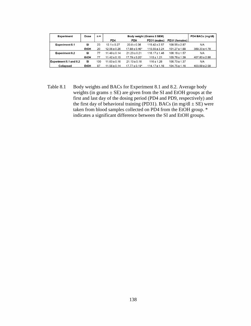
138
Table 8.1 Body weights and BACs for Experiment 8.1 and 8.2. Average body
weights (in grams ± SE) are given from the SI and EtOH groups at the
first and last day of the dosing period (PD4 and PD9, respectively) and
the first day of behavioral training (PD31). BACs (in mg/dl ± SE) were
taken from blood samples collected on PD4 from the EtOH group. *
indicates a significant difference between the SI and EtOH groups.
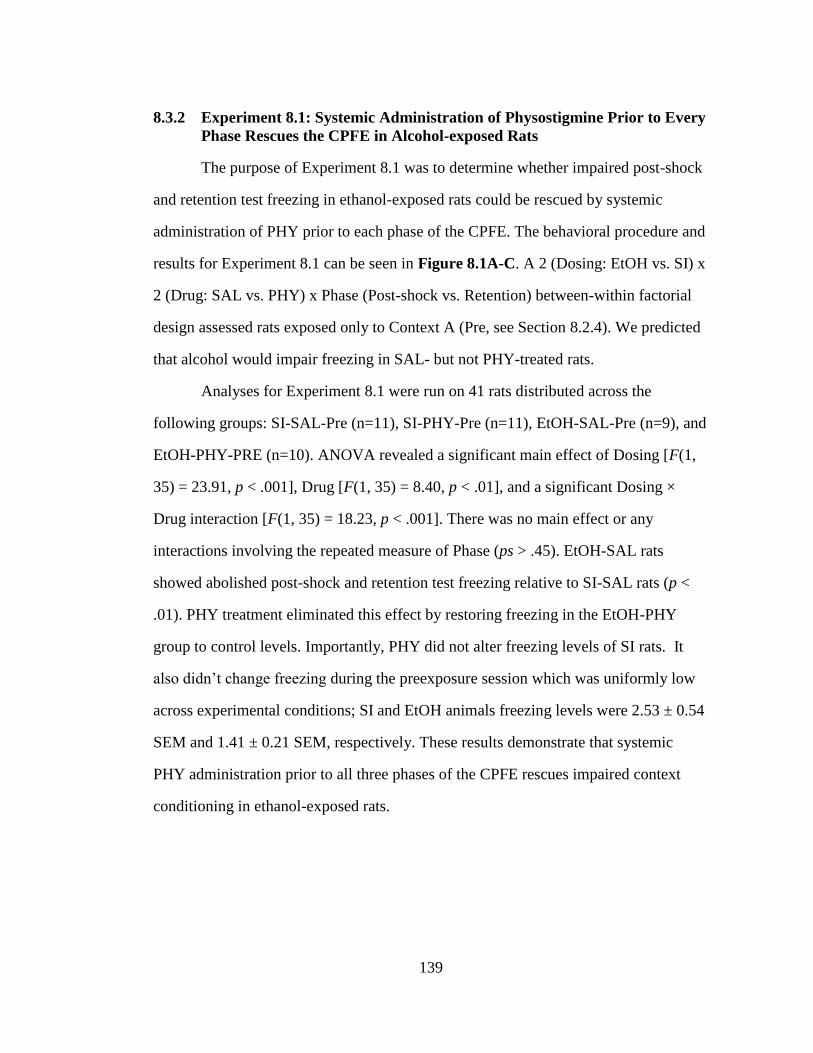
139
8.3.2 Experiment 8.1: Systemic Administration of Physostigmine Prior to Every
Phase Rescues the CPFE in Alcohol-exposed Rats
The purpose of Experiment 8.1 was to determine whether impaired post-shock
and retention test freezing in ethanol-exposed rats could be rescued by systemic
administration of PHY prior to each phase of the CPFE. The behavioral procedure and
results for Experiment 8.1 can be seen in Figure 8.1A-C. A 2 (Dosing: EtOH vs. SI) x
2 (Drug: SAL vs. PHY) x Phase (Post-shock vs. Retention) between-within factorial
design assessed rats exposed only to Context A (Pre, see Section 8.2.4). We predicted
that alcohol would impair freezing in SAL- but not PHY-treated rats.
Analyses for Experiment 8.1 were run on 41 rats distributed across the
following groups: SI-SAL-Pre (n=11), SI-PHY-Pre (n=11), EtOH-SAL-Pre (n=9), and
EtOH-PHY-PRE (n=10). ANOVA revealed a significant main effect of Dosing [F(1,
35) = 23.91, p < .001], Drug [F(1, 35) = 8.40, p < .01], and a significant Dosing ×
Drug interaction [F(1, 35) = 18.23, p < .001]. There was no main effect or any
interactions involving the repeated measure of Phase (ps > .45). EtOH-SAL rats
showed abolished post-shock and retention test freezing relative to SI-SAL rats (p <
.01). PHY treatment eliminated this effect by restoring freezing in the EtOH-PHY
group to control levels. Importantly, PHY did not alter freezing levels of SI rats. It
also didn’t change freezing during the preexposure session which was uniformly low
across experimental conditions; SI and EtOH animals freezing levels were 2.53 ± 0.54
SEM and 1.41 ± 0.21 SEM, respectively. These results demonstrate that systemic
PHY administration prior to all three phases of the CPFE rescues impaired context
conditioning in ethanol-exposed rats.
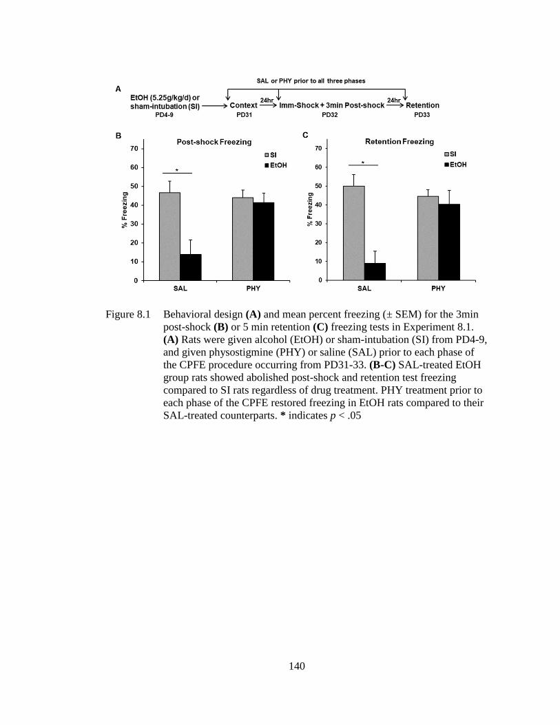
140
Figure 8.1 Behavioral design (A) and mean percent freezing (± SEM) for the 3min
post-shock (B) or 5 min retention (C) freezing tests in Experiment 8.1.
(A) Rats were given alcohol (EtOH) or sham-intubation (SI) from PD4-9,
and given physostigmine (PHY) or saline (SAL) prior to each phase of
the CPFE procedure occurring from PD31-33. (B-C) SAL-treated EtOH
group rats showed abolished post-shock and retention test freezing
compared to SI rats regardless of drug treatment. PHY treatment prior to
each phase of the CPFE restored freezing in EtOH rats compared to their
SAL-treated counterparts. * indicates p < .05
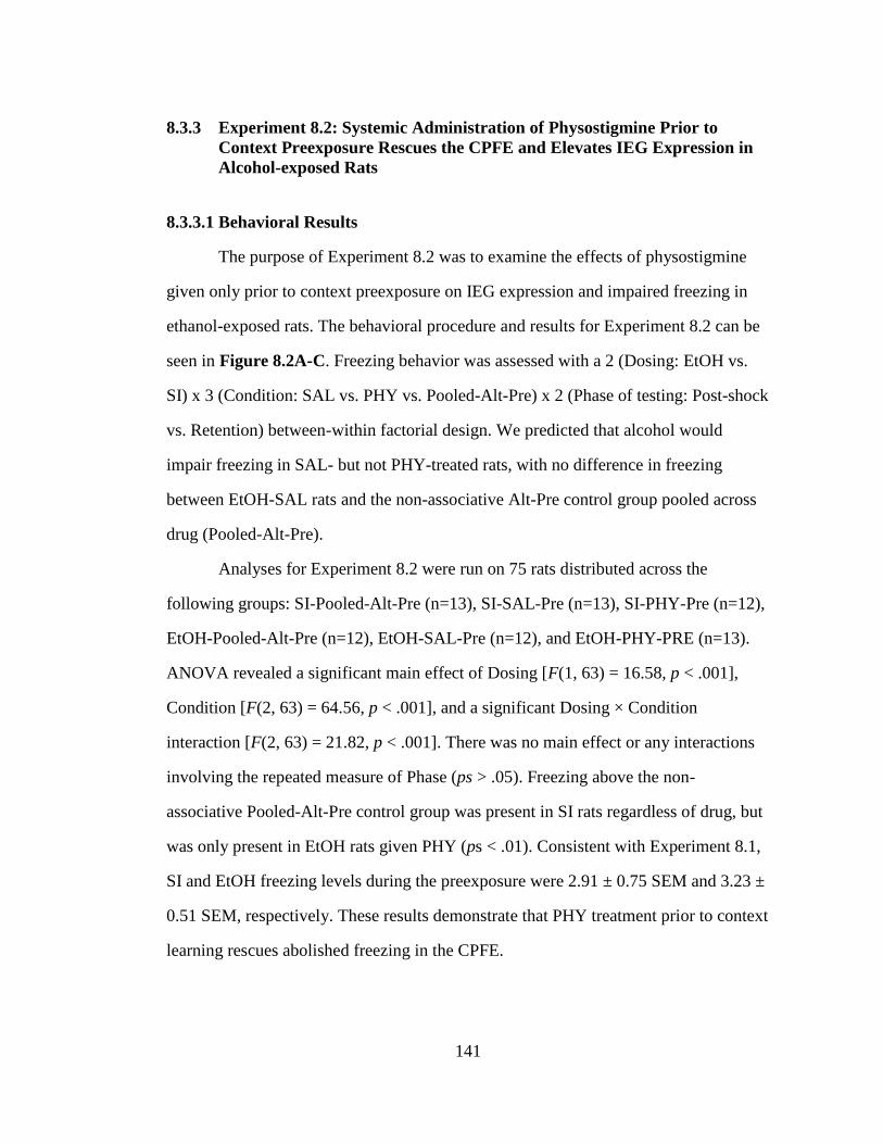
141
8.3.3 Experiment 8.2: Systemic Administration of Physostigmine Prior to
Context Preexposure Rescues the CPFE and Elevates IEG Expression in
Alcohol-exposed Rats
8.3.3.1 Behavioral Results
The purpose of Experiment 8.2 was to examine the effects of physostigmine
given only prior to context preexposure on IEG expression and impaired freezing in
ethanol-exposed rats. The behavioral procedure and results for Experiment 8.2 can be
seen in Figure 8.2A-C. Freezing behavior was assessed with a 2 (Dosing: EtOH vs.
SI) x 3 (Condition: SAL vs. PHY vs. Pooled-Alt-Pre) x 2 (Phase of testing: Post-shock
vs. Retention) between-within factorial design. We predicted that alcohol would
impair freezing in SAL- but not PHY-treated rats, with no difference in freezing
between EtOH-SAL rats and the non-associative Alt-Pre control group pooled across
drug (Pooled-Alt-Pre).
Analyses for Experiment 8.2 were run on 75 rats distributed across the
following groups: SI-Pooled-Alt-Pre (n=13), SI-SAL-Pre (n=13), SI-PHY-Pre (n=12),
EtOH-Pooled-Alt-Pre (n=12), EtOH-SAL-Pre (n=12), and EtOH-PHY-PRE (n=13).
ANOVA revealed a significant main effect of Dosing [F(1, 63) = 16.58, p < .001],
Condition [F(2, 63) = 64.56, p < .001], and a significant Dosing × Condition
interaction [F(2, 63) = 21.82, p < .001]. There was no main effect or any interactions
involving the repeated measure of Phase (ps > .05). Freezing above the non-
associative Pooled-Alt-Pre control group was present in SI rats regardless of drug, but
was only present in EtOH rats given PHY (ps < .01). Consistent with Experiment 8.1,
SI and EtOH freezing levels during the preexposure were 2.91 ± 0.75 SEM and 3.23 ±
0.51 SEM, respectively. These results demonstrate that PHY treatment prior to context
learning rescues abolished freezing in the CPFE.
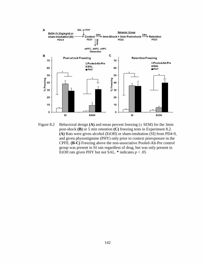
142
Figure 8.2 Behavioral design (A) and mean percent freezing (± SEM) for the 3min
post-shock (B) or 5 min retention (C) freezing tests in Experiment 8.2.
(A) Rats were given alcohol (EtOH) or sham-intubation (SI) from PD4-9,
and given physostigmine (PHY) only prior to context preexposure in the
CPFE. (B-C) Freezing above the non-associative Pooled-Alt-Pre control
group was present in SI rats regardless of drug, but was only present in
EtOH rats given PHY but not SAL. * indicates p < .05
143
8.3.3.2 IEG Results
Littermates of the behavior group were sacrificed 30 min after context
exposure on the preexposure day of the CPFE. The IEG results can be seen in Figure
8.3A-C. The statistical results, group sizes, and number of outliers removed in each
sampling condition can be found in Table 8.2.
In the mPFC (Fig. 8.3A), the EtOH-SAL group showed significantly reduced
mRNA expression of the IEGs c-Fos, Arc, and Npas4 compared to Group SI-SAL (p <
.001), replicating Experiment 7.2 (Chapter 7). Interestingly, PHY administration in
EtOH rats specifically rescued prefrontal expression of c-Fos (i.e., the increase
occurred in EtOH-PHY but not SI-PHY rats; ps < .01). Both SI and EtOH rats given
PHY showed significantly elevated expression of the IEGs Arc and Npas4 above their
respective saline-treated groups (ps < .01), indicating that PHY enhanced overall
expression but failed to reverse ethanol effects on expression of these IEGs. While
PHY treatment did raise prefrontal egr-1 expression in SI rats (p < .05), there was no
difference in expression between the EtOH groups (ps > .20). Dunnett’s tests revealed
that every group was significantly elevated above HC levels (ps < .01) with the
exception of EtOH-SAL for Arc; and EtOH-SAL and EtOH-PHY for egr-1 expression
(p > .05).
In the dHPC (Fig. 8.3B), no main of interaction effects involving alcohol
(dosing) were found for any IEG and a main effect of drug was found only for c-Fos
(Table 8.2). The EtOH-PHY and SI-PHY groups showed significantly higher c-Fos
expression than their saline-treated counterparts (p < .01), with no difference between
the two groups (p > .40). Dunnett’s tests revealed that SI and EtOH rats given either
drug had significantly elevated expression of all IEGs above HC levels. Finally, in the
vHPC (Fig. 8.3C), there were no main or interaction effects of dosing or drug (Table
144
8.2). While Dunnett’s tests revealed that all four treated groups had significantly
higher expression of c-Fos and Arc than HC levels (ps < .05), there was no elevation
above HC in vHPC egr-1 and Npas4 (ps > .10).
Taken together, the results of Experiment 8.2 demonstrate that systemic PHY
administration prior to context preexposure in the CPFE rescues context freezing in
ethanol-exposed rats. A similar effect on IEG expression was found only for c-Fos in
mPFC. PHY administration increased prefrontal Arc and Npas4 expression, and
dHPC c-Fos expression regardless of alcohol treatment. Consistent with previous
findings (Heroux et al., 2019) alcohol impaired IEG expression only in mPFC.
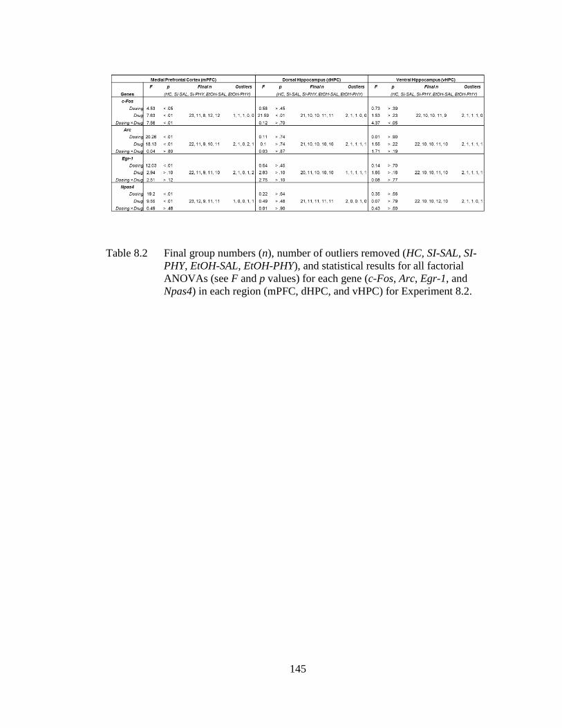
145
Table 8.2 Final group numbers (n), number of outliers removed (HC, SI-SAL, SI-
PHY, EtOH-SAL, EtOH-PHY), and statistical results for all factorial
ANOVAs (see F and p values) for each gene (c-Fos, Arc, Egr-1, and
Npas4) in each region (mPFC, dHPC, and vHPC) for Experiment 8.2.
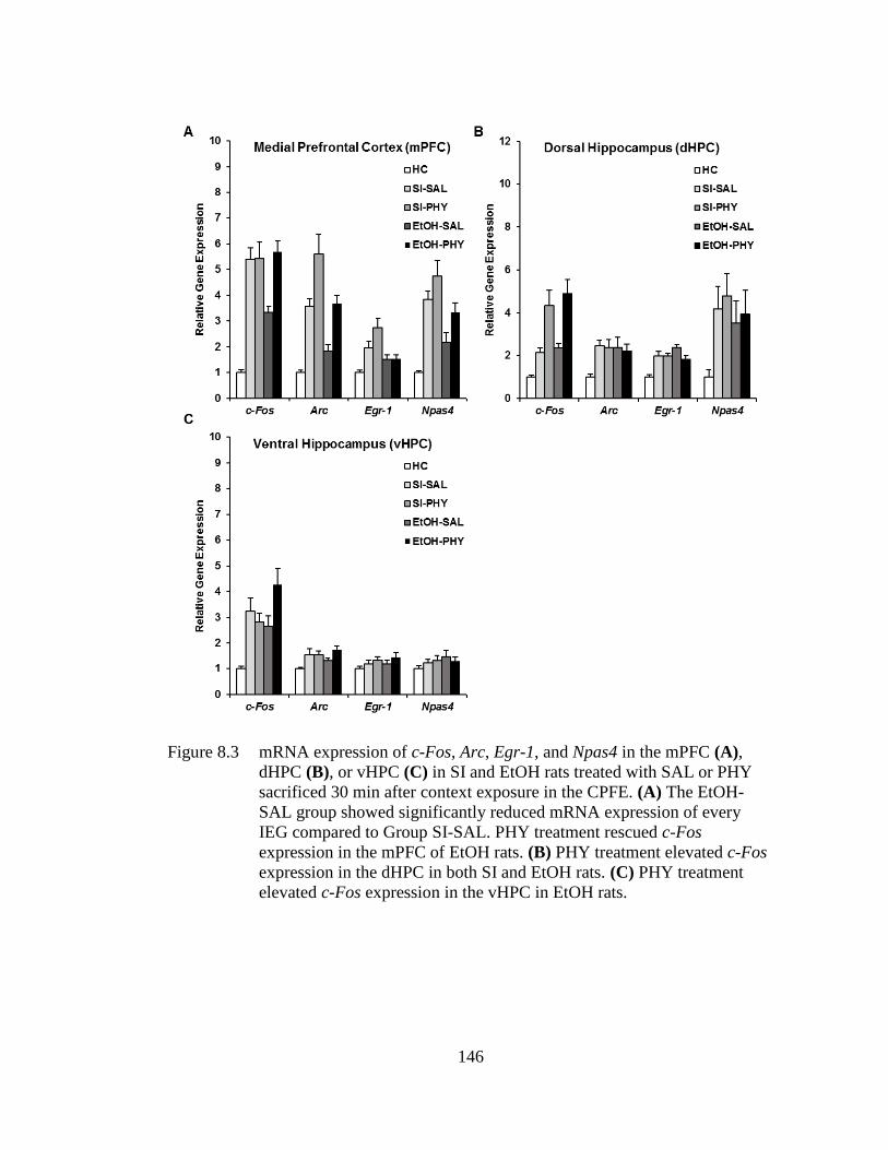
146
Figure 8.3 mRNA expression of c-Fos, Arc, Egr-1, and Npas4 in the mPFC (A),
dHPC (B), or vHPC (C) in SI and EtOH rats treated with SAL or PHY
sacrificed 30 min after context exposure in the CPFE. (A) The EtOH-
SAL group showed significantly reduced mRNA expression of every
IEG compared to Group SI-SAL. PHY treatment rescued c-Fos
expression in the mPFC of EtOH rats. (B) PHY treatment elevated c-Fos
expression in the dHPC in both SI and EtOH rats. (C) PHY treatment
elevated c-Fos expression in the vHPC in EtOH rats.
147
8.4 Discussion
The current set of experiments examined the effects of systemic PHY on
disruptions in contextual fear conditioning and regional IEG expression in adolescent
rats receiving neonatal ethanol exposure during the brain growth spurt. Consistent
with our prior reports (Heroux, Robinson-Drummer, et al., 2019; Murawski et al.,
2012; Murawski & Stanton, 2010, 2011), high binge-like doses of ethanol given over
PD4-9 abolished both post-shock and retention test freezing in the CPFE. This
behavioral disruption in ethanol-exposed rats was rescued by PHY administration
prior to all three phases (Experiment 8.1) or just prior to context preexposure
(Experiment 8.2), with no effects of PHY on behavior of sham-intubated rats.
Furthermore, PHY treatment prior to context learning selectively rescued ethanol-
induced disruptions in prefrontal expression of c-Fos but not Npas4 or Arc, or Egr-1.
Prefrontal expression of Npas4 and Arc was non-specifically boosted by PHY in both
ethanol-exposed and sham-intubated rats, whereas PHY increased Egr-1 in sham but
not ethanol-exposed rats. Hippocampal IEG expression was not impaired by alcohol
or altered by PHY, except for increased c-Fos expression in dHPC regardless of
alcohol exposure. Taken together, augmenting cholinergic signaling rescues neonatal-
alcohol induced impairment of configural learning, memory, and prefrontal gene
expression.
The present study informs the psychological and neural mechanisms through
which neonatal alcohol impairs cognition. As reviewed in Chapter 1, the CPFE
develops between PD17 and PD24 in the rat, after which it depends on activity and
cholinergic muscarinic-receptor cell signaling in the dHPC and mPFC during all three
phases (Heroux et al., 2017; Jablonski et al., 2012; Robinson-Drummer et al., 2016,
2017; Schiffino et al., 2011). While impairment of the CPFE is robust across dosing
148
scenarios, neonatal ethanol exposure has no effect on single-trial sCFCwhich is known
to be hippocampal dependent (Wiltgen et al., 2006; Zelikowsky et al., 2013). Neonatal
ethanol exposure impairs the acquisition and/or consolidation of the conjunctive
context representation on the preexposure day of the CPFE (Goodfellow & Lindquist,
2014; Heroux, Robinson-Drummer, et al., 2019), resulting in abolished post-shock and
retention test freezing [see Experiment 8.1 and 8.2]. Moreover, ethanol-exposed rats
have significantly reduced prefrontal but not hippocampal immediate early gene
expression during context learning in the CPFE [Heroux et al., 2019; see Chapter 7].
In summary, neonatal alcohol impairs incidental context learning or consolidation
through a mechanism that may involve reduced prefrontal activity or plasticity.
In the current study, systemic administration of PHY prior to all three phases
or just prior to context preexposure rescued post-shock and retention test freezing
deficits in ethanol-exposed rats. This extends our prior finding that PHY treatment
prior to all three phases rescues retention freezing in PD7-9 ethanol-exposed rats
(Dokovna et al., 2013). In both cases, the behavioral rescue was specific to ethanol-
exposed rats, without any non-specific boost in performance in sham-intubated rats. In
addition to rescuing behavioral performance, PHY treatment rescued prefrontal c-Fos
expression in ethanol-exposed rats while boosting prefrontal Arc and Npas4 and
hippocampal c-Fos expression in both dosing groups. These behavioral findings
support previous work showing that deficits in trace fear conditioning in rats receiving
PD4-9 ethanol exposure are also dose-dependently rescued by systemic PHY
treatment prior to training (Hunt & Barnet, 2015). While more research is needed,
some mechanisms by which PHY treatment may rescue neurobehavioral deficits
include decreasing neuroinflammation, lowering the threshold for LTP induction, and
149
increasing bioavailability of acetylcholine (Goodfellow et al., 2018; Kalb et al., 2013;
Monk et al., 2012; Pyapali, Turner, Williams, Meck, & Swartzwelder, 1998). Despite
our results suggesting selective prefrontal targeting by alcohol, they do not discount
previous research showing robust cholinergic dysfunction in the hippocampus
resulting from alcohol exposure, they only question whether this dysfunction is
exclusively responsible for cognitive deficits (see Section 3.3 and 3.5 in Chapter 3
for full discussion). Nevertheless, taken together, the results in this chapter further
support our conclusion that impaired context learning or consolidation by PD4-9
ethanol exposure reflects impaired prefrontal activity or plasticity involving
cholinergic signaling.
The current study supports and extends previous literature examining the
neurobiology of contextual fear conditioning and the role of IEGs in learning and
memory. As reviewed in Chapter 2, we chose to examine the IEGs c-Fos, Arc, Egr-1,
and Npas4 because of their involvement in synaptic plasticity supporting LTM
formation and consolidation; and because neonatal ethanol exposure impairs
expression of these IEGs during context learning (Chapter 7; Jablonski et al., 2018).
Despite emerging evidence supporting a role of the prefrontal circuitry across learning
paradigms, especially those relevant to alcohol-induced insult (Gilmartin et al., 2014;
Giustino & Maren, 2015; Heroux, Robinson-Drummer, et al., 2019; Heroux et al.,
2017; Ramanathan et al., 2018; Zelikowsky et al., 2014), examination of the role of
prefrontal IEG-expressing neurons in memory has been largely ignored in favor of the
hippocampus. Although the mechanism is not known, we show that PHY treatment in
ethanol-exposed rats elevates expression of c-Fos, Arc, and Npas4 in the prefrontal
cortex bot only c-Fos in the hippocampus. PHY treatment reversed the alcohol-
150
induced deficit in context fear and in prefrontal c-Fos expression. For other prefrontal
IEG expression as well as for dHPC c-Fos expression, PHY treatment elevated IEG
expression but did not rescue the alcohol-induced deficit. While the current study
cannot fully establish causality between gene expression and behavior, the data
suggest a mechanistic role for prefrontal c-Fos expression. Increased acetylcholine
bioavailability via systemic PHY treatment elevates c-Fos expression by activating
neuronal nicotinic and muscarinic receptors across receptor-populated brain regions,
including the prefrontal cortex (Kaufer, Friedman, Seidman, & Soreq, 2016; Pongrac
& Rylett, 2003; Thomsen et al., 2008). While there is a clear disruption in dHPC
cholinergic receptor function after neonatal alcohol exposure in rats, this disruption
likely extends beyond the dHPC and might inform our findings in the mPFC (Monk et
al., 2012). Given that c-Fos expression is linked to neuronal activity and plasticity
necessary for cellular consolidation of long-term memory (Gallo et al., 2018;
Minatohara et al., 2016), PHY treatment likely recues alcohol-induced deficits by
augmenting plasticity-related protein expression. It is also possible that elevated
prefrontal Arc and hippocampal c-Fos expression in ETOH-PHY rats contributed to
the rescue by exceeding a threshold of expression that was not met in EtOH-SAL rats.
Another contributing factor might be IEG expression differences between neuronal
sub-regions (e.g., IL vs. PL) and sub-types between dosing conditions, which may
contribute to the cholinergic rescue of neurobehavioral effects of alcohol. More
research is needed to establish mechanistic roles of cholinergic signaling, regional
expression of specific IEGs, and behavioral performance in normally developing and
ethanol-exposed rats.
151
In summary, our findings demonstrate that the acetylcholinesterase inhibitor
physostigmine rescues ethanol-induced disruptions in context memory and prefrontal
expression of some immediate early genes in adolescent rats. While the current study
cannot establish a causal link between IEG expression and behavior, these results
suggest that ethanol disrupts behavioral performance by altering activity and/or
plasticity induced by cholinergic signaling in the prefrontal cortex during configural
learning and memory. Taken together with our recent reports (Dokovna et al., 2013;
Heroux, Robinson-Drummer, et al., 2019; Jablonski et al., 2018), these findings are
important because prefrontal dysfunction is an integral hallmark of FASD in humans,
but animal models have thus far largely failed to capture prefrontal dysfunction after
third-trimester equivalent exposure. Building upon an emerging body of animal and
human research linking alcohol and cholinergic dysfunction, future experiments
should characterize the effects of intra-cranial infusions of drugs that augment
cholinergic signaling into mPFC or other discrete brain regions on behavioral
performance of alcohol-exposed rats across the lifespan.

152
SUMMARY AND CONCLUSIONS
This dissertation examined the neural and behavioral mechanisms of context
and contextual fear learning and memory in the CPFE and how these mechanisms are
disrupted by third-trimester equivalent alcohol exposure in rats. Experiments 6.1-6.2
examined prefrontal and hippocampal mechanisms of incidental context learning and
memory during context preexposure in the CPFE. Experiments 7.1-7.2 examined the
effects of PD4-9 alcohol exposure on post-shock and retention test freezing in the
CPFE and sCFC. Experiment 7.3 examined the effects of this exposure on regional
molecular activity during context preexposure in the CPFE. Finally, Experiments 8.1-
8.2 examined the efficacy of the acetylcholinesterase inhibitor physostigmine in
rescuing impaired regional molecular activity and behavioral performance in alcohol-
exposed rats in the CPFE. In the following discussion I summarize the rationale,
hypotheses, findings, and future directions for these experiments and briefly discuss
them in relation to the neurobiology of CFC and rat models of FASD.
9.1 Prefrontal and Hippocampal Mechanisms of Context and Contextual Fear
Learning and Memory in the CPFE
The rationale for the experiments in this dissertation largely stems from our
recent discovery of a necessary role of the prefrontal cortex across all three phases of
the CPFE variant of Pavlovian CFC (see Section 4.2 in Chapter 4; Heroux et al.,
2017). These findings lie in stark contrast to the general lack of an effect of prefrontal
inactivation or lesions on performance of sCFC besides increased fear generalization
Chapter 9
153
(Giustino & Maren, 2015; Xu & Südhof, 2013). While a modulatory role of prefrontal
plasticity in the consolidation of context-shock associations has been previously
established (Rozeske et al., 2015), our study provided novel evidence of prefrontal
circuitry underlying the acquisition and/or consolidation of a context representation in
the absence of aversive stimuli (Heroux et al., 2017; Robinson-Drummer et al., 2017).
Due to a similar dependence of the CPFE on hippocampal activity, we hypothesized
that the mPFC interacts with the HPC via reciprocal connectivity with the VMT to
support the encoding or consolidation of a long-term conjunctive context
representation in the CPFE (see Section 2.1.3 in Chapter 2; Heroux et al., 2017).
Accordingly, in Chapter 6, we first asked the question: does prefrontal inactivation
effect molecular activity in the ventral midline thalamus and hippocampus during
incidental context exposure? In Experiment 6.1, we found that prefrontal inactivation
disrupted IEG expression in the mPFC, VMT, and VHPC but not dHPC during
context preexposure. We then asked the question: Does ventral hippocampal
inactivation effect molecular activity in the prefrontal cortex and dorsal hippocampus
during incidental context exposure? In Experiment 6.2, we found that ventral
hippocampal inactivation disrupted IEG expression in the vHPC, mPFC, and dHPC
during this preexposure. These results did not support our initial mPFC-dHPC
interaction hypothesis, as prefrontal inactivation did not alter dHPC activity during
context exposure. Instead, to our knowledge, this is the first study to suggest a role of
circuitry between the mPFC and vHPC in incidental context learning and memory
during the CPFE.
It’s informative to compare these results to recent studies and models of
prefrontal circuitry underlying CFC (see Section 2.1.2 in Chapter 2; Gilmartin,
154
Balderston, & Helmstetter, 2014; Giustino & Maren, 2015; Rozeske, Valerio,
Chaudun, & Herry, 2015). In particular, Rozeske et al.’s (2015) model suggests a
modulatory role of the PFC in the context specificity and consolidation of the context-
shock association. In this model, during context encoding, the HPC sends contextual
information to the PFC, which regulates feature encoding and specificity of the
context representation via projections to the NR and then HPC (Ramanathan et al.,
2018; Rozeske et al., 2015; Xu & Südhof, 2013). This contextual information is sent
to the amygdala for the acquisition of the context-shock association and is then sent to
the AC, which in turn modulates consolidation and fear expression. In contrast, unlike
in sCFC (see Section 2.1.1 in Chapter 2), we report no compensation occurs after
mPFC or vHPC inactivation in the CPFE (Heroux et al., 2017; Matus-Amat et al.,
2007). It’s unlikely that the PFC is required for encoding contextual cues in the CPFE
as rats can acquire a context-shock association in sCFC under PFC inactivation
(Giustino & Maren, 2015; Heroux et al., 2017). Rozeske et al.’s (2015) model would
predict that prefrontal inactivation prior to context preexposure would not impair the
CPFE per se, but instead result in a less precise context representation and thus more
generalized fear when tested in a non-conditioned context. While our data doesn’t
directly support this, it’s possible to reconcile this difference when considering that,
unlike sCFC, the CPFE requires a configural context representation to be associated
with foot-shock (Jablonski et al., 2012; Rudy & O’Reilly, 1999b). Therefore, via
downstream effects on NR and vHPC, this prefrontal inactivation may result in a less
feature-rich conjunctive context representation, subsequently interfering with rapid
pattern completion and thus context retrieval during immediate-shock training on the
next day. One way to test this hypothesis could be by taking advantage of the finding
155
that, in the CPFE, the context representation does not undergo reconsolidation after
retrieval unless a biologically significant event occurs such as introducing novel
stimuli (Biedenkapp & Rudy, 2004). Thus, if rats under prefrontal inactivation acquire
a less feature-rich representation, then reexposure to the context should trigger
reconsolidation to update with features that were originally missed, rendering the
memory susceptible to protein synthesis inhibition. Therefore, interfering with PFC or
HPC during a second context preexposure should impair the CPFE only in rats that
were under prefrontal inactivation during the first preexposure. This general
interpretation would be consistent with studies done by Xu & Südhof (2013) and
Ramanathan et al. (2018), which suggest that PFC-NR connectivity modulates the
precision of hippocampal-dependent context representations during CFC (see Section
2.1.2 in Chapter 2). One important caveat is that it’s unclear if the roles of the mPFC
and NR would change when contextual learning is incidental vs. driven by shock
reinforcement, as these previous studies fail to dissociate context from contextual fear
learning. If the role of the NR is in specificity of context encoding but not contextual
fear per se, then inactivation of NR (or PFC) during training should have no effect on
the CPFE as the context was encoded on the previous day. While we’ve shown that
pre-training inactivation of the PFC does not impair fear acquisition, it’s unknown if
this manipulation caused generalization to the alternate context (Heroux et al., 2017).
These are all fruitful directions for future research, especially because the CPFE
dissociates these component processes of CFC.
These recent studies from our lab (see Chapter 4) and others motivate the first
working model of the neurobiology of the CPFE variant of CFC (see Figure 9.1 and
9.2). Incorporating prefrontal circuitry, this model significantly expands upon earlier
156
work that almost exclusively focused on hippocampal mechanisms (Matus-Amat et
al., 2004; Rudy, 2009; Rudy et al., 2002; Rudy & O’Reilly, 1999a). The proposed
neurobiology underlying discrete processes that occur during context preexposure can
be seen in Figure 9.1. We propose that encoding of contextual features occurs within
the PHR before being relayed directly to the dorsal and indirectly to the ventral HPC.
This contextual information is sent to the mPFC via direct projections from the vHPC,
which in turn projects back to the vHPC via NR. These specific pathways would be
supported by the results presented in Chapter 6, in which vHPC inactivation disrupted
mPFC but not VMT IEG expression, but mPFC inactivation disrupted VMT and
vHPC IEG expression. The former suggests a direct projection, and the latter suggests
indirect effects of vHPC activity via the NR, in the absence of any direct mPFC-HPC
projections in rats. It’s also possible that that vHPC inactivation alters mPFC
indirectly by impairing dHPC activity and thus any projections from the dHPC to the
AC that convey contextual information, although it’s unlikely that the dHPC would
need vHPC input to achieve this (Fanselow & Dong, 2010). Moreover, these
prefrontal projections to the NR likely contribute to the specificity of the context
representation via modulating the number of features that get incorporated into the
conjunctive context representation (Ramanathan et al., 2018; Xu & Südhof, 2013).
Amygdalar projections to the mPFC originating from the vHPC might communicate
cue saliency during exposure (Gilmartin et al., 2014; Sengupta et al., 2018), and thus
may modulate this incorporation process. Context preexposure requires BLA activity
but not plasticity, suggesting a role of this region during encoding (Huff & Rudy,
2004). Indeed, Kim & Cho (2017) showed that a subset of vHPC neurons projecting to
the amygdala and mPFC preferentially respond to context exposure, and have
157
excitatory influences on these structures during context encoding. In any case, after
receiving information about features from the NR, the vHPC sends this contextual
information back to the dHPC indirectly through the PHR. Finally, consolidation of
the conjunctive context representation is supported by dorsal and ventral hippocampal
communication, as pharmacological disruption of these regions pre- or post- context
preexposure impairs the CPFE (Chang & Liang, 2012; Cullen et al., 2017; Matus-
Amat et al., 2007; Robinson-Drummer et al., 2016; Rudy & Matus-Amat, 2005;
Schiffino et al., 2011).
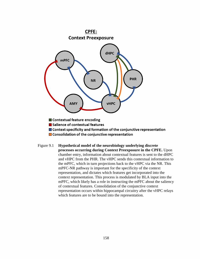
158
Figure 9.1 Hypothetical model of the neurobiology underlying discrete
processes occurring during Context Preexposure in the CPFE. Upon
chamber entry, information about contextual features is sent to the dHPC
and vHPC from the PHR. The vHPC sends this contextual information to
the mPFC, which in turn projections back to the vHPC via the NR. This
mPFC-NR pathway is important for the specificity of the context
representation, and dictates which features get incorporated into the
context representation. This process is modulated by BLA input into the
mPFC, which likely has a role in instructing the mPFC about the saliency
of contextual features. Consolidation of the conjunctive context
representation occurs within hippocampal circuitry after the vHPC relays
which features are to be bound into the representation.
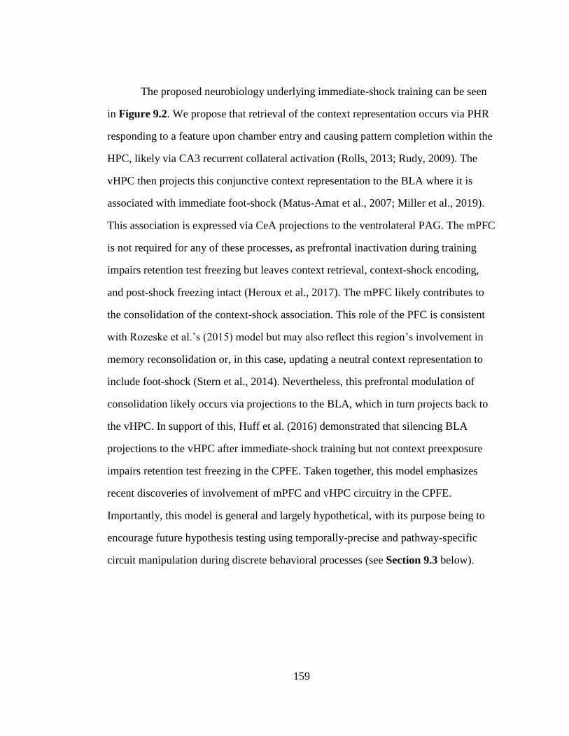
159
The proposed neurobiology underlying immediate-shock training can be seen
in Figure 9.2. We propose that retrieval of the context representation occurs via PHR
responding to a feature upon chamber entry and causing pattern completion within the
HPC, likely via CA3 recurrent collateral activation (Rolls, 2013; Rudy, 2009). The
vHPC then projects this conjunctive context representation to the BLA where it is
associated with immediate foot-shock (Matus-Amat et al., 2007; Miller et al., 2019).
This association is expressed via CeA projections to the ventrolateral PAG. The mPFC
is not required for any of these processes, as prefrontal inactivation during training
impairs retention test freezing but leaves context retrieval, context-shock encoding,
and post-shock freezing intact (Heroux et al., 2017). The mPFC likely contributes to
the consolidation of the context-shock association. This role of the PFC is consistent
with Rozeske et al.’s (2015) model but may also reflect this region’s involvement in
memory reconsolidation or, in this case, updating a neutral context representation to
include foot-shock (Stern et al., 2014). Nevertheless, this prefrontal modulation of
consolidation likely occurs via projections to the BLA, which in turn projects back to
the vHPC. In support of this, Huff et al. (2016) demonstrated that silencing BLA
projections to the vHPC after immediate-shock training but not context preexposure
impairs retention test freezing in the CPFE. Taken together, this model emphasizes
recent discoveries of involvement of mPFC and vHPC circuitry in the CPFE.
Importantly, this model is general and largely hypothetical, with its purpose being to
encourage future hypothesis testing using temporally-precise and pathway-specific
circuit manipulation during discrete behavioral processes (see Section 9.3 below).
160
Figure 9.2 Hypothetical model of the neurobiology underlying discrete
processes occurring during Immediate-shock Training in the CPFE.
Upon chamber entry, PHR responds to features of the context and drives
hippocampal pattern completion and thus context retrieval via projections
to the dHPC and activation of recurrent collaterals in the CA3 sub-region.
The vHPC sends this context representation to the BLA, where it
converges with foot-shock US inputs to support the formation of the
context-shock association. Immediate expression of the context-shock
association via the species-typical freezing response occurs via direct
projections between the CeA and the ventrolateral PAG. Consolidation of
this association is supported by ventral hippocampal and amygdalar
connectivity with the mPFC.
161
9.2 Acute Enhancement of Cholinergic Function Rescues Neurobehavioral
Disruption after Neonatal Alcohol Exposure in Rats
As reviewed in Chapter 3, PD4-9 alcohol exposure abolishes retention test
freezing in the CPFE but leaves cued and un-signaled fear conditioning intact in rats
(Murawski et al., 2012; Murawski & Stanton, 2010). Our lab has shown that this
exposure results in CA1 pyramidal cell loss and a knockdown of hippocampal c-Fos
protein expression during context preexposure, which could underlie disrupted context
learning and/or memory (Murawski et al., 2012). Despite this, these previous studies
are unable to dissociate PD4-9 alcohol effects on preexposure or training day
processes because only retention-test freezing was measured. Post-shock freezing
helps inform which of these processes is impaired (Chapter 1, Jablonski et al., 2013)
and PD7-9 alcohol appears to selectively impair retention-test freezing (Jablonski &
Stanton, 2014). Accordingly, in Chapter 7, we first asked the question: does PD4-9
alcohol exposure disrupt incidental context learning and memory during context
preexposure or contextual fear learning and memory during immediate-shock training
in the CPFE? In Experiment 7.1, alcohol exposure impaired both post-shock and
retention test freezing in the CPFE. These results are still unable to determine if
alcohol-exposed rats are unable to acquire, consolidate, or retrieve a context
representation or if these processes are intact but they are unable to acquire a context-
shock association during training. To examine these possibilities in another paradigm,
we asked the question: does PD4-9 alcohol exposure impair context or contextual fear
learning in sCFC? In Experiment 7.2, alcohol-exposed rats had intact retention test
freezing in single-trial sCFC. This result indicated that: (1) alcohol-exposed rats can
acquire a context representation and associate it with shock, (2) potential
“performance effects” of alcohol exposure such as reduced feature perception or shock
162
sensitivity do not underlie deficits in the CPFE, and (3) these rats are unimpaired in a
hippocampal-dependent (Wiltgen et al., 2006) but prefrontal-independent (Heroux et
al., 2017) variant of CFC. Therefore, we hypothesized that PD4-9 exposure results in
prefrontal dysfunction and impaired consolidation of the conjunctive context
representation in the CPFE. Finally, the third question we asked was: does neonatal
alcohol exposure alter molecular activity in the prefrontal cortex and hippocampus
during context preexposure in the CPFE? In Experiment 7.3, alcohol exposure
impaired expression of the IEGs c-Fos, Arc, Egr-1, and Npas4 in the mPFC but not
dHPC during context exposure. These data support our hypothesis that prefrontal
impairments underlie alcohol-induced CPFE deficits that were historically attributed
to hippocampal impairment(s). These results demonstrate that third-trimester-
equivalent alcohol exposure disrupts behaviorally-driven molecular activity in the
prefrontal cortex that may subserve incidental contextual memory processes in rats.
As reviewed in Chapter 3, neonatal alcohol exposure causes both acute and
persistent disruptions in cholinergic signaling important for successful
neurodevelopment and cognition across the lifespan in rats. Developmental choline
supplementation mitigates alcohol-induced disruptions in MWM, CFC, trace fear
conditioning, and spatial working memory (Hunt & Barnet, 2015; Idrus et al., 2017;
Ryan et al., 2008; Thomas et al., 2009, 2007, 2004, 2010; Wagner & Hunt, 2006).
This treatment is proving to be at least partially efficacious in rescuing cognitive
function in developing children with FASD (Jacobson et al., 2018; Wozniak et al.,
2015, 2013). One major question that emerges from this literature is whether acutely
enhancing cholinergic signaling later after exposure would also rescue impaired
cognition. Our lab has previously shown that systemic administration of the
163
acetylcholinesterase inhibitor physostigmine prior to every phase rescues deficits in
retention test freezing in the CPFE in PD7-9 alcohol-exposed rats (Dokovna et al.,
2013). The exact phase of the CPFE that mediates this rescue effect and whether the
PD7-9 result would generalize to the more severe PD4-9 exposure window is unclear.
Therefore, in Chapter 8, we asked the question: does acute physostigmine treatment
prior to every phase or just during context preexposure rescues PD4-9 alcohol-
induced deficits in the CPFE? In Experiment 8.1, alcohol-exposed rats given
physostigmine prior to every phase showed normalized post-shock and retention test
freezing relative to sham-intubated rats, which did not benefit from treatment. Given
the dependence of preexposure day processes on cholinergic signaling in the mPFC
and dHPC (see Chapter 4; Robinson-Drummer et al., 2016, 2017), we then
hypothesized that this treatment prior to just context exposure would rescue impaired
performance and IEG expression in alcohol-exposed rats. Therefore, we asked: Can
alcohol-induced neurobehavioral disruptions be rescued by enhancing cholinergic
signaling via acute acetylcholinesterase inhibitor treatment prior to context exposure?
In Experiment 8.2, physostigmine treatment prior to just context preexposure rescued
the CPFE in alcohol-exposed rats. This treatment specifically rescued prefrontal c-Fos
expression, in tandem with non-specific elevations in mPFC Arc and Npas4 as well as
dHPC and VHPC c-Fos in both dosing groups. Future studies are needed to establish
regional specificity of this rescue (i.e., whether intra-mPFC physostigmine prior to
context preexposure produces the same effect). Such studies would address the critical
issue of whether there is a causal link between regional alterations in activity or
cholinergic signaling and impaired cognition across in this rodent model of FASD.
164
The results of these two research aims have several broad implications for
developmental FASD research in humans and animal models. The results of Chapter
7 highlight the need, as a field, to re-examine alcohol-induced deficits that have been
attributed solely to hippocampal dysfunction in behavioral tasks that require both the
dHPC and mPFC (see Chapters 3 and 7 for extended discussion). Indeed, in part
because the CPFE was largely used as a “hippocampal-dependent” CFC task, we
previously attributed deficits in the CPFE almost solely to reduced hippocampal
molecular activity and cell counts (G. F. Hamilton et al., 2011; Murawski et al., 2012;
Murawski & Stanton, 2010). While our current results do not rule out hippocampal
involvement, they do suggest a major role of impaired prefrontal molecular and
cholinergic activity in cognitive deficits arising from third-trimester equivalent alcohol
exposure in rats. These findings are particularly important given evidence showing
alcohol deficits are the largest in tasks that engage both the HPC and PFC, such as in
trace fear conditioning (DuPont et al., 2014; Gilmartin et al., 2014; Goodfellow et al.,
2016; Hunt et al., 2009; Wagner & Hunt, 2006). This pattern is especially interesting
given that it was recently discovered that this alcohol exposure results in a dramatic
(>20%) reduction in neurons in the NR, which serves as the major communication
nexus between these two regions (Gursky, Savage, & Klintsova, 2019). Taken
together with our proposed model (see Figure 9.1), it’s likely that PD4-9 alcohol
exposure impairs the CPFE by disrupting communication between the mPFC and
vHPC. The results of Chapter 8 demonstrate that acute treatment with drugs that
enhance cholinergic system function later in development is sufficient to rescue select
neurobehavioral deficits in rat models of FASD. This finding is important for two
reasons. First, this finding complements only two other studies that have examined
165
acute treatment with acetylcholinesterase inhibitors in exposed rats (Dokovna et al.,
2013; Hunt & Barnet, 2015). Importantly, this is the first study to suggest a neural
mechanism of this rescue in enhanced prefrontal activity via increased activation of
muscarinic acetylcholine receptors, which are required for the CPFE (Heroux, Horgan,
Rosen, et al., 2019; Robinson-Drummer et al., 2016, 2017). Second, these results
suggest that acute enhancement of cholinergic function might be efficacious in
attenuating select cognitive deficits in humans with FASD. This demands further
testing, as currently the alcohol field is focused on using cholinergic treatments (e.g.,
choline supplementation) concurrent with or soon after alcohol exposure but not later
in life (Jacobson et al., 2018; Wozniak et al., 2015, 2013). Collectively, this research
promises to shed light on the basic neural mechanisms underlying cognitive
impairment resulting from developmental alcohol exposure.
9.3 Future Directions
9.3.1 Limitations
The neurobiological analyses in this dissertation are subject to several
important limitations discussed in the previous experimental chapters. Notably, these
limitations include 1) examination of mRNA instead of protein expression, 2)
dissecting out entire brain regions vs. individual neural sub-regions, 3) inability to
determine cell-type specific effects, and 4) assessing only one time point (30min) after
experimental manipulations. Furthermore, we are unable to determine the specific
function of the observed IEG expression in supporting behavior in the absence of loss-
and gain-of-function experiments that directly manipulate specific gene expression
during the CPFE. While we measured the expression of several IEGs (c-Fos, Arc, Egr-
166
1, and Npas4), patterns of expression of these genes generally did not differ across the
qPCR experiments. A notable exception to this is the lack of Arc, Egr-1, and Npas4
expression seen in the VMT when compared to robust c-Fos expression observed in
this area. This may suggest that plasticity in this region is not heavily engaged by
context exposure, as c-Fos expression has been tied more heavily to neural activity but
not plasticity supporting LTM (see Section 2.2 in Chapter 2 for more discussion).
Another exception can be seen in the effects of physostigmine on IEG expression in SI
and EtOH rats, which was more heavily tied to a boost in c-Fos and Arc expression
(see Section 8.4 in Chapter 8 for specific discussion). Despite these limitations, the
methodology in this dissertation permits measurement of several IEGs. This provides
novel normative and pharmacological data concerning cholinergic and GABAergic
modulation of IEG expression during behavioral experience (context exposure).
Pursuing these findings with other methodologies that address the limitations
mentioned above is an obvious and important direction for future research. Finally,
another important future direction is to determine whether similar neurobehavioral
impairments are seen after exposure to other teratogens (e.g., such as nicotine and
marijuana) during the same developmental window in rats.
9.3.2 Experimental Predictions
Several experimental predictions can be made when considering the findings
of all three aims of this dissertation and our newly proposed model of the
neurobiology of the CPFE (see Figure 9.1 and 9.2). Some of these predictions,
especially those concerning the specific roles of the mPFC and NR during incidental
context encoding and consolidation appear in Sections 2.1.2 and 2.1.3 in Chapter 2
and Section 9.1 in Chapter 9 . Our model predicts that pre-preexposure but not pre-
167
training inactivation of NR would impair the CPFE by causing a feature-impoverished
context representation (Xu & Südhof, 2013). While inactivation of mPFC prior to
either phase disrupts 24-hr retention of the CPFE (Heroux et al., 2017), post-phase
inactivation should only effect consolidation of training day processes. Moreover,
contrary to our original hypothesis, we also now predict that mPFC-vHPC but not
mPFC-dHPC pharmacological disconnection during either of these phases would
impair the CPFE, as our results strongly suggest that communication between these
structures is critical for these processes. In line with this, our results suggest that
prefrontal communication with the vHPC occurs via the NR, whereas vHPC
communication with mPFC occurs via direct projection (see Chapter 6). Therefore,
pathway-specific silencing of direct mPFC-VMT, VMT-vHPC, or vHPC-mPFC
projections during context preexposure will disrupt the CPFE. In our rat model of
FASD, our results suggest that neonatal alcohol exposure disrupts prefrontal function
that may be important for the formation of a detailed conjunctive context
representation. Taken together with Murawski et al. (2012), these results suggest that
this exposure targets neurobiology underlying both the acquisition and consolidation
of incidentally-encoded contextual information. Despite not observing robust vHPC
IEG expression after alcohol or sham intubation procedures (relative to undisturbed
rats in Experiments 6.1 and 6.2), this effect on prefrontal function likely has adverse
consequences on vHPC signaling, via the NR (which is heavily affected by PD4-9
alcohol exposure; see Gursky et al., 2019). An important test of this hypothesis is to
use gain-of-function manipulations such as optogenetic excitation of specific
prefrontal circuitry with the HPC (e.g., mPFC-NR, NR-mPFC, NR-vHPC) in alcohol-
exposed rats. Whether impairments in similar thalamocortical pathways are seen in
168
humans with FASD remains to be seen, and is also a fruitful direction for future
research.
9.4 Summary Statement
The goals of this dissertation were to elucidate prefrontal mechanisms
underlying contextual learning and memory and how these processes are disrupted by
third-trimester equivalent alcohol exposure in rats. This dissertation also examined the
therapeutic potential of acute enhancement of cholinergic function in rescuing these
deficits. As reviewed above, the results of this dissertation have several broad
implications. First, we demonstrate a novel role of prefrontal and ventral hippocampal
circuitry in incidental context learning and memory. By leveraging these discoveries,
this work allows us to propose the neural circuitry underlying context and contextual
fear learning and memory in the CPFE. This is important because processes of context
and contextual fear are dissociated in this task, so this work allows for elucidation of
the fundamental neural circuitry underlying these distinct processes in normally and
abnormally developing (e.g., alcohol-exposed) animals. Second, we demonstrate the
efficacy of acute physostigmine treatment in adolescence in reversing cognitive
impairments seen in a rodent model of FASD. Taken together with our other studies
(Dokovna et al., 2013; Robinson-Drummer et al., 2016, 2017), this work strongly
suggest impaired cholinergic function within the prefrontal cortex and hippocampus
plays an important causal role in these cognitive deficits. These studies provide a
foundation for future work examining the efficacy of similar treatments in other
models of FASD and in humans, and suggest a potential neural mechanism underlying
the intervention.
169
Alberini, C. M. (2009). Transcription Factors in Long-Term Memory and Synaptic
Plasticity. Physiology Rev, 121–145. https://doi.org/10.1152/physrev.00017.2008.
Amin, E., Pearce, J. M., Brown, M. W., & Aggleton, J. P. (2006). Novel temporal
configurations of stimuli produce discrete changes in immediate-early gene
expression in the rat hippocampus. European Journal of Neuroscience, 24(9),
2611–2621. https://doi.org/10.1111/j.1460-9568.2006.05131.x
Anagnostaras, S. G., Gale, G. D., & Fanselow, M. S. (2001). Hippocampus and
contextual fear conditioning: Recent controversies and advances. Hippocampus,
11(1), 8–17. https://doi.org/10.1002/1098-1063(2001)11:1<8::AID-
HIPO1015>3.0.CO;2-7
Anagnostaras, S. G., Maren, S., & Fanselow, M. S. (1995). Scopolamine selectively
disrupts the acquisition of contextual fear conditioning in rats. Neurobiology of
Learning and Memory, 64(3), 191–194. https://doi.org/10.1006/nlme.1995.0001
Anagnostaras, S. G., Maren, S., Sage, J. R., Goodrich, S., & Fanselow, M. S. (1999).
Scopolamine and Pavlovian Fear Conditioning in Rats: Dose-Effect Analysis.
Neuropsychopharmacology, 21(6), 731–744. https://doi.org/10.1016/S0893-
133X(99)00083-4
Asok, A., Leroy, F., Rayman, J. B., & Kandel, E. R. (2019). Molecular Mechanisms of
the Memory Trace. Trends in Neurosciences, 42(1), 14–22.
https://doi.org/10.1016/j.tins.2018.10.005
Asok, A., Schreiber, W. B., Jablonski, S. A., Rosen, J. B., & Stanton, M. E. (2013).
Egr-1 increases in the prefrontal cortex following training in the context
preexposure facilitation effect (CPFE) paradigm. Neurobiology of Learning and
Memory, 106, 145–153. https://doi.org/10.1016/j.nlm.2013.08.006
Ballesteros, C. I., De Oliveira Galvão, B., Maisonette, S., & Landeira-Fernandez, J.
(2014). Effect of dorsal and ventral hippocampal lesions on contextual fear
conditioning and unconditioned defensive behavior induced by electrical
stimulation of the dorsal periaqueductal gray. PLoS ONE, 9(1).
https://doi.org/10.1371/journal.pone.0083342
REFERENCES
170
Bannerman, D. M., Good, M. A., Yee, B. K., Heupel, M. J., Iversen, S. D., & Rawlins,
J. N. P. (1999). Double dissociation of function within the hippocampus: A
comparison of dorsal, ventral, and complete hippocampal cytotoxic lesions.
Behavioral Neuroscience, 113(6), 1170–1188. https://doi.org/10.1037/0735-
7044.113.6.1170
Beeman, C. L., Bauer, P. S., Pierson, J. L., & Quinn, J. J. (2013). Hippocampus and
medial prefrontal cortex contributions to trace and contextual fear memory
expression over time. Learning and Memory, 20(6), 336–343.
https://doi.org/10.1101/lm.031161.113
Berman, R. F., & Hannigan, J. H. (2000). Effects of prenatal alcohol exposure on the
hippocampus: Spatial behavior, electrophysiology, and neuroanatomy.
Hippocampus, 10(1), 94–110. https://doi.org/10.1002/(SICI)1098-
1063(2000)10:1<94::AID-HIPO11>3.0.CO;2-T
Besnard, A., Caboche, J., & Laroche, S. (2013). Recall and Reconsolidation of
Contextual Fear Memory: Differential Control by ERK and Zif268 Expression
Dosage. PLoS ONE, 8(8), 1–8. https://doi.org/10.1371/journal.pone.0072006
Biedenkapp, J. C., & Rudy, J. W. (2004). Context memories and reactivation:
constraints on the reconsolidation hypothesis. Behavioral Neuroscience, 118(5),
956–964. https://doi.org/10.1037/0735-7044.118.5.956
Biedenkapp, J. C., & Rudy, J. W. (2009). Hippocampal and extrahippocampal systems
compete for control of contextual fear: Role of ventral subiculum and amygdala.
Learning & Memory, (303), 1–22. https://doi.org/10.1101/lm.1099109.16
Bloodgood, B. L., Sharma, N., Browne, H. A., Trepman, A. Z., & Greenberg, M. E.
(2013). The activity-dependent transcription factor NPAS4 regulates domain-
specific inhibition. Nature, 503(7474), 121–125.
https://doi.org/10.1038/nature12743
Bonthius, D. J., & West, J. R. (1990). Alcohol‐Induced Neuronal Loss in Developing
Rats: Increased Brain Damage with Binge Exposure. Alcoholism: Clinical and
Experimental Research, 14(1), 107–118. https://doi.org/10.1111/j.1530-
0277.1990.tb00455.x
Boschen, K. E., Keller, S. M., Roth, T. L., & Klintsova, A. Y. (2018). Epigenetic
mechanisms in alcohol- and adversity-induced developmental origins of
neurobehavioral functioning. Neurotoxicology and Teratology, 66(July 2017),
63–79. https://doi.org/10.1016/j.ntt.2017.12.009
Bozon, B., Davis, S., & Laroche, S. (2003). A requirement for the immediate early
171
gene zif268 in reconsolidation of recognition memory after retrieval. Neuron,
40(4), 695–701. Retrieved from http://www.ncbi.nlm.nih.gov/pubmed/14622575
Brown, K. L., Calizo, L. H., & Stanton, M. E. (2008). Dose-dependent deficits in dual
interstimulus interval classical eyeblink conditioning tasks following neonatal
binge alcohol exposure in rats. Alcoholism: Clinical and Experimental Research,
32(2), 277–293. https://doi.org/10.1111/j.1530-0277.2007.00579.x
Brown, K. L., Goodlett, C. R., & Stanton, M. E. (2007). Neonatal Alcohol Exposure
Impairs Acquisition of Eyeblink Conditioned Responses during Discrimination
Learning and Reversal in Weanling Rats. Developmental Psychobiology, 49,
243–257. https://doi.org/10.1002/dev
Brown, K. L., Kennard, J. A., Sherer, D. J., Comalli, D. M., & Woodruff-Pak, D. S.
(2011). The context preexposure facilitation effect in mice: A dose-response
analysis of pretraining scopolamine administration. Behavioural Brain Research,
225(1), 290–296. https://doi.org/10.1016/j.bbr.2011.07.044
Burman, M. a, Murawski, N. J., Schiffino, F. L., Rosen, J. B., & Stanton, M. E.
(2009). Factors governing single-trial contextual fear conditioning in the
weanling rat. Behavioral Neuroscience, 123(5), 1148–1152.
https://doi.org/10.1037/a0016733
Cai, D. J., Aharoni, D., Shuman, T., Shobe, J., Biane, J. S., Song, W., … Silva, A. J.
(2016). A shared neural ensemble links distinct contextual memories encoded
close in time. Nature, 534(7605), 115–118. https://doi.org/10.1038/nature17955
Chakraborty, T., Asok, A., Stanton, M. E., & Rosen, J. B. (2016). Variants of
contextual fear conditioning induce differential patterns of Egr-1 activity within
the young adult prefrontal cortex. Behavioural Brain Research, 302, 122–130.
https://doi.org/10.1016/j.bbr.2016.01.018
Chang, S. D., & Liang, K. C. (2012). Roles of hippocampal GABAA and muscarinic
receptors in consolidation of context memory and context-shock association in
contextual fear conditioning: A double dissociation study. Neurobiology of
Learning and Memory, 98(1), 17–24. https://doi.org/10.1016/j.nlm.2012.04.004
Cheng, D. T., Jacobson, S. W., Jacobson, J. L., Molteno, C. D., Stanton, M. E., &
Desmond, J. E. (2015). Eyeblink classical conditioning in alcoholism and fetal
alcohol spectrum disorders. Frontiers in Psychiatry, 6(NOV), 1–7.
https://doi.org/10.3389/fpsyt.2015.00155
Cholvin, T., Hok, V., Giorgi, L., Chaillan, F., & Poucet, B. (2017). Ventral midline
thalamus is necessary for hippocampal place field stability and cell firing
172
modulation. The Journal of Neuroscience, 38(1), 2039–17.
https://doi.org/10.1523/jneurosci.2039-17.2017
Chowdhury, N., Quinn, J. J., & Fanselow, M. S. (2005). Dorsal hippocampus
involvement in trace fear conditioning with long, but not short, trace intervals in
mice. Behavioral Neuroscience, 119(5), 1396–1402.
https://doi.org/10.1037/0735-7044.119.5.1396
Coelho, C. A. O., Ferreira, T. L., Kramer-Soares, J. C., Sato, J. R., & Oliveira, M. G.
M. (2018). Network supporting contextual fear learning after dorsal
hippocampal damage has increased dependence on retrosplenial cortex. PLoS
Computational Biology (Vol. 14). https://doi.org/10.1371/journal.pcbi.1006207
Corcoran, K. A., & Quirk, G. J. (2007). Activity in prelimbic cortex is necessary for
the expression of learned, but not innate, fears. Journal of Neuroscience, 27(4),
840–844. https://doi.org/10.1523/JNEUROSCI.5327-06.2007
Costa, L. G., Giordano, G., & Guizzetti, M. (2013). Inhibition of cholinergic
muscarinic signaling by ethanol: Potential mechanism of developmental
neurotoxicity and biological plausibility for the beneficial effects of choline
supplementation. The International Journal of Alcohol and Drug Research, 2(3),
17. https://doi.org/10.7895/ijadr.v2i3.72
Cowansage, K. K., Shuman, T., Dillingham, B. C., Chang, A., Golshani, P., &
Mayford, M. R. (2014). Direct Reactivation of a Coherent Neocortical Memory
of Context. Neuron, 84(2), 432–441.
https://doi.org/10.1016/j.neuron.2014.09.022
Cullen, P. K., Ferrara, N. C., Pullins, S. E., & Helmstetter, F. J. (2017). Context
memory formation requires activity- dependent protein degradation in the
hippocampus. Learning & Memory, 589–596.
https://doi.org/10.1101/lm.045443.117.24
Czerniawski, J. J., Ree, F. F., Chia, C., Ramamoorthi, K., Kumata, Y. Y., & Otto, T.
A. T. A. (2011). The importance of having Arc: expression of the immediate-
early gene Arc is required for hippocampus-dependent fear conditioning and
blocked by NMDA receptor antagonism. Journal of Neuroscience, 31(31),
11200–11207. https://doi.org/10.1523/JNEUROSCI.2211-11.2011
Davis, S., Renaudineau, S., Poirier, R., Poucet, B., Save, E., & Laroche, S. (2010).
The Formation and Stability of Recognition Memory: What Happens Upon
Recall? Frontiers in Behavioral Neuroscience, 4(November), 1–11.
https://doi.org/10.3389/fnbeh.2010.00177
173
DeNardo, L. A., Liu, C. D., Allen, W. E., Adams, E. L., Friedmann, D., Fu, L., …
Luo, L. (2019). Temporal evolution of cortical ensembles promoting remote
memory retrieval. Nature Neuroscience, 22(3), 460–469.
https://doi.org/10.1038/s41593-018-0318-7
Denny, C. A., Kheirbek, M. A., Alba, E. L., Tanaka, K. F., Brachman, R. A.,
Laughman, K. B., … Hen, R. (2014). Hippocampal memory traces are
differentially modulated by experience, time, and adult neurogenesis. Neuron,
83(1), 189–201. https://doi.org/10.1016/j.neuron.2014.05.018
Dokovna, L. B., Jablonski, S. A., & Stanton, M. E. (2013). Neonatal alcohol exposure
impairs contextual fear conditioning in juvenile rats by disrupting cholinergic
function. Behavioural Brain Research, 248, 114–120.
https://doi.org/10.1016/j.bbr.2013.03.043
Dolleman-van der Weel, M. J., Griffin, A. L., Ito, H. T., Shapiro, M. L., Witter, M. P.,
Vertes, R. P., & Allen, T. A. (2019). The nucleus reuniens of the thalamus sits at
the nexus of a hippocampus and medial prefrontal cortex circuit enabling
memory and behavior, (305), 1–48. https://doi.org/10.1101/lm.048389.118.Freely
Donald, K. A., Fouche, J. P., Roos, A., Koen, N., Howells, F. M., Riley, E. P., …
Stein, D. J. (2016). Alcohol exposure in utero is associated with decreased gray
matter volume in neonates. Metabolic Brain Disease, 31(1), 81–91.
https://doi.org/10.1007/s11011-015-9771-0
Donley, M. P., & Rosen, J. B. (2017). Novelty and fear conditioning induced gene
expression in high and low states of anxiety. Learning and Memory, 24(9), 449–
462. https://doi.org/10.1101/lm.044289.116.24
DuPont, C. M., Coppola, J. J., Kaercher, R. M., & Lindquist, D. H. (2014). Impaired
trace fear conditioning and diminished ERK1/2 phosphorylation in the dorsal
hippocampus of adult rats administered alcohol as neonates. Behavioral
Neuroscience, 128(2), 187–198. https://doi.org/10.1037/a0035989
Einarsson, E. O., & Nader, K. (2012). Involvement of the anterior cingulate cortex in
formation, consolidation, and reconsolidation of recent and remote contextual
fear memory. Learning & Memory, 19(10), 449–452.
https://doi.org/10.1101/lm.027227.112
Fabio, M. C., March, S. M., Molina, J. C., Nizhnikov, M. E., Spear, N. E., & Pautassi,
R. M. (2013). Prenatal ethanol exposure increases ethanol intake and reduces C-
fos expression in infralimbic cortex of adolescent rats. Pharmacology
Biochemistry and Behavior, 103(4), 842–852.
https://doi.org/10.1016/j.pbb.2012.12.009
174
Fanselow, M. S. (1990). Factors governing one-trial contextual conditioning. Animal
Learning & Behavior, 18(3), 264–270. https://doi.org/10.3758/BF03205285
Fanselow, M. S. (2010). From contextual fear to a dynamic view of memory systems.
Trends in Cognitive Sciences, 14(1), 7–15.
https://doi.org/10.1016/j.tics.2009.10.008
Fanselow, M. S., & Dong, H.-W. (2010). Are the dorsal and ventral hippocampus
functionally distinct structures? Neuron, 65(1), 7–19.
https://doi.org/10.1016/j.neuron.2009.11.031
Fanselow, M. S., & Poulos, A. M. (2005). The Neuroscience of Mammalian
Associative Learning. Annual Review of Psychology, 56(1), 207–234.
https://doi.org/10.1146/annurev.psych.56.091103.070213
Fantie, B. D., & Kolb, B. (1990). An examination of prefrontal lesion size and the
effects of cortical grafts on performance of the Morris water task by rats.
Psychobiology, 18(1), 74–80. https://doi.org/10.3758/BF03327218
Feldon, Ã. J., Yee, B. K., Pothuizen, H. H. J., Zhang, W., & Jongen-Relo, A. L.
(2004). Dissociation of function between the dorsal and the ventral hippocampus
in spatial learning abilities of the rat : a within-subject , within-task comparison
of reference and working spatial memory. European Journal of Neuroscience, 19,
705–712. https://doi.org/10.1111/j.1460-9568.2004.03170.x
Fendt, M., & Fanselow, M. S. (1999). The neuroanatomical and neurochemical basis
of conditioned fear. Neuroscience and Biobehavioral Reviews, 23(5), 743–760.
https://doi.org/10.1016/S0149-7634(99)00016-0
Ferguson, B. R., & Gao, W.-J. (2014). Development of thalamocortical connections
between the mediodorsal thalamus and the prefrontal cortex and its implication in
cognition. Frontiers in Human Neuroscience, 8(January), 1027.
https://doi.org/10.3389/fnhum.2014.01027
Fleischmann, A., Hvalby, O., Jensen, V., Strekalova, T., Zacher, C., Layer, L. E., …
Gass, P. (2003). Impaired long-term memory and NR2A-type NMDA receptor-
dependent synaptic plasticity in mice lacking c-Fos in the CNS. The Journal of
Neuroscience : The Official Journal of the Society for Neuroscience, 23(27),
9116–9122. Retrieved from http://www.ncbi.nlm.nih.gov/pubmed/14534245
Frankland, P. W., & Bontempi, B. (2005). The organization of recent and remote
memories. Nature Reviews. Neuroscience, 6(2), 119–130.
https://doi.org/10.1038/nrn1607
175
Frankland, Paul W., Cestari, V., Filipkowski, R. K., McDonald, R. J., & Silva, A. J.
(1998). The dorsal hippocampus is essential for context discrimination but not for
contextual conditioning. Behavioral Neuroscience, 112(4), 863–874.
https://doi.org/10.1037/0735-7044.112.4.863
Fryer, S. L., Tapert, S. F., Mattson, S. N., Paulus, M. P., Spadoni, A. D., & Riley, E.
P. (2007). Prenatal alcohol exposure affects frontal-striatal BOLD response
during inhibitory control. Alcoholism: Clinical and Experimental Research,
31(8), 1415–1424. https://doi.org/10.1111/j.1530-0277.2007.00443.x
Fukazawa, Y., Saitoh, Y., Ozawa, F., Ohta, Y., Mizuno, K., & Inokuchi, K. (2003).
Hippocampal LTP is accompanied by enhanced F-actin content within the
dendritic spine that is essential for late LTP maintenance in vivo. Neuron, 38(3),
447–460. https://doi.org/10.1016/S0896-6273(03)00206-X
Gallo, F. T., Katche, C., Morici, J. F., Medina, J. H., & Weisstaub, N. V. (2018).
Immediate Early Genes, Memory and Psychiatric Disorders: Focus on c-Fos,
Egr1 and Arc. Frontiers in Behavioral Neuroscience, 12(April), 1–16.
https://doi.org/10.3389/fnbeh.2018.00079
Gewirtz, J. C., Falls, W. A., & Davis, M. (1997). Normal conditioned inhibition and
extinction of freezing and fear- potentiated startle following electrolytic lesions
of medial prefrontal cortex in rats. Behavioral Neuroscience, 111(4), 712–726.
https://doi.org/10.1037/0735-7044.111.4.712
Ghandour, K., Ohkawa, N., Fung, C. C. A., Asai, H., Saitoh, Y., Takekawa, T., …
Inokuchi, K. (2019). Orchestrated ensemble activities constitute a hippocampal
memory engram. Nature Communications, 10(1), 2637.
https://doi.org/10.1038/s41467-019-10683-2
Gilmartin, M. R., Balderston, N. L., & Helmstetter, F. J. (2014). Prefrontal cortical
regulation of fear learning. Trends in Neurosciences, 37(8), 445–464.
https://doi.org/10.1016/j.tins.2014.05.004
Gilmartin, M. R., & Helmstetter, F. J. (2010). Trace and contextual fear conditioning
require neural activity and NMDA receptor-dependent transmission in the medial
prefrontal cortex. Learning & Memory, 17(6), 289–296.
https://doi.org/10.1101/lm.1597410
Gilmartin, M. R., Kwapis, J. L., & Helmstetter, F. J. (2012). Trace and contextual fear
conditioning are impaired following unilateral microinjection of muscimol in the
ventral hippocampus or amygdala, but not the medial prefrontal cortex.
Neurobiology of Learning and Memory, 97(4), 452–464.
https://doi.org/10.1016/j.nlm.2012.03.009
176
Gilmartin, M. R., Kwapis, J. L., & Helmstetter, F. J. (2013). NR2A- and NR2B-
containing NMDA receptors in the prelimbic medial prefrontal cortex
differentially mediate trace, delay, and contextual fear conditioning. Learning &
Memory, 20(6), 290–294. https://doi.org/10.1101/lm.030510.113
Gilmartin, M. R., Miyawaki, H., Helmstetter, F. J., & Diba, K. (2013). Prefrontal
Activity Links Nonoverlapping Events in Memory. Journal of Neuroscience,
33(26), 10910–10914. https://doi.org/10.1523/JNEUROSCI.0144-13.2013
Giordano, G., Guizzetti, M., Dao, K., Mattison, H. A., & Costa, L. G. (2011). Ethanol
impairs muscarinic receptor-induced neuritogenesis in rat hippocampal slices:
Role of astrocytes and extracellular matrix proteins. Biochemical Pharmacology,
82(11), 1792–1799. https://doi.org/10.1016/j.bcp.2011.08.014
Girard, T. A., Xing, H. C., Ward, G. R., & Wainwright, P. E. (2000). Early postnatal
ethanol exposure has long-term effects on the performance of male rats in a
delayed matching-to-place task in the Morris water maze. Alcoholism: Clinical
and Experimental Research, 24(3), 300–306. https://doi.org/10.1097/00000374-
200003000-00007
Giustino, T. F., & Maren, S. (2015). The Role of the Medial Prefrontal Cortex in the
Conditioning and Extinction of Fear. Frontiers in Behavioral Neuroscience,
9(November), 298. https://doi.org/10.3389/fnbeh.2015.00298
Goeke, C. M., Roberts, M. L., Hashimoto, J. G., Finn, D. A., & Guizzetti, M. (2018).
Neonatal Ethanol and Choline Treatments Alter the Morphology of Developing
Rat Hippocampal Pyramidal Neurons in Opposite Directions. Neuroscience, 374,
13–24. https://doi.org/10.1016/j.neuroscience.2018.01.031
Goodfellow, M. J., Abdulla, K. A., & Lindquist, D. H. (2016). Neonatal Ethanol
Exposure Impairs Trace Fear Conditioning and Alters NMDA Receptor Subunit
Expression in Adult Male and Female Rats. Alcoholism: Clinical and
Experimental Research, 40(2), 309–318. https://doi.org/10.1111/acer.12958
Goodfellow, M. J., & Lindquist, D. H. (2014). Significant long-term, but not short-
term, hippocampal-dependent memory impairment in adult rats exposed to
alcohol in early postnatal life. Developmental Psychobiology, 56(6), 1316–1326.
https://doi.org/10.1002/dev.21210
Goodfellow, M. J., Shin, Y. J., & Lindquist, D. H. (2018). Mitigation of postnatal
ethanol-induced neuroinflammation ameliorates trace fear memory deficits in
juvenile rats. Behavioural Brain Research, 338(October 2017), 28–31.
https://doi.org/10.1016/j.bbr.2017.09.047
177
Goodlett, C. R., & Johnson, T. B. (1997). Neonatal binge ethanol exposure using
intubation: Timing and dose effects on place learning. Neurotoxicology and
Teratology, 19(6), 435–446. https://doi.org/10.1016/S0892-0362(97)00062-7
Goodlett, C. R., & Peterson, S. D. (1995). Sex differences in vulnerability to
developmental spatial learning deficits induced by limited binge alcohol exposure
in neonatal rats. Neurobiology of Learning and Memory, 64(3), 265–275.
https://doi.org/10.1006/nlme.1995.0009
Granato, A., Di Rocco, F., Zumbo, A., Toesca, A., & Giannetti, S. (2003).
Organization of cortico-cortical associative projections in rats exposed to ethanol
during early postnatal life. Brain Research Bulletin, 60(4), 339–344.
https://doi.org/10.1016/S0361-9230(03)00052-2
Granato, A., Palmer, L. M., De Giorgio, A., Tavian, D., & Larkum, M. E. (2012).
Early Exposure to Alcohol Leads to Permanent Impairment of Dendritic
Excitability in Neocortical Pyramidal Neurons. Journal of Neuroscience, 32(4),
1377–1382. https://doi.org/10.1523/JNEUROSCI.5520-11.2012
Grimm, R., Schicknick, H., Riede, I., Gundelfinger, E. D., Herdegen, T., Zuschratter,
W., & Tischmeyer, W. (1997). Suppression of c-fos induction in rat brain impairs
retention of a brightness discrimination reaction. Learning and Memory, 3(5),
402–413. https://doi.org/10.1101/lm.3.5.402
Guizzetti, M., Moore, N. H., Giordano, G., VanDeMark, K. L., & Costa, L. G. (2010).
Ethanol inhibits neuritogenesis induced by astrocyte muscarinic receptors. Glia,
58(12), 1395–1406. https://doi.org/10.1002/glia.21015
Guizzetti, M., Moore, N. H., VanDeMark, K. L., Giordano, G., & Costa, L. G. (2011).
Muscarinic receptor-activated signal transduction pathways involved in the
neuritogenic effect of astrocytes in hippocampal neurons. European Journal of
Pharmacology, 659(2–3), 102–107. https://doi.org/10.1016/j.ejphar.2011.03.019
Gursky, Z. H., Savage, L. M., & Klintsova, A. Y. (2019). Nucleus reuniens of the
midline thalamus of a rat is specifically damaged after early postnatal alcohol
exposure. NeuroReport, 1. https://doi.org/10.1097/wnr.0000000000001270
Guzowski, J. F. (2002). Insights into immediate-early gene function in hippocampal
memory consolidation using antisense oligonucleotide and fluorescent imaging
approaches. Hippocampus, 12(1), 86–104. https://doi.org/10.1002/hipo.10010
Guzowski, J. F., Lyford, G. L., Stevenson, G. D., Houston, F. P., McGaugh, J. L.,
Worley, P. F., & Barnes, C. A. (2000). Inhibition of activity-dependent arc
protein expression in the rat hippocampus impairs the maintenance of long-term
178
potentiation and the consolidation of long-term memory. The Journal of
Neuroscience : The Official Journal of the Society for Neuroscience, 20(11),
3993–4001. https://doi.org/20/11/3993 [pii]
Guzowski, J. F., & McGaugh, J. L. (2002). Antisense oligodeoxynucleotide-mediated
disruption of hippocampal cAMP response element binding protein levels impairs
consolidation of memory for water maze training. Proceedings of the National
Academy of Sciences, 94(6), 2693–2698. https://doi.org/10.1073/pnas.94.6.2693
Guzowski, J. F., Setlow, B., Wagner, E. K., & McGaugh, J. L. (2001). Experience-
dependent gene expression in the rat hippocampus after spatial learning: a
comparison of the immediate-early genes Arc, c-fos, and zif268. The Journal of
Neuroscience : The Official Journal of the Society for Neuroscience, 21(14),
5089–5098. https://doi.org/21/14/5089 [pii]
Hall, J., Thomas, K. L., & Everitt, B. J. (2001). Cellular imaging of zif268 expression
in the hippocampus and amygdala during contextual and cued fear memory
retrieval: selective activation of hippocampal CA1 neurons during the recall of
contextual memories. The Journal of Neuroscience : The Official Journal of the
Society for Neuroscience, 21(6), 2186–2193. https://doi.org/21/6/2186 [pii]
Hallock, H. L., Wang, A., & Griffin, A. L. (2016). Ventral Midline Thalamus Is
Critical for Hippocampal-Prefrontal Synchrony and Spatial Working Memory.
Journal of Neuroscience, 36(32), 8372–8389.
https://doi.org/10.1523/jneurosci.0991-16.2016
Hallock, H. L., Wang, A., Shaw, C. L., & Griffin, A. L. (2013). Transient inactivation
of the thalamic nucleus reuniens and rhomboid nucleus produces deficits of a
working-memory dependent tactile-visual conditional discrimination task.
Behavioral Neuroscience, 127(6), 860–866. https://doi.org/10.1037/a0034653
Hamilton, D. A., Akers, K. G., Rice, J. P., Johnson, T. E., Candelaria-Cook, F. T.,
Maes, L. I., … Savage, D. D. (2010). Prenatal exposure to moderate levels of
ethanol alters social behavior in adult rats: Relationship to structural plasticity
and immediate early gene expression in frontal cortex. Behavioural Brain
Research, 207(2), 290–304. https://doi.org/10.1016/j.bbr.2009.10.012
Hamilton, D. A., Candelaria-Cook, F. T., Akers, K. G., Rice, J. P., Maes, L. I.,
Rosenberg, M. J., … Savage, D. D. (2010). Patterns of social-experience-related
c-fos and Arc expression in the frontal cortices of rats exposed to saccharin or
moderate levels of ethanol during prenatal brain development. Behavioural Brain
Research, 214(1), 66–74. https://doi.org/10.1016/j.bbr.2010.05.048
Hamilton, D. A., Kodituwakku, P., Sutherland, R. J., & Savage, D. D. (2003).
179
Children with Fetal Alcohol Syndrome are impaired at place learning but not
cued-navigation in a virtual Morris water task. Behavioural Brain Research,
143(1), 85–94. https://doi.org/10.1016/S0166-4328(03)00028-7
Hamilton, G. F., Hernandez, I. J., Krebs, C. P., Bucko, P. J., & Rhodes, J. S. (2017).
Neonatal alcohol exposure reduces number of parvalbumin-positive interneurons
in the medial prefrontal cortex and impairs passive avoidance acquisition in mice
deficits not rescued from exercise. Neuroscience, 352, 52–63.
https://doi.org/10.1016/j.neuroscience.2017.03.058
Hamilton, G. F., Murawski, N. J., St. Cyr, S. A., Jablonski, S. A., Schiffino, F. L.,
Stanton, M. E., & Klintsova, A. Y. (2011). Neonatal alcohol exposure disrupts
hippocampal neurogenesis and contextual fear conditioning in adult rats. Brain
Research, 1412, 88–101. https://doi.org/10.1016/j.brainres.2011.07.027
Hamilton, G. F., Whitcher, L. T., & Klintsova, A. Y. (2010). Postnatal binge-like
alcohol exposure decreases dendritic complexity while increasing the density of
mature spines in mPFC layer II/III pyramidal neurons. Synapse, 64(2), 127–135.
https://doi.org/10.1002/syn.20711
Han, C. J., O’Tuathaigh, C. M., van Trigt, L., Quinn, J. J., Fanselow, M. S., Mongeau,
R., … Anderson, D. J. (2003). Trace but not delay fear conditioning requires
attention and the anterior cingulate cortex. Proceedings of the National Academy
of Sciences of the United States of America, 100(22), 13087–13092.
https://doi.org/10.1073/pnas.2132313100
Han, J., Kushner, S. a, Yiu, A. P., Cole, C. J., Matynia, A., Brown, R. a, … Josselyn,
S. a. (2007). Neuronal Competition and Selection During Memory Formation.
Science, 316(April), 457–460. https://doi.org/10.1126/science.1139438
Han, S., Hong, S., Mo, J., Lee, D., Choi, E., Choi, J., … Kim, H. (2014). Impaired
Extinction of Learned Contextual Fear Memory in Early Growth Response 1
Knockout Mice. Molecules and Cells, 37(1), 24–30.
https://doi.org/10.14348/molcells.2014.2206
Hendrickson, T. J., Mueller, B. A., Sowell, E. R., Mattson, S. N., Coles, C. D., Kable,
J. A., … Wozniak, J. R. (2017). Cortical gyrification is abnormal in children with
prenatal alcohol exposure. NeuroImage: Clinical, 15(October 2017), 391–400.
https://doi.org/10.1016/j.nicl.2017.05.015
Heroux, N. A., Horgan, C. J., Pinizzotto, C. C., Rosen, B., & Stanton, M. E. (2019).
Medial prefrontal and ventral hippocampal contributions to incidental context
learning and memory in adolescent rats. Neurobiology of Learning and Memory,
166(July). https://doi.org/10.1016/j.nlm.2019.107091
180
Heroux, N. A., Horgan, C. J., Rosen, J. B., & Stanton, M. E. (2019). Cholinergic
rescue of neurocognitive insult following third-trimester equivalent alcohol
exposure in rats. Neurobiology of Learning and Memory, 163(March).
https://doi.org/10.1016/j.nlm.2019.107030
Heroux, N. A., Osborne, B. F., Miller, L. A., Kawan, M., Buban, K. N., Rosen, J. B.,
& Stanton, M. E. (2018). Differential expression of the immediate early genes c-
Fos , Arc , Egr-1 , and Npas4 during long-term memory formation in the context
preexposure facilitation effect (CPFE). Neurobiology of Learning and Memory,
147(January 2018), 128–138. https://doi.org/10.1016/j.nlm.2017.11.016
Heroux, N. A., Robinson-Drummer, P. A., Kawan, M., Rosen, J. B., & Stanton, M. E.
(2019). Neonatal ethanol exposure impairs long-term context memory formation
and prefrontal immediate early gene expression in adolescent rats. Behavioural
Brain Research, 359(August 2018), 386–395.
https://doi.org/10.1016/j.bbr.2018.11.018
Heroux, N. A., Robinson-Drummer, P. A., Rosen, J. B., & Stanton, M. E. (2016).
NMDA receptor antagonism disrupts acquisition and retention of the context
preexposure facilitation effect in adolescent rats. Behavioural Brain Research,
301, 168–177. https://doi.org/10.1016/j.bbr.2015.12.025
Heroux, N. A., Robinson-Drummer, P. A., Sanders, H. R., Rosen, J. B., & Stanton, M.
E. (2017). Differential involvement of the medial prefrontal cortex across
variants of contextual fear conditioning. Learning & Memory, 3(3), 3–7.
Herry, C., & Johansen, J. P. (2014). Encoding of fear learning and memory in
distributed neuronal circuits. Nature Neuroscience, 17(12), 1644–1654.
https://doi.org/10.1038/nn.3869
Hess, U. S., Lynch, G., & Gall, C. M. (1995). Regional patterns of c-fos mRNA
expression in rat hippocampus following exploration of a novel environment
versus performance of a well-learned discrimination. The Journal of
Neuroscience : The Official Journal of the Society for Neuroscience, 15(12),
7796–7809. Retrieved from http://www.ncbi.nlm.nih.gov/pubmed/8613720
Hok, V., Chah, E., Save, E., & Poucet, B. (2013). Prefrontal cortex focally modulates
hippocampal place cell firing patterns. The Journal of Neuroscience, 33(8),
3443–3451. https://doi.org/10.1523/JNEUROSCI.3427-12.2013
Hok, V., Save, E., Lenck-Santini, P. P., & Poucet, B. (2005). Coding for spatial goals
in the prelimbic/infralimbic area of the rat frontal cortex. Proceedings of the
National Academy of Sciences of the United States of America, 102(12), 4602–
4607. https://doi.org/10.1073/pnas.0407332102
181
Holson, R. R., & Walker, C. (1986). Mesial prefrontal cortical lesions and timidity in
rats. I. Reactivity to novel stimuli. Physiology and Behavior, 37(2), 231–238.
https://doi.org/10.1016/0031-9384(86)90225-8
Hoover, W. B., & Vertes, R. P. (2012). Collateral projections from nucleus reuniens of
thalamus to hippocampus and medial prefrontal cortex in the rat: A single and
double retrograde fluorescent labeling study. Brain Structure and Function,
217(2), 191–209. https://doi.org/10.1007/s00429-011-0345-6
Huff, N. C., Frank, M., Wright-hardesty, K., Sprunger, D., Matus-amat, P., Higgins, E.
A., & Rudy, J. W. (2006). Amygdala Regulation of Immediate-Early Gene
Expression in the Hippocampus Induced by Contextual Fear Conditioning.
Neuroscience, 26(5), 1616–1623. https://doi.org/10.1523/JNEUROSCI.4964-
05.2006
Huff, N. C., & Rudy, J. W. (2004). The amygdala modulates hippocampus-dependent
context memory formation and stores cue-shock associations. Behavioral
Neuroscience, 118(1), 53–62. https://doi.org/10.1037/0735-7044.118.1.53
Hunsaker, M. R., & Kesner, R. P. (2008). Dissociations across the dorsal-ventral axis
of CA3 and CA1 for encoding and retrieval of contextual and auditory-cued fear.
Neurobiology of Learning and Memory, 89(1), 61–69.
https://doi.org/10.1016/j.nlm.2007.08.016
Hunt, P. S., & Barnet, R. C. (2015). An animal model of fetal alcohol spectrum
disorder: Trace conditioning as a window to inform memory deficits and
intervention tactics. Physiology and Behavior, 148, 36–44.
https://doi.org/10.1016/j.physbeh.2014.11.066
Hunt, P. S., Jacobson, S. E., & Torok, E. J. (2009). Deficits in trace fear conditioning
in a rat model of fetal alcohol exposure: dose-response and timing effects.
Alcohol, 43(6), 465–474. https://doi.org/10.1016/j.alcohol.2009.08.004
Hunt, P. S., & Richardson, R. (2007). Pharmacological dissociation of trace and long-
delay fear conditioning in young rats. Neurobiology of Learning and Memory,
87(1), 86–92. https://doi.org/10.1016/j.nlm.2006.06.003
Hyman, J. M., Ma, L., Balaguer-Ballester, E., Durstewitz, D., & Seamans, J. K.
(2012). Contextual encoding by ensembles of medial prefrontal cortex neurons.
Proceedings of the National Academy of Sciences of the United States of
America, 109(13), 5086–5091. https://doi.org/10.1073/pnas.1114415109
Idrus, N. M., Breit, K. R., & Thomas, J. D. (2017). Dietary choline levels modify the
effects of prenatal alcohol exposure in rats. Neurotoxicology and Teratology, 59,
182
43–52. https://doi.org/10.1016/j.ntt.2016.11.007
Ikonomidou, C., Price, M. T., Stefovska, V., & Ho, F. (2000). Ethanol-Induced
Apoptotic Neurodegeneration and Fetal Alcohol Syndrome. Science, 287(5455),
1056–1060. https://doi.org/10.1126/science.287.5455.1056
Jablonski, S. A., Robinson-Drummer, P. A., Schreiber, W. B., Asok, A., Rosen, J. B.,
& Stanton, M. E. (2018). Impairment of the Context Pre-exposure Facilitation
Effect in Juvenile Rats by Neonatal Alcohol Exposure is Associated with
Decreased Egr-1 mRNA Expression in the Prefrontal Cortex. Behavioral
Neuroscience, 132(6), 497–511.
Jablonski, S. A., Schiffino, F. L., & Stanton, M. E. (2012). Role of age, post-training
consolidation, and conjunctive associations in the ontogeny of the context
preexposure facilitation effect. Developmental Psychobiology, 54(7), 714–722.
https://doi.org/10.1002/dev.20621
Jablonski, S. A., & Stanton, M. E. (2014). Neonatal alcohol impairs the context
preexposure facilitation effect in juvenile rats: Dose-response and post-training
consolidation effects. Alcohol, 48(1), 35–42.
https://doi.org/10.1016/j.alcohol.2013.11.002
Jacobson, S. W., Carter, R. C., Molteno, C. D., Stanton, M. E., Herbert, J. S.,
Lindinger, N. M., … Jacobson, J. L. (2018). Efficacy of Maternal Choline
Supplementation During Pregnancy in Mitigating Adverse Effects of Prenatal
Alcohol Exposure on Growth and Cognitive Function: A Randomized, Double-
Blind, Placebo-Controlled Clinical Trial. Alcoholism: Clinical and Experimental
Research, 42(7), 1327–1341. https://doi.org/10.1111/acer.13769
Jacobson, S. W., Stanton, M. E., Dodge, N. C., Pienaar, M., Fuller, D. S., Molteno, C.
D., … Jacobson, J. L. (2011). Impaired Delay and Trace Eyeblink Conditioning
in School-Age Children With Fetal Alcohol Syndrome. Alcoholism: Clinical and
Experimental Research, 35(2), 250–264. https://doi.org/10.1111/j.1530-
0277.2010.01341.x
Jacobson, S. W., Stanton, M. E., Molteno, C. D., Burden, M. J., Fuller, D. S., Hoyme,
H. E., … Jacobson, J. L. (2008). Impaired eyeblink conditioning in children with
fetal alcohol syndrome. Alcoholism: Clinical and Experimental Research, 32(2),
365–372. https://doi.org/10.1111/j.1530-0277.2007.00585.x
Johnson, T. B., & Goodlett, C. R. (2002). Selective and enduring deficits in spatial
learning after limited neonatal binge alcohol exposure in male rats. Alcoholism,
Clinical and Experimental Research, 26(1), 83–93.
https://doi.org/10.1111/j.1530-0277.2002.tb02435.x
183
Jones, M. W., Errington, M. L., & French, P. J. (2001). A requirement for the
immediate early gene Fine, A Bliss, T V Garel, S Charnay, P Bozon, B Laroche,
S Davis, S Zif268 in the expression of late LTP and long-term memoriesTitle.
Nature Neuroscience, 4(3), 289–296.
Josselyn, S. A., & Frankland, P. W. (2018). Memory Allocation : Mechanisms and
Function, (April), 389–413.
Kalb, A., von Haefen, C., Sifringer, M., Tegethoff, A., Paeschke, N., Kostova, M., …
Spies, C. D. (2013). Acetylcholinesterase Inhibitors Reduce Neuroinflammation
and -Degeneration in the Cortex and Hippocampus of a Surgery Stress Rat
Model. PLoS ONE, 8(5), 1–13. https://doi.org/10.1371/journal.pone.0062679
Kalmbach, B. E., Ohyama, T., Kreider, J. C., Riusech, F., & Mauk, M. D. (2009).
Interactions between prefrontal cortex and cerebellum revealed by trace eyelid
conditioning. Learning & Memory, 16(1), 86–95.
https://doi.org/10.1101/lm.1178309
Kandel, E. R., Dudai, Y., & Mayford, M. R. (2014). The molecular and systems
biology of memory. Cell, 157(1), 163–186.
https://doi.org/10.1016/j.cell.2014.03.001
Katche, C., Dorman, G., Gonzalez, C., Kramar, C. P., Slipczuk, L., Rossato, J. I., …
Medina, J. H. (2013). On the role of retrosplenial cortex in long-lasting memory
storage. Hippocampus, 23(4), 295–302. https://doi.org/10.1002/hipo.22092
Katche, C., Goldin, A., Gonzalez, C., Bekinschtein, P., & Medina, J. H. (2012).
Maintenance of long-term memory storage is dependent on late posttraining Egr-
1 expression. Neurobiology of Learning and Memory, 98(3), 220–227.
https://doi.org/10.1016/j.nlm.2012.08.001
Katche, C., & Medina, J. H. (2017). Requirement of an Early Activation of BDNF/c-
Fos Cascade in the Retrosplenial Cortex for the Persistence of a Long-Lasting
Aversive Memory. Cerebral Cortex (New York, N.Y. : 1991), 27(2), 1060–1067.
https://doi.org/10.1093/cercor/bhv284
Kaufer, D., Friedman, A., Seidman, S., & Soreq, H. (2016). Acute stress facilitates
long-lasting changes in cholinergic gene expression. Nature, 531(7592), 373–
378.
Kelly, S. J., & Lawrence, R. C. (2008). Intragastric intubation of alcohol during the
perinatal period (Vol. 447). https://doi.org/10.1007/978-1-59745-242-7
Kemp, A., Tischmeyer, W., & Manahan-Vaughan, D. (2013). Learning-facilitated
184
long-term depression requires activation of the immediate early gene, c-fos, and
is transcription dependent. Behavioural Brain Research, 254, 83–91.
https://doi.org/10.1016/j.bbr.2013.04.036
Kim, W. Bin, & Cho, J.-H. (2017). Synaptic Targeting of Double-Projecting Ventral
CA1 Hippocampal Neurons to the Medial Prefrontal Cortex and Basal Amygdala.
The Journal of Neuroscience, 37(19), 4868–4882.
https://doi.org/10.1523/jneurosci.3579-16.2017
Kim, J. H., Li, S., Hamlin, A. S., McNally, G. P., & Richardson, R. (2012).
Phosphorylation of mitogen-activated protein kinase in the medial prefrontal
cortex and the amygdala following memory retrieval or forgetting in developing
rats. Neurobiology of Learning and Memory, 97(1), 59–68.
https://doi.org/10.1016/j.nlm.2011.09.005
Kim, J. H., & Richardson, R. (2007). A developmental dissociation of context and
GABA effects on extinguished fear in rats. Behavioral Neuroscience, 121(1),
131. https://doi.org/10.1037/0735-7044.121.1.131
Kim, J. J., DeCola, J. P., Landeira-Fernandez, J., & Fanselow, M. S. (1991). N-
methyl-D-aspartate receptor antagonist APV blocks acquisition but not
expression of fear conditioning. Behavioral Neuroscience, 105(1), 126–133.
https://doi.org/10.1037/0735-7044.105.1.126
Kim, J. J., Fanselow, M. S., DeCola, J. P., & Landeira-Fernandez, J. (1992). Selective
impairment of long-term but not short-term conditional fear by the N-methyl-D-
aspartate antagonist APV. Behavioral Neuroscience, 106(4), 591–596.
https://doi.org/10.1037/0735-7044.106.4.591
Kim, J. J., Rison, R. a, & Fanselow, M. S. (1993). Effects of amygdala, hippocampus,
and periaqueductal gray lesions on short- and long-term contextual fear.
Behavioral Neuroscience, 107(6), 1093–1098. https://doi.org/10.1037/0735-
7044.107.6.1093
Klintsova, A. Y., Hamilton, G. F., & Boschen, K. E. (2013). Long-Term
Consequences of Developmental Alcohol Exposure on Brain Structure and
Function: Therapeutic Benefits of Physical Activity. Brain Sci (Vol. 3).
https://doi.org/10.3390/brainsci3010001
Kodituwakku, P. W. (2009). Neurocognitive profile in children with fetal alcohol
spectrum disorders. Developmental Disabilities Research Reviews, 15(3), 218–
224. https://doi.org/10.1002/ddrr.73
Kolb, B., Buhrmann, K., McDonald, R. J., & Sutherland, R. J. (1994). Dissociation of
185
the medial prefrontal, posterior parietal, and posterior temporal cortex for spatial
navigation and recognition memory in the rat. Cerebral Cortex, 4(6), 664–680.
https://doi.org/10.1093/cercor/4.6.664
Kolb, B., & Cioe, J. (1996). Sex-related differences in cortical function after medial
frontal lesions in rats. Behavioral Neuroscience, 110(6), 1271–1281.
https://doi.org/10.1037/0735-7044.110.6.1271
Korb, E., & Finkbeiner, S. (2011). Arc in synaptic plasticity: From gene to behavior.
Trends in Neurosciences, 34(11), 591–598.
https://doi.org/10.1016/j.tins.2011.08.007
Kyd, R. J., & Bilkey, D. K. (2003). Prefrontal cortex lesions modify the spatial
properties of hippocampal place cells. Cerebral Cortex, 13(5), 444–451.
https://doi.org/10.1093/cercor/13.5.444
Kyd, R. J., & Bilkey, D. K. (2005). Hippocampal place cells show increased
sensitivity to changes in the local environment following prefrontal cortex
lesions. Cerebral Cortex, 15(6), 720–731. https://doi.org/10.1093/cercor/bhh173
Lacroix, L., Spinelli, S., Heidbreder, C. A., & Feldon, J. (2000). Differential role of
the medial and lateral prefrontal cortices in fear and anxiety. Behavioral
Neuroscience, 114(6), 1119–1130. https://doi.org/10.1037/0735-7044.114.6.1119
Langston, R. F., Ainge, J. A., Couey, J. J., Canto, C. B., Bjerknes, T. L., Witter, M. P.,
… Moser, M. B. (2010). Development of the Spatial Representation System in
the Rat. Science, 328(5985), 1576–1580. https://doi.org/10.1126/science.1188210
Lawrence, R. C., Otero, N. K. H., & Kelly, S. J. (2012). Selective effects of perinatal
ethanol exposure in medial prefrontal cortex and nucleus accumbens.
Neurotoxicology and Teratology, 34(1), 128–135.
https://doi.org/10.1016/j.ntt.2011.08.002
Lebedeva, J., Zakharov, A., Ogievetsky, E., Minlebaeva, A., Kurbanov, R.,
Gerasimova, E., … Khazipov, R. (2017). Inhibition of Cortical Activity and
Apoptosis Caused by Ethanol in Neonatal Rats In Vivo. Cerebral Cortex (New
York, N.Y. : 1991), 27(2), 1068–1082. https://doi.org/10.1093/cercor/bhv293
Lebel, C., Roussotte, F., & Sowell, E. R. (2011). Imaging the impact of prenatal
alcohol exposure on the structure of the developing human brain.
Neuropsychology Review, 21(2), 102–118. https://doi.org/10.1007/s11065-011-
9163-0
Lee, J. L. (2008). Memory reconsolidation mediates the strengthening of memories by
186
additional learning. Nature Neuroscience, 11(11), 1264–1266.
https://doi.org/10.1038/nn.2205
Lee, J. L. (2010). Memory Reconsolidation Mediates the Updating of Hippocampal
Memory Content. Frontiers in Behavioral Neuroscience, 4(November), 168.
https://doi.org/10.3389/fnbeh.2010.00168
Lee, J. Q., Sutherland, R. J., & McDonald, R. J. (2017). Hippocampal damage causes
retrograde but not anterograde memory loss for context fear discrimination in
rats. Hippocampus, 27(9), 951–958. https://doi.org/10.1002/hipo.22759
Leon, W. C., Bruno, M. A., Allard, S., Nader, K., & Cuello, A. C. (2010).
Engagement of the PFC in consolidation and recall of recent spatial memory.
Learning and Memory, 17(6), 297–305. https://doi.org/10.1101/lm.1804410
Li, S., Kim, J. H., & Richardson, R. (2012). Differential involvement of the medial
prefrontal cortex in the expression of learned fear across development.
Behavioral Neuroscience, 126(2), 217–225. https://doi.org/10.1037/a0027151
Lin, Y., Bloodgood, B. L., Hauser, J. L., Lapan, A. D., Koon, A. C., Kim, T.-K., …
Greenberg, M. E. (2008). Activity-dependent regulation of inhibitory synapse
development by Npas4. Nature, 455(7217), 1198–1204.
https://doi.org/10.1038/nature07319
Lindquist, D. H., Sokoloff, G., Milner, E., & Steinmetz, J. E. (2013). Neonatal ethanol
exposure results in dose-dependent impairments in the acquisition and timing of
the conditioned eyeblink response and altered cerebellar interpositus nucleus and
hippocampal CA1 unit activity in adult rats. Alcohol, 47(6), 447–457.
https://doi.org/10.1016/j.alcohol.2013.05.007
Liu, X., Ramirez, S., Pang, P. T., Puryear, C. B., Govindarajan, A., Deisseroth, K., &
Tonegawa, S. (2012). Optogenetic stimulation of a hippocampal engram activates
fear memory recall. Nature, 484(7394), 381–385.
https://doi.org/10.1038/nature11028
Livy, D. J., Miller, E. K., Maier, S. E., & West, J. R. (2003). Fetal alcohol exposure
and temporal vulnerability: Effects of binge-like alcohol exposure on the
developing rat hippocampus. Neurotoxicology and Teratology, 25(4), 447–458.
https://doi.org/10.1016/S0892-0362(03)00030-8
Lonergan, M. E., Gafford, G. M., Jarome, T. J., & Helmstetter, F. J. (2010). Time-
dependent expression of arc and Zif268 after acquisition of fear conditioning.
Neural Plasticity, 2010, 8–11. https://doi.org/10.1155/2010/139891
187
Maddox, S. A., Monsey, M. S., & Schafe, G. E. (2011). Early growth response gene 1
(Egr-1) is required for new and reactivated fear memories in the lateral amygdala.
Learning & Memory (Cold Spring Harbor, N.Y.), 18(1), 24–38.
https://doi.org/10.1101/lm.1980211
Maisson, D. J. N., Gemzik, Z. M., & Griffin, A. L. (2018). Optogenetic suppression of
the nucleus reuniens selectively impairs encoding during spatial working
memory. Neurobiology of Learning and Memory, 155(June), 78–85.
https://doi.org/10.1016/j.nlm.2018.06.010
Malisza, K. L., Allman, A.-A., Shiloff, D., Jakobson, L., Longstaffe, S., & Chudley,
A. E. (2005). Evaluation of spatial working memory function in children and
adults with fetal alcohol spectrum disorders: a functional magnetic resonance
imaging study. Pediatric Research, 58(6), 1150–1157.
https://doi.org/10.1203/01.pdr.0000185479.92484.a1
Malkani, S., & Rosen, J. B. (2000). Specific induction of early growth response gene 1
in the lateral nucleus of the amygdala following contextual fear conditioning in
rats. Neuroscience, 97(4), 693–702. https://doi.org/10.1016/S0306-
4522(00)00058-0
Malkani, S., & Rosen, J. B. (2001). N-Methyl-D-aspartate receptor antagonism blocks
contextual fear conditioning and differentially regulates early growth response-1
messenger RNA expression in the amygdala: Implications for a functional
amygdaloid circuit of fear. Neuroscience, 102(4), 853–861.
https://doi.org/10.1016/S0306-4522(00)00531-5
Malkani, S., Wallace, K. J., Donley, M. P., & Rosen, J. B. (2004). An egr-1 (zif268)
antisense oligodeoxynucleotide infused into the amygdala disrupts fear
conditioning. Learning & Memory (Cold Spring Harbor, NY), 11(5), 617–624.
https://doi.org/10.1101/lm.73104
Maren, S. (2001). Neurobiology of Pavlovian Fear Conditioning. Annual Review of
Neuroscience, 24, 897–931. https://doi.org/10.1177/097152310801600106
Maren, S., Aharonov, G., & Fanselow, M. S. (1997). Neurotoxic lesions of the dorsal
hippocampus and Pavlovian fear conditioning in rats. Behavioural Brain
Research, 88(2), 261–274. https://doi.org/10.1016/S0166-4328(97)00088-0
Maren, S., & Holt, W. G. (2004). Hippocampus and Pavlovian Fear Conditioning in
Rats: Muscimol Infusions into the Ventral, but Not Dorsal, Hippocampus Impair
the Acquisition of Conditional Freezing to an Auditory Conditional Stimulus.
Behavioral Neuroscience, 118(1), 97–110. https://doi.org/10.1037/0735-
7044.118.1.97

188
Maren, S., Phan, K. L., & Liberzon, I. (2013). The contextual brain: Implications for
fear conditioning, extinction and psychopathology. Nature Reviews
Neuroscience, 14(6), 417–428. https://doi.org/10.1038/nrn3492
Marino, M. D., Aksenov, M. Y., & Kelly, S. J. (2004). Vitamin E protects against
alcohol-induced cell loss and oxidative stress in the neonatal rat hippocampus.
International Journal of Developmental Neuroscience, 22(5–6), 363–377.
https://doi.org/10.1016/j.ijdevneu.2004.04.005
Marquardt, K., & Brigman, J. L. (2016). The impact of prenatal alcohol exposure on
social, cognitive and affective behavioral domains: Insights from rodent models.
Alcohol, 51, 1–15. https://doi.org/10.1016/j.alcohol.2015.12.002
Matsuo, N. (2015). Irreplaceability of neuronal ensembles after memory allocation.
Cell Reports, 11(3), 351–357. https://doi.org/10.1016/j.celrep.2015.03.042
Matus-Amat, P., Higgins, E. A., Barrientos, R. M., & Rudy, J. W. (2004). The Role of
the Dorsal Hippocampus in the Acquisation and Retrieval of Context Memory
Representations. Journal of Neuroscience, 24(10), 2431–2439.
https://doi.org/10.1523/JNEUROSCI.1598-03.2004
Matus-Amat, P., Higgins, E. A., Sprunger, D., Wright-Hardesty, K., & Rudy, J. W.
(2007). The role of dorsal hippocampus and basolateral amygdala NMDA
receptors in the acquisition and retrieval of context and contextual fear memories.
Behavioral Neuroscience, 121(4), 721–731. https://doi.org/10.1037/0735-
7044.121.4.721
May, P. A., Chambers, C. D., Kalberg, W. O., Zellner, J., Feldman, H., Buckley, D.,
… Hoyme, H. E. (2018). Prevalence of Fetal Alcohol Spectrum Disorders in 4
US Communities. JAMA, 319(5), 474. https://doi.org/10.1001/jama.2017.21896
May, P. A., Gossage, J. P., Kalberg, W. O., Robinson, L. K., Buckley, D., Manning,
M., & Hoyme, H. E. (2009). Prevalence and epidemiologic characteristics of
FASD from various research methods with an emphasis on recent in-school
studies. Developmental Disabilities Research Reviews, 15(3), 176–192.
https://doi.org/10.1002/ddrr.68
McDonald, R. J., King, A. L., Foong, N., Rizos, Z., & Hong, N. S. (2008). Neurotoxic
lesions of the medial prefrontal cortex or medial striatum impair multiple-
location place learning in the water task: Evidence for neural structures with
complementary roles in behavioural flexibility. Experimental Brain Research,
187(3), 419–427. https://doi.org/10.1007/s00221-008-1314-z
Messaoudi, E., Kanhema, T., Soulé, J., Tiron, A., Dagyte, G., da Silva, B., &
189
Bramham, C. R. (2007). Sustained Arc/Arg3.1 synthesis controls long-term
potentiation consolidation through regulation of local actin polymerization in the
dentate gyrus in vivo. Journal of Neuroscience, 27(39), 10445–10455.
https://doi.org/10.1523/JNEUROSCI.2883-07.2007
Migeon, J. C., & Nathansons, N. M. (1994). Differential Regulation of cAMP-
mediated Gene Transcription by m1 and m4 Muscarinic Acetylcholine Receptors.
Journal of Biological Chemistry, 269(13), 9767–9773.
Miller, L. A., Heroux, N. A., & Stanton, M. E. (2019). Differential involvement of
amygdalar NMDA receptors across variants of contextual fear conditioning in
adolescent rats. Behavioural Brain Research, 356(August 2018), 236–242.
https://doi.org/10.1016/j.bbr.2018.08.012
Minatohara, K., Akiyoshi, M., & Okuno, H. (2016). Role of Immediate-Early Genes in
Synaptic Plasticity and Neuronal Ensembles Underlying the Memory Trace.
Frontiers in Molecular Neuroscience, 8(January), 1–11.
https://doi.org/10.3389/fnmol.2015.00078
Miyashita, T., Kikuchi, E., Horiuchi, J., & Saitoe, M. (2018). Long-Term Memory
Engram Cells Are Established by c-Fos/CREB Transcriptional Cycling. Cell
Reports, 25(10), 2716-2728.e3. https://doi.org/10.1016/j.celrep.2018.11.022
Monk, B. R., Leslie, F. M., & Thomas, J. D. (2012). The effects of perinatal choline
supplementation on hippocampal cholinergic development in rats exposed to
alcohol during the brain growth spurt. Hippocampus, 22(8), 1750–1757.
https://doi.org/10.1002/hipo.22009
Moore, E. M., Migliorini, R., Infante, M. A., & Riley, E. P. (2014). Fetal Alcohol
Spectrum Disorders: Recent Neuroimaging Findings. Current Developmental
Disorders Reports, 1(3), 161–172. https://doi.org/10.1007/s40474-014-0020-8
Morgan, J. (1991). Stimulus-Transcription Coupling In The Nervous System:
Involvement Of The Inducible Proto-oncogenes fos And jun. Annual Review of
Neuroscience, 14(1), 421–451. https://doi.org/10.1146/annurev.neuro.14.1.421
Morgan, M. A., & LeDoux, J. E. (1995). Differential contribution of dorsal and ventral
medial prefrontal cortex to the acquisition and extinction of conditioned fear in
rats. Behavioral Neuroscience, 109(4), 681–688. https://doi.org/10.1037/0735-
7044.109.4.681
Morgan, M. A., Romanski, L. M., & LeDoux, J. E. (1993). Extinction of emotional
learning: Contribution of medial prefrontal cortex. Neuroscience Letters, 163(1),
109–113. https://doi.org/10.1016/0304-3940(93)90241-C
190
Moser, E. I., Moser, M. B., & Andersen, P. (1993). Spatial learning impairment
parallels the magnitude of dorsal hippocampal lesions, but is hardly present
following ventral lesions. The Journal of Neuroscience : The Official Journal of
the Society for Neuroscience, 13(9), 3916–3925. Retrieved from
http://www.ncbi.nlm.nih.gov/pubmed/8366351
Moser, M. B., Forrest, E., Moser, E. I., Morris, R., & Andersen, P. (1995). Spatial
learning with a minislab in the dorsal hippocampus. Neurobiology, 92(October),
9697–9701.
Muessig, L., Lasek, M., Varsavsky, I., Cacucci, F., & Wills, T. J. (2019). Coordinated
Emergence of Hippocampal Replay and Theta Sequences during Post-natal
Development. Current Biology, 29(5), 834-840.e4.
https://doi.org/10.1016/j.cub.2019.01.005
Mukherjee, D., Ignatowska-Jankowska, B. M., Itskovits, E., Gonzales, B. J., Turm, H.,
Izakson, L., … Citri, A. (2018). Salient experiences are represented by unique
transcriptional signatures in the mouse brain. ELife, 7, 1–20.
https://doi.org/10.7554/elife.31220
Murawski, N. J., Jablonski, S. A., Brown, K. L., & Stanton, M. E. (2013). Effects of
neonatal alcohol dose and exposure window on long delay and trace eyeblink
conditioning in juvenile rats. Behavioural Brain Research, 236(1), 307–318.
https://doi.org/10.1016/j.bbr.2012.08.025
Murawski, N. J., Klintsova, A. Y., & Stanton, M. E. (2012). Neonatal alcohol
exposure and the hippocampus in developing male rats: Effects on behaviorally
induced CA1 c-Fos expression, CA1 pyramidal cell number, and contextual fear
conditioning. Neuroscience, 206, 89–99.
https://doi.org/10.1016/j.neuroscience.2012.01.006
Murawski, N. J., Moore, E. M., Thomas, J. D., & Riley, E. P. (2015). Advances in
Diagnosis and Treatment of Fetal Alcohol Spectrum Disorders: From Animal
Models to Human Studies. Alcohol Research : Current Reviews, 37(1), 97–108.
Retrieved from
http://www.pubmedcentral.nih.gov/articlerender.fcgi?artid=4476607&tool=pmce
ntrez&rendertype=abstract
Murawski, N. J., & Stanton, M. E. (2010). Variants of contextual fear conditioning are
differentially impaired in the juvenile rat by binge ethanol exposure on postnatal
days 4-9. Behavioural Brain Research, 212(2), 133–142.
https://doi.org/10.1016/j.bbr.2010.04.003
Murawski, N. J., & Stanton, M. E. (2011). Effects of dose and period of neonatal
191
alcohol exposure on the context preexposure facilitation effect. Alcoholism:
Clinical and Experimental Research, 35(6), 1160–1170.
https://doi.org/10.1111/j.1530-0277.2011.01449.x
Nagahara, A. H., & Handa, R. J. (1995). Fetal alcohol exposure alters the induction of
immediate early gene mRNA in the rat prefrontal cortex after an alternation task.
Alcoholism: Clinical and Experimental Research, 19(6), 1389–1397.
https://doi.org/10.1111/j.1530-0277.1995.tb00997.x
Nakayama, D., Hashikawa-Yamasaki, Y., Ikegaya, Y., Matsuki, N., Nomura, H.,
Steckler, T., … Teshirogi, C. (2016). Late Arc/Arg3.1 expression in the
basolateral amygdala is essential for persistence of newly-acquired and
reactivated contextual fear memories. Scientific Reports, 6(October 2015), 21007.
https://doi.org/10.1038/srep21007
Nakayama, D., Iwata, H., Teshirogi, C., Ikegaya, Y., Matsuki, N., & Nomura, H.
(2015). Long-delayed expression of the immediate early gene Arc/Arg3.1 refines
neuronal circuits to perpetuate fear memory. Journal of Neuroscience, 35(2),
819–830. https://doi.org/10.1523/JNEUROSCI.2525-14.2015
Nikolaev, E., Kaminska, B., Tischmeyer, W., Matthies, H., & Kaczmarek, L. (1992).
Induction of expression of genes encoding transcription factors in the rat brain
elicited by behavioral training. Brain Research Bulletin, 28(3), 479–484.
https://doi.org/10.1016/0361-9230(92)90050-8
Norman, A. L., Crocker, N., Mattson, S. N., & Riley, E. P. (2009). Neuroimaging and
fetal alcohol spectrum disorders. Developmental Disabilities Research Reviews,
15(3), 209–217. https://doi.org/10.1002/ddrr.72
Nunez, C. C., Roussotte, F., & Sowell, E. R. (2011). Focus on: structural and
functional brain abnormalities in fetal alcohol spectrum disorders. Alcohol
Research & Health : The Journal of the National Institute on Alcohol Abuse and
Alcoholism, 34(1), 121–131. Retrieved from
http://www.ncbi.nlm.nih.gov/pubmed/23580049%0Ahttp://www.pubmedcentral.
nih.gov/articlerender.fcgi?artid=PMC3860550
O’Reilly, R. C., & Rudy, J. W. (2001). Conjunctive representations in learning and
memory: Principles of cortical and hippocampal function. Psychological Review,
108(1), 83–95. https://doi.org/10.1037//0033-295X.
Ohkawa, N., Saitoh, Y., Suzuki, A., Tsujimura, S., Murayama, E., Kosugi, S., …
Inokuchi, K. (2015). Artificial association of pre-stored information to generate a
qualitatively new memory. Cell Reports, 11(2), 261–269.
https://doi.org/10.1016/j.celrep.2015.03.017
192
Otero, N. K. H., Thomas, J. D., Saski, C. A., Xia, X., & Kelly, S. J. (2012). Choline
Supplementation and DNA Methylation in the Hippocampus and Prefrontal
Cortex of Rats Exposed to Alcohol During Development. Alcoholism: Clinical
and Experimental Research, 36(10), 1701–1709. https://doi.org/10.1111/j.1530-
0277.2012.01784.x
Panczakiewicz, A. L., Glass, L., Coles, C. D., Kable, J. A., Sowell, E. R., Wozniak, J.
R., … Mattson, S. N. (2016). Neurobehavioral Deficits Consistent Across Age
and Sex in Youth with Prenatal Alcohol Exposure. Alcoholism: Clinical and
Experimental Research, 40(9), 1971–1981. https://doi.org/10.1111/acer.13153
Patten, A. R., Fontaine, C. J., & Christie, B. R. (2014). A comparison of the different
animal models of fetal alcohol spectrum disorders and their use in studying
complex behaviors. Frontiers in Pediatrics, 2(September), 93.
https://doi.org/10.3389/fped.2014.00093
Paylor, R., Johnson, R. S., Papaioannou, V., Spiegelman, B. M., & Wehner, J. M.
(1994). Behavioral assessment of c-fos mutant mice. Brain Research, 651(1–2),
275–282. https://doi.org/10.1016/0006-8993(94)90707-2
Peebles, C. L., Yoo, J., Thwin, M. T., Palop, J. J., Noebels, J. L., & Finkbeiner, S.
(2010). Arc regulates spine morphology and maintains network stability in vivo.
Proceedings of the National Academy of Sciences of the United States of
America, 107(42), 18173–18178. https://doi.org/10.1073/pnas.1006546107
Perkins, A., Lehmann, C., Lawrence, R. C., & Kelly, S. J. (2013). Alcohol exposure
during development: Impact on the epigenome. International Journal of
Developmental Neuroscience, 31(6), 391–397.
https://doi.org/10.1016/j.ijdevneu.2013.03.010
Petrelli, B., Weinberg, J., & Hicks, G. G. (2018). Effects of prenatal alcohol exposure
(PAE): insights into FASD using mouse models of PAE. Biochemistry and Cell
Biology, 96(2), 131–147. https://doi.org/10.1139/bcb-2017-0280
Pevzner, A., & Guzowski, J. F. (2015). Immediate-early gene transcriptional
activation in hippocampus CA1 and CA3 does not accurately reflect rapid,
pattern completion-based retrieval of context memory. Learning and Memory,
22(1), 1–5. https://doi.org/10.1101/lm.035469.114
Phillips, R. G., & LeDoux, J. E. (1992). Differential contribution of amygdala and
hippocampus to cued and contextual fear conditioning. Behavioral Neuroscience,
106(2), 274–285. Retrieved from http://www.ncbi.nlm.nih.gov/pubmed/1590953
Plath, N., Ohana, O., Dammermann, B., Errington, M. L., Schmitz, D., Gross, C., …
193
Kuhl, D. (2006). Arc/Arg3.1 Is Essential for the Consolidation of Synaptic
Plasticity and Memories. Neuron, 52(3), 437–444.
https://doi.org/10.1016/j.neuron.2006.08.024
Ploski, J. E., Monsey, M. S., Nguyen, T., DiLeone, R. J., & Schafe, G. E. (2011). The
neuronal PAS domain protein 4 (Npas4) is required for new and reactivated fear
memories. PLoS ONE, 6(8). https://doi.org/10.1371/journal.pone.0023760
Ploski, J. E., Park, K. W., Ping, J., Monsey, M. S., & Schafe, G. E. (2010).
Identification of plasticity-associated genes regulated by Pavlovian fear
conditioning in the lateral amygdala. Journal of Neurochemistry, 112(3), 636–
650. https://doi.org/10.1111/j.1471-4159.2009.06491.x
Pongrac, J. L., & Rylett, R. J. (2003). Molecular Mechanisms Regulating NGF-
Mediated Enhancement of Cholinergic Neuronal Phenotype: c-Fos Trans-
Activation of the Choline Acetyltransferase Gene. Journal of Molecular
Neuroscience, 11(1), 79–94. https://doi.org/10.1385/jmn:11:1:79
Pope, C., Karanth, S., & Liu, J. (2005). Pharmacology and toxicology of
cholinesterase inhibitors: Uses and misuses of a common mechanism of action.
Environmental Toxicology and Pharmacology, 19(3), 433–446.
https://doi.org/10.1016/j.etap.2004.12.048
Pugh, C. R., & Rudy, J. W. (1996). A developmental analysis of contextual fear
conditioning. Developmental Psychobiology, 29(2), 87–100.
https://doi.org/10.1002/(SICI)1098-2302(199603)29:2<87::AID-
DEV1>3.0.CO;2-H
Puglia, M. P., & Valenzuela, C. F. (2010a). Ethanol acutely inhibits ionotropic
glutamate receptor-mediated responses and long-term potentiation in the
developing CA1 hippocampus. Alcoholism: Clinical and Experimental Research,
34(4), 594–606. https://doi.org/10.1111/j.1530-0277.2009.01128.x
Puglia, M. P., & Valenzuela, C. F. (2010b). Repeated third trimester-equivalent
ethanol exposure inhibits long-term potentiation in the hippocampal CA1 region
of neonatal rats. Alcohol, 44(3), 283–290.
https://doi.org/10.1016/j.alcohol.2010.03.001
Pyapali, G. K., Turner, D. a, Williams, C. L., Meck, W. H., & Swartzwelder, H. S.
(1998). Prenatal dietary choline supplementation decreases the threshold for
induction of long-term potentiation in young adult rats. Journal of
Neurophysiology, 79(4), 1790–1796. Retrieved from
http://www.ncbi.nlm.nih.gov/pubmed/9535948
194
Quinn, J. J., Loya, F., Ma, Q. D., & Fanselow, M. S. (2005). Dorsal hippocampus
NMDA receptors differentially mediate trace and contextual fear conditioning.
Hippocampus, 15(5), 665–674. https://doi.org/10.1002/hipo.20088
Quirk, G. J., Russo, G. K., Barron, J. L., & Lebron, K. (2000). The role of
ventromedial prefrontal cortex in the recovery of extinguished fear. Journal of
Neuroscience, 20(16), 6225–6231.
Radulovic, J., Kammermeier, J., & Spiess, J. (1998). Relationship between Fos
Production and Classical Fear Conditioning: Effects of Novelty, Latent
Inhibition, and Unconditioned Stimulus Preexposure. Journal of Neuroscience,
18(18), 7452–7461. Retrieved from http://www.ncbi.nlm.nih.gov/pubmed
Rajaprakash, M., Chakravarty, M. M., Lerch, J. P., & Rovet, J. (2014). Cortical
morphology in children with alcohol-related neurodevelopmental disorder. Brain
and Behavior, 4(1), 41–50. https://doi.org/10.1002/brb3.191
Ramamoorthi, K., Fropf, R., Belfort, G. M., Fitzmaurice, H. L., McKinney, R. M.,
Neve, R. L., … Lin, Y. (2011). Npas4 Regulates a Transcriptional Program in
CA3 Required for Contextual Memory Formation. Science, 334(December),
1670. https://doi.org/10.1088/0004-637X/736/2/160
Ramanathan, K. R., Ressler, R. L., Jin, J., & Maren, S. (2018). Nucleus reuniens is
required for encoding and retrieving precise, hippocampal-dependent contextual
fear memories in rats. The Journal of Neuroscience, 38(46), 1429–18.
https://doi.org/10.1523/JNEUROSCI.1429-18.2018
Ramirez, S., Liu, X., Lin, P.-A., Suh, J., Pignatelli, M., Redondo, R. L., … Tonegawa,
S. (2013). Creating a False Memory in the Hippocampus. Science, 341(6144),
387–391. https://doi.org/10.1126/science.1239073
Rashid, A. J., Yan, C., Mercaldo, V., Hsiang, H.-L., Park, S., Cole, C. J.Yu, J., …
Josselyn, S. A. (2016). Competition between engrams influences fear memory
formation and recall. Science, 353(6297), 383–387.
https://doi.org/10.1126/science.aaf0594
Rasmussen, C., Andrew, G., Zwaigenbaum, L., & Tough, S. (2008). Neurobehavioural
outcomes of children with fetal alcohol spectrum disorders: A Canadian
perspective. Paediatrics & Child Health, 13(3), 185–191.
Robinson-Drummer, P. A., Chakraborty, T., Heroux, N. A., Rosen, J. B., & Stanton,
M. E. (2018). Age and experience dependent changes in Egr-1 expression during
the ontogeny of the context preexposure facilitation effect (CPFE). Neurobiology
of Learning and Memory, 150(February), 1–12.
195
https://doi.org/10.1016/j.nlm.2018.02.008
Robinson-Drummer, P. A., Dokovna, L. B., Heroux, N. A., & Stanton, M. E. (2016).
Cholinergic mechanisms of the context preexposure facilitation effect in
adolescent rats. Behavioral Neuroscience, 130(2), 196–205.
https://doi.org/10.1037/bne0000134
Robinson-Drummer, P. A., Heroux, N. A., & Stanton, M. E. (2017). Antagonism of
muscarinic acetylcholine receptors in medial prefrontal cortex disrupts the
context preexposure facilitation effect. Neurobiology of Learning and Memory,
143, 27–35. https://doi.org/10.1016/j.nlm.2017.04.003
Rolls, E. T. (2013). The mechanisms for pattern completion and pattern separation in
the hippocampus. Frontiers in Systems Neuroscience, 7(OCT), 1–21.
https://doi.org/10.3389/fnsys.2013.00074
Rosen, J. B., Fanselow, M. S., Young, S. L., Sitcoske, M., & Maren, S. (1998).
Immediate-early gene expression in the amygdala following footshock stress and
contextual fear conditioning. Brain Research, 796(1–2), 132–142.
https://doi.org/10.1016/S0006-8993(98)00294-7
Rozeske, R. R., Jercog, D., Karalis, N., Chaudun, F., Khoder, S., Girard, D., … Herry,
C. (2018). Prefrontal-Periaqueductal Gray-Projecting Neurons Mediate Context
Fear Discrimination. Neuron, 97(4), 898-910.e6.
https://doi.org/10.1016/j.neuron.2017.12.044
Rozeske, R. R., Valerio, S., Chaudun, F., & Herry, C. (2015). Prefrontal neuronal
circuits of contextual fear conditioning. Genes, Brain and Behavior, 14(1), 22–
36. https://doi.org/10.1111/gbb.12181
Rudy, J. W. (2009). Context representations, context functions, and the
parahippocampal-hippocampal system. Learning & Memory, 16(10), 573–585.
https://doi.org/10.1101/lm.1494409
Rudy, J. W., Barrientos, R. M., & O’Reilly, R. C. (2002). Hippocampal formation
supports conditioning to memory of a context. Behavioral Neuroscience, 116(4),
530–538. https://doi.org/10.1037/0735-7044.116.4.530
Rudy, J. W., & Matus-Amat, P. (2005). The ventral hippocampus supports a memory
representation of context and contextual fear conditioning: Implications for a
unitary function of the hippocampus. Behavioral Neuroscience, 119(1), 154–163.
https://doi.org/10.1037/0735-7044.119.1.154
Rudy, J. W., & Morledge, P. (1994). Ontogeny of contextual fear conditioning in rats:
196
Implications for consolidation, infantile amnesia, and hippocampal system
function. Behavioral Neuroscience, 108(2), 227–234.
https://doi.org/10.1037/0735-7044.108.2.227
Rudy, J. W., & O’Reilly, R. C. (1999a). Conjunctive representations, the
hippocampus, and contextual fear conditioning. Behavioral Neuroscience,
113(5), 867–880. https://doi.org/10.1037/0735-7044.113.5.867
Rudy, J. W., & O’Reilly, R. C. (1999b). Contextual fear conditioning, conjunctive
representations, pattern completion, and the hippocampus. Behavioral
Neuroscience, 113(5), 867. https://doi.org/10.1037/0735-7044.113.5.867
Ryan, S. H., Williams, J. K., & Thomas, J. D. (2008). Choline supplementation
attenuates learning deficits associated with neonatal alcohol exposure in the rat:
Effects of varying the timing of choline administration. Brain Research, 1237,
91–100. https://doi.org/10.1016/j.brainres.2008.08.048
Sanders, M. J., & Fanselow, M. S. (2003). Pre-training prevents context fear
conditioning deficits produced by hippocampal NMDA receptor blockade.
Neurobiology of Learning and Memory, 80(2), 123–129.
https://doi.org/10.1016/S1074-7427(03)00040-6
Sang Jo, Y., Hye Park, E., Hwan Kim, I., Kwon Park, S., Kim, H. T., Taek Kim, H., &
Choi, J.-S. (2007). The Medial Prefrontal Cortex Is Involved in Spatial Memory
Retrieval under Partial-Cue Conditions. The Journal of Neuroscience, 27(49),
13567–13578. https://doi.org/10.1523/JNEUROSCI.3589-07.2007
Schiffino, F. L., Murawski, N. J., Rosen, J. B., & Stanton, M. E. (2011). Ontogeny and
neural substrates of the context preexposure facilitation effect. Neurobiology of
Learning and Memory, 95(2), 190–198.
https://doi.org/10.1016/j.nlm.2010.11.011
Schneider, M. L., Moore, C. F., & Adkins, M. M. (2011). The effects of prenatal
alcohol exposure on behavior: Rodent and primate studies. Neuropsychology
Review. https://doi.org/10.1007/s11065-011-9168-8
Schreiber, W. B., Asok, A., Jablonski, S. A., Rosen, J. B., & Stanton, M. E. (2014).
Egr-1 mRNA expression patterns in the prefrontal cortex, hippocampus, and
amygdala during variants of contextual fear conditioning in adolescent rats. Brain
Research, 1576(1), 63–72. https://doi.org/10.1016/j.brainres.2014.06.007
Sengupta, A., Yau, J. O. Y., Jean-Richard-dit-Bressel, P., Liu, Y., Millan, E. Z.,
Power, J. M., & McNally, G. P. (2018). Basolateral amygdala neurons maintain
aversive emotional salience. Journal of Neuroscience, 38(12), 3001–3012.
197
https://doi.org/10.1523/JNEUROSCI.2460-17.2017
Siegel, J. J., Taylor, W., Gray, R., Kalmbach, B., Zemelman, B. V., Desai, N. S., …
Chitwood, R. A. (2015). Trace Eyeblink Conditioning in Mice Is Dependent
upon the Dorsal Medial Prefrontal Cortex, Cerebellum, and Amygdala:
Behavioral Characterization and Functional Circuitry. ENeuro, 2(4), 1–29.
https://doi.org/10.1523/eneuro.0051-14.2015
Sierra-Mercado, D., Corcoran, K. A., Lebrón-Milad, K., & Quirk, G. J. (2006).
Inactivation of the ventromedial prefrontal cortex reduces expression of
conditioned fear and impairs subsequent recall of extinction. European Journal of
Neuroscience, 24(6), 1751–1758. https://doi.org/10.1111/j.1460-
9568.2006.05014.x
Sierra-Mercado, D., Padilla-Coreano, N., & Quirk, G. J. (2011). Dissociable roles of
prelimbic and infralimbic cortices, ventral hippocampus, and basolateral
amygdala in the expression and extinction of conditioned fear.
Neuropsychopharmacology, 36(2), 529–538.
https://doi.org/10.1038/npp.2010.184
Slipczuk, L., Katche, C., Izquierdo, I. A., Medina, J. H., Goldin, A., Cammarota, M.,
& Bekinschtein, P. (2009). Delayed wave of c-Fos expression in the dorsal
hippocampus involved specifically in persistence of long-term memory storage.
Proceedings of the National Academy of Sciences, 107(1), 349–354.
https://doi.org/10.1073/pnas.0912931107
Sotres-Bayon, F., & Quirk, G. J. (2010). Prefrontal control of fear: More than just
extinction. Current Opinion in Neurobiology, 20(2), 231–235.
https://doi.org/10.1016/j.conb.2010.02.005
Spellman, T., Rigotti, M., Ahmari, S. E., Fusi, S., Gogos, J. A., & Gordon, J. A.
(2015). Hippocampal-prefrontal input supports spatial encoding in working
memory. Nature, 522(7556), 309–314. https://doi.org/10.1038/nature14445
Spottiswoode, B. S., Meintjes, E. M., Anderson, A. W., Molteno, C. D., Stanton, M.
E., Dodge, N. C., … Jacobson, S. W. (2011). Diffusion Tensor Imaging of the
Cerebellum and Eyeblink Conditioning in Fetal Alcohol Spectrum Disorder.
Alcoholism: Clinical and Experimental Research, 35(12), 2174–2183.
https://doi.org/10.1111/j.1530-0277.2011.01566.x
Stanton, M. E., & Goodlett, C. R. (1998). Neonatal ethanol exposure impairs eyeblink
conditioning in weanling rats. Alcoholism: Clinical and Experimental Research,
22(1), 270–275. https://doi.org/10.1111/j.1530-0277.1998.tb03649.x
198
Stern, C. A., Gazarini, L., Vanvossen, A. C., Hames, M. S., & Bertoglio, L. J. (2014).
Activity in prelimbic cortex subserves fear memory reconsolidation over time.
Learning & Memory, 21(1), 14–20. https://doi.org/10.1101/lm.032631.113
Sun, X., & Lin, Y. (2016). Npas4: Linking Neuronal Activity to Memory. Trends in
Neurosciences, 39(4), 264–275. https://doi.org/10.1016/j.tins.2016.02.003
Suttie, M., Wozniak, J. R., Parnell, S. E., Wetherill, L., Mattson, S. N., Sowell, E. R.,
… Hammond, P. (2018). Combined Face–Brain Morphology and Associated
Neurocognitive Correlates in Fetal Alcohol Spectrum Disorders. Alcoholism:
Clinical and Experimental Research, 42(9), 1769–1782.
https://doi.org/10.1111/acer.13820
Takehara-Nishiuchi, K., Kawahara, S., & Kirino, Y. (2005). NMDA receptor-
dependent processes in the medial prefrontal cortex are important for acquisition
and the early stage of consolidation during trace, but not delay eyeblink
conditioning. Learning and Memory, 12(6), 606–614.
https://doi.org/10.1101/lm.5905
Tanaka, K. Z., Pevzner, A., Hamidi, A. B., Nakazawa, Y., Graham, J., & Wiltgen, B.
J. (2014). Cortical Representations Are Reinstated by the Hippocampus during
Memory Retrieval. Neuron, 84(2), 347–354.
https://doi.org/10.1016/j.neuron.2014.09.037
Tang, J., Ko, S. W., Ding, H.-K., Qiu, C.-S., Calejesan, A. a, & Zhuo, M. (2005).
Pavlovian fear memory induced by activation in the anterior cingulate cortex.
Molecular Pain, 1, 6. https://doi.org/10.1186/1744-8069-1-6
Tayler, K. K., Lowry, E., Tanaka, K. Z., Levy, B., Reijmers, L., Mayford, M. R., &
Wiltgen, B. J. (2011). Characterization of NMDAR-Independent Learning in the
Hippocampus. Frontiers in Behavioral Neuroscience, 5(May), 28.
https://doi.org/10.3389/fnbeh.2011.00028
Thomas, J. D., Abou, E. J., & Dominguez, H. D. (2009). Prenatal choline
supplementation mitigates the adverse effects of prenatal alcohol exposure on
development in rats. Neurotoxicology and Teratology, 31(5), 303–311.
https://doi.org/10.1016/j.ntt.2009.07.002
Thomas, J. D., Biane, J. S., O’Bryan, K. A., O’Neill, T. M., & Dominguez, H. D.
(2007). Choline supplementation following third-trimester-equivalent alcohol
exposure attenuates behavioral alterations in rats. Behavioral Neuroscience,
121(1), 120–130. https://doi.org/10.1037/0735-7044.121.1.120
Thomas, J. D., Garrison, M., & O’Neill, T. M. (2004). Perinatal choline
199
supplementation attenuates behavioral alterations associated with neonatal
alcohol exposure in rats. Neurotoxicology and Teratology, 26(1), 35–45.
https://doi.org/10.1016/j.ntt.2003.10.002
Thomas, J. D., Idrus, N. M., Monk, B. R., & Dominguez, H. D. (2010). Prenatal
choline supplementation mitigates behavioral alterations associated with prenatal
alcohol exposure in rats. Birth Defects Research Part A: Clinical and Molecular
Teratology, 88(10), 827–837. https://doi.org/10.1002/bdra.20713
Thomas, J. D., Sather, T. M., & Whinery, L. A. (2008). Voluntary Exercise Influences
Behavioral Development in Rats Exposed to Alcohol During the Neonatal Brain
Growth Spurt. Behavioral Neuroscience, 122(6), 1264–1273.
https://doi.org/10.1037/a0013271
Thomas, J. D., Wasserman, E. A., West, J. R., & Goodlett, C. R. (1996). Behavioral
deficits induced by bingelike exposure to alcohol in neonatal rats: Importance of
developmental timing and number of episodes. Developmental Psychobiology,
29(5), 433–452. https://doi.org/10.1002/(SICI)1098-
2302(199607)29:5<433::AID-DEV3>3.0.CO;2-P
Thomsen, M. S., Mikkelsen, J. D., Timmermann, D. B., Peters, D., Hay-Schmidt, A.,
Martens, H., & Hansen, H. H. (2008). The selective α7 nicotinic acetylcholine
receptor agonist A-582941 activates immediate early genes in limbic regions of
the forebrain: Differential effects in the juvenile and adult rat. Neuroscience,
154(2), 741–753. https://doi.org/10.1016/j.neuroscience.2008.03.083
Tonegawa, S., Liu, X., Ramirez, S., & Redondo, R. L. (2015). Memory Engram Cells
Have Come of Age. Neuron, 87(5), 918–931.
https://doi.org/10.1016/j.neuron.2015.08.002
Tovote, P., Fadok, J. P., & Luthi, A. (2015). Neuronal circuits for fear and anxiety.
Nat Rev Neurosci, 16(6), 317–331. https://doi.org/10.1038/nrn3945
Tran, T. D., & Kelly, S. J. (2003). Critical periods for ethanol-induced cell loss in the
hippocampal formation. Neurotoxicology and Teratology, 25(5), 519–528.
https://doi.org/10.1016/S0892-0362(03)00074-6
Travaglia, A., Bisaz, R., Sweet, E. S., Blitzer, R. D., & Alberini, C. M. (2016).
Infantile amnesia reflects a developmental critical period for hippocampal
learning. Nature Neuroscience, 19(9), 1225–1233.
https://doi.org/10.1038/nn.4348
Uecker, a, & Nadel, L. (1996). Spatial locations gone awry: object and spatial
memory deficits in children with fetal alcohol syndrome. Neuropsychologia,
200
34(3), 209–223. https://doi.org/10.1016/0028-3932(95)00096-8
van Eden, C. G., & Uylings, H. B. M. (1985). Cytoarchitectonic development of the
prefrontal cortex in the rat. Journal of Comparative Neurology, 241(3), 253–267.
https://doi.org/10.1002/cne.902410302
Vann, S. D., Brown, M. W., & Aggleton, J. P. (2000). Fos expression in the rostral
thalamic nuclei and associated cortical regions in response to different spatial
memory tests. Neuroscience, 101(4), 983–991. https://doi.org/10.1016/S0306-
4522(00)00288-8
Vann, S. D., Brown, M. W., Erichsen, J. T., & Aggleton, J. P. (2000). Fos imaging
reveals differential patterns of hippocampal and parahippocampal subfield
activation in rats in response to different spatial memory tests. The Journal of
Neuroscience : The Official Journal of the Society for Neuroscience, 20(7),
2711–2718. Retrieved from http://www.ncbi.nlm.nih.gov/pubmed/10729352
Varela, C., Kumar, S., Yang, J. Y., & Wilson, M. A. (2014). Anatomical substrates for
direct interactions between hippocampus, medial prefrontal cortex, and the
thalamic nucleus reuniens. Brain Structure and Function, 219(3), 911–929.
https://doi.org/10.1007/s00429-013-0543-5
Vetere, G., Restivo, L., Cole, C. J., Ross, P. J., Ammassari-Teule, M., Josselyn, S. A.,
& Frankland, P. W. (2011). Spine growth in the anterior cingulate cortex is
necessary for the consolidation of contextual fear memory. Proceedings of the
National Academy of Sciences of the United States of America, 108(20), 8456–
8460. https://doi.org/10.1073/pnas.1016275108
Wagner, A. F., & Hunt, P. S. (2006). Impaired trace fear conditioning following
neonatal ethanol: reversal by choline. Behavioral Neuroscience, 120(2), 482–487.
https://doi.org/10.1037/0735-7044.120.2.482
Weng, F. J., Garcia, R. I., Lutzu, S., Alviña, K., Zhang, Y., Dushko, M., … Lin, Y.
(2018). Npas4 Is a Critical Regulator of Learning-Induced Plasticity at Mossy
Fiber-CA3 Synapses during Contextual Memory Formation. Neuron, 97(5),
1137-1152.e5. https://doi.org/10.1016/j.neuron.2018.01.026
Whitcher, L. T., & Klintsova, A. Y. (2008). Postnatal binge-like alcohol exposure
reduces spine density without affecting dendritic morphology in rat mPFC.
Synapse, 62(8), 566–573. https://doi.org/10.1002/syn.20532
Wilhelm, C. J., & Guizzetti, M. (2015). Fetal Alcohol Spectrum Disorders: An
Overview from the Glia Perspective. Frontiers in Integrative Neuroscience,
9(January), 65. https://doi.org/10.3389/fnint.2015.00065
201
Willoughby, K. A., Sheard, E. D., Nash, K., & Rovet, J. (2008). Effects of prenatal
alcohol exposure on hippocampal volume, verbal learning, and verbal and spatial
recall in late childhood. J Int Neuropsychol Soc, 14(6), 1022–1033.
https://doi.org/S1355617708081368 [pii]\r10.1017/S1355617708081368 [doi]
Wills, T. J., Barry, C., & Cacucci, F. (2012). The abrupt development of adult-like
grid cell firing in the medial entorhinal cortex. Frontiers in Neural Circuits,
6(April), 21. https://doi.org/10.3389/fncir.2012.00021
Wills, T. J., Cacucci, F., Burgess, N., & O’Keefe, J. (2010). Development of the
Hippocampal Cognitive Map in Preweanling Rats. Science, 328(5985), 1573–
1576. https://doi.org/10.1126/science.1188224
Wiltgen, B. J., Sanders, M. J., Anagnostaras, S. G., Sage, J. R., & Fanselow, M. S.
(2006). Context Fear Learning in the Abssence of the Hippocampus. Journal of
Neuroscience, 26(20), 5484–5491.
Wozniak, J. R., A.J., F., J.K., E., B.A., F., H.L., H., C.J., B., … S.H., Z. (2015).
Choline supplementation in children with fetal alcohol spectrum disorders: A
randomized, double-blind, placebo-controlled trial. American Journal of Clinical
Nutrition, 102(5), 1113–1125. https://doi.org/10.3945/ajcn.114.099168
Wozniak, J. R., Fuglestad, A. J., Eckerle, J. K., Kroupina, M. G., Miller, N. C., Boys,
C. J., … Georgieff, M. K. (2013). Choline supplementation in children with fetal
alcohol spectrum disorders has high feasibility and tolerability. Nutrition
Research, 33(11), 897–904. https://doi.org/10.1016/j.nutres.2013.08.005
Wozniak, J. R., Mueller, B. A., Mattson, S. N., Coles, C. D., Kable, J. A., Jones, K. L.,
… The Cifasd. (2016). Functional connectivity abnormalities and associated
cognitive deficits in fetal alcohol Spectrum disorders (FASD). Brain Imaging and
Behavior, 1–14. https://doi.org/10.1007/s11682-016-9624-4
Xu, W., & Südhof, T. C. (2013). A neural circuit for memory specificity and
generalization. Science, 339(6125), 1290–1295.
https://doi.org/10.1126/science.1229534
Yang, Y., Roussotte, F., Kan, E., Sulik, K. K., Mattson, S. N., Riley, E. P., … Sowell,
E. R. (2012). Abnormal cortical thickness alterations in fetal alcohol spectrum
disorders and their relationships with facial dysmorphology. Cerebral Cortex,
22(5), 1170–1179. https://doi.org/10.1093/cercor/bhr193
Zelikowsky, M., Bissiere, S., & Fanselow, M. S. (2012). Contextual Fear Memories
Formed in the Absence of the Dorsal Hippocampus Decay Across Time. Journal
of Neuroscience, 32(10), 3393–3397. https://doi.org/10.1523/JNEUROSCI.4339-
202
11.2012.Contextual
Zelikowsky, M., Bissiere, S., Hast, T. A., Bennett, R. Z., Abdipranoto, A., Vissel, B.,
& Fanselow, M. S. (2013). Prefrontal microcircuit underlies contextual learning
after hippocampal loss. Proceedings of the National Academy of Sciences of the
United States of America, 110(24), 9938–9943.
https://doi.org/10.1073/pnas.1301691110/-
/DCSupplemental.www.pnas.org/cgi/doi/10.1073/pnas.1301691110
Zelikowsky, M., Hersman, S., Chawla, M. K., Barnes, C. A., & Fanselow, M. S.
(2014). Neuronal Ensembles in Amygdala, Hippocampus, and Prefrontal Cortex
Track Differential Components of Contextual Fear. Journal of Neuroscience,
34(25), 8462–8466. https://doi.org/10.1523/JNEUROSCI.3624-13.2014
Zhang, W.-N., Bast, T., Xu, Y., & Feldon, J. (2014). Temporary inhibition of dorsal or
ventral hippocampus by muscimol: Distinct effects on measures of innate anxiety
on the elevated plus maze, but similar disruption of contextual fear conditioning.
Behavioural Brain Research, 262, 47–56.
https://doi.org/10.1016/j.bbr.2013.10.044
Zhang, Z. wei. (2004). Maturation of Layer V Pyramidal Neurons in the Rat Prefrontal
Cortex: Intrinsic Properties and Synaptic Function. Journal Of Neurophysiology,
91(3), 1171–1182. https://doi.org/10.1152/jn.00855.2003
Zhao, M. G., Toyoda, H., Lee, Y. S., Wu, L. J., Ko, S. W., Zhang, X. H., … Zhuo, M.
(2005). Roles of NMDA NR2B subtype receptor in prefrontal long-term
potentiation and contextual fear memory. Neuron, 47(6), 859–872.
https://doi.org/10.1016/j.neuron.2005.08.014
Zhu, H., Pleil, K. E., Urban, D. J., Moy, S. S., Kash, T. L., & Roth, B. L. (2014).
Chemogenetic inactivation of ventral hippocampal glutamatergic neurons
disrupts consolidation of contextual fear memory. Neuropsychopharmacology,
39(8), 1880–1892. https://doi.org/10.1038/npp.2014.35
203
PERMISSIONS AND APPROVALS
Appendix A
204
205
206
207

208
209
210
211
212
213
IACUC Approval
Appendix B
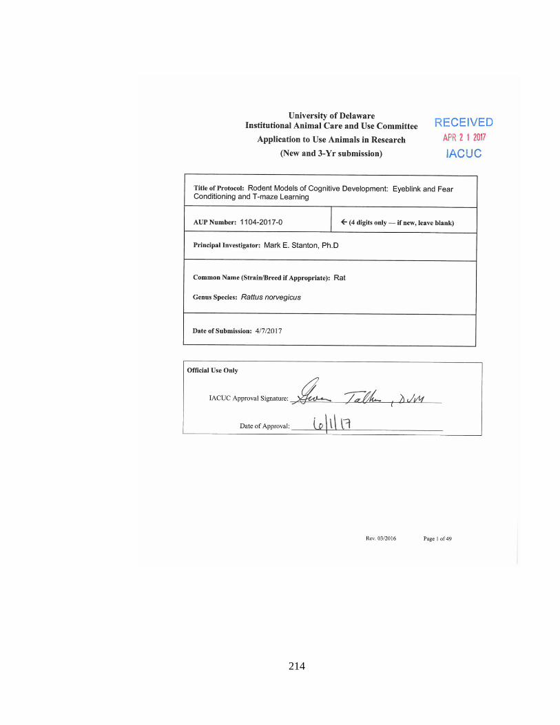
214