association of dorsolateral prefrontal cortex dysfunction with disrupted coordinated brain activity...
TRANSCRIPT

Article
1006 Am J Psychiatry 165:8, August 2008ajp.psychiatryonline.org
This article is featured in this month’s AJP Audio, is the subject of a CME course (p. 1079), and is discussed in an editorial by Drs. Mathalon and Ford (p. 944) and in an editorial by Dr. Tamminga (p. 949).
Association of Dorsolateral Prefrontal Cortex Dysfunction With Disrupted Coordinated Brain Activity in
Schizophrenia: Relationship With Impaired Cognition, Behavioral Disorganization, and Global Function
Jong H. Yoon, M.D.
Michael J. Minzenberg, M.D.
Stefan Ursu, M.D., Ph.D.
Ryan Walters, B.A.
Carter Wendelken, Ph.D.
J. Daniel Ragland, Ph.D.
Cameron S. Carter, M.D.
Objective: Although deficits in cognitivecontrol are thought to contribute to thediverse cognitive and behavioral abnor-malities in individuals with schizophrenia,the neural mechanisms underlying thesedeficits remain unclear. In this event-re-lated functional magnetic resonance im-aging (fMRI) study, the authors tested thehypothesis that during cognitive controltasks, impaired activation of the dorsolat-eral prefrontal cortex in schizophrenia pa-tients is associated with disrupted coordi-nated activity between this prefrontalregion and a distributed brain networkthat supports cognitive control.
Method: Through the use of an event-related design, 25 patients with first-episode schizophrenia and 24 healthycomparison subjects, matched on demo-graphic characteristics, were assessedwhile performing a version of the AX con-tinuous performance task. Functionalneuroimaging data were analyzed using1) univariate (region-of-interest blood-ox-ygen-level-dependent [BOLD] time seriesand whole brain voxel-wise regression)analysis to confirm the presence of dorso-lateral prefrontal cortex dysfunction and2) multivariate analysis to examine dorso-lateral prefrontal cortex functional con-nectivity. In addition, correlations be-tween dorsolateral prefrontal cortex
functional connectivity and the followingvariables were investigated: clinical symp-toms, task performance, and coordinatedbrain activity associated with cognitivecontrol.
Results: Schizophrenia patients exhib-ited a specific deficit in cognitive control,with significantly reduced accuracy in theBX condition relative to any other condi-tion. Univariate fMRI revealed dorsolat-eral prefrontal cortex dysfunction duringthe high cognitive control condition. Mul-tivariate analysis revealed significant im-pairment in functional connectivity be-tween the dorsolateral prefrontal cortexand task-relevant brain regions. Signi-ficant correlations were also foundbetween dorsolateral prefrontal cortexfunctional connectivity and cognitive per-formance, behavioral disorganization,and global functioning.
Conclusions: These findings suggest thatthere is an association between de-creased dorsolateral prefrontal cortex ac-tivity and connectivity and a task-relatedneural network. This deficit in coordi-nated brain activity may result in the dis-abling disorganization symptoms relatedto impaired cognition in individuals withschizophrenia.
(Am J Psychiatry 2008; 165:1006–1014)
Kraepelin (1) described the behavioral disorganiza-tion and cognitive deficits in schizophrenia as “an orches-tra without a conductor,” which presciently suggests thatcognitive control—the coordination of thoughts and ac-tions that facilitates goal-oriented behavior—is one of thekey higher-order cognitive processes impaired in individ-uals with schizophrenia. The findings of basic studies incognitive neuroscience suggest that the neural correlate ofcognitive control is the coordinated activity of task-rele-vant brain regions, and the dorsolateral prefrontal cortexplays a key role in this process (2–4).
Although dorsolateral prefrontal cortex dysfunction hasbeen one of the most replicated findings in schizophreniaresearch, particularly during tasks that require cognitivecontrol (5–11), the mechanisms by which impairment ofthis prefrontal region translates into cognitive control def-icits and related behavioral abnormalities in schizophre-nia remain poorly understood. The objective of thepresent study was to examine one potential mechanismby testing the hypothesis that reduced dorsolateral pre-frontal cortex activity is associated with altered coordi-

Am J Psychiatry 165:8, August 2008 1007
YOON, MINZENBERG, URSU, ET AL.
ajp.psychiatryonline.org
nated activity of task-relevant cortical processing acrossbrain regions in individuals with schizophrenia.
In this functional magnetic resonance imaging (fMRI)study, 25 first-episode schizophrenia patients and 24healthy comparison subjects were assessed while theycompleted the AX continuous performance task, which al-lows the measurement of performance and brain activityassociated with specific aspects of cognitive control (6, 10,12, 13). This paradigm was optimized in order to accom-modate an event-related functional connectivity analysiswhile providing efficient sampling of both A and B cue tri-als. Under conditions that required greater cognitive con-trol, we expected schizophrenia patients to show reduceddorsolateral prefrontal cortex engagement and reducedcorrelated activity in the distributed brain network thatsupports task performance. We investigated the clinicalsignificance of the result by measuring the association ofthe deficit in top-down modulation with behavioral per-formance and clinical measures. We predicted that alteredtop-down modulation would be significantly correlatedwith impaired behavioral performance and clinical mea-sures of disorganization in schizophrenia patients.
Method
Participants
Twenty five patients with schizophrenia and related disorders(schizophrenia, 23 patients; schizophreniform disorder, 1 patient;schizoaffective disorder, 1 patient) and 24 healthy comparisonsubjects were recruited from the community. The age range forparticipants in both study groups was between 14 and 32 years.Demographic and clinical characteristics of the two groups areprovided in Table 1. At the time of testing, all subjects in theschizophrenia group were outpatients and within the first year ofillness onset. To confirm the diagnosis of schizophrenia in pa-tients and to exclude the presence of a major psychiatric illness incomparison subjects, all subjects were evaluated using the Struc-tured Clinical Interview for DSM-IV-TR. Schizophrenia patientswho were <16 years old were assessed using the Schedule for Af-fective Disorders and Schizophrenia for School-Age Children—Present and Lifetime Version. Master’s- and doctoral-level clini-cians conducted the diagnostic evaluations, and diagnoses wereconfirmed via consensus conference. Illness onset was deter-mined by establishing the time at which DSM-IV criteria A and Bfor schizophrenia were met. Individuals with an illness onset >12months from the time of testing were excluded from the study.Symptoms were quantified using the following assessment mea-sures: 24-item Brief Psychiatric Rating Scale (BPRS), Scale for theAssessment of Negative Symptoms (SANS), Scale for the Assess-ment of Positive Symptoms (SAPS), Strauss Carpenter OutcomeScale, and Global Assessment Scale (GAS). Subscores from theBPRS, SANS, and SAPS were used to derive indexes for the follow-ing three major domains of symptoms: reality distortion, disorga-nization, and negative symptoms (12). Nine schizophrenia pa-tients were medication-naive. The remaining 16 patients werebeing treated with antipsychotics (risperidone [N=10], aripipra-zole [N=4], quetiapine [N=2], olanzapine [N=1]) at the time oftesting, with one subject receiving two neuroleptics. Exclusioncriteria for comparison subjects were 1) the presence of a lifetimediagnosis of an axis I disorder or 2) a first-degree relative with apsychotic disorder. Exclusion criteria for both study groups wereas follows: 1) IQ <70, 2) history of drug or alcohol dependence or
abuse within the 3 months before the start of the study or a posi-tive urine drug screen at the start of the study, 3) significant headtrauma, or 4) any known contraindication to MRI. Both groupswere well-matched, except in years of education (schizophreniagroup versus comparison group: 12.4 years [SD=2.7] versus 14.6years [SD=4.0], p<0.05) and IQ score (schizophrenia group versuscomparison group: 98.2 [SD=13.3] versus 112.4 [SD=8.5], p<0.01).After complete description of the study was given, written in-formed consent was obtained. The study was approved by theUniversity of California Davis Institutional Review Board.
Cognitive Task
Subjects completed the AX continuous performance task,which has been demonstrated to reliably measure specific cogni-tive control deficits in individuals with schizophrenia (10–12). Inthis performance task, the cue letter is presented for a 500-msecduration. Following a 3,500-msec delay, the probe letter is shownfor a duration of 500 msec. Subjects are required to make a targetresponse (using a right-index-finger button press) to the probeletter X only when it follows the cue letter A. All other stimuli re-quire a nontarget response (using a right-middle-finger buttonpress), including trials in which the letter X is preceded by any let-ter other than A (collectively referred to as B). Trials with targetcue-probe pairings (e.g., AX) occur with high frequency (70%) andset up the tendency for the subject to make a target response tothe probe letter X. The condition with the highest cognitive con-trol demand is the BX cue-probe trial, in which subjects mustovercome the tendency to make a target response to X.
Neuroimaging
Acquisition. Coplanar T1-weighted structural images were ob-tained prior to echoplanar imaging scans. Functional scans (T2-weighted echoplanar imaging: TR=2,000 msec, echo time=40msec, flip angle=90°, field of view=22 cm) were acquired using a1.5T GE scanner. Twenty-four contiguous 4.0-mm axial slices(with a 3.4-mm2 in-plane resolution from 80 mm above to 16 mmbelow the anterior commissure-posterior commissure line) werealso obtained.
Preprocessing. Preprocessing steps, implemented using Auto-mated Image Registration software (http://bishopw.loni.ucla.edu/AIR), were as follows: 1) removal of linear tendencies and signifi-cant outliers in the time series, 2) spatial realignment, 3) normal-ization to the Montreal Neurological Institute (MNI) templatethrough the use of a nonlinear warping algorithm, and 4) spatialsmoothing through the use of a Gaussian 8-mm full-width half-maximum kernel. Two comparison subjects were excluded be-cause of a >4-mm within-run movement. A multivariate analysis ofvariance (MANOVA) on the linear and angular movement parame-ters revealed no significant effect by group.
Statistical Analysis
Dorsolateral prefrontal cortex region-of-interest time se-ries. We conducted a region-of-interest analysis with a function-ally defined dorsolateral prefrontal cortex region of interest identi-fied in a previous study (14). Trial-averaged blood-oxygen-level-dependent (BOLD) time series were generated for each subject andaveraged by group for statistical comparison. For this and all func-tional neuroimaging analyses, only correct trials were assessed.
Whole brain analysis. In this exploratory voxel-wise analysis,conducted using Statistical Parametric Mapping-2 (http://www.fil.ion.ucl.ac.uk/spm), we performed a multiple regression,with covariates representing cue and probe events. In the first-level analysis, covariates were convolved with a canonical hemo-dynamic response function, using the temporal derivative to ac-count for intersubject variability in BOLD signal time to peak (13,15). In the second-level analysis, we conducted a random-effects

1008 Am J Psychiatry 165:8, August 2008
DORSOLATERAL PREFRONTAL CORTEX DYSFUNCTION
ajp.psychiatryonline.org
comparison of the linear contrast between B and A cue trials be-tween the two study groups. For between-group comparisons,nontask-related regions were excluded by imposing a mask of sig-nificant regions of activity from either group. Thresholding of allmaps followed the procedure for determining cluster-level signif-icance (corrected for multiple comparisons at p<0.05 family-wiseerror outlined in Friston et al. [16]).
Dorsolateral prefrontal cortex-correlated brain networkactivity. We measured functional connectivity of the dorsolat-eral prefrontal cortex using the beta series correlation method(17). Hemodynamic response function-convolved trial-specificcovariates modeled activity during the component stages of eachindividual trial in order to generate beta series. Voxel-wise bivari-ate Pearson’s correlations between a “seed” region (i.e., dorsolat-eral prefrontal cortex) and all other brain regions were computedfor a task component and condition of interest (e.g., B cue). Be-tween-group random-effects analysis was then conducted forgroup-averaged correlations. The same procedure used for deter-mining univariate whole brain analysis was also used to deter-mine statistically significant activity. All correlation analyses wereconducted using Statistical Parametric Mapping-2, with Matlab(MathWorks, Natick, Mass.) scripts specifically designed for thepresent study.
Results
AX Continuous Performance Task In-Scanner Performance
Accuracy results revealed a specific deficit in contextprocessing in schizophrenia patients (Figure 1). Analysisof variance (ANOVA), with between-subject factor bygroup and within-subject factor by condition (AX, AY, BX,and BY), showed a significant main effect by group (F=9.240, df=1, 47, p=0.004) (with schizophrenia patients per-forming worse than comparison subjects) and condition(F=16.982, df=3, 141, p<0.001) and a significant group-by-condition interaction (F=4.033, df=3, 141, p=0.009). Posthoc two-sample t tests demonstrated significantly bettercognitive control performance among healthy compari-son subjects relative to schizophrenia patients in the BX
(t=3.349, df=47, p=0.002) and AX conditions (t=2.295, df=47, p=0.03) but not in the BY or AY conditions (p>0.15).
fMRI
Dorsolateral prefrontal cortex region-of-interesttime series. Healthy comparison subjects exhibited ro-bust enhancement in dorsolateral prefrontal cortex activ-ity during the B cue condition relative to the A cue condi-tion, with maximal difference occurring at scan 3 (theexpected peak of cue-related activity). Schizophrenia pa-tients showed minimal enhancement of dorsolateral pre-frontal cortex activity across cue types (Figure 1). ANOVAof the BOLD signal at scan 3 revealed a significant main ef-fect of group (F=33.725, df=1, 47, p<0.001) and condition(F=11.014, df=1, 47, p=0.002) and a significant group-by-condition interaction (F=4.685, df=1, 47, p=0.04). Acrossscans 1 to 3, there was a significant group-by-condition-by-scan interaction (F=3.172, df=2, 94, p=0.05). A two-tailed, paired-sample t test showed that the scan 3 signalwas significantly greater during the B cue trial relative tothe A cue trial in comparison subjects (t=4.437, df=23,p<0.001) but not in schizophrenia patients (t=0.74, df=24,p=0.47). A two-tailed, two-sample t test of the differencebetween B and A cue signals during scan 3 revealed a sig-nificant difference between groups (t=2.164, df=47, p=0.04). ANOVA of the dorsolateral prefrontal cortex signal inschizophrenia patients, with regard to medication status,revealed no significant effect of medication on the signalat scan 3 (p=0.26) or scans 1 to 3 (p=0.42).
Whole brain analysis. To confirm the differential in-volvement of the dorsolateral prefrontal cortex in healthycomparison subjects relative to schizophrenia patients,we conducted a whole brain voxel-wise analysis, an ap-proach complementary to the region-of-interest BOLDtime-series analysis (Table 2, Figure 1). In comparisonsubjects, a diverse network involving the middle frontal
TABLE 1. Demographic and Clinical Characteristics of Schizophrenia Patients and Healthy Comparison Subjects
Characteristic
Group
Schizophrenia Patients (N=25) Healthy Comparison Subjects (N=24)Mean SD Mean SD
Age (years) 19.6 3.80 21.6 4.24Parental education (years) 15.0 2.37 15.1 2.88Participant education (years) 12.4 2.71 14.6 4.02WAIS IQ scorea 98.2 13.93 112.4 8.46Symptom severity (scale scores)
BPRS 46.5 8.95 -- --SANS 30.1 13.31 -- --SAPS 30.4 16.99 -- --Strauss Carpenter Outcome Scale 9.9 2.84 -- --Global Assessment Scale 53.7 10.15 -- --
N % N %Male 17 68 13 54Right-handed 24 96 23 96Antipsychotic medication
Atypical 16 64 -- --Typical 0 0 -- --Unmedicated 9 36 -- --
History of substance abuse or dependence 7 28 3 12.5a Significant between-group difference (p≤0.01).

Am J Psychiatry 165:8, August 2008 1009
YOON, MINZENBERG, URSU, ET AL.
ajp.psychiatryonline.org
gyrus, anterior and posterior cingulate, and precuneuswas engaged. In schizophrenia patients, a network limitedto subcortical regions—the thalamus and putamen—wasactivated. Notably, there was an absence of engagement ofthe middle frontal gyrus in schizophrenia patients. Thisdifference between groups in middle frontal gyrus activitywas confirmed by the between-group results. In the B cueminus A cue contrast, only the left middle frontal gyrusand inferior frontal gyrus showed significantly greater ac-tivity in healthy comparison subjects relative to schizo-phrenia patients, while no regions showed greater activityin schizophrenia patients relative to healthy comparisonsubjects.
Dorsolateral prefrontal cortex-correlated brain net-work activity. We conducted a dorsolateral prefrontalcortex functional connectivity analysis using the beta se-ries method and predicted that schizophrenia patients
would show diminished dorsolateral prefrontal cortexfunctional connectivity (Table 3, Figure 2). In healthy com-parison subjects, the right inferior parietal lobule and leftpremotor cortex (including the frontal eye fields) were re-gions showing greater functional connectivity with thedorsolateral prefrontal cortex in the B cue condition rela-tive to the A cue condition. In schizophrenia patients, noregions exhibited enhanced dorsolateral prefrontal cortexfunctional connectivity. In a direct comparison betweenthe two groups in B cue minus A cue contrast, the left pre-motor cortex and right inferior parietal lobule showedgreater dorsolateral prefrontal cortex connectivity inhealthy comparison subjects. The identification of theright inferior parietal lobule raises questions as to whetherdeficits in this region rather than the dorsolateral prefron-tal cortex regulate network connectivity differences, giventhat the connectivity measure cannot provide directional-ity of influences between these two higher-order brain re-
FIGURE 1. AX Continuous Performance Task In-Scanner Behavioral and Univariate fMRI Results in Schizophrenia Patientsand Healthy Comparison Subjectsa
a Accuracy and reaction times are displayed for all conditions of the AX continuous performance task in the left column. The overall patternshows a selective deficit in context processing in schizophrenia patients, with the worst performance occurring in the BX condition. The percentchange in the BOLD signal in the left dorsolateral prefrontal cortex in schizophrenia patients and healthy comparison subjects for the B and Acue conditions are shown in the top right. Note that this time series was derived from a functional region of interest identified in healthy sub-jects in a previous study and not from the dorsolateral prefrontal cortex region identified from the whole brain analysis in the present study.Healthy comparison subjects exhibited the predicted up-regulation of dorsolateral prefrontal cortex activity during the B cue relative to the Acue conditions, while patients did not demonstrate this effect. Results of whole brain, voxel-wise multiple regression analysis are shown in thelower right. These surface-rendered t maps of the contrast B cue condition minus A cue condition, thresholded at the cluster level of p<0.05(corrected), depict regions showing significantly greater activity in healthy comparison subjects relative to schizophrenia patients.
∗p<0.05.
6
Mean
(±
SD) R
eact
ion
Tim
e (m
sec)
AX Continuous Performance Task
900
800
700
600
500
400
300
200
AX AY BXTrial Target
BY
Mean
(±
SD) A
ccu
racy
(%
)
1.00
Comparisonsubjects (N=24)
Schizophreniapatients (N=25)
0.90
0.80
0.70
0.60
0.50
1 2 3 5 7Scan
Comparison Subjects > Schizophrenia Patients
BO
LD (%
)
0.20
0.15
0.10
0.05
0
4
*
*
*
R L
*
Schizophrenia A CueComparison B CueSchizophrenia B CueComparison A Cue
*
1000
1100
1200
1.10*

1010 Am J Psychiatry 165:8, August 2008
DORSOLATERAL PREFRONTAL CORTEX DYSFUNCTION
ajp.psychiatryonline.org
gions. To address this issue, we conducted a logistic re-gression analysis in order to predict diagnostic status fordorsolateral prefrontal cortex and right inferior parietallobule activity. Activity in the dorsolateral prefrontal cor-tex was a significant negative predictor of schizophrenia(beta=–2.67, p=0.009), with every unit increase in dorso-lateral prefrontal cortex activity resulting in a >14:1 de-crease in the odds of schizophrenia. Activity in the rightinferior parietal lobule was not a significant predictor(beta=0.12, p=0.85).
To evaluate the possibility that the functional connec-tivity results were confounded by low univariate activity inthe “seed” (i.e., dorsolateral prefrontal cortex) region inschizophrenia patients, we compared trial-to-trial vari-ability of the BOLD signal in the left dorsolateral prefrontalcortex. We found equivalent variance between groupsduring A and B cues (p=0.75 and p=0.31, respectively). Inother words, the lower mean BOLD signal in schizophre-nia patients did not constrain the functional connectivityanalysis, since patients and healthy comparison subjectsexhibited equivalent levels of trial-to-trial variability of theBOLD signal.
Behavioral and clinical correlations with dorsolat-eral prefrontal cortex functional connectivity mea-sures. To evaluate the relevance of our dorsolateral pre-frontal cortex functional connectivity results, we calcu-lated the correlation between dorsolateral prefrontal cor-tex connectivity and behavior (Figure 2). Dorsolateralprefrontal cortex connectivity showed moderate correla-tion with performance on the AX continuous performancetask across all subjects, and thus connectivity was signifi-
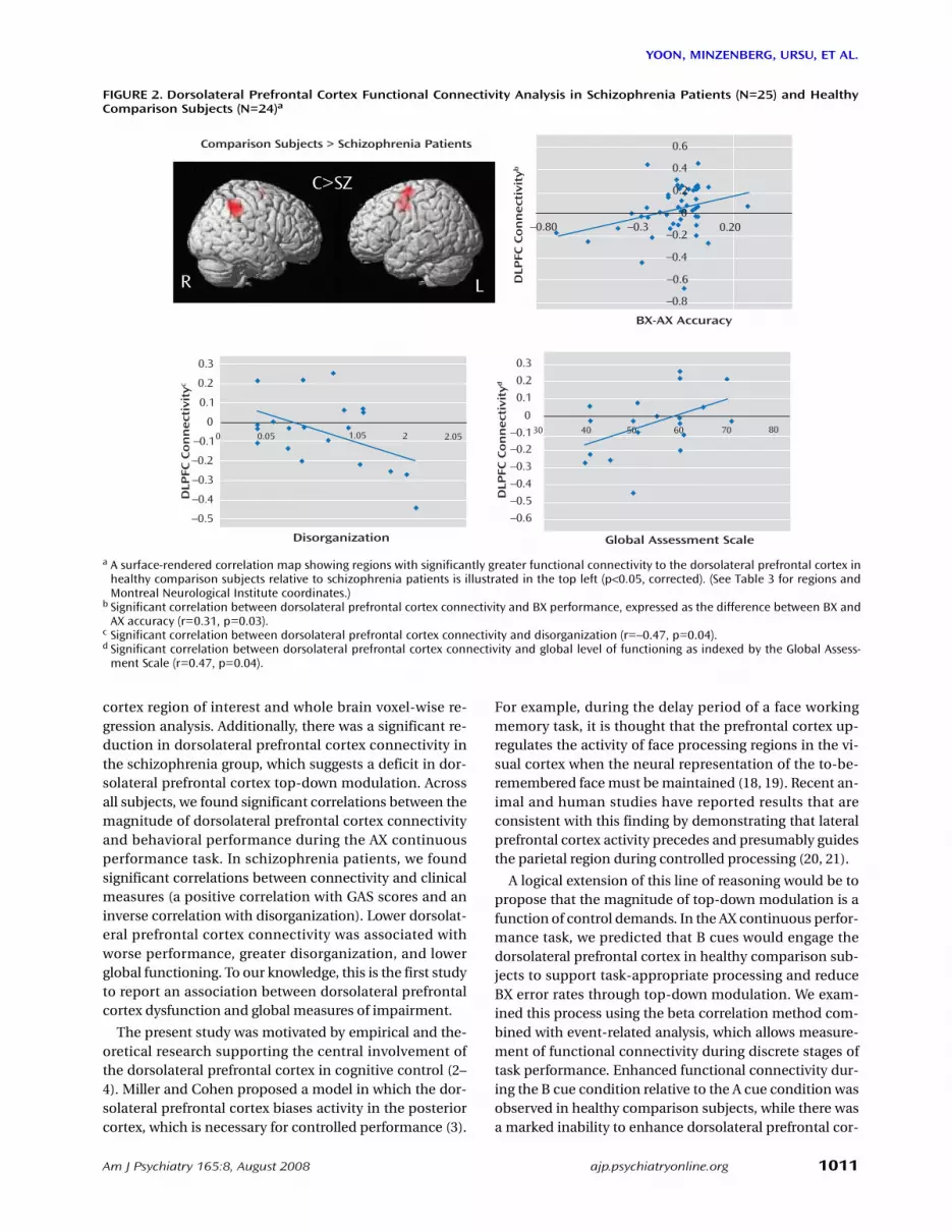
cantly associated with better performance in the BX con-dition, with either BX accuracy (r=0.35, p=0.01) or the dif-ference in accuracy between the BX and AX conditions (r=0.31, p=0.03). There was a significant inverse correlationbetween disorganization and dorsolateral prefrontal cor-tex connectivity (r=–0.47, p=0.04). This relationship wasspecific in that the correlation between dorsolateral pre-frontal cortex connectivity and other symptom domains(behavioral poverty and reality distortion) was low andnonsignificant (behavioral poverty: r=0.12, p=0.63; realitydistortion: r=–0.29, p=0.21). Finally, dorsolateral prefron-tal cortex connectivity was significantly correlated withglobal level of functioning as assessed by GAS (r=0.47, p=0.04).
Discussion
In a large sample of subjects with first-episode schizo-phrenia, we demonstrated a specific deficit in cognitivecontrol, dysfunction of the dorsolateral prefrontal cortex,and significant correlations between impaired dorsolat-eral prefrontal cortex functional connectivity and behav-ior and clinical status. Schizophrenia patients exhibitedpoor performance during the BX condition, a conditionthat requires a high degree of cognitive control. However,patients did not differ from healthy comparison subjectsin the AY and BY trials (12). Schizophrenia patients wereunable to up-regulate activity in the dorsolateral prefron-tal cortex during B cues, as indicated by the results of thefollowing two separate lines of analyses: BOLD time seriesderived from an a priori-identified dorsolateral prefrontal
TABLE 2. Univariate Voxel-Wise Whole Brain fMRI Analysis in Schizophrenia Patients (N=25) and Healthy Comparison Sub-jects (N=24)
Group and Brain Region MNI Coordinates (x, y, z)
Analysis
t zHealthy comparison subjects
Left posterior cingulate –3, –72, 12 5.81 4.51Left middle frontal gyrus –45, 52, 12 5.80 4.51Right middle frontal gyrus 45, 17, 32 5.49 4.34Right precuneus 21, –72, 48 5.39 4.29Anterior cingulate –3, 34, 40 3.89 3.38
Schizophrenia patientsLeft thalamus –7, –7, 4 4.44 3.76Right thalamus 31, –14, 0 3.91 3.40
Healthy comparison subjects > schizophrenia patientsa
Left middle frontal gyrus 48, 31, 28 3.89 3.61Left inferior frontal gyrus 41, 10, 16 3.63 3.39
a There were no regions in which greater activity was seen in schizophrenia patients relative to healthy comparison subjects.
TABLE 3. Multivariate Dorsolateral Prefrontal Cortex Functional Connectivity Analysis in Schizophrenia Patients (N=25)and Healthy Comparison Subjects (N=24)
Group and Brain Region MNI Coordinates (x, y, z)
Analysis
t zHealthy comparison subjects
Right inferior parietal lobule 43, –44, 45 5.97 4.59Left premotor cortex –16, –6, 57 5.84 4.53
Healthy comparison subjects > schizophrenia patientsRight inferior parietal lobule 43, –44, 45 4.30 3.93Left premotor cortex –19, –6, 65 4.17 3.83
Am J Psychiatry 165:8, August 2008 1011
YOON, MINZENBERG, URSU, ET AL.
ajp.psychiatryonline.org
cortex region of interest and whole brain voxel-wise re-gression analysis. Additionally, there was a significant re-duction in dorsolateral prefrontal cortex connectivity inthe schizophrenia group, which suggests a deficit in dor-solateral prefrontal cortex top-down modulation. Acrossall subjects, we found significant correlations between themagnitude of dorsolateral prefrontal cortex connectivityand behavioral performance during the AX continuousperformance task. In schizophrenia patients, we foundsignificant correlations between connectivity and clinicalmeasures (a positive correlation with GAS scores and aninverse correlation with disorganization). Lower dorsolat-eral prefrontal cortex connectivity was associated withworse performance, greater disorganization, and lowerglobal functioning. To our knowledge, this is the first studyto report an association between dorsolateral prefrontalcortex dysfunction and global measures of impairment.
The present study was motivated by empirical and the-oretical research supporting the central involvement ofthe dorsolateral prefrontal cortex in cognitive control (2–4). Miller and Cohen proposed a model in which the dor-solateral prefrontal cortex biases activity in the posteriorcortex, which is necessary for controlled performance (3).
For example, during the delay period of a face workingmemory task, it is thought that the prefrontal cortex up-regulates the activity of face processing regions in the vi-sual cortex when the neural representation of the to-be-remembered face must be maintained (18, 19). Recent an-imal and human studies have reported results that areconsistent with this finding by demonstrating that lateralprefrontal cortex activity precedes and presumably guidesthe parietal region during controlled processing (20, 21).
A logical extension of this line of reasoning would be topropose that the magnitude of top-down modulation is afunction of control demands. In the AX continuous perfor-mance task, we predicted that B cues would engage thedorsolateral prefrontal cortex in healthy comparison sub-jects to support task-appropriate processing and reduceBX error rates through top-down modulation. We exam-ined this process using the beta correlation method com-bined with event-related analysis, which allows measure-ment of functional connectivity during discrete stages oftask performance. Enhanced functional connectivity dur-ing the B cue condition relative to the A cue condition wasobserved in healthy comparison subjects, while there wasa marked inability to enhance dorsolateral prefrontal cor-
FIGURE 2. Dorsolateral Prefrontal Cortex Functional Connectivity Analysis in Schizophrenia Patients (N=25) and HealthyComparison Subjects (N=24)a
a A surface-rendered correlation map showing regions with significantly greater functional connectivity to the dorsolateral prefrontal cortex inhealthy comparison subjects relative to schizophrenia patients is illustrated in the top left (p<0.05, corrected). (See Table 3 for regions andMontreal Neurological Institute coordinates.)
b Significant correlation between dorsolateral prefrontal cortex connectivity and BX performance, expressed as the difference between BX andAX accuracy (r=0.31, p=0.03).
c Significant correlation between dorsolateral prefrontal cortex connectivity and disorganization (r=–0.47, p=0.04).d Significant correlation between dorsolateral prefrontal cortex connectivity and global level of functioning as indexed by the Global Assess-
ment Scale (r=0.47, p=0.04).
0.3
0.2
0.1
030 40 50 60 70 80–0.1
–0.2
–0.3
–0.4
–0.5
–0.6
Global Assessment Scale
BX-AX Accuracy
DLP
FC C
on
nect
ivit
yd
DLP
FC C
on
nect
ivit
yb
DLP
FC C
on
nect
ivit
yc
0.3
0.2
0.1
0.05 1.05 2 2.05
00–0.1
–0.2
–0.3
–0.4
–0.5
Disorganization
Comparison Subjects > Schizophrenia Patients
R L
0.6
0.4
–0.20.20
–0.4
–0.3–0.80
–0.6
–0.8
0
0.2C>SZ
R L

1012 Am J Psychiatry 165:8, August 2008
DORSOLATERAL PREFRONTAL CORTEX DYSFUNCTION
ajp.psychiatryonline.org
tex connectivity in schizophrenia patients. Previous stud-ies have also shown altered interactions between corticalregions, suggesting the presence of disordered connectiv-ity in schizophrenia (22–25). Accordingly, not only are ourresults consistent with the hypothesis that connectivity isimpaired in individuals with schizophrenia, but they sug-gest that there is a potential fundamental neural mecha-nism by which dorsolateral prefrontal cortex dysfunctionleads to cognitive impairment (i.e., top-down modulationthrough impaired connectivity with the dorsolateral pre-frontal cortex).
As a measure of covariance, the beta series correlationmethod cannot determine the directionality of interac-tions. However, the fact that 1) only the dorsolateral pre-frontal cortex (not other elements of the task-related neu-ral network) showed differential activity in the univariatecontrast in schizophrenia patients and healthy compari-son subjects and 2) logistic regression analysis selectivelyassociated decreased dorsolateral prefrontal cortex activ-ity but not right inferior parietal lobule activity with aschizophrenia diagnosis provides converging support forour model. However, we cannot rule out the possibilitythat altered function in another element of the distributednetwork, such as the right inferior parietal lobule, under-lies the altered pattern of connectivity. Future studies us-ing tasks that selectively engage prefrontal and parietalcognitive control functions or other methods capable ofdetecting directionality of effects are needed to addressthis issue in a more definitive manner.
There may be concerns that our functional connectivityresults were confounded by decreased dorsolateral pre-frontal cortex activity in schizophrenia patients. However,the most important parameter constraining dorsolateralprefrontal cortex connectivity is not its mean activity butrather the magnitude of trial-to-trial variability. In fMRI,these are largely independent parameters, and lower dor-solateral prefrontal cortex connectivity in patients is notnecessarily the result of low univariate activity. This wasverified in our data by the demonstration of equivalentdorsolateral prefrontal cortex variance between groups.Other examples of the divergence between the meanunivariate and functional connectivity properties of a re-gion are present in the extant literature (17).
In the present study, we included adolescent subjects inorder to ensure that we investigated a representative sam-ple of first-episode schizophrenia patients (26). In thisstudy and a previous report (27), we closely matched thestudy groups on age to ensure that there were no develop-mental differences, aside from those that may be relatedto the illness, between the groups. Further, in a reanalysisof our data in the present study, which excluded subjects<16 years old, we found an identical pattern of results. Toaddress the possibility that lower dorsolateral prefrontalcortex activity in schizophrenia patients might have beenthe result of lower accuracy on the AX continuous perfor-mance task among these individuals (and hence a lower
number of correct BX trials), we conducted an analysis inwhich only performance-matched subjects were in-cluded. This analysis yielded fMRI results that were identi-cal to those from the full sample.
An examination of the putative functions of cortical re-gions identified in the dorsolateral prefrontal cortex con-nectivity analysis revealed that each of these regions maysupport task-relevant processes during the AX continuousperformance task. The right inferior parietal lobule hasstrong anatomical connections to the dorsolateral pre-frontal cortex (28), and recent functional neuroimagingstudies in humans suggest that the right inferior parietallobule supports sustained attention (29, 30). Similarly, thefrontal eye field is thought to support visual attention, inaddition to its well-recognized role in eye-movement mo-tor control (31, 32). It is possible that functional impair-ment in one of these regions could lead to disrupted con-nectivity with the dorsolateral prefrontal cortex.
Limitations to the present study include the inferencesthat could be made pertaining to the relative importanceof brain regions other than the dorsolateral prefrontal cor-tex (33–35) in the broader pathophysiology of schizophre-nia. Although the demonstration of a significant associa-tion between dorsolateral prefrontal cortex connectivityand behavioral and clinical impairments (particularlywith disorganization symptoms and GAS) suggests broadrelevance of dorsolateral prefrontal cortex dysfunction,the determination of the relative importance of the dorso-lateral prefrontal cortex compared with other regions in-volving other cognitive processes needs to be examined infuture studies.
Other possible limitations are the effects of medicationon fMRI measures. The available evidence argues againstmedication effects as a major contributor to our findings.Prior studies have shown that antipsychotic drugs do notsuppress the prefrontal cortex BOLD signal during higher-order cognition (27, 36). In the present study, there wereno differences between medicated and unmedicated pa-tients in cue-related dorsolateral prefrontal cortex activity.Furthermore, since our patients were very early in thecourse of illness, it is reasonable to conclude that impaireddorsolateral prefrontal cortex function is fundamentaland unrelated to the long-term effects of medication ex-posure or illness chronicity.
Although the physiological mechanisms underlying im-paired dorsolateral prefrontal cortex activity are unknown,the asynchronous activity of neurons as the result of per-turbations in the local circuitry is a cogent potential mech-anism (37, 38). Simultaneous recording and fMRI studiesin primates suggest that synchronous firing of corticalneurons, particularly in the high-frequency range (40–80Hz or gamma), is highly correlated with BOLD response(39). In a recent EEG study, which parallels the presentfMRI study, we reported a decrease in induced gamma-band activity associated with impaired cognitive controlperformance and disorganization in schizophrenia pa-

Am J Psychiatry 165:8, August 2008 1013
YOON, MINZENBERG, URSU, ET AL.
ajp.psychiatryonline.org
tients (40). The results of the present study are consistentwith the findings of our previous EEG study and with amodel of impaired synchronous neuronal firing in thedorsolateral prefrontal cortex, which is associated withdisruption in top-down-regulation of neural networksthat support cognitive control.
The convergence of behavioral, functional neuroimag-ing, and clinical findings in the present study supports thedorsolateral prefrontal cortex dysfunction model of cogni-tive control deficits in schizophrenia. Furthermore, the re-ported functional connectivity results provide new in-sights into how dorsolateral prefrontal cortex dysfunctionmay lead to impaired cognitive control, behavioral disor-ganization, and functional impairment in individuals withschizophrenia by suggesting a physiological model inwhich a failure to recruit and maintain a coordinated net-work may underlie the cognitive impairment and relatedaspects of the psychopathology of schizophrenia.
Previously presented at the International Congress of SchizophreniaResearch Biennial Meeting, Colorado Springs, Colo., 2007. ReceivedJune 16, 2007; revisions received Jan. 19 and March 12, 2008;accepted March 14, 2008 (doi: 10.1176/appi.ajp.2008.07060945).From the Department of Psychiatry and Imaging Research Center,University of California, Davis, Sacramento, Calif. Address correspon-dence and reprint requests to Dr. Carter, UC Davis Imaging ResearchCenter, 4701 X St., Sacramento, CA 95817; [email protected] (e-mail).
Dr. Minzenberg is a shareholder with Elan Pharmaceuticals. Dr.Carter has served as a consultant to Pfizer, Hoffman La Roche, and EliLilly. Drs. Yoon, Ursu, Wendelken, and Ragland and Mr. Walters re-port no competing interests.
Supported by NIH grant R01 MH-66629, NIMH KO2 award, and theBurroughs Wellcome Foundation Translational Clinical ScientistAward (Dr. Carter).
References
1. Kraepelin E: Dementia Praecox and Paraphrenia. New York,Robert D. Krieger Publishing,1919
2. Desimone R: Neural mechanisms for visual memory and theirrole in attention. Proc Natl Acad Sci U S A 1996; 93:13494–13499
3. Miller EK, Cohen JD: An integrative theory of prefrontal cortexfunction. Annu Rev Neurosci 2001; 24:167–202
4. Tomita H, Ohbayashi M, Nakahara K, Hasegawa I, Miyashita Y:Top-down signal from prefrontal cortex in executive control ofmemory retrieval. Nature 1999; 401:699–703
5. Berman KF, Zec RF, Weinberger DR: Physiologic dysfunction ofdorsolateral prefrontal cortex in schizophrenia, II: role of neu-roleptic treatment, attention, and mental effort. Arch Gen Psy-chiatry 1986; 43:126–135
6. MacDonald AW 3rd, Carter CS, Kerns JG, Ursu S, Barch DM,Holmes AJ, Stenger VA, Cohen JD: Specificity of prefrontal dys-function and context processing deficits to schizophrenia innever-medicated patients with first-episode psychosis. Am JPsychiatry 2005; 162:475–484
7. Callicott JH, Bertolino A, Mattay VS, Langheim FJ, Duyn J, Cop-pola R, Goldberg TE, Weinberger DR: Physiological dysfunctionof the dorsolateral prefrontal cortex in schizophrenia revisited.Cereb Cortex 2000; 10:1078–1092
8. Cannon TD, Glahn DC, Kim J, Van Erp TG, Karlsgodt K, CohenMS, Nuechterlein KH, Bava S, Shirinyan D: Dorsolateral pre-frontal cortex activity during maintenance and manipulation
of information in working memory in patients with schizo-phrenia. Arch Gen Psychiatry 2005; 62:1071–1080
9. Glahn DC, Ragland JD, Abramoff A, Barrett J, Laird AR, BeardenCE, Velligan DI: Beyond hypofrontality: a quantitative meta-analysis of functional neuroimaging studies of working mem-ory in schizophrenia. Hum Brain Mapp 2005; 25:60–69
10. Barch DM, Carter CS, Braver TS, Sabb FW, MacDonald A 3rd,Noll DC, Cohen JD: Selective deficits in prefrontal cortex func-tion in medication-naive patients with schizophrenia. Arch GenPsychiatry 2001; 58:280–288
11. MacDonald AW 3rd, Carter CS: Event-related fMRI study of con-text processing in dorsolateral prefrontal cortex of patientswith schizophrenia. J Abnorm Psychol 2003; 112:689–697
12. Barch DM, Carter CS, MacDonald AW 3rd, Braver TS, Cohen JD:Context-processing deficits in schizophrenia: diagnostic speci-ficity, 4-week course, and relationships to clinical symptoms. JAbnorm Psychol 2003; 112:132–143
13. Barch DM, Mathews JR, Buckner RL, Maccotta L, Csernansky JG,Snyder AZ: Hemodynamic responses in visual, motor, and so-matosensory cortices in schizophrenia. Neuroimage 2003; 20:1884–1893
14. MacDonald AW 3rd, Cohen JD, Stenger VA, Carter CS: Dissociat-ing the role of the dorsolateral prefrontal and anterior cingu-late cortex in cognitive control. Science 2000; 288:1835–1838
15. Ford JM, Johnson MB, Whitfield SL, Faustman WO, MathalonDH: Delayed hemodynamic responses in schizophrenia. Neu-roimage 2005; 26:922–931
16. Friston KJ, Holmes A, Poline JB, Price CJ, Frith CD: Detecting ac-tivations in PET and fMRI: levels of inference and power. Neu-roimage 1996; 4(3 pt 1):223–235
17. Rissman J, Gazzaley A, D’Esposito M: Measuring functional con-nectivity during distinct stages of a cognitive task. Neuroimage2004; 23:752–763
18. Gazzaley A, Cooney JW, McEvoy K, Knight RT, D’Esposito M: Top-down enhancement and suppression of the magnitude andspeed of neural activity. J Cogn Neurosci 2005; 17:507–517
19. Ranganath C, DeGutis J, D’Esposito M: Category-specific modu-lation of inferior temporal activity during working memory en-coding and maintenance. Brain Res Cogn Brain Res 2004; 20:37–45
20. Brass M, Ullsperger M, Knoesche TR, von Cramon DY, PhillipsNA: Who comes first? The role of the prefrontal and parietalcortex in cognitive control. J Cogn Neurosci 2005; 17:1367–1375
21. Buschman TJ, Miller EK: Top-down versus bottom-up control ofattention in the prefrontal and posterior parietal cortices. Sci-ence 2007; 315:1860–1862
22. Fletcher P, McKenna PJ, Friston KJ, Frith CD, Dolan RJ: Abnormalcingulate modulation of fronto-temporal connectivity inschizophrenia. Neuroimage 1999; 9:337–342
23. Meyer-Lindenberg AS, Olsen RK, Kohn PD, Brown T, Egan MF,Weinberger DR, Berman KF: Regionally specific disturbance ofdorsolateral prefrontal-hippocampal functional connectivity inschizophrenia. Arch Gen Psychiatry 2005; 62:379–386
24. Wolf DH, Gur RC, Valdez JN, Loughead J, Elliott MA, Gur RE, Rag-land JD: Alterations of fronto-temporal connectivity duringword encoding in schizophrenia. Psychiatry Res 2007; 154:221–232
25. Friston KJ, Frith CD: Schizophrenia: A disconnection syndrome?Clin Neurosci 1995; 3:89–97
26. an der Heiden W, Hafner H: The epidemiology of onset andcourse of schizophrenia. Eur Arch Psychiatry Clin Neurosci2000; 250:292–303
27. Snitz BE, MacDonald A 3rd, Cohen JD, Cho RY, Becker T, CarterCS: Lateral and medial hypofrontality in first-episode schizo-phrenia: functional activity in a medication-naive state and ef-

1014 Am J Psychiatry 165:8, August 2008
DORSOLATERAL PREFRONTAL CORTEX DYSFUNCTION
ajp.psychiatryonline.org
fects of short-term atypical antipsychotic treatment. Am J Psy-chiatry 2005; 162:2322–2329
28. Cavada C, Goldman-Rakic PS: Posterior parietal cortex inrhesus monkey, I: parcellation of areas based on distinctivelimbic and sensory corticocortical connections. J Comp Neurol1989; 287:393–421
29. Husain M, Nachev P: Space and the parietal cortex. TrendsCogn Sci 2007; 11:30–36
30. Husain M, Rorden C: Non-spatially lateralized mechanisms inhemispatial neglect. Nat Rev Neurosci 2003; 4:26–36
31. Thompson KG, Biscoe KL, Sato TR: Neuronal basis of covertspatial attention in the frontal eye field. J Neurosci 2005; 25:9479–9487
32. Wardak C, Ibos G, Duhamel JR, Olivier E: Contribution of themonkey frontal eye field to covert visual attention. J Neurosci2006; 26:4228–4235
33. Dierks T, Linden DE, Jandl M, Formisano E, Goebel R, Lanfer-mann H, Singer W: Activation of Heschl’s gyrus during auditoryhallucinations. Neuron 1999; 22:615–621
34. Heckers S, Rauch SL, Goff D, Savage CR, Schacter DL, FischmanAJ, Alpert NM: Impaired recruitment of the hippocampus dur-ing conscious recollection in schizophrenia. Nat Neurosci1998; 1:318–323
35. Onitsuka T, Shenton ME, Salisbury DF, Dickey CC, Kasai K, TonerSK, Frumin M, Kikinis R, Jolesz FA, McCarley RW: Middle and in-
ferior temporal gyrus gray matter volume abnormalities inchronic schizophrenia: an MRI study. Am J Psychiatry 2004;161:1603–1611
36. Honey GD, Bullmore ET, Soni W, Varatheesan M, Williams SC,Sharma T: Differences in frontal cortical activation by a work-ing memory task after substitution of risperidone for typicalantipsychotic drugs in patients with schizophrenia. Proc NatlAcad Sci U S A 1999; 96:13432–13437
37. Volk DW, Austin MC, Pierri JN, Sampson AR, Lewis DA: De-creased glutamic acid decarboxylase67 messenger RNA expres-sion in a subset of prefrontal cortical gamma-aminobutyricacid neurons in subjects with schizophrenia. Arch Gen Psychia-try 2000; 57:237–245
38. Woo TU, Whitehead RE, Melchitzky DS, Lewis DA: A subclass ofprefrontal gamma-aminobutyric acid axon terminals are selec-tively altered in schizophrenia. Proc Natl Acad Sci U S A 1998;95:5341–5346
39. Logothetis NK, Pauls J, Augath M, Trinath T, Oeltermann A:Neurophysiological investigation of the basis of the fMRI signal.Nature 2001; 412:150–157
40. Cho RY, Konecky RO, Carter CS: Impairments in frontal corticalgamma synchrony and cognitive control in schizophrenia.Proc Natl Acad Sci U S A 2006; 103:19878–19883