development and calcification of enamel* - citeseerx
TRANSCRIPT
Development andCalcification of Enamel*
Antonio Nanci and Charles E. Smith
*Supported by the Medical Research Council of Canada
Reprinted from CALCIFICATION IN BIOLOGICAL SYSTEMS,CRC Press, Boca Raton, FL
313
Chapter 13
DEVELOPMENT AND CALCIFICATION OF ENAMEL*
Antonio Nanci and Charles E. Smith
TABLE OF CONTENTS
I. Introduction 314
II. Morphological Events of Amelogenesis 314
III. Proteins Secreted by Ameloblasts Into Developing Enamel 320
IV. Removal of Proteins From Developing Enamel 323
V. Formation and Growth of Enamel Crystals 331
VI. References 335
* Supported by the Medical Research Council of Canada
314 Calcification in Biological Systems
I. INTRODUCTION
Enamel was one of the first biological tissues to be examined in the lightmicroscope1 and has since fascinated and mystified researchers with its com-plexity. The creation of this tissue exemplifies many fundamental cellularevents such as proliferation and differentiation, protein synthesis and secre-tion, post-translational processing, endocytosis, lysosomal protein targetingand degradation, ion (calcium) transport, and mineralization. There are alsoseveral unusual cellular events associated with amelogenesis such as (1) re-versal of secretory polarity by ameloblasts; (2) formation and destruction, andeventual recreation of a basal lamina along a surface that constitutes theembryonic bases of preameloblasts but later occupies the functional apices ofdifferentiated ameloblasts; and (3) bulk destruction of almost all of the proteinssecreted by these cells. A comprehensive understanding of these phenomenais essential in understanding how enamel forms and mineralizes.
Amelogenesis has been described in as many as six phases,2 but it isgenerally subdivided into three main functional stages universally referred toas the presecretory, secretory and maturation stages of amelogenesis.3 Eachstage can be divided into regions based on characteristic morphological fea-tures of ameloblasts and/or the appearance of the extracellular matrix theyproduce.4 These regions can themselves be subdivided into smaller functionalunits as, for example, the region of ameloblasts facing dentin which can bepartitioned into three subregions.5 Classically, ameloblasts from each stageare portrayed as fulfilling more or less exclusive functions: first, during thepresecretory stage differentiating ameloblasts acquire their phenotype, changepolarity, develop an extensive protein synthetic apparatus, and prepare tosecrete the organic matrix of enamel; second, during the secretory stageameloblasts elaborate the entire enamel thickness by appositional growth andsimultaneously organize it into characteristic rod/interrod regions; and third,during the maturation stage the ameloblasts modulate and transport specificions required for the concurrent accretion of mineral.2,6 These combinedactivities result in the formation of a highly ordered tissue containing over96% mineral by weight, the hardest in the body. Over the past ten years orso, the cellular activities occurring during each stage have been more exten-sively investigated, and ameloblasts must now be viewed as cells which carryout multiple activities throughout their life cycle and which upregulate, ordownregulate some or all of them accordingly.
II. MORPHOLOGICAL EVENTS OF AMELOGENESIS
The compartmentalization of the ameloblast life cycle into stages impliesthat enamel development occurs in precisely defined steps and that extracel-lular matrix proteins are produced and released exclusively at one point intime — the secretory stage. Early ultrastructural studies described the exist-
315
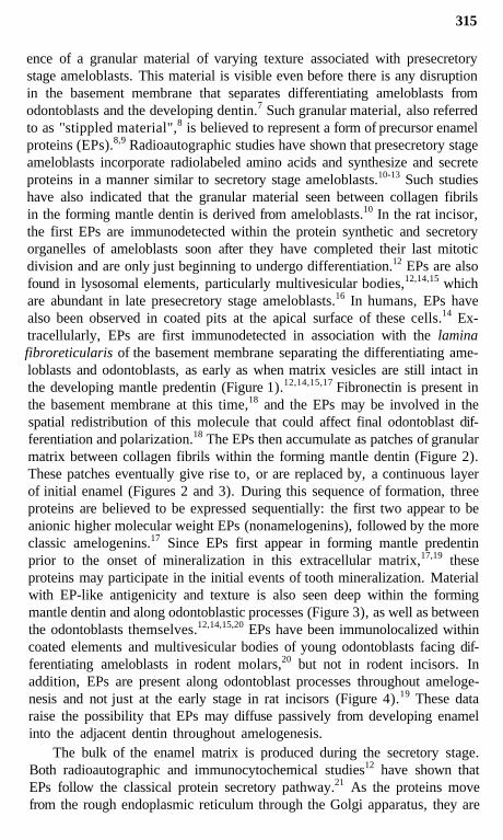
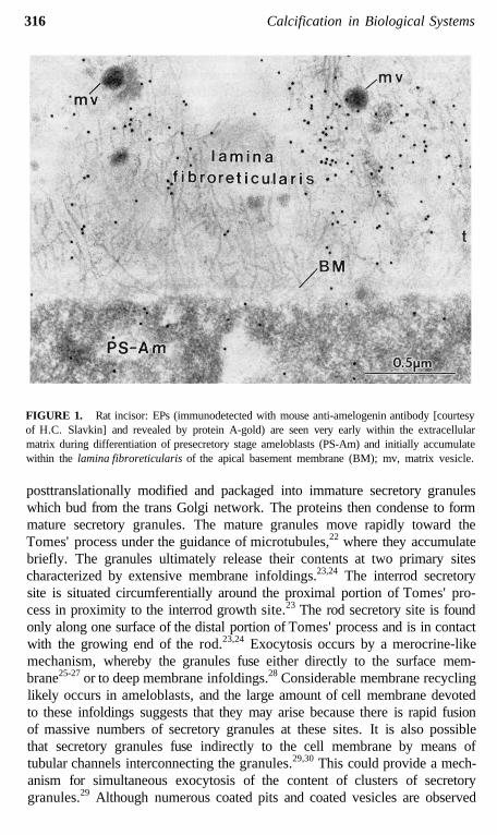
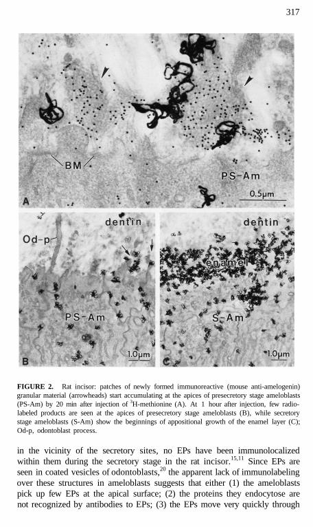
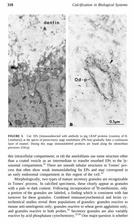
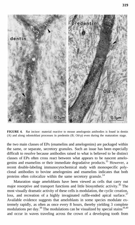
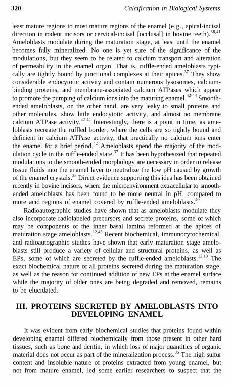
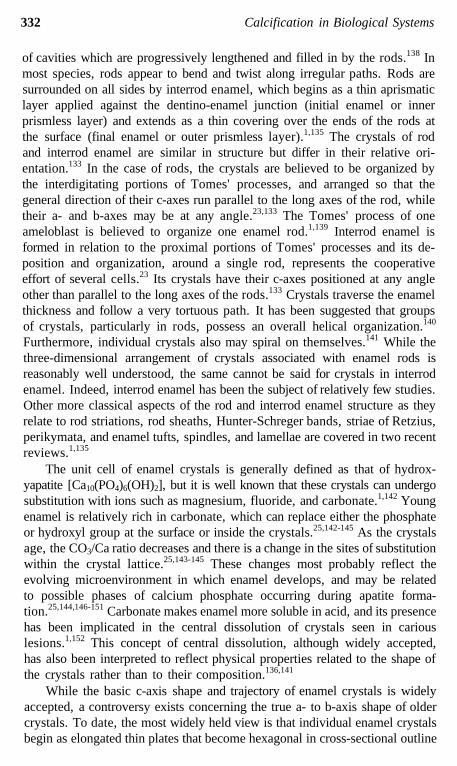
ence of a granular material of varying texture associated with presecretorystage ameloblasts. This material is visible even before there is any disruptionin the basement membrane that separates differentiating ameloblasts fromodontoblasts and the developing dentin.7 Such granular material, also referredto as ''stippled material",8 is believed to represent a form of precursor enamelproteins (EPs).8,9 Radioautographic studies have shown that presecretory stageameloblasts incorporate radiolabeled amino acids and synthesize and secreteproteins in a manner similar to secretory stage ameloblasts.10-13 Such studieshave also indicated that the granular material seen between collagen fibrilsin the forming mantle dentin is derived from ameloblasts.10 In the rat incisor,the first EPs are immunodetected within the protein synthetic and secretoryorganelles of ameloblasts soon after they have completed their last mitoticdivision and are only just beginning to undergo differentiation.12 EPs are alsofound in lysosomal elements, particularly multivesicular bodies,12,14,15 whichare abundant in late presecretory stage ameloblasts.16 In humans, EPs havealso been observed in coated pits at the apical surface of these cells.14 Ex-tracellularly, EPs are first immunodetected in association with the laminafibroreticularis of the basement membrane separating the differentiating ame-loblasts and odontoblasts, as early as when matrix vesicles are still intact inthe developing mantle predentin (Figure 1).12,14,15,17 Fibronectin is present inthe basement membrane at this time,18 and the EPs may be involved in thespatial redistribution of this molecule that could affect final odontoblast dif-ferentiation and polarization.18 The EPs then accumulate as patches of granularmatrix between collagen fibrils within the forming mantle dentin (Figure 2).These patches eventually give rise to, or are replaced by, a continuous layerof initial enamel (Figures 2 and 3). During this sequence of formation, threeproteins are believed to be expressed sequentially: the first two appear to beanionic higher molecular weight EPs (nonamelogenins), followed by the moreclassic amelogenins.17 Since EPs first appear in forming mantle predentinprior to the onset of mineralization in this extracellular matrix,17,19 theseproteins may participate in the initial events of tooth mineralization. Materialwith EP-like antigenicity and texture is also seen deep within the formingmantle dentin and along odontoblastic processes (Figure 3), as well as betweenthe odontoblasts themselves.12,14,15,20 EPs have been immunolocalized withincoated elements and multivesicular bodies of young odontoblasts facing dif-ferentiating ameloblasts in rodent molars,20 but not in rodent incisors. Inaddition, EPs are present along odontoblast processes throughout ameloge-nesis and not just at the early stage in rat incisors (Figure 4).19 These dataraise the possibility that EPs may diffuse passively from developing enamelinto the adjacent dentin throughout amelogenesis.
The bulk of the enamel matrix is produced during the secretory stage.Both radioautographic and immunocytochemical studies12 have shown thatEPs follow the classical protein secretory pathway.21 As the proteins movefrom the rough endoplasmic reticulum through the Golgi apparatus, they are
316 Calcification in Biological Systems
FIGURE 1. Rat incisor: EPs (immunodetected with mouse anti-amelogenin antibody [courtesyof H.C. Slavkin] and revealed by protein A-gold) are seen very early within the extracellularmatrix during differentiation of presecretory stage ameloblasts (PS-Am) and initially accumulatewithin the lamina fibroreticularis of the apical basement membrane (BM); mv, matrix vesicle.
posttranslationally modified and packaged into immature secretory granuleswhich bud from the trans Golgi network. The proteins then condense to formmature secretory granules. The mature granules move rapidly toward theTomes' process under the guidance of microtubules,22 where they accumulatebriefly. The granules ultimately release their contents at two primary sitescharacterized by extensive membrane infoldings.23,24 The interrod secretorysite is situated circumferentially around the proximal portion of Tomes' pro-cess in proximity to the interrod growth site.23 The rod secretory site is foundonly along one surface of the distal portion of Tomes' process and is in contactwith the growing end of the rod.23,24 Exocytosis occurs by a merocrine-likemechanism, whereby the granules fuse either directly to the surface mem-brane25-27 or to deep membrane infoldings.28 Considerable membrane recyclinglikely occurs in ameloblasts, and the large amount of cell membrane devotedto these infoldings suggests that they may arise because there is rapid fusionof massive numbers of secretory granules at these sites. It is also possiblethat secretory granules fuse indirectly to the cell membrane by means oftubular channels interconnecting the granules.29,30 This could provide a mech-anism for simultaneous exocytosis of the content of clusters of secretorygranules.29 Although numerous coated pits and coated vesicles are observed
317
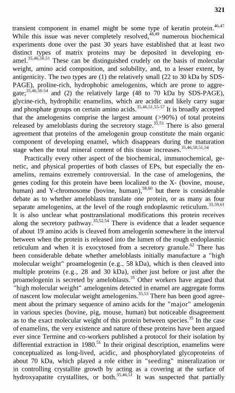
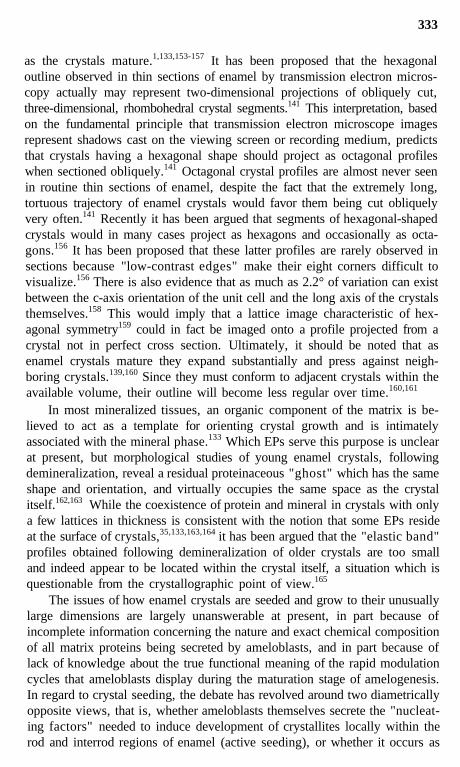
FIGURE 2. Rat incisor: patches of newly formed immunoreactive (mouse anti-amelogenin)granular material (arrowheads) start accumulating at the apices of presecretory stage ameloblasts(PS-Am) by 20 min after injection of 3H-methionine (A). At 1 hour after injection, few radio-labeled products are seen at the apices of presecretory stage ameloblasts (B), while secretorystage ameloblasts (S-Am) show the beginnings of appositional growth of the enamel layer (C);Od-p, odontoblast process.
in the vicinity of the secretory sites, no EPs have been immunolocalizedwithin them during the secretory stage in the rat incisor.15,11 Since EPs areseen in coated vesicles of odontoblasts,20 the apparent lack of immunolabelingover these structures in ameloblasts suggests that either (1) the ameloblastspick up few EPs at the apical surface; (2) the proteins they endocytose arenot recognized by antibodies to EPs; (3) the EPs move very quickly through
318 Calcification in Biological Systems
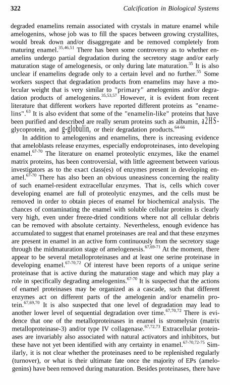
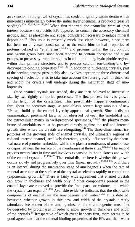
FIGURE 3. Cat: EPs (immunodetected with antibody to pig LRAP proteins [courtesy of H.Limeback]) at the apices of presecretory stage ameloblasts (PS-Am) gradually form a continuouslayer of enamel. During this stage immunolabeled products are found along the odontoblastprocesses (Od-p).
this intracellular compartment; or (4) the ameloblasts use some structure otherthan a coated vesicle as an intermediate to transfer resorbed EPs to the ly-sosomal compartment.32 There are smooth tubular structures in Tomes' pro-cess that often show weak immunolabeling for EPs and may correspond toan early endosomal compartment in this region of the cell.33
Morphologically, two types of mature secretory granules are recognizablein Tomes' process. In calcified specimens, these clearly appear as granuleswith a pale or dark content. Following incorporation of 3H-methionine, onlya portion of the granules are labeled, a finding which is consistent with fastturnover for these granules. Combined immunocytochemical and lectin cy-tochemical studies reveal three populations of granules: granules reactive tomouse anti-amelogenin only, granules reactive to wheat germ agglutinin only,and granules reactive to both probes.34 Secretory granules are also variablyreactive by acid phosphatase cytochemistry.29,30 One major question is whether
319
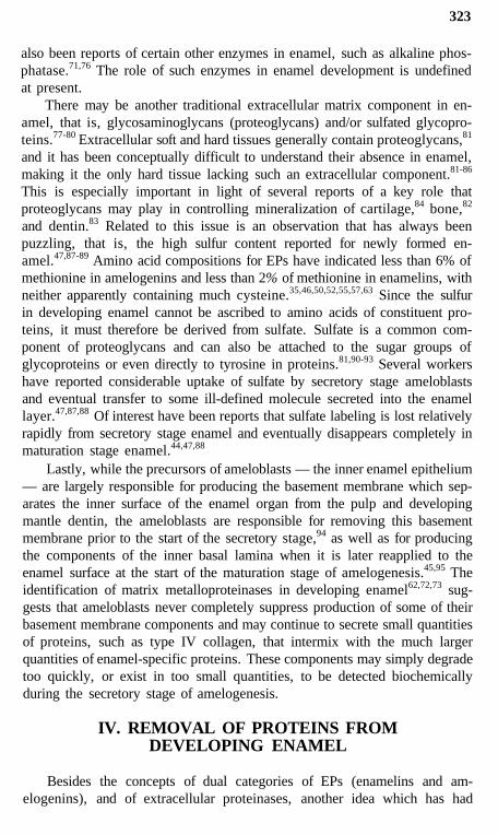
FIGURE 4. Rat incisor: material reactive to mouse amelogenin antibodies is found in dentin(A) and along odontoblast processes in predentin (B; Od-p) even during the maturation stage.
the two main classes of EPs (enamelins and amelogenins) are packaged withinthe same, or separate, secretory granules. Such an issue has been especiallydifficult to resolve because antibodies raised to what is believed to be distinctclasses of EPs often cross react between what appears to be nascent amelo-genins and enamelins or their immediate degradative products.35 However, arecent double-labeling immunocytochemical study with monospecific poly-clonal antibodies to bovine amelogenins and enamelins indicates that bothproteins often colocalize within the same secretory granule.14
Maturation stage ameloblasts have been viewed as cells that carry outmajor resorptive and transport functions and little biosynthetic activity.36 Themost visually dramatic activity of these cells is modulation, the cyclic creation,loss, and recreation of a highly invaginated ruffle-ended apical surface.37
Available evidence suggests that ameloblasts in some species modulate ex-tremely rapidly, as often as once every 8 hours, thereby yielding 3 completemodulations per day.38 The modulations can be visualized by special stains39,40
and occur in waves traveling across the crown of a developing tooth from
320 Calcification in Biological Systems
least mature regions to most mature regions of the enamel (e.g., apical-incisaldirection in rodent incisors or cervical-incisal [occlusal] in bovine teeth).38,41
Ameloblasts modulate during the maturation stage, at least until the enamelbecomes fully mineralized. No one is yet sure of the significance of themodulations, but they seem to be related to calcium transport and alterationof permeability in the enamel organ. That is, ruffle-ended ameloblasts typi-cally are tightly bound by junctional complexes at their apices.37 They showconsiderable endocytotic activity and contain numerous lysosomes, calcium-binding proteins, and membrane-associated calcium ATPases which appearto promote the pumping of calcium ions into the maturing enamel.42-44 Smooth-ended ameloblasts, on the other hand, are very leaky to small proteins andother molecules, show little endocytotic activity, and almost no membranecalcium ATPase activity.42-44 Interestingly, there is a point in time, as ame-loblasts recreate the ruffled border, where the cells are so tightly bound anddeficient in calcium ATPase activity, that practically no calcium ions enterthe enamel for a brief period.42 Ameloblasts spend the majority of the mod-ulation cycle in the ruffle-ended state.37 It has been hypothesized that repeatedmodulations to the smooth-ended morphology are necessary in order to releasetissue fluids into the enamel layer to neutralize the low pH caused by growthof the enamel crystals.38 Direct evidence supporting this idea has been obtainedrecently in bovine incisors, where the microenvironment extracellular to smooth-ended ameloblasts has been found to be more neutral in pH, compared tomore acid regions of enamel covered by ruffle-ended ameloblasts.40
Radioautographic studies have shown that as ameloblasts modulate theyalso incorporate radiolabeled precursors and secrete proteins, some of whichmay be components of the inner basal lamina reformed at the apices ofmaturation stage ameloblasts.12,45 Recent biochemical, immunocytochemical,and radioautographic studies have shown that early maturation stage amelo-blasts still produce a variety of cellular and structural proteins, as well asEPs, some of which are secreted by the ruffle-ended ameloblasts.12,13 Theexact biochemical nature of all proteins secreted during the maturation stage,as well as the reason for continued addition of new EPs at the enamel surfacewhile the majority of older ones are being degraded and removed, remainsto be elucidated.
III. PROTEINS SECRETED BY AMELOBLASTS INTODEVELOPING ENAMEL
It was evident from early biochemical studies that proteins found withindeveloping enamel differed biochemically from those present in other hardtissues, such as bone and dentin, in which loss of major quantities of organicmaterial does not occur as part of the mineralization process.35 The high sulfurcontent and insoluble nature of proteins extracted from young enamel, butnot from mature enamel, led some earlier researchers to suspect that the
321
transient component in enamel might be some type of keratin protein.46,47
While this issue was never completely resolved,48,49 numerous biochemicalexperiments done over the past 30 years have established that at least twodistinct types of matrix proteins may be deposited in developing en-amel.35,46,50,51 These can be distinguished crudely on the basis of molecularweight, amino acid composition, and solubility, and, to a lesser extent, byantigenicity. The two types are (1) the relatively small (22 to 30 kDa by SDS-PAGE), proline-rich, hydrophobic amelogenins, which are prone to aggre-gate;35,46,50-54 and (2) the relatively large (48 to 70 kDa by SDS-PAGE),glycine-rich, hydrophilic enamelins, which are acidic and likely carry sugarand phosphate groups on certain amino acids.35,46,51,55-57 It is broadly acceptedthat the amelogenins comprise the largest amount (>90%) of total proteinsreleased by ameloblasts during the secretory stage.35,51 There is also generalagreement that proteins of the amelogenin group constitute the main organiccomponent of developing enamel, which disappears during the maturationstage when the total mineral content of this tissue increases.35,46,50,51,54
Practically every other aspect of the biochemical, immunochemical, ge-netic, and physical properties of both classes of EPs, but especially the en-amelins, remains extremely controversial. In the case of amelogenins, thegenes coding for this protein have been localized to the X- (bovine, mouse,human) and Y-chromosome (bovine, human),58,60 but there is considerabledebate as to whether ameloblasts translate one protein, or as many as fourseparate amelogenins, at the level of the rough endoplasmic reticulum.35,59,61
It is also unclear what posttranslational modifications this protein receivesalong the secretory pathway.35,52,54 There is evidence that a leader sequenceof about 19 amino acids is cleaved from amelogenin somewhere in the intervalbetween when the protein is released into the lumen of the rough endoplasmicreticulum and when it is exocytosed from a secretory granule.62 There hasbeen considerable debate whether ameloblasts initially manufacture a "highmolecular weight" proamelogenin (e.g., 58 kDa), which is then cleaved intomultiple proteins (e.g., 28 and 30 kDa), either just before or just after theproamelogenin is secreted by ameloblasts.35 Other workers have argued that"high molecular weight" amelogenins detected in enamel are aggregate formsof nascent low molecular weight amelogenins.35,53 There has been good agree-ment about the primary sequence of amino acids for the "major" amelogeninin various species (bovine, pig, mouse, human) but noticeable disagreementas to the exact molecular weight of this protein between species.35 In the caseof enamelins, the very existence and nature of these proteins have been arguedever since Termine and co-workers published a protocol for their isolation bydifferential extraction in 1980.51 In their original description, enamelins wereconceptualized as long-lived, acidic, and phosphorylated glycoproteins ofabout 70 kDa, which played a role either in "seeding" mineralization orin controlling crystallite growth by acting as a covering at the surface ofhydroxyapatite crystallites, or both.35,46,51 It was suspected that partially
322 Calcification in Biological Systems
degraded enamelins remain associated with crystals in mature enamel whileamelogenins, whose job was to fill the spaces between growing crystallites,would break down and/or disaggregate and be removed completely frommaturing enamel.35,46,51 There has been some controversy as to whether en-amelins undergo partial degradation during the secretory stage and/or earlymaturation stage of amelogenesis, or only during late maturation.35 It is alsounclear if enamelins degrade only to a certain level and no further.55 Someworkers suspect that degradation products from enamelins may have a mo-lecular weight that is very similar to "primary" amelogenins and/or degra-dation products of amelogenins.35,53,57 However, it is evident from recentliterature that different workers have reported different proteins as "ename-lins".63 It is also evident that some of the "enamelin-like" proteins that havebeen purified and described are really serum proteins such as albumin, a 2 H S -glycoprotein, and g-globulin, or their degradation products.64-66
In addition to amelogenins and enamelins, there is increasing evidencethat ameloblasts release enzymes, especially endoproteinases, into developingenamel.67-70 The literature on enamel proteolytic enzymes, like the enamelmatrix proteins, has been controversial, with little agreement between variousinvestigators as to the exact class(es) of enzymes present in developing en-amel.67-70 There has also been an obvious uneasiness concerning the realityof such enamel-resident extracellular enzymes. That is, cells which coverdeveloping enamel are full of proteolytic enzymes, and the cells must beremoved in order to obtain pieces of enamel for biochemical analysis. Thechances of contaminating the enamel with soluble cellular proteins is clearlyvery high, even under freeze-dried conditions where not all cellular debriscan be removed with absolute certainty. Nevertheless, enough evidence hasaccumulated to suggest that enamel proteinases are real and that these enzymesare present in enamel in an active form continuously from the secretory stagethrough the midmaturation stage of amelogenesis.67,69-71 At the moment, thereappear to be several metalloproteinases and at least one serine proteinase indeveloping enamel.67-70,72 Of interest have been reports of a unique serineproteinase that is active during the maturation stage and which may play arole in specifically degrading amelogenins.67-70 It is suspected that the actionsof enamel proteinases may be organized as a cascade, such that differentenzymes act on different parts of the amelogenin and/or enamelin pro-tein.67,69,70 It is also suspected that one level of degradation may lead toanother lower level of sequential degradation over time.67,70,72 There is evi-dence that one of the metalloproteinases in enamel is stromelysin (matrixmetalloproteinase-3) and/or type IV collagenase.67,72,73 Extracellular protein-ases are invariably also associated with natural activators and inhibitors, butthese have not yet been identified with any certainty in enamel.67-70,72-75 Sim-ilarly, it is not clear whether the proteinases need to be replenished regularly(turnover), or what is their ultimate fate once the majority of EPs (amelo-genins) have been removed during maturation. Besides proteinases, there have
323
also been reports of certain other enzymes in enamel, such as alkaline phos-phatase.71,76 The role of such enzymes in enamel development is undefinedat present.
There may be another traditional extracellular matrix component in en-amel, that is, glycosaminoglycans (proteoglycans) and/or sulfated glycopro-teins.77-80 Extracellular soft and hard tissues generally contain proteoglycans,81
and it has been conceptually difficult to understand their absence in enamel,making it the only hard tissue lacking such an extracellular component.81-86
This is especially important in light of several reports of a key role thatproteoglycans may play in controlling mineralization of cartilage,84 bone,82
and dentin.83 Related to this issue is an observation that has always beenpuzzling, that is, the high sulfur content reported for newly formed en-amel.47,87-89 Amino acid compositions for EPs have indicated less than 6% ofmethionine in amelogenins and less than 2% of methionine in enamelins, withneither apparently containing much cysteine.35,46,50,52,55,57,63 Since the sulfurin developing enamel cannot be ascribed to amino acids of constituent pro-teins, it must therefore be derived from sulfate. Sulfate is a common com-ponent of proteoglycans and can also be attached to the sugar groups ofglycoproteins or even directly to tyrosine in proteins.81,90-93 Several workershave reported considerable uptake of sulfate by secretory stage ameloblastsand eventual transfer to some ill-defined molecule secreted into the enamellayer.47,87,88 Of interest have been reports that sulfate labeling is lost relativelyrapidly from secretory stage enamel and eventually disappears completely inmaturation stage enamel.44,47,88
Lastly, while the precursors of ameloblasts — the inner enamel epithelium— are largely responsible for producing the basement membrane which sep-arates the inner surface of the enamel organ from the pulp and developingmantle dentin, the ameloblasts are responsible for removing this basementmembrane prior to the start of the secretory stage,94 as well as for producingthe components of the inner basal lamina when it is later reapplied to theenamel surface at the start of the maturation stage of amelogenesis.45,95 Theidentification of matrix metalloproteinases in developing enamel62,72,73 sug-gests that ameloblasts never completely suppress production of some of theirbasement membrane components and may continue to secrete small quantitiesof proteins, such as type IV collagen, that intermix with the much largerquantities of enamel-specific proteins. These components may simply degradetoo quickly, or exist in too small quantities, to be detected biochemicallyduring the secretory stage of amelogenesis.
IV. REMOVAL OF PROTEINS FROMDEVELOPING ENAMEL
Besides the concepts of dual categories of EPs (enamelins and am-elogenins), and of extracellular proteinases, another idea which has had
324 Calcification in Biological Systems
significant impact on enamel research has been the hypothesis that ameloblastsresorb intact or partially degraded EPs directly from maturing enamel anddispose of them within their lysosomes.88 At the time it was proposed, andfor the following 20 years, this concept provided a reasonable explanationfor why ameloblasts possess such a complicated lysosomal system and whymaturation stage ameloblasts, especially those which are ruffled-ended, fre-quently show numerous large acid phosphatase- and trimetaphosphatase-pos-itive lysosomes.29,30,44,45,96-100 Studies with exogenous protein tracers haveclearly demonstrated that ruffle-ended ameloblasts avidly endocytose intra-vascularly injected tracers and transfer them rapidly to their lyso-somes.42,44,101,102 Endocytotic activity in ruffle-ended ameloblasts seems greaterthan in smooth-ended ameloblasts, or compared to secretory stage amelo-blasts.101-103 Recent immunocytochemical studies, using different monoclonaland polyclonal antibodies to either amelogenins or enamelins, have consist-ently demonstrated the presence of immunoreactive material within lysosomesof ameloblasts.14,15,31,104 While highly suggestive of resorptive activity, thisevidence, as well as that obtained by cytochemical techniques (tracers), proveonly that ameloblasts display active fluid-phase endocytosis and that thereare EPs, or their immediate degradative products, within the lysosomes ofameloblasts.
Enzyme inhibitor studies have revealed that extracellular degradation ofEPs can be blocked using natural compounds such as aprotinin, an inhibitorfor trypsin-like serine proteinases.70 It is also possible to block intracellulardegradation of EPs within lysosomes of ameloblasts using other natural com-pounds such as leupeptin, an inhibitor for thiol proteinases (cathepsins B/H/L), and serine proteinases.13,70 Immunocytochemical studies have indicatedthat the lysosomes of leupeptin-treated ameloblasts are filled with immuno-reactive material as monitored by an anti-amelogenin antibody.105 Radioau-tographic studies have further shown that much of the excess material ac-cumulating within these lysosomes corresponds to newly formed proteins.13,105
In contrast, there is no solid evidence from radioautographic studies for anatural increase in lysosomal labeling in ruffle-ended ameloblasts at 4 to 8days after a single pulse injection of 3H-methionine, 3H-leucine, or 3H-glycine,when aged, radioactive EPs are lost from maturing enamel.13,70,104,106 Theseresults suggest that newly formed material passing into the lysosomal systemof ameloblasts is degraded rapidly and the byproducts are likely not conservedbut are returned to the general circulation. It should be noted that the anti-amelogenin antibodies used in many published studies cannot reveal proteinsless than 14 kDa in molecular weight as determined by immunoblotting.1213
Consequently, it cannot be stated with absolute certainty that EPs degradedto this size or below do not gain access to the lysosomes of ameloblasts atsome point in time. Existing radioautographic data suggest that this does notoccur but such evidence remains largely circumstantial.104,106
325
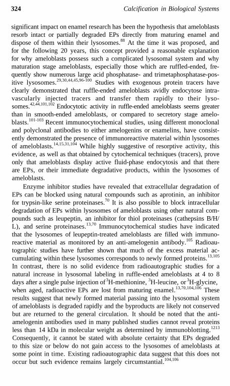
FIGURE 5. Schematic drawing illustrating the concept of vectorial secretion in ameloblasts.While most secretory products go to the functional apex (large arrow), a portion may get routedbasolaterally (short arrows) as a consequence of the reverse polarity this cell undergoes duringdifferentiation.
There is another possible explanation for the origin of immunoreactiveEPs detected in lysosomes of ameloblasts. That is, ameloblasts may secreteas much as 10% of the proteins they biosynthesize to their lateral and basalsurfaces, and then quickly reincorporate this material back into lysosomes byfluid-phase endocytosis. It is well documented that polarized epithelial cellsdirect some proteins exclusively to their apical surfaces, while others aredirected to the basal and lateral surfaces.107-111 A special case could exist inameloblasts because of the reverse polarity they undergo during differentiation(few epithelial cells do this; Figures 5 and 6). Before inner enamel epithelialcells become preameloblasts, the Golgi apparatus is located on the side ofthe nucleus situated toward the stratum intermedium. As preameloblasts dif-ferentiate, the Golgi apparatus moves to the opposite pole of the nucleustoward the basement membrane with preodontoblasts. Consequently, whatconstituted the base of the cell becomes the functional apex, and it is along
326 Calcification in Biological Systems
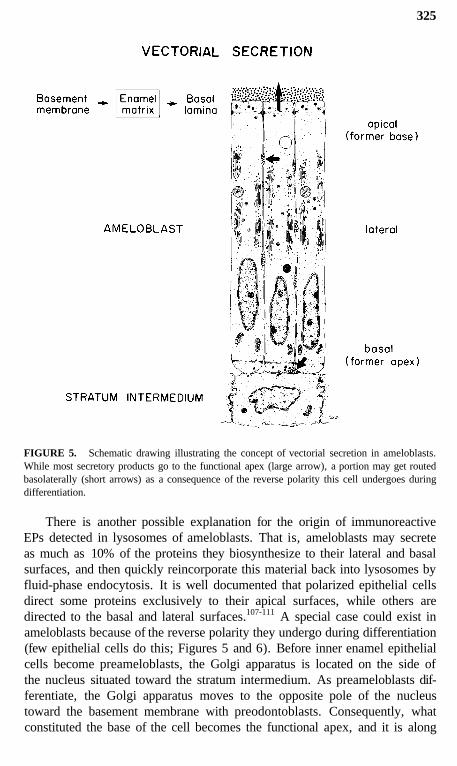
FIGURE 6. Schematic drawing illustrating possible directions of flow of proteins out of thesecretory pathway (numbers with thin circles) or into the lysosomal pathway (numbers with thickcircles) of ameloblasts. Secretory stage: Out, 1 = rod; 2 = interrod; 3 = lateral; 4 = lysosomal;5 = basal; In, 1 = distal portion Tome's process; 2 = proximal portion Tome's process; 3 =lateral; 4 = basal. Maturation stage: Out, 1 = apical; 2 = lateral; 3 = lysosomal; 4 = basal;In, 1 = lateral; 2 = basal; 3 = apical.
327
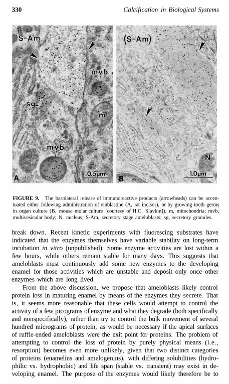
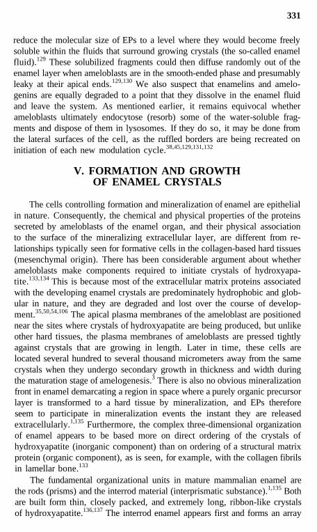
this surface where the bulk of the EPs (and later proteinases) are ultimatelysecreted (Figures 5 and 6).12,29,31,35,104,106 This restructuring may upset thenormal targeting mechanisms which direct appropriate "apical proteins" to"apical surfaces" (Figures 5 and 6). Secretory granules are sometimes seenin clusters in the functional base of ameloblasts (most prominent during thesecretory stage), and it is possible that some or all of these granules may beexocytosed and replaced by new granules that are also exocytosed. Pools ofrecently formed immunoreactive material are often seen naturally in the ba-solateral spaces between ameloblasts (Figures 7 to 9),14,112 and this could bederived from the contents of such basolaterally exocytosed granules. Anextreme example of basolateral secretion by ameloblasts is seen in animalstreated with compounds that disrupt microtubules and filaments (Figure 9).These ameloblasts often show large amounts of immunoreactive, granularmaterial surrounding the sides and base of the cell (Figure 9).113,119 Thispresumably happens because secretory granules that would normally go tothe apical surface (90% of secretory products) become "disoriented" andrelease their contents at the first plasma membrane encountered. This, in fact,would most often be the lateral sides of the cell, since the Golgi apparatusbecomes fragmented and dispersed throughout the cytoplasm of the ameloblastaffected by these compounds.115-119
The possibility that some EPs in lysosomes also derive from endocytosisat the apical surface and/or from direct targeting of secretory proteins tolysosomes cannot be ruled out completely (Figure 6). Indeed, the 10% of EPssuspected to be cycling through the lysosomal system is consistent with quan-tities of exportable proteins degraded in other active secretory cell types, suchas fibroblasts.120 The predominance of multivesicular bodies in the Golgiregion of ameloblasts (Figure 6), and their relative paucity at sites close towhere endocytosis may occur (e.g., basal surface), are not inconsistent withthe above theory, considering recent evidence for funnelling of apical andbasolateral endocytotic pathways to a common lysosomal compartment as-sociated with the apical region of polarized cells.108,121 It must also be rec-ognized that several distinct types of multivesicular bodies can be identifiedin ameloblasts and some may be related to endocytosis, while others may beinvolved in posttranslational degradation.122-124 Pale multivesicular bodies oftenrepresent an intermediate in the evolution of endosome to lysosome32,121,123,125
and their relatively weak immunolabeling104 may reflect endocytosis of ex-tracellularly processed proteins which are no longer recognizable by immu-nocytochemical methods, at least with the antibodies we have used.
Lastly, it is important to recall that the original concept of resorptiveactivity by ameloblasts was proposed many years ago, before it was realizedthat ameloblasts may secrete extracellular proteolytic enzymes,67-73,106 andbefore it was widely accepted that amelogenins likely undergo considerabledegradation by these enzymes in a "top-down" fashion from some precur-sor (e.g., 25 kDa by SDS-PAGE) toward a much lower molecular weight
328 Calcification in Biological Systems
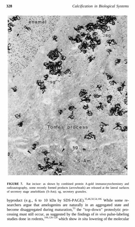
FIGURE 7. Rat incisor: as shown by combined protein A-gold immunocytochemistry andradioautography, some recently formed products (arrowheads) are released at the lateral surfacesof secretory stage ameloblasts (S-Am); sg, secretory granules.
byproduct (e.g., 6 to 10 kDa by SDS-PAGE).35,46,50,54,106 While some re-searchers argue that amelogenins are naturally in an aggregated state andbecome disaggregated during maturation,53 the "top-down" proteolytic pro-cessing must still occur, as suggested by the findings of in vivo pulse-labelingstudies done in rodents,106,126-128 which show in situ lowering of the molecular
329
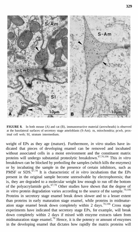
FIGURE 8. In both mouse (A) and cat (B), immunoreactive material (arrowheads) is observedat the basolateral surfaces of secretory stage ameloblasts (S-Am). m, mitochondria; pcwb, prox-imal cell web; SI, stratum intermedium.
weight of EPs as they age (mature). Furthermore, in vitro studies have in-dicated that pieces of developing enamel can be removed and incubatedwithout associated cells in a moist environment and the constituent matrixproteins will undergo substantial proteolytic breakdown.67,70,106 This in vitrobreakdown can be blocked by preboiling the samples (which kills the enzymes)or by incubating the sample in the presence of certain inhibitors, such asPMSF or SDS.67,70 It is characteristic of in vitro incubations that the EPspresent in the original sample become unresolvable by electrophoresis; thatis, they are degraded to a molecular weight low enough to run off the bottomof the polyacrylamide gels.67,70 Other studies have shown that the degree ofin vitro protein degradation varies according to the source of the sample.70,106
Proteins in secretory stage enamel break down slower and to a lesser extentthan proteins in early maturation stage enamel, while proteins in midmatur-ation stage enamel break down completely within 2 days.70,106 Cross stageexperiments have indicated that secretory stage EPs, for example, will breakdown completely within 2 days if mixed with enzyme extracts taken frommidmaturation stage enamel.70 Hence, it is the potency or amount of enzymesin the developing enamel that dictates how rapidly the matrix proteins will
330 Calcification in Biological Systems
FIGURE 9. The basolateral release of immunoreactive products (arrowheads) can be accen-tuated either following administration of vinblastine (A, rat incisor), or by growing tooth germsin organ culture (B, mouse molar culture [courtesy of H.C. Slavkin]). m, mitochondria; mvb,multivesicular body; N, nucleus; S-Am, secretory stage ameloblasts; sg, secretory granules.
break down. Recent kinetic experiments with fluorescing substrates haveindicated that the enzymes themselves have variable stability on long-termincubation in vitro (unpublished). Some enzyme activities are lost within afew hours, while others remain stable for many days. This suggests thatameloblasts must continuously add some new enzymes to the developingenamel for those activities which are unstable and deposit only once otherenzymes which are long lived.
From the above discussion, we propose that ameloblasts likely controlprotein loss in maturing enamel by means of the enzymes they secrete. Thatis, it seems more reasonable that these cells would attempt to control theactivity of a few picograms of enzyme and what they degrade (both specificallyand nonspecifically), rather than try to control the bulk movement of severalhundred micrograms of protein, as would be necessary if the apical surfacesof ruffle-ended ameloblasts were the exit point for proteins. The problem ofattempting to control the loss of protein by purely physical means (i.e.,resorption) becomes even more unlikely, given that two distinct categoriesof proteins (enamelins and amelogenins), with differing solubilities (hydro-philic vs. hydrophobic) and life span (stable vs. transient) may exist in de-veloping enamel. The purpose of the enzymes would likely therefore be to
331
reduce the molecular size of EPs to a level where they would become freelysoluble within the fluids that surround growing crystals (the so-called enamelfluid).129 These solubilized fragments could then diffuse randomly out of theenamel layer when ameloblasts are in the smooth-ended phase and presumablyleaky at their apical ends.129,130 We also suspect that enamelins and amelo-genins are equally degraded to a point that they dissolve in the enamel fluidand leave the system. As mentioned earlier, it remains equivocal whetherameloblasts ultimately endocytose (resorb) some of the water-soluble frag-ments and dispose of them in lysosomes. If they do so, it may be done fromthe lateral surfaces of the cell, as the ruffled borders are being recreated oninitiation of each new modulation cycle.38,45,129,131,132
V. FORMATION AND GROWTHOF ENAMEL CRYSTALS
The cells controlling formation and mineralization of enamel are epithelialin nature. Consequently, the chemical and physical properties of the proteinssecreted by ameloblasts of the enamel organ, and their physical associationto the surface of the mineralizing extracellular layer, are different from re-lationships typically seen for formative cells in the collagen-based hard tissues(mesenchymal origin). There has been considerable argument about whetherameloblasts make components required to initiate crystals of hydroxyapa-tite.133,134 This is because most of the extracellular matrix proteins associatedwith the developing enamel crystals are predominately hydrophobic and glob-ular in nature, and they are degraded and lost over the course of develop-ment.35,50,54,106 The apical plasma membranes of the ameloblast are positionednear the sites where crystals of hydroxyapatite are being produced, but unlikeother hard tissues, the plasma membranes of ameloblasts are pressed tightlyagainst crystals that are growing in length. Later in time, these cells arelocated several hundred to several thousand micrometers away from the samecrystals when they undergo secondary growth in thickness and width duringthe maturation stage of amelogenesis.3 There is also no obvious mineralizationfront in enamel demarcating a region in space where a purely organic precursorlayer is transformed to a hard tissue by mineralization, and EPs thereforeseem to participate in mineralization events the instant they are releasedextracellularly.1,135 Furthermore, the complex three-dimensional organizationof enamel appears to be based more on direct ordering of the crystals ofhydroxyapatite (inorganic component) than on ordering of a structural matrixprotein (organic component), as is seen, for example, with the collagen fibrilsin lamellar bone.133
The fundamental organizational units in mature mammalian enamel arethe rods (prisms) and the interrod material (interprismatic substance).1,135 Bothare built form thin, closely packed, and extremely long, ribbon-like crystalsof hydroxyapatite.136,137 The interrod enamel appears first and forms an array
332 Calcification in Biological Systems
of cavities which are progressively lengthened and filled in by the rods.138 Inmost species, rods appear to bend and twist along irregular paths. Rods aresurrounded on all sides by interrod enamel, which begins as a thin aprismaticlayer applied against the dentino-enamel junction (initial enamel or innerprismless layer) and extends as a thin covering over the ends of the rods atthe surface (final enamel or outer prismless layer).1,135 The crystals of rodand interrod enamel are similar in structure but differ in their relative ori-entation.133 In the case of rods, the crystals are believed to be organized bythe interdigitating portions of Tomes' processes, and arranged so that thegeneral direction of their c-axes run parallel to the long axes of the rod, whiletheir a- and b-axes may be at any angle.23,133 The Tomes' process of oneameloblast is believed to organize one enamel rod.1,139 Interrod enamel isformed in relation to the proximal portions of Tomes' processes and its de-position and organization, around a single rod, represents the cooperativeeffort of several cells.23 Its crystals have their c-axes positioned at any angleother than parallel to the long axes of the rods.133 Crystals traverse the enamelthickness and follow a very tortuous path. It has been suggested that groupsof crystals, particularly in rods, possess an overall helical organization.140
Furthermore, individual crystals also may spiral on themselves.141 While thethree-dimensional arrangement of crystals associated with enamel rods isreasonably well understood, the same cannot be said for crystals in interrodenamel. Indeed, interrod enamel has been the subject of relatively few studies.Other more classical aspects of the rod and interrod enamel structure as theyrelate to rod striations, rod sheaths, Hunter-Schreger bands, striae of Retzius,perikymata, and enamel tufts, spindles, and lamellae are covered in two recentreviews.1,135
The unit cell of enamel crystals is generally defined as that of hydrox-yapatite [Ca10(PO4)6(OH)2], but it is well known that these crystals can undergosubstitution with ions such as magnesium, fluoride, and carbonate.1,142 Youngenamel is relatively rich in carbonate, which can replace either the phosphateor hydroxyl group at the surface or inside the crystals.25,142-145 As the crystalsage, the CO3/Ca ratio decreases and there is a change in the sites of substitutionwithin the crystal lattice.25,143-145 These changes most probably reflect theevolving microenvironment in which enamel develops, and may be relatedto possible phases of calcium phosphate occurring during apatite forma-tion.25,144,146-151 Carbonate makes enamel more soluble in acid, and its presencehas been implicated in the central dissolution of crystals seen in cariouslesions.1,152 This concept of central dissolution, although widely accepted,has also been interpreted to reflect physical properties related to the shape ofthe crystals rather than to their composition.136,141
While the basic c-axis shape and trajectory of enamel crystals is widelyaccepted, a controversy exists concerning the true a- to b-axis shape of oldercrystals. To date, the most widely held view is that individual enamel crystalsbegin as elongated thin plates that become hexagonal in cross-sectional outline
333
as the crystals mature.1,133,153-157 It has been proposed that the hexagonaloutline observed in thin sections of enamel by transmission electron micros-copy actually may represent two-dimensional projections of obliquely cut,three-dimensional, rhombohedral crystal segments.141 This interpretation, basedon the fundamental principle that transmission electron microscope imagesrepresent shadows cast on the viewing screen or recording medium, predictsthat crystals having a hexagonal shape should project as octagonal profileswhen sectioned obliquely.141 Octagonal crystal profiles are almost never seenin routine thin sections of enamel, despite the fact that the extremely long,tortuous trajectory of enamel crystals would favor them being cut obliquelyvery often.141 Recently it has been argued that segments of hexagonal-shapedcrystals would in many cases project as hexagons and occasionally as octa-gons.156 It has been proposed that these latter profiles are rarely observed insections because "low-contrast edges" make their eight corners difficult tovisualize.156 There is also evidence that as much as 2.2° of variation can existbetween the c-axis orientation of the unit cell and the long axis of the crystalsthemselves.158 This would imply that a lattice image characteristic of hex-agonal symmetry159 could in fact be imaged onto a profile projected from acrystal not in perfect cross section. Ultimately, it should be noted that asenamel crystals mature they expand substantially and press against neigh-boring crystals.139,160 Since they must conform to adjacent crystals within theavailable volume, their outline will become less regular over time.160,161
In most mineralized tissues, an organic component of the matrix is be-lieved to act as a template for orienting crystal growth and is intimatelyassociated with the mineral phase.133 Which EPs serve this purpose is unclearat present, but morphological studies of young enamel crystals, followingdemineralization, reveal a residual proteinaceous "ghost" which has the sameshape and orientation, and virtually occupies the same space as the crystalitself.162,163 While the coexistence of protein and mineral in crystals with onlya few lattices in thickness is consistent with the notion that some EPs resideat the surface of crystals,35,133,163,164 it has been argued that the "elastic band"profiles obtained following demineralization of older crystals are too smalland indeed appear to be located within the crystal itself, a situation which isquestionable from the crystallographic point of view.165
The issues of how enamel crystals are seeded and grow to their unusuallylarge dimensions are largely unanswerable at present, in part because ofincomplete information concerning the nature and exact chemical compositionof all matrix proteins being secreted by ameloblasts, and in part because oflack of knowledge about the true functional meaning of the rapid modulationcycles that ameloblasts display during the maturation stage of amelogenesis.In regard to crystal seeding, the debate has revolved around two diametricallyopposite views, that is, whether ameloblasts themselves secrete the "nucleat-ing factors" needed to induce development of crystallites locally within therod and interrod regions of enamel (active seeding), or whether it occurs as
334 Calcification in Biological Systems
an extension in the growth of crystallites seeded originally within dentin whichmineralizes immediately before the initial layer of enamel is produced (passiveseeding).129,133,134,146,166,167 When first reported, the enamelins were of greatinterest because these acidic EPs appeared to contain the accessory chemicalgroups, such as phosphate and sugar, considered necessary to induce mineraldeposition.51 This issue is presently much less clear, however, since therehas been no universal consensus as to the exact biochemical properties ofproteins defined as "enamelins",63-66 and proteins within the hydrophobicamelogenin group have since been reported to contain phosphate and sugargroups, to possess hydrophilic regions in addition to long hydrophobic regionswithin their primary structure, and to possess calcium ion-binding and hy-droxyapatite-binding properties.34,35,129 In addition to nucleating crystals, partof the seeding process presumably also involves appropriate three-dimensionalspacing of nucleation sites to take into account the future growth in thicknessand width the crystals will undergo during the maturation stage of ame-loqenesis.
Once enamel crystals are seeded, they are then believed to increase insize by two tightly controlled processes. The first process involves growthin the length of the crystallites. This presumably happens continuouslythroughout the secretory stage, as ameloblasts secrete large amounts of newEPs and build up the enamel layer by appositional growth. Since a distinctunmineralized preenamel layer is not observed between the ameloblast andthe extracellular matrix in well-preserved specimens,168,169 the plasma mem-brane of ameloblasts must be pressed directly against the rod and interrodgrowth sites where the crystals are elongating.144 The three-dimensional tra-jectories of the growing ends of enamel crystals, and ultimately regions ofrod and interrod enamel, are likely therefore, greatly influenced by the chem-ical nature of proteins embedded within the plasma membranes of ameloblastsor deposited near the surface of the membranes at these sites.129,133 The secondprocess occurs later in time and involves expansion in the thickness and widthof the enamel crystals.133,153-155 The central dispute here is whether this growthoccurs slowly and progressively over time (linear growth),44,153-155 or if thereis an interval during the maturation stage of amelogenesis when the rate ofmineral accretion at the surface of the crystal accelerates rapidly to completion(exponential growth).50 There is fairly wide agreement that enamel crystalscan grow in thickness and width only if other components present in theenamel layer are removed to provide the free space, or volume, into whichthe crystals can expand.50,170 Available evidence indicates that the disposablecomponents of enamel are the amelogenins and water.35,50 It is debated,however, whether growth in thickness and width of the crystals directlystimulates breakdown of the amelogenins, or if the amelogenins must firstbe degraded by proteinases in order to allow growth in thickness and widthof the crystals.35 Irrespective of which event happens first, there seems to begood agreement that the mineral binding properties of the EPs and their water
335
solubility likely exert considerable local control over the rate of crystal growthin thickness and width, until these proteins begin to disappear.35,129,170 Ulti-mately, it must be remembered that the formation of crystals of hydroxyapatitewith unusually large dimensions is a process that takes many weeks to com-plete,50 as opposed to only hours, as seen in those hard tissues using amineralization front to produce small plate-like crystals of hydroxyapatite ina collagenous matrix.133 It is now well established that ameloblasts rhyth-mically alter their morphology at the surface of enamel over the same periodof time that the enamel crystals expand in thickness and width.37,38 Suchcyclic modulations represent a biological process unlike anything seen in anyother of the hard tissues. This phenomenon presumably, therefore, providesthe unique mechanism which allows additional mineral to be deposited con-tinuously over the long periods of time at the surface of maturing enamelcrystals.
REFERENCES
1. Boyde, A., Enamel, in Handbook of Microscopic Anatomy, Vol. V/6: Teeth, Oksche, A.and Vollrath, L., Eds., Springer-Verlag, Berlin, 1989, 310.
2. Yaeger, J. A., Enamel, in Orban's Oral Histology and Embryology, 9th ed., Bhashkar,S. N., Ed., C. V. Mosby, St. Louis, 1980, 46.
3. Leblond, C. P. and Warshawsky, H., Dynamics of enamel formation in the rat incisortooth, J. Dent. Res., 58(B), 950, 1979.
4. Warshawsky, H. and Smith, C. E., Morphological classification of rat incisor ame-loblasts, Anat. Rec., 179, 423, 1974.
5. Josephsen, K., Fejerskov, O., Baelum, V., and Weile, V., The effect of a single doseof l-hydroxyethylidene-l,l-bisphosphonate (HEBP) on presecretory ameloblast differ-entiation in rat incisors, J. Biol. Buccale, 18, 321, 1990.
6. Eisenmann, D. R., Amelogenesis, in Oral Histology: Development, Structure, andFunction, 3rd ed., Ten Cate, A. R., Ed., C. V. Mosby, St. Louis, 1989, 197.
7. Kallenbach, E., Electron microscopy of the differentiating rat incisor ameloblast, J.Ulstrastruct. Res., 35, 508, 1971.
8. Watson, M. L., The extracellular nature of enamel in the rat, J. Biophys. Biochem.Cytol., 7, 489, 1960.
9. Fearnhead, R. W., Electron microscopy of forming enamel, Arch. Oral Biol., 4, 24,1961.
10. Slavkin, H. C., Mino, W., and Bringas, P., Jr., The biosynthesis and secretion ofprecursor enamel protein by ameloblasts as visualized by autoradiography after tryptophanadministration, Anat. Rec., 185, 289, 1976.
11. Warshawsky, H. and Vugman, I., A comparison of the protein synthetic activity ofpresecretory and secretory ameloblasts in rat incisors, Anat. Rec., 188, 143, 1977.
12. Nanci, A., Ahluwalia, J. P., Pompura, J. R., and Smith, C. E., Biosynthesis andsecretion of enamel proteins in the rat incisor, Anat. Rec., 224, 277, 1989.
13. Smith, C. E. and Nanci, A., Secretory activity as a function of the development andmaturation of ameloblasts, Connect. Tissue Res., 22, 147, 1989.
336 Calcification in Biological Systems
14. Inage, T., Shimokawa, H., Teranishi, Y., Iwase, T., and Toda, Y., Immunocyto-chemical demonstration of amelogenins and enamelins secreted by ameloblasts duringthe secretory and maturation stages, Arch. Histol. Cytol., 52, 213, 1989.
15. Uchida, T., Tanabe, T., and Fukae, M., Immunocytochemical localization of amelo-genins in the deciduous tooth germs of the human fetus, Arch. Histol. Cytol., 52, 543,1989.
16. Smith, C. E., Stereological analysis of organelle distribution within rat incisor enamelorgan at successive stages of amelogenesis, INSERM, 125, 273, 1984.
17. Slavkin, H. C., Bessem, C., Bringas, P., Jr., Zeichner-David, M., Nanci, A., andSnead, M. L., Sequential expression and differential function of multiple enamel proteinsduring fetal, neonatal, and early postnatal stages of mouse molar organogenesis, Differ-entiation, 37, 26, 1988.
18. Meyer, J.-M., Lesot, H., Staubli, A., and Ruch, J.-V., Immunoperoxidase localizationof fibronectin during odontoblast differentiation. An ultrastructural study, Biol. Struct.Morphog., 2, 19, 1989.
19. Suga, S., On the penetration of the enamel matrix substances to the dentine duringamelogenesis, with special reference to its functional significance, Arch. Histol. Jpn.,20, 477, 1960.
20. Inai, T., Kukita, T., Ohsaki, Y., Nagata, K., Kukita, A., and Kurisu, K., Immu-nohistochemical demonstration of amelogenin penetration toward the dental pulp in theearly stages of ameloblast development in rat molar tooth germs, Anat. Rec., 229, 259,1991.
21. Burgess, T. L. and Kelly, R. B., Constitutive and regulated secretion of proteins, Annu.Rev. Cell Biol., 3, 243, 1987.
22. Kelly, R. B., Microtubules, membrane traffic, and cell organization, Cell, 61, 5, 1990.23. Nanci, A. and Warshawsky, H., Characterization of putative secretory sites on ame-
loblasts of the rat incisor, Am. J. Anat., 171, 163, 1984.24. Kallenbach, E., Fine structure of secretory ameloblasts in the kitten, Am. J. Anat., 148,
479, 1977.25. Frank, R. M. and Nalbandian, J., Ultrastructure of amelogenesis, in Structural and
Chemical Organization of Teeth, Miles, A. E. W., Ed., Academic Press, New York,1967, 399.
26. Reith, E. J., The early stage of amelogenesis as observed in molar teeth of young rats,J. Ultrastruct. Res., 17, 503, 1991.
27. Matthiessen, M. E. and von Bülow, F. A., The ultrastructure of human secretoryameloblasts, Zellforschung, 101, 232, 1969.
28. Simmelink, J. W., Mode of enamel matrix secretion, J. Dent. Res., 61(Sp Iss), 1483,1982.
29. Smith, C. E., Ameloblasts: secretory and resorptive functions, J. Dent. Res., 58(B),695, 1979.
30. Ozawa, H., Yamada, M., Uchida, T., Yamamoto, T., and Takano, Y., Fine structuraland cytochemical studies on the Golgi-SER system of ameloblasts with special referenceto its resorptive function, in Mechanisms of Tooth Enamel Formation, Suga, S., Ed.,Quintessence Publishing, Tokyo, 1983, 1.
31. Nanci, A., Bendayan, M., and Slavkin, H. C., Enamel protein biosynthesis and se-cretion in mouse incisor secretory ameloblasts as revealed by high-resolution immuno-cytochemistry, J. Histochem. Cytochem., 33, 1153, 1985.
32. Griffiths, G., Back, R., and Marsh, M., A quantitative analysis of the endocyticpathway in baby hamster kidney cells, J. Cell Biol., 109, 2703, 1989.
33. Ishii, M., Vroman, B., and Larusso, N. F., Fluid-phase endocytosis by intrahepaticbile duct epithelial cells isolated from normal rat liver, J. Histochem. Cytochem., 38,515, 1990.
337
34. Nanci, A., Ahluwalia, J. P., Zalzal, S., and Smith, C. E., Cytochemical and bio-chemical characterization of glycoproteins in forming and maturing enamel of the ratincisor, J. Histochem. Cytochem., 370, 1619, 1989.
35. Deutsch, D., Structure and function of enamel gene products, Anat. Rec., 224, 189,1989.
36. Kallenbach, E., Fine structure of rat incisor ameloblasts in transition between enamelsecretion and maturation stages, Tissue Cell, 6, 173, 1974.
37. Josephsen, K. and Fejerskov, O., Ameloblast modulation in the maturation zone of therat incisor enamel organ. A light and electron microscopic study, J. Anat., 124, 45, 1977.
38. Smith, C. E., McKee, M. D., and Nanci, A., Cyclic induction and rapid movementof sequential waves of new smooth-ended ameloblast modulation bands in rat incisors asvisualized by polychrome fluorescent labeling and GBHA-staining of maturing enamel,Adv. Dent. Res., 1, 162, 1987.
39. McKee, M. D., Nanci, A., Smith, C. E., and Warshawsky, H., Cyclical aspects ofenamel maturation and the role of ruffle-ended and smooth-ended ameloblasts, in ToothEnamel V, Florence Publishers, Yokohama, 1989, 41.
40. Sasaki, S., Takagi, T., and Suzuki, M., Cyclical changes in pH in bovine developingenamel as sequential bands, Arch. Oral Biol., 36, 227, 1991.
41. Takano, Y., Matsuo, S., Wakisaka, S., Ichikawa, S., Nishikawa, S., and Akai, M.,Cyclic changes in the properties of maturing enamel in the bovine permanent incisor asrevealed by glyoxal bis(2-hydroxyanil) staining, Arch. Oral Biol., 33, 231, 1988.
42. Takano, Y., Ozawa, H., and Crenshaw, M., The mechanism of calcium and phosphatetransport to the enamel, in Mechanisms of Tooth Enamel Formation, Suga, S., Ed.,Quintessence Publishing, Tokyo, 1983, 49.
43. Salama, A. H., El-Moneim, A., Zaki, E., and Eisenmann, D. R., Cytochemicallocalization of Ca2 + -Mg2+ adenosine triphosphatase in rat incisor ameloblasts duringenamel secretion and maturation, J. Histochem. Cytochem., 35, 471, 1987.
44. Sasaki, T., Cell biology of tooth enamel formation, in Monographs in Oral Science,Myers, H. M., Ed., S. Karger, Basel, 1990, 1.
45. Takano, Y., Cytochemical studies of ameloblasts and the surface layer of enamel of therat incisor at the maturation stage, Arch. Histol. Jpn., 42, 11, 1979.
46. Eastoe, J. E., The amino acid composition of proteins from the oral tissues. II. Thematrix proteins in dentine and enamel from developing human deciduous teeth, Arch.Oral Biol., 8, 633, 1963.
47. Blumen, G. and Merzel, J., Autoradiographic study with [35]-sodium sulphate of lossof sulphated glycosaminoglycans during amelogenesis in the guinea pig, Arch. Oral Biol.,21, 513, 1976.
48. Lesot, H., Smith, A. J., Matthews, J. B., and Ruch, J.-V., An extracellular matrixprotein of dentine, enamel, and bone shares common antigenic determinants with keratins,Calcif. Tissue Int., 42, 53, 1988.
49. Robinson, C., Shore, R. C., and Kirkham, J., Tuft protein: its relationship with thekeratins and the developing enamel matrix, Calcif. Tissue Int., 44, 393, 1989.
50. Robinson, C. and Kirkham, J., Dynamics of amelogenesis as revealed by proteincompositional studies, in The Chemistry and Biology of Mineralized Tissue, Butler, W.T., Ed., EBSCO Media, Birmingham, AL, 1985, 248.
51. Termine, J. D., Belcourt, A. B., Christner, P. J., Conn, K. M., and Nylen, M. U.,Properties of dissociatively extracted fetal tooth matrix proteins, J. Biol. Chem., 20,9760, 1980.
52. Fincham, A. G., Hu, Y., Lau, E., Pavlova, Z., Slavkin, H. C., and Snead, M. L.,Isolation and partial characterization of a human amelogenin from a single fetal dentitionusing HPLC techniques, Calcif. Tissue Int., 47, 105, 1990.
53. Limeback, H. and Simic, A., Biochemical characterization of stable high molecular-weight aggregates of amelogenins formed during porcine enamel development, Arch.Oral Biol., 35, 459, 1990.
338 Calcification in Biological Systems
54. Fincham, A. G., Hu, Y., Lau, E. C, Slavkin, H. C., and Snead, M. L., Amelogeninpost-secretory processing during biomineralization in the postnatal mouse molar tooth,Arch. Oral Biol., 36, 305, 1991.
55. Limeback, H., Isolation and characterization of pig enamelins, Biochem. J., 243, 385,1987.
56. Menanteau, J., Meflah, K., and Strecker, G., The carbohydrate moiety of mineral-bound proteins from fetal enamel: a basis for enamelins heterogeneity, Calcif. TissueInt., 42, 196, 1988.
57. Ogata, Y., Shimokawa, H., and Sasaki, S., Purification, characterization, and bio-synthesis of bovine enamelins, Calcif. Tissue Int., 43, 389, 1988.
58. Snead, M. L., Lau, E. C., Fincham, A. G., Zeichner-David, M., and Slavkin,H. C., Knowledge based molecular dissection of amelogenesis: the chromosomal locationof the amelogenin genes, in Tooth Enamel V, Fearnhead, R. W., Ed., Florence Publishers,Yokohama, 1989, 261.
59. Gibson, C., Golub, E., Herold, R., Risser, M., Ding, W., Shimokawa, H., Young,M., Termine, J., and Rosenbloom, J., Structure and expression of the bovine ame-logenin gene, Biochemistry, 30, 1075, 1991.
60. Fincham, A. G., Bessem, C. C., Lau, E. C., Pavlova, Z., Shuler, C., Slavkin,H. C., and Snead, M. L., Human developing enamel proteins exhibit a sex-linkeddimorphism, Calcif. Tissue Int., 48, 288, 1991.
61. Shimokawa, H., Tamura, M., Ibaraki, K., Ogata, Y., and Sasaki, S., Human ame-logenin gene, in Tooth Enamel V, Fearnhead, R. W., Ed., Florence Publishers, Yokohama,1989, 301.
62. Shimokawa, H., Ogata, Y., Sasaki, S., Sobel, M. E., McQuillan, C. I., Termine,J. D., and Young, M. F., Molecular cloning of bovine amelogenin cDNA, Adv. Dent.Res., 1, 293, 1987.
63. Robinson, C., Kirkham, J., and Fincham, A., The enamelin/non-amelogenin problem.A brief review, Calcif. Tissue Int., 22, 93, 1989.
64. Limeback, H., Sakarya, H., Chu, W., and MacKinnon, M., Serum albumin and itsacid hydrolysis peptide dominate preparations of mineral-bound enamel proteins, J. BoneMiner. Res., 4, 235, 1989.
65. Strawich, E. and Glimcher, M. J., Tooth 'enamelins' identified mainly as serumproteins — major 'enamelin' is albumin, Eur. J. Biochem., 191, 47, 1990.
66. Menanteau, J., Dajean, S., Laboux, O., and Aubry, J., Proteins of the mineralcompartment of bovine fetal enamel share common antigenic determinants with serumproteins, Calcif. Tissue Int., 47, 251, 1990.
67. Overall, C. M. and Limeback, H., Identification and characterization of enamel pro-teinases isolated from developing enamel. Amelogeninolytic serine proteinases are as-sociated with enamel maturation in pig, Biochem. J., 256, 965, 1988.
68. Carter, J., Smillie, A. C., and Shepherd, M. G., Purification and properties of theprotease from developing porcine dental enamel, Arch. Oral Biol., 34, 195, 1989.
69. DenBesten, P. K. and Heffernan, L. M., Separation by polyacrylamide gel electro-phoresis of multiple proteases in rat and bovine enamel, Arch. Oral Biol., 34, 399, 1989.
70. Smith, C. E., Borenstein, S., Fazel, A., and Nanci, A., In vitro studies of the pro-teinases which degrade amelogenins in developing rat incisor enamel, in Tooth EnamelV, Florence Publishers, Yokohama, 1989, 286.
71. Robinson, C., Kirkham, J., Stonehouse, N. J., and Shore, R. C., Extracellularprocessing of enamel matrix and origin and function of tuft protein, in Tooth Enamel V,Fearnhead, R. W., Ed., Florence Publishers, Yokohama, 1989, 59.
72. DenBesten, P. K., Heffernan, L. M., Treadwell, B. V., and Awbrey, B. J., Thepresence and possible functions of the matrix metalloproteinase collagenase activatorprotein in developing enamel matrix, Biochem. J., 264, 917, 1989.
339
73. DenBesten, P. K., Awbrey, B. J., and Treadwell, B. V., Similarities between a pro-teinase in secretory enamel matrix and a neutral metalloproteinase found in cartilage, inTooth Enamel V, Fearnhead, R. W., Ed., Florence Publishers, Yokohama, 1989, 278.
74. Termine, J. D., Miyamoto, M. S., and Kuettner, K. E., Lysosome, protease, andprotease inhibitor proteins in fetal bovine enamel matrix extracts, J. Dent. Res., 59,1523, 1980.
75. Robinson, R. M., Taylor, R. E., and Birkedal-Hansen, H., Evidence for an extra-cellular plasmin-dependent proteolytic system in mineralizing matrices, Calcif. TissueInt., 36, 31, 1984.
76. Fukae, M., Alkaline phosphatase extracted from porcine immature enamel, in ToothEnamel IV, Fearnhead, R. W. and Suga, S., Eds., Elsevier, Amsterdam, 1984, 120.
77. Yoshiki, S. and Umeda, T., Histochemical demonstration of acid mucopolysaccharidesin rat enamel matrix at the stage of matrix formation after treatment with proteases, Arch.Oral Biol., 17, 1765, 1972.
78. Goldberg, M. and Septier, D., Ultrastructural location of complex carbohydrates indeveloping rat incisor enamel, Anat. Rec., 216, 181, 1986.
79. Goldberg, M. and Septier, D., Visualization of proteoglycans and membrane-associatedcomponents in rat incisor enamel organ using ruthenium hexamine trichloride, J. Biol.Buccale, 15, 59, 1987.
80. Kogaya, Y. and Furuhashi, K., Sulfated glycoconjugates in rat incisor secretory ame-loblasts and developing enamel matrix, Calcif. Tissue Int., 43, 307, 1988.
81. Poole, A. R., Proteoglycans in health and disease: structures and functions, Biochem.J., 236, 1, 1986.
82. Goldberg, H. A., Domenicucci, C., Pringle, G. A., and Sodek, J., Mineral-bindingproteoglycans of fetal porcine calvarial bone, J. Biol. Chem., 263, 12092, 1988.
83. Linde, A., Dentin matrix proteins: composition and possible functions in calcification,Anat. Rec., 224, 154, 1989.
84. Poole, A. R., Matsui, Y., Hinek, A., and Lee, E. R., Cartilage macromolecules andthe calcification of cartilage matrix, Anat. Rec., 224, 167, 1989.
85. Bartold, P. M., Reinboth, B., Nakae, H., Narayanan, A. S., and Page, R. C.,Proteoglycans of bovine cementum: isolation and characterization, Matrix, 10, 10, 1990.
86. Fedarko, N. S., Termine, J. D., Young, M. F., and Gehron Robey, P., Temporalregulation of hyaluronan and proteoglycan metabolism by human bone cells in vitro, J.Biol. Chem., 265, 12200, 1990.
87. Suga, S., Murayama, Y., and Musashi, T., A study of the mineralization process inthe developing enamel of guinea pigs, Arch. Oral Biol., 15, 597, 1970.
88. Reith, E. J. and Cotty, V. F., The absorptive activity of ameloblasts during the maturationof enamel, Anat. Rec., 157, 577, 1967.
89. Sasaki, T., Debari, K., and Garant, P. R., Ameloblast modulation and changes in theCa, P, and S content of developing enamel matrix as revealed by SEM-EDX, J. Dent.Res., 66, 778, 1987.
90. Capasso, J. M. and Hirschbeg, C. B., Mechanisms of glycosylation and sulfation inthe Golgi apparatus: evidence for nucleotide sugar/nucleoside monophosphate and nu-cleotide sulfate/nucleoside monophosphate antiports in the Golgi apparatus membrane,Proc. Natl. Acad. Sci. U.S.A., 81, 7051, 1984.
91. Baeuerle, P. A. and Huttner, W. B., Tyrosine sulfation is a trans-Golgi-specific proteinmodification, J. Cell Biol., 105, 2655, 1987.
92. Huttner, W. B., Protein tyrosine sulfatation, TIBS, 12, 361, 1987.93. Hille, A., Braulke, T., von Figura, K., and Huttner, W. B., Occurence of tyrosine
sulfate in proteins — a balance sheet. I. Secretory and lysosomal proteins, Eur. J.Biochem., 188, 577, 1990.
94. Sawada, T., Yamamoto, T., Yanagisawa, T., Takuma, S., Hasegawa, H., and Wa-tanabe, K., Evidence for uptake of basement membrane by differentiating ameloblastsin the rat incisor enamel organ, J. Dent. Res., 69, 1508, 1990.
340 Calcification in Biological Systems
95. Nanci, A., Zalzal, S., and Smith, C. E., Application of backscattered electron imagingand lectin-gold cytochemistry to visualize the distribution of glycoconjugates in a basallamina, Scanning Microsc., 1, 1963, 1987.
96. Katchburian, E. and Holt, S. J., Role of lysosomes in amelogenesis, Nature, 223,1367, 1969.
97. Matsuo, S., Nakahara, H., Takano, Y., Ichikawa, H., Wakisaka, S., and Akai, M.,Localization of two distinct acid phosphatases in secretory ameloblasts of rat molar toothgerms, Arch. Oral Biol., 34, 599, 1989.
98. Salama, A. H., Zaki, A. E.-M. E., and Eisenmann, D. R., Tubular lysosomes inruffle-ended ameloblasts associated with enamel maturation in rat incisor, J. Histochem.Cytochem., 37, 801, 1989.
99. Salama, A. H., Bailey, R. L., Eisenmann, D. R., and Zaki, A. E., Quantitativecytochemistry of lysosomal structures in rat incisor maturation enamel organ, Arch. OralBiol., 35, 535, 1990.
100. Salama, A. H., Eisenmann, D. R., and Zaki, A. E., Effect of colchicine on lysosomalstructures in maturation-ameloblasts of the rat incisor, Cell Tissue Res., 260, 565, 1990.
101. Kallenbach, E., Access of horseradish peroxidase (HRP) to the extracellular spaces ofthe maturation zone of the rat incisor enamel organ, Tissue Cell, 12, 165, 1980.
102. Kallenbach, E., Fate of horseradish peroxidase in the secretion zone of the rat incisorenamel organ, Tissue Cell, 12, 491, 1980.
103. Takano, Y. and Ozawa, H., Ultrastructural and cytochemical observations on the al-ternating morphologic changes of the ameloblasts at the stage of enamel maturation, Arch.Histol. Jpn., 43, 385, 1980.
104. Nanci, A., Slavkin, H. C., and Smith, C. E., Immunocytochemical and radioauto-graphic evidence for secretion and intracellular degradation of enamel proteins by ame-loblasts during the maturation stage of amelogenesis in rat incisors, Anat. Rec., 217,107, 1987.
105. Nanci, A., Bitton, G. M., Ahluwalia, J. P., and Smith, C. E., Degradation of newlyformed enamel proteins in relation to the secretory activity of ameloblasts, in ToothEnamel V, Florence Publishers, Yokohama, 1989, 69.
106. Smith, C. E., Pompura, J. R., Borenstein, S., Fazel, A., and Nanci, A., Degradationand loss of matrix proteins from developing enamel, Anat. Rec., 224, 292, 1989.
107. Unemori, E. N., Bouhana, K. S., and Werb, Z., Vertorial secretion of extracellularmatrix proteins, matrix-degrading proteinases, and tissue inhibitor of metalloproteinasesby endothelial cells, J. Biol. Chem., 265, 445, 1990.
108. Hughson, E. J. and Hopkins, C. R., Endocytic pathways in polarized caco-2 cells:identification of an endosomal compartment accessible from both apical and basolateralsurfaces, J. Cell Biol., 110, 337, 1990.
109. Simons, K. and Wandinger-Ness, A., Polarized sorting in epithelia, Cell, 62, 207,1990.
110. Watson, A. J., Damsky, C. H., and Kidner, G. M., Differentiation of an epithelium:factors affecting the polarized distribution of Na+,K + -ATPase in mouse, Dev. Biol.,141, 104, 1990.
111. Arvan, P. and Lee, J., Regulated and constitutive protein targeting can be distinguishedby secretory polarity in thyroid epithelial cells, J. Cell Biol., 112, 365, 1991.
112. Nanci, A., Slavkin, H. C., and Smith, C. E., Application of high-resolution immu-nocytochemistry to the study of the secretory, resorptive, and degradative functions ofameloblasts, Adv. Dent. Res., 1, 148, 1987.
113. Moe, H. and Mikkelsen, H., Light microscopical and ultrastructural observations onthe effect of vinblastine on ameloblasts of rat incisors in vivo, Acta Pathol. Microbiol.Immunol. Scand. Sect. A, 85, 73, 1977.
114. Karim, A. and Warshawsky, H., The effect of colcemid on the structure and secretoryactivity of ameloblasts in the rat incisor as shown by radioautography after injection of3H-proline, Anat. Rec., 195, 587, 1979.
341
115. Takuma, S., Savvada, T., and Yanagisawa, T., Ultrastructural changes of secretingrat-incisor ameloblasts following administration of vincristine and vinblastine, J. Dent.Res., 61, 1472, 1982.
116. Akita, H. and Kagayama, M., Ultrastructure of mouse incisor ameloblasts after vascularperfusion with colchicine, Calcif. Tissue Int., 239, 567, 1985.
117. Nanci, A., Uchida, T., and Warshawsky, H., The effects of vinblastine on the secretoryameloblasts: an ultrastructural, cytochemical, and immunocytochemical study in the ratincisor, Anat. Rec., 219, 113, 1987.
118. Matsuo, S., Takano, Y., Wakisaka, S., Ichikawa, H., Nishikawa, S., and Akai, M.,Effect of colchicine on the transport of precursor enamel protein in secretory ameloblastsstudied by 3H-proline radioautography in vitro, Anat. Rec., 221, 812, 1988.
119. Matsuo, S., Ichikawa, H., Wakisaka, S., and Akai, M., Influence of colchicine onthe addition of a sugar to the enamel protein in secretory ameloblasts of cultured germsof rat molar tooth by 3H-galactose radioautography, Cell Tissue Res., 260, 521, 1990.
120. Bienkowski, R. S., Intracellular degradation of newly synthesized secretory proteins,Biochem. J., 214, 1, 1983.
121. Parton, R. G., Pryzd, K., Bomsel, M., Simons, K., and Griffiths, G., Meeting ofthe apical and basolateral endocytic pathways of the Madin-Darby canine kidney cell inlate endosomes, J. Cell Biol., 109, 3259, 1989.
122. Selmi, S. and Rousset, B., Identification of two subpopulations of thyroid lysosomes:relation to the thyroglobulin proteolytic pathway, Biochem. J., 253, 523, 1988.
123. Hopkins, C. R., Gibson, A., Shipman, M., and Miller, K., Movement of internalizedligand-receptor complexes along a continuous endosomal reticulum, Nature, 346, 335,1990.
124. Kindberg, G. M., Stang, E., Andersen, K.-J., Roos, N., and Berg, T., Intracellulartransport of endocytosed proteins in rat liver endothelial cells, Biochem. J., 270, 205,1990.
125. Burwen, S. J. and Jones, A. L., Hepatocellular processing of endocytosed proteins, J.Electron Microsc. Tech., 14, 140, 1990.
126. Sasaki, S., Shimokawa, H., and Tanaka, K., Biosynthesis of the rat enamel matrixcomponents in vivo, J. Dent. Res., 61, 1479, 1982.
127. Robinson, C., Kirkham, J., Briggs, H. D., and Atkinson, P. J., Enamel proteins:from secretion to maturation, J. Dent. Res., 61, 1490, 1982.
128. Aoba, T. and Moreno, E. C., The enamel fluid in the early secretory stage of porcineamelogenesis: chemical composition and saturation with respect to enamel mineral, Calcif.Tissue Int., 41, 86, 1987.
129. Aoba, T. and Moreno, E. C., Structural relationship of amelogenin proteins to theirregulatory function of enamel mineralization, in Surface Reactive Peptides and Polymers,Sikes, C. S. and Wheeler, A. P., Eds., American Chemical Society, Washington, D.C.,1991, 86.
130. McKee, M. D., Martineau-Doize, B., and Warshawsky, H., Penetration of variousmolecular-weight proteins into the enamel organ and enamel of the rat incisor, Arch.Oral Biol., 31, 287, 1986.
131. Garant, P. R., Nagy, A., and Cho, M. I., A freeze-fracture study of ruffle-ended post-secretory ameloblasts, J. Dent. Res., 63, 622, 1984.
132. Goldberg, M. and Sasaki, T., Intramembrane particle distribution on the plasma mem-brane of ruffle-ended and smooth ended maturation ameloblasts of the rat incisors, J.Biol. Buccale, 13, 251, 1985.
133. Weiner, S., Organization of extracellularly mineralized tissues: a comparative study ofbiological crystal growth, in Crit. Rev. Biochem., 20, 365, 1986.
134. Arsenault, A. L. and Robinson, B. W., The dentino-enamel junction: a structural andmicroanalytical study of early mineralization, Calcif. Tissue Int., 45, 111, 1989.
135. Warshawsky, H., Formation of enamel and dentin: a critical review, Crit. Rev. Anat.Cell Biol., 1, 425, 1988.
342 Calcification in Biological Systems
136. Warshawsky, H. and Nanci, A., Stereo electron microscopy of enamel crystallites, J.Dent. Res., 61, 1504, 1982.
137. Daculsi, G., Menanteau, J., Kerebel, L. M., and Mitre, D., Length and shape ofenamel crystals, Calcif. Tissue Int., 36, 550, 1984.
138. Warshawsky, H., Josephsen, K., Thylstrup, A., and Fejerskov, O., The developmentof enamel structure in rat incisors as compared to the teeth of monkey and man, Anat.Rec., 200, 371, 1981.
139. Warshawsky, H., Ultrastructural studies on amelogenesis, in The Chemistry and Biologyof Mineralized Tissues, Butler, W. T., Ed., EBSCO Media, Birmingham, AL, 1985, 33.
140. Warshawsky, H., Bai, P., and Nanci, A., Lack of evidence for rhythmicity in enameldevelopment, INSERM, 125, 241, 1984.
141. Warshawsky, H., Organization of crystals in enamel, Anat. Rec., 224, 242, 1989.142. Robinson, C., Briggs, H. D., Atkinson, P. J., and Weatherell, J. A., Matrix and
mineral changes in developing enamel, J. Dent. Res., 58(B), 871, 1979.143. Landis, W. J. and Navarro, M., Correlated physicochemical and age changes in em-
bryonic bovine enamel, Calcif. Tissue Int., 35, 48, 1983.144. Aoba, T. and Moreno, E. C., Changes in the nature and composition of enamel mineral
during porcine amelogenesis, Calcif. Tissue Int., 47, 356, 1990.145. Sydney-Zax, M., Mayer, I., and Deutsch, D., Carbonate content in developing human
and bovine enamel, J. Dent. Res., 70, 913, 1991.146. Nancollas, G. H., Enamel apatite nucleation and crystal growth, J. Dent. Res., 58(B),
861, 1979.147. Brown, W. E., Chow, L. C., Siew, C., and Gruninger, S., Acidic calcium phosphate
precursors in formation of enamel mineral, in Tooth Enamel IV, Fearnhead, R. W. andSuga, S., Eds., Elsevier, Amsterdam, 1984, 8.
148. LeGeros, R. Z., Daculsi, G., Orly, I., Abergas, T., and Torres, W., Solution-mediatedtransformation of octocalcium phosphate (OCP) to apatite, Scanning Microsc., 3, 129,1989.
149. Rey, C., Shimizu, M., Collins, B., and Glimcher, M. J., Resolution-enhanced Fouriertransform infrared spectroscopy study of the environment of phosphate ions in the earlydeposits of a solid phase of calcium-phosphate in bone and enamel, and their evolutionwith age. I. Investigations of the v4 PO4, Calcif. Tissue Int., 46, 384, 1990.
150. Larsen, M. J. and Jensen, S. J., The hydroxyapatite solubility product of human dentalenamel as a function of pH in the range of 4.6-7.6 at 20°C, Arch. Oral Biol., 34, 957,1989.
151. Larsen, M. J. and Jensen, S. J., Stability and mutual conversion of enamel apatite andbrushite at 20°C as a function of pH of the acqueous phase, Arch. Oral Biol., 34, 963,1989.
152. Boyde, A., Carbonate concentration, crystal centers, core dissolution, caries, cross stria-tions, circadian rhythms, and compositional contrast in the SEM, J. Dent. Res., 58(B),981, 1979.
153. Nylen, M. U., Eanes, E. D., and Omnell, K.-A., Crystal growth in rat enamel, J. CellBiol., 18, 109, 1963.
154. Kerebel, B., Daculsi, G., and Kerebel, M., Ultrastructural studies of enamel crystallites,J. Dent. Res., 58(B), 844, 1979.
155. Weiss, M. P., Voegel, J. C, and Frank, R. M., Enamel crystallite growth: width andthickness study related to the possible presence of octocalcium phosphate during ame-logenesis, J. Ultrastruct. Res., 76, 286, 1981.
156. Kallenbach, E., Evidence that apatite crystals of rat incisor enamel have hexagonal crosssections, Anat. Rec., 228, 132, 1990.
157. Miake, Y., Aoba, T., Moreno, E. C., Shimoda, S., Prostak, K., and Suga, S.,Ultrastructural studies on crystal growth of enameloid minerals in elasmobranch andteleost fish, Calcif. Tissue Int., 48, 204, 1991.
343
158. Hirai, G., The N-pattern of the enamel crystallite, in Tooth Enamel, Stack, M. V. andFearnhead, R. W., Eds., John Wright and Sons, Bristol, 1965, 182.
159. Selvig, K. A., The crystal structure of hydroxyapatite in dental enamel as seen with theelectron microscope, J. Ultrastruct. Res., 41, 369, 1972.
160. Selvig, K. A. and Halse, A., Mineral content and crystal size in mature rat incisorenamel. A correlated electron microprobe and electron microscope study, J. Ultrastruct.Res., 40, 527, 1972.
161. Apkarian, R. B., Gutekunst, M. D., and Joy, D. C., High resolution SE-I SEM studyof enamel crystal morphology, J. Electron Microsc. Tech., 14, 70, 1990.
162. Nanci, A., Bai, P., and Warshawsky, H., The effect of osmium postfixation and uranyland lead staining on the ultrastructure of young enamel in the rat incisor, Anat. Rec.,207, 1, 1983.
163. Hayashi, Y., Bianco, P., Shimokawa, H., Termine, J. D., and Bonucci, E., Organic-inorganic relationships, and immunohistochemical localization of amelogenins and ena-melins in developing enamel, Basic Appl. Histochem., 30, 291, 1986.
164. Nylen, M. U., Matrix-mineral relationships — a morphologist's viewpoint, J. Dent.Res., 58(B), 922, 1979.
165. Bai, P. and Warshawsky, H., Morphological studies on the distribution of enamel matrixproteins using routine electron microscopy and freeze-fracture replicas in the rat incisor,Anat. Rec., 212, 1, 1985.
166. Bernard, G. W., Ultrastructural observations of initial calcification in dentine and en-amel, J. Ultrastruct. Res., 41, 1, 1972.
167. Höhling, H. J., Krefting, E. R., and Barckhaus, R., Does correlation exist betweenmineralization in collagen-rich hard tissues and that in enamel?, J. Dent. Res., 61, 1496,1982.
168. Nanci, A. and Warshawsky, H., Relationship between the quality of fixation and thepresence of stippled material in newly formed enamel of the rat incisor, Anat. Rec., 208,15, 1984.
169. Takano, Y., Akai, M., Matsuo, S., Wakisaka, H., and Nishikawa, S., Temperature-related ultrastructural changes in early stage amelogenesis in vascularly perfused ratincisors, Adv. Dent. Res., 1, 371, 1987.
170. Robinson, C., Kirkham, J., Stonehouse, N. J., and Shore, R. C., Control of crystalgrowth during enamel maturation, Calcif. Tissue Res., 22, 139, 1989.