enamel wear caused by three different restorative materials
TRANSCRIPT
E n a m e l w e a r c a u s e d by t h r e e d i f f eren t r e s t o r a t i v e m a t e r i a l s
J a m e s D. H u d s o n , D M D , a G a r y R . G o l d s t e i n , D D S , b a n d M a r i a G e o r g e s c u , M S c
College of Dentistry, New York University, New York, N. Y.
The idea l r e s t o r a t i v e m a t e r i a l s h o u l d c a u s e m i n i m a l w e a r o f o p p o s i n g e n a m e l . This s t u d y c o m p a r e d the e f f ec t s o f go ld al loy, g l a z e d porce la in , a n d a l a b o r a t o r y - p r o c e s s e d c o m p o s i t e on o p p o s i n g ename l . T e n s a m p l e s o f a t y p e III go ld al loy, a p o r c e l a i n , a n d a v i s ib le - l ight , hea t , and v a c u u m - p r o c e s s e d c o m p o s i t e w e r e a b r a d e d a g a i n s t c u s p s o f e x t r a c t e d m o l a r s for 10,000 c y c l e s o n an a b r a d i n g m a c h i n e . P r e t e s t a n d p o s t t e s t p r o f i l o m e t r i c m e a s u r e m e n t s o f the r e s t o r a t i v e m a t e r i a l s d e m o n s t r a t e d n o s ta t i s t i ca l d i f f erence . P r e t e s t and p o s t t e s t t r a c i n g s o f the c u s p s w e r e m a d e o n an o p t i c a l c o m p a r a t o r to d e t e r m i n e l o s s o f v e r t i c a l h e i g h t a n d su r fa c e area. Th e p o r c e l a i n c a u s e d s i g n i f i c a n t l y m o r e loss o f v e r t i c a l h e i g h t a n d s u r f a c e area t h a n the go ld a l l o y or the c o m p o s i t e , w h i c h w e r e s imi lar . (J PROSTHET DENT 1995;74:647-54.)
I t is the desire of the restorative dentist to provide a mater ial tha t has the function and appearance of the enamel tha t it replaces, but the goal has been elusive be- cause the materials tha t function most like enamel do not resemble it esthetically, and materials tha t resemble enamel do not necessarily function like enamel. This dis- pari ty is readily apparent when the wear of enamel surfaces, opposed by different restorative materials, is considered.
All materials (including enamel) can wear or abrade other materials. Ideally, a restorative material that re- places enamel and/or opposing enamel should have func- t ional characteristics similar to enamel. Seghi et al.1 stated tha t such a material should wear at the same rate as enamel and should not cause more wear to the enamel it opposes t han enamel itself would. Lambrechts et al. 2 reported that the wear of enamel opposing enamel is approximately 20 to 40 ~m per year.
Excessive wear of a single tooth, restoration, or an entire denti t ion has been associated with overeruption of oppos- ing teeth, mesial drift of teeth distal to an eroding con-
3 4 tact, , t raumat ic occlusion, 5 and temporomandibular dis- orders. 6
Gold alloy has been and is still considered the most ideal
Presented before the Greater New York Academy of Prosthodon- tics Spring meeting, June 10, 1994; and the International As- sociation for Dental Research, 72nd General Session, Seattle, Wash., March, 1994.
aClinical Assistant Professor, Division of Restorative and Pros- thodontic Sciences.
bDirector of Prosthodontic Research, Division of Restorative and Prosthodontic Sciences.
CAdjunct Assistant Professor, Division of Restorative and Pros- thodontic Sciences.
Copyright �9 1995 by The Editorial Council of THE JOURNAL OF PROSTHETIC DENTISTRY.
0022-3913/95/$5.00 + 0. 10/1/67764
restorative mater ial because it most resembles enamel in function and wear characteristics. 7-9 However, its esthetic
l imitations often cause it to be overlooked in favor of the more "natural" appearing alternatives.
Porcelain has been used for many years, and in many forms, as the esthetic alternative to gold alloy. Feldspathic porcelain fused to a metal substructure is the most widely used form of this restoration. Its greatest shortcoming is its abrasiveness. 9 If it is well polished or glazed it becomes less abrasive, but it is still more abrasive than an enamel-to- enamel coupling. 7' 9, 10 Modern porcelain systems, such as
Dicor (Dentsply Internat ional , York, Pa.), Cerestore (Johnson & Johnson Dental Products, Inc., East Windsor, N. J.), and Optec (Jeneric/Pentron, Wallingford, Conn.) have sought to address this problem but with only limited results.i, s, 10 The severity of this problem is best noted by Wiley, 11 who stated, "Group function in porcelain can elicit
group destruction." The search for other acceptable esthetic alternatives has
focused on composite materials. The ability of composites to cause enamel wear has been documented clinically by Chapman and Nathanson 12 and recently in the laboratory by Suzuki and LeinfelderJ 3 However, composites have most often been reported excessively worn by enamel and/or other restorative materials. 14-1s This limitation has
led to the development of newer composite materials that are more resis tant to wear from the opposing dentition. The most promising of these newer materials requires tha t the restoration be fabricated in a dental laboratory much as a porcelain restoration.
This study compared the wear rate of enamel abraded against a new composite system, porcelain, and gold alloy.
M A T E R I A L A N D M E T H O D S
Three different commercially available restorative sys- tems were used in this study: a type III gold alloy, a por- celain, and a visible-light, heat, and vacuum-treated com- posite (VLHC) (Table I). The VLHC material is a microhy- brid oligocarbonate dimethylacrylic ester resin based
DECEMBER 1995 THE JOURNAL OF P R O S T H E T I C DENTISTRY 647
THE JOURNAL OF PROSTHETIC DENTISTRY HUDSON, GOLDSTEIN, AND GEORGESCU
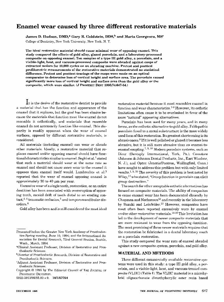
Fig. 1. Prepared type III gold sample in autopolymerizing resin matrix.
Fig. 3. Prepared composite sample in autopolymerizing resin matrix.
Fig. 2. Prepared porcelain sample in autopolymerizing resin matrix.
Fig. 4. Prepared restorative material in autopolymeriz- ing resin mat~x with screw to attach to weighted cup and rod.
material 19 classified as a direct/indirect composite resin. The gold alloy samples (Fig. 1) were used as they were
received from the manufacturer. Each 2 pennyweight (dwt) ingot was sectioned in half and the surface repolished with tripoli and rouge. The porcelain samples (Fig. 2) were prepared by the manufacturer and fired to a medium glaze. The composite samples (Fig. 3) were prepared by com- pressing the material in a split stainless steel ring (20 mm diameter, 2 mm high) between two glass slides, light cured (Optilux 400, Demetron, Danbury, Conn.) for 40 seconds on each side, and then placed in the curing unit (Conquest curing unit, model 001, Jeneric/Pentron) and fired at 107 ~ C for 15 minutes in 29 inches of mercury vacuum followed by a 2-minute cooldown. The composite samples were then finished with medium, fine, and superfine Soflex disks (3M Dental Products; St. Paul, Minn.).
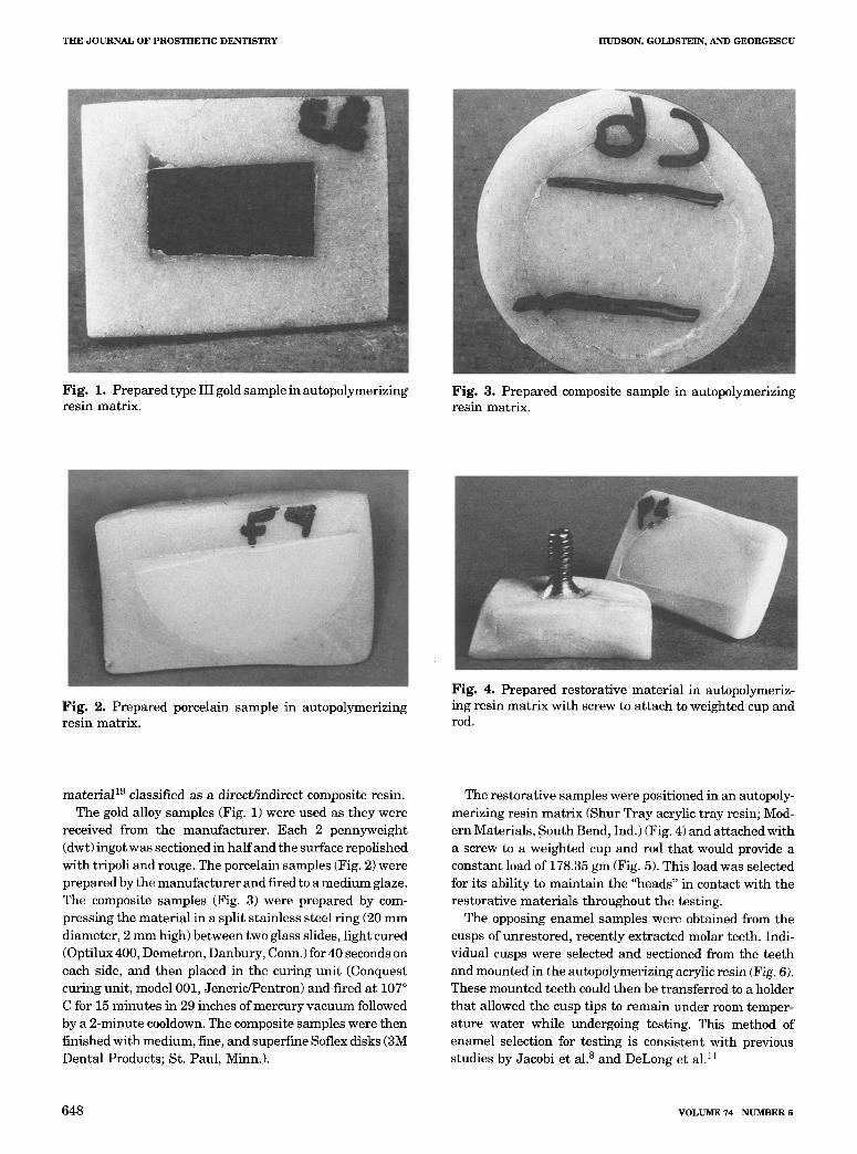
The restorative samples were positioned in an autopoly- merizing resin matrix (Shur Tray acrylic tray resin; Mod- em Materials, South Bend, Ind.) (Fig. 4) and attached with a screw to a weighted cup and rod that would provide a constant load of 178.35 gm (Fig. 5). This load was selected for its ability to maintain the "heads" in contact with the restorative materials throughout the testing.
The opposing enamel samples were obtained from the cusps of unrestored, recently extracted molar teeth. Indi- vidual cusps were selected and sectioned from the teeth and mounted in the autopolymerizing acrylic resin (Fig. 6). These mounted teeth could then be transferred to a holder that allowed the cusp tips to remain under room temper- ature water while undergoing testing. This method of enamel selection for testing is consistent with previous studies by Jacobi et al. 8 and DeLong et al. 11
6 4 8 VOLUME 74 NUMBER 6
HUDSON, GOLDSTEIN, AND GEORGESCU THE JOURNAL OF PROSTHETIC DENTISTRY
Fig . 6. P repa red enamel sample mounted in autopoly- merizing acrylic resin.
F ig . 5. Weighted cup and rod (178.35 gm) with restor- at ive sample at tached.
Table I. Restorat ive mate r i a l s included in wear tes t ing
Material Manufacturer Composit ion
Gold Degussa, Degular C (type III) South Plainfield, (74% Au) N.J.
Porcelain Dentsply Int. Biobond body porcelain York, Pa. (shade A2)
VLHC Jeneric/Pentron Conquest Crown and Bridge Wallingford, Conn. (Body shade A3)
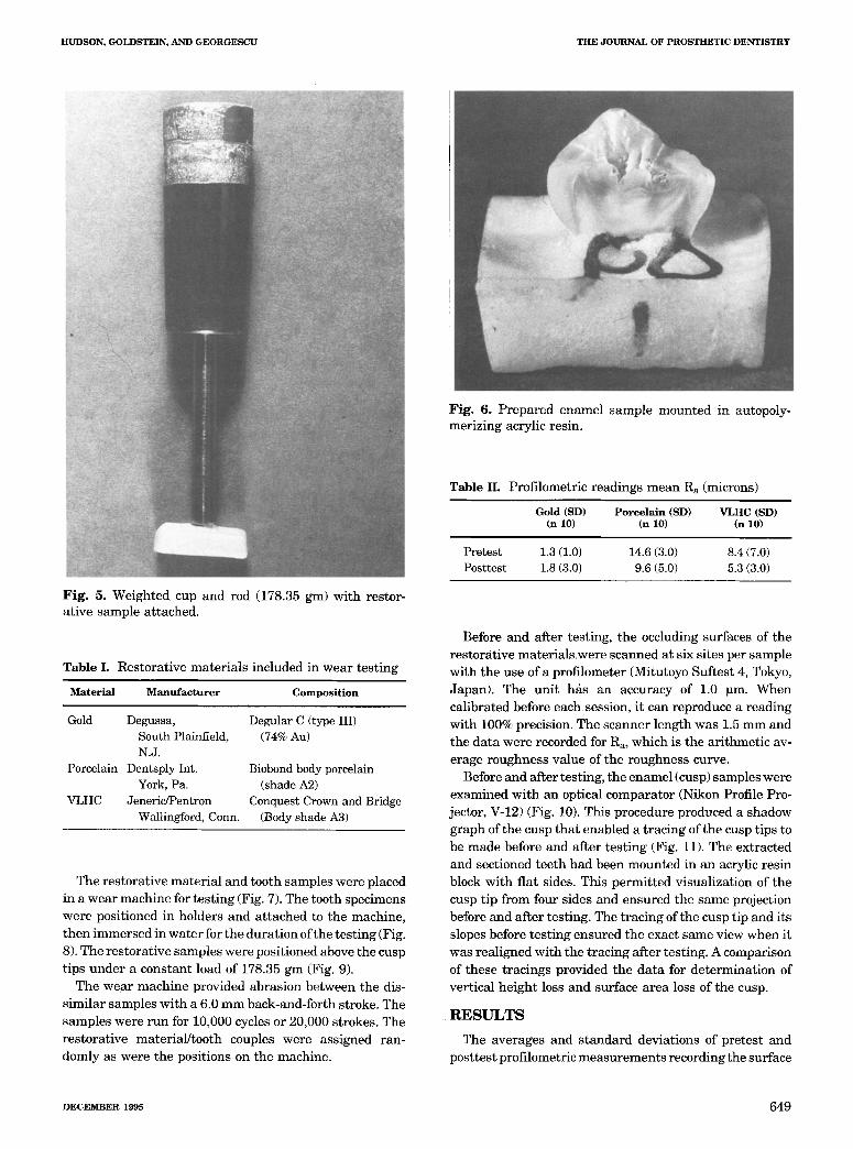
The res torat ive mate r ia l and tooth samples were placed in a wear machine for tes t ing (Fig. 7). The tooth specimens were posit ioned in holders and a t tached to the machine, then immersed in wate r for the dura t ion of the tes t ing (Fig. 8). The res tora t ive samples were posit ioned above the cusp t ips under a constant load of 178.35 gm (Fig. 9).
The wear machine provided abras ion between the dis- s imi lar samples wi th a 6.0 m m back-and-forth stroke. The samples were run for 10,000 cycles or 20,000 strokes. The res tora t ive mater ia l / tooth couples were assigned ran- domly as were the positions on the machine.
Table II. Profilometric readings mean Ra (microns)
Gold (SD) Porce la in (SD) V L H C (SD) (n 10) (n 10) (n 10)
Pretest 1.3 (1.0) 14.6 (3.0) 8.4 (7.0) Posttest 1.8 (3.0) 9.6 (5.0) 5.3 (3.0)
Before and after testing, the occluding surfaces of the restorat ive mater ia ls ,were scanned at six sites per sample wi th the use of a profilometer (Mitutoyo Suftest 4, Tokyo, Japan). The uni t has an accuracy of 1.0 pro. When cal ibrated before eachsess ion , i t can reproduce a reading with 100% precision. The scanner length was 1.5 mm and the da ta were recorded for Ra, which is the ar i thmet ic av- erage roughness value of the roughness curve.
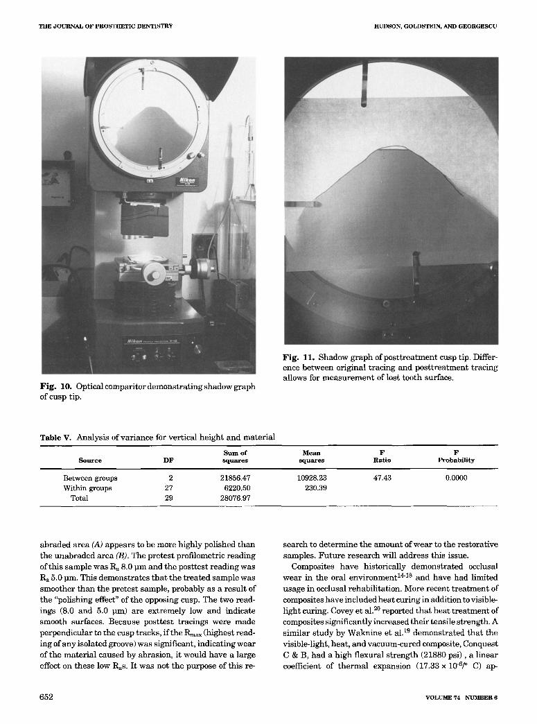
Before and after testing, the enamel (cusp) samples were examined wi th an optical comparator (Nikon Profile Pro- jector, V-12) (Fig. 10). This procedure produced a shadow graph of the cusp tha t enabled a t racing of the cusp t ips to be made before and after tes t ing (Fig. 11). The extracted and sectioned teeth had been mounted in an acrylic resin block with f lat sides. This permi t ted visual izat ion of the cusp tip from four sides and ensured the same projection before and af ter testing. The t racing of the cusp tip and its slopes before tes t ing ensured the exact same view when it was rea l igned with the t racing af ter testing. A comparison of these t racings provided the da t a for de terminat ion of vert ical he ight loss and surface a rea loss of the cusp.
R E S U L T S
The averages and s t andard deviat ions of pretes t and post tes t profilometric measurements recording the surface
DECEMBER 1 ~ 5 6 4 9
THE J O U R N A L OF P R O S T H E T I C DENTISTRY HUDSON, GOLDSTEIN, AND GEORGESCU
Fig. 7. Wear machine used in testing with weighted cups and rods in place.
Fig. 8. Close-up view of weighted cups and rods with restorative sample attached.
smoothness of the gold alloy, porcelain, and VLHC samples are presented in Table II. An ANOVA and Scheffe analy- sis (p < 0.01) demonstrated no significant difference be- tween the pretest and posttest profilometric readings.
Comparison of pretest and posttest tracings of the cusp tips allowed the determination of the wear of enamel as measured by both surface area loss and the vertical height loss of the cusps. The ANOVA and Scheffe analysis (p < 0.01) are presented in Tables III through VI. Porcelain
caused a statistically significant loss of vertical height and surface area of the opposing enamel when compared with either gold alloy or VLHC. There was no significant differ- ence in the wear of enamel opposing gold alloy or VLHC.
D I S C U S S I O N
In this experiment, feldspathic porcelain, polished and glazed, caused significantly more enamel wear than the gold alloy, and a relatively new composite restorative
6 5 0 VOLUME 74 NUMBER 6
HUDSON, GOLDSTEIN, AND GEORGESCU THE JOURNAL OF PROSTHETIC DENTISTRY
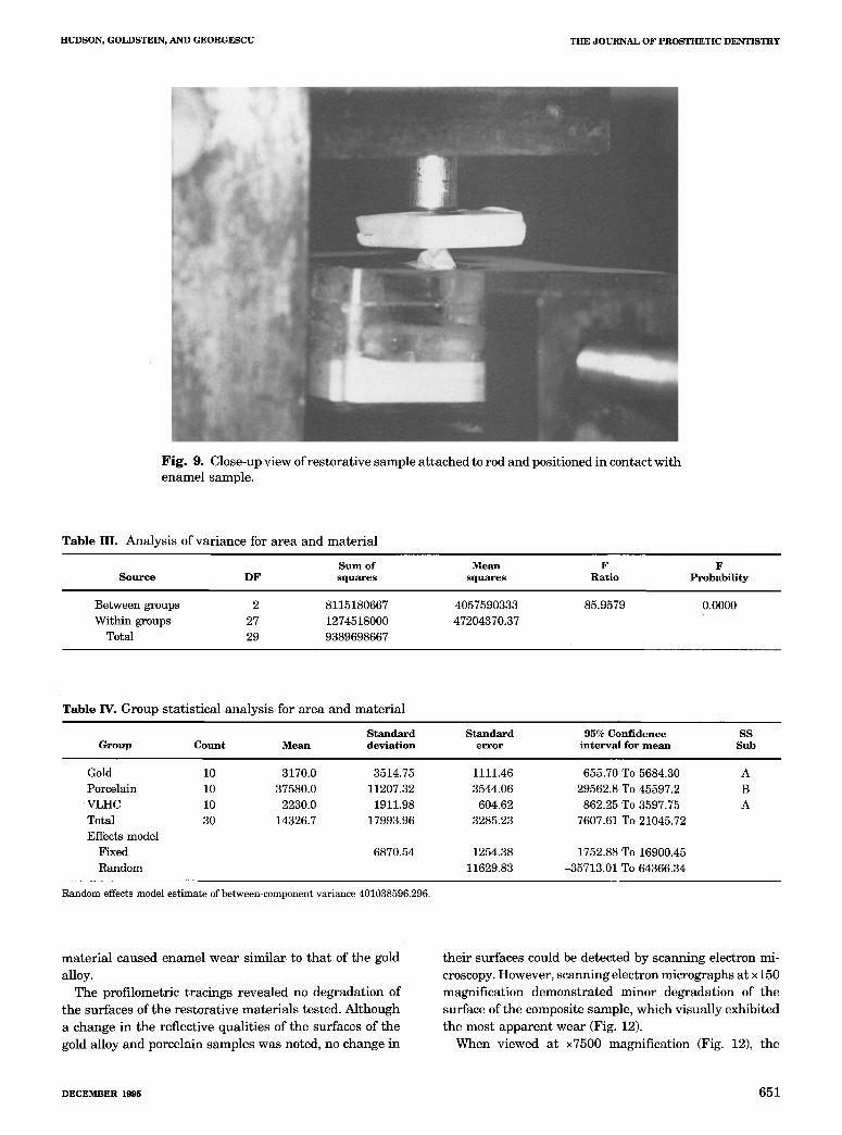
Fig. 9. Close-up view of restorative sample attached to rod and positioned in contact with enamel sample.
Table III. Analysis of variance for area and material
S u m of M e a n F F S o u r c e D F s q u a r e s s q u a r e s Rat io Probab i l i t y
Between groups 2 8115180667 4057590333 85.9579 0.0000 Within groups 27 1274518000 47204370.37
Total 29 9389698667
Table IV. Group statistical analysis for area and material
S t a n d a r d S t a n d a r d 95% C o n f i d e n c e SS G r o u p C o u n t M e a n d e v i a t i o n e r r o r i n t e r v a l for m e a n Sub
Gold Porcelain VLHC Total Effects model
Fixed Random
10 3170.0 3514.75 1111.46 655.70 To 5684.30 A 10 37580.0 11207.32 3544.06 29562.8 To 45597.2 B 10 2230.0 1911.98 604.62 862.25 To 3597.75 A 30 14326.7 17993.96 3285.23 7607.61 To 21045.72
6870.54 1254.38 1752.88 To 16900.45 11629.83 -35713.01 To 64366.34
Random effects model est imate of between-component variance 401038596.296.
material caused enamel wear similar to that of the gold
alloy. The profilometric tracings revealed no degradation of
the surfaces of the restorative materials tested. Although a change in the reflective qualities of the surfaces of the gold alloy and porcelain samples was noted, no change in
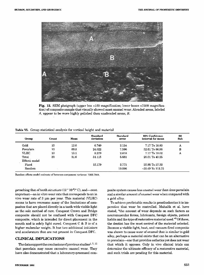
their surfaces could be detected by scanning electron mi- croscopy. However, scanning electron micrographs at x 150 magnification demonstrated minor degradation of the surface of the composite sample, which visually exhibited the most apparent wear (Fig. 12).
When viewed at • magnification (Fig. 12), the
DECEMBER 1~5 6 5 1
THE JOURNAL OF PROSTHETIC DENTISTRY HUDSON, GOLDSTEIN, AND GEORGESCU
Fig. 10. Optical comparitor demonstrating shadow graph of cusp tip.
Fig. 11. Shadow graph ofposttreatment cusp tip. Differ- ence between original tracing and posttreatment tracing allows for measurement of lost tooth surface.
Table V. Analysis of variance for vertical height and material
S u m of Mean F F S o u r c e D F s q u a r e s s q u a r e s Rat io Probab i l i t y
Between groups 2 21856.47 10928.23 47.43 0.0000 Within groups 27 6220.50 230.39
Total 29 28076.97
abraded area (A) appears to be more highly polished than the unabraded area (B). The pretest profilometric reading of this sample was R a 8.0 Dm and the posttest reading was Ra 5.0 Din. This demonstrates that the treated sample was smoother than the pretest sample, probably as a result of the "polishing effect" of the opposing cusp. The two read- ings (8.0 and 5.0 l~m) are extremely low and indicate smooth surfaces. Because posttest tracings were made perpendicular to the cusp tracks, if the Rmax (highest read- ing of any isolated groove) was significant, indicating wear of the material caused by abrasion, it would have a large effect on these low Ras. It was not the purpose of this re-
search to determine the amount of wear to the restorative samples. Future research will address this issue.
Composites have historically demonstrated occlusal wear in the oral environment 14-B and have had limited usage in occlusal rehabilitation. More recent treatment of composites have included heat curing in addition to visible- light curing. Covey et al. 2~ reported that heat treatment of composites significantly increased their tensile strength. A similar study by Waknine et al. m demonstrated that the visible-light, heat, and vacuum-cured composite, Conquest C & B, had a high flexural strength (21880 psi), a linear coefficient of thermal expansion (17.33 • 106/~ C) ap-
6 5 2 VOLUME 74 NUMBER 6
HUDSON, GOLDSTEIN, AND GEORGESCU THE JOURNAL OF PROSTHETIC DENTISTRY
Fig . 12. SEM photgraph (upper box • magnification; lower boxes • magnifica- tion) of composite sample tha t visual ly showed most enamel wear. Abraded areas , labeled A, appear to be more highly polished t han unabraded areas , B.
Table VI. Group s ta t is t ical analysis for vert ical height and mate r ia l
S t a n d a r d S t a n d a r d 95% C o n f i d e n c e SS G r o u p C o u n t Mean d e v i a t i o n e r r o r i n t e r v a l for m e a n Sub
Gold Porcelain VLHC Total Effects model
Fixed Random
10 12.0 6.749 2.134 7.17 To 16.83 A 10 69.8 24.022 7.596 52.61 To 86.98 B 10 13.1 8.279 2.618 7.17 To 19.02 A 30 31.6 31.115 5.681 20.01 To 43.25
15.179 2.771 25.95 To 37.32 19.086 -50.49 To 113.75
Random effects model es t imate of between-component variance: 1069.7844.
proaching tha t of tooth s t ructure (12 o 10-6/o C), and most impor tant - - -an in vi tro wear ra te tha t corresponds to an in vivo wear ra te of 3 p m per year. This mate r ia l (VLHC) seems to have overcome many of the l imitat ions of com- posites tha t are placed directly in a tooth with visible l ight as the sole method of cure. Conquest Crown and Bridge composite should not be confused wi th Conquest DFC composite, which is in tended for direct p lacement in the mouth and is solely l ight cured. Conquest C & B is of a h igher molecular weight. I t has two addi t ional ini t ia tors and accelerators tha t are not present in Conquest DFC.
C L I N I C A L I M P L I C A T I O N S
The da t a support the conclusions of previous studiesi , 7, i i t ha t porcelain may cause excessive enamel wear. They have also demons t ra ted tha t a laboratory-processed com-
posite sys tem causes less enamel wear than does porcelain and a s imi lar amount of enamel wear when compared with a gold alloy.
To achieve predictable resul ts in prosthodontics i t is im- perat ive t ha t wear be controlled. Maha l ik et al. have stated, "the amount of wear depends on such factors as neuromuscular forces, lubricants , foreign objects, pa t ient habi ts and the type of res tora t ive mate r ia l used. "9 Of these, the dent i s t has the most control of the ma te r i a l selected. Because a visible-light, heat , and vacuum-fired composite was shown to cause wear of enamel tha t is s imi lar to gold alloy, perhaps a mate r ia l exists tha t can be an a l ternat ive to porce la in- -one t ha t provides esthetics ye t does not wear tha t which i t opposes. Only in vivo clinical t r ials can de termine the u l t imate efficacy of a res tora t ive mater ia l , and such t r ia ls are pending for this mater ia l .
DECEMBER 1995 6 5 3
THE JOURNAL OF PROSTHETIC DENTISTRY HUDSON, GOLDSTEIN, AND GEORGESCU
C O N C L U S I O N S
Under the conditions of this study, the following conclu- sions were made.
1. Gold alloy and a visible-light, heat, and vacuum-fired composite caused similar wear of enamel.
2. Polished and glazed feldspathic porcelain caused a significant amount of enamel wear when compared with gold alloy or a visible-light, heat, and vacuum-fired com- posite.
R E F E R E N C E S
1. Seghi RR, Rosenstiel SF, Bauer P. Abrasion of human enamel by dif- ferent dental ceramics in vitro. J Dent Res 1991;70:221-5.
2. Lambrechts P, Braem M, Vanherle G. Evaluation of clinical perfor- mance for posterior composite resin and dental adhesives. Oper Dent 1987;12:53-78.
3. Leinfelder KF. Composite resin in posterior teeth. Dent Clin North Am 1981;25:357-64.
4. Wilder AD, May KN Jr, Leinfelder KF. Three year clinical study of UV-cured composite resins in posterior teeth. J PROSTHET DENT 1983;50:26-30.
5. Ramtjord S, Ash MM. Occlusion. 3rd ed. Philadelphia: WB Saunders, 1983:203.
6. Okeson JP. Management of temporomandibular disorders and occlu- sion. 2nd ed. St Louis: CV Mosby, 1989:204.
7. Monasky GE, Taylor DF. Studies on the wear of porcelain, enamel and gold. J Dent Res 1971;25:299-306.
8. Jacobi R, Shillingburg HT, Duncanson MG Jr. A comparison of the abrasiveness of six ceramic surfaces and gold. J PROSTHET DENT 1991; 66:303-9.
9. Mahalick JA, Knap FJ, Weiter EJ. Occlusal wear in prosthodontics. J Am Dent Assoc 1971;82:154-9.
10. DeLong R, Sasik C, Pintado MR, Douglas WH. The wear of enamel when opposed by ceramic systems. Dent Mater 1989;5:266-71.
11. Wiley MG. Effects of porcelain on occluding surf ~ces of restored teeth. J PROSTHET DENT 1989;61:133-7.
12. Chapman RJ, Nathanson D. Excessive wear of natural tooth structure by opposing composite restorations. J Am Dent Assoc 1983;106:51-3.
13. Suzuki S, Leinfelder KF. Wear of enamel cusps opposed by posterior composite resin. Quintessence Int 1993;24:885-90.
14. Waknine S, Gold AJ, Leinfelder KF. Comparative evaluation of clini- cal and laboratory wear of posterior composites [Abstract]. J Dent Res 1988;67:361.
15. Sakaguchi RL, Douglas WH, DeLong R, Pintado MR. The wear of a posterior composite in an artificial mouth: a clinical correlation. Dent Mater 1986;2:235-40.
16. Embong A, Glyn Jones J, Harrison A. The wear effects of selected com- posites on restorative materials and enamel. Dent Mater 1987;3:236- 40.
17. Powell JM, Phillips RW, Norman RD. In vitro wear response of com- posite resin, amalgam and enamel. J Dent Res 1975;54:1183-95.
18. Sulong MZAM, Aziz RA. Wear of materials used in dentistry: a review of the literature. J PROSTHET DENT 1990;63:342-9.
19. Waknine S, Prasad A, Jia W, Schulman A. Direct/indirect commercial composites: characterization of strength, shrinkage and wear [Ab- stract]. J Dent Res 1991;70:481.
20. Covey DA, Tahaney SR, Davenport JM. Mechanical properties of heat- treated composite resin restorative materials. J PROSTHET DENT 1992;68:458-61.
Reprint requests to: DR. JAMES D. HUDSON 630 FIFTH AVE. SUITE 1810 NEW YORK, NY 10111
6 5 4 VOLUME 74 NUMBER 6