vascular calcification: key roles of phosphate and ... - mdpi
TRANSCRIPT
International Journal of
Molecular Sciences
Review
Vascular Calcification: Key Roles of Phosphateand Pyrophosphate
Ricardo Villa-Bellosta 1,2
�����������������
Citation: Villa-Bellosta, R. Vascular
Calcification: Key Roles of Phosphate
and Pyrophosphate. Int. J. Mol. Sci.
2021, 22, 13536. https://doi.org/
10.3390/ijms222413536
Academic Editors: José
Luis Martin-Ventura and Luis
M. Blanco-Colio
Received: 24 November 2021
Accepted: 14 December 2021
Published: 17 December 2021
Publisher’s Note: MDPI stays neutral
with regard to jurisdictional claims in
published maps and institutional affil-
iations.
Copyright: © 2021 by the author.
Licensee MDPI, Basel, Switzerland.
This article is an open access article
distributed under the terms and
conditions of the Creative Commons
Attribution (CC BY) license (https://
creativecommons.org/licenses/by/
4.0/).
1 Center for Research in Molecular Medicine and Chronic Diseases (CiMUS), Av Barcelona. Campus Vida,Universidade de Santiago de Compostela, 15782 Santiago de Compostela, Spain; [email protected]
2 Department of Biochemistry and Molecular Biology, Universidade de Santiago de Compostela,Plaza do Obradoiro s/n, 15782 Santiago de Compostela, Spain
Abstract: Cardiovascular complications due to accelerated arterial stiffening and atherosclerosis arethe leading cause of morbimortality in Western society. Both pathologies are frequently associatedwith vascular calcification. Pathologic calcification of cardiovascular structures, or vascular calcifica-tion, is associated with several diseases (for example, genetic diseases, diabetes, and chronic kidneydisease) and is a common consequence of aging. Calcium phosphate deposition, mainly in the form ofhydroxyapatite, is the hallmark of vascular calcification and can occur in the medial layer of arteries(medial calcification), in the atheroma plaque (intimal calcification), and cardiac valves (heart valvecalcification). Although various mechanisms have been proposed for the pathogenesis of vascularcalcification, our understanding of the pathogenesis of calcification is far from complete. However, inrecent years, some risk factors have been identified, including high serum phosphorus concentration(hyperphosphatemia) and defective synthesis of pyrophosphate (pyrophosphate deficiency). Thebalance between phosphate and pyrophosphate, strictly controlled by several genes, plays a key rolein vascular calcification. This review summarizes the current knowledge concerning phosphate andpyrophosphate homeostasis, focusing on the role of extracellular pyrophosphate metabolism in aorticsmooth muscle cells and macrophages.
Keywords: vascular calcification; pyrophosphate; phosphate; ATP; calcium
1. Introduction
Pathologic calcification of cardiovascular structures, or vascular calcification, is asso-ciated with several diseases (for example, genetic diseases, diabetes, and chronic kidneydisease), and is a common consequence of aging [1,2]. Vascular calcification, the depositionof phosphate-calcium crystals on the cardiovascular system, mainly in blood vessels, my-ocardium, and cardiac valves, is one of the most important factors determining patients’morbidity and mortality worldwide [3].
In blood vessels, calcified deposits are found in distinct layers of the aortic wall andare associated with specific pathologies. Intimal calcification occurs in atheroscleroticlesions and is associated with vascular smooth muscle cells and macrophages [4]; whereasmedial calcification (so-called “Monckeberg’s medial sclerosis”) occurs in the medial layerof the aortic wall and is associated with the collagen/elastin fibers and vascular smoothmuscle cells [5,6].
Different mechanisms regarding the pathogenesis of vascular calcification have beenproposed [2], including (1) loss of inhibitions, (2) calcium and phosphorus homeostasis,(3) osteochondrogenic differentiation of vascular cells, (4) apoptosis, (5) circulating nucleationcomplexes/paracrine factors, and (6) matrix degradation. However, despite major advancesin recent years, our understanding of calcification pathogenesis is far from complete.
Int. J. Mol. Sci. 2021, 22, 13536. https://doi.org/10.3390/ijms222413536 https://www.mdpi.com/journal/ijms
Int. J. Mol. Sci. 2021, 22, 13536 2 of 14
2. Role of Phosphate
Inorganic phosphate is essential for a variety of cellular processes, such as energymetabolism, bone formation, and synthesis of biomolecules, including phospholipids andnucleic acids. However, elevated serum phosphorus (in the form of inorganic phosphate)has emerged as a key risk factor for vascular calcification [7,8]. During the past decade,in vitro experiments have shown calcium-phosphate deposits in vascular smooth musclecells incubated with high phosphate concentration [8]. Logically, this observation was firstinterpreted as the consequence of an increase in phosphate transport, with the consequentincrease in the intracellular phosphate concentration [9]. However, several studies showthat phosphate transporters are saturated with normal serum phosphate levels [10,11].Moreover, additional studies show that the formation of calcium-phosphate crystals is apassive physicochemical process that does not require any cellular activity, suggesting animportant role of phosphate homeostasis [6,11,12]. Notably, there are two major conse-quences regarding the fate of vascular smooth muscle cells in phosphate-induced vascularcalcification. The first involves apoptosis-dependent matrix mineralization, which hasbeen detected both in cultured human vascular smooth muscle cells and in arteries frompediatric dialysis patients [13–15]. The second invokes a profound transition to a bone-forming phenotype [16]. In support of this notion, in vitro studies have shown that elevatedphosphate results in the expression of osteochondrogenic markers (such as BMP-2 andRunx2/Cbfa1, a transcription factor that induces the expression of major components of thebone matrix) [17,18]. However, recent studies show that calcium-phosphate deposits caninduce both the transition to a bone-forming phenotype and apoptosis in vascular smoothmuscle cells and the aortic wall, suggesting that the active mechanisms described could bea response to the excessive formation and deposit of calcium-phosphate crystals [5,6,19,20].
2.1. Biomineralization Process
The formation and deposition of inorganic minerals within or outside the cells ofvarious organisms is known as biological mineralization or biomineralization. Biomineral-ization in hard tissues (such as in bone or dentine) is normally considered a physiologicalprocess; however, the accumulation of inorganic minerals in soft tissues (such as bloodvessels, joints, and internal organs, including muscle, liver, or brain) is considered patho-logical or ectopic biomineralization. Under normal conditions, the soft tissues are notmineralized, but due to aging and other pathological conditions, soft tissues become calci-fied, which leads to morbidity and mortality. The main biominerals found in mineralizedvertebrate connective tissue are calcium-phosphate salts. In an aqueous system of calciumand phosphate, there are several known non-ion-substituted calcium phosphates, whichhave also been found in calcified tissues. The phosphate ion is a central phosphorus atomsurrounded by four oxygen atoms in a tetrahedral arrangement. In biological systems, it isfound as a free phosphate ion in solution (inorganic phosphate) or bound with differentbiological molecules, including proteins, sugars, lipids, and nucleic acid. Aqueous inor-ganic phosphate exists in four forms according to its triprotic equilibrium: (1) trihydrogenphosphate ion (H3P04), (2) dihydrogen phosphate ion (H2PO4
−), (3) hydrogen phosphateion (HPO4
2−), and (4) phosphate ion (PO42−); (see Figure 1). Inorganic phosphate is quite
strong with respect to the first dissociation (pKa1 = 2.1), moderately weak with respectto the second (pKa2 = 6.9), and very weak with respect to the third (pKa3 = 12.4). Understrongly basic or acidic conditions, the phosphate ion or trihydrogen phosphate dominates,respectively. In extracellular fluid (pH = 7.4), only H2PO4
− and HPO42− ions are present
in significant amounts in a proportion of 1:4, respectively. Whereas, in cytosol (pH = 7) andlysosome (pH = 4.8), this proportion is inverted (1.6:1 and 99:1, respectively).
Int. J. Mol. Sci. 2021, 22, 13536 3 of 14Int. J. Mol. Sci. 2021, 22, x FOR PEER REVIEW 3 of 14
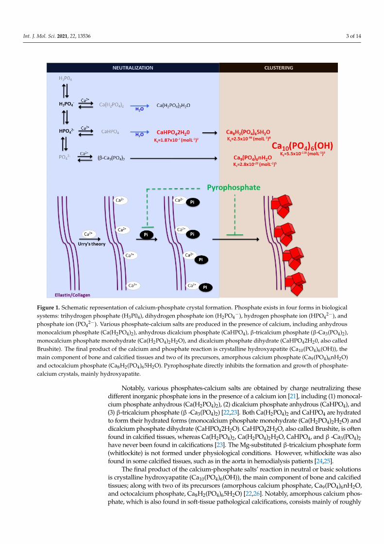
Figure 1. Schematic representation of calcium-phosphate crystal formation. Phosphate exists in four forms in biological systems: trihydrogen phosphate (H3P04), dihydrogen phosphate ion (H2PO4−), hydrogen phosphate ion (HPO42−), and phosphate ion (PO42−). Various phosphate-calcium salts are produced in the presence of calcium, including anhydrous monocalcium phosphate (Ca(H2PO4)2), anhydrous dicalcium phosphate (CaHPO4), β-tricalcium phosphate (β-Ca3(PO4)2), monocalcium phosphate monohydrate (Ca(H2PO4)2H2O), and dicalcium phosphate dihydrate (CaHPO42H20, also called Brushite). The final product of the calcium and phosphate reaction is crystalline hydroxyapatite (Ca10(PO4)6(OH)), the main component of bone and calcified tissues and two of its precursors, amorphous calcium phosphate (Ca9(PO4)6nH2O) and octocalcium phosphate (Ca8H2(PO4)65H2O). Pyrophosphate directly inhibits the formation and growth of phosphate-cal-cium crystals, mainly hydroxyapatite.
Notably, various phosphates-calcium salts are obtained by charge neutralizing these different inorganic phosphate ions in the presence of a calcium ion [21], including (1) mon-ocalcium phosphate anhydrous (Ca(H2PO4)2), (2) dicalcium phosphate anhydrous (Ca-HPO4), and (3) β-tricalcium phosphate (β -Ca3(PO4)2) [22,23]. Both Ca(H2PO4)2 and Ca-HPO4 are hydrated to form their hydrated forms (monocalcium phosphate monohydrate (Ca(H2PO4)2H2O) and dicalcium phosphate dihydrate (CaHPO42H2O). CaHPO42H2O, also called Brushite, is often found in calcified tissues, whereas Ca(H2PO4)2, Ca(H2PO4)2H2O, CaHPO4, and β -Ca3(PO4)2 have never been found in calcifications [23]. The Mg-substi-tuted β-tricalcium phosphate form (whitlockite) is not formed under physiological condi-tions. However, whitlockite was also found in some calcified tissues, such as in the aorta in hemodialysis patients [24,25].
The final product of the calcium-phosphate salts’ reaction in neutral or basic solu-tions is crystalline hydroxyapatite (Ca10(PO4)6(OH)), the main component of bone and cal-cified tissues; along with two of its precursors (amorphous calcium phosphate, Ca9(PO4)6nH2O, and octocalcium phosphate, Ca8H2(PO4)65H2O) [22,26]. Notably,
Figure 1. Schematic representation of calcium-phosphate crystal formation. Phosphate exists in four forms in biologicalsystems: trihydrogen phosphate (H3P04), dihydrogen phosphate ion (H2PO4
−), hydrogen phosphate ion (HPO42−), and
phosphate ion (PO42−). Various phosphate-calcium salts are produced in the presence of calcium, including anhydrous
monocalcium phosphate (Ca(H2PO4)2), anhydrous dicalcium phosphate (CaHPO4), β-tricalcium phosphate (β-Ca3(PO4)2),monocalcium phosphate monohydrate (Ca(H2PO4)2H2O), and dicalcium phosphate dihydrate (CaHPO42H20, also calledBrushite). The final product of the calcium and phosphate reaction is crystalline hydroxyapatite (Ca10(PO4)6(OH)), themain component of bone and calcified tissues and two of its precursors, amorphous calcium phosphate (Ca9(PO4)6nH2O)and octocalcium phosphate (Ca8H2(PO4)65H2O). Pyrophosphate directly inhibits the formation and growth of phosphate-calcium crystals, mainly hydroxyapatite.
Notably, various phosphates-calcium salts are obtained by charge neutralizing thesedifferent inorganic phosphate ions in the presence of a calcium ion [21], including (1) monocal-cium phosphate anhydrous (Ca(H2PO4)2), (2) dicalcium phosphate anhydrous (CaHPO4), and(3) β-tricalcium phosphate (β -Ca3(PO4)2) [22,23]. Both Ca(H2PO4)2 and CaHPO4 are hydratedto form their hydrated forms (monocalcium phosphate monohydrate (Ca(H2PO4)2H2O) anddicalcium phosphate dihydrate (CaHPO42H2O). CaHPO42H2O, also called Brushite, is oftenfound in calcified tissues, whereas Ca(H2PO4)2, Ca(H2PO4)2H2O, CaHPO4, and β -Ca3(PO4)2have never been found in calcifications [23]. The Mg-substituted β-tricalcium phosphate form(whitlockite) is not formed under physiological conditions. However, whitlockite was alsofound in some calcified tissues, such as in the aorta in hemodialysis patients [24,25].
The final product of the calcium-phosphate salts’ reaction in neutral or basic solutionsis crystalline hydroxyapatite (Ca10(PO4)6(OH)), the main component of bone and calcifiedtissues; along with two of its precursors (amorphous calcium phosphate, Ca9(PO4)6nH2O,and octocalcium phosphate, Ca8H2(PO4)65H2O) [22,26]. Notably, amorphous calcium phos-phate, which is also found in soft-tissue pathological calcifications, consists mainly of roughly

Int. J. Mol. Sci. 2021, 22, 13536 4 of 14
spherical Ca9(PO4)6 clusters (called Posner’s clusters) that appear to be energetically favoredcompared to (Ca3(PO4) and Ca6(PO4)4 clusters [21,22]. Therefore, the structure of hydroxyap-atite [26] can be interpreted as an aggregation of Posner’s clusters [27,28] (Figure 1). Notably,Mg2+ and ATP are critical for the stabilization of amorphous calcium phosphate [29,30].
According to the charge neutralization theory of calcification [31], the high glycinecontent of elastin and collagen proteins favors the formation of beta-turns that are known tointeract with calcium ions. Therefore, the deposition of these calcium-phosphate salts, bothin vitro and in vivo, takes place on these extracellular matrix proteins [5,6]. For example,in bone and connective tissues, these salts are predominantly deposited on the elastic andtype I collagen fibers. Moreover, in the aorta wall, calcium-phosphate crystals are depositedon elastin, the main component of the elastic fibers in the medial layer [6]. Notably, a studyshowed that a mouse model of elastin haploinsufficiency exhibited a significant reductionin arterial calcification [32]. In contrast, phosphate-induced mineralization, both in vitroand in vivo, is accelerated by the products derived from elastin degradation [33].
Finally, the depositions of calcium-phosphate crystals in soft tissues can be classified intothree main categories: (1) calcinosis, (2) dystrophic calcification, and (3) metastatic calcification.In the presence of normal homeostasis of phosphate, calcinosis and/or dystrophic calcificationoccur, most often in subcutaneous tissues, skin, and related connective tissues, whereas, inthe second case, calcification occurs in degenerated or necrotic tissue. Metastatic calcificationoccurs in normal tissues when the calcium levels are elevated in serum.
2.2. Phosphate Homeostasis
In adults, normal phosphate concentration in serum or plasma is mainly 2.5 to 4.5 mg/dL(0.81 to 1.45 mmol/L). Elevated serum phosphate (hyperphosphatemia) is a key risk factorfor pathologic calcification in cardiovascular structures [1]. Treatment of hyperphosphatemiawith phosphate binders is associated with the slow progression of cardiovascular calcificationin hemodialysis patients [34]. Therefore, the homeostasis of phosphate plays a critical role inthe initiation and progression of calcification [35] (see Figure 2).
Int. J. Mol. Sci. 2021, 22, x FOR PEER REVIEW 5 of 14
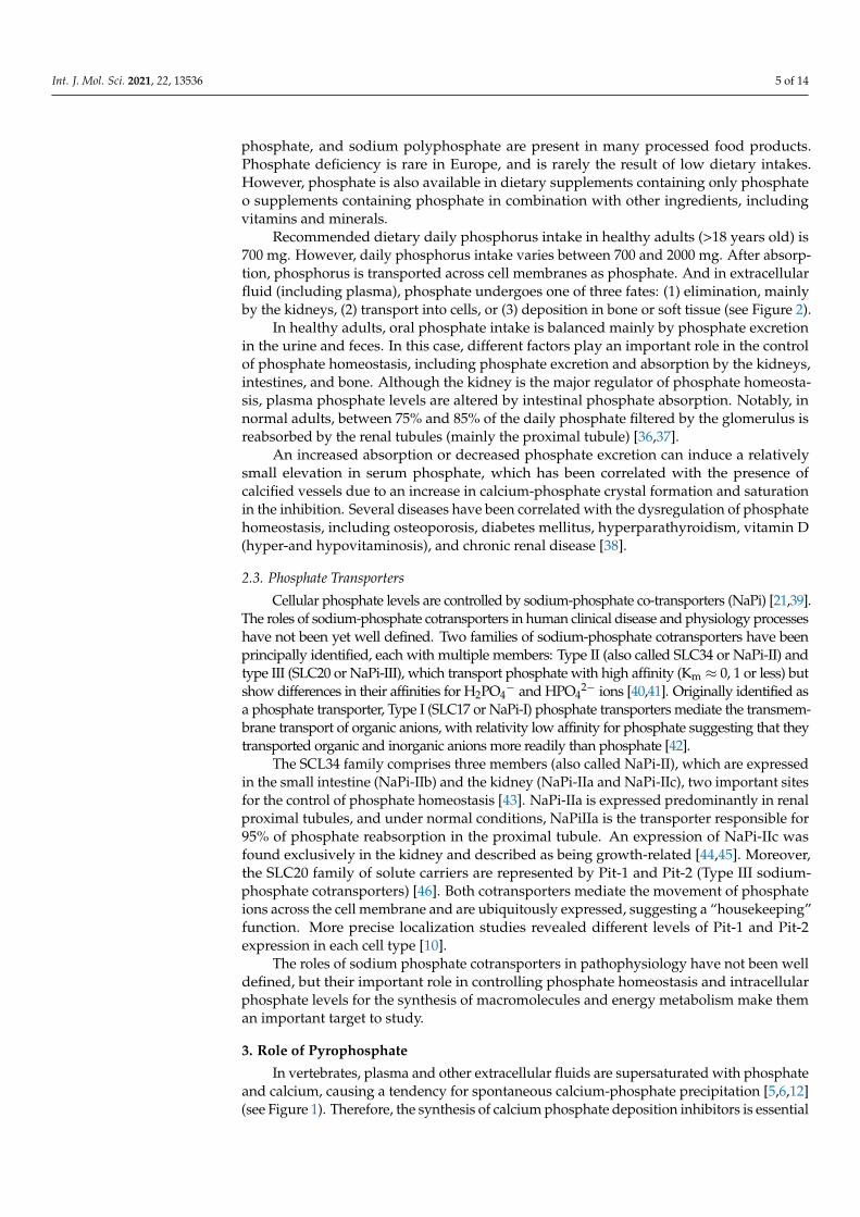
Figure 2. Phosphate flux between body compartments. Phosphate balance is a complex process involving bone intestinal absorption and dietary phosphate and renal excretion of phosphate.
In healthy adults, oral phosphate intake is balanced mainly by phosphate excretion in the urine and feces. In this case, different factors play an important role in the control of phosphate homeostasis, including phosphate excretion and absorption by the kidneys, intestines, and bone. Although the kidney is the major regulator of phosphate homeosta-sis, plasma phosphate levels are altered by intestinal phosphate absorption. Notably, in normal adults, between 75% and 85% of the daily phosphate filtered by the glomerulus is reabsorbed by the renal tubules (mainly the proximal tubule) [36,37].
An increased absorption or decreased phosphate excretion can induce a relatively small elevation in serum phosphate, which has been correlated with the presence of calci-fied vessels due to an increase in calcium-phosphate crystal formation and saturation in the inhibition. Several diseases have been correlated with the dysregulation of phosphate homeostasis, including osteoporosis, diabetes mellitus, hyperparathyroidism, vitamin D (hyper-and hypovitaminosis), and chronic renal disease [38].
2.3. Phosphate Transporters Cellular phosphate levels are controlled by sodium-phosphate co-transporters (NaPi)
[21,39]. The roles of sodium-phosphate cotransporters in human clinical disease and phys-iology processes have not been yet well defined. Two families of sodium-phosphate co-transporters have been principally identified, each with multiple members: Type II (also called SLC34 or NaPi-II) and type III (SLC20 or NaPi-III), which transport phosphate with high affinity (Km ≈ 0,1 or less) but show differences in their affinities for H2PO4− and HPO42− ions [40,41]. Originally identified as a phosphate transporter, Type I (SLC17 or NaPi-I) phosphate transporters mediate the transmembrane transport of organic anions, with rel-ativity low affinity for phosphate suggesting that they transported organic and inorganic anions more readily than phosphate [42].
The SCL34 family comprises three members (also called NaPi-II), which are ex-pressed in the small intestine (NaPi-IIb) and the kidney (NaPi-IIa and NaPi-IIc), two im-portant sites for the control of phosphate homeostasis [43]. NaPi-IIa is expressed predom-inantly in renal proximal tubules, and under normal conditions, NaPiIIa is the transporter responsible for 95% of phosphate reabsorption in the proximal tubule. An expression of
Figure 2. Phosphate flux between body compartments. Phosphate balance is a complex process involving bone intestinalabsorption and dietary phosphate and renal excretion of phosphate.
Many different foods contain phosphorus, including vegetables, grains, legumes,eggs, fish, and meats. In addition, phosphate additives such as phosphoric acid, sodium
Int. J. Mol. Sci. 2021, 22, 13536 5 of 14
phosphate, and sodium polyphosphate are present in many processed food products.Phosphate deficiency is rare in Europe, and is rarely the result of low dietary intakes.However, phosphate is also available in dietary supplements containing only phosphateo supplements containing phosphate in combination with other ingredients, includingvitamins and minerals.
Recommended dietary daily phosphorus intake in healthy adults (>18 years old) is700 mg. However, daily phosphorus intake varies between 700 and 2000 mg. After absorp-tion, phosphorus is transported across cell membranes as phosphate. And in extracellularfluid (including plasma), phosphate undergoes one of three fates: (1) elimination, mainlyby the kidneys, (2) transport into cells, or (3) deposition in bone or soft tissue (see Figure 2).
In healthy adults, oral phosphate intake is balanced mainly by phosphate excretionin the urine and feces. In this case, different factors play an important role in the controlof phosphate homeostasis, including phosphate excretion and absorption by the kidneys,intestines, and bone. Although the kidney is the major regulator of phosphate homeosta-sis, plasma phosphate levels are altered by intestinal phosphate absorption. Notably, innormal adults, between 75% and 85% of the daily phosphate filtered by the glomerulus isreabsorbed by the renal tubules (mainly the proximal tubule) [36,37].
An increased absorption or decreased phosphate excretion can induce a relativelysmall elevation in serum phosphate, which has been correlated with the presence ofcalcified vessels due to an increase in calcium-phosphate crystal formation and saturationin the inhibition. Several diseases have been correlated with the dysregulation of phosphatehomeostasis, including osteoporosis, diabetes mellitus, hyperparathyroidism, vitamin D(hyper-and hypovitaminosis), and chronic renal disease [38].
2.3. Phosphate Transporters
Cellular phosphate levels are controlled by sodium-phosphate co-transporters (NaPi) [21,39].The roles of sodium-phosphate cotransporters in human clinical disease and physiology processeshave not been yet well defined. Two families of sodium-phosphate cotransporters have beenprincipally identified, each with multiple members: Type II (also called SLC34 or NaPi-II) andtype III (SLC20 or NaPi-III), which transport phosphate with high affinity (Km ≈ 0, 1 or less) butshow differences in their affinities for H2PO4
− and HPO42− ions [40,41]. Originally identified as
a phosphate transporter, Type I (SLC17 or NaPi-I) phosphate transporters mediate the transmem-brane transport of organic anions, with relativity low affinity for phosphate suggesting that theytransported organic and inorganic anions more readily than phosphate [42].
The SCL34 family comprises three members (also called NaPi-II), which are expressedin the small intestine (NaPi-IIb) and the kidney (NaPi-IIa and NaPi-IIc), two important sitesfor the control of phosphate homeostasis [43]. NaPi-IIa is expressed predominantly in renalproximal tubules, and under normal conditions, NaPiIIa is the transporter responsible for95% of phosphate reabsorption in the proximal tubule. An expression of NaPi-IIc wasfound exclusively in the kidney and described as being growth-related [44,45]. Moreover,the SLC20 family of solute carriers are represented by Pit-1 and Pit-2 (Type III sodium-phosphate cotransporters) [46]. Both cotransporters mediate the movement of phosphateions across the cell membrane and are ubiquitously expressed, suggesting a “housekeeping”function. More precise localization studies revealed different levels of Pit-1 and Pit-2expression in each cell type [10].
The roles of sodium phosphate cotransporters in pathophysiology have not been welldefined, but their important role in controlling phosphate homeostasis and intracellularphosphate levels for the synthesis of macromolecules and energy metabolism make theman important target to study.
3. Role of Pyrophosphate
In vertebrates, plasma and other extracellular fluids are supersaturated with phosphateand calcium, causing a tendency for spontaneous calcium-phosphate precipitation [5,6,12](see Figure 1). Therefore, the synthesis of calcium phosphate deposition inhibitors is essential
Int. J. Mol. Sci. 2021, 22, 13536 6 of 14
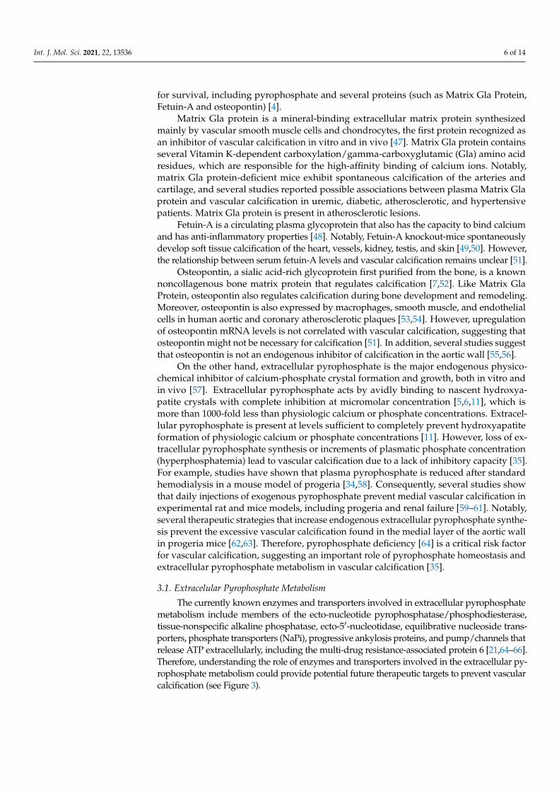
for survival, including pyrophosphate and several proteins (such as Matrix Gla Protein,Fetuin-A and osteopontin) [4].
Matrix Gla protein is a mineral-binding extracellular matrix protein synthesizedmainly by vascular smooth muscle cells and chondrocytes, the first protein recognized asan inhibitor of vascular calcification in vitro and in vivo [47]. Matrix Gla protein containsseveral Vitamin K-dependent carboxylation/gamma-carboxyglutamic (Gla) amino acidresidues, which are responsible for the high-affinity binding of calcium ions. Notably,matrix Gla protein-deficient mice exhibit spontaneous calcification of the arteries andcartilage, and several studies reported possible associations between plasma Matrix Glaprotein and vascular calcification in uremic, diabetic, atherosclerotic, and hypertensivepatients. Matrix Gla protein is present in atherosclerotic lesions.
Fetuin-A is a circulating plasma glycoprotein that also has the capacity to bind calciumand has anti-inflammatory properties [48]. Notably, Fetuin-A knockout-mice spontaneouslydevelop soft tissue calcification of the heart, vessels, kidney, testis, and skin [49,50]. However,the relationship between serum fetuin-A levels and vascular calcification remains unclear [51].
Osteopontin, a sialic acid-rich glycoprotein first purified from the bone, is a knownnoncollagenous bone matrix protein that regulates calcification [7,52]. Like Matrix GlaProtein, osteopontin also regulates calcification during bone development and remodeling.Moreover, osteopontin is also expressed by macrophages, smooth muscle, and endothelialcells in human aortic and coronary atherosclerotic plaques [53,54]. However, upregulationof osteopontin mRNA levels is not correlated with vascular calcification, suggesting thatosteopontin might not be necessary for calcification [51]. In addition, several studies suggestthat osteopontin is not an endogenous inhibitor of calcification in the aortic wall [55,56].
On the other hand, extracellular pyrophosphate is the major endogenous physico-chemical inhibitor of calcium-phosphate crystal formation and growth, both in vitro andin vivo [57]. Extracellular pyrophosphate acts by avidly binding to nascent hydroxya-patite crystals with complete inhibition at micromolar concentration [5,6,11], which ismore than 1000-fold less than physiologic calcium or phosphate concentrations. Extracel-lular pyrophosphate is present at levels sufficient to completely prevent hydroxyapatiteformation of physiologic calcium or phosphate concentrations [11]. However, loss of ex-tracellular pyrophosphate synthesis or increments of plasmatic phosphate concentration(hyperphosphatemia) lead to vascular calcification due to a lack of inhibitory capacity [35].For example, studies have shown that plasma pyrophosphate is reduced after standardhemodialysis in a mouse model of progeria [34,58]. Consequently, several studies showthat daily injections of exogenous pyrophosphate prevent medial vascular calcification inexperimental rat and mice models, including progeria and renal failure [59–61]. Notably,several therapeutic strategies that increase endogenous extracellular pyrophosphate synthe-sis prevent the excessive vascular calcification found in the medial layer of the aortic wallin progeria mice [62,63]. Therefore, pyrophosphate deficiency [64] is a critical risk factorfor vascular calcification, suggesting an important role of pyrophosphate homeostasis andextracellular pyrophosphate metabolism in vascular calcification [35].
3.1. Extracelular Pyrophosphate Metabolism
The currently known enzymes and transporters involved in extracellular pyrophosphatemetabolism include members of the ecto-nucleotide pyrophosphatase/phosphodiesterase,tissue-nonspecific alkaline phosphatase, ecto-5′-nucleotidase, equilibrative nucleoside trans-porters, phosphate transporters (NaPi), progressive ankylosis proteins, and pump/channels thatrelease ATP extracellularly, including the multi-drug resistance-associated protein 6 [21,64–66].Therefore, understanding the role of enzymes and transporters involved in the extracellular py-rophosphate metabolism could provide potential future therapeutic targets to prevent vascularcalcification (see Figure 3).

Int. J. Mol. Sci. 2021, 22, 13536 7 of 14
Int. J. Mol. Sci. 2021, 22, x FOR PEER REVIEW 7 of 14
synthesis prevent the excessive vascular calcification found in the medial layer of the aor-tic wall in progeria mice [62,63]. Therefore, pyrophosphate deficiency [64] is a critical risk factor for vascular calcification, suggesting an important role of pyrophosphate homeo-stasis and extracellular pyrophosphate metabolism in vascular calcification [35].
3.1. Extracelular Pyrophosphate Metabolism The currently known enzymes and transporters involved in extracellular pyrophos-
phate metabolism include members of the ecto-nucleotide pyrophosphatase/phos-phodiesterase, tissue-nonspecific alkaline phosphatase, ecto-5′-nucleotidase, equilibrative nucleoside transporters, phosphate transporters (NaPi), progressive ankylosis proteins, and pump/channels that release ATP extracellularly, including the multi-drug resistance-associated protein 6 [21,64–66]. Therefore, understanding the role of enzymes and trans-porters involved in the extracellular pyrophosphate metabolism could provide potential future therapeutic targets to prevent vascular calcification (see Figure 3).
Pyrophosphate is mainly produced during the extracellular hydrolysis of ATP [6,67,68]. The major generator of endogenous extracellular pyrophosphate in several tis-sues, including the aorta, is the enzyme ecto-nucleotide pyrophosphatase/phos-phodiesterase (eNPP), which hydrolyzes extracellular ATP to generate pyrophosphate and AMP [67] (see Figure 3). Three members of the eNPP activity have been found (eNPP1-3); they exist both as membrane proteins, with an extracellular active site, and as soluble proteins in body fluids (also known as PC-1, autotaxin, and B10, respectively) [65]. In aorta and vascular smooth muscle cells, eNPP1 is the main source of extracellular py-rophosphate [6,60,68]. Mutations in eNPP1 result in generalized arterial calcification of infancy (see Table 1), characterized by an excessive calcification of the internal elastic lam-ina of large and medium-sized arteries [67]. Moreover, eNPP1-null mice develop ectopic artery calcification [69]. Notably, ATP is also a direct inhibitor of calcification [70], with a physicochemical mechanism similar to pyrophosphate, bisphosphonates (non-hydrolyz-able analogous of pyrophosphate), and polyphosphates [57,71].
Figure 3. Schematic representation of the ectoenzymes and transporters involved in the extracellular pyrophosphate me-tabolism. Ectonucleotide pyrophosphatase phosphodiesterase (eNPP) hydrolyze ATP releasing pyrophosphate (PPi) and adenosine-5′-monophosphate (AMP). Pyrophosphate is degraded to phosphate (Pi) by tissue non-specific alkaline phos-phatase (TNAP). ATP is released by cells via exocytotic mechanisms and via multiple types of membrane channels, in-cluding ABCC6. The progressive ankylosis (ANK) protein can contribute to extracellular pyrophosphate by transporting either ATP or pyrophosphate. Equilibrative nucleoside transporter 1 (ENT1). Sodium-phosphate co-transporter (NaPi). Ecto-5`nucletotidase (5NT). Oxidative phosphorylation pathway (OXPHOS).
Figure 3. Schematic representation of the ectoenzymes and transporters involved in the extracellular pyrophosphatemetabolism. Ectonucleotide pyrophosphatase phosphodiesterase (eNPP) hydrolyze ATP releasing pyrophosphate (PPi)and adenosine-5′-monophosphate (AMP). Pyrophosphate is degraded to phosphate (Pi) by tissue non-specific alkalinephosphatase (TNAP). ATP is released by cells via exocytotic mechanisms and via multiple types of membrane channels,including ABCC6. The progressive ankylosis (ANK) protein can contribute to extracellular pyrophosphate by transportingeither ATP or pyrophosphate. Equilibrative nucleoside transporter 1 (ENT1). Sodium-phosphate co-transporter (NaPi).Ecto-5′nucletotidase (5NT). Oxidative phosphorylation pathway (OXPHOS).
Pyrophosphate is mainly produced during the extracellular hydrolysis of ATP [6,67,68].The major generator of endogenous extracellular pyrophosphate in several tissues, includingthe aorta, is the enzyme ecto-nucleotide pyrophosphatase/phosphodiesterase (eNPP), whichhydrolyzes extracellular ATP to generate pyrophosphate and AMP [67] (see Figure 3). Threemembers of the eNPP activity have been found (eNPP1-3); they exist both as membraneproteins, with an extracellular active site, and as soluble proteins in body fluids (also known asPC-1, autotaxin, and B10, respectively) [65]. In aorta and vascular smooth muscle cells, eNPP1is the main source of extracellular pyrophosphate [6,60,68]. Mutations in eNPP1 result in gen-eralized arterial calcification of infancy (see Table 1), characterized by an excessive calcificationof the internal elastic lamina of large and medium-sized arteries [67]. Moreover, eNPP1-nullmice develop ectopic artery calcification [69]. Notably, ATP is also a direct inhibitor of calcifi-cation [70], with a physicochemical mechanism similar to pyrophosphate, bisphosphonates(non-hydrolyzable analogous of pyrophosphate), and polyphosphates [57,71].
Table 1. Genetic Diseases involved in extracellular pyrophosphate metabolism that produces ectopic Calcification.
Genetic Disease Ectopic Calcification Protein Affected Main Reference Role
Generalized ArterialCalcification of Infancy Medial Arterial eNPP1 Rutsch et al., 2003 Synthesis of
pyrophosphateMedial Arterial
and Periarticular 5NT St Hilaire et al., 2011 Hydrolysis of AMP
Idiopatic Skeletal Hypertosis Spinal Tissues ENT1 Warraich et al., 2013 Ado TransporterFamilial Idiopathic basal
Ganglia CalcificationBasal Ganglia
and‘cortex Pit-2 Wang et al., 2012 PhosphateTransporter
Pseudoxanthoma ellasticum Elastic fibers in skin,arteries and retine. ABCC6 La Seux et al., 2000
Bergen et al., 2000 ATP transporter
Craniometaphysealdysplasia Craniofacial Bones ANK Nürnberg et al., 2001 ?
Condrocalcinosis Articular cartilage ANK Pendleton et al., 2002 ?
Moreover, pyrophosphate is degraded to phosphate mainly by tissue non-specificalkaline phosphatase (TNAP) in tissues and extracellular fluids (see Figure 3). Cells over-expressing TNAP, or the addition of alkaline phosphatase in culture media, is sufficient tocause medial vascular calcification in the aortic ring ex vivo [68,72]. Notably, TNAP activityis increased in models of medial vascular calcification, such as in uremic rats or in a mousemodel of progeria [34,60,73]. Additionally, several studies have shown that phosphatase
Int. J. Mol. Sci. 2021, 22, 13536 8 of 14
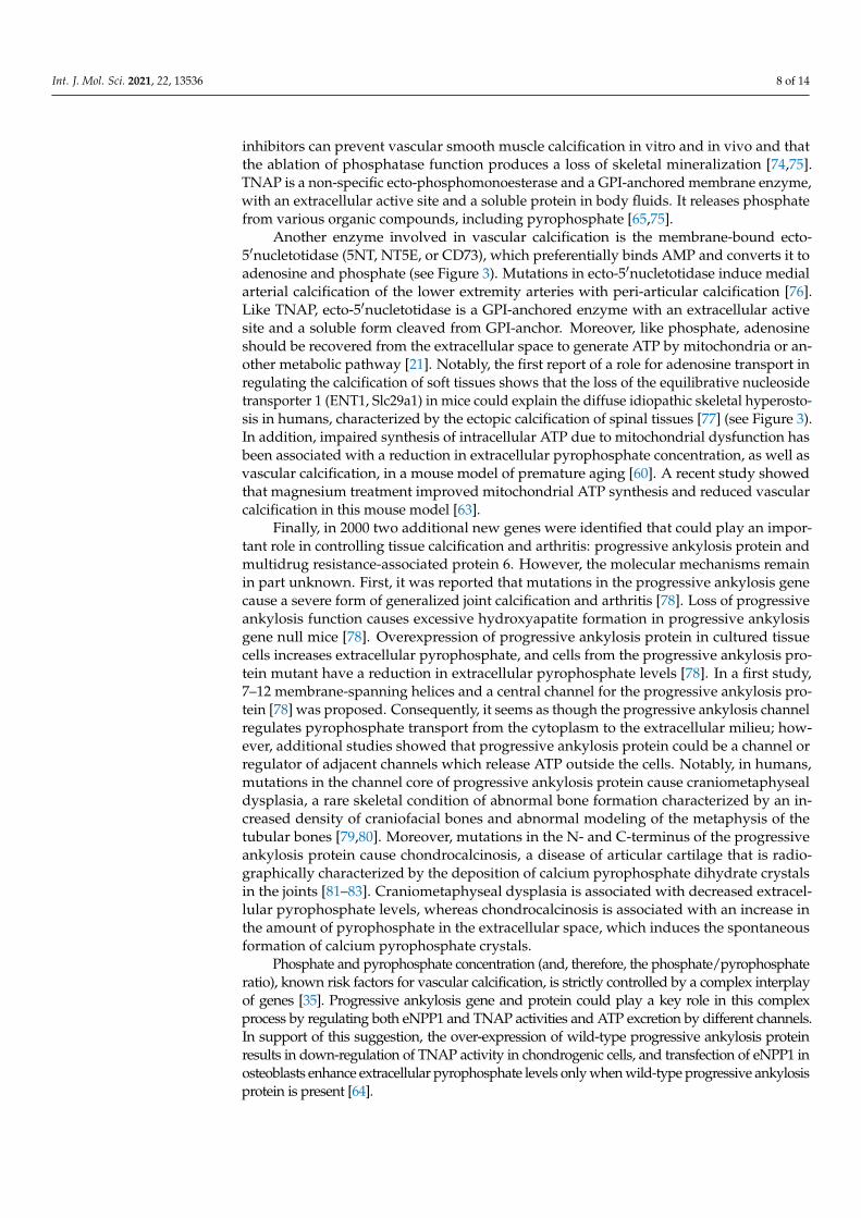
inhibitors can prevent vascular smooth muscle calcification in vitro and in vivo and thatthe ablation of phosphatase function produces a loss of skeletal mineralization [74,75].TNAP is a non-specific ecto-phosphomonoesterase and a GPI-anchored membrane enzyme,with an extracellular active site and a soluble protein in body fluids. It releases phosphatefrom various organic compounds, including pyrophosphate [65,75].
Another enzyme involved in vascular calcification is the membrane-bound ecto-5′nucletotidase (5NT, NT5E, or CD73), which preferentially binds AMP and converts it toadenosine and phosphate (see Figure 3). Mutations in ecto-5′nucletotidase induce medialarterial calcification of the lower extremity arteries with peri-articular calcification [76].Like TNAP, ecto-5′nucletotidase is a GPI-anchored enzyme with an extracellular activesite and a soluble form cleaved from GPI-anchor. Moreover, like phosphate, adenosineshould be recovered from the extracellular space to generate ATP by mitochondria or an-other metabolic pathway [21]. Notably, the first report of a role for adenosine transport inregulating the calcification of soft tissues shows that the loss of the equilibrative nucleosidetransporter 1 (ENT1, Slc29a1) in mice could explain the diffuse idiopathic skeletal hyperosto-sis in humans, characterized by the ectopic calcification of spinal tissues [77] (see Figure 3).In addition, impaired synthesis of intracellular ATP due to mitochondrial dysfunction hasbeen associated with a reduction in extracellular pyrophosphate concentration, as well asvascular calcification, in a mouse model of premature aging [60]. A recent study showedthat magnesium treatment improved mitochondrial ATP synthesis and reduced vascularcalcification in this mouse model [63].
Finally, in 2000 two additional new genes were identified that could play an impor-tant role in controlling tissue calcification and arthritis: progressive ankylosis protein andmultidrug resistance-associated protein 6. However, the molecular mechanisms remainin part unknown. First, it was reported that mutations in the progressive ankylosis genecause a severe form of generalized joint calcification and arthritis [78]. Loss of progressiveankylosis function causes excessive hydroxyapatite formation in progressive ankylosisgene null mice [78]. Overexpression of progressive ankylosis protein in cultured tissuecells increases extracellular pyrophosphate, and cells from the progressive ankylosis pro-tein mutant have a reduction in extracellular pyrophosphate levels [78]. In a first study,7–12 membrane-spanning helices and a central channel for the progressive ankylosis pro-tein [78] was proposed. Consequently, it seems as though the progressive ankylosis channelregulates pyrophosphate transport from the cytoplasm to the extracellular milieu; how-ever, additional studies showed that progressive ankylosis protein could be a channel orregulator of adjacent channels which release ATP outside the cells. Notably, in humans,mutations in the channel core of progressive ankylosis protein cause craniometaphysealdysplasia, a rare skeletal condition of abnormal bone formation characterized by an in-creased density of craniofacial bones and abnormal modeling of the metaphysis of thetubular bones [79,80]. Moreover, mutations in the N- and C-terminus of the progressiveankylosis protein cause chondrocalcinosis, a disease of articular cartilage that is radio-graphically characterized by the deposition of calcium pyrophosphate dihydrate crystalsin the joints [81–83]. Craniometaphyseal dysplasia is associated with decreased extracel-lular pyrophosphate levels, whereas chondrocalcinosis is associated with an increase inthe amount of pyrophosphate in the extracellular space, which induces the spontaneousformation of calcium pyrophosphate crystals.
Phosphate and pyrophosphate concentration (and, therefore, the phosphate/pyrophosphateratio), known risk factors for vascular calcification, is strictly controlled by a complex interplayof genes [35]. Progressive ankylosis gene and protein could play a key role in this complexprocess by regulating both eNPP1 and TNAP activities and ATP excretion by different channels.In support of this suggestion, the over-expression of wild-type progressive ankylosis proteinresults in down-regulation of TNAP activity in chondrogenic cells, and transfection of eNPP1 inosteoblasts enhance extracellular pyrophosphate levels only when wild-type progressive ankylosisprotein is present [64].
Int. J. Mol. Sci. 2021, 22, 13536 9 of 14
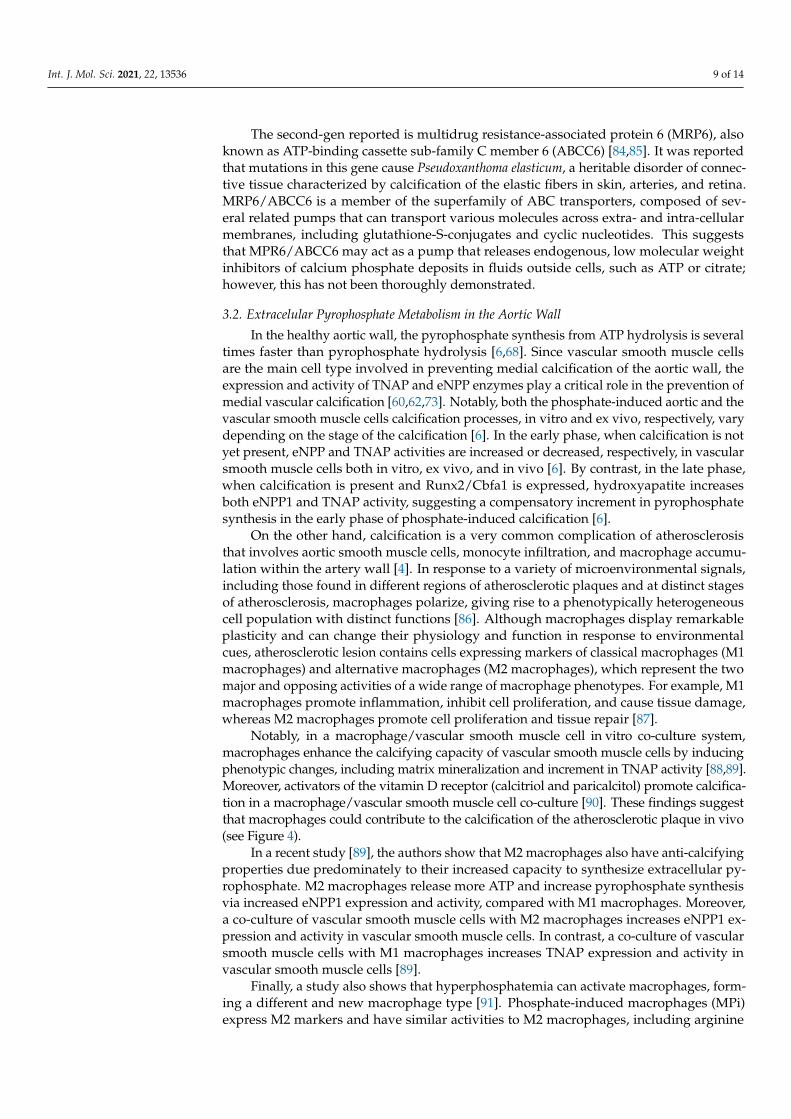
The second-gen reported is multidrug resistance-associated protein 6 (MRP6), alsoknown as ATP-binding cassette sub-family C member 6 (ABCC6) [84,85]. It was reportedthat mutations in this gene cause Pseudoxanthoma elasticum, a heritable disorder of connec-tive tissue characterized by calcification of the elastic fibers in skin, arteries, and retina.MRP6/ABCC6 is a member of the superfamily of ABC transporters, composed of sev-eral related pumps that can transport various molecules across extra- and intra-cellularmembranes, including glutathione-S-conjugates and cyclic nucleotides. This suggeststhat MPR6/ABCC6 may act as a pump that releases endogenous, low molecular weightinhibitors of calcium phosphate deposits in fluids outside cells, such as ATP or citrate;however, this has not been thoroughly demonstrated.
3.2. Extracelular Pyrophosphate Metabolism in the Aortic Wall
In the healthy aortic wall, the pyrophosphate synthesis from ATP hydrolysis is severaltimes faster than pyrophosphate hydrolysis [6,68]. Since vascular smooth muscle cellsare the main cell type involved in preventing medial calcification of the aortic wall, theexpression and activity of TNAP and eNPP enzymes play a critical role in the prevention ofmedial vascular calcification [60,62,73]. Notably, both the phosphate-induced aortic and thevascular smooth muscle cells calcification processes, in vitro and ex vivo, respectively, varydepending on the stage of the calcification [6]. In the early phase, when calcification is notyet present, eNPP and TNAP activities are increased or decreased, respectively, in vascularsmooth muscle cells both in vitro, ex vivo, and in vivo [6]. By contrast, in the late phase,when calcification is present and Runx2/Cbfa1 is expressed, hydroxyapatite increasesboth eNPP1 and TNAP activity, suggesting a compensatory increment in pyrophosphatesynthesis in the early phase of phosphate-induced calcification [6].
On the other hand, calcification is a very common complication of atherosclerosisthat involves aortic smooth muscle cells, monocyte infiltration, and macrophage accumu-lation within the artery wall [4]. In response to a variety of microenvironmental signals,including those found in different regions of atherosclerotic plaques and at distinct stagesof atherosclerosis, macrophages polarize, giving rise to a phenotypically heterogeneouscell population with distinct functions [86]. Although macrophages display remarkableplasticity and can change their physiology and function in response to environmentalcues, atherosclerotic lesion contains cells expressing markers of classical macrophages (M1macrophages) and alternative macrophages (M2 macrophages), which represent the twomajor and opposing activities of a wide range of macrophage phenotypes. For example, M1macrophages promote inflammation, inhibit cell proliferation, and cause tissue damage,whereas M2 macrophages promote cell proliferation and tissue repair [87].
Notably, in a macrophage/vascular smooth muscle cell in vitro co-culture system,macrophages enhance the calcifying capacity of vascular smooth muscle cells by inducingphenotypic changes, including matrix mineralization and increment in TNAP activity [88,89].Moreover, activators of the vitamin D receptor (calcitriol and paricalcitol) promote calcifica-tion in a macrophage/vascular smooth muscle cell co-culture [90]. These findings suggestthat macrophages could contribute to the calcification of the atherosclerotic plaque in vivo(see Figure 4).
In a recent study [89], the authors show that M2 macrophages also have anti-calcifyingproperties due predominately to their increased capacity to synthesize extracellular py-rophosphate. M2 macrophages release more ATP and increase pyrophosphate synthesisvia increased eNPP1 expression and activity, compared with M1 macrophages. Moreover,a co-culture of vascular smooth muscle cells with M2 macrophages increases eNPP1 ex-pression and activity in vascular smooth muscle cells. In contrast, a co-culture of vascularsmooth muscle cells with M1 macrophages increases TNAP expression and activity invascular smooth muscle cells [89].
Finally, a study also shows that hyperphosphatemia can activate macrophages, form-ing a different and new macrophage type [91]. Phosphate-induced macrophages (MPi)express M2 markers and have similar activities to M2 macrophages, including arginine
Int. J. Mol. Sci. 2021, 22, 13536 10 of 14
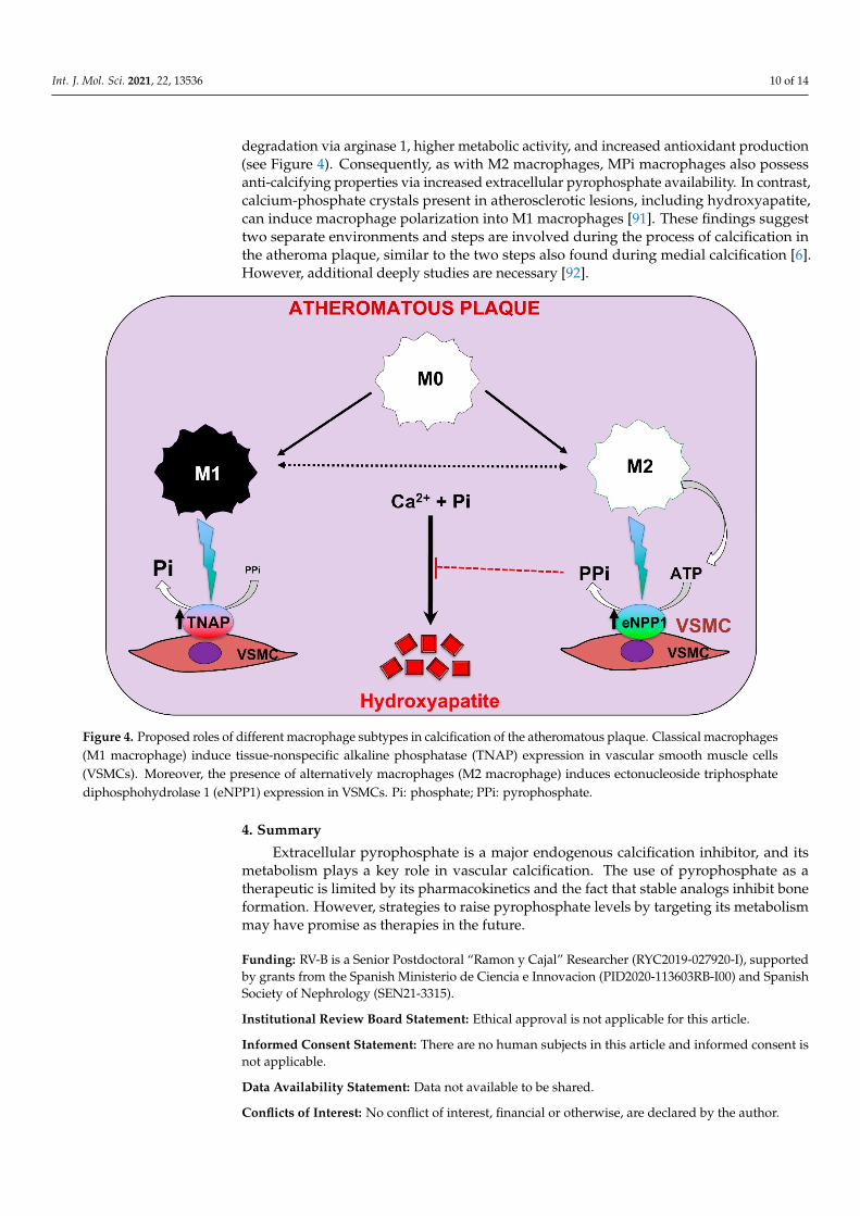
degradation via arginase 1, higher metabolic activity, and increased antioxidant production(see Figure 4). Consequently, as with M2 macrophages, MPi macrophages also possessanti-calcifying properties via increased extracellular pyrophosphate availability. In contrast,calcium-phosphate crystals present in atherosclerotic lesions, including hydroxyapatite,can induce macrophage polarization into M1 macrophages [91]. These findings suggesttwo separate environments and steps are involved during the process of calcification inthe atheroma plaque, similar to the two steps also found during medial calcification [6].However, additional deeply studies are necessary [92].
Int. J. Mol. Sci. 2021, 22, x FOR PEER REVIEW 10 of 14
plasticity and can change their physiology and function in response to environmental cues, atherosclerotic lesion contains cells expressing markers of classical macrophages (M1 macrophages) and alternative macrophages (M2 macrophages), which represent the two major and opposing activities of a wide range of macrophage phenotypes. For exam-ple, M1 macrophages promote inflammation, inhibit cell proliferation, and cause tissue damage, whereas M2 macrophages promote cell proliferation and tissue repair [87].
Notably, in a macrophage/vascular smooth muscle cell in vitro co-culture system, macrophages enhance the calcifying capacity of vascular smooth muscle cells by inducing phenotypic changes, including matrix mineralization and increment in TNAP activity [88,89]. Moreover, activators of the vitamin D receptor (calcitriol and paricalcitol) promote calcification in a macrophage/vascular smooth muscle cell co-culture [90]. These findings suggest that macrophages could contribute to the calcification of the atherosclerotic plaque in vivo (see Figure 4).
In a recent study [89], the authors show that M2 macrophages also have anti-calcify-ing properties due predominately to their increased capacity to synthesize extracellular pyrophosphate. M2 macrophages release more ATP and increase pyrophosphate synthe-sis via increased eNPP1 expression and activity, compared with M1 macrophages. More-over, a co-culture of vascular smooth muscle cells with M2 macrophages increases eNPP1 expression and activity in vascular smooth muscle cells. In contrast, a co-culture of vascu-lar smooth muscle cells with M1 macrophages increases TNAP expression and activity in vascular smooth muscle cells [89].
Figure 4. Proposed roles of different macrophage subtypes in calcification of the atheromatous plaque. Classical macro-phages (M1 macrophage) induce tissue-nonspecific alkaline phosphatase (TNAP) expression in vascular smooth muscle cells (VSMCs). Moreover, the presence of alternatively macrophages (M2 macrophage) induces ectonucleoside triphos-phate diphosphohydrolase 1 (eNPP1) expression in VSMCs. Pi: phosphate; PPi: pyrophosphate.
Finally, a study also shows that hyperphosphatemia can activate macrophages, form-ing a different and new macrophage type [91]. Phosphate-induced macrophages (MPi) express M2 markers and have similar activities to M2 macrophages, including arginine
Figure 4. Proposed roles of different macrophage subtypes in calcification of the atheromatous plaque. Classical macrophages(M1 macrophage) induce tissue-nonspecific alkaline phosphatase (TNAP) expression in vascular smooth muscle cells(VSMCs). Moreover, the presence of alternatively macrophages (M2 macrophage) induces ectonucleoside triphosphatediphosphohydrolase 1 (eNPP1) expression in VSMCs. Pi: phosphate; PPi: pyrophosphate.
4. Summary
Extracellular pyrophosphate is a major endogenous calcification inhibitor, and itsmetabolism plays a key role in vascular calcification. The use of pyrophosphate as atherapeutic is limited by its pharmacokinetics and the fact that stable analogs inhibit boneformation. However, strategies to raise pyrophosphate levels by targeting its metabolismmay have promise as therapies in the future.
Funding: RV-B is a Senior Postdoctoral “Ramon y Cajal” Researcher (RYC2019-027920-I), supportedby grants from the Spanish Ministerio de Ciencia e Innovacion (PID2020-113603RB-I00) and SpanishSociety of Nephrology (SEN21-3315).
Institutional Review Board Statement: Ethical approval is not applicable for this article.
Informed Consent Statement: There are no human subjects in this article and informed consent isnot applicable.
Data Availability Statement: Data not available to be shared.
Conflicts of Interest: No conflict of interest, financial or otherwise, are declared by the author.
Int. J. Mol. Sci. 2021, 22, 13536 11 of 14
References1. Shanahan, C.M.; Crouthamel, M.H.; Kapustin, A.; Giachelli, C.M. Arterial calcification in chronic kidney disease: Key roles for
calcium and phosphate. Circ. Res. 2011, 109, 697–711. [CrossRef] [PubMed]2. Rutsch, F.; Nitschke, Y.; Terkeltaub, R. Genetics in arterial calcification: Pieces of a puzzle and cogs in a wheel. Circ. Res. 2011, 109, 578–592.
[CrossRef] [PubMed]3. Rocha-Singh, K.J.; Zeller, T.; Jaff, M.R. Peripheral arterial calcification: Prevalence, mechanism, detection, and clinical implications.
Catheter. Cardiovasc. Interv. 2014, 83, E212–E220. [CrossRef] [PubMed]4. Villa-Bellosta, R. New insights into endogenous mechanisms of protection against arterial calcification. Atherosclerosis 2020, 306, 68–74.
[CrossRef]5. Villa-Bellosta, R.; Millan, A.; Sorribas, V. Role of calcium-phosphate deposition in vascular smooth muscle cell calcification. Am. J.
Physiol. Cell Physiol. 2011, 300, C210–C220. [CrossRef] [PubMed]6. Villa-Bellosta, R. Synthesis of Extracellular Pyrophosphate Increases in Vascular Smooth Muscle Cells During Phosphate-Induced
Calcification. Arterioscler. Thromb. Vasc. Biol. 2018, 38, 2137–2147. [CrossRef] [PubMed]7. Giachelli, C.M.; Speer, M.Y.; Li, X.; Rajachar, R.M.; Yang, H. Regulation of vascular calcification: Roles of phosphate and
osteopontin. Circ. Res. 2005, 96, 717–722. [CrossRef]8. Jono, S.; McKee, M.D.; Murry, C.E.; Shioi, A.; Nishizawa, Y.; Mori, K.; Morii, H.; Giachelli, C.M. Phosphate regulation of vascular
smooth muscle cell calcification. Circ. Res. 2000, 87, E10–E17. [CrossRef]9. Li, X.; Yang, H.-Y.; Giachelli, C.M. Role of the sodium-dependent phosphate cotransporter, Pit-1, in vascular smooth muscle cell
calcification. Circ. Res. 2006, 98, 905–912. [CrossRef]10. Villa-Bellosta, R.; Bogaert, Y.E.; Levi, M.; Sorribas, V. Characterization of phosphate transport in rat vascular smooth muscle cells:
Implications for vascular calcification. Arterioscler. Thromb. Vasc. Biol. 2007, 27, 1030–1036. [CrossRef]11. Villa-Bellosta, R.; Sorribas, V. Calcium phosphate deposition with normal phosphate concentration. -Role of pyrophosphate-.
Circ. J. 2011, 75, 2705–2710. [CrossRef] [PubMed]12. Schinke, T.; Karsenty, G. Vascular calcification—A passive process in need of inhibitors. Nephrol. Dial. Transplant. 2000, 15, 1272–1274.
[CrossRef]13. Proudfoot, D.; Skepper, J.N.; Hegyi, L.; Bennett, M.R.; Shanahan, C.M.; Weissberg, P.L. Apoptosis regulates human vascular
calcification in vitro: Evidence for initiation of vascular calcification by apoptotic bodies. Circ. Res. 2000, 87, 1055–1062. [CrossRef][PubMed]
14. Shroff, R.C.; McNair, R.; Figg, N.; Skepper, J.N.; Schurgers, L.; Gupta, A.; Hiorns, M.; Donald, A.E.; Deanfield, J.; Rees, L.; et al. Dialysisaccelerates medial vascular calcification in part by triggering smooth muscle cell apoptosis. Circulation 2008, 118, 1748–1757. [CrossRef]
15. Mansfield, K.; Teixeira, C.C.; Adams, C.S.; Shapiro, I.M. Phosphate ions mediate chondrocyte apoptosis through a plasmamembrane transporter mechanism. Bone 2001, 28, 1–8. [CrossRef]
16. Steitz, S.A.; Speer, M.Y.; Curinga, G.; Yang, H.Y.; Haynes, P.; Aebersold, R.; Schinke, T.; Karsenty, G.; Giachelli, C.M. Smoothmuscle cell phenotypic transition associated with calcification: Upregulation of Cbfa1 and downregulation of smooth musclelineage markers. Circ. Res. 2001, 89, 1147–1154. [CrossRef] [PubMed]
17. Li, X.; Yang, H.-Y.; Giachelli, C.M. BMP-2 promotes phosphate uptake, phenotypic modulation, and calcification of humanvascular smooth muscle cells. Atherosclerosis 2008, 199, 271–277. [CrossRef]
18. Speer, M.Y.; Li, X.; Hiremath, P.G.; Giachelli, C.M. Runx2/Cbfa1, but not loss of myocardin, is required for smooth muscle celllineage reprogramming toward osteochondrogenesis. J. Cell. Biochem. 2010, 110, 935–947. [CrossRef]
19. Lei, Y.; Sinha, A.; Nosoudi, N.; Grover, A.; Vyavahare, N. Hydroxyapatite and calcified elastin induce osteoblast-like differentiationin rat aortic smooth muscle cells. Exp. Cell Res. 2014, 323, 198–208. [CrossRef]
20. Sage, A.P.; Lu, J.; Tintut, Y.; Demer, L.L. Hyperphosphatemia-induced nanocrystals upregulate the expression of bone morpho-genetic protein-2 and osteopontin genes in mouse smooth muscle cells in vitro. Kidney Int. 2011, 79, 414–422. [CrossRef]
21. Villa-Bellosta, R. Vascular Calcification Revisited: A New Perspective for Phosphate Transport. Curr. Cardiol. Rev. 2015, 11, 341–351.[CrossRef]
22. Kanzaki, N.; Treboux, G.; Onuma, K.; Tsutsumi, S.; Ito, A. Calcium phosphate clusters. Biomaterials 2001, 22, 2921–2929. [CrossRef]23. Johnsson, M.S.; Nancollas, G.H. The role of brushite and octacalcium phosphate in apatite formation. Crit. Rev. Oral Biol. Med.
1992, 3, 61–82. [CrossRef]24. P’ng, C.H.; Boadle, R.; Horton, M.; Bilous, M.; Bonar, F. Magnesium whitlockite of the aorta. Pathology 2008, 40, 539–540.
[CrossRef]25. Reid, J.D.; Andersen, M.E. Medial calcification (whitlockite) in the aorta. Atherosclerosis 1993, 101, 213–224. [CrossRef]26. Kay, M.I.; Young, R.A.; Posner, A.S. Crystal structure of hydroxyapatite. Nature 1964, 204, 1050–1052. [CrossRef]27. Posner, A.S. The structure of bone apatite surfaces. J. Biomed. Mater. Res. 1985, 19, 241–250. [CrossRef] [PubMed]28. Posner, A.S.; Beebe, R.A. The surface chemistry of bone mineral and related calcium phosphates. Semin. Arthritis Rheum. 1975, 4, 267–291.
[CrossRef]29. Posner, A.S.; Betts, F.; Blumenthal, N.C. Role of ATP and Mg in the stabilization of biological and synthetic amorphous calcium
phosphates. Calcif. Tissue Res. 1977, 22, 208–212. [CrossRef] [PubMed]30. Blumenthal, N.C.; Betts, F.; Posner, A.S. Stabilization of amorphous calcium phosphate by Mg and ATP. Calcif. Tissue Res. 1977, 23, 245–250.
[CrossRef]
Int. J. Mol. Sci. 2021, 22, 13536 12 of 14
31. Urry, D.W. Neutral sites for calcium ion binding to elastin and collagen: A charge neutralization theory for calcification and itsrelationship to atherosclerosis. Proc. Natl. Acad. Sci. USA 1971, 68, 810–814. [CrossRef]
32. Khavandgar, Z.; Roman, H.; Li, J.; Lee, S.; Vali, H.; Brinckmann, J.; Davis, E.C.; Murshed, M. Elastin haploinsufficiency impedesthe progression of arterial calcification in MGP-deficient mice. J. Bone Miner. Res. 2014, 29, 327–337. [CrossRef]
33. Hosaka, N.; Mizobuchi, M.; Ogata, H.; Kumata, C.; Kondo, F.; Koiwa, F.; Kinugasa, E.; Akizawa, T. Elastin degradation acceleratesphosphate-induced mineralization of vascular smooth muscle cells. Calcif. Tissue Int. 2009, 85, 523–529. [CrossRef] [PubMed]
34. Azpiazu, D.; González-Parra, E.; Egido, J.; Villa-Bellosta, R. Hydrolysis of Extracellular Pyrophosphate increases in post-hemodialysis plasma. Sci. Rep. 2018, 8, 11089. [CrossRef]
35. Villa-Bellosta, R.; Egido, J. Phosphate, pyrophosphate, and vascular calcification: A question of balance. Eur. Heart J. 2017, 38, 1801–1804.[CrossRef]
36. Manghat, P.; Sodi, R.; Swaminathan, R. Phosphate homeostasis and disorders. Ann. Clin. Biochem. 2014, 51 Pt 6, 631–656.[CrossRef] [PubMed]
37. Christov, M.; Jüppner, H. Phosphate homeostasis disorders. Best Pract. Res. Clin. Endocrinol. Metab. 2018, 32, 685–706. [CrossRef]38. Bergwitz, C.; Jüppner, H. Regulation of phosphate homeostasis by PTH, vitamin D, and FGF23. Annu. Rev. Med. 2010, 61, 91–104.
[CrossRef] [PubMed]39. Gonzalo, S.; Villa-Bellosta, R. The role of sodium phosphate cotransporters in ectopic calcification. Endokrynol. Pol. 2019, 70, 496–503.
[CrossRef]40. Forster, I.C.; Hernando, N.; Biber, J.; Murer, H. Phosphate transporters of the SLC20 and SLC34 families. Mol. Asp. Med. 2013, 34, 386–395.
[CrossRef]41. Forster, I.C.; Hernando, N.; Biber, J.; Murer, H. Phosphate transport kinetics and structure-function relationships of SLC34 and
SLC20 proteins. Curr. Top. Membr. 2012, 70, 313–356.42. Reimer, R.J. SLC17: A functionally diverse family of organic anion transporters. Mol. Asp. Med. 2013, 34, 350–359. [CrossRef]
[PubMed]43. Wagner, C.A.; Hernando, N.; Forster, I.C.; Biber, J. The SLC34 family of sodium-dependent phosphate transporters. Pflugers Arch.
2014, 466, 139–153. [CrossRef]44. Silverstein, D.M.; Barac-Nieto, M.; Murer, H.; Spitzer, A. A putative growth-related renal Na(+)-Pi cotransporter. Am. J. Physiol.
1997, 273 Pt 2, R928–R933. [CrossRef] [PubMed]45. Segawa, H.; Kaneko, I.; Takahashi, A.; Kuwahata, M.; Ito, M.; Ohkido, I.; Tatsumi, S.; Miyamoto, K.-I. Growth-related renal type II
Na/Pi cotransporter. J. Biol. Chem. 2002, 277, 19665–19672. [CrossRef]46. Collins, J.F.; Bai, L.; Ghishan, F.K. The SLC20 family of proteins: Dual functions as sodium-phosphate cotransporters and viral
receptors. Pflugers Arch. 2004, 447, 647–652. [CrossRef] [PubMed]47. Bjorklund, G.; Svanberg, E.; Dadar, M.; Card, D.J.; Chirumbolo, S.; Harrington, D.J.; Aaseth, J. The Role of Matrix Gla Protein
(MGP) in Vascular Calcification. Curr. Med. Chem. 2020, 27, 1647–1660. [CrossRef] [PubMed]48. Mori, K.; Emoto, M.; Inaba, M. Fetuin-A: A multifunctional protein. Recent Pat. Endocr. Metab. Immune Drug Discov. 2011, 5, 124–146.
[CrossRef]49. Jahnen-Dechent, W.; Schinke, T.; Trindl, A.; Müller-Esterl, W.; Sablitzky, F.; Kaiser, S.; Blessing, M. Cloning and targeted deletion
of the mouse fetuin gene. J. Biol. Chem. 1997, 272, 31496–31503. [CrossRef]50. Schäfer, C.; Heiss, A.; Schwarz, A.; Westenfeld, R.; Ketteler, M.; Floege, J.; Müller-Esterl, W.; Schinke, T.; Jahnen-Dechent, W. The
serum protein alpha 2-Heremans-Schmid glycoprotein/fetuin-A is a systemically acting inhibitor of ectopic calcification. J. Clin.Investig. 2003, 112, 357–366. [CrossRef]
51. Ulutas, O.; Taskapan, M.C.; Dogan, A.; Baysal, T.; Taskapan, H. Vascular calcification is not related to serum fetuin-A andosteopontin levels in hemodialysis patients. Int. Urol. Nephrol. 2018, 50, 137–142. [CrossRef] [PubMed]
52. Franzén, A.; Heinegård, D. Isolation and characterization of two sialoproteins present only in bone calcified matrix. Biochem. J.1985, 232, 715–724. [CrossRef] [PubMed]
53. O’Brien, E.R.; Garvin, M.R.; Stewart, D.K.; Hinohara, T.; Simpson, J.B.; Schwartz, S.M.; Giachelli, C.M. Osteopontin is synthesizedby macrophage, smooth muscle, and endothelial cells in primary and restenotic human coronary atherosclerotic plaques.Arterioscler. Thromb. J. Vasc. Biol. 1994, 14, 1648–1656. [CrossRef] [PubMed]
54. Ikeda, T.; Shirasawa, T.; Esaki, Y.; Yoshiki, S.; Hirokawa, K. Osteopontin mRNA is expressed by smooth muscle-derived foamcells in human atherosclerotic lesions of the aorta. J. Clin. Investig. 1993, 92, 2814–2820. [CrossRef] [PubMed]
55. Proudfoot, D.; Skepper, J.N.; Shanahan, C.M.; Weissberg, P.L. Calcification of human vascular cells in vitro is correlated with highlevels of matrix Gla protein and low levels of osteopontin expression. Arterioscler. Thromb. Vasc. Biol. 1998, 18, 379–388. [CrossRef]
56. Jono, S.; Peinado, C.; Giachelli, C.M. Phosphorylation of osteopontin is required for inhibition of vascular smooth muscle cellcalcification. J. Biol. Chem. 2000, 275, 20197–20203. [CrossRef] [PubMed]
57. Schibler, D.; Russell, R.G.; Fleisch, H. Inhibition by pyrophosphate and polyphosphate of aortic calcification induced by vitaminD3 in rats. Clin. Sci. 1968, 35, 363–372.
58. Lomashvili, K.A.; Khawandi, W.; O’Neill, W.C. Reduced plasma pyrophosphate levels in hemodialysis patients. J. Am. Soc.Nephrol. 2005, 16, 2495–2500. [CrossRef]
59. O’Neill, W.C.; Lomashvili, K.A.; Malluche, H.H.; Faugere, M.-C.; Riser, B.L. Treatment with pyrophosphate inhibits uremicvascular calcification. Kidney Int. 2011, 79, 512–517. [CrossRef]
Int. J. Mol. Sci. 2021, 22, 13536 13 of 14
60. Villa-Bellosta, R.; Rivera-Torres, J.; Osorio, F.G.; Acín-Pérez, R.; Enriquez, J.A.; López-Otín, C.; Andrés, V. Defective extracellularpyrophosphate metabolism promotes vascular calcification in a mouse model of Hutchinson-Gilford progeria syndrome that isameliorated on pyrophosphate treatment. Circulation 2013, 127, 2442–2451. [CrossRef] [PubMed]
61. Riser, B.L.; Barreto, F.C.; Rezg, R.; Valaitis, P.W.; Cook, C.S.; White, J.A.; Gass, J.H.; Maizel, J.; Louvet, L.; Drueke, T.B.; et al. Dailyperitoneal administration of sodium pyrophosphate in a dialysis solution prevents the development of vascular calcification in amouse model of uraemia. Nephrol. Dial. Transplant. 2011, 26, 3349–3357. [CrossRef]
62. Villa-Bellosta, R. ATP-based therapy prevents vascular calcification and extends longevity in a mouse model of Hutchinson-Gilford progeria syndrome. Proc. Natl. Acad. Sci. USA 2019, 116, 23698–23704. [CrossRef] [PubMed]
63. Villa-Bellosta, R. Dietary magnesium supplementation improves lifespan in a mouse model of progeria. EMBO Mol. Med. 2020,12, e12423. [CrossRef] [PubMed]
64. Villa-Bellosta, R.; O’Neill, W.C. Pyrophosphate deficiency in vascular calcification. Kidney Int. 2018, 93, 1293–1297. [CrossRef][PubMed]
65. Zimmermann, H.; Zebisch, M.; Sträter, N. Cellular function and molecular structure of ecto-nucleotidases. Purinergic Signal. 2012,8, 437–502. [CrossRef]
66. Zimmermann, H. Extracellular metabolism of ATP and other nucleotides. Naunyn Schmiedebergs Arch. Pharmacol. 2000, 362, 299–309.[CrossRef] [PubMed]
67. Rutsch, F.; Ruf, N.; Vaingankar, S.; Toliat, M.R.; Suk, A.; Höhne, W.; Schauer, G.; Lehmann, M.; Roscioli, T.; Schnabel, D.; et al.Mutations in ENPP1 are associated with «idiopathic» infantile arterial calcification. Nat. Genet. 2003, 34, 379–381. [CrossRef][PubMed]
68. Villa-Bellosta, R.; Wang, X.; Millán, J.L.; Dubyak, G.R.; O’Neill, W.C. Extracellular pyrophosphate metabolism and calcification invascular smooth muscle. Am. J. Physiol. Heart Circ. Physiol. 2011, 301, H61–H68. [CrossRef] [PubMed]
69. Zhang, J.; Dyment, N.A.; Rowe, D.W.; Siu, S.Y.; Sundberg, J.P.; Uitto, J.; Li, Q. Ectopic mineralization of cartilage and collagen-richtendons and ligaments in Enpp1asj-2J mice. Oncotarget 2016, 7, 12000–12009. [CrossRef] [PubMed]
70. Villa-Bellosta, R.; Sorribas, V. Prevention of vascular calcification by polyphosphates and nucleotides-role of ATP. Circ. J. 2013,77, 2145–2151. [CrossRef] [PubMed]
71. Villa-Bellosta, R.; Sorribas, V. Phosphonoformic acid prevents vascular smooth muscle cell calcification by inhibiting calcium-phosphate deposition. Arterioscler. Thromb. Vasc. Biol. 2009, 29, 761–766. [CrossRef] [PubMed]
72. Lomashvili, K.A.; Cobbs, S.; Hennigar, R.A.; Hardcastle, K.I.; O’Neill, W.C. Phosphate-induced vascular calcification: Role ofpyrophosphate and osteopontin. J. Am. Soc. Nephrol. 2004, 15, 1392–1401. [CrossRef]
73. Lomashvili, K.A.; Garg, P.; Narisawa, S.; Millan, J.L.; O’Neill, W.C. Upregulation of alkaline phosphatase and pyrophosphatehydrolysis: Potential mechanism for uremic vascular calcification. Kidney Int. 2008, 73, 1024–1030. [CrossRef] [PubMed]
74. Narisawa, S.; Harmey, D.; Yadav, M.C.; O’Neill, W.C.; Hoylaerts, M.F.; Millán, J.L. Novel inhibitors of alkaline phosphatasesuppress vascular smooth muscle cell calcification. J. Bone Miner. Res. 2007, 22, 1700–1710. [CrossRef]
75. Azpiazu, D.; Gonzalo, S.; Villa-Bellosta, R. Tissue Non-Specific Alkaline Phosphatase and Vascular Calcification: A PotentialTherapeutic Target. Curr. Cardiol. Rev. 2019, 15, 91–95. [CrossRef] [PubMed]
76. St Hilaire, C.; Ziegler, S.G.; Markello, T.C.; Brusco, A.; Groden, C.; Gill, F.; Carlson-Donohoe, H.; Lederman, R.J.; Chen, M.Y.; Yang, D.; et al.NT5E mutations and arterial calcifications. N. Engl. J. Med. 2011, 364, 432–442. [CrossRef] [PubMed]
77. Warraich, S.; Bone, D.; Quinonez, D.; Ii, H.; Choi, D.-S.; Holdsworth, D.; Drangova, M.; Dixon, S.J.; Séguin, C.A.; Hammond, J.Loss of equilibrative nucleoside transporter 1 in mice leads to progressive ectopic mineralization of spinal tissues resemblingdiffuse idiopathic skeletal hyperostosis in humans. J. Bone Miner. Res. 2013, 28, 1135–1149. [CrossRef] [PubMed]
78. Ho, A.M.; Johnson, M.D.; Kingsley, D.M. Role of the mouse ank gene in control of tissue calcification and arthritis. Science 2000,289, 265–270. [CrossRef] [PubMed]
79. Nürnberg, P.; Thiele, H.; Chandler, D.; Höhne, W.; Cunningham, M.L.; Ritter, H.; Leschik, G.; Uhlmann, K.; Mischung, C.;Harrop, K.; et al. Heterozygous mutations in ANKH, the human ortholog of the mouse progressive ankylosis gene, result incraniometaphyseal dysplasia. Nat. Genet. 2001, 28, 37–41. [CrossRef]
80. Reichenberger, E.; Tiziani, V.; Watanabe, S.; Park, L.; Ueki, Y.; Santanna, C.; Baur, S.T.; Shiang, R.; Grange, D.K.; Beighton, P.; et al.Autosomal dominant craniometaphyseal dysplasia is caused by mutations in the transmembrane protein ANK. Am. J. Hum.Genet. 2001, 68, 1321–1326. [CrossRef] [PubMed]
81. Pendleton, A.; Johnson, M.D.; Hughes, A.; Gurley, K.A.; Ho, A.M.; Doherty, M.; Dixey, J.; Gillet, P.; Loeuille, D.; McGrath, R.; et al.Mutations in ANKH cause chondrocalcinosis. Am. J. Hum. Genet. 2002, 71, 933–940. [CrossRef]
82. Williams, C.J.; Zhang, Y.; Timms, A.; Bonavita, G.; Caeiro, F.; Broxholme, J.; Cuthbertson, J.; Jones, Y.; Marchegiani, R.; Reginato, A.; et al.Autosomal dominant familial calcium pyrophosphate dihydrate deposition disease is caused by mutation in the transmembrane proteinANKH. Am. J. Hum. Genet. 2002, 71, 985–991. [CrossRef]
83. Williams, C.J.; Pendleton, A.; Bonavita, G.; Reginato, A.J.; Hughes, A.E.; Peariso, S.; Doherty, M.; Mccarty, D.J.; Ryan, L.M.Mutations in the amino terminus of ANKH in two US families with calcium pyrophosphate dihydrate crystal deposition disease.Arthritis Rheum. 2003, 48, 2627–2631. [CrossRef]
84. Le Saux, O.; Urban, Z.; Tschuch, C.; Csiszar, K.; Bacchelli, B.; Quaglino, D.; Pasquali-Ronchetti, I.; Pope, F.M.; Richards, A.; Terry, S.; et al.Mutations in a gene encoding an ABC transporter cause pseudoxanthoma elasticum. Nat. Genet. 2000, 25, 223–227. [CrossRef]
Int. J. Mol. Sci. 2021, 22, 13536 14 of 14
85. Bergen, A.A.; Plomp, A.S.; Schuurman, E.J.; Terry, S.F.; Breuning, M.H.; Dauwerse, H.G.; Swart, J.; Kool, M.; Van Soest, S.; Baas, F.;·et al.Mutations in ABCC6 cause pseudoxanthoma elasticum. Nat. Genet. 2000, 25, 228–231. [CrossRef] [PubMed]
86. Locati, M.; Curtale, G.; Mantovani, A. Diversity, Mechanisms, and Significance of Macrophage Plasticity. Annu. Rev. Pathol. 2020,15, 123–147. [CrossRef] [PubMed]
87. Colin, S.; Chinetti-Gbaguidi, G.; Staels, B. Macrophage phenotypes in atherosclerosis. Immunol. Rev. 2014, 262, 153–166. [CrossRef]88. Tintut, Y.; Patel, J.; Territo, M.; Saini, T.; Parhami, F.; Demer, L.L. Monocyte/macrophage regulation of vascular calcification
in vitro. Circulation 2002, 105, 650–655. [CrossRef] [PubMed]89. Villa-Bellosta, R.; Hamczyk, M.R.; Andrés, V. Alternatively activated macrophages exhibit an anticalcifying activity dependent on
extracellular ATP/pyrophosphate metabolism. Am. J. Physiol. Cell Physiol. 2016, 310, C788–C799. [CrossRef] [PubMed]90. Li, X.; Speer, M.Y.; Yang, H.; Bergen, J.; Giachelli, C.M. Vitamin D receptor activators induce an anticalcific paracrine program in
macrophages: Requirement of osteopontin. Arterioscler. Thromb. Vasc. Biol. 2010, 30, 321–326. [CrossRef] [PubMed]91. Villa-Bellosta, R.; Hamczyk, M.R.; Andrés, V. Novel phosphate-activated macrophages prevent ectopic calcification by increasing
extracellular ATP and pyrophosphate. PLoS ONE 2017, 12, e0174998. [CrossRef] [PubMed]92. Wang, C.; Li, Y.; Shi, L.; Ren, J.; Patti, M.; Wang, T.; De Oliveira, J.R.M.; Sobrido, M.-J.; Quintáns, B.; Baquero, M.; et al. Mutations
in SLC20A2 link familial idiopathic basal ganglia calcification with phosphate homeostasis. Nat. Genet. 2012, 44, 254–256.[CrossRef] [PubMed]