on the multiple functional roles of the active site histidine in catalysis and allosteric regulation...
TRANSCRIPT
Subscriber access provided by RUTGERS UNIV
Biochemistry is published by the American Chemical Society. 1155 Sixteenth StreetN.W., Washington, DC 20036
ArticleOn the Multiple Functional Roles of the Active SiteHistidine in Catalysis and Allosteric Regulation of
Escherichia coli Glucosamine 6-Phosphate Deaminase!
Gabriela M. Montero-Morn, Samuel Lara-Gonzlez, Laura I.lvarez-Aorve, Jacqueline A. Plumbridge, and Mario L. Calcagno
Biochemistry, 2001, 40 (34), 10187-10196• DOI: 10.1021/bi0105835 • Publication Date (Web): 03 August 2001Downloaded from http://pubs.acs.org on March 8, 2009
More About This Article
Additional resources and features associated with this article are available within the HTML version:
• Supporting Information• Access to high resolution figures• Links to articles and content related to this article• Copyright permission to reproduce figures and/or text from this article

On the Multiple Functional Roles of the Active Site Histidine in Catalysis andAllosteric Regulation of Escherichia coli Glucosamine 6-Phosphate Deaminase†
Gabriela M. Montero-Moran,‡ Samuel Lara-Gonzalez,‡ Laura I. AÄ lvarez-Anorve,‡ Jacqueline A. Plumbridge,§ andMario L. Calcagno*,‡
Departamento de Bioquımica, Laboratorio de Fisicoquımica y Diseno de Proteınas, Facultad de Medicina, UniVersidadNacional Autonoma de Mexico (UNAM), P. O. Box 70-159, Mexico City 04510, D.F., Mexico, and Institut de Biologie
Physico-Chimique (CNRS, UPR9073), 75005 Paris, France
ReceiVed March 22, 2001; ReVised Manuscript ReceiVed June 20, 2001
ABSTRACT: The active site of glucosamine-6-phosphate deaminase (EC 3.5.99.6, formerly 5.3.1.10) fromEscherichia coli was first characterized on the basis of the crystallographic structure of the enzyme boundto the competitive inhibitor 2-amino-2-deoxy-glucitol 6-phosphate. The structure corresponds to the Rallosteric state of the enzyme; it shows the side-chain of His143 in close proximity to the O5 atom of theinhibitor. This arrangement suggests that His143 could have a role in the catalysis of the ring-openingstep of glucosamine 6-phosphate whose R-anomer is the true substrate. The imidazole group of this active-site histidine contacts the carboxy groups from Glu148 and Asp141, via its N!1 atom [Oliva et al. (1995)Structure 3, 1323-1332]. These interactions change in the T state because the side chain of Glu148moves toward the allosteric site, leaving at the active site the dyad Asp141-His143 [Horjales et al. (1999)Structure 7, 527-536]. In this research, a dual approach using site-directed mutagenesis and controlledchemical modification of histidine residues has been used to investigate the role of the active-site histidine.Our results support a multifunctional role of His143; in the forward reaction, it is involved in the catalysisof the ring-opening step of the substrate, glucosamine 6-P. In the reverse reaction, the substrate fructose6-P binds in its open chain, carbonylic form. The role of His143 in the binding of both glucosamine 6-Pand reaction intermediates in their extended-chain forms was demonstrated by binding experiments usingthe reaction intermediate analogue, 2-amino-2-deoxy-D-glucitol 6-phosphate. His143 was also shown tobe a critical residue for the conformational coupling between active and allosteric sites. From the pHdependence of the reactivity of the active site histidine to diethyl dicarbonate, we observed a pKa changeof 1.2 units to the acid side when the enzyme undergoes the allosteric T to R transition during which theside chain of Glu148 moves toward the active site. The kinetic study of the Glu148-Gln mutant deaminaseshows that the loss of the carboxy group and its replacement with the corresponding amide modifies thekcat versus pH profile of the enzyme, suggesting that the catalytic step requiring the participation of His143has become rate-limiting. This, in turn, indicates that the interaction Glu148-His143 in the wild-typeenzyme in the R state contributes to make the enzyme functional over a wide pH range.
The enzyme glucosamine-6-phosphate deaminase (EC3.5.99.6, formerly glucosamine 6-phosphate isomerase, EC5.3.1.10) from Escherichia coli, catalyzes the reversibleconversion of D-glucosamine 6-phosphate (GlcN6P)1 intoD-fructose 6-phosphate (Fru6P) and ammonia (1, 2). It showsan absolute specificity for the R-anomer of GlcN6P (2), andits reaction mechanism involves a ring-opening step, followedby an enolization step that proceeds through a cis-enolamine
(2-amino-2-deoxy-D-arabino-hex-2-enitol 6-phosphate) andits tautomeric imine (2-deoxy-2-imino-D-arabino-hexitol6-phosphate) as reaction intermediates (2). This mechanismis similar to that of other ketose-aldose isomerases (2, 3),and it is illustrated in Figure 1, which also summarizes themain results of this research. The enzyme from E. coli isthe most studied GlcN6P deaminase, and it has beenstructurally (4, 5) and functionally characterized (6-8). Itis an allosteric enzyme activated by N-acetylglucosamine6-phosphate (GlcNAc6P), and its allosteric kinetics can bewell accounted for by the Monod-Wyman-Changeux (MWC)model (9, as shown by Altamirano et al., 7). Like mostallosteric enzymes, it is a K-system, i.e., regulation is theresult of a change in the apparent affinity of the enzymecaused by the allosteric transition, without any change inthe catalytic constant, kcat. Structural models of the enzymein its R [PDB 1dea, 1hor, 1hot, (4)] and T allosteric states[PDB 1cd5, (5)] were obtained. The crystallographic structureof the enzyme in its R-state complexed to the dead-end
† This research was supported by grants from the National Autono-mous University of Mexico (UNAM) DGAPA-UNAM IN201295 andfrom The National Council of Science and Technology CONACYT(Mexico), project 25258-N. G.M.M. was the recipient of a Ph.D. studentfellowship from CONACYT, Mexico.* Address correspondence to this author: email calcagno@
bq.unam.mx; fax + (52) 5616 2419,‡ Universidad Nacional Autonoma de Mexico, Mexico City, DF,
Mexico.§ Institut de Biologie Physico-Chimique, Paris, France.1 Abbreviations: GlcN6P, glucosamine 6-phosphate; GlcNAc6P,
N-acetyl- glucosamine-6-phosphate; GlcN-ol-6P, 2-amino-2-deoxy-D-glucitol 6-phosphate; DEDC, diethyl dicarbonate.
10187Biochemistry 2001, 40, 10187-10196
10.1021/bi0105835 CCC: $20.00 © 2001 American Chemical SocietyPublished on Web 08/03/2001

inhibitor 2-amino-2-deoxy-D-glucitol 6-phosphate (GlcN-ol-6P) (PDB 1hor) allowed the precise identification of thecatalytic pocket (Figures 2 and 3). The enzyme is a hexamerof identical subunits, arranged as a dimer of trimers. Thecomparison of the structure of the hexamer in the R and Tconformations provided a first geometrical description of theallosteric transition. This consists of a concerted change inthe quaternary structure, as predicted by the MWC model(9), accompanied by well-defined internal rearrangementsin each monomer (5). The catalytic mechanism of GlcN6Pdeaminase, as originally proposed by Midelfort and Rose
(2), has been reexamined in the light of the crystallographicstructure (4). Using the three-dimensional structure of theenzyme complexed with GlcN-ol-6P as well as the modeledcomplex of the enzyme bound to R-D-pyranosyl-GlcN6P, itwas possible to propose some functional roles for severalactive-site residues (4). The complex of the enzyme withthe inhibitor GlcN-ol-6P (PDB 1hor) shows the ligand inan almost extended conformation. This conformation shouldbe similar to that adopted by the bound aldehydo-GlcN6P.The proximity of the carboxylate group of Asp72 to the C1-C2 bond of GlcN-ol-6P suggests that this acid residue is the
FIGURE 1: Scheme of the chemical mechanism of the reaction catalyzed by E. coli GlcN6P deaminase, which resumes data from previousresearch (2, 4) and the results of the present paper. The dotted arrows correspond to the equilibrium of the enzyme with aldehydo-GlcN6P.The contribution of this form of the substrate to the reaction rate is discussed in the text.
FIGURE 2: Stereo drawing showing the active site of E. coli GlcN6P deaminase in the R allosteric state. A molecule of GlcN-ol-6P, adead-end inhibitor of the enzyme, appears bound to the site, and the side chains of the functionally significant residues discussed in the text,are depicted using stick and ball representation. This figure was generated from the coordinates of the deaminase-GlcN-ol-6P complex(PDB 1hor), using the graphics program MolScript (38) compiled for Linux and rendered to the present version with Raster-3D (39).
10188 Biochemistry, Vol. 40, No. 34, 2001 Montero-Moran et al.

general-base catalyzing the GlcN6P enolization by abstrac-tion of the C-2 proton. Indeed, its replacement by asparaginedecreased the kcat for the forward reaction by 4 orders ofmagnitude (S. Lara-Gonzalez, unpublished). The O5 in theinhibitor molecule corresponds to O5 of the open-chainsubstrate and becomes the pyranose oxygen in the cyclicform of the sugar. This atom contacts with N!2 of His143,suggesting that this histidine residue could play a role in thering opening of R-D-GlcN6P (4). The imidazolic N!1 ofHis143 side-chain appears hydrogen-bonded to the carboxy-lates of Asp141 and Glu148 when the enzyme is in the Rallosteric conformation. This arrangement changes when theenzyme goes from the R to the T allosteric state. A localdisplacement of the loop 144-154 occurs, resulting in areorientation of the side chain of Glu148, which movestoward the allosteric site and forms a salt bridge with theside chain of Lys160 (Figure 3). This lysine residue is partof the allosteric site and contributes to the binding of thephospho group of GlcNAc6P. This is the most conspicuoustertiary structural change produced by the R-T transitionin the deaminase monomer, and it occurs independently ofthe quaternary structural change, which consists of an overallrotation of the enzyme subunits (5). One of the consequencesof the local structural change is that the triad Asp141-His143-Glu148, present in the R state, becomes the dyad Asp141-His143 in the T-state (Figure 3). The arrangement aroundHis143 in the active site of GlcN6P deaminase resemblesthe active site of serine proteases. In both cases, the N!1atom of the active-site histidine makes contact with anactivating carboxy group, while the N!2 is able to transfera proton, after being transitorily protonated during thecatalytic cycle (4). In the R state of GlcN6P deaminase,which is the conformation expected to be responsible formost of its catalytic activity, there are two carboxygroups simultaneously interacting with the N!1 atom fromHis143. The importance of this triadic arrangement aroundHis143 can be inferred from the sequence alignment shownin Figure 4. In those deaminases known to be allosteric, thetriad Asp141-His143-Glu148 is absolutely conserved (Figure4A). Histidine-aspartate pairs are common in the active site
FIGURE 3: Stereopair showing the displacement of the side chain of Glu148 in the allosteric transition of E. coli GlcN6P deaminase. Itscarboxylate group appears at interaction distance with the N!1 atom of His143 when the enzyme is in the R allosteric state (gray; N!1-(His143)-O!2(Glu148) distance ) 2.71 Å) and moves toward the allosteric site where it forms a salt bridge with the ammonium group ofLys160 (black). The interaction Asp141-His143 is conserved in both conformational states; the corresponding distances are N!1(His143)-O!2(Asp141) ) 3.33 Å (R form) and N!1(His143)-O!2(Asp141) ) 2.6 Å (T form). This image was generated from the coordinates of thedeaminase with the active site occupied by GlcN-ol-6P (PDB 1hor) and from those for the ligand-free structure (PDB 1cd5), as describedin Figure 2. Overlapping of both set of coordinates was performed with the program Swiss-PdbViewer (41).
FIGURE 4: Alignment of several orthologous sequences of GlcN6Pdeaminases from bacteria and eukaryota. Escherichia coli, (Ecoli);Vibrio choleræ, (Vchole); Hæmophillus influenzæ, (Haein); Borreliaburgdorferi, (Borbu); Homo sapiens, (Human); Mesocricætusauratus (Mesau);Mus musculus (Mouse); Drosophila melanogaster,(Droso); Cænorabditis elegans, (Caeel); Candida albicans, (Canal);Bacillus subtilis, (Bacsu); Clostridium aceticum, (Clostac); Giardialamblia, gene 1 (Giala1). (A) The orthologous group of GlcN6Pdeaminases known to be allosteric (E. coli, mammals, Drosophila)or predicted to be; all have the residues involved in binding theallosteric phosphate. These allosteric functional residues are Arg158,Lys160, Ser151, as well as Thr161 and Tyr254 (40), not shownhere. In this group, the triad 141-143-148 is invariant. (B)Deaminases known to be nonallosteric (Giardia, Candida) or thoselacking the signature for the binding site for the allosteric phosphate(Gram positive bacteria). Sequences were obtained from GeneBank(http://www.ncbi.nlm.nih.gov/genbank) and TIGR (http://www.ti-gr.org) databases. Identical amino acids within each subset areshown on a black background while similarities appear in boldcharacters. The segment 136-158 in E. coli deaminase is a loopconnecting the sixth and the seventh "-strands (E and C′ strands inref 4). The position of the active-site triad and the phospho groupbinding residues in the allosteric site are indicated with arrows.This alignment was produced with the program Multalin 5.4.1 (42).
Role of His143 in the Active Site of GlcN6P Deaminase Biochemistry, Vol. 40, No. 34, 2001 10189

of many enzymes; they are present in the famed catalytictriad in serine-proteases (10) and in other enzymes such asD-glucarate dehydratase (11), L-lactate dehydrogenase (fla-vocytochrome b2) (12), and several hydrolytic enzymes (13).A ring-opening mechanism of cyclic sugar substrates using
a histidine imidazole as a general acid or base catalyst hasbeen proposed for those aldose-ketose isomerases whichcontain a histidine-carboxylic amino acid pair, such asphosphoglucose isomerase (14), GlcN6P synthase (Fru6P-glutamine amidotransferase) (15), L-arabinose isomerase (16),and D-xylose (D-glucose) isomerase. However, in the lastenzyme the catalytic role of the active-site histidine in sugarring-opening, based on the crystallographic structure (17),was not supported by site-directed mutagenesis experiments(18).The purpose of this research is to characterize the
functional role of His143 and the carboxylic residues whichcontact it in E. coli GlcN6P deaminase and to correlate theirstructural changes with the allosteric properties of theenzyme. For this, we have used a combined approach of site-directed mutagenesis and chemical modification. Our resultsdemonstrate that His143 and its flanking carboxylic residueshave multiple roles in the catalytic and allosteric propertiesof GlcN6P deaminase.
MATERIALS AND METHODS
Biochemicals. Diethyl dicarbonate, (EtO-CO-)2O (DEDC),also called diethyl pyrocarbonate (DEPC), and most bio-chemicals were purchased from Sigma-Aldrich Quımica S.A. de C. V. (Toluca, Edo. Mex., Mexico). N-6-aminohex-anoylglucosamine-6-P agarose, GlcNAc6P, GlcN-ol-6P, 14C-labeled GlcNAc6P and 3H-labeled GlcN-ol-6P, were allprepared as previously described (7). The oxime of Fru6P(2-(hydroxyimino)-2-deoxy-D-arabino-hexitol 6-phosphate)was prepared from Fru6P according to Finch and Merchant(19). 2,5 anhydro-D-mannitol 6-P was prepared from thecorresponding nonphosphorylated compound (Sigma-Ald-rich) by phosphorylation with 1.2 molar excess of MgATP2-and yeast hexokinase (Sigma H5625, 2.5 U mL-1). Theproduct was passed through a column packed with Dowex50 H+ equilibrated and eluted with water. Then, 2,5 anhydro-D-mannitol 6-P was purified on DEAE cellulose (Whatman),packed in 100 mM pyridine: 1 M formic acid and elutedwith the same buffer. The O-methyl glycosides of Fru6P(mixed R and " glycosides) were prepared by methylationof Fru6P, according to Fishbein et al. (20). Phosphate esterswere detected and quantified according to Ben-Yoseph etal. (21). All other chemical reagents were of suitable gradeand purity for their immediate use.Bacterial Strains and Mutagenesis. Site-directed mutations
were constructed by the technique of oligonucleotide-directedmutagenesis, using the Kunkel method, as described bySambrook et al. (22) with the nagB gene inserted in thesingle-strand vector pTZ18R. The single mutations createdwere His143 (CAT) to Gln(CAA), Asp141 (GAC) to Asn(AAC), and Glu (GAA to Gln(CAA)). The replacement ofeach carboxylic amino acid for its corresponding amide waschosen as the most conservative mutation, changing the pH-dependent electrostatic properties of the active site butmaintaining the side chain polarity in order to minimize anyactive site distortion. Phagemids, pTZ(NagB), carrying themutations were verified by sequencing and were used to
transform the !nag strain IBPC590 (23). This strain is !lacIand expresses the deaminase constitutively from the plasmidlac promoter. Details of the procedure and a more completedescription of the bacterial strain were already reported (7).Enzyme Purification and Assays. Wild-type GlcN6P
deaminase and the mutants used in this research were allpurified by allosteric affinity chromatography, as previouslyreported (6, 8). The purity of the enzymes was verified bySDS-PAGE. These mutations are not expected to producea significant change of the molar absorptivity of the enzyme,and thus, the value for the wild-type enzyme (20.0 ! 104M-1 cm-1) was used to calculate the molar concentration ofthese mutant forms. GlcN6P deaminase assays and analysesof kinetic data were performed as previously reported (6,7). Rate measurements were made at pH 7.7, at 30 °C in 50mM Tris-HCl buffer, unless otherwise indicated. Whenkinetic parameters were measured as a function of pH, theternary ACES-Tris-ethanolamine buffer was used to keepionic strength variations at a minimum (24). The measure-ment of the reverse reaction rate was performed in anincubation mixture prepared in the same buffer and contain-ing variable concentrations of Fru6P and NH4HCO3. TheGlcN6P formed was measured at fixed times, using amodified Morgan-Elson procedure, as already described (6).Measurement of the Dissociation Constant of the Allosteric
ActiVator. The dissociation constant for GlcNAc6P andGlcN-ol-6P was determined by ultracentrifugation accordingto the procedure of Howlett (25) as modified by Montero-Moran et al. (8).Chemical Modification by DEDC of the Wild-Type and
Mutant GlcN6P Deaminases. Kinetic Analysis of its pH-Dependence. Diethyl dicarbonate shows a good specificityfor the carboxyethylation of histidine residues near neutralpH (26), but it is unstable in aqueous media; its decomposi-tion rate depends on temperature, pH and buffer composition(27). Solutions of approximately 50 mM DEDC in coldabsolute ethanol were prepared immediately prior to use. Theexact DEDC concentration was calculated by means of itsreaction with imidazole, using the known molar absorptivityfor N-carboxyethylhistidine (!230 ) 3.0 ! 103 M-1 cm-1,27).Carboxyethylation of GlcN6P deaminase was carried out
by incubating the protein (1-5 µM) with DEDC in 0.2 MHEPES buffer at variable pH. The final ethanol concentrationin the reaction mixture never exceeded 5%. The reaction wasbrought to a halt by the addition of histidine, to obtain afinal concentration of 1 mM. Then, a series of rate measure-ments at variable GlcN6P concentrations were run, todetermine the kinetic parameters of the enzyme. Despite thegreat molar excess of DEDC over the enzyme, pseudo-first-order kinetics were never attained because of the instabilityof the reagent in aqueous solution. To correct the kineticdata to allow for hydrolysis of DEDC, the pseudo-first-orderrate-constants for this side reaction were determined throughthe pH range explored. These data were used to correct thereaction rates of deaminase modification, according to Gomiand Fujioka (28).
RESULTS
The Replacement His143-Gln Drastically Impairs theActiVity of the Enzyme in the Forward but not in theBackward Direction of the Reaction. The curves of initial
10190 Biochemistry, Vol. 40, No. 34, 2001 Montero-Moran et al.

velocities versus GlcN6P concentration for His143-Glndeaminase in the forward direction of the reaction arehyperbolic. This result contrasts with the kinetic behaviorof the wild-type enzyme, which displays strong homotropiccooperativity (6). The addition of a saturating concentrationof GlcNAc6P did not produce significant changes in itskinetic parameters (Figure 5 and Table 1B). The mutationproduces a remarkable loss of activity; the correspondingkcat is 3 orders of magnitude lower than the value for thewild-type protein, while the corresponding Km for GlcN6Pshows only a small increase (less than 2-fold) compared tothe allosterically activated wild-type enzyme. In agreementwith the loss of homotropic cooperativity, GlcN-ol-6P, whichacts as a dead-end inhibitor by binding at the active site,binds hyperbolically to this mutant enzyme. The correspond-ing Kd, determined by direct binding, is 17 times higher thanthe value for the wild-type enzyme (Table 1A). This mutantform, although it is much less active than the wild-type,behaves kinetically as if it were locked in a conformationsimilar to the R state of the wild-type enzyme, displaying aKm value for GlcN6P that is close to that for the wild-typeenzyme, either in the presence or the absence of the allostericactivator (Figure 5 and Table 1B). Although there is nosubstrate homotropic cooperativity, some cooperativity isobserved in the GlcNAc6P binding curves. The [GlcNAc6P]0.5values for the wild-type enzyme and the mutant are the same(Table 1A).Substrate homotropic cooperativity is also absent in the
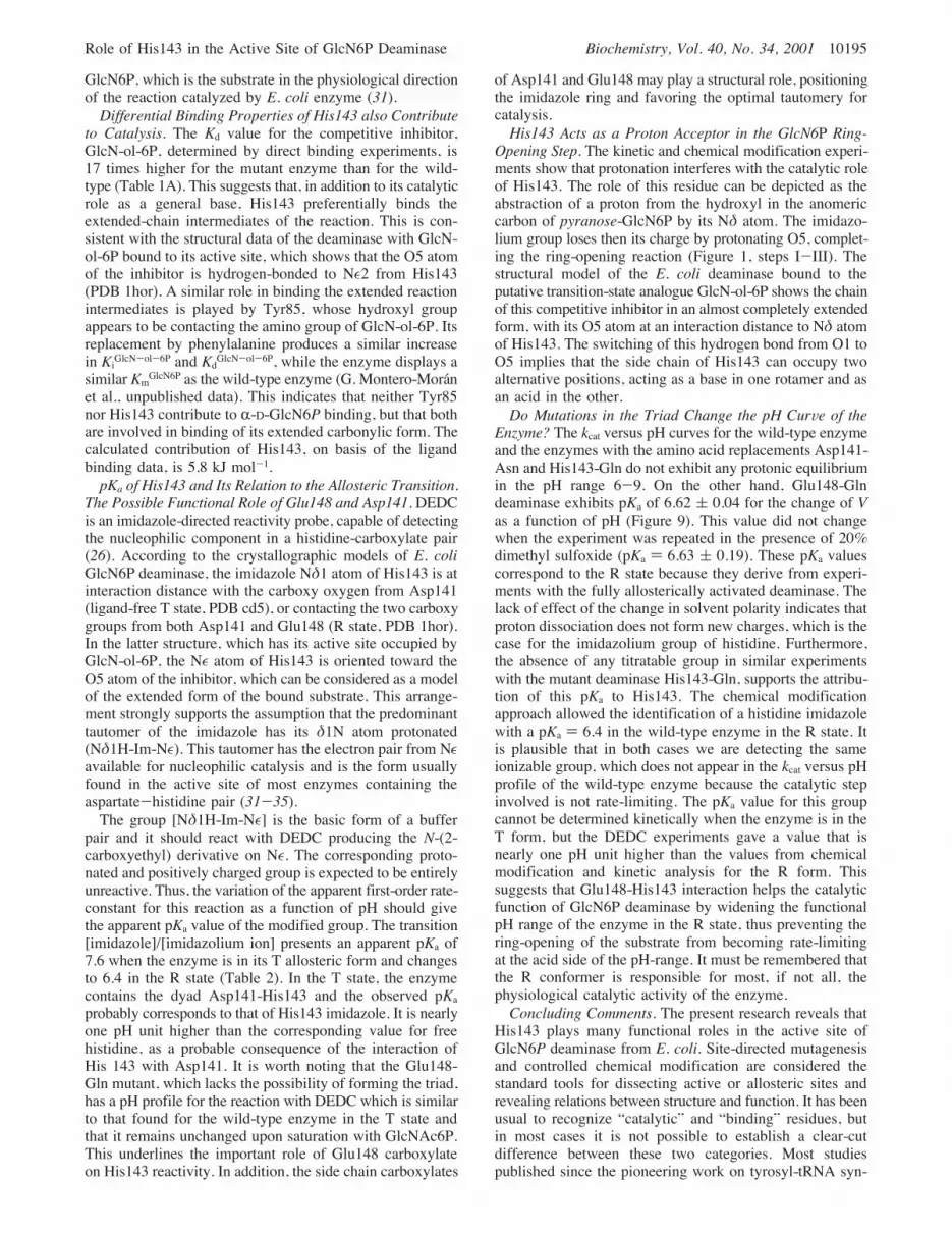
His143-Gln mutant enzyme when studied in the reversedirection of the reaction. Plots of velocity against substrateconcentration show substrate inhibition by ammonium ionand even by Fru6P, at high concentrations. To determinethe kcat value for the reverse reaction, we performed the
kinetic measurements out of the inhibitory range (Figure 6).Data were fitted to the following equation:
The kinetic parameters obtained are shown in Table 1C. Thefitted kcat value for the reverse reaction catalyzed by theHis143-Gln mutant, is 10.3 s-1, which is close to the valuefor the wild-type enzyme (15.2 s-1). This contrasts with thedecrease of 3 orders of magnitude in the kcat for the forwardreaction (Table 1B).Inhibition by Analogues of Fru6P. To know more about
the form of Fru6P interacting with the active site of theenzyme, we tested the following Fru6P analogues as potentialGlcN6P deaminase inhibitors in the forward direction of thereaction (Figure 7): (I) 2,5 anhydro-D-mannitol 6-P, whichis structurally similar to the "-anomer of furanose-Fru6P,the most abundant Fru6P anomer in solution (29); (II) TheO-methyl glycosides of Fru6P (mixed R- and "-isomers)which are structurally related to the corresponding anomersof furanose-Fru6P; (III) The oxime of Fru6P, (2-hydroxy-imino)-2-deoxy-D-arabino-hexitol 6-phosphate, which can beconsidered as an analogue of the open-chain form of thissubstrate. Its structure is also closely related to 2-deoxy-2-imino-D-arabino-hexitol 6-phosphate, a postulated reactionintermediate (VI in Figure 1). Oximes are formed in eitherZ or E isomers, but E forms are predominant (30). Thekinetic study of the wild-type GlcN6P deaminase, in thepresence of these compounds gave the inhibition constantsshown in Figure 7.pKa of the ActiVe Site Histidine and Its Modification by
the Allosteric Transition. GlcN6P deaminase largely existsin the T conformation and it crystallizes in this state in theabsence of ligands (5). This is reflected in the high valuefor the allosteric constant (L) 104) for the wild-type enzyme(7, 8). The allosteric equilibrium is shifted to the R state bysaturation with the allosteric activator, which binds exclu-sively to the R form (7). Taking advantage of this fact, wecould analyze the pH-dependence of the reaction rate ofGlcN6P deaminase with DEDC, when the enzyme is in eitherallosteric state. The treatment of the wild-type deaminasewith this reagent results in its complete inactivation. Whendata were corrected for DEDC hydrolysis (28), pseudo-first-order kinetics were always obtained. The number of chemi-cally modified histidine residues was determined spectro-photometrically; 4.9 residues per chain were ethoxycar-bonylated at pH 8.0 with a second-order rate-constant of 1.40( 0.3 M-1 s-1.Saturation with the competitive inhibitor, GlcN-ol-6P,
completely protects the enzyme activity against DEDCinactivation. On the other hand, displacing the allostericequilibrium toward the R state by saturation with GlcNAc6P,produces a partial protection (Figure 8). This is evidencefor the heterotropic conformational change at the active siteand shows that the empty active site in the R state is notclosed and inaccessible to DEDC.The spectrophotometric assay measures 3.6 residues modi-
fied by DEDC per chain in the GlcN-ol-6P protected GlcN6Pdeaminase. This modified enzyme displays similar kinetic
FIGURE 5: Initial velocities versus substrate concentration curvesfor the His143-Gln mutant form of GlcN6P deaminase. Data wereobtained in the absence (broken line, O) or in the presence (solidline, b) of 2 mM GlcNAc6P. Assays were performed using mutantGlcN6P deaminase (0.2 µM) in a final volume of 200 µL in 50mM Tris-HCl buffer (pH 7.7) at 30 °C, as described (7). Data werefitted to the hyperbola. The fitted kinetic parameters are shown inTable 1B.
V0 ) [kcat [Et][NH4+][Fru6P]]/[Km
[NH4+]Km[Fru6P] +
Km[Fru6P][NH4
+] + Km[NH4+][Fru6P] + [NH4
+][Fru6P]](1)
Role of His143 in the Active Site of GlcN6P Deaminase Biochemistry, Vol. 40, No. 34, 2001 10191
parameters and the same allosteric activation pattern as thewild-type protein. This implies that the residue whosechemical modification inactivates the enzyme and which isprotected by saturation with GlcN-ol-6P should be locatedat or near the active site. This is His143 because it is theonly histidine present in the active site. This result also provesthat the other four modified residues are irrelevant forenzyme function.
The pKa of the essential histidine residue was calculatedfrom the effect of pH on the rate of modification by DEDC.The apparent pseudo-first-order rate-constant of the reactionwith DEDC decreases with H+ ion concentration, but thecurve is not asymptotic to the abscissa and the kapp presentsa finite value at saturating [H+]. Calling k1 the pseudo-first-order rate constant independent of [H+], Ka the dissociationconstant of the group whose protonation causes the change
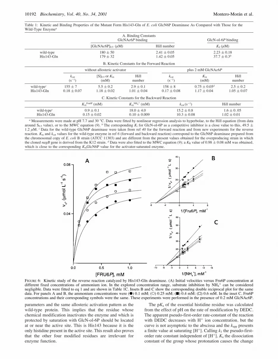
Table 1: Kinetic and Binding Properties of the Mutant Form His143-Gln of E. coli GlcN6P Deaminase As Compared with Those for theWild-Type Enzymea
A. Binding ConstantsGlcNAc6P binding GlcN-ol-6P binding
[GlcNAc6P]0.5 (µM) Hill number Kd (µM)wild-type 180 ( 50 2.41 ( 0.05 2.23 ( 0.18His143-Gln 179 ( 32 1.42 ( 0.05 37.7 ( 0.3b
B. Kinetic Constants for the Forward Reactionwithout allosteric activator plus 2 mM GlcNAc6P
kcat(s-1)
[S]0.5 or Km(mM)
Hillnumber
kcat(s-1)
Km(mM)
Hillnumber
wild-typec 155 ( 7 5.5 ( 0.2 2.9 ( 0.1 158 ( 8 0.75 ( 0.05d 2.5 ( 0.2His143-Gln 0.18 ( 0.07 1.18 ( 0.02 1.01 ( 0.04 0.17 ( 0.08 1.17 ( 0.04 1.05 ( 0.07
C. Kinetic Constants for the Backward ReactionKmFru6P (mM) KmNH4+ (mM) kcat (s-1) Hill number
wild-typec 0.9 ( 0.1 18.0 ( 4.0 15.2 ( 0.8 1.6 ( 0. 05His143-Gln 0.15 ( 0.02 0.10 ( 0.009 10.3 ( 0.08 1.02 ( 0.01
aMeasurements were made at pH 7.7 and 30 °C. Data were fitted by nonlinear regression analysis to hyperbolae, to the Hill equation (from dataaround S0.5 value), or to the MWC equation (9). b The corresponding Ki for GlcN-ol-6P as a competitive inhibitor is a close value to this, 49.5 (1.2 µM. c Data for the wild-type GlcN6P deaminase were taken from ref 40 for the forward reaction and from new experiments for the reversereaction. Km and kcat values for the wild-type enzyme in ref 6 (forward and backward reaction) correspond to the GlcN6P deaminase prepared fromthe chromosomal copy of E. coli B strain (ATCC 11303) and are different from the present values obtained for the overproducing strain in whichthe cloned nagB gene is derived from the K12 strain. d Data were also fitted to the MWC equation (9); a KR value of 0.98 ( 0.08 mM was obtained,which is close to the corresponding KmGlcN6P value for the activator-saturated enzyme.
FIGURE 6: Kinetic study of the reverse reaction catalyzed by His143-Gln deaminase. (A) Initial velocities versus Fru6P concentration atdifferent fixed concentrations of ammonium ion. In the explored concentration range, substrate inhibition by NH4+ can be considerednegligible. Data were fitted to eq 1 and are shown in Table 1C. Insets B and C show the corresponding double reciprocal plot for the samedata. For panels A and B, the ammonium concentrations were (b) 0.1 mM; (O) 0.25 mM; (9) 0.4 mM; (0) 0.6 mM. In the inset C, Fru6Pconcentrations and their corresponding symbols were the same. These experiments were performed in the presence of 0.2 mM GlcNAc6P.
10192 Biochemistry, Vol. 40, No. 34, 2001 Montero-Moran et al.

in kapp and k2 the limit value for kapp at infinite [H+], we canwrite the following expression for the pH dependence of thepseudo-first-order rate-constant:
It is at first sight surprising that protonation of the histidinedoes not completely stop its acylation by DEDC, because itdepends on the nucleophilic attack of the histidine residueon a carbonyl group of the reagent. Nevertheless, it ispossible that a nearby base, e.g., a carboxylate group, mayremove H+ from it. Thus, the reactive species could be thecarboxylate-imidazole pair -COO-‚‚‚Im-. This pair couldbe protonated with the observed molecular pKa, to form theequilibrium mixture of COOH‚‚‚Im-h -COO-‚‚‚ImH+-which will display a lower rate constant for reaction withDEDC. The pKa for the further protonation of this systemto yield -COOH‚‚‚ImH+- may well be too low to observe.Table 2 summarizes the results of these titration experi-
ments based on DEDC reactivity. The apparent pKa of thisgroup is 7.6 ( 0.2 when the enzyme is in the T state. Asimilar pKa value, which does not change upon GlcNAc6Paddition was found for the mutant Glu148-Gln. It should benoted that the wild-type enzyme in the T-state and theGlu148-Gln mutant, both lack the interaction His143-Glu148.On the other hand, the wild-type enzyme in the R statedisplays a pKa of 6.4. Similar measurements were performedusing the mutant forms Asp141-Asn and Asp141-Asn:Glu148-Gln. The single mutant Asp141-Asn does not changeits kinetic properties upon DEDC treatment, while the doublemutant Asp141-Asn:Glu148-Gln behaves similarly to Glu148-Gln deaminase, that is, it shows an apparent pKa of 7.7 thatdoes not change by saturation with GlcNAc6P (Table 2).These data indicate that Glu148 is the residue affecting thereactivity of His143 by changing its pKa from 7.6 in the Tstate to 6.4 in the R state.
Effect of the Mutations in Residues Asp141 and Glu148on the pH CurVe of the Enzyme. The replacement of eitherAsp141 or Glu148 by their corresponding amides affects thekcat values similarly. In both cases, they are in the range 6.6-7.8 s-1, that is 40 times higher than the value for the mutantforms involving His143, but 20 times lower than thecorresponding kcat for the wild-type enzyme (not shown).Both mutants display substrate inhibition, which was takeninto account for data fitting.The low kcat values of the His143-Gln enzyme were found
to be constant over the pH interval 6 to 9. Similarly, thewild-type enzyme does not reveal any protonic equilibriumin the same range (Figure 9A). In contrast, the mutationGlu148-Gln (Figure 9B) and the double mutation Asp141-Asn:Glu148-Gln (not shown) show a decrease of kcat valueson the acid side of the range. From the analysis of thesedata, a pKa of 6.62 ( 0.04 was obtained. These values didnot change in the presence of 2.8 M dimethyl sulfoxide. TheAsp141-Asn mutant did not present any apparent pKa overthe same pH range.
FIGURE 7: Some structural analogues of Fru6P that were tested asinhibitors of GlcN6P deaminase. I and III behave as weakcompetitive inhibitors with respect to GlcN6P, while IV is a stronginhibitor and probably corresponds to a transition state analogue.The corresponding Ki values are given.
kapp ) (k1Ka + k2[H+])/(Ka + [H+]) (2)
FIGURE 8: Effect of ligands on the inactivation of wild-type GlcN6Pdeaminase by DEDC. Semilogarithmic plot of the time-course ofthe fraction of remaining activity of the enzyme. This is defined asthe V/Vo ratio, where Vo is the maximal velocity determined in theabsence of ligands at zero time. (9) In the absence of any ligand,GlcN6P deaminase is totally inactivated by DEDC. (b) A saturatingconcentration of the dead-end inhibitor GlcN-ol-6P, fully protectedthe enzyme. (O) a saturating (2 mM) concentration of the allostericactivator GlcNAc6P slows the inactivation reaction.
Table 2: pKa Values of the Imidazole Group of the Active SiteHistidine (His143), Calculated from the Effect of [H+] on ItsReactivity toward DEDC
ligand-free(T state)
with 2 mM GlcNAc6P(R state)
wild-type enzyme 7.60 ( 0.2 6.40 ( 0.1His143-Gln no change no changeGlu148-Gln 7.56 ( 0.5 7.63 ( 0.4Asp141-Asn:Glu148-Gln 7.66 ( 0.58 7.70 ( 0.01Asp141-Asn no change no change
Role of His143 in the Active Site of GlcN6P Deaminase Biochemistry, Vol. 40, No. 34, 2001 10193

DISCUSSION
The Role of His143 in Functional Coupling of ActiVe andAllosteric Site. The substitution of the catalytic histidine bya glutamine residue produces marked functional changes inGlcN6P deaminase, with respect to the wild-type enzyme.The mutation impairs the enzyme activity and causes thecomplete loss of the homotropic cooperativity. The mutantenzyme behaves as if it were locked in the R state, displayinghyperbolic kinetics and a Km value for GlcN6P close to thatfor the wild-type in the R state (Table 1B). A saturatingconcentration of GlcNAc6P does not change the kinetics ofthis mutant deaminase, and both enzymes, wild-type andmutant, bind the allosteric activator with the same affinity(Table 1A). Some degree of cooperativity is present in theGlcNAc6P binding curve of the His143-Gln deaminase,indicating that the allosteric transition is not entirely abol-
ished by the mutation, even if active-site homotropic effectsare absent. It is possible that His143-Gln deaminase has astructure close to that of the wild-type R state. A crystal-lographic study could help to clarify this point. These resultsemphasize the importance of His143 and the flexible loopcontaining it, in coupling the local tertiary-structure changesin the active site, to the quaternary concerted transition.His143 Catalyzes the Ring-Opening of R-D-GlcN6P but
Does not Participate in Ring-Opening of the FuranoseFru6P. The mutation His143-Gln severely interferes withthe catalysis of the deamination reaction, as shown by thedecrease of 3 orders of magnitude in the corresponding kcatvalue. In contrast, the kcat for the reverse reaction is two-thirds the value for the wild-type enzyme (Table 1B), and itis close to the first-order rate-constant for the spontaneousring-opening of either R- or "-D-Fru6P, which is 20 s-1 (29).The most abundant Fru6P anomer in aqueous solution isR-D-Fru6P (mole fraction 0.81), while the rest is mainly "-D-Fru6P accompanied by a small concentration of the freecarbonylic species (mole fraction, 0.022), which must be thetrue substrate for the reverse reaction (Figure 1, VIII). Fromthe data in Table 1, we can see that the ratio Vforward/Vbackwardchanges from 10.2 for the wild-type enzyme to 0.02 for theHis143-Gln mutant. Binding of the aldehydo form of GlcN6P(Figure 1, IV), which is present in a small concentration atequilibrium, should easily occur (dotted arrows in Figure 1).The enzyme recognizes the open-chain analogues of thesubstrate, and it is expected that it also easily binds the openform of the substrate. After the rapid exhaustion of thisspecies, the spontaneous ring-opening in the solution, whichis very low (2), becomes the rate-limiting step for thecatalyzed forward reaction. Therefore, the contribution ofaldehydo-GlcN6P to the rate of the forward reaction isnegligible and uncatalyzed ring-opening does not contributeto the measured reaction rate. On the other hand, the absenceof the aldehydo-GlcN6P cyclization step does not affect therate of the reverse reaction, indicating that the open-chainGlcN6P can diffuse to the solution, where it slowly formsthe pyranose sugar (Figure 1).Additional evidence supporting the hypothesis that the
enzyme binds or releases the open carbonylic form of Fru6P,comes from inhibition experiments by Fru6P analogues(Figure 7). 2,5-Anhydro-D-mannitol 6-P, a close analogueof "-D-Fru6P, is a low-affinity competitive inhibitor, and theR- and "-O-methyl glycosides of Fru6P are not inhibitors atall. On the other hand, the oxime of Fru6P, which is ananalogue of its open form, inhibits competitively the enzymewith respect to GlcN6P, with a Ki of 1.7 mM. This oxime isstructurally similar to the reaction intermediate 2-deoxy-2-imino-D-arabino-hexitol 6-phosphate (VI, Figure 1), whichis a tautomer of the corresponding cis-enolamine (V).Midelfort and Rose (2) had proposed that this imineintermediate is the species adding a water molecule in thereaction. The inhibition by the analogue of intermediate VIsupports the presence of this intermediate in the reactionsequence.It is also worth mentioning that docking experiments on
the crystallographic model of the enzyme show that thefuranose form of Fru6P built into the active site, presentsunfavorable steric interactions with His143 (E. Rudino-Pineraand E. Horjales, personal communication). It is, after all,rationale that a ring-opening step has evolved only for
FIGURE 9: (A) kcat versus pH curves for the wild-type deaminase(O) and His143-Gln mutant (b). (B) kcat versus pH curves forGlu146-Gln deaminase. Inset: Double reciprocal plot in which theinhibition pattern by [H+] is shown. The [H+] explored were (4)3.20 ! 10-8 M; (2) 1 ! 10-8 M; (O) 6.3 ! 10-7 M; (b) 2.5 !10-7 M; (0) 5.01 ! 10-6 M; (9) 2.51 ! 10-6 M. Data were fittedto noncompetitive inhibition by nonlinear multiple regressionanalysis. The fitted value for the involved pKa is 6.63 ( 0.04 forthe Glu148-Gln mutant. The noncompetitive inhibition patternindicates that this protonic equilibrium affects only the kcat values.In the presence of 20% dimethyl sulfoxide, the pKa value forGlu148-Gln deaminase remains unchanged (6.63( 0.2). Essentiallysimilar results were obtained with the double mutant Asp141-Asn:Glu148-Gln (not shown).
10194 Biochemistry, Vol. 40, No. 34, 2001 Montero-Moran et al.
GlcN6P, which is the substrate in the physiological directionof the reaction catalyzed by E. coli enzyme (31).Differential Binding Properties of His143 also Contribute
to Catalysis. The Kd value for the competitive inhibitor,GlcN-ol-6P, determined by direct binding experiments, is17 times higher for the mutant enzyme than for the wild-type (Table 1A). This suggests that, in addition to its catalyticrole as a general base, His143 preferentially binds theextended-chain intermediates of the reaction. This is con-sistent with the structural data of the deaminase with GlcN-ol-6P bound to its active site, which shows that the O5 atomof the inhibitor is hydrogen-bonded to N!2 from His143(PDB 1hor). A similar role in binding the extended reactionintermediates is played by Tyr85, whose hydroxyl groupappears to be contacting the amino group of GlcN-ol-6P. Itsreplacement by phenylalanine produces a similar increasein KiGlcN-ol-6P and KdGlcN-ol-6P, while the enzyme displays asimilar KmGlcN6P as the wild-type enzyme (G. Montero-Moranet al., unpublished data). This indicates that neither Tyr85nor His143 contribute to R-D-GlcN6P binding, but that bothare involved in binding of its extended carbonylic form. Thecalculated contribution of His143, on basis of the ligandbinding data, is 5.8 kJ mol-1.pKa of His143 and Its Relation to the Allosteric Transition.
The Possible Functional Role of Glu148 and Asp141.DEDCis an imidazole-directed reactivity probe, capable of detectingthe nucleophilic component in a histidine-carboxylate pair(26). According to the crystallographic models of E. coliGlcN6P deaminase, the imidazole N!1 atom of His143 is atinteraction distance with the carboxy oxygen from Asp141(ligand-free T state, PDB cd5), or contacting the two carboxygroups from both Asp141 and Glu148 (R state, PDB 1hor).In the latter structure, which has its active site occupied byGlcN-ol-6P, the N! atom of His143 is oriented toward theO5 atom of the inhibitor, which can be considered as a modelof the extended form of the bound substrate. This arrange-ment strongly supports the assumption that the predominanttautomer of the imidazole has its !1N atom protonated(N!1H-Im-N!). This tautomer has the electron pair from N!available for nucleophilic catalysis and is the form usuallyfound in the active site of most enzymes containing theaspartate-histidine pair (31-35).The group [N!1H-Im-N!] is the basic form of a buffer
pair and it should react with DEDC producing the N-(2-carboxyethyl) derivative on N!. The corresponding proto-nated and positively charged group is expected to be entirelyunreactive. Thus, the variation of the apparent first-order rate-constant for this reaction as a function of pH should givethe apparent pKa value of the modified group. The transition[imidazole]/[imidazolium ion] presents an apparent pKa of7.6 when the enzyme is in its T allosteric form and changesto 6.4 in the R state (Table 2). In the T state, the enzymecontains the dyad Asp141-His143 and the observed pKaprobably corresponds to that of His143 imidazole. It is nearlyone pH unit higher than the corresponding value for freehistidine, as a probable consequence of the interaction ofHis 143 with Asp141. It is worth noting that the Glu148-Gln mutant, which lacks the possibility of forming the triad,has a pH profile for the reaction with DEDC which is similarto that found for the wild-type enzyme in the T state andthat it remains unchanged upon saturation with GlcNAc6P.This underlines the important role of Glu148 carboxylateon His143 reactivity. In addition, the side chain carboxylates
of Asp141 and Glu148 may play a structural role, positioningthe imidazole ring and favoring the optimal tautomery forcatalysis.His143 Acts as a Proton Acceptor in the GlcN6P Ring-
Opening Step. The kinetic and chemical modification experi-ments show that protonation interferes with the catalytic roleof His143. The role of this residue can be depicted as theabstraction of a proton from the hydroxyl in the anomericcarbon of pyranose-GlcN6P by its N! atom. The imidazo-lium group loses then its charge by protonating O5, complet-ing the ring-opening reaction (Figure 1, steps I-III). Thestructural model of the E. coli deaminase bound to theputative transition-state analogue GlcN-ol-6P shows the chainof this competitive inhibitor in an almost completely extendedform, with its O5 atom at an interaction distance to N! atomof His143. The switching of this hydrogen bond from O1 toO5 implies that the side chain of His143 can occupy twoalternative positions, acting as a base in one rotamer and asan acid in the other.Do Mutations in the Triad Change the pH CurVe of the
Enzyme? The kcat versus pH curves for the wild-type enzymeand the enzymes with the amino acid replacements Asp141-Asn and His143-Gln do not exhibit any protonic equilibriumin the pH range 6-9. On the other hand, Glu148-Glndeaminase exhibits pKa of 6.62 ( 0.04 for the change of Vas a function of pH (Figure 9). This value did not changewhen the experiment was repeated in the presence of 20%dimethyl sulfoxide (pKa ) 6.63 ( 0.19). These pKa valuescorrespond to the R state because they derive from experi-ments with the fully allosterically activated deaminase. Thelack of effect of the change in solvent polarity indicates thatproton dissociation does not form new charges, which is thecase for the imidazolium group of histidine. Furthermore,the absence of any titratable group in similar experimentswith the mutant deaminase His143-Gln, supports the attribu-tion of this pKa to His143. The chemical modificationapproach allowed the identification of a histidine imidazolewith a pKa ) 6.4 in the wild-type enzyme in the R state. Itis plausible that in both cases we are detecting the sameionizable group, which does not appear in the kcat versus pHprofile of the wild-type enzyme because the catalytic stepinvolved is not rate-limiting. The pKa value for this groupcannot be determined kinetically when the enzyme is in theT form, but the DEDC experiments gave a value that isnearly one pH unit higher than the values from chemicalmodification and kinetic analysis for the R form. Thissuggests that Glu148-His143 interaction helps the catalyticfunction of GlcN6P deaminase by widening the functionalpH range of the enzyme in the R state, thus preventing thering-opening of the substrate from becoming rate-limitingat the acid side of the pH-range. It must be remembered thatthe R conformer is responsible for most, if not all, thephysiological catalytic activity of the enzyme.Concluding Comments. The present research reveals that
His143 plays many functional roles in the active site ofGlcN6P deaminase from E. coli. Site-directed mutagenesisand controlled chemical modification are considered thestandard tools for dissecting active or allosteric sites andrevealing relations between structure and function. It has beenusual to recognize “catalytic” and “binding” residues, butin most cases it is not possible to establish a clear-cutdifference between these two categories. Most studiespublished since the pioneering work on tyrosyl-tRNA syn-
Role of His143 in the Active Site of GlcN6P Deaminase Biochemistry, Vol. 40, No. 34, 2001 10195

thetase by Fersht et al. (36) have shown that this functionaldissection is a difficult task because replacement of residuesstrictly labeled as binding have been shown to causesignificant changes in catalytic parameters. This is the casefor His143 in E. coli GlcN6P deaminase, and the presentresearch shows that this histidine plays multiple roles. Itparticipates in bond breaking and forming in ring-openingof R-D-GlcN6P and also contributes to catalysis by adifferential binding activity (37) in favor of the open-chainand extended reaction intermediates in the subsequent stepof the reaction. The present study also implicates His143and the loop containing it, in the transmission of conforma-tional changes between the active and the allosteric sites.We cannot quantify the contribution of this histidine to sitecoupling, but the analysis of the structural models showsthat this residue is the center of an intricate network ofinteractions in the R allosteric state, involving the active-site lid and the intersubunit contacts in the allosteric cleft.The loop 136-158 connects the sixth and seventh "-strandsin deaminase molecule (4). It contains the active-site triadas well as Ser151, one of the residues whose side-chaincontributes to bind the phospho-group in the allosteric site.The following short strand (158-161) contains the residuesArg158 and Lys160, which are also part of the phospho-group binding subsite in the allosteric site (Figure 4). It alsohas Thr161, which forms an intersubunit link in the allostericsite with Tyr254 of the facing subunit in the R state (8).His143 contacts one of the O# atoms of Glu148 in the Rstate, whose other O# forms hydrogen bonds with the O"atoms from Thr166 and Thr163 which are located in theactive site lid (segment 162-185) (observation communi-cated by E. Rudino-Pinera). In the T state, His143 retainsits contact with Asp141, and the displacement of the loop144-154 puts it in a more open and water-rich environment(5). Deciphering this web of multiple interactions to describecooperative effects in E. coli GlcN6P deaminase is achallenging task.
ACKNOWLEDGMENT
The authors gratefully acknowledge Juan-Pablo Pardo andRosario Munoz-Clares (National University of Mexico) andalso H.B.F. Dixon and Myriam M. Altamirano (CambridgeUniversity, UK) for stimulating and helpful discussions andthe critical reading of the manuscript. Enrique Rudino-Pineraand Eduardo Horjales contributed to this research withfruitful discussions and the communication of unpublishedcrystallographic results.
REFERENCES1. Comb, D. G., and Roseman, S. (1958) J. Biol. Chem. 232,807-827.
2. Midelfort, C., and Rose, I. A. (1977) Biochemistry 16, 1590-1596.
3. Rose, I. A. (1975) AdV. Enzymol. 43, 491-517.4. Oliva, G., Fontes, M. R. M., Garratt, R. C., Altamirano, M.M., Calcagno, M. L., and Horjales, E. (1995) Structure 3,1323-1332.
5. Horjales, E., Altamirano, M. M, Calcagno, M. L., Garratt R.C., Glaucius O. (1999) Structure 7, 527-537.
6. Calcagno, M. L., Campos, P. J., Mulliert, G., and Suastegui,J. (1984) Biochim. Biophys. Acta 787, 165-173.
7. Altamirano, M. M., Plumbridge, J. A., Horjales, E., andCalcagno, M. L. (1995) Biochemistry 34, 6074-6082.
8. Montero-Moran, G. M., Horjales E., Calcagno, M. L., andAltamirano, M. M. (1998) Biochemistry 37, 7844-7849.
9. Monod, J., Wyman, J., and Changeux, J. P. (1965) J. Mol.Biol. 12, 88-118.
10. Dodson, G., and Wlodaver, A. (1998) Trends Biol. Sci. 23,347-352.
11. Gulick, A. M., Hubbard, B. K., Gerlt, J. A., and Rayment, I.(2000) Biochemistry 39, 4590-602.
12. Gondry, M., and Lederer, F. (1996) Biochemistry 35, 8587-94.
13. Quirk, D. J., Park, C., Thompson, J. E., and Raines, R. T.(1998) Biochemistry 37, 17958-17964.
14. Malaisse-Lagae, F., Liemans, V., Yayali, B., Sener, A., andMalaisse, W. J. (1989) Biochim. Biophys. Acta 998, 118-125.
15. Tepliakov, A., Obmolova, G., Badet-Denisot, M. A., andBadet, B. (1999) Protein Sci. 8, 596-602.
16. Banerjee, S., Anderson, F., and Farber, G. K. (1995) ProteinEng. 8, 1189-1195.
17. Blow, D. M., Collyer, C. A., Goldberg, J. D., and Smart, O.S. (1992) Faraday Discuss. 93, 67-73.
18. Whitaker, R. B., Cho, Y., Cha, J., Carrell, H. L. Glusker, J.P., Karplus P. A., and Batt, C. A. (1995) J. Biol. Chem. 270,22895-22906.
19. Finch, P., and Merchant, Z. (1979) Carbohydr. Res. 76, 225-232.
20. Fishbein, R., Benkovic, P. A., Schray, K. J., Siewers, I. J.,Steffens, J. J., and Benkovic, S. J. (1974) J. Biol. Chem. 249,6047-6051.
21. Ben-Yoseph, O., Sparkes, M. J.; Dixon, H. B. F. (1993) Anal.Biochem. 210, 195-198.
22. Sambrook, J., Fritsch, E. F., and Maniatis, P. (1989) MolecularCloning, 2nd Ed., Cold Spring Harbor Laboratory Press,Plainview, NY.
23. Plumbridge, J. A. (1992) J. Gen. Microbiol. 138, 1011-1017.24. Ellis, K. J., and Morrison, J. F. (1982) Methods Enzymol. 87,
405-426.25. Howlett, G. J., Yeh, E., and Schachman, H. K. (1978) Arch.
Biochem. Biophys. 190, 809-819.26. Miles, E. W. (1977) Methods Enzymol. 47, 431-422.27. Berger, S. L. (1975) Anal. Biochem. 67, 428-437.28. Gomi, T., Fujioka, M. N. (1983) Biochemistry 22, 137-14329. Pierce, J., Serianni, A. S., and Barker, R. (1985) J. Am. Chem.
Soc. 107, 2448-2456.30. Bearne, S., and Blouin, C. (2000) J. Biol. Chem. 275, 135-
140.31. Vogler, A. P., Trentman, S., Lengeler, J. W. J. (1989)
Bacteriology 171, 6586-6592.32. Kossiakoff, A. A., and Spencer, S. A. (1981) Biochemistry
20, 6462-74.33. Bachovchin, W. W. (1985) Proc. Natl. Acad. Sci. U.S.A. 82,
7948-51.34. Bachovchin, W.W. (1986) Biochemistry 25, 7751-9.35. Ash E. L., Sudmeier J. L., De Fabo E. C., and Bachovchin
W. W. (1997) Science 278, 1128.36. Fersht, A. R., Shi, J. P., Knill-Jones, J., Lowe, D. M.,
Wilkinson, A. J., Blow, D. M., Brick, P., Carter, P., Waye,M. M., and Winter, G. (1985) Nature 314, 235-238.
37. Albery, W. J., and Knowles, J. R. (1976) Biochemistry 15,5631-5640.
38. Kraulis, P. (1991) J. Appl. Crystalogr. 24, 946-950.39. Merritt, E. A., and Bacon, D. J. (1997)Methods Enzymol. 277,
505-524.40. Lara-Gonzalez S., Dixon B. F. H., Mendoza-Hernandez G.,
Altamirano M. M., and Calcagno M. L. (2000) J. Mol. Biol.301, 219-228.
41. Guex, N., and Pertsch, M. C. (1997) Electrophoresis 18,2714-2723.
42. Corpet F. (1988) Nucleic Acids Res. 16, 10881-10890.
BI0105835
10196 Biochemistry, Vol. 40, No. 34, 2001 Montero-Moran et al.