enamel ultrastructure in fossil cetaceans (cetacea: archaeoceti and odontoceti)
TRANSCRIPT
RESEARCH ARTICLE
Enamel Ultrastructure in Fossil Cetaceans(Cetacea: Archaeoceti and Odontoceti)Carolina Loch1,2*, Jules A. Kieser2†, R. Ewan Fordyce1
1 Department of Geology, University of Otago, Dunedin, New Zealand, 2 Sir JohnWalsh Research Institute,Faculty of Dentistry, University of Otago, Dunedin, New Zealand
†Deceased.* [email protected]
AbstractThe transition from terrestrial ancestry to a fully pelagic life profoundly altered the body sys-
tems of cetaceans, with extrememorphological changes in the skull and feeding apparatus.
The Oligocene Epoch was a crucial time in the evolution of cetaceans when the ancestors of
modern whales and dolphins (Neoceti) underwent major diversification, but details of dental
structure and evolution are poorly known for the archaeocete-neocete transition. We report the
morphology of teeth and ultrastructure of enamel in archaeocetes, and fossil platanistoids and
delphinoids, ranging from late Oligocene (Waitaki Valley, New Zealand) to Pliocene (Caldera,
Chile). Teeth were embedded in epoxy resin, sectioned in cross and longitudinal planes, pol-
ished, etched, and coated with gold palladium for scanning electron microscopy (SEM) obser-
vation. SEM images showed that in archaeocetes, squalodontids and Prosqualodon (taxa with
heterodont and nonpolydont/limited polydont teeth), the inner enamel was organized in Hunt-
er-Schreger bands (HSB) with an outer layer of radial enamel. This is a common pattern in
most large-bodiedmammals and it is regarded as a biomechanical adaptation related to food
processing and crack resistance. Fossil Otekaikea sp. and delphinoids, which were polydont
and homodont, showed a simpler structure, with inner radial and outer prismless enamel. Radi-
al enamel is regarded as more wear-resistant and has been retained in several mammalian
taxa in which opposing tooth surfaces slide over each other. These observations suggest that
the transition from a heterodont and nonpolydont/limited polydont dentition in archaeocetes
and early odontocetes, to homodont and polydont teeth in crownward odontocetes, was also
linked to a marked simplification in the enamel Schmelzmuster. These patterns probably re-
flect functional shifts in food processing from shear-and-mastication in archaeocetes and early
odontocetes, to pierce-and-grasp occlusion in crownward odontocetes, with the implication of
less demanding feeding biomechanics as seen in most extant odontocetes.
IntroductionCetaceans—whales, dolphins—are unusual mammals that are adapted to a fully aquatic lifethrough a range of physiological and morphological modifications in respiration, circulation,
PLOSONE | DOI:10.1371/journal.pone.0116557 January 28, 2015 1 / 14
OPEN ACCESS
Citation: Loch C, Kieser JA, Fordyce RE (2015)Enamel Ultrastructure in Fossil Cetaceans (Cetacea:Archaeoceti and Odontoceti). PLoS ONE 10(1):e0116557. doi:10.1371/journal.pone.0116557
Academic Editor:Matthew C. Mihlbachler, NYIT Col-lege of Osteopathic Medicine, UNITED STATES
Received: July 23, 2014
Accepted: December 9, 2014
Published: January 28, 2015
Copyright: © 2015 Loch et al. This is an open ac-cess article distributed under the terms of theCreative Commons Attribution License, which permitsunrestricted use, distribution, and reproduction in anymedium, provided the original author and source arecredited.
Data Availability Statement: All relevant data arewithin the paper and its Supporting Information files.
Funding: CL received an University of Otago PhDscholarship and Sir John Walsh Research InstitutePostdoctoral Fellowship. The funders had no role instudy design, data collection and analysis, decision topublish, or preparation of the manuscript.
Competing Interests: The authors have declaredthat no competing interests exist.
feeding and locomotion [1]. The evolutionary history of the group began with the rise ofarchaeocetes (archaic stem Cetacea) in the ancient Tethys seaway in the Early Eocene morethan 50 million years ago [2]. The earliest fossils include amphibious species from Eocene sedi-ments of fluvial, brackish and fully marine origin. Crown cetaceans (Odontoceti and Mysticeti,or Neoceti) appeared probably in the late Eocene, by 35–36 Ma, and diversified rapidly duringthe Oligocene. Odontocetes and mysticetes most likely originated from a group of widespreadpelagic archaeocetes, the Basilosauridae [3,4].
Archaeocetes typically had elongated rostra bearing a heterodont dentition. The anteriorteeth were conical, high-crowned, and separated by prominent diastemata, while the posteriorteeth were lower-crowned, more-heavily built and, in the later-appearing group Basilosauridae,ornamented with multiple neomorphic denticles on the mesial and distal keels. Archaeoceteswere diphyodont, with no evidence for the polydonty seen in crown Cetacea [5]. The morphol-ogy of their teeth and feeding apparatus suggest that most archaeocetes could shear and alsosnap prey items [3,6].
Crown Cetacea (Mysticeti and Odontoceti) are polydont and monophyodont. Extantmysticetes, however, have their set of multiple tooth buds resorbed before birth and bear filter-ing baleen in their upper jaws from later foetal stage [7]. Most Odontoceti (fossil and living)have elongated rostra bearing a polydont dentition. Archaic odontocetes were heterodont andpolydont, with an accompanying trend towards homodonty and increased polydonty duringthe evolution of the group. In contrast, some fossil and extant odontocetes developed short,broad and robust rostra (e.g. extant Globicephala and extinct Prosqualodon), and reduced boththe number of teeth and the role of teeth in food processing (e.g. in the groups Ziphiidae,Kogiidae, Monodontidae, Globicephalinae) [3,8]. For most living odontocetes, the feedingapparatus is specialized for food acquisition, with little or no reduction and processing of foodin the oral cavity. Many odontocete species grasp and eat prey whole, but some species mayslash or tear pieces from large prey; many species may use suction feeding to catch andtransport food items [9]. The morphology of teeth and wear patterns suggest that archaicheterodont odontocetes used their teeth for more than just grasping and eating whole prey;teeth often functioned for the mechanical reduction of food items taken and for predation onhard food [3].
Teeth form a prominent part of mammal remains in paleontological and archaeologicalsites because of the good preservation potential conferred by high mineral content and densityof enamel and dentine compared to other skeletal elements. Further, teeth have long been usedto elucidate aspects of the ecology, functional morphology and systematics of fossil and recentmammal species [10]. Teeth play a major role in studies of dietary adaptations, and may eluci-date behaviors including social activities, defense and sexual signaling, representing a powerfultool in mammalian evolutionary biology [11,12].
Enamel is a highly mineralized tissue that forms the outermost layer of reptilian and mam-malian tooth crowns. Mammalian enamel is organized into bundles of bounded hydroxyapa-tite crystals known as enamel prisms, which can vary in morphology, diameter, density,patterns of organization, and packaging [13]. The 3-dimensional arrangement of enameltypes may vary between different taxa or different tooth types in heterodont species [13]. Thediversity of complex structures in mammalian enamel reflects many determinants, particularlybiomechanical/functional adaptation, geometric packing, and phylogeny. The relationship be-tween biomechanical stresses and enamel structures reflects functional constraints; howeverphylogeny may influence enamel differentiation and variability, as some specific structures areclosely correlated with particular mammalian taxa [14]. Many previous studies have sought todescribe the enamel ultrastructure in fossil mammals to address issues of systematics and func-tional morphology/lifestyle (e.g. [11,15–17]).
Enamel Ultrastructure in Fossil Cetaceans
PLOS ONE | DOI:10.1371/journal.pone.0116557 January 28, 2015 2 / 14
Structural changes in cetacean teeth occurred during adaptation of the skeletal and bodysystems to an increasingly pelagic lifestyle, as considered in other studies on cetacean teeth.The ultrastructure of enamel has been studied for Eocene basal archaeocetes [18–20], while theenamel of recent odontocetes was reportedly both prismatic and prismless [20–22]. Details arevirtually unknown for cetacean dental structure and evolution in Oligocene times, when therewas a major diversification of the Neoceti (Mysticeti and Odontoceti). The early echolocatingdolphins and filter feeding whales rapidly diversified in response to changing ocean ecosystemsand new ecological opportunities. A few relict Oligocene archaeocetes are also known. Modernfamilies of mysticetes and odontocetes appeared progressively from possibly the late Oligocene,and definitely the early Miocene to the Pliocene [3,4].
In spite of the ever-increasing diversity of fossil species, there are few published accountsof dental and enamel ultrastructure in fossil Neoceti. A major limitation in such work is theneed for destructive sampling. Well-preserved fossil cetacean skulls with teeth in place are un-common, scientifically valuable, and are not usually available from Museums for destructivesampling, as the literature on tooth structure reveals. Skulls are conserved because they are fun-damental to identify the species, and to use in studies of phylogeny and function. Isolatedteeth, conversely, are relatively common and more-readily available for destructive sectioning,but unless associated with a taxonomically-diagnostic skull, the exact species is commonly un-certain. Nevertheless, the genus or family can often be identified based on the similarity of anisolated tooth to teeth seen in situ in identifiable skulls, especially for more-archaic Cetaceawhich have feature-laden heterodont teeth, and especially for localities or strata which havealready produced diverse assemblages of reliably identified toothed cetaceans.
This study describes the arrangement and ultrastructure of teeth in 7 species of fossil ceta-ceans of Oligocene to Pliocene age. Our main focus is on enamel, with brief accounts on thestructure of dentine and on the diagenesis of tooth tissues. Functional aspects and biomechani-cal implications of the enamel arrangement in fossil cetaceans are examined and discussed,with reference to extant cetaceans and other mammals.
Material and Methods
Material ExaminedFossil cetacean teeth analyzed here are from archaeocetes, platanistoids and delphinoids,and were collected in strata ranging in age from late Oligocene (Waitaki Valley, New Zealand)to Pliocene (Caldera, Chile). Specimens were made available for destructive sampling fromthe collections of the Geology Museum, University of Otago (Dunedin, New Zealand—OU),Museo Nacional de Historia Natural de Chile (Santiago, Chile—SGO-PV) and MuseoPaleontológico Egidio Feruglio (Trelew, Argentina—MPEF-PV). One specimen belongs to theformally named species, Prosqualodon australis. For the other specimens not named tospecies-level (Table 1), the teeth of OU 22023, OU 22457, OU 22257, OU 22306, OU 22108and SGO-PV-754 were associated with taxonomically-diagnostic rostra or skulls and/ortympanoperiotics. A list of key features which diagnose the study teeth to family-level is in theSupporting Information (S1 Table). No permits were required for the described study, whichcomplied with all relevant regulations.
MethodsSilicone rubber molds of the study teeth were made, and epoxy resin replicas produced, to re-cord the original 3-dimensional structure before destructive sampling; replicas are held in theGeology Museum, University of Otago. Fossil cetacean teeth were surface-cleaned with alcoholand embedded in epoxy resin (Epofix Cold-Setting Embedding Resin, Struers, Copenhagen,
Enamel Ultrastructure in Fossil Cetaceans
PLOS ONE | DOI:10.1371/journal.pone.0116557 January 28, 2015 3 / 14
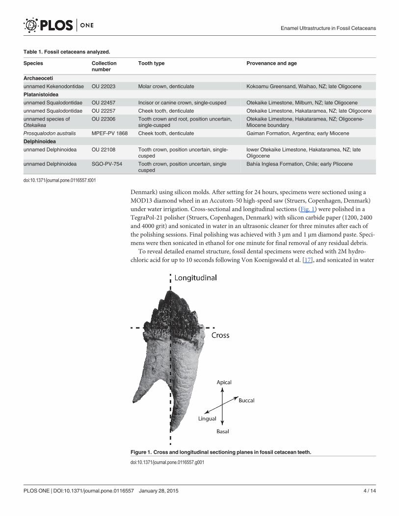
Denmark) using silicon molds. After setting for 24 hours, specimens were sectioned using aMOD13 diamond wheel in an Accutom-50 high-speed saw (Struers, Copenhagen, Denmark)under water irrigation. Cross-sectional and longitudinal sections (Fig. 1) were polished in aTegraPol-21 polisher (Struers, Copenhagen, Denmark) with silicon carbide paper (1200, 2400and 4000 grit) and sonicated in water in an ultrasonic cleaner for three minutes after each ofthe polishing sessions. Final polishing was achieved with 3 μm and 1 μm diamond paste. Speci-mens were then sonicated in ethanol for one minute for final removal of any residual debris.
To reveal detailed enamel structure, fossil dental specimens were etched with 2M hydro-chloric acid for up to 10 seconds following Von Koenigswald et al. [17], and sonicated in water
Table 1. Fossil cetaceans analyzed.
Species Collectionnumber
Tooth type Provenance and age
Archaeoceti
unnamed Kekenodontidae OU 22023 Molar crown, denticulate Kokoamu Greensand, Waihao, NZ; late Oligocene
Platanistoidea
unnamed Squalodontidae OU 22457 Incisor or canine crown, single-cusped Otekaike Limestone, Milburn, NZ; late Oligocene
unnamed Squalodontidae OU 22257 Cheek tooth, denticulate Otekaike Limestone, Hakataramea, NZ; late Oligocene
unnamed species ofOtekaikea
OU 22306 Tooth crown and root, position uncertain,single-cusped
Otekaike Limestone, Hakataramea, NZ; Oligocene-Miocene boundary
Prosqualodon australis MPEF-PV 1868 Cheek tooth, denticulate Gaiman Formation, Argentina; early Miocene
Delphinoidea
unnamed Delphinoidea OU 22108 Tooth crown, position uncertain, single-cusped
lower Otekaike Limestone, Hakataramea, NZ; lateOligocene
unnamed Delphinoidea SGO-PV-754 Tooth crown, position uncertain, singlecusped
Bahía Inglesa Formation, Chile; early Pliocene
doi:10.1371/journal.pone.0116557.t001
Figure 1. Cross and longitudinal sectioning planes in fossil cetacean teeth.
doi:10.1371/journal.pone.0116557.g001
Enamel Ultrastructure in Fossil Cetaceans
PLOS ONE | DOI:10.1371/journal.pone.0116557 January 28, 2015 4 / 14
for one minute, to avoid damage to fragile specimens. Samples were then coated with gold pal-ladium for scanning electron microscopy (SEM) observation. Secondary and backscatter elec-tron microscopic images were obtained in a JEOL JSM-6700F Field Emission SEM (JEOL Ltd.,Tokyo, Japan), operating at 5 kV and 10 μA. Magnifications in the SEM ranged from 30X to5000X.
Sampling practicalitiesPrevious studies of enamel ultrastructure in mammals have demonstrated that overall enamelorganization is generally consistent among individuals of the same species, but may varyamong species [15,19]. All taxa in this study were represented by a single specimen because ofthe rarity of fossils readily identifiable to taxon and also available for destructive sampling. Wepresume that the structural features described were typical of each species, but the conclusionswere made with some caution. Here we mainly analyzed the shape and size of prisms andprism sheaths, the spatial organization of prisms and interprismatic matrix, and the overall or-ganization of the enamel in each tooth (according to Carlson and Krause, Maas and Thewissen[15,19]). Anatomical terminology followed Von Koenigswald and Sander [23].
Results
MacromorphologyTeeth analyzed in this study consisted of diverse tooth types. Cheek teeth (premolar and molarequivalents) were analyzed for the Waihao Kekenodontidae (OU 22023), the HakatarameaSqualodontidae (OU 22257) and the Patagonian Prosqualodon australis (Fig. 2A–C). Theseteeth had a main cusp and small accessory denticles on the anterior (mesial) and posterior(distal) faces. The anterior face had fewer denticles than the posterior. The crown was bucco-lingually compressed and in the OU 22257 Squalodontidae and Prosqualodon it was orna-mented with cristae rugosae (sensu Rothausen [24]), which were more prominent at the base ofthe crown, where enamel nodules were formed. The other Squalodontidae specimen examined(Milburn tooth; OU 22457), was represented by an incisor crown which was triangular andsubconical, bucco-lingually compressed, with vertical subparallel ridges on the enamel, andwithout accessory denticles (Fig. 2D).
Less complex tooth morphologies were observed in Otekaikea sp. (cf. Squalodelphinidae)and Delphinoidea. Otekaikea sp. (OU 22306) had a slender simple crown, conical in shape,bucco-lingually compressed and gently curved lingually (Fig. 2E). The two Delphinoidea ana-lyzed (Hakataramea tooth—OU 22108; and SGO-PV-754) had a conical and featureless dentalcrown also curved lingually (Fig. 2F and G). The external surface of the enamel was featurelessat macroscopic scale in both Otekaikea sp. and the Delphinoidea specimens.
Enamel thickness and overall organizationEnamel thickness varied among the species analyzed (Table 2). The thinnest enamel was ob-served in the conical-crowned Otekaikea sp. OU 22306 and in the Delphinoidea OU 22108.Moderately thick enamel was observed both in the denticulate Kekenodontidae OU 22023and in the geologically young conical-crowned Chilean Delphinoidea SGO-PV-754. Thickerenamel was found in the squalodontids OU 22257 (denticulate crown, cheek-tooth) and OU22457 (conical crown, incisor), as well as in the denticulate Prosqualodon australis. On average,enamel thickness varied from 350–380 μm in these specimens, but it reached up to 580 μm inthickness in zones where enamel was heavily wrinkled to form strong subvertical ridges.
Enamel Ultrastructure in Fossil Cetaceans
PLOS ONE | DOI:10.1371/journal.pone.0116557 January 28, 2015 5 / 14

The enamel Schmelzmuster (the spatial distribution of enamel types) for the fossil cetaceansstudied, consisted of two different patterns of organization, both of them double-layered. ThePlatanistoidea and Kekenodontidae shared a Schmelzmuster consisting in an inner layer of
Figure 2. Specimens examined in lingual view. All fossils other than Prosqualodon australis have been coated with sublimed ammonium chloride. a)Unnamed Kekenodontidae (OU 22023). b) Prosqualodon australis (MPEF-PV 1868). c) Unnamed Squalodontidae (OU 22257). d) Unnamed Squalodontidae(OU 22457). e)Otekaikea sp. (cf. Squalodelphinidae) (OU 22306). f) Unnamed Delphinoidea (OU 22108). g) Unnamed Delphinoidea (SGO-PV-754). Scalebars = 1 cm. Images of Fig. 2A and 2C–F used with permission of Geology Museum, University of Otago.
doi:10.1371/journal.pone.0116557.g002
Table 2. Enamel thickness in the fossil cetaceans analyzed.
Species Collection number Enamel thickness
Archaeoceti
unnamed Kekenodontidae OU 22023 180–210 μm
Platanistoidea
unnamed Squalodontidae OU 22457 350–380 μm
unnamed Squalodontidae OU 22257 350–380 μm, up to 580 μm at location of enamel ridges
Otekaikea sp. OU 22306 75–85 μm
Prosqualodon australis MPEF-PV 1868 350–380 μm, up to 580 μm at location of enamel ridges
Delphinoidea
unnamed Delphinoidea OU 22108 100–125 μm
unnamed Delphinoidea SGO-PV-754 250–300 μm
doi:10.1371/journal.pone.0116557.t002
Enamel Ultrastructure in Fossil Cetaceans
PLOS ONE | DOI:10.1371/journal.pone.0116557 January 28, 2015 6 / 14
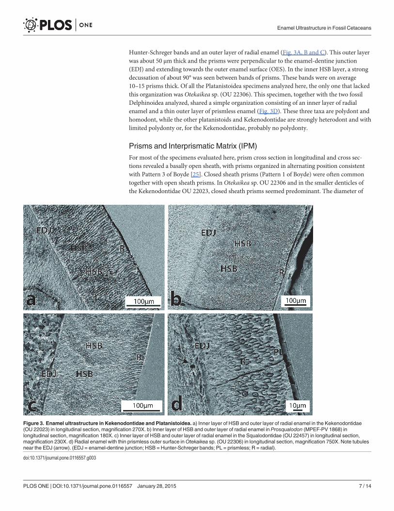
Hunter-Schreger bands and an outer layer of radial enamel (Fig. 3A, B and C). This outer layerwas about 50 μm thick and the prisms were perpendicular to the enamel-dentine junction(EDJ) and extending towards the outer enamel surface (OES). In the inner HSB layer, a strongdecussation of about 90° was seen between bands of prisms. These bands were on average10–15 prisms thick. Of all the Platanistoidea specimens analyzed here, the only one that lackedthis organization was Otekaikea sp. (OU 22306). This specimen, together with the two fossilDelphinoidea analyzed, shared a simple organization consisting of an inner layer of radialenamel and a thin outer layer of prismless enamel (Fig. 3D). These three taxa are polydont andhomodont, while the other platanistoids and Kekenodontidae are strongly heterodont and withlimited polydonty or, for the Kekenodontidae, probably no polydonty.
Prisms and Interprismatic Matrix (IPM)For most of the specimens evaluated here, prism cross section in longitudinal and cross sec-tions revealed a basally open sheath, with prisms organized in alternating position consistentwith Pattern 3 of Boyde [25]. Closed sheath prisms (Pattern 1 of Boyde) were often commontogether with open sheath prisms. In Otekaikea sp. OU 22306 and in the smaller denticles ofthe Kekenodontidae OU 22023, closed sheath prisms seemed predominant. The diameter of
Figure 3. Enamel ultrastructure in Kekenodontidae and Platanistoidea. a) Inner layer of HSB and outer layer of radial enamel in the Kekenodontidae(OU 22023) in longitudinal section, magnification 270X. b) Inner layer of HSB and outer layer of radial enamel in Prosqualodon (MPEF-PV 1868) inlongitudinal section, magnification 180X. c) Inner layer of HSB and outer layer of radial enamel in the Squalodontidae (OU 22457) in longitudinal section,magnification 230X. d) Radial enamel with thin prismless outer surface inOtekaikea sp. (OU 22306) in longitudinal section, magnification 750X. Note tubulesnear the EDJ (arrow). (EDJ = enamel-dentine junction; HSB = Hunter-Schreger bands; PL = prismless; R = radial).
doi:10.1371/journal.pone.0116557.g003
Enamel Ultrastructure in Fossil Cetaceans
PLOS ONE | DOI:10.1371/journal.pone.0116557 January 28, 2015 7 / 14
prisms varied from 3 to 5 μm both in open and closed sheath prisms. Interprismatic matrix(IPM) surrounded the prisms and IPM crystallites were parallel to one another, but at a slightangle to the prism long axes.
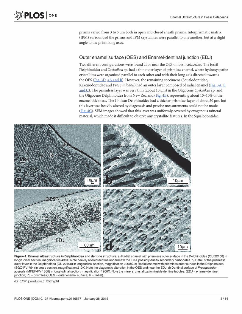
Outer enamel surface (OES) and Enamel-dentinal junction (EDJ)Two different configurations were found at or near the OES of fossil cetaceans. The fossilDelphinoidea andOtekaikea sp. had a thin outer layer of prismless enamel, where hydroxyapatitecrystallites were organized parallel to each other and with their long axis directed towardsthe OES (Fig. 3D, 4A and B). However, the remaining specimens (Squalodontidae,Kekenodontidae and Prosqualodon) had an outer layer composed of radial enamel (Fig. 3A, Band C). The prismless layer was very thin (about 10 μm) in the Oligocene Otekaikea sp. andthe Oligocene Delphinoidea from New Zealand (Fig. 4B), representing about 15–10% of theenamel thickness. The Chilean Delphinoidea had a thicker prismless layer of about 50 μm, butthis layer was heavily altered by diagenesis and precise measurements could not be made(Fig. 4C). SEM images showed that this layer was uniformly covered by exogenous mineralmaterial, which made it difficult to observe any crystallite features. In the Squalodontidae,
Figure 4. Enamel ultrastructure in Delphinoidea and dentine structure. a) Radial enamel with prismless outer surface in the Delphinoidea (OU 22108) inlongitudinal section, magnification 430X. Note heavily altered dentine underneath the EDJ, possibly due to secondary carbonates. b) Detail of the prismlessouter layer in the Delphinoidea (OU 22108) in longitudinal section, magnification 2200X. c) Radial enamel with prismless outer surface in the Delphinoidea(SGO-PV-754) in cross section, magnification 210X. Note the diagenetic alteration in the OES and near the EDJ. d) Dentinal surface of Prosqualodonaustralis (MPEF-PV 1868) in longitudinal section, magnification 1200X. Note the mineral crystallization inside dentine tubules. (EDJ = enamel-dentinejunction; PL = prismless; OES = outer enamel surface; R = radial).
doi:10.1371/journal.pone.0116557.g004
Enamel Ultrastructure in Fossil Cetaceans
PLOS ONE | DOI:10.1371/journal.pone.0116557 January 28, 2015 8 / 14
Prosqualodon and Kekenodontidae, the radial outer layer was about 50 μm thick, representingfrom 15 to about 25% of the enamel thickness.
The EDJ was characterized by a sharp and well-defined boundary. A thin layer of prismlessenamel a few μm thick was often just above the EDJ. Diagenetic alteration, indicated by exogenousmineralization, was often common at dentine underneath the EDJ zone (Fig. 3C and D, 4A).The subsurface mineralization was not studied in its own right, but surfaces of other teeth andbones sourced from the same strata as the studied teeth have shown secondary carbonates,phosphates, and iron and manganese minerals.
Tubules and tuftsTubules and tuft-like structures were seen at the EDJ in some fossil cetaceans both in cross-sectional and longitudinal sections (Fig. 3D). Tubules seen at the EDJ or at the basal enamelmeasured roughly 0.5 μm or less in diameter. Tuft-like structures were seen at the EDJ and ex-tended to the basal portion of enamel. Tufts were ribbon-like in shape and ran longitudinallytowards the OES. Tubules and tuft-like structures were often associated with diageneticchanges and cracks, probably acting as nuclei for later diagenetic changes.
DentineFossil cetacean dentine was heavily affected by diagenesis, mainly in the form of mineral in-fillings, mineral growth inside dentine tubules and loss of expected detail of dentine structure(Fig. 4A and D). In less altered regions, fossil dentine was relatively featureless with an unevensurface and irregularly distributed dentinal tubules measuring about 1 μm in diameter.
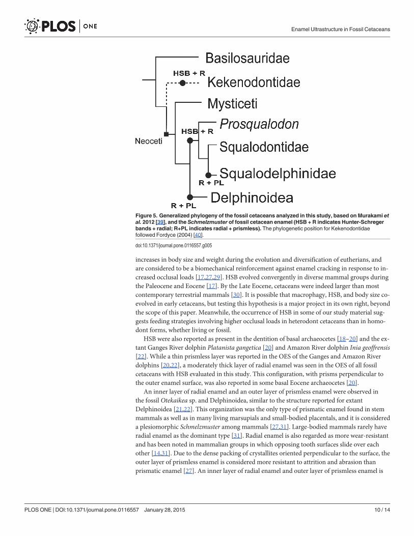
Enamel ultrastructure and phylogenetic patternPrismatic enamel was observed in all fossil specimens, with Pattern 3 (open) prisms and Pat-tern 1 (closed) predominant (patterns from Boyde [25]). At the Schmelzmuster level, two maintrends were evident when plotted on a simplified cetacean phylogeny (Fig. 5). An inner layerof HSB and an outer layer of radial enamel were observed in Kekenodontidae and in bothProsqualodon and Squalodontidae. Conversely, Otekaikea sp. (cf. Squalodelphinidae) and bothspecimens of Delphinoidea had a simpler Schmelzmuster composed of radial enamel and anouter layer of prismless enamel.
DiscussionThe dominant prism pattern observed in the fossil cetaceans was an open prism sheath, withprisms horizontally arranged in alternating positions. This arrangement is consistent with thePattern 3 of Boyde’s classification [25]. Closed prisms were sometimes found near the outerenamel surface (notably in Otekaikea sp. and Kekenodontidae), but always less frequent thanopen sheath prisms. This arrangement, in which open sheath prisms predominate with closedprisms scattered close to the tooth surface, is also a common trend recorded in many terrestrialmammals and archaeocete cetaceans [19,26,27]. The average prism size (maximum diameter)of about 5 μmwas similar among all fossil species, independent of differences in tooth size andpresumed body sizes, and it was consistent with what was reported for modern humans andother land mammals [28].
At the Schmelzmuster level, we observed a complex organization resulting from an innerlayer of HSB and an outer layer of radial enamel in the Kekenodontidae (Archaeoceti), and inthe Squalodontidae and Prosqualodon (Odontoceti). These three extinct taxa are heterodont,with differentiated teeth that include denticulate cheek teeth. HSB have been related to
Enamel Ultrastructure in Fossil Cetaceans
PLOS ONE | DOI:10.1371/journal.pone.0116557 January 28, 2015 9 / 14
increases in body size and weight during the evolution and diversification of eutherians, andare considered to be a biomechanical reinforcement against enamel cracking in response to in-creased occlusal loads [17,27,29]. HSB evolved convergently in diverse mammal groups duringthe Paleocene and Eocene [17]. By the Late Eocene, cetaceans were indeed larger than mostcontemporary terrestrial mammals [30]. It is possible that macrophagy, HSB, and body size co-evolved in early cetaceans, but testing this hypothesis is a major project in its own right, beyondthe scope of this paper. Meanwhile, the occurrence of HSB in some of our study material sug-gests feeding strategies involving higher occlusal loads in heterodont cetaceans than in homo-dont forms, whether living or fossil.
HSB were also reported as present in the dentition of basal archaeocetes [18–20] and the ex-tant Ganges River dolphin Platanista gangetica [20] and Amazon River dolphin Inia geoffrensis[22]. While a thin prismless layer was reported in the OES of the Ganges and Amazon Riverdolphins [20,22], a moderately thick layer of radial enamel was seen in the OES of all fossilcetaceans with HSB evaluated in this study. This configuration, with prisms perpendicular tothe outer enamel surface, was also reported in some basal Eocene archaeocetes [20].
An inner layer of radial enamel and an outer layer of prismless enamel were observed inthe fossil Otekaikea sp. and Delphinoidea, similar to the structure reported for extantDelphinoidea [21,22]. This organization was the only type of prismatic enamel found in stemmammals as well as in many living marsupials and small-bodied placentals, and it is considereda plesiomorphic Schmelzmuster among mammals [27,31]. Large-bodied mammals rarely haveradial enamel as the dominant type [31]. Radial enamel is also regarded as more wear-resistantand has been noted in mammalian groups in which opposing tooth surfaces slide over eachother [14,31]. Due to the dense packing of crystallites oriented perpendicular to the surface, theouter layer of prismless enamel is considered more resistant to attrition and abrasion thanprismatic enamel [27]. An inner layer of radial enamel and outer layer of prismless enamel is
Figure 5. Generalized phylogeny of the fossil cetaceans analyzed in this study, based on Murakami etal. 2012 [39], and the Schmelzmuster of fossil cetacean enamel (HSB + R indicates Hunter-Schregerbands + radial; R+PL indicates radial + prismless). The phylogenetic position for Kekenodontidaefollowed Fordyce (2004) [40].
doi:10.1371/journal.pone.0116557.g005
Enamel Ultrastructure in Fossil Cetaceans
PLOS ONE | DOI:10.1371/journal.pone.0116557 January 28, 2015 10 / 14
the most common Schmelzmuster in extant Delphinoidea; such dolphins commonly possess asimplified polydont-homodont dentition in which upper and lower teeth interdigitate to graspand secure prey but probably do not masticate [22].
In most of the specimens analyzed, dentine was characterized by subparallel tubules embed-ded in a matrix of intertubular dentine rich in fibers, which may represent collagen fibers thatbecame secondarily mineralized after burial [17,32]. At the EDJ, structures similar to tubulesand enamel tufts were often identified. In earlier studies, tubules were also recognized inarchaeocetes [20] and recent delphinoids [21,22], possibly related to a low degree of enamelmineralization at the EDJ. Tufts are also regarded as hypomineralized areas of enamel andhave been considered as intrinsic crack-like structures that play a major role in damage toler-ance and mechanical response due to occlusal loading [33].
The dentine and EDJ were areas that showed clear diagenetic alteration in most of the fossilspecimens analyzed. These alterations include recrystallization and presumed-secondary min-eral growths (mineral without obvious biogenic microstructure), commonly not observed inthe dentine and EDJ of extant cetacean teeth. The composition of these minerals was notdetermined, but there is independent evidence of secondary carbonates and phosphates in thesource rocks. The dentine and EDJ contain dentinal tubules, tuft-like structures, and tubules,all of which are known to act as conduits for diagenetic fluids during fossilization [17]. In thefossils, the enamel layer preserved microstructural details more reliably than dentine, butsome fossil specimens also had their enamel layer slightly altered by diagenesis (e.g. ChileanDelphinoidea, which is from a sequence known for its secondary phosphate mineralization).These modifications include fusing of enamel crystallites to form a uniform matrix that wasmore resistant to acid etching, particularly in the OES and close to the EDJ. However, besidesthese alterations, the overall enamel ultrastructure could be reasonably reconstructed in theChilean delphinoid.
Major changes in tooth morphology normally result in simplification of enamel microstruc-ture in response to changed biomechanical demands [14]. The simplification in tooth formduring the evolution of cetaceans was also reflected in the thickness of the enamel cover.For the fossil specimens analyzed here, enamel was moderately thick in the Squalodontidae,Prosqualodon and Chilean Delphinoidea (about 300–380 μm thick), but thinner in theKekenodontidae, Otekaikea sp., and New Zealand Delphinoidea (ranging from 75–200 μmthick). Thus, enamel thickness shows no strong correlation with phylogeny, or tooth-size, orheterodont versus homodont form. In Eocene archaeocetes, enamel thickness was reportedas ranging from 400–500 μm, with well-developed HSB and a wide variation of prism cross-section [19,20]. For most mammals, the presence of a thick enamel layer would enhance resis-tance to contact-induced fracture, and would prolong tooth lifetime in case of progressive wear[34]. Thus, the moderately thin layer of enamel in fossil cetaceans suggests limited utility infeeding and relaxed selection for tooth microstructure in comparison with other eutherians [9].
The occurrence of a double-layered Schmelzmuster consisting of an inner layer of HSB andan outer layer of radial enamel in the Kekenodontidae, Squalodontidae and Prosqualodon, allof which were heterodont, suggests that Oligocene archaeocetes and early odontocetes inher-ited the Schmelzmuster of more-basal archaeocetes [18–20]. These clades were heterodont andnon-polydont (kekenodontids), or heterodont with limited polydonty (Squalodontidae andProsqualodon). Archaeocetes and most stem Oligocene odontocetes probably used their anteri-or conical teeth to grasp and restrain prey, including fish and other marine vertebrates, whilethe low-crowned, laterally compressed, denticulate, and ornamented posterior teeth were pos-sibly used to slice and shear [6,35,36]. External surface ornamentation of the enamel seen inmany fossil cetaceans could help to grip and seize prey, but the functional and phylogenetic sig-nificance of ornament is still unknown. The heavily built, subtriangular, shark-like teeth of
Enamel Ultrastructure in Fossil Cetaceans
PLOS ONE | DOI:10.1371/journal.pone.0116557 January 28, 2015 11 / 14
squalodontids and other archaic odontocetes suggested to Kellogg (1928) a predaceous feedinghabit on bigger fish or even small pelagic mammals [37]. The rostrum in squalodontids and rel-atives was long and attenuated, suggesting a fast-snap but not necessarily a powerful bite. Theshort and robust rostrum of Prosqualodon would allow a more powerful bite although possiblyslower; probably allowing the removal of large chunks of prey [3].
A Schmelzmuster consisting of an inner layer of radial enamel and an outer layer ofprismless enamel was observed in the squalodelphinid-like dolphinOtekaikea sp. and inDelphinoidea, both of which are more-crownward than the Kekenodontidae, Squalodontidae, andProsqualodon. This configuration was also reported as common in extant Delphinida, a cladecharacterized by a polydont and homodont dentition [22]. Otekaikea sp. and the Delphinoideaspecimens analyzed here have tooth morphologies consistent with a grasping and piercingaction, but not mastication, implying limited food processing [36]. These odontocetes, withmany slender and pointed teeth, were probably raptorial predators which used their elongatepincer-like jaws and rostra to secure and pierce grasped prey. Prey items were probably swal-lowed after limited food processing, as seen in most extant Delphinida [9,38].
The transition from a heterodont and nonpolydont (or limited polydont) dentition inarchaeocetes and early odontocetes, to homodont and polydont teeth in more-crownwardodontocetes, also marked a simplification in the enamel Schmelzmuster. Such trends were likelyrelated to functional changes such as the lack of mastication-related occlusion and less con-strained feeding biomechanical demands in the fossil Otekaikea sp. and Delphinoidea, as seenin extant Delphinida [22]. These modifications involved particularly the shift in food process-ing from shear-and-mastication in archaeocetes and early odontocetes, to pierce-and-graspocclusion in most crownward cetaceans.
It is widely accepted that the evolution of mammalian enamel has been driven by a combi-nation of developmental and geometric constraints, functional influences and phylogenetichistory [27]. The phylogenetic significance of some enamel features might be obscured by ho-moplasy (e.g. convergent presence of HSB in different groups), but some evolutionary trendscan still be unveiled in clades with common functional adaptations [27]. Further studies con-sidering more specimens of late Oligocene and early Miocene odontocetes would help in eluci-dating how the transition from HSB to radial enamel has occurred and the role of phylogenyand dental functional biomechanics in the macro and ultrastructure of cetacean enamel.
Supporting InformationS1 Table. List of diagnostic characters used to identify species to family-level.(DOCX)
AcknowledgmentsC. Loch and R.E. Fordyce gratefully acknowledge the late Jules A. Kieser for his inspirationalmentoring and scientific insights throughout this research. His dynamic presence and generouscontribution at both academic and personal level are sorely missed and will never be forgotten.Thanks are also extended to David Rubilar-Rogers (MNCN, Santiago, Chile) and Pablo Puerta(MPEF, Trelew, Argentina) for allowing access to some specimens used in this study. CarolinaGutstein (Universidad de Chile) and Mario Cozzuol (UFMG, Brazil) facilitated the access tospecimens in Chile and Argentina. Yoshihiro Tanaka kindly agreed that one tooth from hisPhD study specimen of Otekaikea could be analyzed. New Zealand fossils were collected withsupport from National Geographic Society grants 4024–88 and 4341–90 to R.E. Fordyce.Ludwig Jansen van Vuuren (Oral Rehabilitation, University of Otago) kindly helped in the
Enamel Ultrastructure in Fossil Cetaceans
PLOS ONE | DOI:10.1371/journal.pone.0116557 January 28, 2015 12 / 14
sample embedding process and Brent Pooley (Department of Geology, University of Otago)was instrumental in sample polishing. Thanks also to Liz Girvan (OCEM, University of Otago)who provided technical assistance with SEM facilities. Alexander Werth, Mark Clementz andSteve Dawson provided valuable comments on early drafts of this manuscript. Wighart vonKoenigswald and two anonymous reviewers contributed important suggestions during thereview stage. C. Loch acknowledges the University of Otago for a PhD scholarship and the SirJohnWalsh Research Institute for a Postdoctoral Fellowship.
Author ContributionsConceived and designed the experiments: CL JAK REF. Performed the experiments: CL. Ana-lyzed the data: CL JAK REF. Contributed reagents/materials/analysis tools: CL JAK REF.Wrote the paper: CL JAK REF.
References1. Simpson GG (1945) The principles of classification and a classification of mammals. Bull AmMus Nat
Hist 85: 1–350.
2. Gingerich PD, ul Haq M, Zalmout IS, Khan IH, Malkani MS (2001) Origin of whales from earlyartiodactyls: hands and feet of Eocene Protocetidae from Pakistan. Science 293: 2239–2242. PMID:11567134
3. Fordyce RE, de Muizon C (2001) Evolutionary history of cetaceans: a review. In: Mazin J-M, deBuffrénil V, editors. Secondary adaptation of tetrapods to life in water. München: Verlag Dr. FriedrichPfeil. pp. 169–233.
4. Fordyce RE (2009) Cetacean evolution. In: PerrinWF, Würsig B, Thewissen JGM, editors. Encyclopediaof Marine Mammals, Second Edition. San Diego: Academic Press. pp. 201–207.
5. Uhen MD (2000) Replacement of deciduous first premolars and dental eruption in archaeocete whales.J Mammal 81: 123–133.
6. Fahlke JM, Bastl KA, Semprebon GM, Gingerich PD (2013) Paleoecology of archaeocete whalesthroughout the Eocene: dietary adaptations revealed by microwear analysis. Palaeogeog, Palaeoclimatol,Palaeoecol 386: 690–701.
7. Karlsen K (1962) Development of tooth germs and adjacent structures in the whalebone whale(Balaenoptera physalus (L.)). Hvalradets skrifter 45: 5–56.
8. Werth AJ (2006) Mandibular and dental variation and the evolution of suction feeding in Odontoceti.J Mammal 87: 579–588.
9. Werth AJ (2000) Feeding in marine mammals. In: Schwenk K, editor. Feeding: form, function and evolu-tion in tetrapod vertebrates. San Diego: Academic Press. pp. 487–526.
10. Bergqvist LP (2003) The role of teeth in mammal history. Braz J Oral Sci 2: 249–257.
11. Rensberger J, Pfretzschner HU (1992) Enamel structure in astrapotheres and its functional implica-tions. Scan Microsc 6: 495–508. PMID: 1462135
12. Ungar PS (2010) Mammal teeth: origin, evolution and diversity. Baltimore: The Johns Hopkins Univer-sity Press. p. 304.
13. Von Koenigswald W, ClemensW (1992) Levels of complexity in the microstructure of mammalianenamel and their application in studies of systematics. Scan Microsc 6: 195–218. PMID: 1626241
14. Von Koenigswald W (1997) Evolutionary trends in the differentiation of mammalian enamel ultrastruc-ture. In: Von Koenigswald W, Sander PM, editors. Tooth enamel microstructure. Rotterdam: Balkema.pp. 203–235.
15. Carlson SJ, Krause DW (1985) Enamel ultrastructure of multituberculate mammals: an investigation ofvariability. Cont Mus Paleo Univ Mich 27: 1–50.
16. Wood C, Dumont E, Crompton A (1999) New studies of enamel microstructure in Mesozoic mammals:a review of enamel prisms as a mammalian synapomorphy. J MammEvol 6: 177–213.
17. Von Koenigswald W, Kalthoff DC, Semprebon GM (2010) The microstructure of enamel, dentine andcementum in advanced Taeniodonta (Mammalia) with comments on their dietary adaptations. J VertPaleo 30: 1797–1804.
18. Sahni A (1981) Enamel ultrastructure of fossil Mammalia: Eocene Archaeoceti from Kutch. J PalaeoSoc India 25: 33–37.
Enamel Ultrastructure in Fossil Cetaceans
PLOS ONE | DOI:10.1371/journal.pone.0116557 January 28, 2015 13 / 14
19. MaasMC, Thewissen J (1995) Enamel microstructure ofPakicetus (Mammalia: Archaeoceti). J Paleontol:1154-1163.
20. Sahni A, Von Koenigswald W (1997) The enamel structure of some fossil and recent whales from theIndian subcontinent. In: Von Koenigswald W, Sander PM, editors. Tooth enamel microstructure.Rotterdam: Balkema. pp. 177–191.
21. Ishiyama M (1987) Enamel structure in odontocete whales. Scan Microsc 1: 1071–1079. PMID:3659855
22. Loch C, DuncanW, Simões-Lopes PC, Kieser JA, Fordyce RE (2013) Ultrastructure of enamel anddentine in extant dolphins (Cetacea: Delphinoidea and Inioidea). Zoomorphology 132: 215–225.doi: 10.1016/j.jsb.2013.11.006 PMID: 24287325
23. Von Koenigswald W, Sander PM (1997) Glossary of terms used for enamel microstructures. In VonKoenigswald W, Sander PM, editors. Tooth enamel microstructure. Rotterdam: Balkema. pp. 267–280.
24. Rothausen K (1968) Die systematische stellung der europäischen Squalodontidae (Odontoceti,Mamm.). Paläontologische Zeitschrift 42: 83–104.
25. Boyde A (1967) The development of enamel structure. Proc R Soc Med 60: 923–928.
26. Hillson S (2005) Teeth. Cambridge: Cambridge University Press. p. 373.
27. Maas MC, Dumont ER (1999) Built to last: the structure, function, and evolution of primate dentalenamel. Evol Anthropol 8: 133–152.
28. Nanci A (2007) Ten Cate’s Oral Histology: Development, Structure, and Function. Philadelphia:Elsevier Health Sciences. p. 418.
29. Von Koenigswald W, Pfretzschner H (1991) Biomechanics in the enamel of mammalian teeth. In:Schmidt-Kittler N, Vogel K, editors. Constructional morphology and evolution. Berlin: Springer-Verlag.pp. 113–125.
30. Gingerich PD (1998) Paleobiological perspectives on Mesonychia, Archaeoceti, and the origin ofwhales. In: Thewissen JGM, editor. The emergence of whales: Evolutionary patterns in the origin ofCetacea. New York: Springer. pp. 423–449.
31. Von Koenigswald W (2000) Two different strategies in enamel differentiation: Marsupialia versusEutheria. In: Teaford MF, Smith MM, Ferguson MW, editors. Development, function and evolution ofteeth. Cambridge: Cambridge University Press. pp. 107–118.
32. Boyde A (1980) Histological studies of dental tissues of odontocetes. Rep Int Whaling Comm SpecialIssue 3: 65–87.
33. Lawn BR, Lee JJ (2009) Analysis of fracture and deformation modes in teeth subjected to occlusalloading. Acta Biomater 5: 2213–2221. doi: 10.1016/j.actbio.2009.02.001 PMID: 19268644
34. Lucas P, Constantino P, Wood B, Lawn B (2008) Dental enamel as a dietary indicator in mammals.BioEssays 30: 374–385. PMID: 18348196
35. Carpenter K, White D (1986) Feeding in the archaeocete whale Zygorhiza kochii (Cetacea: Archaeoceti).Mississippi Geol 7: 1–14.
36. Ciampaglio C, Wray G, Corliss B (2005) A toothy tale of evolution: convergence in tooth morphologyamong marine Mesozoic-Cenozoic sharks, reptiles, and mammals. Sed Rec 3: 4–8. doi: 10.1371/journal.pone.0063586 PMID: 23874378
37. Kellogg R (1928) The history of whales-their adaptation to life in the water. Q Rev Biol 3: 29–76.
38. Tomilin AG (1957) Adaptive types in the Order Cetacea: The problem of an ecological classification ofCetacea. Can J Fish Aquat Sci 95: 1–13.
39. Murakami M, Shimada C, Hikida Y, Hirano H (2012) A new basal porpoise, Pterophocaena nishinoi(Cetacea, Odontoceti, Delphinoidea), from the upper Miocene of Japan and its phylogenetic relation-ships. J Vert Paleo 32: 1157–1171.
40. Fordyce RE (2004) The transition from Archaeoceti to Neoceti: Oligocene archaeocetes in the southwestPacific. J Vert Paleo 24, supplement 3A:59a.
Enamel Ultrastructure in Fossil Cetaceans
PLOS ONE | DOI:10.1371/journal.pone.0116557 January 28, 2015 14 / 14