a phase i multiple dose, dose escalation study of cg250 monoclonal antibody in patients with...
TRANSCRIPT
Cancer Immun 1424
Cancer Immunity (17 Submitted: 20 July 200
-9634Academy of Cancer Immunology
www.cancerimmunity.org
07071315487888
Article
A phase I multiple dose, dose escalation study of cG250 monoclonal antibody in patients with advanced renal cell carcinoma
Ian D. Davis1,2, Gregory A. Wiseman3, Fook-Thean Lee1, Denise N. Gansen3, Wendie Hopkins1, Anthony T. Papenfuss4, ZhanqiLiu1, Timothy J. Moynihan3, Gary A. Croghan3, Alex A. Adjei3, Eric W. Hoffman5, James N. Ingle3, Lloyd J. Old5 and Andrew M.Scott1,21Ludwig Institute for Cancer Research, Victoria, Australia2Austin Hospital, Victoria, Australia3Mayo Clinic, Rochester, MN, USA4Walter and Eliza Hall Institute of Medical Research, Victoria, Australia5Ludwig Institute for Cancer Research, New York, NY, USA
Contributed by: LJ Old
August 2007) Vol. 7, p. 137. Accepted: 20 July 2007.
Copyright © 2007 by Ian D. Davis
The chimeric monoclonal antibody cG250 recognises the G250/CAIX/MN antigen found on 95% of clear cell renal cell carcinomas (RCCs). We performed a phase I clinical trial to evaluate the safety, blood pharmacokinetics (PK), and biodistribution of repeated doses of cG250. The primary endpoint was toxicity. Secondary endpoints were cG250 biodistribution and PK; measurement of human anti-chimeric-antibodies (HACA); and tumour response rates. Eligible patients had unresectable or metastatic clear cell RCC. Doses of 5, 10, 25, or 50 mg/m2 were given weekly by intravenous infusion for six weeks. Three patients were treated at each dose level. Trace 131I-labelled cG250 was administered on weeks 1 and 5. Thirteen patients participated and were evaluable. One patient developed brain metastases and was replaced. No grade 3 or 4 toxicities and no dose-limiting toxicity occurred. One patient died due to progressive disease within 30 days of receiving the study drug. One patient developed HACA during the second six-week cycle. PK analysis showed mean whole body and blood alpha and beta half-lives of cG250 of 18.99 ± 6.84 and 180.19 ± 86.68 hours, respectively. All patients had cG250 tumour localization by gamma camera imaging in week 1 and 5. One patient had a complete response, nine patients had stable disease, and three had progressive disease. One patient received 11 six-week cycles of treatment with no toxicity or HACA. In conclusion, repeated intravenous doses of up to 50 mg/m2 of cG250 are safe. Furthermore cG250 has a long half-life and targets clear cell RCC effectively.
Keywords: phase I clinical trial, renal cell carcinoma, human CA9 protein, cG250, chimeric antibody
IntroductionRenal cell carcinoma (RCC) is a significant public health issue.
In 2007, over 51,000 people in the USA are expected to develop RCC or cancer of the renal pelvis, with more than 12,000 deaths predicted (1). Both the incidence of and mortality from RCC is increasing in frequency, although RCC is now found more commonly at an earlier stage (2). When resectable, five-year survival rates of more than 90% are seen (3); however, patients with metastatic disease have 5-year survival rates of approximately 30% (3) with a median survival of 10 months (4).
Until recently, no treatments have been shown to extend survival. Recent data suggest a survival advantage with the use of the mTOR inhibitor temsirolimus in highly selected patients (5) and significant improvements in progression-free survival with the receptor tyrosine kinase inhibitors sunitinib or sorafenib (6, 7). However, not all patients are suitable for or respond to these treatments and more effective therapies are still required for RCC.
The G250 antigen (carbonic anhydrase IX; CAIX, MN) is found on more than 85% of RCCs and particularly on clear cell RCCs (8). G250 is a heat-sensitive transmembrane cell-surface antigen with homology to carbonic anhydrase (9). G250 is also expressed on gastric epithelium, biliary ducts, and some pancreatic acini (10, 11). G250 expression is linked to overexpression of hypoxia-related molecules such as HIF-1α(12). Expression of G250/CAIX correlates with responsiveness to IL-2 therapy and to longevity of survival after therapy, independently of tumour grade and stage (13).
The murine monoclonal IgG1 antibody G250 recognises the G250 antigen and has been used in clinical trials (11, 14, 15). However, repeated doses of this antibody were not possible due to development of human anti-mouse antibody (HAMA) responses. cG250 is an IgG1 kappa chimeric antibody (16) with identical specificity to the murine antibody. cG250 has been shown to have intrinsic immune effector function on renal cell carcinoma cell lines, which suggests a further potential mechanism of action for an anti-tumour effect (17). cG250, either alone or conjugated with radioisotopes, has been used in several clinical trials in various doses and schedules (18-24). Much of this work has been reviewed previously (15). In general, these trials showed that cG250 or its labelled derivates are well tolerated, minimally immunogenic in terms of development of human anti-chimeric antibody (HACA) responses, and able to target clear cell RCC efficiently. Localization of the antibody to the tumour [up to 0.52% injected dose per gram (ID/gm)] was amongst the highest reported for a monoclonal antibody directed to a solid tumour (18). In these studies, activity of cG250 was evident with stable disease in some patients, and three partial remissions and one complete remission have been reported (19, 23). Despite its expression on biliary epithelium,
1 of 8
Cancer Immunity (17 August 2007) Vol. 7, p. 13
Table 1Patient characteristics.
radiolabelled cG250 does not bind significantly to biliary tract cancers (25).
In view of the promising results of targeting of cG250 to RCC, we performed a phase I clinical trial to determine the safety of repeated doses of cG250. Secondary objectives were to evaluate pharmacokinetic, biodistribution and immunological effects, as well as antitumour effects. This study was performed between late 1999 and early 2001. Early results of this study have already been presented in abstract form (26).
ResultsPatients
Patient characteristics are shown in Table 1. All patients had metastatic clear cell renal cell carcinoma but were not required to have documentation of progressive disease at study entry. Eight patients participated at Mayo Clinic and five at Austin Hospital. The male:female ratio was 8:5 and all but two patients were of Caucasian background. The median age at study entry was 64 (range 46-73). Most patients were Karnofsky performance status 90-100%; one patient was 80% at study entry. A total of 11/13 patients had received prior therapy. One patient (patient 9) was replaced due to the development of brain metastases during the first treatment cycle after three weeks of treatment.
ToxicityToxicities are summarized in Table 2. In general, treatment
with cG250 was very well tolerated and most toxicities were grade 1. Patients 3, 4 and 7 experienced pain at tumour sites during infusion of cG250 that was of mild or moderate severity and was thought possibly or probably related to study drug; these patients were treated at the 5, 10 and 25 mg/m2 dose levels respectively. No grade 3 or 4 toxicities were observed, although one patient had grade 3 bone pain thought due to the underlying disease. Serious adverse events occurred in five patients, including one fatal event (acute renal failure) occurring after the patient had completed the study. This patient had pre-existing
grade 2 renal impairment that worsened during the study, not reaching grade 3 until after completion of the study. The decline in renal function was shown to be due to progressive cancer. All serious adverse events were deemed unrelated or unlikely related to cG250.
Table 2 Summary of toxicities.
Biodistribution of cG250
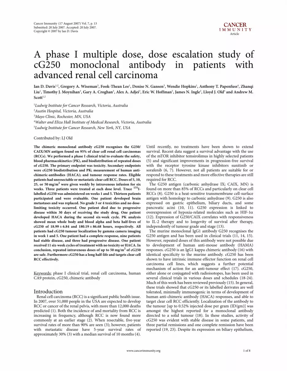
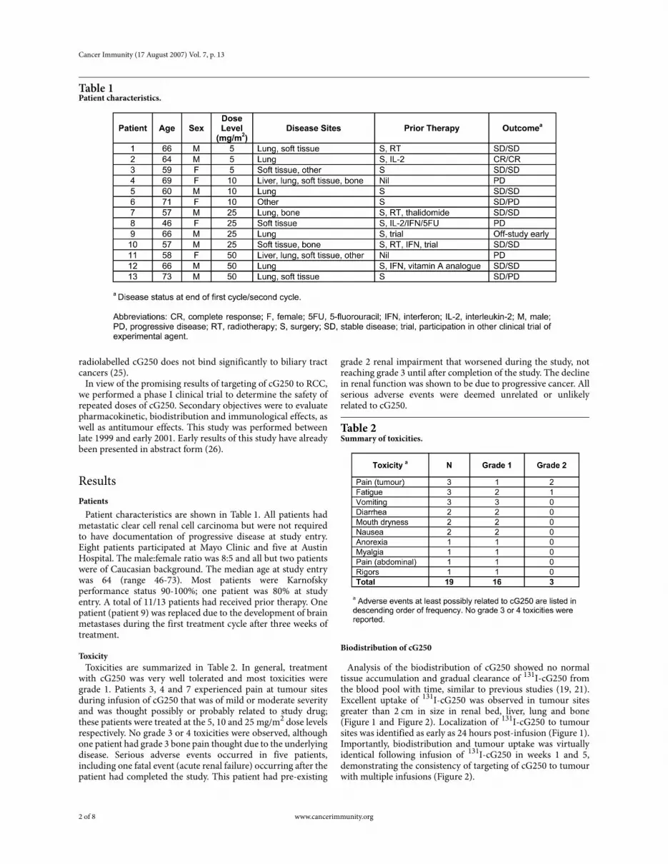
Analysis of the biodistribution of cG250 showed no normal tissue accumulation and gradual clearance of 131I-cG250 from the blood pool with time, similar to previous studies (19, 21). Excellent uptake of 131I-cG250 was observed in tumour sites greater than 2 cm in size in renal bed, liver, lung and bone (Figure 1 and Figure 2). Localization of 131I-cG250 to tumour sites was identified as early as 24 hours post-infusion (Figure 1). Importantly, biodistribution and tumour uptake was virtually identical following infusion of 131I-cG250 in weeks 1 and 5, demonstrating the consistency of targeting of cG250 to tumour with multiple infusions (Figure 2).
2 of 8 www.cancerimmunity.org
Davis et al.
Figure 1
Gamma camera biodistribution images of 131I-cG250. (A) Whole body anterior image showing uptake of 131I-cG250 in hepatic metastases (arrows), also evident on CT scan. Some bowel excretion of 131I is also evident. (B) Whole body anterior image showing uptake showing uptake of 131I-cG250 in a lung metastasis (arrow), seen also on chest radiograph.
Figure 2
Consistent targeting of 131I-cG250 to metastatic disease in the sacrum (arrow) with multiple infusions. Consistent targeting is demonstrated following infusion 1 (A) anterior and (B) posterior images, and infusion 5 (C) anterior and (D) posterior images. (E) SPECT image in the same transaxial plane as (F) CT scan also shows excellent targeting to metastatic disease in the sacrum in this patient. The patient had stable disease at restaging.
www.cancerimmunity.org 3 of 8
Cancer Immunity (17 August 2007) Vol. 7, p. 13
Table 3Pharmacokinetic parameters for 131I-cG250.
Table 4 cG250 ELISA: Cmax and Cmin cG250 serum levels.
Pharmacokinetics of cG250Thirteen patients were evaluable for pharmacokinetics.
Pharmacokinetic analyses were performed by measurement of serum 131I activity and by ELISA assay of serum cG250. Summarized data from the first cycle of treatment for infusions 1 and 5 of 131I-cG250 are presented in Table 3, and ELISA peak and trough values for all infusions are presented in Table 4.
No statistical differences were found between dose levels for the mean (± SD) values of T½α, T½β, CL or MRT for 131I-cG250 (ANOVA, P > 0.112). The V1 (volume of central compartment) and Vss (volume of distribution at steady state) results also showed no differences between protein dose levels (Table 3) and were consistent with a two-compartment model for pharmacokinetic analysis.
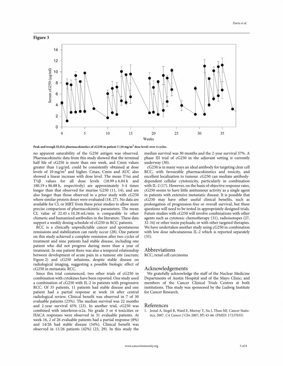
Cmax was calculated from the radiolabelled cG250 pharmacokinetics based upon the protein dose infused for infusions 1 and 5 (Table 3). Serum cG250 Cmax and Cmin were also determined directly by ELISA, and mean ± SD values for all infusions are shown in Table 4. Although some variation in the peak serum level results was evident when individual patient data were compared between the two methods, no statistical difference was observed between either method for any dose level for infusion 1 (P > 0.226; data not shown). The Cmax and AUC values showed a linear relationship to protein dose of cG250 (Table 3 and Table 4). Infusions of cG250 within a cycle were associated with increasing values for Cmax and Cmin (Figure 3). The Cmin data results revealed that the 10 mg/m2
dose level produced serum values consistently greater than 1 µg/
mL in all patients at one week post-infusion of cG250, which is a concentration that reproducibly produces antibody-dependent cellular-cytotoxicity of G250 target cells in in vitro assays (17).
HACAOne patient developed low titer detectable HACA activity
(50 ng/mL) at the end of the second cycle of treatment and was removed from the study. No further patients developed any detectable HACA. HACA was not associated with any clinical sequelae in this patient.
Clinical outcomesA total of 34 cycles of treatment were administered to the 13
patients on this study. One patient received two cycles of treatment at the 5 mg/m2 dose level and had a complete response. Eight patients had stable disease for at least one cycle, although this commonly occurs spontaneously in RCC and cannot necessarily be attributed to the study drug. One of these patients maintained stable disease for a total of 11 cycles of treatment before progressing (66 weeks). Three patients had progressive disease after one cycle and were removed from the study. Another patient (patient 9) was removed from the study due to the development of brain metastases before completing one cycle of treatment and was replaced.
DiscussionThis phase I study has shown that treatment of patients with
metastatic clear cell RCC with cG250 was safe and well tolerated. The frequency of adverse events did not correlate with the doses of cG250 used. One patient developed low level HACA with no adverse clinical outcome and was removed from the study. Importantly, patients were able to receive multiple cycles of cG250 without significant toxicity or development of clinically significant HACA. This is similar to other unconjugated cG250 trials; however HACA appears to be more common in studies using iodinated cG250, for reasons that remain unclear.
Biodistribution studies of 131I-cG250 showed excellent targeting of sites of RCC. When 131I-cG250 was administered in weeks 1 and 5, gamma camera imaging showed tumour localization of cG250 in all patients. Uptake of 131I-cG250 in tumour was similar despite disparate locations of tumour, and
4 of 8 www.cancerimmunity.org
Davis et al.
Figure 3
Peak and trough ELISA pharmacokinetics of cG250 in patient 5 (10 mg/m2 dose level) over 4 cycles.
no apparent saturability of the G250 antigen was observed. Pharmacokinetic data from this study showed that the terminal half life of cG250 is more than one week, and Cmin values greater than 1 µg/mL could be consistently obtained at dose levels of 10 mg/m2 and higher. Cmax, Cmin and AUC also showed a linear increase with dose level. The mean T½α and T½β values for all dose levels (18.99 ± 6.84 h and 180.19 ± 86.88 h, respectively) are approximately 3-4 times longer than that observed for murine G250 (11, 14), and are also longer than those observed in a prior study with cG250 where similar protein doses were evaluated (18, 27). No data are available for CL or MRT from these prior studies to allow more precise comparison of pharmacokinetic parameters. The mean CL value of 22.45 ± 10.28 mL/min is comparable to other chimeric and humanized antibodies in the literature. These data support a weekly dosing schedule of cG250 in RCC patients.
RCC is a clinically unpredictable cancer and spontaneous remissions and stabilization can rarely occur (28). One patient on this study achieved a complete remission after two cycles of treatment and nine patients had stable disease, including one patient who did not progress during more than a year of treatment. In one patient there was also a temporal relationship between development of acute pain in a tumour site (sacrum; Figure 2) and cG250 infusions, despite stable disease on radiological imaging, suggesting a possible biologic effect of cG250 in metastatic RCC.
Since this trial commenced, two other trials of cG250 in combination with cytokines have been reported. One study used a combination of cG250 with IL-2 in patients with progressive RCC. Of 35 patients, 11 patients had stable disease and one patient had a partial response at week 16 after central radiological review. Clinical benefit was observed in 7 of 30 evaluable patients (23%). The median survival was 22 months and 2-year survival 45% (23). In another trial, cG250 was combined with interferon-α2a. No grade 3 or 4 toxicities or HACA responses were observed in 31 evaluable patients. At week 16, 2 of 26 evaluable patients had a partial response (8%) and 14/26 had stable disease (54%). Clinical benefit was observed in 11/26 patients (42%) (23, 29). In this study the
median survival was 30 months and the 2-year survival 57%. A phase III trial of cG250 in the adjuvant setting is currently underway (30).
cG250 is in many ways an ideal antibody for targeting clear cell RCC, with favourable pharmacokinetics and toxicity, and excellent localization to tumour. cG250 can mediate antibody-dependent cellular cytotoxicity, particularly in combination with IL-2 (17). However, on the basis of objective response rates, cG250 seems to have little antitumour activity as a single agent in patients with extensive metastatic disease. It is possible that cG250 may have other useful clinical benefits, such as prolongation of progression-free or overall survival, but these questions will need to be tested in appropriately designed trials. Future studies with cG250 will involve combinations with other agents such as cytotoxic chemotherapy (31), radioisotopes (27, 32-34) or other toxin payloads; or with other targeted therapies. We have undertaken another study using cG250 in combination with low dose subcutaneous IL-2 which is reported separately (35).
AbbreviationsRCC, renal cell carcinoma
AcknowledgementsWe gratefully acknowledge the staff of the Nuclear Medicine
Departments of Austin Hospital and of the Mayo Clinic; and members of the Cancer Clinical Trials Centres at both institutions. This study was sponsored by the Ludwig Institute for Cancer Research.
References1. Jemal A, Siegel R, Ward E, Murray T, Xu J, Thun MJ. Cancer Statis-
tics, 2007. CA Cancer J Clin 2007; 57: 43-66. (PMID: 17237035)
www.cancerimmunity.org 5 of 8
Cancer Immunity (17 August 2007) Vol. 7, p. 13
2. Pantuck AJ, Zisman A, Belldegrun AS. The changing natural history of renal cell carcinoma. J Urol 2001; 166: 1611-1623. (PMID: 11586189)
3. Tsui KH, Shvarts O, Smith RB, Figlin RA, deKernion JB, BelldegrunA. Prognostic indicators for renal cell carcinoma: a multivariate analysis of 643 patients using the revised 1997 TNM staging criteria.J Urol 2000; 163: 1090-1095. (PMID: 10737472)
4. Motzer RJ, Mazumdar M, Bacik J, Berg W, Amsterdam A, Ferrara J. Survival and prognostic stratification of 670 patients with advanced renal cell carcinoma. J Clin Oncol 1999; 17: 2530-2540. (PMID: 10561319)
5. Hudes G, Carducci M, Tomczak P, Dutcher J, Figlin R, Kapoor A, Staroslawska E, Sosman J, McDermott D, Bodrogi I, Kovacevic Z, Lesovoy V, Schmidt-Wolf IG, Barbarash O, Gokmen E, O'Toole T, Lustgarten S, Moore L, Motzer RJ ; Global ARCC Trial. Temsiroli-mus, interferon alfa, or both for advanced renal-cell carcinoma. N Engl J Med 2007; 356: 2271-2281. (PMID: 17538086)
6. Motzer RJ, Hutson TE, Tomczak P, Michaelson MD, Bukowski RM, Rixe O, Oudard S, Negrier S, Szczylik C, Kim ST, Chen I, BycottPW, Baum CM, Figlin RA. Sunitinib versus interferon alfa in meta-static renal-cell carcinoma. N Engl J Med 2007; 356: 115-124. (PMID: 17215529)
7. Escudier B, Eisen T, Stadler WM, Szczylik C, Oudard S, Siebels M, Negrier S, Chevreau C, Solska E, Desai AA, Rolland F, Demkow T, Hutson TE, Gore M, Freeman S, Schwartz B, Shan M, Simantov R, Bukowski RM; TARGET Study Group. Sorafenib in advanced clear-cell renal-cell carcinoma. N Engl J Med 2007; 356: 125-134. (PMID: 17215530)
8. Bui MH, Seligson D, Han KR, Pantuck AJ, Dorey FJ, Huang Y, Hor-vath S, Leibovich BC, Chopra S, Liao SY, Stanbridge E, Lerman MI, Palotie A, Figlin RA, Belldegrun AS. Carbonic anhydrase IX is an independent predictor of survival in advanced renal clear cell carci-noma: implications for prognosis and therapy. Clin Cancer Res 2003; 9: 802-811. (PMID: 12576453)
9. Uemura H, Nakagawa Y, Yoshida K, Saga S, Yoshikawa K, Hirao Y, Oosterwijk E. MN/CA IX/G250 as a potential target for immuno-therapy of renal cell carcinomas. Br J Cancer 1999; 81: 741-746. (PMID: 10574265)
10. Oosterwijk E, Ruiter DJ, Hoedemaeker PJ, Pauwels EK, Jonas U, Zwartendijk J, Warnaar SO. Monoclonal antibody G250 recognizes a determinant present in renal-cell carcinoma and absent from nor-mal kidney. Int J Cancer 1986; 38: 489-494. (PMID: 2428759)
11. Oosterwijk E, Bender NH, Divgi CR, Welt S, Wakka JC, Finn RD, Carswell EA, Larson SM, Warnaar SO, Fleuren GJ, Oettgen HF, OldLJ. Antibody localization in human renal cell carcinoma: a phase I study of monoclonal antibody G250. J Clin Oncol 1993; 11: 738-750. (PMID: 8478666)
12. Grabmaier K, A de Weijert MC, Verhaegh GW, Schalken JA, Oost-erwijk E. Strict regulation of CAIX (G250/MN) by HIF-1alpha in clear cell renal cell carcinoma. Oncogene 2004; 23: 5624-5631. (PMID: 15184875)
13. Atkins M, Regan M, McDermott D, Mier J, Stanbridge E, YoumansA, Febbo P, Upton M, Lechpammer M, Signoretti S. Carbonic anhy-
drase IX expression predicts outcome of interleukin 2 therapy for renal cancer. Clin Cancer Res 2005; 11: 3714-3721. (PMID: 15897568)
14. Divgi CR, Bander NH, Scott AM, O'Donoghue JA, Sgouros G, WeltS, Finn RD, Morrissey F, Capitelli P, Williams JM, Deland D, Nakhre A, Oosterwijk E, Gulec S, Graham MC, Larson SM, Old LJ. Phase I/II radioimmunotherapy trial with iodine-131-labeled mon-oclonal antibody G250 in metastatic renal cell carcinoma. Clin Cancer Res 1998; 4: 2729-2739. (PMID: 9829736)
15. Oosterwijk E, Divgi CR, Brouwers A, Boerman OC, Larson SM, Mulders P, Old LJ. Monoclonal antibody-based therapy for renal cell carcinoma. Urol Clin North Am 2003; 30: 623-631. (PMID: 12953760)
16. Surfus JE, Hank JA, Oosterwijk E, Welt S, Lindstrom MJ, AlbertiniMR, Schiller JH, Sondel PM. Anti-renal-cell carcinoma chimeric antibody G250 facilitates antibody-dependent cellular cytotoxicity with in vitro and in vivo interleukin-2-activated effectors. J Immu-nother Emphasis Tumor Immunol 1996; 19: 184-191. (PMID: 8811493)
17. Liu Z, Smyth FE, Renner C, Lee FT, Oosterwijk E, Scott AM. Anti-renal cell carcinoma chimeric antibody G250: cytokine enhance-ment of in vitro antibody-dependent cellular cytotoxicity. Cancer Immunol Immunother 2002; 51: 171-177. (PMID: 11941456)
18. Steffens MG, Boerman OC, Oosterwijk-Wakka JC, Oosterhof GO, Witjes JA, Koenders EB, Oyen WJ, Buijs WC, Debruyne FM, Cor-stens FH, Oosterwijk E. Targeting of renal cell carcinoma with iodine-131-labeled chimeric monoclonal antibody G250. J Clin Oncol 1997; 15: 1529-1537. (PMID: 9193349)
19. Steffens MG, Boerman OC, de Mulder PH, Oyen WJ, Buijs WC, Witjes JA, van den Broek WJ, Oosterwijk-Wakka JC, Debruyne FM, Corstens FH, Oosterwijk E. Phase I radioimmunotherapy of meta-static renal cell carcinoma with 131I-labeled chimeric monoclonal antibody G250. Clin Cancer Res 1999; 5(10 Suppl): 3268s-3274s. (PMID: 10541374)
20. Varga Z, de Mulder P, Kruit W, Hegele A, Hofmann R, Lamers C, Warnaar S, Mala C, Ullrich S, Mulders P. A prospective open-label single-arm phase II study of chimeric monoclonal antibody cG250 in advanced renal cell carcinoma patients. Folia Biol (Praha) 2003; 49: 74-77. (PMID: 12779016)
21. Divgi CR, O'Donoghue JA, Welt S, O'Neel J, Finn R, Motzer RJ, Jungbluth A, Hoffman E, Ritter G, Larson SM, Old LJ. Phase I clini-cal trial with fractionated radioimmunotherapy using 131I-labeled chimeric G250 in metastatic renal cancer. J Nucl Med 2004; 45: 1412-1421. (PMID: 15299069)
22. Bleumer I, Knuth A, Oosterwijk E, Hofmann R, Varga Z, Lamers C, Kruit W, Melchior S, Mala C, Ullrich S, De Mulder P, Mulders PF, Beck J. A phase II trial of chimeric monoclonal antibody G250 for advanced renal cell carcinoma patients. Br J Cancer 2004; 90: 985-990. (PMID: 14997194)
23. Neville N, Bevan P, Klöpfer P, Mala C, Hofmann R, Kindler M, Sie-bels M, Oberneder R. Treatment with monoclonal antibody cG250 (Rencarex®) in combination with IFNα-2a significantly prolongs survival in patients with metastatic renal cell cancer patients. 5th Kidney Cancer Symposium 2006; September 22-23; Chicago (IL).
6 of 8 www.cancerimmunity.org
Davis et al.
24. Divgi CR, Pandit-Taskar N, Jungbluth AA, Reuter VE, Gönen M, Ruan S, Pierre C, Nagel A, Pryma DA, Humm J, Larson SM, Old LJ, Russo P. Preoperative characterisation of clear-cell renal carcinoma using iodine-124-labelled antibody chimeric G250 (124I-cG250) and PET in patients with renal masses: a phase I trial. Lancet Oncol2007; 8: 304-310. (PMID: 17395103)
25. Hendrickx BW, Punt CJ, Boerman OC, Postema EJ, Oosterwijk E, Mavridu A, Corstens FH, Oyen WJ. Targeting of biliary cancer with radiolabeled chimeric monoclonal antibody CG250. Cancer Biother Radiopharm 2006; 21: 263-268. (PMID: 16918303)
26. Wiseman GA, Scott AM, Lee FT, Gansen W, Hopkins W, SteinmetzS, Ingle JN, Croghan GA, Burch PA, Davis I, Moynihan TJ, WarnaarS, Ullrich S, Mason JV, Pugliese L, Divgi CR, Hoffman EW, Old LJ. Chimeric G250 (cG250) monoclonal antibody phase I dose escala-tion trial in patients with advanced renal cell carcinoma (RCC).Proc Am Soc Clin Oncol 2001; May 12-15; San Francisco (CA); 257a. Abstract 1027.
27. Brouwers AH, Mulders PF, de Mulder PH, van den Broek WJ, BuijsWC, Mala C, Joosten FB, Oosterwijk E, Boerman OC, Corstens FH, Oyen WJ. Lack of efficacy of two consecutive treatments of radio-immunotherapy with 131I-cG250 in patients with metastasized clear cell renal cell carcinoma. J Clin Oncol 2005; 23: 6540-6548. (PMID: 16170161)
28. Gleave ME, Elhilali M, Fradet Y, Davis I, Venner P, Saad F, KlotzLH, Moore MJ, Paton V, Bajamonde A. Interferon gamma-1b com-pared with placebo in metastatic renal-cell carcinoma. N Engl J Med1998; 338: 1265-1271. (PMID: 9562580)
29. Bevan P, Mala C, Kindler M, Siebels M, Oberneder R, Beck HJ. Results of a phase I/II study with monoclonal antibody CG250 in combination with IFN α-2a in metastatic renal cell carcinoma patients. J Clin Oncol 2004; 22(14S): 4606.
30. WILEX AG - Pipeline. Accessed 18 July 2007. URL: http://www.wilex.com/R&D/Pipeline.htm
31. Al-Batran SE, Neumann A, Atmaca A, Ruppert M, Karbach J, RitterG, Hoffman E, Old L, Knuth A, Jaeger E. LUD01-014: Phase 1/2 study of chimeric monoclonal antibody cG250 in combination with vinblastine in patients with advanced renal cell carcinoma (ARCC).J Clin Oncol 2004; 22(14S): 2531.
32. Brouwers AH, Buijs WC, Mulders PF, de Mulder PH, van den BroekWJ, Mala C, Oosterwijk E, Boerman OC, Corstens FH, Oyen WJ. Radioimmunotherapy with [131I]cG250 in patients with metasta-sized renal cell cancer: dosimetric analysis and immunologic response. Clin Cancer Res 2005; 11: 7178s-7186s. (PMID: 16203819)
33. Brouwers AH, Buijs WC, Oosterwijk E, Boerman OC, Mala C, De Mulder PH, Corstens FH, Mulders PF, Oyen WJ. Targeting of meta-static renal cell carcinoma with the chimeric monoclonal antibody G250 labeled with 131I or 111In: an intrapatient comparison. Clin Cancer Res 2003; 9: 3953s-3960s. (PMID: 14506194)
34. Postema EJ, Frielink C, Oyen WJ, Raemaekers JM, Goldenberg DM, Corstens FH, Boerman OC. Biodistribution of 131I-, 186Re-, 177Lu-, and 88Y-labeled hLL2 (Epratuzumab) in nude mice with CD22-positive lymphoma. Cancer Biother Radiopharm 2003; 18: 525-533. (PMID: 14503946)
35. Davis ID, Liu Z, Saunders W, Lee FT, Spirkoska V, Hopkins W, Smyth FE, Chong G, Papenfuss AT, Chappell B, Poon A, SaunderTH, Hoffman EW, Old LJ, Scott AM. A pilot study of monoclonal antibody cG250 and low dose subcutaneous IL-2 in patients with advanced renal cell carcinoma. Cancer Immun 2007; 7: 14. URL: http://www.cancerimmunity.org/v7p14/070714.htm
36. Uemura H, Beniers AJ, Okajima E, Debruyne FM, Oosterwijk E. Vaccination with anti-idiotype antibodies mimicking a renal cell carcinoma-associated antigen induces tumor immunity. Int J Cancer1994; 58: 555-561. (PMID: 8056452)
Materials and methodsTrial design
Trial LUD98-011 was an open-label, non-randomized, dose-escalation phase 1 study. Four cohorts of three patients were planned to receive weekly doses of cG250 for six weeks, with escalation of the dose of cG250 between cohorts. The primary objective was safety. Secondary objectives were to evaluate pharmacokinetic parameters (biodistribution, 131I-cG250 serum concentration, half-life, area under the curve, concentration-time curve), immunological effects (HACA), and antitumour effects (response rate).
Eligibility criteria included the following. Inclusion criteria: unresectable or metastatic clear cell renal cell carcinoma; Karnofsky performance status of 70% or more; life expectancy of more than 12 weeks; adequate major organ function; 18 years of age or more. Patients were not required to have measurable or evaluable disease or to have progressive disease at study entry. Exclusion criteria: active central nervous system metastases (unless adequately treated and stable); chemotherapy, immunotherapy, biologic therapy, or radiation therapy within four weeks prior to study entry; prior antibody exposure (unless no evidence of HAMA, HACA or HAHA); failure to fully recover from effects of prior cancer therapy; concurrent use of systemic corticosteroids or immunosuppressive agents; uncontrolled infection or other serious disease; pregnancy or lactation; women of childbearing potential not using medically acceptable means of contraception. The trial protocol was approved by the Human Research Ethics Committees of Austin Health and of the Mayo Clinic. The trial was performed under the Australian Therapeutic Goods Administration Clinical Trials Notification (CTN) scheme and FDA IND BB-8023. All patients were treated at the Austin Hospital in Melbourne, Australia, or at the Mayo Clinic, Rochester, MN, USA.
The following dose levels were planned: cG250 doses of 5, 10, 25, or 50 mg/m2. Doses were administered weekly by i.v. infusion for 6 weeks. Patients were then observed for toxicities weeks 7-10 and tumour response was evaluated at week 8 using WHO guidelines. The first and fifth weekly doses were trace-labelled with 131I to facilitate the evaluation of biodistribution and pharmacokinetics of cG250. Patients received potassium iodide solution, 10 drops orally twice a day for seven days, beginning on the morning of the 131I-cG250 infusions (day 1 and 29). If no dose-limiting toxicity (DLT) was observed within 28 days of the first dose of cG250 in three patients in a cohort, escalation to the next dose level would occur. Dose modifications of cG250 (reduction in infusion rate or cessation of infusion) were permitted if adverse events occurred during the infusion. Patients who were stable, responding clinically or by assessment of measurable disease during the study or at re-staging (week 8), and who had not developed HACA were
www.cancerimmunity.org 7 of 8
Cancer Immunity (17 August 2007) Vol. 7, p. 13
eligible for continued weekly treatment at the dose they were initially receiving. Treatment beyond four cycles was approved where applicable on a case-by-case basis, depending upon treatment response, absence of HACA, and availability of study drug.
Blood samples were collected before treatment and at various time points during the study as described below. Tumour evaluations were performed prior to treatment and two weeks after the sixth dose.
131I-cG250 biodistribution
Clinical grade cG250 was obtained from the Biological Production Facility, Ludwig Institute for Cancer Research, Melbourne, Australia. Infusions of cG250 trace-labelled with 200-320 MBq (5-8 mCi) of 131I were administered in weeks 1 and 5 for determination of tumour targeting and normal organ biodistribution, and pharmacokinetics. Gamma camera imaging with anterior and posterior whole body scans using conjugate view methodology were performed on five occasions after each trace-labelled infusion: within two hours after 131I-cG250 infusion (day 1) and on day 2, days 3 or 4, 5 or 6 and 7 or 8. A standard was included in the field of view at each imaging time point. Single-photon emission computed tomographic (SPECT) imaging of relevant areas of disease were performed on at least one occasion following 131I-cG250 infusion. All data were archived in digital format in a secure electronic database.
Pharmacokinetic studiesPharmacokinetics of cG250 were determined by measurement
of radioactivity in blood samples after 131I-cG250 infusion (weeks 1 and 5). Blood samples for these assays were drawn in weeks 1 and 5 on the day of infusion (immediately prior to the infusion, 10 min, 1 h, 2 h and 4 h after completion of the infusion), day 2, days 3 or 4, 5 or 6 and 7 or 8, in conjunction with gamma camera imaging. Blood samples for measurement of cG250 protein in blood by ELISA were drawn prior to each infusion and 10 minutes after completion of each infusion.
Samples (5 mL of clotted blood) were collected in an unheparinized (clotted) tube, centrifuged at 400 x g for 10 minutes, serum pipetted into a cryovial and stored at -70°C to -80°C for analysis of serum cG250 and calculation of pharmacokinetic parameters after each patient had completed the study. Measurement of cG250 in serum was performed by ELISA. In brief, serum samples were incubated in anti-cG250 anti-idiotype NUH82-coated microtiter plates. After washing, biotinylated-NH82 was used as tracer antibody. Serial dilutions of cG250 were used to obtain a standard curve. The lower quantitation limit was 30 ng/L cG250. All results were expressed as mean ± standard deviation in µg/mL.
Serum obtained from patients following infusion of radiolabelled cG250 was aliquoted in duplicate and counted in a gamma counter. Triplicate standards prepared from the injected material were counted for 131I at each time point with serum samples to enable calculations to be corrected for the isotope’s physical decay. The results of the serum counting were expressed as % injected dose per liter (%ID/L), and converted to µg/mL for pharmacokinetic calculations.
Pharmacokinetic parameters were estimated from radiolabelled patient serum clearance data using a curve fitting program (WinNonlin, Pharsight Co., Mountain View, CA). A two-compartment model was used to calculate pharmacokinetic parameters of Cmax (maximum serum concentration); AUC (area under the serum concentration curve extrapolated to
infinite time); CL (total serum clearance); T½α and T½β (half lives of the initial and terminal phases of disposition); and V1(volume of central compartment).
HACAPatient serum samples for HACA assessment were collected
prior to each infusion, and at weeks 10 and 18. Assay of anti-cG250 antibodies in human serum was performed by ELISA based upon previously published methods (18). Briefly, serum samples (450 µL) were diluted 1:2 and then serially diluted for analysis in duplicate. The cG250 anti-idiotype monoclonal antibody NUH-82 (36) was included as a positive control. The limit of quantitation (in samples at minimum dilution) was 16 ng/mL for NUH-82 anti-idiotype and 150 ng/mL for anti-IgG antibodies. Samples with values greater than the anti-idiotype NUH-82 limit of quantitation (16 ng/mL) were considered HACA positive. Results were reported as "negative" (<16 ng/mL) or "positive" (with the titer of anti-cG250 antibodies provided in ng/mL). Samples that contained human anti-chimeric antibody tested positive in at least two independent assay runs.
Tumour responsesTumour responses were evaluated according to WHO criteria
and based on the computed tomography (CT) scans read by an independent radiologist. Responses were rated as follows: Complete response (CR), disappearance of all known disease; partial response (PR), a decrease of 50% or more in the sum of products of largest and perpendicular diameters of measurable lesions without appearance of new lesions or progression of any lesion; stable disease (SD), <50% decrease in total tumour size or <25% increase in size of one or more measurable lesions; progressive disease (PD), an increase of 25% or more in size of one or more measurable lesions or appearance of new lesions.
StatisticsThe study was an open label, single arm, phase I study. The
primary objective was to determine the safety of repeated doses of cG250 monoclonal antibody in patients with advanced renal cell carcinoma. The study was designed to detect dose-limiting toxicity (DLT). If the true underlying probability of an individual developing DLT was 0.1, the probability that at most one of six patients at a given dose level would develop DLT was 0.89. Secondary outcome measures were recorded descriptively and (where appropriate) tabulated as means and standard deviations.
ContactAddress correspondence to:
Associate Professor Ian D. DavisLudwig Institute for Cancer ResearchAustin HospitalStudley Rd.Heidelberg, Victoria 3084AustraliaTel.: + 61 3 9496-5726Fax: + 61 3 9457-6698E-mail: [email protected]
8 of 8 www.cancerimmunity.org