navitoclax, a targeted high-affinity inhibitor of bcl-2, in lymphoid malignancies: a phase 1...
TRANSCRIPT

Safety, Pharmacokinetics, Pharmacodynamics, and Activity ofNavitoclax, a Targeted High Affinity Inhibitor of BCL-2, inLymphoid Malignancies
Wyndham H. Wilson, M.D.1,*, Owen A. O’Connor, M.D.2,*, Myron S. Czuczman, M.D.3, AnnS. LaCasce, M.D.4, John F. Gerecitano, M.D.5, John P. Leonard, M.D.6,*, Anil Tulpule, M.D.7, Kieron Dunleavy, M.D.1, Hao Xiong, Ph.D.8, Yi-Lin Chiu, Ph.D.8, Yue Cui, Ph.D.8, ToddBusman, M.S.8, Steven W. Elmore, Ph.D.8, Saul H. Rosenberg, Ph.D.8, Andrew P.Krivoshik, M.D.8, Sari H. Enschede, M.D.8, and Rod A. Humerickhouse, M.D.8
1National Cancer Institute, NIH, Bethesda, Maryland 2NYU Cancer Institute, NYU LangoneMedical Center, New York, New York 3Roswell Park Cancer Institute, Buffalo, New York 4Dana-Farber Cancer Institute, Boston, Massachusetts 5Memorial Sloan-Kettering Cancer Center, NewYork, New York 6Cornell University, New York, New York 7University of South California, LosAngeles, California 8Abbott Laboratories, Abbott Park, Illinois
SUMMARYBackground—BCL-2 family proteins play a central role in regulating clonal selection andsurvival of lymphocytes and are frequently over expressed in lymphomas. Navitoclax (ABT-263)is a targeted high-affinity small molecule that occupies the BH3 binding groove of BCL-2 andBCL-XL and inhibits their anti-apoptotic activity. Experimentally, navitoclax kills cells in a BAX/BAK-dependent manner and results in regression of lymphoid tumors in xenograft models.
Methods—This is a phase I dose-escalation study of navitoclax in patients with relapsed orrefractory lymphoid malignancies. Study endpoints included safety, maximum tolerated dose(MTD), pharmacokinetic profile and clinical activity. In addition, mechanism-basedpharmacodynamic effects on platelets and lymphocytes were assessed. Navitoclax was orally
© 2010 Elsevier Ltd. All rights reserved.Correspondence: Wyndham H. Wilson, M.D, Ph.D, Senior Investigator, Head, Lymphoma Therapeutics Section, MetabolismBranch, Center for Cancer Research, National Cancer Institute, Bethesda, MD, Tel: 301-435-2415, [email protected].*Full Professor Rank.Publisher's Disclaimer: This is a PDF file of an unedited manuscript that has been accepted for publication. As a service to ourcustomers we are providing this early version of the manuscript. The manuscript will undergo copyediting, typesetting, and review ofthe resulting proof before it is published in its final citable form. Please note that during the production process errors may bediscovered which could affect the content, and all legal disclaimers that apply to the journal pertain.Author ContributionsWyndham H. Wilson, Owen A. O’Connor, Myron S. Czucman, Ann S. La Casce, John F. Gerecitano, John P. Leonard, Anil Tulpuleand Kieron Dunleavy were responsible for patient enrollment and collection and assembly of data. Wyndham H. Wilson, Sari H.Enschede, Andrew P. Krivoshik, Rod A. Humerickhouse, Hao Xiong, Yi-Lin Chiu, Yue Cui, Todd B. Busman, Steven W. Elmore,and Saul H. Rosenberg were responsible for data analysis and interpretation. All authors were responsible for writing, editing and finalapproval of the manuscript.Conflict of InterestWHW received funds from Abbott Laboratories to support his travel to one protocol meeting for this study. MSC receives grant andconsulting fees and honorarium from Abbott Laboratories. HX, YLC, YC, TB, SWE, SHR, APK, SHE and RAH are employee ofAbbott Laboratories; HX, YLC, TB, SWE, APK, SHE and RAH have stock in the company, and APK holds patents assigned to andreceives funds for travel, accommodation and meeting expenses from Abbott Laboratories. JFG, JPL, KD, have no conflicts todeclare. OAO, ASL and AT have yet to provide their conflict of interest and financial disclosures.
NIH Public AccessAuthor ManuscriptLancet Oncol. Author manuscript; available in PMC 2011 October 1.
Published in final edited form as:Lancet Oncol. 2010 December ; 11(12): 1149–1159. doi:10.1016/S1470-2045(10)70261-8.
NIH
-PA Author Manuscript
NIH
-PA Author Manuscript
NIH
-PA Author Manuscript

administered and assessed on an intermittent schedule of once daily for 14 days followed by 7days off (14/21 days) or on a continuous once daily schedule (21/21 days). This trial is registeredwith ClinicalTrials.gov, number NCT00406809.
Findings—Fifty-five patients were enrolled, (median age 59 years, IQR 51–67), of whom twodid not complete the first cycle and were not evaluable for assessment of dose-limiting toxicity(DLT). Common toxicities included grade 1/2 diarrhea and fatigue in 31 and 21 patients,respectively. Thrombocytopenia and neutropenia were the serious common toxicities with grade3/4 observed in 29 and 17 patients, respectively. On the intermittent schedule (14/21), 5 DLT’swere observed; two due to hospitalizations for bronchitis and pleural effusion, and one each due tograde 3 transaminase elevation, grade 4 thrombocytopenia and grade 3 cardiac arrhythmia.Navitoclax caused a rapid and dose-dependent decline in peripheral platelets following initial drugexposure, followed by a rebound. To reduce the platelet nadir associated with intermittent dosing,a lead-in dose followed by continuous dosing (21/21 schedule) was examined. Three DLT’s wereobserved on this schedule (21/21); one each due to grade 4 thrombocytopenia, grade 3transaminase elevation and grade 3 gastrointestinal bleed. Navitoclax showed a pharmacodynamiceffect on circulating platelets and T-cells. Based on these findings, a 150 mg 7-day lead-in dosefollowed by 325 mg dose administered on a continuous (21/21) schedule was selected for phase IIstudy. Clinical responses occurred at all dose levels and in multiple histologies. Partial responseswere observed in 10 of 46 patients with evaluable disease, and the responders had a medianprogression-free survival of 455 days (IQR 40-218).
INTRODUCTIONBCL-2 family proteins play a central role in lymphocyte biology where they regulate clonalselection and survival. (1–3) It is therefore not unexpected that pro-survival BCL-2 proteinsare benefactors of upstream driver mutations or are themselves over expressed throughtranslocation or amplification in many lymphoma subtypes.(4–7) The importance of theseproteins in normal and malignant lymphoid biology has driven the search for inhibitors. Aneffective strategy to develop a highly specific inhibitor involves high-throughput NMR-based screening, parallel synthesis and structure-based design to identify small moleculesthat bind BCL-XL.(8,9) This effort yielded ABT-737, which showed high affinity binding toBH3-only proteins with an affinity two to three orders of magnitude greater than previouslyreported compounds. Mechanistic studies showed that ABT-737 does not directly initiateapoptosis but enhances the effect of death signal and is synergistic with cytotoxic agents andradiation.(10) To overcome the low solubility and oral bioavailability of ABT-737, theABT-263 analog (navitoclax) was developed for clinical investigation. Pre-clinical studiesconfirmed that like ABT-737, navitoclax had a high affinity for the anti-apoptotic BCL-2family proteins and killed in a BAX/BAK-dependent manner.
Navitoclax demonstrated broad activity against a panel of human tumor cell lines including11 of 23 hematological cell lines at an EC50 < 1 µmol/L.(10,11) In vivo, navitoclax induceddurable and complete tumor regressions in a murine xenograft model of acute lymphocyticleukemia and significantly improved the cure rate of rituximab plus chemotherapy in axenograft model of mantle cell lymphoma.(11) We report the first in-human phase 1 andpharmacodynamic results of navitoclax, which induced durable responses in drug resistantlymphoid malignancies and mechanism specific pharmacodynamic adverse effects.
METHODSStudy Design
This phase 1 dose-escalation study utilized a modified Fibonacci 3+3 design to evaluate thesafety, pharmacokinetics, pharmacodynamics, and preliminary efficacy of navitoclax in
Wilson et al. Page 2
Lancet Oncol. Author manuscript; available in PMC 2011 October 1.
NIH
-PA Author Manuscript
NIH
-PA Author Manuscript
NIH
-PA Author Manuscript

relapsed/refractory lymphoid malignancies. Eligibility included subjects with ahistologically confirmed lymphoid malignancy as defined in the World Health Organization(WHO) classification; at least 1 prior chemotherapy regimen and relapsed or refractorydisease; an Eastern Cooperative Oncology Group (ECOG) performance status 0–1; age ≥ 18years; adequate bone marrow (platelets ≥ 100,000/µl; absolute neutrophil count ≥ 1000/µl;hemoglobin ≥ 9.0 g/dL); serum creatinine ≤ 2.0 mg/dL or calculated creatinine clearance ≥50; adequate hepatic function (AST and ALT ≤ 3.0 upper limit of normal (ULN); bilirubin ≤1.5 × ULN unless presence of Gilbert’s Syndrome); and adequate coagulation (PTT, and PT≤ 1.2 × ULN). Initial evaluation included a history and physical examination, standard bloodtests, whole body computed tomography (CT), and bone marrow biopsy. Tumor responseswere evaluated by CT, bone marrow biopsy and peripheral lymphocyte counts after Cycle(C) 2 and C4, and every third cycle thereafter, and followed International Working Groupcriteria.(12,13) Overall survival and progression-free survival were calculated by theKaplan-Meier method.(14)
This study was Institutional Review Board approved, complied with the Declaration ofHelsinki, and patients gave written informed consent. The study was conducted at sevensites and co-sponsored by Abbott Laboratories and Genentech, and All authors had access tothe primary data and approved the manuscript.
Dose Escalation and ToxicityNavitoclax was orally administered over multiple dose levels (Table 1). Two schedules wereevaluated: (i) an intermittent schedule on days 1–14, every 21 days (14/21) and (ii) acontinuous schedule (21/21) that was preceded by a lead-in dose of 150 mg for 7–14 days toreduce acute thrombocytopenia (Figure 1). Navitoclax was escalated in cohorts of 3–9patients. Treatment was continued until tumor progression or unacceptable toxicity asdefined below. The maximum tolerated dose (MTD) was defined as the dose level at whichno more than one of six patients experienced dose-limiting toxicity (DLT). DLT was definedas grade 4 thrombocytopenia, and/or at least grade 2 bleeding. Other grade 3, 4, or 5 adverseevents were considered dose-limiting except grade 3 or 4 neutropenia less than or equal to 7days without fever, grade 3 or 4 lymphopenia, and grade 3 nausea, vomiting or diarrheaunless treatment unresponsive. Unexpected grade 2 toxicity that required dose modificationor delay of ≥ 1 week was also dose limiting. Adverse events were graded using NCICTCAE V3.
The dose of navitoclax was interrupted for any pre-dose platelet count < 25,000/µl and couldbe restarted at a reduced dose level if the platelets recovered to > 50,000//µl. Navitoclax wasalso interrupted for any clinically significant bleeding, defined as Grade 2 or higherhemorrhage and/or a DLT and could be restarted at a reduced dose level if the toxicitiesresolved. The investigator and study sponsor jointly determined the reduced dose level.Patients were removed from study if they underwent more than three dose reductions andhad no objective response.
Pharmacokinetic ProfileBlood samples for pharmacokinetics (PK) were collected fasting on C1, Day (D) –3 andnon-fasting on D1 and 14 pre-infusion, and on C2–6 on Day 14 pre-infusion. A 24-hoururine collection was obtained after dosing on C1 D–3. Navitoclax levels were determinedusing a liquid chromatography method with Tandem Mass Spectrometric detection. PKparameters assessed included maximum concentration (Cmax) time to Cmax, (Tmax0), oralclearance (CL/F), and area under the concentration curve (AUC).
Wilson et al. Page 3
Lancet Oncol. Author manuscript; available in PMC 2011 October 1.
NIH
-PA Author Manuscript
NIH
-PA Author Manuscript
NIH
-PA Author Manuscript
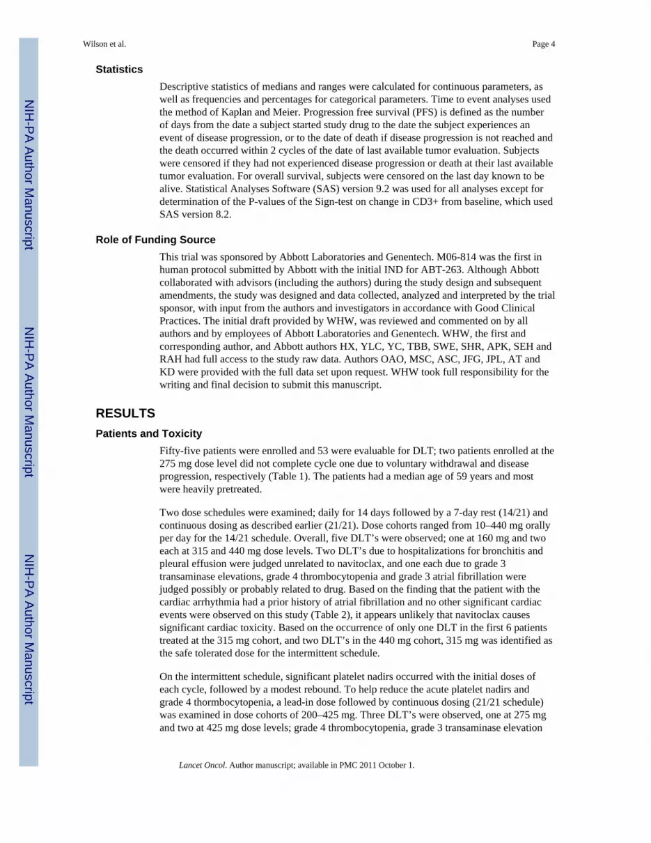
StatisticsDescriptive statistics of medians and ranges were calculated for continuous parameters, aswell as frequencies and percentages for categorical parameters. Time to event analyses usedthe method of Kaplan and Meier. Progression free survival (PFS) is defined as the numberof days from the date a subject started study drug to the date the subject experiences anevent of disease progression, or to the date of death if disease progression is not reached andthe death occurred within 2 cycles of the date of last available tumor evaluation. Subjectswere censored if they had not experienced disease progression or death at their last availabletumor evaluation. For overall survival, subjects were censored on the last day known to bealive. Statistical Analyses Software (SAS) version 9.2 was used for all analyses except fordetermination of the P-values of the Sign-test on change in CD3+ from baseline, which usedSAS version 8.2.
Role of Funding SourceThis trial was sponsored by Abbott Laboratories and Genentech. M06-814 was the first inhuman protocol submitted by Abbott with the initial IND for ABT-263. Although Abbottcollaborated with advisors (including the authors) during the study design and subsequentamendments, the study was designed and data collected, analyzed and interpreted by the trialsponsor, with input from the authors and investigators in accordance with Good ClinicalPractices. The initial draft provided by WHW, was reviewed and commented on by allauthors and by employees of Abbott Laboratories and Genentech. WHW, the first andcorresponding author, and Abbott authors HX, YLC, YC, TBB, SWE, SHR, APK, SEH andRAH had full access to the study raw data. Authors OAO, MSC, ASC, JFG, JPL, AT andKD were provided with the full data set upon request. WHW took full responsibility for thewriting and final decision to submit this manuscript.
RESULTSPatients and Toxicity
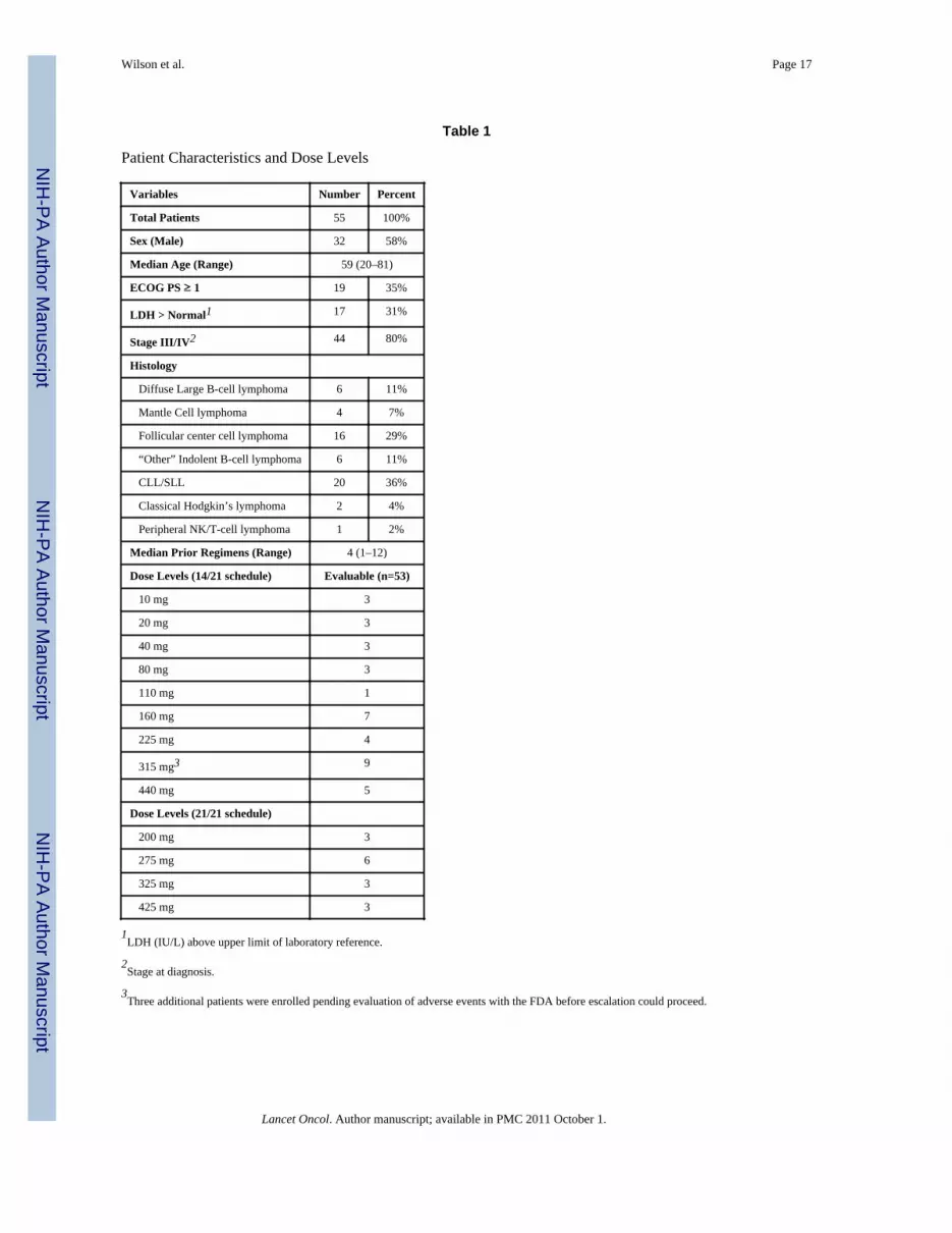
Fifty-five patients were enrolled and 53 were evaluable for DLT; two patients enrolled at the275 mg dose level did not complete cycle one due to voluntary withdrawal and diseaseprogression, respectively (Table 1). The patients had a median age of 59 years and mostwere heavily pretreated.
Two dose schedules were examined; daily for 14 days followed by a 7-day rest (14/21) andcontinuous dosing as described earlier (21/21). Dose cohorts ranged from 10–440 mg orallyper day for the 14/21 schedule. Overall, five DLT’s were observed; one at 160 mg and twoeach at 315 and 440 mg dose levels. Two DLT’s due to hospitalizations for bronchitis andpleural effusion were judged unrelated to navitoclax, and one each due to grade 3transaminase elevations, grade 4 thrombocytopenia and grade 3 atrial fibrillation werejudged possibly or probably related to drug. Based on the finding that the patient with thecardiac arrhythmia had a prior history of atrial fibrillation and no other significant cardiacevents were observed on this study (Table 2), it appears unlikely that navitoclax causessignificant cardiac toxicity. Based on the occurrence of only one DLT in the first 6 patientstreated at the 315 mg cohort, and two DLT’s in the 440 mg cohort, 315 mg was identified asthe safe tolerated dose for the intermittent schedule.
On the intermittent schedule, significant platelet nadirs occurred with the initial doses ofeach cycle, followed by a modest rebound. To help reduce the acute platelet nadirs andgrade 4 thormbocytopenia, a lead-in dose followed by continuous dosing (21/21 schedule)was examined in dose cohorts of 200–425 mg. Three DLT’s were observed, one at 275 mgand two at 425 mg dose levels; grade 4 thrombocytopenia, grade 3 transaminase elevation
Wilson et al. Page 4
Lancet Oncol. Author manuscript; available in PMC 2011 October 1.
NIH
-PA Author Manuscript
NIH
-PA Author Manuscript
NIH
-PA Author Manuscript

and grade 3 gastrointestinal bleed, respectively, all possibly or probably related tonavitoclax. Based on these findings, a 150 mg 7-day lead-in dose followed by 325 mg doseadministered on a continuous (21/21) schedule was selected for the phase II study.
When all dose levels are considered, the most common toxicities were grade 1/2gastrointestinal complaints likely due to the drug vehicle (Table 2). Nausea and vomitingwere managed with anti-emetics and diarrhea was managed with diphenoxylate and atropine(lomotil). Fatigue was relatively common but not dose limiting. Eight patients developedrespiratory infections and/or bronchitis (Table 2), none of which were associated with grade4 neutropenia. Thrombocytopenia and neutropenia were the serious common toxicities withgrade 3 or 4 thrombocytopenia or neutropenia observed in 29 and 17 patients, respectively(Table 2). Unlike the thrombocytopenia, neutropenia was not clearly associated with doselevel and tended to occur on the later cycles; it was also reversible upon navitoclaxdiscontinuation. Grade 4 thrombocytopenia and neutropenia were managed by temporarysuspension and dose reduction of navitoclax and filgrastim was used for persistent grade 4neutropenia. These results indicate that navitoclax may cause unacceptable hematologicaltoxicity in patients with limited bone marrow reserve. Overall dose levels, 11 patientsrequired at least one dose reduction and 6 patients withdrew their consent for treatment, twoof which were due to toxicity.
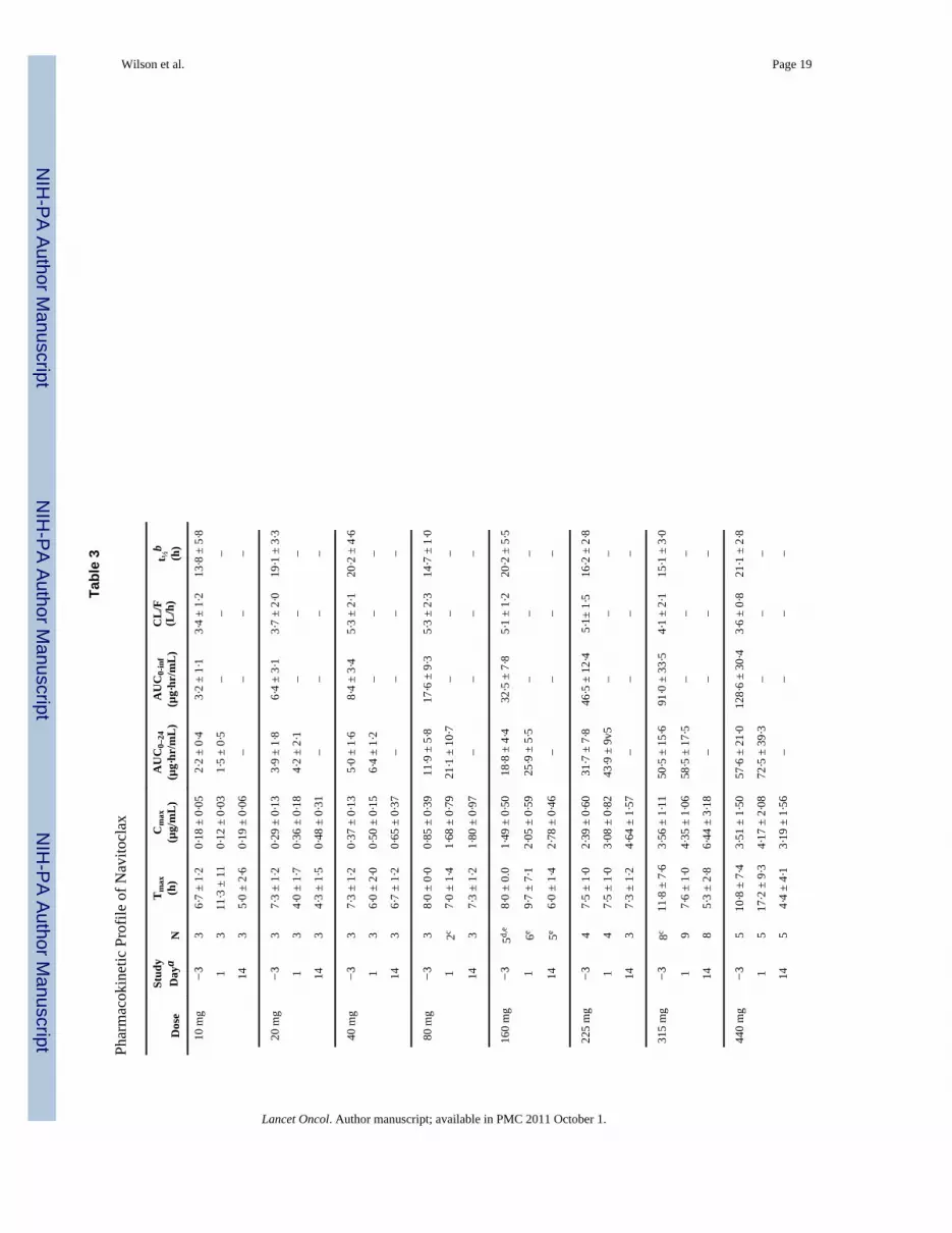
Pharmacokinetic ProfileNavitoclax PK was assessed on the 14/21-day schedule (Figure 2 and Table 3). To assessfood effects on absorption, navitoclax was administered fasting on day –3 and non-fastingon days 1–14 of cycle one. Overall, food increased the oral bioavailability by approximately20% with the current lipid formulation. Exposure was dose-proportional between 10 mg and440 mg with approximately 40% interpatient variability in the plasma AUC. Navitoclaxachieved peak concentrations (Cmax) at approximately 9 hours post-dose with a half-life ofapproximately 17 hours, and first order elimination kinetics. The projected effectiveexposure of 55–88 µg·hr/mL based on animal models was achieved at dose levels of at least315 mg/day in patients. Navitoclax could not be detected in urine, indicating negligible renalelimination.
Pharmacodynamic EffectsWe hypothesized that if navitoclax functionally inhibited BCL-2 and BCL-XL, it shoulddecrease the survival of normal cells in which these proteins regulate survival such as in T-cells and platelets, respectively.(15,16) To assess this, we measured the absolute number ofCD3+ cells in the circulation before, on day 14 of cycle 1, and at the end of treatment(Figure 3A). Patients showed a relatively rapid decrease in T-cells after only 14 days of drugexposure (median reduction 241 CD3+ cells/µl), which did not worsen with furthertreatment (Figure 3A). Importantly, the loss of T-cells was modest and not associated withopportunistic infections.
We also examined the kinetics of circulating platelets during navitoclax exposure. There wasa significant reduction following a single dose of navitoclax, which was observed to begin assoon as one hour after dosing (data not shown) (Figure 3B). With continued dosing, therewas a modest rise from nadir levels. To obviate the acute nadirs associated with intermittentdosing (14/21), we tested a lead-in dose followed by continuous dosing (21/21) andobserved higher nadirs. Consistent with a pharmacodynamic effect, the severity of theplatelet nadirs was concentration dependent (Figure 3C) and related to dose level (data notshown).
Wilson et al. Page 5
Lancet Oncol. Author manuscript; available in PMC 2011 October 1.
NIH
-PA Author Manuscript
NIH
-PA Author Manuscript
NIH
-PA Author Manuscript

Treatment OutcomeOverall, 21 of 46 patients with evaluable adenopathy showed some tumor reduction.Furthermore, 10 of these patients achieved a partial response (PR) lasting a median (range)of 455 days (IQR 40–218) (Figure 4). Responses occurred across dose levels and tumortypes and were observed after a median (range) of 3.5 (2–10) cycles. Chronic lymphocyticleukemia/lymphoma (CLL/SLL), a disease of B-cell accumulation, showed the greatestsensitivity. Among the seven patients with CLL, all achieved at least a 50% reduction intheir leukemia cells, and 8 of 16 patients with measurable adenopathy achieved a PR, whichincluded patients with bulky adenopathy. Including all 20 patients with CLL/SLL, themedian (range) PFS was 246 days (IQR 49-309) and the median overall survival has notbeen reached. Measurable tumor reduction was also observed in 6 of 16 patients withfollicular lymphoma. Overall, the 16 patients with follicular lymphoma had a median PFS of88 days (IQR 42-92). A PR was achieved in a single patient with an NK/T-cell lymphoma.The patients with CLL/SLL or follicular lymphomas had received a similar number of priorregimens with a median (range) of 4 (1–12) and 5 (1–11), respectively.
DISCUSSIONFifty-three evaluable patients received navitoclax on two different treatment schedules.Navitoclax demonstrated a high therapeutic index with a low incidence of off-targettoxicities. The major off-target toxicity was gastrointestinal, which appears likely related tothe phosphatidylcholine solubilizer. Though uncommon, transaminase elevation at higherdose levels and neutropenia after prolonged drug exposure were also observed. Navitoclaxalso demonstrated favorable pharmacokinetic properties and therapeutic index. Due to goodoral absorption, exposure was dose proportional, and the approximate 17-hour half-lifeallowed daily dosing. Furthermore, concentrations shown to be effective in preclinicalmodels were achieved at the recommended phase 2 dose of 325 g/day.
The pharmacodynamic effects of navitoclax on circulating lymphocytes and platelets arenovel and consistent with on-target mechanisms. Based on preclinical evidence that plateletsenescence entails an apoptosis-like process mediated through BCL-XL, it is likely thatintravascular apoptosis is responsible for the acute thrombocytopenia following navitoclax.(17,18) Furthermore, the relative resistance of younger platelets to navitoclax appears to bedue to their higher levels of BCL-XL, which explains the platelet kinetics observed inpatients and in pre-clinical animal models using ABT-737.(18). It is also likely thatnavitoclax induces apoptosis of normal lymphocytes through its inhibitory effect on BCL-2.These results suggest that the pharmacodynamic effects of navitoclax are biomarkers ofpharmacological inhibition of BCL-2 and BCL-XL, and should be observed with alleffective inhibitors. We also observed grade 3 or 4 neutropenia in 17 patients, which raisesthe question of whether navitoclax may also have a pharmacodynamic effect on myeloidcells. To help assess this, we performed preliminary methylcellulose-based in vitro colonyforming assays to assess the effects of ABT-737, a navitoclax analog, on humanhematopoietic progenitors. In the presence of multilineage cytokines, ABT-737 inhibited thegrowth of both erythroid and common myeloid precursors (IC50 of 1.0 µM and 0.9 µM,respectively) (data not shown). Furthermore, BCL-2 has been shown to be necessary for thein vitro survival of myeloid progenitors under conditions of cytokine withdrawal, suggestingthe neutropenia we observed in our study could be due to inhibition of BCL-2 by navitoclax.(19)
Navitoclax demonstrated clinical activity at all dose levels and across tumor types with thegreatest activity seen in CLL. Letai et al. recently proposed a model, termed BH3 profiling,which may explain the differential sensitivity of lymphoma cells to BCL-2 inhibition.(20,21)They proposed that sequestration of the activator BH3 only proteins, BIM or BID, by BCL-2
Wilson et al. Page 6
Lancet Oncol. Author manuscript; available in PMC 2011 October 1.
NIH
-PA Author Manuscript
NIH
-PA Author Manuscript
NIH
-PA Author Manuscript

produces a “primed” state in which BCL-2 inhibition releases activator proteins and inducesapoptosis. Using BH3 profiling, the group found a pattern of BCL-2 dependence in CLLwith high BIM:BCL-2 complex levels and exquisite sensitivity to ABT-737.(20)Interestingly, we observed few bone marrow responses with navitoclax, even among patientswith robust nodal and blood responses, which may be due to the influence of themicroenvironment on increased expression of MCL-1, BCL-XL or BCL-2A1.(22) Thoughthe activity of navitoclax was less apparent in other lymphoma subtypes, it has synergisticactivity with chemotherapeutic agents in preclinical models. Given the complexity of theprimed BCL-2 phenotype, and the influence of the microenvironment and upstreampathways, we hypothesize that the greatest benefit of navitoclax will be observed incombination with other agents.
Several other inhibitors of BCL-2 have undergone clinical testing. Two of these, obatoclaxand gossypol, are small molecules that are reported to be pan-BCL-2 inhibitors.(23,24) Thusfar, they have shown little to no clinical activity. Furthermore, they have been shown to killcells in a BAX/BAK-independent manner, challenging the functional significance of theirweak affinity for BCL-2 family proteins and their true mechanisms of action.(25–27) Areview of their toxicities also showed no on-target pharmacodynamic effects on platelets,suggesting they do not achieve effective inhibition of BCL-XL.(23,25–28) Gossypolprimarily caused gastrointestinal side effects, which were dose limiting, but caused nosignificant laboratory abnormalities.(23) While navitoclax also had gastrointestinal sideeffects, they were low grade and likely due to the drug solubilizer. Infusion-relatedsomnolence and neurologic symptoms were the most common toxicities associated withobatoclax, and were dose limiting.(24) Modest hematological effects were also observed,including mild thrombocytopenia, which were attributed to progression of the patients’underlying CLL.(24) A third drug, oblimersen, is an antisense oligodeoxyribonucleotide thatdown regulates BCL-2 translation.(29) A phase I study demonstrated limited single agentactivity with only one response in 21 patients.(30) Interestingly, like navitoclax, oblimersencaused thrombocytopenia, which progressively worsened during the drug infusion andcorrelated with its plasma concentration.(30) While these results indicate that oblimersenhas a pharmacodynamic effect on platelets, its indirect effect on BCL-2 levels throughinhibition of BCL-2 mRNA is inconsistent with a direct inhibition of BCL-XL. In contrast tothese other putative BCL-2 inhibitors, induction of apoptosis by navitoclax in vitro can beattributed to inhibition of BCL-2 family proteins. Coimmunoprecipitation studies show thatnavitoclax induced a dose-dependent decrease in BIM:BCL-2 family protein interactions inBCL-XL and BCL-2 over expressing prolymphocytic murine cell lines. Navitoclax alsoinduced a dose-dependent decrease in cytosolic BAX and an increase in cytochrome cwithin 2 hours of treatment of a BCL-2 dependent human small cell lung cancer (SCLC) cellline.(11,27)
The importance of BCL-2 family proteins in lymphoid biology and pathogenesis has driventhe search for small molecule inhibitors of this pathway. With few exceptions, lymphomasexpress increased BCL-2, which may be physiological or pathogenetic. In follicularlymphoma and the germinal center subset of diffuse large B-cell lymphoma (DLBCL),translocation of the BCL-2 locus and immunoglobulin heavy chain promoter (t(14;18))drives BCL-2 production, whereas in the post-germinal center subset of DLBCL, BCL-2may be over expressed through amplification or transcriptional activation.(6,31–33) CLLemploys yet another mechanism whereby deletion or down regulation of miRNA miR-16-1and miR-15a drives post-transcriptional increases in BCL-2.(34,35) The occurrence of suchvaried pathogenetic mechanisms that increase BCL-2 expression points to the evolutionarysignificance of this pathway in lymphomagenesis, and the potential importance of this targetin lymphoid malignancies.
Wilson et al. Page 7
Lancet Oncol. Author manuscript; available in PMC 2011 October 1.
NIH
-PA Author Manuscript
NIH
-PA Author Manuscript
NIH
-PA Author Manuscript

Our study provides the first clinical insights to our knowledge into a pharmacologicallyactive BCL-2 family inhibitor. We are currently investigating navitoclax in an expandedcohort of indolent and aggressive B-cell lymphomas. There are ongoing phase I studies ofnavitoclax with other agents including rituximab (CD20 monoclonal antibody),bendamustine and rituximab, and fludarabine, cyclophosphamide and rituximabcombinations in lymphoma and CLL. Furthermore, to overcome the pharmacodynamiceffect of navitoclax on circulating platelets, which will limit its ability to be combined withcytotoxic agents, a selective inhibitor of BCL-2 is under development.(36)
RESEARCH IN CONTEXTSystematic Review
There exists an extensive literature that demonstrates apoptosis, or programmed cell death isthe principal mechanism through which unwanted or damaged cells are safely eliminated.(37–40) Although cancer has historically been considered a disease of uncontrolled celldivision, abnormal resistance to apoptosis is now understood to contribute to tumorinitiation, progression, and resistance to chemotherapy. Defects in the apoptotic pathwayconfer a survival advantage that allows a net increase in tumor cell number and theaccumulation of oncogenic mutations, which gives rise to highly aggressive tumors.Interactions between pro-apoptotic (pro-death) and antiapoptotic (pro-survival) BCL-2family proteins regulate the initiation of the intrinsic apoptosis pathway. The pro-deathproteins of BAX and BAK are direct mediators of apoptosis and are absolutely required forthe initiation of the mitochondrial apoptosis pathway.(41) Over expression of anti-apoptoticBCL-2 family proteins (BCL-XL, BCL-2, BCL-W, A1, MCL-1) suppresses BAX and BAKand prevents the initiation of the apoptosis, thereby protecting cancer cells from respondingto proapoptotic signals.
Compelling evidence for the role of BCL-2 family proteins in lymphoid biology andpathogenesis has driven the search for inhibitors.(42) With infrequent exception, lymphomasexpress BCL-2, which may be physiological and/or pathogenetic. The search for BCL-2inhibitors has primarily relied on cytotoxicity screening. While such methodologies havelead to the identification of small molecules with low affinity inhibition and/or off targeteffects, these agents have shown relatively little single agent activity.(23,26) An alternativestrategy employed in the development of navitoclax entailed a structure-based design toidentify small molecules that bind BCL-XL, which lead to the high affinity inhibitornavitoclax.(8,10)
InterpretationIn the present study, we report that navitoclax, a high affinity inhibitor of BCL-2 familyproteins, has clinical activity in lymphoid malignancies and has on-target pharmacodynamiceffects on platelets and T-cells, where BCL-XL and BCL-2 regulate survival. While otherputative BCL-2 inhibitors have undergone clinical testing, they have not shown significantclinical activity or targeted pharmacodynamic effects, which likely reflects low inhibition ofBCL-2 family proteins. Thus, the present study provides the first proof of concept inhumans, to our knowledge, that inhibition of BCL-2 family proteins leads to tumor celldeath and targeted cell death of platelets and T-cells. As most cytotoxic agents induceapoptosis as a primary mechanism of cell kill, modulation of the apoptotic “threshold” withagents such as navitoclax is hypothesized to significantly increase the efficacy of currentcytotoxic treatments. Presently, phase I trials are underway to assess the safety of navitoclaxwith cytotoxic agents. Further studies will be necessary to determine if navitoclax is safe andeffective before it can be used in standard treatment.
Wilson et al. Page 8
Lancet Oncol. Author manuscript; available in PMC 2011 October 1.
NIH
-PA Author Manuscript
NIH
-PA Author Manuscript
NIH
-PA Author Manuscript

AcknowledgmentsThe authors would like to thank Juliann M. Dziubinski, Katherine Papp, Lori Gressick, Michael D. Dawson andRenee Greco, for operational support; Di Li, and Joseph E. Beason for statistical analyses; Christin Tse, Morey L.Smith, Stephen K. Tahir, Kennan C. Marsh, Joy L. Bauch, Sherry J. Morgan, Joel Leverson, and Anne H. Illi-Lovefor their knowledge of the preclinical data referenced and their contributions during manuscript preparation, and AiQ. Lockard for editorial assistance for the manuscript. The authors would also like to thank the contributions of theresearch data managers, coordinators and nurses including Margaret Shovlin, Barbara MacGregor Cortelli, AmeetNarwal, Barbara Anderson, Alice Mohr, Hazel Reynolds, Susan Twohig, Jennifer Pappanicholaou, Payal Dixit,June Greenberg, and Nancy Berman. This study was funded by Abbott Laboratories and Genentech, Inc.
REFERENCES1. Merino R, Ding L, Veis DJ, Korsmeyer SJ, Nunez G. Developmental regulation of the Bcl-2 protein
and susceptibility to cell death in B lymphocytes. EMBO J 1994 Feb 1;13(3):683–691. [PubMed:8313913]
2. Tsujimoto Y, Finger LR, Yunis J, Nowell PC, Croce CM. Cloning of the chromosome breakpoint ofneoplastic B cells with the t(14;18) chromosome translocation. Science 1984 Nov 30;226(4678):1097–1099. [PubMed: 6093263]
3. Tsujimoto Y, Gorham J, Cossman J, Jaffe E, Croce CM. The t(14;18) chromosome translocationsinvolved in B-cell neoplasms result from mistakes in VDJ joining. Science 1985 Sep 27;229(4720):1390–1393. [PubMed: 3929382]
4. Davis RE, Ngo VN, Lenz G, Tolar P, Young RM, Romesser PB, et al. Chronic active B-cell-receptor signalling in diffuse large B-cell lymphoma. Nature 2010 Jan 7;463(7277):88–92.[PubMed: 20054396]
5. Hockenbery D, Nunez G, Milliman C, Schreiber RD, Korsmeyer SJ. Bcl-2 is an inner mitochondrialmembrane protein that blocks programmed cell death. Nature 1990 Nov 22;348(6299):334–336.[PubMed: 2250705]
6. McDonnell TJ, Deane N, Platt FM, Nunez G, Jaeger U, McKearn JP, et al. bcl-2-immunoglobulintransgenic mice demonstrate extended B cell survival and follicular lymphoproliferation. Cell 1989Apr 7;57(1):79–88. [PubMed: 2649247]
7. Kondo E, Nakamura S, Onoue H, Matsuo Y, Yoshino T, Aoki H, et al. Detection of bcl-2 proteinand bcl-2 messenger RNA in normal and neoplastic lymphoid tissues by immunohistochemistry andin situ hybridization. Blood 1992 Oct 15;80(8):2044–2051. [PubMed: 1391959]
8. Degterev A, Lugovskoy A, Cardone M, Mulley B, Wagner G, Mitchison T, et al. Identification ofsmall-molecule inhibitors of interaction between the BH3 domain and Bcl-xL. Nat Cell Biol 2001Feb;3(2):173–182. [PubMed: 11175750]
9. Huang JW, Zhang Z, Wu B, Cellitti JF, Zhang X, Dahl R, et al. Fragment-based design of smallmolecule X-linked inhibitor of apoptosis protein inhibitors. J Med Chem 2008 Nov 27;51(22):7111–7118. [PubMed: 18956862]
10. Oltersdorf T, Elmore SW, Shoemaker AR, Armstrong RC, Augeri DJ, Belli BA, et al. An inhibitorof Bcl-2 family proteins induces regression of solid tumours. Nature 2005 Jun 2;435(7042):677–681. [PubMed: 15902208]
11. Tse C, Shoemaker AR, Adickes J, Anderson MG, Chen J, Jin S, et al. ABT-263: a potent andorally bioavailable Bcl-2 family inhibitor. Cancer Res 2008 May 1;68(9):3421–3428. [PubMed:18451170]
12. Cheson BD, Horning SJ, Coiffier B, Shipp MA, Fisher RI, Connors JM, et al. Report of aninternational workshop to standardize response criteria for non-Hodgkin's lymphomas. J ClinOncol 1999;17(4):1244. [PubMed: 10561185]
13. Hallek M, Cheson BD, Catovsky D, Caligaris-Cappio F, Dighiero G, Dohner H, et al. Guidelinesfor the diagnosis and treatment of chronic lymphocytic leukemia: a report from the InternationalWorkshop on Chronic Lymphocytic Leukemia updating the National Cancer Institute-WorkingGroup 1996 guidelines. Blood 2008 Jun 15;111(12):5446–5456. [PubMed: 18216293]
14. Kaplan E, Meier P. Nonparametric estimation from incomplete observations. Journal of theAmerican Statictical Association 1958;53:457–481.
Wilson et al. Page 9
Lancet Oncol. Author manuscript; available in PMC 2011 October 1.
NIH
-PA Author Manuscript
NIH
-PA Author Manuscript
NIH
-PA Author Manuscript

15. Akbar AN, Borthwick N, Salmon M, Gombert W, Bofill M, Shamsadeen N, et al. The significanceof low bcl-2 expression by CD45RO T cells in normal individuals and patients with acute viralinfections. The role of apoptosis in T cell memory. J Exp Med 1993 Aug 1;178(2):427–438.[PubMed: 8340752]
16. Zhang H, Nimmer PM, Tahir SK, Chen J, Fryer RM, Hahn KR, et al. Bcl-2 family proteins areessential for platelet survival. Cell Death Differ 2007 May;14(5):943–951. [PubMed: 17205078]
17. Chipuk JE, Fisher JC, Dillon CP, Kriwacki RW, Kuwana T, Green DR. Mechanism of apoptosisinduction by inhibition of the anti-apoptotic BCL-2 proteins. Proc Natl Acad Sci U S A 2008 Dec23;105(51):20327–20332. [PubMed: 19074266]
18. Mason KD, Carpinelli MR, Fletcher JI, Collinge JE, Hilton AA, Ellis S, et al. Programmedanuclear cell death delimits platelet life span. Cell 2007 Mar 23;128(6):1173–1186. [PubMed:17382885]
19. Villunger A, Scott C, Bouillet P, Strasser A. Essential role for the BH3-only protein Bim butredundant roles for Bax, Bcl-2, and Bcl-w in the control of granulocyte survival. Blood 2003 Mar15;101(6):2393–2400. [PubMed: 12433687]
20. Del Gaizo Moore V, Brown JR, Certo M, Love TM, Novina CD, Letai A. Chronic lymphocyticleukemia requires BCL2 to sequester prodeath BIM, explaining sensitivity to BCL2 antagonistABT-737. J Clin Invest 2007 Jan;117(1):112–121. [PubMed: 17200714]
21. Deng J, Carlson N, Takeyama K, Dal Cin P, Shipp M, Letai A. BH3 profiling identifies threedistinct classes of apoptotic blocks to predict response to ABT-737 and conventionalchemotherapeutic agents. Cancer Cell 2007 Aug;12(2):171–185. [PubMed: 17692808]
22. Vogler M, Butterworth M, Majid A, Walewska RJ, Sun XM, Dyer MJ, et al. Concurrent up-regulation of BCL-XL and BCL2A1 induces approximately 1000-fold resistance to ABT-737 inchronic lymphocytic leukemia. Blood 2009 Apr 30;113(18):4403–4413. [PubMed: 19008458]
23. Liu G, Kelly WK, Wilding G, Leopold L, Brill K, Somer B. An open-label, multicenter, phase I/IIstudy of single-agent AT-101 in men with castrate-resistant prostate cancer. Clin Cancer Res 2009May 1;15(9):3172–3176. [PubMed: 19366825]
24. O'Brien SM, Claxton DF, Crump M, Faderl S, Kipps T, Keating MJ, et al. Phase I study ofobatoclax mesylate (GX15-070), a small molecule pan-Bcl-2 family antagonist, in patients withadvanced chronic lymphocytic leukemia. Blood 2009 Jan 8;113(2):299–305. [PubMed: 18931344]
25. Kitada S, Leone M, Sareth S, Zhai D, Reed JC, Pellecchia M. Discovery, characterization, andstructure-activity relationships studies of proapoptotic polyphenols targeting B-cell lymphocyte/leukemia-2 proteins. J Med Chem 2003 Sep 25;46(20):4259–4264. [PubMed: 13678404]
26. Nguyen M, Marcellus RC, Roulston A, Watson M, Serfass L, Murthy Madiraju SR, et al. Smallmolecule obatoclax (GX15-070) antagonizes MCL-1 and overcomes MCL-1-mediated resistanceto apoptosis. Proc Natl Acad Sci U S A 2007 Dec 4;104(49):19512–19517. [PubMed: 18040043]
27. Vogler M, Weber K, Dinsdale D, Schmitz I, Schulze-Osthoff K, Dyer MJ, et al. Different forms ofcell death induced by putative BCL2 inhibitors. Cell Death Differ 2009 Jul;16(7):1030–1039.[PubMed: 19390557]
28. Hwang JJ, Kuruvilla J, Mendelson D, Pishvaian MJ, Deeken JF, Siu LL, et al. Phase I dose findingstudies of obatoclax (GX15-070), a small molecule pan-BCL-2 family antagonist, in patients withadvanced solid tumors or lymphoma. Clin Cancer Res 2010 Aug 1;16(15):4038–4045. [PubMed:20538761]
29. Klasa RJ, Bally MB, Ng R, Goldie JH, Gascoyne RD, Wong FM. Eradication of human non-Hodgkin's lymphoma in SCID mice by BCL-2 antisense oligonucleotides combined with low-dosecyclophosphamide. Clin Cancer Res 2000 Jun;6(6):2492–2500. [PubMed: 10873104]
30. Waters JS, Webb A, Cunningham D, Clarke PA, Raynaud F, di Stefano F, et al. Phase I clinicaland pharmacokinetic study of bcl-2 antisense oligonucleotide therapy in patients with non-Hodgkin's lymphoma. J Clin Oncol 2000 May;18(9):1812–1823. [PubMed: 10784621]
31. Gascoyne RD, Adomat SA, Krajewski S, Krajewska M, Horsman DE, Tolcher AW, et al.Prognostic significance of Bcl-2 protein expression and Bcl-2 gene rearrangement in diffuseaggressive non-Hodgkin's lymphoma. Blood 1997 Jul 1;90(1):244–251. [PubMed: 9207459]
Wilson et al. Page 10
Lancet Oncol. Author manuscript; available in PMC 2011 October 1.
NIH
-PA Author Manuscript
NIH
-PA Author Manuscript
NIH
-PA Author Manuscript

32. Iqbal J, Sanger WG, Horsman DE, Rosenwald A, Pickering DL, Dave B, et al. BCL2Translocation Defines a Unique Tumor Subset within the Germinal Center B-Cell-Like DiffuseLarge B-Cell Lymphoma. Am J Pathol 2004 July 1;165(1):159–166. 2004. [PubMed: 15215171]
33. Wilson WH, Teruya-Feldstein J, Fest T, Harris C, Steinberg SM, Jaffe ES, et al. Relationship ofp53, bcl-2, and tumor proliferation to clinical drug resistance in non-Hodgkin's lymphomas. Blood1997;89(2):601–609. [PubMed: 9002964]
34. Aqeilan RI, Calin GA, Croce CM. miR-15a and miR-16-1 in cancer: discovery, function and futureperspectives. Cell Death Differ 2010 Feb;17(2):215–220. [PubMed: 19498445]
35. Calin GA, Ferracin M, Cimmino A, Di Leva G, Shimizu M, Wojcik SE, et al. A MicroRNAsignature associated with prognosis and progression in chronic lymphocytic leukemia. N Engl JMed 2005 Oct 27;353(17):1793–1801. [PubMed: 16251535]
36. Petros AM, Huth JR, Oost T, Park CM, Ding H, Wang X, et al. Discovery of a potent and selectiveBcl-2 inhibitor using SAR by NMR. Bioorganic & medicinal chemistry letters 2010 Nov15;20(22):6587–6591. [PubMed: 20870405]
37. Brenner C, Kroemer G. Apoptosis. Mitochondria--the death signal integrators. Science 2000 Aug18;289(5482):1150–1151. [PubMed: 10970229]
38. Arends MJ, Wyllie AH. Apoptosis: mechanisms and roles in pathology. Int Rev Exp Pathol1991;32:223–254. [PubMed: 1677933]
39. Reed JC. Apoptosis and cell death. Foreword. Oncogene 2008 Oct 20;27(48):6192–6193.[PubMed: 18931686]
40. Danial NN, Korsmeyer SJ. Cell death: critical control points. Cell 2004 Jan 23;116(2):205–219.[PubMed: 14744432]
41. Reed JC. Mechanisms of apoptosis. Am J Pathol 2000 Nov;157(5):1415–1430. [PubMed:11073801]
42. Reed JC. Apoptosis mechanisms: implications for cancer drug discovery. Oncology (WillistonPark) 2004 Nov;18(13 Suppl 10):11–20. [PubMed: 15651172]
Wilson et al. Page 11
Lancet Oncol. Author manuscript; available in PMC 2011 October 1.
NIH
-PA Author Manuscript
NIH
-PA Author Manuscript
NIH
-PA Author Manuscript

Figure 1. Navitoclax Dosing Schedule(a). Intermittent (14/21) dosing schedule. For Cycle 1, navitoclax was administered on day –3 to assess pharmacokinetics (PK) and food effect. Beginning on Cycle 1 day 1, patientswere dosed non-fasting for 14 consecutive days on days 1–14 of a 21- day dosing cycle. Insubsequent cycles, patients receive drug on D1–14 followed by 7 days off. (b). Continuous(21/21) dosing schedule. On cycle 1, patients received a 150 mg lead-in dose for 7–14 daysfollowed by continuous dosing at their assigned dose level.
Wilson et al. Page 12
Lancet Oncol. Author manuscript; available in PMC 2011 October 1.
NIH
-PA Author Manuscript
NIH
-PA Author Manuscript
NIH
-PA Author Manuscript
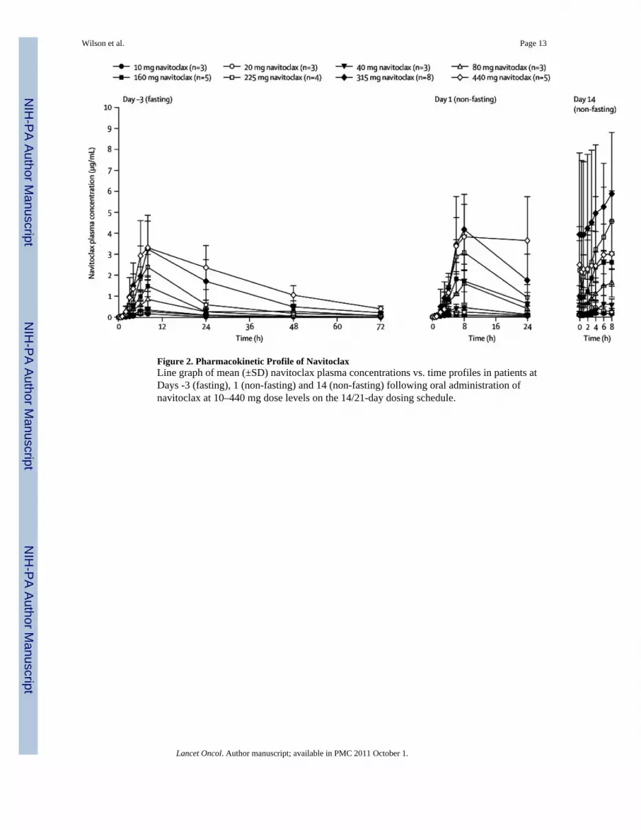
Figure 2. Pharmacokinetic Profile of NavitoclaxLine graph of mean (±SD) navitoclax plasma concentrations vs. time profiles in patients atDays -3 (fasting), 1 (non-fasting) and 14 (non-fasting) following oral administration ofnavitoclax at 10–440 mg dose levels on the 14/21-day dosing schedule.
Wilson et al. Page 13
Lancet Oncol. Author manuscript; available in PMC 2011 October 1.
NIH
-PA Author Manuscript
NIH
-PA Author Manuscript
NIH
-PA Author Manuscript

Figure 3. Pharmacodynamic Effects of Navitoclax(a) Bar graph of median absolute circulating CD3 cells/µl at baseline (n=20 patients), day 14of cycle 1 (n=19 patients) and the final visit (n=16 patients) at the end of navitoclaxtreatment, and the median change on day 14 of cycle 1 and final visit. Patients received atleast 200 mg navitoclax for at least two cycles on either the 14/21 or 21/21day schedule. Themean (range) time between baseline and final visit was 89 (29–332) days and the mean(range) daily drug exposure was 255 (137–388) mg of navitoclax. P-value was calculatedusing a Sign-test because the data were skewed. (b) Line graph of platelet nadirs over time at315 mg on the 14/21-day schedule (n=9 patients) (left panel) and at 275 mg on the 21/21-day schedule (n=6 patients) (right panel). (c) Platelet nadir versus navitoclax area under the
Wilson et al. Page 14
Lancet Oncol. Author manuscript; available in PMC 2011 October 1.
NIH
-PA Author Manuscript
NIH
-PA Author Manuscript
NIH
-PA Author Manuscript

curve at multiple dose levels (n=35 patients). R-value was calculated by using SigmaPlot9.0.
Wilson et al. Page 15
Lancet Oncol. Author manuscript; available in PMC 2011 October 1.
NIH
-PA Author Manuscript
NIH
-PA Author Manuscript
NIH
-PA Author Manuscript

Figure 4. Antitumor Activity of NavitoclaxWaterfall plot of maximal percent change in tumor size from baseline using standardcriteria.(12,13) Tumor subtypes are shown on the x-axis: DLBCL-diffuse large B-celllymphoma; FL-follicular lymphoma; MCL-mantle cell lymphoma; CLL/SLL-chroniclymphocytic leukemia/small lymphocytic lymphoma; MZL-marginal zone lymphoma; HL-classical Hodgkin’s lymphoma; and NK/T-natural killer-T-cell lymphoma.
Wilson et al. Page 16
Lancet Oncol. Author manuscript; available in PMC 2011 October 1.
NIH
-PA Author Manuscript
NIH
-PA Author Manuscript
NIH
-PA Author Manuscript
NIH
-PA Author Manuscript
NIH
-PA Author Manuscript
NIH
-PA Author Manuscript
Wilson et al. Page 17
Table 1
Patient Characteristics and Dose Levels
Variables Number Percent
Total Patients 55 100%
Sex (Male) 32 58%
Median Age (Range) 59 (20–81)
ECOG PS ≥ 1 19 35%
LDH > Normal1 17 31%
Stage III/IV2 44 80%
Histology
Diffuse Large B-cell lymphoma 6 11%
Mantle Cell lymphoma 4 7%
Follicular center cell lymphoma 16 29%
“Other” Indolent B-cell lymphoma 6 11%
CLL/SLL 20 36%
Classical Hodgkin’s lymphoma 2 4%
Peripheral NK/T-cell lymphoma 1 2%
Median Prior Regimens (Range) 4 (1–12)
Dose Levels (14/21 schedule) Evaluable (n=53)
10 mg 3
20 mg 3
40 mg 3
80 mg 3
110 mg 1
160 mg 7
225 mg 4
315 mg3 9
440 mg 5
Dose Levels (21/21 schedule)
200 mg 3
275 mg 6
325 mg 3
425 mg 3
1LDH (IU/L) above upper limit of laboratory reference.
2Stage at diagnosis.
3Three additional patients were enrolled pending evaluation of adverse events with the FDA before escalation could proceed.
Lancet Oncol. Author manuscript; available in PMC 2011 October 1.

NIH
-PA Author Manuscript
NIH
-PA Author Manuscript
NIH
-PA Author Manuscript
Wilson et al. Page 18
Tabl
e 2
Clin
ical
and
Lab
orat
ory
Toxi
city
Eve
nts
Inte
rmitt
ent (
14/2
1)Sc
hedu
leA
ll Pa
tient
s (N
=38)
Con
tinuo
us (2
1/21
)Sc
hedu
leA
ll Pa
tient
s (N
=17)
Inte
rmitt
ent (
14/2
1)Sc
hedu
leM
TD
315
mg
(N=9
)
Con
tinuo
us (2
1/21
)Sc
hedu
leM
TD
325
mg
(N=5
)
Gra
de 1
/2G
rade
3/4
Gra
de 1
/2G
rade
3/4
Gra
de 1
/2G
rade
3/4
Gra
de 1
/2G
rade
3/4
Car
diac
Atri
al a
rrhy
thm
ia1
(3%
)1
(3%
)0
00
00
0
Bra
dyca
rdia
00
1 (6
%)
00
01
(20%
)0
Hea
dach
e4
(11%
)0
4 (2
4%)
01
(11%
)0
1 (2
0%)
0
Gas
troi
ntes
tinal
Con
stip
atio
n5
(13%
)0
00
3 (3
3%)
00
0
Dia
rrhe
a19
(50%
)1
(3%
)12
(71%
)0
6 (6
7%)
04
(80%
)0
Abd
omin
al P
ain
7 (1
8%)
06
(35.
%)
03
(33%
)0
1 (2
0%)
0
Nau
sea
19 (5
0%)
010
(59%
)0
6 (6
7%)
02
(40%
)0
Vom
iting
8 (2
1%)
06
(35%
)0
4 (4
4%)
00
0
Dys
peps
ia6
(16%
)0
2 (1
2%)
01
(11%
)0
00
Fatig
ue15
(40%
)0
6 (3
5%)
03
(33%
)0
1 (2
0%)
0
All
Infe
ctio
ns25
(66%
)6
(16%
)14
(82%
)0
8 (8
9%)
1 (1
1%)
5 (1
00%
)0
Bro
nchi
tis1
(3%
)1
(3%
)0
00
00
0
Her
pes Z
oste
r0
1 (3
%)
1 (6
%)
01
(11%
)0
1 (2
0%)
0
Pneu
mon
ia2
(5%
)4
(11%
)0
00
00
0
Hem
atol
ogic
Plat
elet
s15
(39%
)18
(47%
)5
(29%
)11
(65%
)2
(22%
)7
(78%
)2
(40%
)3
(60%
)
Neu
troph
ils7
(18%
)11
(29%
)6
(35%
)7
(41%
)2
(22%
)3
(33%
)2
(40%
)3
(60%
)
Lym
phoc
ytes
10 (2
6%)
14 (3
7%)
3 (1
8%)
4 (2
4%)
5 (5
6%)
4 (4
4%)
2 (4
0%)
2 (4
0%)
Hem
oglo
bin
29 (7
6%)
1 (3
%)
12 (7
1%)
2 (1
2%)
7 (7
8%)
05
(100
%)
0
Hep
atic
Ala
nine
Am
ino-
trans
fera
se17
(45%
)3
(8%
)9
(53%
)1
(6%
)4
(44%
)2
(22.
%)
4 (8
0%)
0
Asp
arta
te A
min
o-tra
nsfe
rase
24 (6
3%)
1 (3
%)
14 (8
2%)
06
(67%
)0
4 (8
0%)
0
Lancet Oncol. Author manuscript; available in PMC 2011 October 1.
NIH
-PA Author Manuscript
NIH
-PA Author Manuscript
NIH
-PA Author Manuscript
Wilson et al. Page 19
Tabl
e 3
Phar
mac
okin
etic
Pro
file
of N
avito
clax
Dos
eSt
udy
Day
aN
Tm
ax(h
)C
max
(µg/
mL
)A
UC
0–24
(µg·
hr/m
L)
AU
C0-
inf
(µg·
hr/m
L)
CL
/F(L
/h)
t ½b
(h)
10 m
g−3
36·
7 ±
1·2
0·18
± 0
·05
2·2
± 0·
43·
2 ±
1·1
3·4
± 1·
213
·8 ±
5·8
13
11·3
± 1
10·
12 ±
0·0
31·
5 ±
0·5
––
–
143
5·0
± 2·
60·
19 ±
0·0
6–
––
–
20 m
g−3
37·
3 ±
1·2
0·29
± 0
·13
3·9
± 1·
86·
4 ±
3·1
3·7
± 2·
019
·1 ±
3·3
13
4·0
± 1·
70·
36 ±
0·1
84·
2 ±
2·1
––
–
143
4·3
± 1·
50·
48 ±
0·3
1–
––
–
40 m
g−3
37·
3 ±
1·2
0·37
± 0
·13
5·0
± 1·
68·
4 ±
3·4
5·3
± 2·
120
·2 ±
4·6
13
6·0
± 2·
00·
50 ±
0·1
56·
4 ±
1·2
––
–
143
6·7
± 1·
20·
65 ±
0·3
7–
––
–
80 m
g−3
38·
0 ±
0·0
0·85
± 0
·39
11·9
± 5
·817
·6 ±
9·3
5·3
± 2·
314
·7 ±
1·0
12c
7·0
± 1·
41·
68 ±
0·7
921
·1 ±
10·
7–
––
143
7·3
± 1·
21·
80 ±
0·9
7–
––
–
160
mg
−3
5d,e
8·0
± 0.
01·
49 ±
0·5
018
·8 ±
4·4
32·5
± 7
·85·
1 ±
1·2
20·2
± 5
·5
16e
9·7
± 7·
12·
05 ±
0·5
925
·9 ±
5·5
––
–
145e
6·0
± 1·
42·
78 ±
0·4
6–
––
–
225
mg
−3
47·
5 ±
1·0
2·39
± 0
·60
31·7
± 7
·846
·5 ±
12·
45·
1± 1
·516
·2 ±
2·8
14
7·5
± 1·
03·
08 ±
0·8
243
·9 ±
9v5
––
–
143
7·3
± 1·
24·
64 ±
1·5
7–
––
–
315
mg
−3
8c11
·8 ±
7·6
3·56
± 1
·11
50·5
± 1
5·6
91·0
± 3
3·5
4·1
± 2·
115
·1 ±
3·0
19
7·6
± 1·
04·
35 ±
1·0
658
·5 ±
17·
5–
––
148
5·3
± 2·
86·
44 ±
3·1
8–
––
–
440
mg
−3
510
·8 ±
7·4
3·51
± 1
·50
57·6
± 2
1·0
128·
6 ±
30·4
3·6
± 0·
821
·1 ±
2·8
15
17·2
± 9
·34·
17 ±
2·0
872
·5 ±
39·
3–
––
145
4·4
± 4·
13·
19 ±
1·5
6–
––
–
Lancet Oncol. Author manuscript; available in PMC 2011 October 1.

NIH
-PA Author Manuscript
NIH
-PA Author Manuscript
NIH
-PA Author Manuscript
Wilson et al. Page 20
Dos
eSt
udy
Day
aN
Tm
ax(h
)C
max
(µg/
mL
)A
UC
0–24
(µg·
hr/m
L)
AU
C0-
inf
(µg·
hr/m
L)
CL
/F(L
/h)
t ½b
(h)
All
dose
s−3
348·
9 ±
4·8
––
–4·
5 ±
1·7
17·1
± 4
·3
135
9·0
± 6·
2–
––
––
CL/
F =
appa
rent
ora
l cle
aran
ce, t
1/2
= ha
lf-lif
e
a Dos
ing
unde
r fas
ting
(Day
–3)
and
non
fast
ing
(Day
s 1 th
roug
h 14
) con
ditio
ns. P
harm
acok
inet
ic p
aram
eter
s for
Day
com
pute
d us
ing
conc
entra
tion
valu
es a
djus
ted
for c
arry
over
for t
he d
ose
on D
ay -3
.
b Har
mon
ic m
ean
± ps
eudo
stan
dard
dev
iatio
n.
Lancet Oncol. Author manuscript; available in PMC 2011 October 1.