weekly docetaxel and concomitant boost radiotherapy for non-small cell lung cancer. a phase i/ii...
TRANSCRIPT
Original Paper
Weekly Docetaxel and Concomitant Boost Radiotherapy forNon-small Cell Lung Cancer. A Phase I/II Dose Escalation
Trial
M.I. Koukourakis,1 C. Kourousis,2 M. Kamilaki,3 S. Koukouraki,1 A. Giatromanolaki,2
S. Kakolyris,2 A. Kotsakis,2 N. Androulakis,2 N. Bahlitzanakis3 and V. Georgoulias2
1Department of Radiation Oncology and 2Medical Oncology, University Hospital of Heraklion; and 3Department
of Lung Disease, Venizelion General Hospital, Heraklion, Heraklion 711 10, PO Box 1352, Crete, Greece
In this phase I/II study, we investigated the radiosensitising eVects of docetaxel in non-small cell lung
cancer (NSCLC). 30 patients with stage IIIb (18 patients) and IV (12 patients) NSCLC were treated
with 64 Gy of accelerated chest radiotherapy (5-week schedule using a concomitant boost technique)
and docetaxel on a weekly basis. The docetaxel starting dose level was 20 mg/m2/week and was esca-
lated by 10 mg/m2 increments in cohorts of 10 patients. Dose-limiting toxicity (grade 3 asthenia) was
observed in 6 of 10 patients treated at the 40 mg/m2/week dose level, enforcing a 50% dose reduction in
4 patients. Grade 3 neutropenia was observed in 5 of 30 patients (17%), 3 of which were treated at the
high dose level. Peripheral neuropathy occurred in 3 (10%) patients. A signi®cant decrease in the
absolute lymphocyte count was observed in all patients; the nadir was reached on day 28 (mean �
standard deviation (S.D.) = 539 � 363/ml) compared with pretreatment values (mean � S.D. = 1842 � 863/
ml; P = 0.002). 6 out of 30 patients (20%) experienced grade 3 oesophagitis, resulting in a 1±2 week delay
in overall treatment time. Complete response of the primary tumour was observed in 8 (27%) patients
assessed 2 months after treatment. 4 of these patients had disease resistant to previous docetaxel-
containing chemotherapy. A partial response occurred in 15 of 30 patients (50%) for an overall
response rate of 77% (95% con®dence interval (CI) 60±92%). Radiosensitisation with docetaxel is fea-
sible and the recommended dose for further phase II studies is 30 mg/m2/week. Further phase II stu-
dies are required to con®rm the remarkably high response rate observed in the present trial. # 1998
Elsevier Science Ltd. All rights reserved.
Key words: docetaxel, radiotherapy, lung cancer
Eur J Cancer, Vol. 34, No. 6, pp. 838±844, 1998
INTRODUCTION
Docetaxel (Taxotere) is a novel semisynthetic agent of the
taxoid class that acts by enhancing tubulin polymerisation
and inhibiting microtubule depolymerisation [1, 2]. This
leads to cell cycle arrest in the G2/M phase, known to be 2.5
times more sensitive to radiation than the G1/S phase [3].
The radiation sensitising eVects of docetaxel have been con-
®rmed in vitro and are probably related to the cell synchroni-
sation eVect [4]. Docetaxel has also shown remarkable
response rates (23±33%) in phase II studies for advanced
non-small-cell lung cancer (NSCLC).
Recent studies have demonstrated that shorter high-dose
radiotherapy schedules cause a statistically signi®cant
increase in local tumour control in NSCLC [5, 6]. The
rationale for such an event is that 2±4 weeks after the begin-
ning of fractionated radiotherapy, tumour clonogenic cells
may enter a phase of rapid tumour repopulation [7]. Delivery
of the total radiation dose within 3±5 weeks would, therefore,
minimize the adverse impact of rapid tumour repopulation on
radiotherapy eYcacy. We make the assumption that agents
able to block active cells in the radiosensitive G2/M cell cycle
phase could also be important in abrogating the signi®cance
of this phenomenon. Thus, despite the acceleration of clono-
genic cell proliferation, synchronising agents would block
cells in the cell cycle sensitive phase and fractionated
European Journal of Cancer, Vol. 34, No. 6, pp. 838±844, 1998# 1998 Elsevier Science Ltd. All rights reserved
Pergamon Printed in Great Britain
PII: S0959-8049(97)10101-0
0959-8049/98 $19.00+0.00
838
Correspondence to M.I. Koukourakis.Received 21 Jul. 1997; revised 2 Oct. 1997; accepted 28 Oct. 1997.

radiotherapy would be more eYcient in eradicating this
important tumour cell component.
To determine whether docetaxel can safely be adminis-
tered together with radiotherapy in NSCLC is, therefore, of
particular interest. Administration of docetaxel on a weekly
basis seems to be consistent with the rationale of cell cycle
synchronisation. In this phase I/II study, we assessed the
toxicity and response rate of escalated docetaxel weekly doses
during the course of a relatively accelerated 5-week radio-
therapy schedule for locally advanced NSCLC.
PATIENTS AND METHODS
Recruitment criteria
30 patients with histologically con®rmed NSCLC entered
this phase I/II study. 18 patients had locally advanced stage
IIIb disease. 12 had metastatic disease together with locally
advanced disease and were referred for radiotherapy because
of chest symptoms (haemoptysis, superior vena cava syn-
drome or chest wall invasion). Patients needed a performance
status of equal to or less than 2 (WHO) as an entry require-
ment for the study. Written informed consent was obtained
from all patients. Patients previously treated with radio-
therapy to another chest site or chemotherapy (including
paclitaxel or docetaxel) completed at least 2 months before
recruitment were also eligible. Patients with a white blood cell
count < 2500/ml and a platelet count < 120 000/ml were
excluded. Patients with haemoglobin < 10 g/ml were trans-
fused until haemoglobin levels were raised > 11 g/ml. Pregnant
women or patients with major heart, lung, liver, renal, psy-
chiatric disease, or haematological malignancies were also
excluded. Patients known to present severe allergic response
to any drug or substance were excluded. Table 1 shows the
patients' characteristics.
Pretreatment and treatment evaluation
Baseline studies included physical examination, chest
radiography, whole blood count with diVerential and platelet
count, complete biochemical pro®le, bone scan and com-
puted tomography (CT) of the chest and upper abdomen.
Whole blood cell count, serum urea and creatinine, and liver
enzymes were analysed once a week during the radiotherapy
period and for 4 weeks thereafter. Chest X-rays and electro-
cardiogram were performed every 2 weeks. Acute radiation
toxicity was registered twice weekly, and radiotherapy delay
was enforced in cases of grade 3 oesophagitis. The World
Health Organization (WHO) scale was used to assess che-
motherapy and acute radiation toxicity [8].
Response to treatment was assessed with a CT scan of the
chest lesion on day 25 (to allow eventual modi®cation of the
radiotherapy ®elds) and 45±60 days after treatment comple-
tion. Duration of response was measured from the time the
criteria for the objective response were ®rst met, with the CT
scan done every 2 months for the ®rst 6 months and every 3±
4 months (or earlier if necessary) thereafter. Complete
response was de®ned as the disappearance of a measurable
chest lesion within 2 months after treatment completion that
lasted for at least 2 months after response documentation. A
remnant scar on the CT scan measuring less than 5% of the
initial tumour volume and with no signs of progression within
2 months after response documentation was considered a
complete response. Similarly, partial and minimal response
refers to a 50±95% and a 25±49% reduction in tumour size,
respectively. Small reductions in tumour size (0±24%) that
lasted at least 2 months after response documentation were
considered stable disease. All other cases were considered
progressive disease, irrespective of the initial response.
Radiotherapy schedule
Radiotherapy treatment planning was based on recent
chest CT scans. Anteroposterior radiation portals encom-
passing the primary tumour and part of the mediastinum
were used to deliver a daily dose of 1.6±1.8 Gy. The homo-
lateral supraclavicular area was included in patients with an
upper lobe mass. One or two oblique ®elds directed to the
bulky tumour area were used to increase the tumour dose per
fraction to 2.4 Gy. This additional dose was given immedi-
ately after the treatment of the two anteroposterior ®elds
(concomitant boost technique) with no interfraction interval.
Patients whose whole hemithorax was irradiated received
18 Gy with 1.2 Gy per fraction, with the concomitant boost
technique applied afterwards. Patients received a normalised
total dose calculated with time correction (for a/bratio = 10 Gy) of 60±64 Gy [6, 9]. The planned overall treat-
ment time was 5 weeks. The radiation dose delivered to the
spinal cord (a/b ratio = 2 Gy) was less than 44 Gy.
Docetaxel administration and dose escalation
At 12 h and at 30 min before chemotherapy, patients
received 32 mg orally (p.o.) and 125 mg intravenous (i.v.)
bolus methylprednisolone, respectively. Ranitidine 300 mg
p.o. was given daily throughout the 5-week treatment period.
Docetaxel was diluted in 250 ml normal saline and infused
within 20 min. Tropisetron (5 mg i.v.) was given as anti-
emetic treatment. Blood pressure was monitored and symp-
toms were assessed every 5 min during infusion and every
15 min for the following 1 h. No steroids were used thereafter
if no allergic reaction occurred. Whenever an allergic reaction
was observed, patients were given methylprednisolone (32 mg
p.o.) 12 h after chemotherapy.
The docetaxel starting dose level was 20 mg/m2/week and
was escalated by 10 mg/m2/week increments in every 10
patients. There was no interpatient escalation. Five weekly
cycles of the drug were delivered during the 5-week course of
radiotherapy. If grade 3/4 toxicity occurred, the dose was
Table 1. Patient and disease characteristics
Number of patients 30
Age (years; mean, range) 65 (42±82)
Sex
Male 30
Histology
Squamous cell 19
Adenocarcinoma 8
UndiVerentiated 3
Stage
IIIb 18
IV 12
Radiotherapy ®eld dimensions
Large ®eld mean 272 cm2 (range: 225±360 cm2)
Boost mean 52 cm2 (range: 30±132 cm2)
Previous chemotherapy
None 12
Pretreated 18
Taxoid-based 8
Platinum-based 14
Doxorubicin-based 5
Docetaxel and Radiotherapy for Lung Cancer 839

reduced by 50% or chemotherapy was interrupted, depend-
ing on severity. The dose level of a cohort in which at least
40% of patients expressed grade 3/4 non-haematological
toxicity was considered as the maximum tolerated dose level.
Phenotypic analysis of peripheral blood lymphocytes
The manufacturer's recommended volume of the appro-
priate monoclonal antibody was aliquotted in individual
tubes and 100ml of peripheral blood was added directly to
each tube, vortexed, and incubated for 20 min at 4�C. After
the addition of ¯uorescence-activated cell sorter lysing solu-
tion (Immunoprep Kit, Coulter, U.S.A.) samples were
washed twice with phosphate buVered saline containing azide
(0.1% v/v) and stored in phosphate buVered saline containing
paraformaldehyde (1% v/v). Cells were either analysed
directly or stored at 4�C overnight before analysis. The fol-
lowing monoclonal antibodies were used for phenotypic
analysis: anti-CD3 (IOT3), anti-CD4 (IOT4), anti-CD8
(IOT8), anti-CD20 (IOT20), and anti-CD57 (IOT57).
Irrelevant murine monoclonal antibodies of the IgG1, IgG2a
and IgG2b subclasses, used to de®ne background staining,
were ¯uorescein isothiocyanate (FITC) coupled. All mono-
clonal antibodies were obtained from Immunotech
(Lumigny, France). Flow cytometry was performed on the
Elite scan apparatus (Coulter) equipped with Elite Software
4.1. After daily calibration, a total of 10 000 cells contained
within the lymphocyte gate were analysed for each tube. A
control sample obtained from normal blood donors was ana-
lysed concurrently with each experimental sample.
Statistical analysis
The statistical analysis and graph presentation of survival
curves was performed using the GraphPad Prism 2.01 ver-
sion package. Survival curves were plotted using the Kaplan±
Meier method, and the log-rank test was used to determine
statistical diVerences between life tables. Haematological
measurements were compared using the paired two-tailed t-
test. P values < 0.05 were considered to be statistically sig-
ni®cant.
RESULTS
Non-haematological docetaxel-related toxicity
Severe hypersensitivity reactions during or immediately
after docetaxel infusion were rare. 2 of 30 patients experi-
enced acute dorsal pain immediately after the beginning of
infusion of the ®rst cycle, leading to the interruption of the
infusion. Methylprednisolone 250 mg i.v. was given immedi-
ately. Both patients received their treatment 10 min later with
no further complications. Subsequent courses were not asso-
ciated with a similar adverse event. One other patient experi-
enced an asthma-like crisis during the third cycle and was
successfully treated with oxygen and inhaled bronchodilators.
3 of 30 patients (10%) developed a mild skin rash in the few
days after the ®rst cycle and were treated with antihistamines.
Hot ¯ushes were observed in 8 of 30 patients (27%).
Steroid-related toxicity was minimal because the high dose
of methylprednisolone was restricted to 1 day per week. 1
patient presented with steroid-related gastroplegia that
rapidly regressed. 5 patients showed an increase in glucose
levels to 250 mg%, but this was transient, lasting 1±2 days
after steroid administration and no insulin was necessary.
Table 2 summarises the non-haematological dose-limiting
toxicity. Docetaxel-related non-haematological toxicity was
minimal in the 20 and 3 mg/m2/week dose level cohorts. At
these dose levels, grade 2 peripheral neuropathy was observed
late in the course of treatment, after the third or fourth cycle,
in 2 of 20 patients and regressed within 2±3 weeks after
treatment completion. Mild (grade 1/2) asthenia, observed in
17 of 20 patients (85%), was never the cause of treatment
interruption or delay. Grade 1 alopecia was observed in 15 of
20 (75%) patients, and grade 2 hypotension in 1 of 20 (5%).
Pronounced asthenia and anorexia were the main side-
eVects observed in 10 patients treated with the 40 mg/m2/
week dose level. Grade 3 asthenia was the cause of treatment
interruption in 1 of 10 patients and of 50% dose reduction in
3 of 10 patients. Asthenia started during the third or fourth
week of treatment. 6 patients who completed the ®ve cycles
of the programmed dose experienced severe fatigue (grade 3),
anorexia and weight loss (5±12 kg) that lasted for more than 4
weeks after treatment completion. This was the reason why
no further dose escalation was considered. Grade 2 hypoten-
sion was observed in 1 of 10 patients (10%) concurrently
with severe fatigue. One patient interrupted treatment
because of diVuse dry skin desquamation observed an day 21.
One patient had bilateral grade 2 leg oedema and 1 had grade
2 peripheral neutropathy. Grade 2 alopecia was observed in 6
of 10 patients.
No headache, arthralgia, myalgia, nausea, vomiting, diar-
rhoea, mucositis or nail disorders were observed at any of the
three dose levels.
`In ®eld' radiotherapy-related toxicity
Radiation-induced grade 3 oesophagitis that resulted in a
1±2 week treatment delay was observed in 6 of 30 patients
(20%). However, delayed grade 3 oesophagitis that started
immediately after treatment completion (week 6) was
observed in another 4 patients treated with a 40 mg/m2/week
dose level. 5 of 20 (25%) and 8 of 10 patients of the 20±30
and 40 mg/m2/week cohorts, respectively, complained of
chest pain and a burning sensation that lasted up to 3 weeks
after treatment completion. 2 patients (7%) presented with
pneumothorax on weeks 6 and 7, respectively, and were suc-
cessfully treated. `In ®eld' grade 2 early radiation skin toxicity
was observed in 1 of 30 patients (3%). No patients developed
neurological or heart-related late sequelae among the 8
patients who completed 8±12 months of follow-up. More-
over, localised pulmonary ®brosis was common, but was
symptomatic in only 1 of the 8 patients.
Haematological toxicity
Haematological toxicity is shown in Table 3. Haemoglobin
and neutrophil toxicity was minimal in all cohorts. Grade 3
neutropenia was observed in 2 of 20 patients (10%; 1 com-
plicated with sepsis) treated with the 20 and 30 mg/m2/week
dose levels. 3 of 10 patients of the 40 mg/m2/week cohort
Table 2. Non-haematological dose-limiting toxicity
Toxicity (grade)
Dose level No. of Asthenia Oesophagitis Hypotension
(mg/m2) patients 0/1 2 3 0/1 2 3 0/1 2 3
20 10 8 2 0 6 3 1 10 0 0
30 10 7 3 0 5 4 1 10 0 0
40 10 0 4 6 0 4 6 8 2 0
840 M.I. Koukourakis et al.
expressed grade 3 neutropenia (1 with sepsis). Patients with
sepsis were successfully treated with antibiotics and human
recombinant granulocyte colony-stimulating factor (5mg/kg/
day subcutaneously (s.c.)). Stable haemoglobin levels were
maintained throughout the treatment at all dose levels, with a
median reduction of 0.7 g/ml (nadir on week 4 or 5). 4 of 30
(13%) patients (all 4 with metastatic disease) were given red
blood cell transfusions to maintain haemoglobin levels > 11 g/ml.
No platelet toxicity was observed.
Severe lymphocytopenia was observed in all three cohorts.
Starting from initial counts of 1842 � 863 ml, lymphocyte
counts were reduced to 539 � 363 ml on day 28 (P = 0.001). 2
cases of interstitial pneumonia, but no cases of opportunistic
pneumonia, were observed.
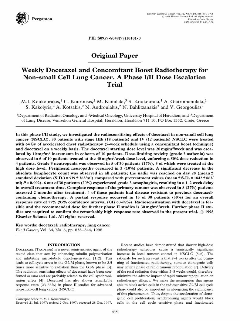
Figure 1 shows the mean absolute lymphocyte counts
(assessed weekly for 8 weeks after the beginning of treatment)
for the 30 lung cancer patients treated with docetaxel and
radiotherapy; after treatment completion, a gradual increase
in the absolute number of lymphocytes was observed, which
was time-dependent. Moreover, phenotypic analysis of the
peripheral blood lymphocytes from 10 patients (treated with
the 30 and 40 mg/m2/week dose levels) revealed that all
lymphocyte subpopulations (CD3, CD4, CD8, CD20, and
CD57) were signi®cantly decreased on day 28 (Table 4).
Response
Table 5 shows the responses observed at the three dose
levels. Complete response of the chest disease was observed
in 8 of 30 (27%) patients and partial response in 15 (50%).
The overall response rate was 77% (95% con®dence interval
(CI) 60±92%). One of the complete response patients had
known refractory disease to taxoids. 5 and 10 patients with
stage IIIb tumours showed a complete and partial response,
respectively, for an overall response rate of 83%. The overall
local (in radiotherapy ®eld) response rate in stage IV cases
was 67% (3 complete responses and 5 partial responses).
Minimal response was seen in 4 (13%) patients and stable or
progressive disease in 3 patients (10%). 7 of 30 patients had
metastatic disease not previously exposed to docetaxel and
none of these patients responded to treatment.
2 patients with stage IIIb disease who had responded to
treatment died because of massive haemoptysis (1 week and 2
months after treatment completion, respectively). One of
these patients showed necrotic tumour cavitation on CT
scan. 3 stage IIIb patients died of local relapse (at 2, 4 and 6
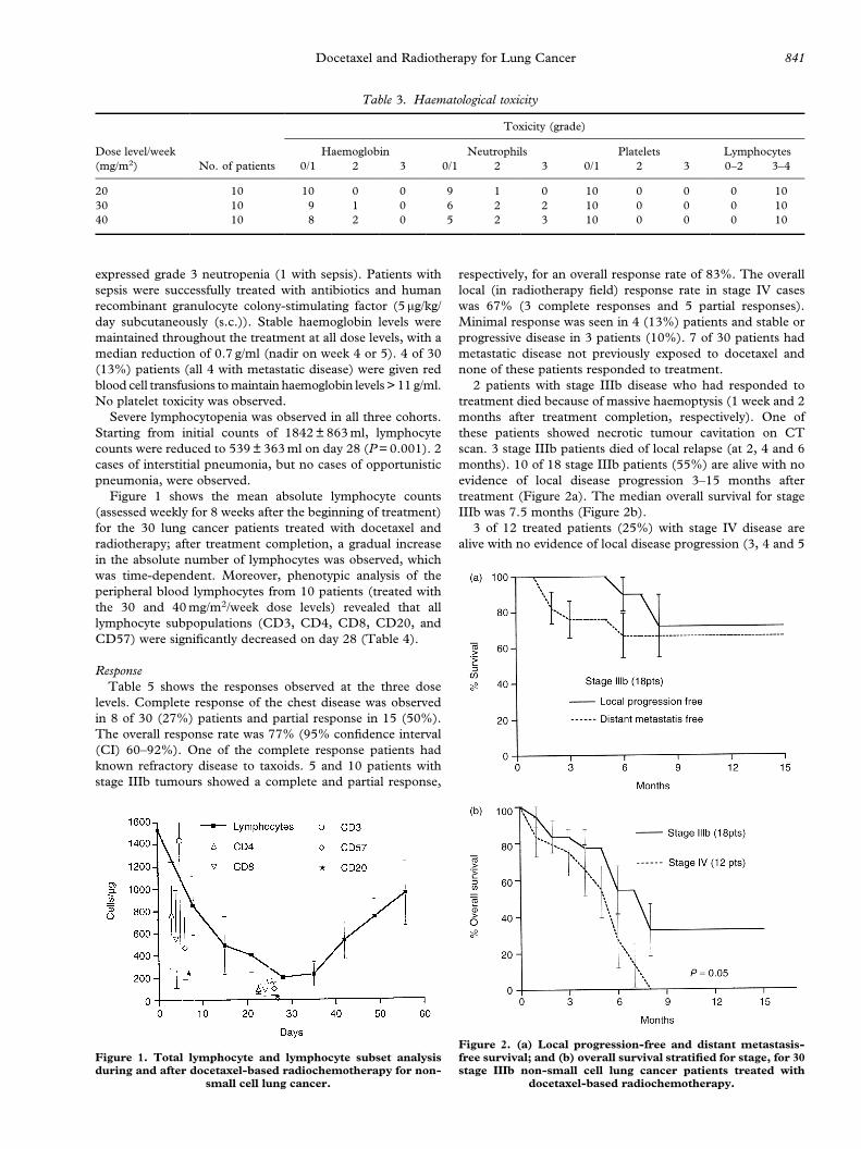
months). 10 of 18 stage IIIb patients (55%) are alive with no
evidence of local disease progression 3±15 months after
treatment (Figure 2a). The median overall survival for stage
IIIb was 7.5 months (Figure 2b).
3 of 12 treated patients (25%) with stage IV disease are
alive with no evidence of local disease progression (3, 4 and 5
Table 3. Haematological toxicity
Toxicity (grade)
Dose level/week Haemoglobin Neutrophils Platelets Lymphocytes
(mg/m2) No. of patients 0/1 2 3 0/1 2 3 0/1 2 3 0±2 3±4
20 10 10 0 0 9 1 0 10 0 0 0 10
30 10 9 1 0 6 2 2 10 0 0 0 10
40 10 8 2 0 5 2 3 10 0 0 0 10
Figure 2. (a) Local progression-free and distant metastasis-free survival; and (b) overall survival strati®ed for stage, for 30stage IIIb non-small cell lung cancer patients treated with
docetaxel-based radiochemotherapy.
Figure 1. Total lymphocyte and lymphocyte subset analysisduring and after docetaxel-based radiochemotherapy for non-
small cell lung cancer.
Docetaxel and Radiotherapy for Lung Cancer 841

months after treatment). 9 patients died of distant metastases
1±7 months after treatment completion, although no patients
had local disease progression con®rmed by CT scan. The
median survival for all stage IV patients was 5.5 months
(Figure 2b), which was signi®cantly lower than that for stage
IIIb patients (P = 0.05). Figure 3 shows the resolution of large
masses seen even before completion of treatment.
DISCUSSION
Locally advanced inoperable NSCLC is a major thera-
peutic problem because radiotherapy results are disappointing,
with 5-year survival less than 10%. The value of chemother-
apy combined with radiotherapy remains controversial. Three
of ®ve randomised studies have shown no survival advantage
of chemoradiotherapy over radiotherapy alone [10±14]. In a
recent study, we showed that 45% of patients with locally
advanced disease treated with radiotherapy alone will die
from local relapse with no evidence of distant metastases
[15]. Moreover, short schedules of high-dose radiotherapy
delivered within less than 5 weeks substantially improved the
overall and local disease-free survival [5, 6]. An additional
treatment that would increase the local control rate without
increasing toxicity would therefore result in further bene®t.
In the present study, we established a well-tolerated doce-
taxel scheme that could be administered concurrently with
radiotherapy in patients with NSCLC. Weekly doses of up to
30 mg/m2 were well tolerated with minimal haematological
toxicity and without severe asthenia, which was observed in
higher doses. This regimen, which delivers a total dose of
90 mg/m2 within 3 weeks, is in accordance with a previous
phase I study of docetaxel given on days 1 and 8 every 3
weeks where the maximum tolerated dose was 100 mg/m2
[16]. The radiation-induced acute toxicity, although higher
than expected from conventional radiotherapy alone, was
similar to that reported by accelerated regimens [17]. It
mainly concerned oesophagitis, which was the cause of 1±2
week treatment delay in 20% of patients. However, severe
oesophagitis was more frequent in the high dose level (40 mg/
m2/week) and was dose-limiting at this dose level. Reduction of
the radiation dose intensity delivered to the oesophagus, when-
ever possible, is strongly recommended to avoid treatment
delay and protect patients' quality of life during treatment.
The other dose-limiting toxicity associated with docetaxel
administration was grade 3 fatigue, which was observed in 4
of 10 patients treated with the high (40 mg/m2/week) dose
level. Asthenia resulted in treatment refusal in 1 patient and
docetaxel dose reduction in 3 other patients. Moreover,
asthenia was observed after the third or fourth week of treat-
ment, suggesting that it is cumulative. Administration of
vitamins and antidepressants did not prove to be of any help;
the patients recovered from this toxicity 3±4 weeks post-
treatment.
Another potentially important side-eVect is the severe
lymphocytopenia observed at all dose levels. In a previous
study of paclitaxel chemoradiotherapy for lung cancer, Reck-
zeh and colleagues [18] observed a similar eVect on lympho-
cyte counts which resulted in interstitial pneumonia outside
the radiation ®eld in 7 of 15 patients. Phenotypic analysis of
lymphocyte subsets revealed that T (CD4+ and CD8+), B
(CD20+) and NK (CD58+) cells are equally aVected during
the weekly administration of the docetaxel±radiotherapy
combination. This ®nding strongly suggests that the regimen
results in non-speci®c inhibition of lymphopoiesis. Whether
lymphocytopenia is directly related to docetaxel or to its
combination with corticosteroids or radiotherapy or both
cannot be answered from the present study. However, a simi-
lar decrease in the absolute number of CD4+ cells was also
observed in patients with NSCLC treated with docetaxel±
cisplatin or docetaxel±vinorelbine combination (not shown).
Therefore, this CD4+ lymphocytopenia is probably related to
the docetaxel administration rather than to radiotherapy.
The onset of asthenia and loss of appetite occurred in the
third week, together with the appearance of a severe drop in
lymphocyte count. The mechanisms of the appearance of
asthenia during cytotoxic treatment are unknown. Further
studies are needed to determine whether immunological
mechanisms may contribute to the appearance of fatigue.
In spite of the low absolute number of CD4+ cells, no case
of opportunistic infection was observed in this group of 30
patients; on the contrary, symptomatic interstitial pneumo-
nia, which was successfully treated with antibiotics, was
detected in only 2 of 30 patients (6.6%). This is in overt
contrast to the ®ndings of Reckzeh and colleagues, in which
many of their patients developed interstitial pneumonia.
Docetaxel has shown substantial activity against NSCLC
[19±21]. In previous phase II studies of ®rst-line treatment
Table 5. Response of the primary tumour
Complete response Partial response Minimal response
Stable or progressive
disease
Response rate
(%)
Dose level (mg/m2/week)
20 (n = 10) 3 4 3 0 70
30 (n = 10) 4 5 0 1 90
40 (n = 10) 1 6 1 2 80
Stage
IIIb (n = 18) 5 10 2 1 83
IV (n = 12) 3 5 2 2 67
Total 8 15 4 3 77
Table 4. Analysis of lymphocyte subsets
Lymphocytes
Day 0
(mean � S.D.)
Day 28
(mean � S.D.) P value
Total 1842 � 863* 539 � 363 0.002
CD3 1423 � 824 334 � 231 0.002
CD4 798 � 422 197 � 137 0.001
CD20 110 � 54 22 � 8 0.0003
CD57 168 � 230 134 � 62 0.002
S.D., standard deviation. *Results are expressed as the mean value
� S.D. of the observed absolute individual numbers of cells/ml.
842 M.I. Koukourakis et al.

with docetaxel combined with cisplatin or vinorelbine, we
con®rmed a 48 and 36% response rate, respectively [22, 23].
The overall response rate of 77% with 27% complete
responses observed in the present study is encouraging, as
most patients had disease unresponsive to chemotherapy. A
dramatic resolution of large masses was seen even before
treatment completion (Figure 3), showing an important
activity of the regimen in a subset of tumours. One complete
response and three partial responses were observed in
patients previously treated with taxoid-based chemotherapy.
Additional comparative studies in stage III patients are nee-
ded to fully demonstrate this bene®cial eVect of docetaxel.
The high response rate and the unexpected early complete
response (before the completion of 35 Gy) observed in 3 of
30 cases (10%) may be relevant to the recently reported
activity of taxanes on apoptosis-related proteins. Taxanes
induce phosphorylation of the anti-apoptotic protein bcl-2
[24]. Moreover, a p53-independent taxane-inducible apop-
tosis pathway has been suggested [25] and, indeed p53
mutations in NSCLC seem not to aVect the sensitivity to
Figure 3. (a) Patient with locally advanced high-grade squamous cell lung carcinoma before treatment and after 24 Gy ofradiotherapy (18 Gy delivered to the whole hemithorax) together with docetaxel (30 mg/m2/weekly); (b) complete responsedocumented 4 weeks after the beginning of treatment. Bars on (a) and (b) represent 10 cm and 5 cm respectively of actual length.
Docetaxel and Radiotherapy for Lung Cancer 843

radiotherapy [26]. Since radiation kills cancer cells by apop-
tosis induction, the combined radiation and taxane therapy
may become a unique model to investigate novel approaches
in radiosensitisation based on apoptosis modulation.
We conclude that docetaxel-sensitised radiotherapy is fea-
sible and this treatment option looks promising for stage IIIb
NSCLC, as the high response rate (even after low radiation
doses), the patterns of failure and overall survival (approxi-
mately 65% local and distant progression-free survival 15
months after treatment) were far beyond expectations [27].
Because 20 mg/m2/week was as eVective as higher dose levels,
a twice-weekly regimen (i.e. 15±20 mg/m2 twice a week) that
could better induce cell synchronisation would be a challen-
ging regimen to test.
1. Ringel I, Horwitz SB. Studies with RP-56976 (Taxotere): asemi-synthetic analogue of Taxol. J Natl Cancer Inst 1991, 83,288±291.
2. Geuritte-Voegelein F, Guenard D, Lavelle F, et al. Relationshipsbetween the structure of Taxol analogues and their antimitoticactivity. J Med Chem 1991, 34, 992±998.
3. ChaVey JT, Hellman S. DiVering responses to radiation of mur-ine bone marrow stem cells in relation to the cell cycle. CancerRes 1971, 31, 1513±1616.
4. Choy H, Rodriguez F, Wilcox B, et al. Radiation sensitizingeVects of Taxotere. Proc Am Assoc Cancer Res 1992, 33, 500.
5. Saunders MI, Dische S, Barrett A, et al. Randomized multicentertrials of CHART vs conventional radiotherapy in head-neck andnon-small cell lung cancer. An interim report. Br J Cancer 1996,73, 1455±1462.
6. Koukourakis M, Hlouverakis G, Kosma L, et al. The impact ofoverall treatment time on the results of radiotherapy for non-small cell lung cancer. Int J Radiat Oncol Biol Phys 1996, 34,315±322.
7. Withers HR, Taylor JMG, Maciejewski B. The hazards of accel-erated tumour clonogene repopulation during radiotherapy. ActaOncol 1988, 27, 131±146.
8. World Health Organization: Handbook for reporting results ofcancer treatment. Geneva, WHO, 1979.
9. Koukourakis M, Dimitakis J. LQ-based model for biologicalradiotherapy planning. Med Dosim 1994, 19, 269±277.
10. Mattson K, Holsti R, Holsti P, et al. Inoperable non-small celllung cancer radiation with or without chemotherapy. Int J ClinOncol 1988, 24, 477±482.
11. Dillman RO, Seagien SL, Propert KJ, et al. A randomized trial ofinduction chemotherapy plus high dose radiation versus radia-tion alone in stage III non-small cell lung cancer. N Engl J Med1990, 323, 940±945.
12. Le Chevalier T, Jett JR, Arriagada R, et al. Signi®cant eVectof adjuvant chemotherapy on survival in locally advanced non-small cell lung carcinoma (letter). J Natl Cancer Inst 1992, 84, 58.
13. Morton RF, Jett JR, McGinnis WL, et al. Thoracic radiationtherapy alone compared with combined chemoradiotherapy forlocally unresectable non-small cell lung cancer. Ann Intern Med1991, 115, 681±686.
14. Brodin O, Nou E, Mercke C, et al. Comparison of inductionchemotherapy before radiotherapy with radiotherapy only inpatients with locally advanced squamous cell carcinomas of thelung. Eur J Cancer 1996, 32, 1892±1900.
15. Koukourakis M, Skarlatos J, Kosma L, et al. Radiotherapy alonefor non-small cell lung carcinoma. Five year disease-free survivaland patterns of failure. Acta Oncol 1995, 34, 525±530.
16. Tomiak EPM, Kerger S, et al. Phase I study of docetaxeladministration as 1 hour intravenous infusion on a weekly basis.J Clin Oncol 1994, 12, 1458±1467.
17. Bishop JF, Ball D, Crennan E, et al. Radiation and carboplatincombined-modality therapy in non-small cell lung cancer. SeminOncol 1994, 21(Suppl. 6), 91±96.
18. Reckzeh B, Merte H, P¯uger K, et al. Severe lymphocytopeniaand interstitial pneumonia in patients treated with paclitaxel andsimultaneous radiotherapy for non-small cell lung cancer. J ClinOncol 1996, 14, 1071±1076.
19. Fossella FV, Lee JS, Murphy WK, et al. Phase II study of doc-etaxel for recurrent or metastatic non-small cell lung cancer. JClin Oncol 1994, 12, 1238±1244.
20. Cerny T, Kaplan S, Pavlidis N, et al. Docetaxel (Taxotere) isactive in non-small cell lung cancer. A phase II trial of theEORTC Early Clinical Trials Group (ECTG). Br J Cancer 1994,70, 384±387.
21. Francis P, Schneider J, Hann L, et al. Phase III trial of docetaxelin patients with platinum-refractory advanced ovarian cancer. JClin Oncol 1994, 12, 2301±2308.
22. Kourousis C, Androulakis N, Kakolyris S, et al. First-line treat-ment of non-small cell lung carcinoma (NSCLC) with docetaxeland vinorelbine; a phase II study. In Antypas G, ed. SecondInternational Congress on Lung Cancer, Crete, 9±13 November1996. Bologna, Italy, Monduzzi Editore S.p.A., 1996, 673±679.
23. Androulakis N, Kourousis C, Kakolyris S, et al. First-line treat-ment of non-small cell lung carcinoma (NSCLC) with docetaxeland cisplatin: preliminary results of a phase II study. In AntypasG, ed. Second International Congress on Lung Cancer, Crete, 9±13November 1996. Bologna, Italy, Monduzzi Editore S.p.A., 1996,661±667.
24. Haldar S, Chintapalli J, Croce CM. Taxol induces bcl-2 phos-phorylation and death of prostate cancer cells. Cancer Res 1996,56, 1253±1255.
25. Walf AF, Donaldson KL, Fairchild C, et al. Loss of normal p53function confers sensitization to Taxol by increasing G2/M arrestand apoptosis. Nat Med 1996, 2, 72±79.
26. Safran H, King-T, Choy H, et al. p53 mutations do not predictresponse to paclitaxel/radiation for nonsmall cell lung carcinoma.Cancer 1996, 78, 1203-1210.
27. Green MR, Cox JD, Ardizzoni A, et al. Endpoints for multi-modal clinical trials in stage III non small cell lung cancer(NSCLC): a consensus report. Lung Cancer 1994, 11(Suppl. 3),S11±S13.
844 M.I. Koukourakis et al.