intrago: intraoperative radiotherapy in glioblastoma multiforme - a phase i/ii dose escalation study
TRANSCRIPT
Giordano et al. BMC Cancer 2014, 15:992http://www.biomedcentral.com/1471-2407/14/992
STUDY PROTOCOL Open Access
INTRAGO: intraoperative radiotherapy inglioblastoma multiforme – a Phase I/II doseescalation studyFrank A Giordano1*, Stefanie Brehmer2, Yasser Abo-Madyan1,3, Grit Welzel1, Elena Sperk1, Anke Keller1,Frank Schneider1, Sven Clausen1, Carsten Herskind1, Peter Schmiedek2 and Frederik Wenz1
Abstract
Background: Glioblastoma multiforme (GBM) is the most frequent primary malignant brain tumor in adults. Despitemultimodal therapies, almost all GBM recur within a narrow margin around the initial resected lesion. Thus, noveltherapeutic intensification strategies must target both, the population of dispersed tumor cells around the cavityand the postoperative microenvironment. Intraoperative radiotherapy (IORT) is a pragmatic and effective approachto sterilize the margins from persistent tumor cells, abrogate post-injury proliferative stimuli and to bridge thetherapeutic gap between surgery and radiochemotherapy. Therefore, we have set up INTRAGO, a phase I/IIdose-escalation study to evaluate the safety and tolerability of IORT added to standard therapy in newly diagnosedGBM. In contrast to previous approaches, the study involves the application of isotropic low-energy (kV) x-raysdelivered by spherical applicators, providing optimal irradiation properties to the resection cavity.
Methods/Design: INTRAGO includes patients aged 50 years or older with a Karnofsky performance status of atleast 50% and a histologically confirmed (frozen sections) supratentorial GBM. Safety and tolerability (i.e., themaximum tolerated dose, MTD) will be assessed using a classical 3 + 3 dose-escalation design. Dose-limiting toxicities(DLT) are wound healing deficits or infections requiring surgical intervention, IORT-related cerebral bleeding or ischemia,symptomatic brain necrosis requiring surgical intervention and early termination of external beam radiotherapy (beforethe envisaged dose of 60 Gy) due to radiotoxicity. Secondary end points are progression-free and overall survival.
Trial registration: The study is registered with clinicaltrials.gov, number: NCT02104882 (Registration Date: 03/26/2014).
BackgroundDespite recent advances in therapy, Glioblastoma multi-forme (GBM) is a lethal disease in most cases with arelatively short overall survival of roughly 15 months[1,2]. In virtually all cases, GBM recur locally within anarrow margin (2–3 cm) around the tumor cavity [3-5].Although GBM are highly invasive and able to migratealong pre-existing structures such as blood vessels orwhite matter tracts [6,7] most (if not all) recurrent tumorsparadoxically grow in close proximity to the resectionmargin [8]. Thus, though novel surgical techniques (suchas fluorescence-guided resection) may have improved the
* Correspondence: [email protected] of Radiation Oncology, University Medical Center Mannheim,University of Heidelberg, Theodor-Kutzer-Ufer 1-3, 68167 Mannheim,GermanyFull list of author information is available at the end of the article
© 2014 Giordano et al.; licensee BioMed CentrCommons Attribution License (http://creativecreproduction in any medium, provided the orDedication waiver (http://creativecommons.orunless otherwise stated.
rates of macroscopic complete resections [9], and ad-vanced radiotherapy techniques are at hand, no single orcombined approach is sufficient to deplete microscopicallydispersed tumor cells around the tumor cavity.One of the techniques employed to tackle this challen-
ging feature of GBM is intraoperative radiotherapy (IORT).IORT allows the delivery of high doses of electrons(IOERT) or low energy x-rays to the tumor bed whilethe surrounding healthy tissue is spared from radiationdue to steep dose gradients [10]. This could lead to instantsterilization of the cavity surface from remaining tumorcells and delayed, or impaired, tumor cell proliferationbetween surgery and adjuvant therapies in deeper areasreceiving lower doses. Furthermore, high single doses mayelicit local (tumor bed) and systemic (immunogenic)responses which are not observed in this extent afterconventionally fractionated radiotherapy [11,12].
al. This is an Open Access article distributed under the terms of the Creativeommons.org/licenses/by/4.0), which permits unrestricted use, distribution, andiginal work is properly credited. The Creative Commons Public Domaing/publicdomain/zero/1.0/) applies to the data made available in this article,
Age ≥ 50 yearsKPS ≥ 50%
informed consent
First visitPhysical ExamLENT-SOMAMedication
Maximum safe resection
IORTfeasible
IORT not feasible
IORT 20 - 40 Gy
Pre-EBRTPhysical ExamLENT-SOMAMedication
early MRI (24-48h)
Chemo-RadiotherapyEBRT (30 x 2 Gy)
+ 50 mg/m2/d temozolomide
post-EBRTPhysical ExamLENT-SOMAMedication
Follow-up MRIsevery 3 months
adjuvant Chemotherapy6 cycles 150-200 mg/m2/d/cycle temozolomide
FUPhysical ExamLENT-SOMAMedication
Early D
LT (3 w
eeks)
Early-d
elayed D
LT (3 m
on
ths)
Histologically confirmed GBM (frozen section)
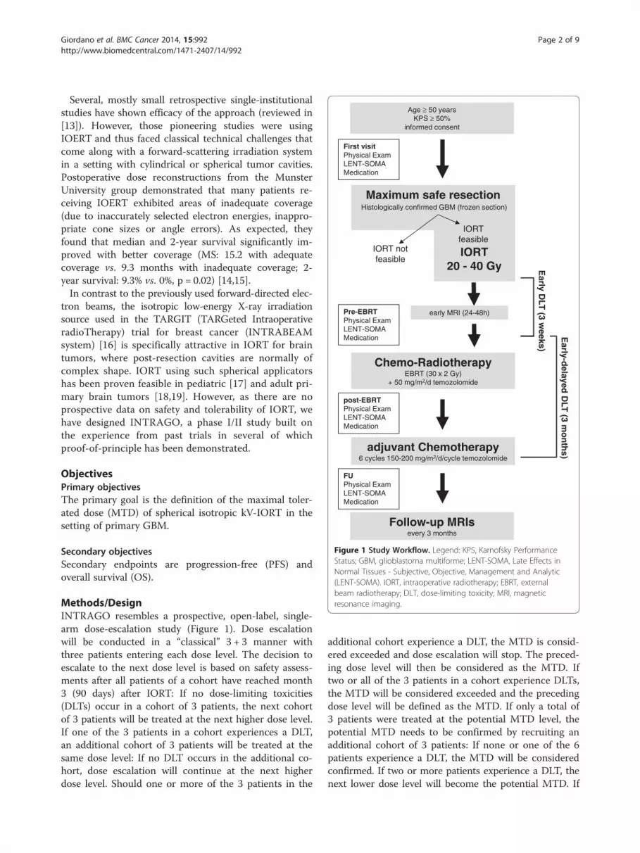
Figure 1 Study Workflow. Legend: KPS, Karnofsky PerformanceStatus; GBM, glioblastoma multiforme; LENT-SOMA, Late Effects inNormal Tissues - Subjective, Objective, Management and Analytic(LENT-SOMA). IORT, intraoperative radiotherapy; EBRT, externalbeam radiotherapy; DLT, dose-limiting toxicity; MRI, magneticresonance imaging.
Giordano et al. BMC Cancer 2014, 15:992 Page 2 of 9http://www.biomedcentral.com/1471-2407/14/992
Several, mostly small retrospective single-institutionalstudies have shown efficacy of the approach (reviewed in[13]). However, those pioneering studies were usingIOERT and thus faced classical technical challenges thatcome along with a forward-scattering irradiation systemin a setting with cylindrical or spherical tumor cavities.Postoperative dose reconstructions from the MunsterUniversity group demonstrated that many patients re-ceiving IOERT exhibited areas of inadequate coverage(due to inaccurately selected electron energies, inappro-priate cone sizes or angle errors). As expected, theyfound that median and 2-year survival significantly im-proved with better coverage (MS: 15.2 with adequatecoverage vs. 9.3 months with inadequate coverage; 2-year survival: 9.3% vs. 0%, p = 0.02) [14,15].In contrast to the previously used forward-directed elec-
tron beams, the isotropic low-energy X-ray irradiationsource used in the TARGIT (TARGeted IntraoperativeradioTherapy) trial for breast cancer (INTRABEAMsystem) [16] is specifically attractive in IORT for braintumors, where post-resection cavities are normally ofcomplex shape. IORT using such spherical applicatorshas been proven feasible in pediatric [17] and adult pri-mary brain tumors [18,19]. However, as there are noprospective data on safety and tolerability of IORT, wehave designed INTRAGO, a phase I/II study built onthe experience from past trials in several of whichproof-of-principle has been demonstrated.
ObjectivesPrimary objectivesThe primary goal is the definition of the maximal toler-ated dose (MTD) of spherical isotropic kV-IORT in thesetting of primary GBM.
Secondary objectivesSecondary endpoints are progression-free (PFS) andoverall survival (OS).
Methods/DesignINTRAGO resembles a prospective, open-label, single-arm dose-escalation study (Figure 1). Dose escalationwill be conducted in a “classical” 3 + 3 manner withthree patients entering each dose level. The decision toescalate to the next dose level is based on safety assess-ments after all patients of a cohort have reached month3 (90 days) after IORT: If no dose-limiting toxicities(DLTs) occur in a cohort of 3 patients, the next cohortof 3 patients will be treated at the next higher dose level.If one of the 3 patients in a cohort experiences a DLT,an additional cohort of 3 patients will be treated at thesame dose level: If no DLT occurs in the additional co-hort, dose escalation will continue at the next higherdose level. Should one or more of the 3 patients in the
additional cohort experience a DLT, the MTD is consid-ered exceeded and dose escalation will stop. The preced-ing dose level will then be considered as the MTD. Iftwo or all of the 3 patients in a cohort experience DLTs,the MTD will be considered exceeded and the precedingdose level will be defined as the MTD. If only a total of3 patients were treated at the potential MTD level, thepotential MTD needs to be confirmed by recruiting anadditional cohort of 3 patients: If none or one of the 6patients experience a DLT, the MTD will be consideredconfirmed. If two or more patients experience a DLT, thenext lower dose level will become the potential MTD. If
Giordano et al. BMC Cancer 2014, 15:992 Page 3 of 9http://www.biomedcentral.com/1471-2407/14/992
no DLT occurs at all, the highest dose level will be definedas the MTD. Thus, the MTD is defined as the highestdose at which one or no DLT will have been observedamong 6 patients.
Patient selectionPatients aged 50 years or older with a Karnofsky per-formance status (KPS) of at least 50% and a histologi-cally confirmed (frozen sections) supratentorial unifocalGBM are included. The tumor location should allowmaximum safe resection. However, patients with tumorsthat are likely to be only partially removable are also eli-gible for the study.
Inclusion criteria
� Histologically confirmed glioblastoma multiforme infrozen sections
� Age ≥50 years� Karnofsky Performance Index ≥ 50%� Written Informed consent� Adequate birth control (e.g., oral contraceptives)
Exclusion criteria
� Astrocytoma ≤WHO grade III� Gliomatosis cerebri� Multifocal lesions� Infratentorial localization� Previous cranial radiation therapy (any location)� Uncontrolled intercurrent illnesses including, but
not limited to, ongoing or active infection orpsychiatric illness/social situations that would limitcompliance with study requirements.
� Contraindications for general anaesthesia� Bleeding or clotting disorders� Contraindications for MRI or CT scans� Pregnant or breastfeeding women
Assessment of the primary objective (Safety)The primary goal is to determine the MTD, which willbe assessed on the basis of pre-defined dose-limitingtoxicities (DLT).Two types of DLT are defined:
1) Early DLTs (≤3 weeks after IORT):
– wound infections/wound healing difficultiesrequiring surgical intervention– IORT-related cerebral bleeding or ischemia
2) Early-delayed DLTs (≤3 months after IORT):– Symptomatic brain necrosis requiring surgical
intervention– Early termination of EBRT (before the envisaged
dose of 60 Gy) due to radiotoxicity
DLTs will be assessed on the basis of clinical presen-tation (physical examination, KPS, current medication),imaging studies (MRI) and on the basis of a neuro-logical assessment using the Late Effects in NormalTissues Subjective, Objective, Management and Analytic(LENT-SOMA) scales defined by the EORTC/RTOG[20,21].
Evaluation of DLT: clinical exams, medicationEach follow-up visit has to include the most recent med-ical history, an inspection of the wound/scar and a thor-ough clinical exam. Episodes of partial or complex seizuresmust be documented. Wound healing (and the scar at FU)is followed with photo documentation. When performinga physical exam, there should be a specific emphasis onneurological functions. Specific awareness is advised forsigns of cerebral edema (for example alterations in thelevel of consciousness, bradycardia, high blood pressure orinequality of pupillary size). All current medication and allchanges made in the medication schedule have to bedocumented. Detailed information on doses (and dosechanges) has to be only documented for corticosteroidsand anticonvulsants.
Evaluation of DLT: MRIEach follow-up visit includes MRI with contrast-enhanced(gadolinium) T1, axial T2 and axial T2-FLAIR sequences.Ischemic areas can be delineated using perfusion diffusion-weighted imaging. It is challenging to distinguish earlypost-treatment blood–brain barrier permeability impair-ment from tumor recurrence and “true” brain necrosis.Here, methods such as Lesion Quotient (LQ), which re-sembles a ratio of the area delineated in a T2 sequence andthe area of the corresponding contrast-enhanced T1 se-quence may be applied [22]. In case MRI scans are incon-clusive, positron emission tomography with amino acidtracers (such as 18F-fluoro-ethyl-tyrosine) can be offered aspreferential modality.
Management of DLTWound infections must be adequately treated, e.g. withdry absorbent dressing and, in case of positive woundswabs, systemic antibiotics should be administered match-ing to the antibiogram. In all cases of wound infection,blood workup (Complete blood counts, white bloodcounts, CRP) and imaging studies should be performed(CT or MRI) to rule out intracranial abscesses. Each caseof wound infection (our healing difficulty) where conser-vative therapy fails and surgical revision is required is de-fined as DLT.Due to the highly flexible positioning system of the
IORT device, cases of cerebral ischemia or bleeding in-duced by the applicator are unexpected. Nevertheless,both were included into the protocol as DLTs and cases

Giordano et al. BMC Cancer 2014, 15:992 Page 4 of 9http://www.biomedcentral.com/1471-2407/14/992
where (venous or arterial) ischemia or intracranialhaemorrhage occur as a consequence of the IORT pro-cedure (addition to the baseline risk), adequate therapyis required. Intracranial haemorrhages must be generallysurgically removed if mass effects are exceeding the pri-mary lesion volume. There is no symptomatic therapyfor arterial or venous ischemia post resection. However,diffusion-weighted imaging (DWI) should be performedto document these events during follow-ups.If radiation necrosis is suspected in MRI scans and no
correlating clinical deterioration is noted, symptomatictherapy may not be necessary and observation is appro-priate. For patients with mass effects or neurologicalsymptoms, treatment options include conservative ther-apy with corticosteroids or anti-angiogenic substances(e.g., bevacizumab) or surgical debulking of the necroticareas. The decision for either therapy should be made ininterdisciplinary consensus (e.g., in interdisciplinarytumor boards). If surgery is required, the correspondingcase will be defined as DLT.All patients will undergo radiochemotherapy and will
present five times per week at the linear accelerators.Complete blood counts (CBC), a chemistry panel, liverfunction tests (LFT) as well as renal function tests (RFT)are regularly performed to screen for hematotoxicity,hepatotoxicity or renal toxicity under temozolomidechemotherapy. Upon intolerance, incompliance or de-terioration of CBCs, LFTs or RFTs, the therapy withtemozolomide can be discontinued at any time point byany physician in charge. Any discontinuation of chemo-therapy for more than 5 consecutive days has to bedocumented.Cranial irradiation can elicit acute (side) effects occur-
ring during treatment or early-delayed effects that appearwithin three months after radiochemotherapy [23]. In mostcases, both acute and early delayed side effects largely re-semble symptoms of mass effects (e.g., headache, nausea,worsening neurological symptoms), they are responsive tocorticosteroids and they either gradually decrease in sever-ity during daily radiotherapy (acute effects) or spontan-eously resolve under cortisol (early-delayed). However, anycase of radiotherapy-associated symptom deteriorationthat requires pausing of radiotherapy for more than 5 con-secutive days has to be documented. If radiotherapy is en-tirely discontinued due to radiotherapy-associated sideeffects before the total dose of 60 Gy is reached, the caseis considered to exhibit a DLT. As for chemotherapy,EBRT may be discontinued by any physician in charge.
Assessment of secondary end points (efficacy)Secondary end points are progression-free (PFS) andoverall survival (OS). PFS is defined as the interval (indays or months) from IORT to the date of first detectionof progressive disease according to updated RANO
criteria [24] or the date of last follow-up. The RANOworking group recommended tight restrictions forevaluating progression within 12 weeks after radiother-apy as irradiation transiently increases the permeabilityof the (peri)tumoral vasculature which in turn impairsthe validity of MRI scans during this period [25].In INTRAGO, only cases where new T1-enhancing le-
sions are detected beyond the 80% isodose or new lesionsthat are histologically proven to be new manifestations ofGBM are defined as true progression within a timeframeof 12 weeks after EBRT.After this period, progressive disease is defined if one
of the following RANO criteria applies:
– New T1-contrast-enhancing lesions outside ofradiation field on decreasing, stable, or increasingdoses of corticosteroids
– Increase by ≥ 25% in the sum of the products ofperpendicular diameters between the first post-radiotherapy scan, or a subsequent scan with smallertumor size, and the scan at 12 weeks or later onstable or increasing doses of corticosteroids.
– Clinical deterioration attributable to tumor progress(and not to concurrent medication or diseases)
– Increased T2/FLAIR compared with baseline scan orbest response after initiation of therapy on stable orincreasing doses of corticosteroids.
OS is defined as the interval (in days or months) fromIORT to the date of death from any cause.
Imaging, interventions, follow-upFirst visitPatients that are eligible for the study will be initially ex-amined and the general medical condition (height, weight,general condition, Karnofsky index, medical history, medi-cation) and the neurological (baseline) status (neurologicalexam and LENT-SOMA scales) will be assessed. All pa-tients have to be willing and able to undergo repetitiveMRI scans. Written informed consent must be obtained atleast 24 h prior to surgery and IORT.
Preceding imaging studiesAll patients enrolled will receive preoperative MRI im-aging (minimum 1.5 T) including contrast-enhanced T1-weighted magnetization-prepared rapid gradient echo(MP-RAGE) sequences to provide a 3D data set forimage-guided surgery. Advanced imaging (such as perfu-sion or diffusion weighted imaging) may be performedat this stage but is not mandatory.
IORT planning, risk structuresThe optic nerve(s) (or the chiasm, respectively) and thebrain stem are defined as risk structures and have to be

Giordano et al. BMC Cancer 2014, 15:992 Page 5 of 9http://www.biomedcentral.com/1471-2407/14/992
identified on pre-operative scans. If any risk structure islocated ≤2 cm to a T1-enhancing lesion in pre-operativeMRIs, intraoperative imaging (e.g. intraoperative ultra-sound or in-room CT/MRI) should be used after re-moval of the tumor (to account for potential brainshifts) to document distances to the applicator surfaceand to allow dose definition.
SurgeryThe resection procedure should be performed as image-(neuronavigation) guided surgery with techniques thatmeet individual center standards and preferences. Resec-tion techniques may include suction, bi-/monopolar cau-tery or ultrasound aspiration. Intraoperative application of5-aminolevulinic acid (5-ALA) can be used to determineresidual tumor tissue. A maximum safe resection approachis recommended, but not mandatory. Due to the possibilityof liquor accumulation (or retention) around the applica-tor and subsequent lowering of doses to the target volume(i.e. the resection cavity wall), ventricular opening duringsurgery should be avoided whenever possible.
Frozen sectionTo establish the diagnosis of GBM, representative tissuesamples have to be sent for histopathological examin-ation. The frozen section/cryosection procedure can beperformed according to local standards. In case histo-pathological hallmarks of grade 4 are present, the patientfulfils all inclusion criteria. If the diagnosis of GBM cannot be reliably established with cryosection or if add-itional analyses are necessary, IORT must be omitted.
Dose prescription and application of IORTFollowing establishment of the diagnosis via frozen sectionand after the surgeon defines the macroscopic (or 5-ALA-delineated) tumor to be satisfactorily removed, IORT willbe prepared. All potentially involved risk structures thathave been defined in pre-operative imaging (see above)may have displaced consequent to neuro-shifting (i.e.reduced intracerebral pressure and loss of liquor afterresection) and should be re-identified with intraopera-tive imaging (ultrasound or in-room CT/MRI).For the two risk structures (Optical nerve/chiasm and
brain stem), dose constraints of 12 Gy (Optical nerve) and12.5 Gy (brain stem) are commonly accepted in LINAC-based EBRT according to the QUANTEC (QuantitativeAnalyses of Normal Tissue Effects in the Clinic) recom-mendations [26]. Since kV-irradiation shows an increasedrelative biological effectiveness (RBE) [27], adapted doseconstraints (DMax) of 10 Gy apply for both structures dur-ing IORT with the (kV-based) INTRABEAM-System.DMax to these structures are then defined intraoperativelyon the basis of the dose-depth profiles of the correspond-ing applicator. If the doses to the risk structures at IORT
(DIORT) exceed 10 Gy, IORT is technically not feasible andhas to be omitted (screening failure). If any risk structureis likely to receive a DIORT of > 10 Gy (e.g., the risk struc-ture has direct contact to the applicator surface) it mustbe sufficiently shielded with cut-to-size tungsten-filed sili-cone shielding strips. Shielding with one layer tungsten-silicone strips will reduce the DIORT by 90%.Based on the cavity geometry and adjacent functional
brain areas, the most suitable applicator will be chosenby the team of surgeons and radiation oncologists (sizesrage from 1.5- 5.0 cm in 0.5 cm steps). The applicatorwill be inserted in the cavity correct positioning and ad-jacent risk structures will then be again visualized usingintraoperative imaging (ultrasound or in-room CT/MRI).Fluids surrounding the surface should be ruled out or
removed. The applicator is then taken out and mountedonto the INTRABEAM system. Next, the arm and thesource are covered with the sterile drape and the mountedapplicator is again fitted into the resection cavity. Radi-ation will then be initiated by a radiation oncologist for adefined time span as calculated by the machine software.After IORT, surgery will be continued in a regular
fashion without specific additional requirements.
Radiation protection issuesIORT has to be delivered in accordance with federal,state and/or local regulations on radiation protection.IORT with the INTRABEAM® System does not requirestructural alterations if the operating room is approvedfor C-arm fluoroscopy [28].
Early postoperative MRIEarly postoperative MRIs must be performed within awindow of 24–48 h after surgery and must be analyzed ina standardized assessment. Before contrast applicationT1-hyperintense lesions must be used to evaluate residualblood/heme and T2-TSE, T2-FLAIR, and DWI to evaluateischemia. After contrast application, T1-hyperintensemasses or nodules (residual tumor tissue) have to bequantified and the following has to be documented:
– Complete resection: removal of at least 98% of theT1-enhancing lesions.
– Subtotal resection: removal of 88-98% of theT1-enhancing lesions.
– Partial resection: removal of less than 88% of theT1-enhancing lesions.
Pre-EBRT visitBefore EBRT and concomitant chemotherapy is initiated,all patients will be re-examined (including a neurologicalexam, an update on medication and a re-assessmentbased on LENT-SOMA scales) to document changes
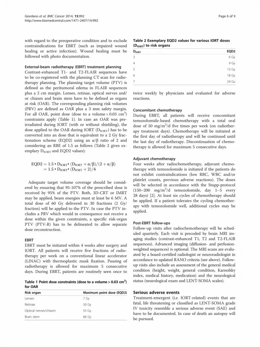
Table 2 Exemplary EQD2 values for various IORT doses(DIORT) to risk organs
DIORT EQD2
3 6 Gy
4 9 Gy
5 13 Gy
6 18 Gy
7 24 Gy
Giordano et al. BMC Cancer 2014, 15:992 Page 6 of 9http://www.biomedcentral.com/1471-2407/14/992
with regard to the preoperative condition and to excludecontraindications for EBRT (such as impaired woundhealing or active infection). Wound healing must befollowed with photo documentation.
External-beam radiotherapy (EBRT) treatment planningContrast-enhanced T1- and T2-FLAIR sequences haveto be co-registered with the planning CT scan for radio-therapy planning. The planning target volume (PTV) isdefined as the peritumoral edema in FLAIR sequencesplus a 2 cm margin. Lenses, retinae, optical nerves and/or chiasm and brain stem have to be defined as organsat risk (OAR). The corresponding planning risk volumes(PRV) are defined as OAR plus a 3 mm safety margin.For all OAR, point dose (dose to a volume > 0.03 cm3)constraints apply (Table 1). In case an OAR was pre-irradiated during IORT (with or without shielding), thedose applied to the OAR during IORT (DIORT) has to beconverted into an dose that is equivalent to a 2 Gy frac-tionation scheme (EQD2) using an α/β ratio of 2 andconsidering an RBE of 1.5 as follows (Table 2 gives ex-emplary DIORT and EQD2 values):
EQD2 ¼ 1:5 • DIORT• DIORT þ α=βð Þ= 2þ α=βð Þ¼ 1:5 • DIORT• DIORT þ 2ð Þ=4
Adequate target volume coverage should be consid-ered by ensuring that 95-107% of the prescribed dose isreceived by 95% of the PTV. Both, 3D-CRT or IMRTmay be applied, beam energies must at least be 6 MV. Atotal dose of 60 Gy delivered in 30 fractions (2 Gy/fraction) will be applied to the PTV. In case the PTV in-cludes a PRV which would in consequence not receive adose within the given constraints, a specific risk-organPTV (PTV-R) has to be delineated to allow separatedose reconstruction.
EBRTEBRT must be initiated within 4 weeks after surgery andIORT. All patients will receive five fractions of radio-therapy per week on a conventional linear accelerator(LINAC) with thermoplastic mask fixation. Pausing ofradiotherapy is allowed for maximum 5 consecutivedays. During EBRT, patients are routinely seen once to
Table 1 Point dose constraints (dose to a volume> 0.03 cm3)for OAR
Risk organ Maximum point dose (EQD2)
Lenses 7 Gy
Retinae 50 Gy
Optical nerves/chiasm 55 Gy
Brain stem 66 Gy
twice weekly by physicians and evaluated for adversereactions.
Concomitant chemotherapyDuring EBRT, all patients will receive concomitanttemozolomide-based chemotherapy with a total oraldose of 50 mg/m2/d five times per week (on radiother-apy treatment days). Chemotherapy will be initiated atthe first day of radiotherapy and will be continued untilthe last day of radiotherapy. Discontinuation of chemo-therapy is allowed for maximum 5 consecutive days.
Adjuvant chemotherapyFour weeks after radiochemotherapy, adjuvant chemo-therapy with temozolomide is initiated if the patients donot exhibit contraindications (low RBC, WBC and/orplatelet counts, previous adverse reactions). The doseswill be selected in accordance with the Stupp-protocol(150–200 mg/m2/d temozolomide, day 1–5 every28 days) [2]. At least six cycles of chemotherapy shouldbe applied. If a patient tolerates the cycling chemother-apy with temozolomide well, additional cycles may beapplied.
Post-EBRT follow-upsFollow-up visits after radiochemotherapy will be sched-uled quarterly. Each visit is preceded by brain MRI im-aging studies (contrast-enhanced T1, T2 and T2-FLAIRsequences). Advanced imaging (diffusion- and perfusion-weighted sequences) is optional. The MRI scans are evalu-ated by a board-certified radiologist or neuroradiologist inaccordance to updated RANO criteria (see above). Follow-up visits also include an assessment of the general medicalcondition (height, weight, general condition, Karnofskyindex, medical history, medication) and the neurologicalstatus (neurological exam and LENT-SOMA scales).
Serious adverse eventsTreatment-emergent (i.e. IORT-related) events that arefatal, life threatening or classified as LENT-SOMA gradeIV toxicity resemble a serious adverse event (SAE) andhave to be documented. In case of death an autopsy willbe pursued.
Giordano et al. BMC Cancer 2014, 15:992 Page 7 of 9http://www.biomedcentral.com/1471-2407/14/992
Ethical aspects, trial registrationINTRAGO is approved by the local ethics committee(Medical Ethics Commission II of the Faculty of Medi-cine Mannheim, University of Heidelberg; 2013-548S-MA) and the Federal Office of Radiation Protection(Z 5-22462/2-2013-063). The trial is registered withclinicaltrials.gov, number: NCT02104882.
DiscussionWe here present the first dose-finding study on low-energy intraoperative radiotherapy for glioblastoma mul-tiforme. Previous approaches used forward-directed elec-tron beams that only inconsistently provided sufficienttarget volume coverage, leading to inconsistency in re-ported outcomes [15,29-37]. Within INTRAGO, thespherically irradiating devices of the INTRABEAM sys-tem are used to enable geometry-optimized IORT. Thismay for the first time enable sufficient dose delivery tothe resection cavity and to remaining tumor cells.On may argue that there is no benefit of further dose
escalation after resection due to failure of trials involvingdose escalation or additional radiosurgery to the tumorbed [38]. We challenge this conclusion on the basis oftwo facts that involve the (crucial) time between surgeryand adjuvant therapy:First, it is known that the mean doubling time of a
GBM stem cell may be as fast as 24 hours and conse-quently, the waiting time for EBRT correlates with over-all survival [39,40]. IORT is embedded in the surgicalremoval of the mass and thus will likely prevent and/orslow down this exceptionally fast tumor growth in thistimeframe. Second, GBM growth shows dose-dependencyas early studies showed that doses of at least 50 Gy are re-quired to improve overall survival [41,42]. In the RTOG98–03 trial, patients treated with 66 Gy showed the worst(11.6 months) and those receiving 84 Gy showed the bestsurvival rates (19.3 months) without increased rates oftoxicities [43]. Of note, this study was conducted with ra-ther “old” techniques and in the era of more advancedfunctional imaging (such as PET) and irradiation tech-niques (intensity-modulated radiotherapy) dose-escalationmay be even more safely and efficiently conducted [44].We think that the reason why several other dose-escalation trials were failing is likely to be related toprocesses occurring during the time between surgeryand adjuvant therapy: any wounded site creates a spe-cific stimulatory environment to promote healing,which inadvertently provides remaining cancer cellswith strong pro-proliferative, pro-migratory and anti-apoptotic stimuli [45,46]. In breast cancer, it has beenshown that this unwanted response of the injuredmicroenvironment can be attenuated with IORT [47].This had direct consequences on the clinical outcome:patients that received IORT in a sequential operation, i.
e. after the first surgery confirmed the diagnosis (‘post-pathology’ cohort) showed higher local recurrence rates(5.4%) compared with patients that received IORT dur-ing first surgery (2.1%; ‘pre-pathology’ cohort) [16].Whether or not traumatic brain injury has a similarstrong influence on GBM cell proliferation post surgeryas observed in breast cancer has not been demonstratedyet. However, as the injured brain is very well known torespond with an impressive cytokine cocktail that effi-ciently promotes astrocytic activation and proliferation[48-50], we believe that similar (and, regarding survival,likely highly beneficial) quenching of the tumor micro-environment may be achievable after IORT for GBM.Symptomatic brain necrosis requiring surgical inter-
vention was defined as DLT. Brain necroses appeared inmultiple IOERT trials and they mostly correlated withimproved survival [29]. This, together with the fact thatbevacizumab is a novel and effective option to conserva-tively treat brain necroses [51] was prompting us to onlyconsider brain necroses as a DLT if they become symp-tomatic and if they require surgery. However, althoughwe do expect cases of brain necrosis, highly elevatedrates are less likely as it is well known that the irradiatedvolume of brain is the key determinant for this side ef-fect [52]. The device used for IORT in INTRAGO useslow-energy (kV) photons that show exponential attenu-ation along their path [53]. Logically, the area receivinghigh(est) doses is a margin of maximum 1–1.5 cm widtharound the tumor cavity (IORT with 40 Gy surface doseat dose level III would result in doses of 12 Gy at 1 cm,8 Gy at 1.5 cm and 4 Gy in 2 cm depth), which is a vol-ume that is eventually not large enough to become clin-ical apparent.INTRAGO is the first prospective IORT study in the
era of temozolomide. The alkylating agent has become acrucial part of standard treatment after the pivotalEORTC/NCIC study showed a considerable improve-ment of both overall and long-term survival rates if thesubstance is added to radiotherapy and given as adjuvantchemotherapy [1]. It is believed that the increased ratesof blood–brain barrier permeability impairments thatare seen after radiochemotherapy (and which are oftenmisinterpreted as progressive disease) are predominantlycaused by temozolomide [25,54]. This abnormal local re-actions together with pre-clinical investigations point toadditive and/or even synergistic activity of both modal-ities and it will be of specific interest to see whetherIORT can further modulate these interactions [55,56].In conclusion, INTRAGO is the first dose-finding
study on low-kV-IORT for newly diagnosed GBM in thetemozolomide era with optimized geometry adaptation.It should provide a robust basis for subsequent random-ized (phase II or III) trials, in which superiority overstandard treatment must be tested.
Giordano et al. BMC Cancer 2014, 15:992 Page 8 of 9http://www.biomedcentral.com/1471-2407/14/992
Abbreviations3D-CRT: Three-Dimensional Conformal Radiation Therapy; CBC: CompleteBlood Count; CT: Computer Tomography; DIORT: Dose applied to an OARduring IORT; DLT: Dose-Limiting Toxicity; DWI: Diffusion-Weighted (MagneticResonance) Imaging; EBRT: External Beam Radiotherapy; EORTC: EuropeanOrganisation for Research and Treatment of Cancer; EQD2: Equivalent Dosein 2 Gy Fractions; FLAIR: Fluid Attenuated Inversion Recovery; FET: 18 F-fluoro-Ethyl-Tyrosine; FU: Follow-Up; GBM: Glioblastoma Multiforme; IMRT: Intensity-Modulated Radiation Therapy; INTRAGO: Intraoperative Radiotherapy inGlioblastoma Multiforme; IOERT: Intraoperative Electron Radiotherapy;IORT: Intraoperative Radiotherapy; KPS: Karnofsky Performance Status;kV: Kilovoltage (103 V); LENT-SOMA: Late Effects in Normal Tissues - Subjective,Objective, Management and Analytic; LINAC: Linear Accelerator; LFT: LiverFunction Tests; LQ: Lesion Quotient; MP-RAGE: Magnetization-PreparedRapid Gradient Echo; MRI: Magnetic Resonance Imaging; MTD: MaximumTolerated Dose; OAR: Organ at Risk; OS: Overall Survival; PET: PositronEmission Tomography; PFS: Progression-Free Survival; PTV: Planning TargetVolume; QUANTEC: Quantitative Analyses of Normal Tissue Effects in theClinic; RANO: Response Assessment in Neuro-Oncology; RBE: RelativeBiological Effectiveness; RFT: Renal Function Tests; RTOG: Radiation TherapyOncology Group; SAE: Serious Adverse Event; TARGIT: TargetedIntraoperative Radiotherapy (randomized phase III study); WBC: White BloodCell Count.
Competing interestsCarl Zeiss Medical AG supports radiobiological research at UMM. FG receivestravel grants and/or speaker’s honoraria from Carl Zeiss Medical AG, MerckSerono GmbH and Roche Pharma AG. The authors declare that they have nocompeting interests.
Authors’ contributionsFG, SB, FW, PS and FW designed the study; FG, SB, YAM, GW and FW wrotethe protocol; FS, SC and CH contributed and reviewed radiobiological andphysical aspects; ES and AK handled ethics and regulatory affairs; FG, SB,YAM and FW wrote the paper draft and all authors have contributed andapproved the final version of the manuscript.
Author details1Department of Radiation Oncology, University Medical Center Mannheim,University of Heidelberg, Theodor-Kutzer-Ufer 1-3, 68167 Mannheim,Germany. 2Department of Neurosurgery, University Medical CenterMannheim, University of Heidelberg, Mannheim, Germany. 3Department ofClinical Oncology and Nuclear Medicine (NEMROCK), Cairo University, Cairo,Egypt.
Received: 15 July 2014 Accepted: 16 December 2014Published: 22 December 2014
References1. Stupp R, Mason WP, van den Bent MJ, Weller M, Fisher B, Taphoorn MJ,
Belanger K, Brandes AA, Marosi C, Bogdahn U, Curschmann J, Janzer RC,Ludwin SK, Gorlia T, Allgeier A, Lacombe D, Cairncross JG, Eisenhauer E,Mirimanoff RO, European Organisation for Research and Treatment of CancerBrain Tumour and Radiation Oncology Groups, National Cancer Institute ofCanada Clinical Trials Group: Radiotherapy plus concomitant and adjuvanttemozolomide for glioblastoma. N Engl J Med 2005, 352(10):987–996.
2. Stupp R, Hegi ME, Mason WP, van den Bent MJ, Taphoorn MJ, Janzer RC,Ludwin SK, Allgeier A, Fisher B, Belanger K, Hau P, Brandes AA, Gijtenbeek J,Marosi C, Vecht CJ, Mokhtari K, Wesseling P, Villa S, Eisenhauer E, Gorlia T,Weller M, Lacombe D, Cairncross JG, Mirimanoff RO, European Organisationfor Research and Treatment of Cancer Brain Tumour and RadiationOncology Groups, National Cancer Institute of Canada Clinical Trials Group:Effects of radiotherapy with concomitant and adjuvant temozolomideversus radiotherapy alone on survival in glioblastoma in a randomisedphase III study: 5-year analysis of the EORTC-NCIC trial. Lancet Oncol2009, 10(5):459–466.
3. Wallner KE, Galicich JH, Krol G, Arbit E, Malkin MG: Patterns of failurefollowing treatment for glioblastoma multiforme and anaplasticastrocytoma. Int J Radiat Oncol Biol Phys 1989, 16(6):1405–1409.
4. Gaspar LE, Fisher BJ, Macdonald DR, LeBer DV, Halperin EC, Schold SC Jr,Cairncross JG: Supratentorial malignant glioma: patterns of recurrence
and implications for external beam local treatment. Int J Radiat Oncol BiolPhys 1992, 24(1):55–57.
5. Choucair AK, Levin VA, Gutin PH, Davis RL, Silver P, Edwards MS, Wilson CB:Development of multiple lesions during radiation therapy andchemotherapy in patients with gliomas. J Neurosurg 1986, 65(5):654–658.
6. Scherer J: The forms of growth in gliomas and their practicalsignificance. Brain 1940, 40:631–635.
7. Holland EC: Glioblastoma multiforme: the terminator. Proc Natl Acad SciU S A 2000, 97(12):6242–6244.
8. Petrecca K, Guiot MC, Panet-Raymond V, Souhami L: Failure patternfollowing complete resection plus radiotherapy and temozolomide is atthe resection margin in patients with glioblastoma. J Neurooncol 2013,111(1):19–23.
9. Stummer W, Pichlmeier U, Meinel T, Wiestler OD, Zanella F, Reulen HJ:Fluorescence-guided surgery with 5-aminolevulinic acid for resection ofmalignant glioma: a randomised controlled multicentre phase III trial.Lancet Oncol 2006, 7(5):392–401.
10. Calvo FA, Meirino RM, Orecchia R: Intraoperative radiation therapy firstpart: rationale and techniques. Crit Rev Oncol Hematol 2006, 59(2):106–115.
11. Veldwijk MR, Zhang B, Wenz F, Herskind C: The biological effect of largesingle doses: a possible role for non-targeted effects in cell inactivation.PLoS One 2014, 9(1):e84991.
12. Herskind C, Wenz F: Radiobiological aspects of intraoperative tumour-bedirradiation with low-energy X-rays (LEX-IORT). Translational CancerResearch 2014, 3(1):3–17.
13. Giordano FA, Abo-Madyan Y, Brehmer S, Herskind C, Sperk E, Schneider F,Clausen S, Welzel G, Schmiedek P, Wenz F: Intraoperative radiotherapy(IORT)—a resurrected option for treating glioblastoma? TranslationalCancer Research 2014, 3(1):94–105.
14. Schueller P, Palkovic S, Moustakis C, Kónemann S, Wassmann H, Willich N: Clinicalresults and isodose planning of neuronavigation-guided intraoperativeradiotherapy (IORT) in 77 brain tumor patients: adequate target volumecoverage improve results. Rev Cancer (Madrid) 2008, 22(extra):1–58.
15. Schueller P, Micke O, Palkovic S, Schroeder J, Moustakis C, Bruns F, SchuckA, Wassmann H, Willich N: 12 years' experience with intraoperativeradiotherapy (IORT) of malignant gliomas. Strahlenther Onkol 2005,181(8):500–506.
16. Vaidya JS, Wenz F, Bulsara M, Tobias JS, Joseph DJ, Keshtgar M, Flyger HL,Massarut S, Alvarado M, Saunders C, Eiermann W, Metaxas M, Sperk E,Sütterlin M, Brown D, Esserman L, Roncadin M, Thompson A, Dewar JA,Holtveg HM, Pigorsch S, Falzon M, Harris E, Matthews A, Brew-Graves C,Potyka I, Corica T, Williams NR, Baum M, TARGIT trialists' group: Risk-adaptedtargeted intraoperative radiotherapy versus whole-breast radiotherapy forbreast cancer: 5-year results for local control and overall survival from theTARGIT-A randomised trial. Lancet 2014, 383(9917):603–613.
17. Kalapurakal JA, Goldman S, Stellpflug W, Curran J, Sathiaseelan V, MarymontMH, Tomita T: Phase I study of intraoperative radiotherapy with photonradiosurgery system in children with recurrent brain tumors: preliminaryreport of first dose level (10 Gy). Int J Radiat Oncol Biol Phys 2006,65(3):800–808.
18. Takakura K, Kubo O: Treatment of malignant brain tumors. Gan To KagakuRyoho 2000, 27(Suppl 2):449–453.
19. Lyons M, Phang I, Eljamel S: The effects of PDT in primary malignant braintumours could be improved by intraoperative radiotherapy.Photodiagnosis Photodyn Ther 2012, 9(1):40–45.
20. Pavy JJ, Denekamp J, Letschert J, Littbrand B, Mornex F, Bernier J, Gonzales-Gonzales D, Horiot JC, Bolla M, Bartelink H: EORTC Late Effects WorkingGroup. Late effects toxicity scoring: the SOMA scale. Radiother Oncol1995, 35(1):11–15.
21. Pavy JJ, Denekamp J, Letschert J, Littbrand B, Mornex F, Bernier J, Gonzales-Gonzales D, Horiot JC, Bolla M, Bartelink H: EORTC Late Effects WorkingGroup. Late Effects toxicity scoring: the SOMA scale. Int J Radiat Oncol BiolPhys 1995, 31(5):1043–1047.
22. Dequesada IM, Quisling RG, Yachnis A, Friedman WA: Can standardmagnetic resonance imaging reliably distinguish recurrent tumor fromradiation necrosis after radiosurgery for brain metastases? Aradiographic-pathological study. Neurosurgery 2008, 63(5):898–903.discussion 904.
23. Giordano FA, Welzel G, Abo-Madyan Y, Wenz F: Potential toxicities ofprophylactic cranial irradiation. Translational Lung Cancer Research 2012,1(4):254–262.

Giordano et al. BMC Cancer 2014, 15:992 Page 9 of 9http://www.biomedcentral.com/1471-2407/14/992
24. Wen PY, Macdonald DR, Reardon DA, Cloughesy TF, Sorensen AG, Galanis E,Degroot J, Wick W, Gilbert MR, Lassman AB, Tsien C, Mikkelsen T, Wong ET,Chamberlain MC, Stupp R, Lamborn KR, Vogelbaum MA, van den Bent MJ,Chang SM: Updated response assessment criteria for high-grade gliomas:response assessment in neuro-oncology working group. J Clin Oncol2010, 28(11):1963–1972.
25. Brandsma D, Stalpers L, Taal W, Sminia P, van den Bent MJ: Clinicalfeatures, mechanisms, and management of pseudoprogression inmalignant gliomas. Lancet Oncol 2008, 9(5):453–461.
26. Marks LB, Yorke ED, Jackson A, Ten Haken RK, Constine LS, Eisbruch A,Bentzen SM, Nam J, Deasy JO: Use of normal tissue complicationprobability models in the clinic. Int J Radiat Oncol Biol Phys 2010,76(3 Suppl):S10–S19.
27. Herskind C, Wenz F: Radiobiological comparison of hypofractionatedaccelerated partial-breast irradiation (APBI) and single-dose intraoperativeradiotherapy (IORT) with 50-kV X-rays. Strahlenther Onkol 2010,186(8):444–451.
28. Schneider F, Clausen S, Jahnke A, Steil V, Bludau F, Sutterlin M, Obertacke U,Wenz F: Radiation protection for an intraoperative x-ray sourcecompared to C-arm fluoroscopy. Z Med Phys 2014, 24(3):243–251.
29. Matsutani M, Nakamura O, Nagashima T, Asai A, Fujimaki T, Tanaka H,Nakamura M, Ueki K, Tanaka Y, Matsuda T: Intra-operative radiation therapyfor malignant brain tumors: rationale, method, and treatment results ofcerebral glioblastomas. Acta Neurochir (Wien) 1994, 131(1–2):80–90.
30. Sakai N, Yamada H, Andoh T, Takada M, Hirata T, Funakoshi T, Doi H,Yanagawa S: Intraoperative radiation therapy for malignant glioma.Neurol Med Chir (Tokyo) 1989, 29(4):312–318.
31. Fujiwara T, Honma Y, Ogawa T, Irie K, Kuyama H, Nagao S, Takashima H,Hosokawa A, Ohkawa M, Tanabe M: Intraoperative radiotherapy forgliomas. J Neurooncol 1995, 23(1):81–86.
32. Shibamoto Y, Yamashita J, Takahashi M, Abe M: Intraoperative radiationtherapy for brain tumors with emphasis on retreatment for recurrencefollowing full-dose external beam irradiation. Am J Clin Oncol 1994,17(5):396–399.
33. Ortiz de Urbina D, Santos M, Garcia-Berrocal I, Bustos JC, Samblas J,Gutierrez-Diaz JA, Delgado JM, Donckaster G, Calvo FA: Intraoperativeradiation therapy in malignant glioma: early clinical results. Neurol Res1995, 17(4):289–294.
34. Nemoto K, Ogawa Y, Matsushita H, Takeda K, Takai Y, Yamada S, Kumabe T:Intraoperative radiation therapy (IORT) for previously untreatedmalignant gliomas. BMC Cancer 2002, 2:1.
35. Wagner W, Schuller P, Willich N, Schober O, Palkovic S, Morgenroth C,Bartenstein P, Prott FJ, Niewohner U: Intraoperative radiotherapy (IORT) inmalignant brain tumors. Strahlenther Onkol 1995, 171(3):154–164.
36. Gouda J, Brown J, Carter D, Dobelbower RR Jr: Malignant brain tumorstreated with IORT. Front Radiat Ther Oncol 1997, 31:87–91.
37. Sakai N, Yamada H, Andoh T, Hirata T, Nishimura Y, Miwa Y, Shimizu K,Yanagawa S: Intraoperative radiation therapy for malignant glioma.Neurol Med Chir (Tokyo) 1991, 31(11):702–707.
38. Souhami L, Seiferheld W, Brachman D, Podgorsak EB, Werner-Wasik M,Lustig R, Schultz CJ, Sause W, Okunieff P, Buckner J, Zamorano L, MehtaMP, Curran WJ Jr: Randomized comparison of stereotactic radiosurgeryfollowed by conventional radiotherapy with carmustine to conventionalradiotherapy with carmustine for patients with glioblastoma multiforme:report of Radiation Therapy Oncology Group 93–05 protocol. Int JRadiat Oncol Biol Phys 2004, 60(3):853–860.
39. Burnet NG, Jena R, Jefferies SJ, Stenning SP, Kirkby NF: Mathematicalmodelling of survival of glioblastoma patients suggests a role forradiotherapy dose escalation and predicts poorer outcome after delay tostart treatment. Clin Oncol (R Coll Radiol) 2006, 18(2):93–103.
40. Do V, Gebski V, Barton MB: The effect of waiting for radiotherapy forgrade III/IV gliomas. Radiother Oncol 2000, 57(2):131–136.
41. Walker MD, Strike TA, Sheline GE: An analysis of dose-effect relationship inthe radiotherapy of malignant gliomas. Int J Radiat Oncol Biol Phys 1979,5(10):1725–1731.
42. Bleehen NM, Stenning SP: A Medical Research Council trial of tworadiotherapy doses in the treatment of grades 3 and 4 astrocytoma. TheMedical Research Council Brain Tumour Working Party. Br J Cancer 1991,64(4):769–774.
43. Tsien C, Moughan J, Michalski JM, Gilbert MR, Purdy J, Simpson J, Kresel JJ,Curran WJ, Diaz A, Mehta MP: Phase I three-dimensional conformal
radiation dose escalation study in newly diagnosed glioblastoma:Radiation Therapy Oncology Group Trial 98–03. Int J Radiat Oncol BiolPhys 2009, 73(3):699–708.
44. Tsien CI, Brown D, Normolle D, Schipper M, Piert M, Junck L, Heth J,Gomez-Hassan D, Ten Haken RK, Chenevert T, et al: Concurrent temozolomideand dose-escalated intensity-modulated radiation therapy in newlydiagnosed glioblastoma. Clin Cancer Res 2012, 18(1):273–279.
45. Fisher B, Gunduz N, Coyle J, Rudock C, Saffer E: Presence of a growth-stimulating factor in serum following primary tumor removal in mice.Cancer Res 1989, 49(8):1996–2001.
46. Tsuchiya Y, Sawada S, Yoshioka I, Ohashi Y, Matsuo M, Harimaya Y, TsukadaK, Saiki I: Increased surgical stress promotes tumor metastasis. Surgery2003, 133(5):547–555.
47. Belletti B, Vaidya JS, D'Andrea S, Entschladen F, Roncadin M, Lovat F,Berton S, Perin T, Candiani E, Reccanello S, et al: Targeted intraoperativeradiotherapy impairs the stimulation of breast cancer cell proliferationand invasion caused by surgical wounding. Clin Cancer Res 2008,14(5):1325–1332.
48. Goodman JC, Van M, Gopinath SP, Robertson CS: Pro-inflammatory andpro-apoptotic elements of the neuroinflammatory response are activatedin traumatic brain injury. Acta Neurochir Suppl 2008, 102:437–439.
49. Hutchinson PJ, O'Connell MT, Rothwell NJ, Hopkins SJ, Nortje J, CarpenterKL, Timofeev I, Al-Rawi PG, Menon DK, Pickard JD: Inflammation in humanbrain injury: intracerebral concentrations of IL-1alpha, IL-1beta, and theirendogenous inhibitor IL-1ra. J Neurotrauma 2007, 24(10):1545–1557.
50. Schultzberg M, Lindberg C, Aronsson AF, Hjorth E, Spulber SD, Oprica M:Inflammation in the nervous system–physiological andpathophysiological aspects. Physiol Behav 2007, 92(1–2):121–128.
51. Gonzalez J, Kumar AJ, Conrad CA, Levin VA: Effect of bevacizumab onradiation necrosis of the brain. Int J Radiat Oncol Biol Phys 2007,67(2):323–326.
52. Emami B, Lyman J, Brown A, Coia L, Goitein M, Munzenrider JE, Shank B,Solin LJ, Wesson M: Tolerance of normal tissue to therapeutic irradiation.Int J Radiat Oncol Biol Phys 1991, 21(1):109–122.
53. Herskind C, Steil V, Kraus-Tiefenbacher U, Wenz F: Radiobiological aspectsof intraoperative radiotherapy (IORT) with isotropic low-energy X raysfor early-stage breast cancer. Radiat Res 2005, 163(2):208–215.
54. Chamberlain MC, Glantz MJ, Chalmers L, Van Horn A, Sloan AE: Earlynecrosis following concurrent Temodar and radiotherapy in patientswith glioblastoma. J Neurooncol 2007, 82(1):81–83.
55. Wedge SR, Porteous JK, Glaser MG, Marcus K, Newlands ES: In vitroevaluation of temozolomide combined with X-irradiation. AnticancerDrugs 1997, 8(1):92–97.
56. van Rijn J, Heimans JJ, van den Berg J, van der Valk P, Slotman BJ: Survivalof human glioma cells treated with various combination oftemozolomide and X-rays. Int J Radiat Oncol Biol Phys 2000, 47(3):779–784.
doi:10.1186/1471-2407-14-992Cite this article as: Giordano et al.: INTRAGO: intraoperative radiotherapyin glioblastoma multiforme – a Phase I/II dose escalation study. BMCCancer 2014 15:992.
Submit your next manuscript to BioMed Centraland take full advantage of:
• Convenient online submission
• Thorough peer review
• No space constraints or color figure charges
• Immediate publication on acceptance
• Inclusion in PubMed, CAS, Scopus and Google Scholar
• Research which is freely available for redistribution
Submit your manuscript at www.biomedcentral.com/submit