exon skipping and dystrophin restoration in patients with duchenne muscular dystrophy after systemic...
TRANSCRIPT

Exon skipping and dystrophin restoration in patients withDuchenne muscular dystrophy after systemicphosphorodiamidate morpholino oligomer treatment: an open-label, phase 2, dose-escalation study
Sebahattin Ciraka,‡, Virginia Arechavala-Gomezaa,‡, Michela Guglierib, Lucy Fenga, SilviaTorellia, Karen Anthonya, Stephen Abbsc, Maria Elena Garraldad, John Bourkeb, Dominic JWellse, George Dicksonf, Matthew JA Woodg, Steve D Wiltonh, Volker Straubb, RyszardKolei, Stephen B Shrewsburyi, Caroline Sewrya,j, Jennifer E Morgana, Kate Bushbyb, andFrancesco Muntonia,*
aDubowitz Neuromuscular Centre, UCL Institute of Child Health, London, UKbInstitute of Human Genetics, Newcastle University, Newcastle, UKcDNA Laboratory Genetics Centre, Guy's Hospital, London, UKdAcademic Unit of Child and Adolescent Psychiatry, Imperial College St Mary's Campus, London,UKeRoyal Veterinary College, London, UKfRoyal Holloway University of London, London, UKgDepartment of Physiology, Anatomy and Genetics, University of Oxford, Oxford, UKhCentre for Neuromuscular and Neurological Disorders, University of Western Australia, Perth,WA, AustraliaiAVI BioPharma, Bothell, WA, USAjCentre for Inherited Neuromuscular Disorders, RJAH Orthopaedic Hospital, Oswestry, UK
SummaryBackground—We report clinical safety and biochemical efficacy from a dose-ranging study ofintravenously administered AVI-4658 phosphorodiamidate morpholino oligomer (PMO) inpatients with Duchenne muscular dystrophy.
Method—We undertook an open-label, phase 2, dose-escalation study (0·5, 1·0, 2·0, 4·0, 10·0,and 20·0 mg/kg bodyweight) in ambulant patients with Duchenne muscular dystrophy aged 5–15years with amenable deletions in DMD. Participants had a muscle biopsy before starting treatmentand after 12 weekly intravenous infusions of AVI-4658. The primary study objective was to assesssafety and tolerability of AVI-4658. The secondary objectives were pharmacokinetic properties
© 2011 Elsevier Ltd. All rights reserved.*Correspondence to: Prof Francesco Muntoni, Dubowitz Neuromuscular Centre, UCL Institute of Child Health, London WC1N 1EH,UK [email protected].‡These authors contributed equallyThis document was posted here by permission of the publisher. At the time of deposit, it included all changes made during peerreview, copyediting, and publishing. The U.S. National Library of Medicine is responsible for all links within the document and forincorporating any publisher-supplied amendments or retractions issued subsequently. The published journal article, guaranteed to besuch by Elsevier, is available for free, on ScienceDirect.
Sponsored document fromLancet
Published as: Lancet. 2011 August 13; 378(9791): 595–605.
Sponsored Docum
ent Sponsored D
ocument
Sponsored Docum
ent

and the ability of AVI-4658 to induce exon 51 skipping and dystrophin restoration by RT-PCR,immunohistochemistry, and immunoblotting. The study is registered, number NCT00844597.
Findings—19 patients took part in the study. AVI-4658 was well tolerated with no drug-relatedserious adverse events. AVI-4658 induced exon 51 skipping in all cohorts and new dystrophinprotein expression in a significant dose-dependent (p=0·0203), but variable, manner in boys fromcohort 3 (dose 2 mg/kg) onwards. Seven patients responded to treatment, in whom meandystrophin fluorescence intensity increased from 8·9% (95% CI 7·1–10·6) to 16·4% (10·8–22·0) ofnormal control after treatment (p=0·0287). The three patients with the greatest responses totreatment had 21%, 15%, and 55% dystrophin-positive fibres after treatment and these findingswere confirmed with western blot, which showed an increase after treatment of protein levels from2% to 18%, from 0·9% to 17%, and from 0% to 7·7% of normal muscle, respectively. Thedystrophin-associated proteins α-sarcoglycan and neuronal nitric oxide synthase were also restoredat the sarcolemma. Analysis of the inflammatory infiltrate indicated a reduction of cytotoxic Tcells in the post-treatment muscle biopsies in the two high-dose cohorts.
Interpretation—The safety and biochemical efficacy that we present show the potential ofAVI-4658 to become a disease-modifying drug for Duchenne muscular dystrophy.
Funding—UK Medical Research Council; AVI BioPharma.
IntroductionDuchenne muscular dystrophy is a progressive, severely disabling neuromuscular diseasethat affects one in 3500 newborn boys and causes premature death. In Duchenne musculardystrophy, the open reading frame of the X-linked dystrophin gene (DMD) is disrupted bydeletions (roughly 65%), duplications (10%), point mutations (10%), or other smallerrearrangements. Dystrophin is located underneath the sarcolemma and assembles withsarcolemmal proteins such as dystroglycan, α-sarcoglycan, and neuronal nitric oxidesynthase (NOS) to form the dystrophin-associated glycoprotein complex. The essentialfunction of dystrophin in muscle is to connect the subsarcolemmal cytoskeleton to thesarcolemma by binding N-terminally to F-actin and C-terminally to β-dystroglycan. Loss ofdystrophin results in inflammation, muscle degeneration, and replacement of muscle withfibroadipose tissue.
In the milder allelic Becker muscular dystrophy, dystrophin mutations do not disrupt theopen reading frame, a shortened but functional dystrophin protein is produced, and mostpatients are able to walk into late adulthood and have a normal lifespan. Therefore,induction of exon skipping to restore the open reading frame is an attractive therapeuticstrategy in Duchenne muscular dystrophy that can be achieved with splice-switchingoligomers. These oligomers are typically 20–30 nucleotides in length and arecomplementary in sequence to regions of the pre-mRNA transcript relevant for targetedDMD exon skipping. Splice-switching oligomers targeting dystrophin exons have beensuccessfully used to restore dystrophin expression in vitro and in various animal models ofDuchenne muscular dystrophy. In the mdx mouse, administration of 2′O-methyl-ribooligonucleoside-phosphorothioate (2′OMe) and phosphorodiamidate morpholinooligomers (PMOs) identified PMOs as more effective for induction of exon skipping andrestoration of long-lasting dystrophin production after intramuscular or intravenousadministration. In the X-linked muscular dystrophy dog, PMO administration was followedby dystrophin restoration and clinical benefit without adverse reactions.
Two proof-of-principle clinical trials in patients with Duchenne muscular dystrophy, whoreceived one intramuscular administration of either 2′OMe or PMO targeted to skip exon 51,showed efficient dystrophin restoration. More recently, in an open-label, dose-escalationstudy in 12 boys with Duchenne muscular dystrophy, weekly subcutaneous injections of
Cirak et al. Page 2
Published as: Lancet. 2011 August 13; 378(9791): 595–605.
Sponsored Docum
ent Sponsored D
ocument
Sponsored Docum
ent

PRO051, a 2′OMe splice switching oligomer, at 0·5, 2, 4, and 6 mg/kg bodyweight for 5weeks induced skipping of exon 51 and increased dystrophin concentrations. A 12-weekextension with a dose of 6 mg/kg bodyweight of PRO051 was well tolerated and wasfollowed by stabilisation of muscle function, but no significant improvement in a 6-minwalk test. We report biochemical efficacy and clinical safety from a dose-ranging study ofthe first intravenous systemically administered PMO, AVI-4658, in patients with Duchennemuscular dystrophy.
MethodsStudy design and participants
This open-label phase 2 study was approved by the UK Medicines and Healthcare ProductsRegulatory Agency. The UK Gene Therapy Advisory Committee provided ethics approvaland site-specific approval under the number GTAC157 (EudraCT number 2007-004695-39)for both active trial sites in London and Newcastle, UK. Additionally, the study was adoptedby the institutional review boards at both sites. Participants were aged 5–15 years and hadgenetically confirmed diagnosis of Duchenne muscular dystrophy with an out-of-framedeletion eligible for correction by skipping of exon 51. The absence of additional deletionsand variants in the splice switching oligomer duplex formation region was confirmed in allparticipants by dystrophin exonic multiplex ligation-dependent probe amplification andDNA sequencing. Participants were enrolled after written informed assent from the childand written informed consent of a parent or legal guardian was obtained. Further studydetails are shown in the webappendix pp 1–3; the protocol is available online. If no originaldiagnostic muscle biopsy sample was available, a sample was obtained from the bicepsbrachii muscle (16 participants) and a post-treatment sample was taken from thecontralateral biceps 2 weeks after the last dose of the study drug.
Figure 1 shows the dose escalation and cohort assignment. The data safety monitoring boardmet with clinical investigators and the sponsor to review safety before dose escalations tookplace. Each cohort led with a single patient and, after three doses, a safety monitoringcommittee consisting of clinical investigators, the sponsor medical monitor, and the medicalstudy monitor reviewed all safety data from that patient before enrolment of subsequentpatients to expand that cohort. Dose escalation occurred after informed discussion of thedata safety monitoring board when all patients in the previous cohort had received at least 3weeks of treatment.
ProceduresAVI-4658 (sequence CTCCAACATCAAGGAAGATGGCATTTCTAG), an exon 51-targeting PMO, was provided by AVI BioPharma (Bothell, WA, USA) at 100 mg/mL inphosphate-buffered saline (1 mL/vial). It was diluted in up to 50 mL saline (NaCl 0·9% v/w)for intravenous infusion over 1 h.
Exon 51 skipping in muscle biopsy samples was assessed by RT-PCR. Dystrophinexpression in pretreatment and post-treatment muscle was investigated first byimmunohistochemical detection of dystrophin with MANDYS106 (MDA MonoclonalAntibody Resource, Glenn Morris, Oswestry, UK) and Dys2 (Novacastra, UK) antibodies,initially assessed by two masked investigators. To control for the presence of trace levels ofdystrophin in the pretreatment muscle, as well as revertant fibres, we set a baseline usingsections of pretreatment muscles from each patient when counting dystrophin-positive fibresin post-treatment muscles, so that only any revertant fibres were seen as positive. We thenused this threshold to count the dystrophin-positive fibres in sections of post-treatmentmuscle of that patient. The investigator had to be unmasked to which section came from the
Cirak et al. Page 3
Published as: Lancet. 2011 August 13; 378(9791): 595–605.
Sponsored Docum
ent Sponsored D
ocument
Sponsored Docum
ent

pretreatment and post-treatment biopsies. The semiquantitative measurements of dystrophin,α-sarcoglycan, and neuronal NOS expression levels were done as previously described.Finally, western blotting was done with the antidystrophin antibody Dys1 and quantified asexplained in the webappendix p 4.
Inflammatory infiltrates in muscle biopsy samples were examined by immunohistochemistrywith antibodies against CD3 pan T cells, CD4 T-helper cells, and CD8 cytotoxic T cells(webappendix p 3). Antidystrophin antibody induction in patients' sera was tested at week 1and week 12. Pharmacokinetic parameters of AVI-4658 were established from plasma andurine taken over 24 h at the first, sixth, and 12th doses. Experimental details are provided inthe webappendix p 3; pharmacokinetic parameters were calculated with WinNonlinProfessional (version 5.2.1). Muscle function testing (North Star ambulatory assessment,myometry, step activity monitoring, and 6-min walk test) was undertaken to identify anydose-dependent changes.
The primary objective of this study was to assess the safety of systemic administration ofescalation of doses of AVI-4658. The secondary objectives were to investigate thepharmacokinetics of AVI-4658 and the biochemical efficacy as assessed by induction ofdystrophin expression in muscle. The functionality of restored dystrophin was assessed bystudy of both the relocalisation of dystrophin-associated glycoprotein complex proteins tothe sarcolemma and the effect of restored dystrophin on muscle inflammation.
Statistical analysisNo formal sample size calculations were done. All descriptive statistical analyses were donewith Graph Pad Prism 4 statistical software. We calculated p values using a paired t test tocompare pretreatment versus post-treatment samples. The exact Cochran-Armitage trend testwas used to show the dose response to AVI-4658.
The study was registered at ClinicalTrials.gov, number NCT00844597.
Role of the funding sourceThe UK Medical Research Council funded the study teams, patients' travel, and theacquisition and analysis of the clinical and biochemical outcome measures. AVI BioPharmafunded the regulatory submission, the study sponsorship including clinical and medicalmonitoring, data collection, and provision of the study drug. FM, KB, SC, and SBS hadcomplete access to the data and FM as corresponding author was responsible for submissionfor publication.
Results19 patients were enrolled within the UK: 12 at Great Ormond Street Hospital for SickChildren (London) and seven at the Royal Victoria Infirmary (Newcastle). Participantsfulfilled eligibility criteria, had a mean age of 8·7 years (range 6–13), and had the clinicalphenotype of Duchenne muscular dystrophy (table 1). A pretreatment muscle biopsyconfirmed less than 5% revertant fibres in all patients. Adverse events were generally mild(63%) to moderate (32%), consistent with complications related to the disorder and to thepaediatric age range, and showed no dose-dependent increase in frequency or severity(webappendix p 7). Difficult venous access largely due to cushingoid features resulted infailure to administer four of the planned 228 doses. Five other doses were not administeredbecause of an adverse event in participant P9 leading to discontinuation of treatment afterthe seventh dose. In total, 219 doses of AVI-4658 were administered. Participant P11 wasenrolled to ensure that at least two participants in each cohort (dose) completed the dosingperiod and had a post-treatment muscle biopsy.
Cirak et al. Page 4
Published as: Lancet. 2011 August 13; 378(9791): 595–605.
Sponsored Docum
ent Sponsored D
ocument
Sponsored Docum
ent

The 219 intravenous PMO administrations, including cannulation, were generally welltolerated. Laboratory safety assessments did not show any effect of AVI-4658 onpulmonary, kidney, liver, or bone-marrow functions. We did not identify any widespreadrash or other clinical signs indicating an adverse reaction; four infusions were associatedwith a local rash due to application of local anaesthetic cream before venepuncture. Twoserious adverse events were reported, both unrelated to the study drug (table 1). ParticipantP9 had normal cardiac fractional shortening (34%) before study entry, although mildregional impairment of the left ventricular inferior segment was detected on Dopplerimaging. Over the first few weeks of study treatment, mild intermittent sinus tachycardia(maximum 125 beats per min) was noted. After the seventh dose, an echocardiogramrevealed that his fractional shortening had fallen to 22%, and treatment with ACE inhibitorand β blocker was started. The investigators and study sponsor agreed to discontinue PMOadministration and that this patient should not undergo general anaesthesia for the post-treatment muscle biopsy. At the final visit at week 26, his heart function had stabilised witha fractional shortening of 26%. This complication was judged most likely to be part of thewell described pattern of cardiomyopathy associated with Duchenne muscular dystrophy.Cardiac investigations in the remaining participants were consistent with findings in thedisorder. Forced vital capacity did not change significantly during the study (webappendixpp 10–11, 24).
Creatine kinase concentrations did not show any dose-dependent trends. There was noinduction of antidystrophin antibodies after treatment (data not shown). Muscle functiontesting did not reveal any dose-dependent changes (webappendix pp 12–21, 25–27). Theseassessments showed that most patients remained stable during the study period; four lostambulation during follow-up, as was expected on the basis of their ambulatory stage at studyentry (table 1).
The plasma half-life of AVI-4658 was short (1·62–3·60 h; figure 2), and no accumulationbetween doses was recorded. Clearance was 233–615 mL/h per kg, and volume ofdistribution was 450–981 mL/kg. As a result of the small sample size, a clear relationbetween dose and clearance and volume of distribution could not be established. Renalclearance of AVI-4658 ranged between 116 and 229 mL/h per kg and was between 32·3%and 46·2% of total (plasma) clearance at doses between 0·5 and 4 mg/kg. At 10 and 20 mg/kg doses, renal clearance accounted for 60·5% and 63·8% of total clearance from the plasmain the first 24 h after dosing.
Experiments on MyoD-converted patient fibroblasts treated with a 2′OMe congener ofAVI-4658 were done in all participants to confirm before the trial that the patients were ableto correctly skip exon 51. This preliminary step was done with a 2′OMe antisenseoligonucleotide because PMO cannot be used to transfect cells in culture, whereas 2′OMecan be efficiently delivered by transfection. The results of this experiment were positive inall patients' cells and no qualitative differences in exon skipping between patients werenoted (data not shown). When post-treatment muscle biopsies were assayed by RT-PCR, allpatients had variable AVI-4658 induced skipping of exon 51, confirmed by sequencing(webappendix p 22).
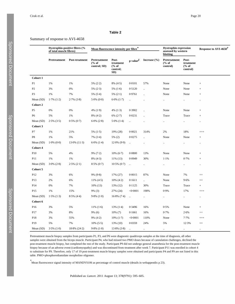
Seven patients had a post-treatment increase in dystrophin protein expression compared withtheir pretreatment biopsies, evidenced by at least two of the three methods of quantification:number of dystrophin-positive fibres, western blotting, and semiquantitativeimmunocytochemistry measurements (figure 3, webappendix p 23). In these seven patients,mean dystrophin fluorescence intensity increased from 8·9% (95% CI 7·1–10·6) to 16·4%(10·8–22·0) of normal control after treatment (p=0·0287). In the low-dose cohorts 1 to 4,there was no increase in dystrophin expression, with the exception of participant P7 in
Cirak et al. Page 5
Published as: Lancet. 2011 August 13; 378(9791): 595–605.
Sponsored Docum
ent Sponsored D
ocument
Sponsored Docum
ent

cohort 3. However, six of eight patients in the two high-dose cohorts 5 and 6 showed anincrease in protein expression. Three patients, one in each of cohorts 3 (P7), 5 (P15), and 6(P18), had a very substantial response to AVI-4658, having 21%, 15%, and 55% dystrophin-positive fibres, respectively. These three patients had also 314%, 198%, and 110% increasesin dystrophin intensity compared with pretreatment biopsy on semiquantitativeimmunocytochemistry. Western blot analysis of these patients also showed an increase aftertreatment of protein levels from 2% to 18%, from 0·9% to 17%, and from 0% to 7·7% ofnormal muscle, respectively (table 2).
A dose-dependent significant linear increase in dystrophin expression was noted (table 2;exact Cochran-Armitage trend test, p=0·0203). Additionally, AVI-4658 administrationinduced significant increase of dystrophin expression on immunocytochemistry pretreatmentversus post-treatment in cohorts 5 and 6 (paired two-sided t test, p=0·04). Participant P19had dystrophin levels on blot of 12·3% of control muscle, but a smaller increase influorescence intensity and percentage of dystrophin-positive fibres, probably reflectingvariability in different blocks of the muscle biopsy sample studied with the differenttechniques (table 2). The functional properties of restored dystrophin were confirmed byquantification of α-sarcoglycan and neuronal NOS expression. Dystrophin-positive fibreshad roughly a 30% average increase in α-sarcoglycan expression compared with dystrophin-negative fibres in the patient with the best response to treatment, P18 (deletion 49–50).Dystrophin restoration was followed by restoration of neuronal NOS at the sarcolemma,more so in patients with exon 49–50 deletions than in those with 45–50 deletions (figure4A), which is consistent with the observation that the neuronal NOS binding domain islocated in dystrophin exons 42–45.
Finally, the inflammatory infiltrate was investigated to establish whether dystrophinrestoration had any effect on the prominent inflammatory response seen in Duchennemuscular dystrophy (figure 4B). A reduction in inflammatory infiltrates was recorded incohorts 5 and 6, apart from in participant P15 in whom the CD8 cell count was increased,but not around dystrophin-positive fibres. Furthermore, this patient did not haveantidystrophin antibodies. Despite the small number of samples, the significant reduction inCD3 cell count (paired two-tailed t test, p=0·0115) shows that restored dystrophin istolerated by the immune system. The quotient between CD3% and dystrophin intensity inthe seven patients who responded to treatment showed a significant reduction in the post-treatment muscle biopsy samples (one-tailed paired t test, p=0·0078), confirming thecorrelation with the increase in dystrophin expression.
DiscussionWe show for the first time that repeated systemic administration of a PMO splice switchingoligomer (AVI-4658) induces targeted exon skipping in skeletal muscle in patients withDuchenne muscular dystrophy, restoring correctly localised dystrophin at the sarcolemma(panel). The administration of AVI-4658 was very well tolerated, without clear drug-induced adverse events with single doses of up to 900 mg and cumulative exposureexceeding 10 000 mg. The absence of drug-related adverse events after 12 weeks isencouraging, but caution is still needed because any splice switching oligomer would needto be given lifelong.
A clear and significant dose response was recorded in terms of dystrophin proteinexpression, leading to seven patients who responded to treatment at higher doses. Thisfinding was accompanied by a significant reduction of inflammatory infiltrates in patients inthe two highest dose cohorts. Patients with the highest levels of dystrophin also hadincreased sarcolemmal expression of proteins of the dystrophin-associated glycoprotein
Cirak et al. Page 6
Published as: Lancet. 2011 August 13; 378(9791): 595–605.
Sponsored Docum
ent Sponsored D
ocument
Sponsored Docum
ent

complex. This outcome included restoration of neuronal NOS to the sarcolemma in patientswith deletions that did not disrupt the NOS binding site localised in spectrin repeats 16/17 ofthe rod domain. The restoration of neuronal NOS is beneficial for patients in whom ongoingmuscle damage is compounded by paradoxical exercise-induced vasoconstriction as a resultof dysfunctional fine-tuning of blood flow.
The reduced inflammation in muscle could be related to reduced necrosis due to improvedsarcolemmal function with better resistance to mechanical load, induced by the restoreddystrophin. We noted variable levels of protein restoration in the seven patients whoresponded to treatment and considered possible reasons for this variability. Variability dueto specific deletions and intronic breakpoints of individual patients could not be detected invitro, because the response to splice switching oligomer in all patients' myotubes wasqualitatively similar. The stability of the resulting protein might be relevant since thepatients with the three greatest responses to treatment all had 49–50 deletions and mildlyaffected patients with Becker muscular dystrophy or even asymptomatic individuals withdeletion of exons 49–51 have been described, suggesting that this shortened protein is highlyfunctional. However two patients who did not respond to treatment (cohort 1, P3, who hadno increase in either dystrophin-positive fibres or dystrophin expression, and cohort 3, P8,who had an increase in dystrophin-positive fibres, but not in dystrophin expression) and onewith a small response (cohort 5, P12) also have the deletion 49–50; additionally, patientswith exon 45–50 deletions did not have more protein than did patients with other genotypes,although asymptomatic individuals with 45–51 deletions are on record. An aspect toconsider is the genetic background, which in humans is variable, including the intronicdeletions breakpoints. A further variable could be differences in pharmacodynamics ofPMO, although our analysis suggests that there was not a clear correlation between responseand maximum concentration and area under the curve for AVI-4658. When concomitanttreatments, age, and extent of muscle pathological changes were taken into account, noobvious pattern emerged. Immune response to novel dystrophin epitopes induced by exonskipping could be another reason for the variability; however, this explanation seemsunlikely because we did not detect humoral immunity in any of the patients and furthermorewe documented a reduction in T cells in muscle biopsy samples in cohorts 5 and 6, whoshowed the highest dystrophin levels.
In a recent systemic exon skipping trial in Duchenne muscular dystrophy using the 2′OMechemistry, high numbers of dystrophin-positive fibres but low dystrophin levels weredescribed. However, the study had no pretreatment muscle biopsy in patients in whom adystrophin response was reported, making establishment of a proper baseline to distinguishminimally positive from negative fibres difficult. Indeed, in Duchenne muscular dystrophy,sections of muscle often have discernible trace levels after immunostaining with theantibodies to dystrophin used, and these pretreatment levels need to be taken into account toprovide an accurate measurement of both number of positive fibres and dystrophin intensity.In our study, we first established the level of dystrophin in the pretreatment muscle biopsysample, and regarded those levels as the baseline for that individual—ie, judged fibres aspositive only if they exceeded the intensity levels of the pretreatment biopsy.
With respect to the variable dystrophin restoration we have reported, the most plausibleconclusion is that stochastic events affect muscle splice switching oligomer targeting andcontribute to variability. Animal models treated with both 2′OMe and PMO also showedsubstantial variability of dystrophin expression, even in contralateral muscles from the sameanimal. Because splice switching oligomers do not target skeletal muscle specifically, theiruptake is partly dependent on local events such as muscle perfusion, damage, andinflammation. To obtain more uniform protein production, either high doses of PMO orprolonged frequent administration, or both, could be considered because PMOs seem to be
Cirak et al. Page 7
Published as: Lancet. 2011 August 13; 378(9791): 595–605.
Sponsored Docum
ent Sponsored D
ocument
Sponsored Docum
ent

well tolerated. The safety profile we noted with AVI-4658 at doses of 20 mg/kg, supportedby animal testing at up to human equivalent doses of 100 mg/kg, is encouraging and bodeswell for longer administration periods and higher clinical dose. Preclinical data suggest thatrepeated administration of even small doses over an extended time achieves morehomogeneous restoration of dystrophin than does the same cumulative dose administered asa bolus injection of PMO. This finding suggests that a long period of administration will benecessary to achieve homogeneous dystrophin expression.
In terms of the clinical efficacy, the PRO051 study claims that eight of 11 boys who wereambulant at entry to the extension study showed improvement in the 6-min walking test of35·2 m (SD 28·7) after 12 weeks' treatment; however, this change was not significant.Moreover, several of these children were younger than 7 years and, according tolongitudinal observation, boys younger than 7 years with Duchenne muscular dystrophygain motor function. Additional confounding factors are the variability in the walking test(SD 36 m) and the powerful placebo effect of open-label studies. Despite these limitations,this observation is encouraging.
In our study, boys remained mostly stable during follow-up, but because the period duringwhich AVI-4658 was administered was only 12 weeks, we did not observe any significantclinical improvement, and the lack of a study extension was a limitation of our study, sinceonly extended exposure to the drug is able to affect progression of the disease. Nevertheless,our results are very encouraging because they prove that doses of 10 mg/kg and 20 mg/kg ofAVI-4658, which were very well tolerated, consistently induced dystrophin expression inthe seven patients who responded to treatment up to levels typically found in patients withBecker muscular dystrophy or disease of intermediate severity between Duchenne musculardystrophy and Becker muscular dystrophy. The restoration of the dystrophin-associatedglycoprotein complex suggests that the produced dystrophin is functional. Because of thevariability of dystrophin restoration, MRI or spectroscopy of muscle are promising methodsto assess the effect of systemic treatment in Duchenne muscular dystrophy. Recent studieshave described the correlation between MRI and the degree of dystrophic muscle changes,the effect of exercise in Duchenne muscular dystrophy, and reduction of muscleinflammation after PMO treatment in dogs.
On the basis of our data and recent preclinical data, we expect that extended administrationof AVI-4658 at doses of 10 mg/kg or higher will result in sufficient dystrophin expression tohave a positive effect on the prevention of muscle degeneration in Duchenne musculardystrophy. Indeed, chronic administration (1 year) of doses of PMO similar to the one usedin our study produced significant improvement in muscle pathology and function in mdxmice. AVI-4658 has the potential to ameliorate the progressive natural history of Duchennemuscular dystrophy and now needs to be investigated in clinical efficacy trials.
References1. Bushby K, Finkel R, Birnkrant DJ, for the DMD Care Considerations Working Group. Diagnosis
and management of Duchenne muscular dystrophy, part 1: diagnosis, and pharmacological andpsychosocial management. Lancet Neurol. 2010; 9:77–93. [PubMed: 19945913]
2. Hoffman EP, Brown RH Jr, Kunkel LM. Dystrophin: the protein product of the Duchenne musculardystrophy locus. Cell. 1987; 51:919–928. [PubMed: 3319190]
3. Bushby KM, Gardner-Medwin D, Nicholson LV. The clinical, genetic and dystrophincharacteristics of Becker muscular dystrophy. II. Correlation of phenotype with genetic and proteinabnormalities. J Neurol. 1993; 240:105–112. [PubMed: 8437017]
4. Sazani, P.; Graziewicz, MA.; Kole, R. Splice switching oligonucleotides as potential therapeutics.In: Crooke, ST., editor. Antisense drug technology, principles, strategies and applications. CBCPress; Boca Raton, FL, USA: 2008. p. 89-114.
Cirak et al. Page 8
Published as: Lancet. 2011 August 13; 378(9791): 595–605.
Sponsored Docum
ent Sponsored D
ocument
Sponsored Docum
ent

5. Lu QL, Rabinowitz A, Chen YC. Systemic delivery of antisense oligoribonucleotide restoresdystrophin expression in body-wide skeletal muscles. Proc Natl Acad Sci USA. 2005; 102:198–203.[PubMed: 15608067]
6. Yokota T, Lu QL, Partridge T. Efficacy of systemic morpholino exon-skipping in Duchennedystrophy dogs. Ann Neurol. 2009; 65:667–676. [PubMed: 19288467]
7. Fletcher S, Honeyman K, Fall AM, Harding PL, Johnsen RD, Wilton SD. Dystrophin expression inthe mdx mouse after localised and systemic administration of a morpholino antisenseoligonucleotide. J Gene Med. 2006; 8:207–216. [PubMed: 16285002]
8. van Deutekom JC, Janson AA, Ginjaar IB. Local dystrophin restoration with antisenseoligonucleotide PRO051. N Engl J Med. 2007; 357:2677–2686. [PubMed: 18160687]
9. Kinali M, Arechavala-Gomeza V, Feng L. Local restoration of dystrophin expression with themorpholino oligomer AVI-4658 in Duchenne muscular dystrophy: a single-blind, placebo-controlled, dose-escalation, proof-of-concept study. Lancet Neurol. 2009; 8:918–928. [PubMed:19713152]
10. Goemans NM, Tulinius M, van den Akker JT. Systemic administration of PRO051 in Duchenne'smuscular dystrophy. N Engl J Med. 2011; 364:1513–1522. [PubMed: 21428760]
11. Arechavala-Gomeza V, Graham IR, Popplewell LJ. Comparative analysis of antisenseoligonucleotide sequences for targeted skipping of exon 51 during dystrophin pre-mRNA splicingin human muscle. Hum Gene Ther. 2007; 18:798–810. [PubMed: 17767400]
12. Nguyen TM, Morris GE. Use of epitope libraries to identify exon-specific monoclonal antibodiesfor characterization of altered dystrophins in muscular dystrophy. Am J Hum Genet. 1993;52:1057–1066. [PubMed: 7684887]
13. Arechavala-Gomeza V, Kinali M, Feng L. Immunohistological intensity measurements as a tool toassess sarcolemma-associated protein expression. Neuropathol Appl Neurobiol. 2010; 36:265–274. [PubMed: 20002311]
14. Arechavala-Gomeza V, Kinali M, Feng L. Revertant fibres and dystrophin traces in Duchennemuscular dystrophy: implication for clinical trials. Neuromuscul Disord. 2010; 20:295–301.[PubMed: 20395141]
15. Neri M, Torelli S, Brown S. Dystrophin levels as low as 30% are sufficient to avoid musculardystrophy in the human. Neuromuscul Disord. 2007; 17:913–918. [PubMed: 17826093]
16. Mazzone ES, Messina S, Vasco G. Reliability of the North Star Ambulatory Assessment in amulticentric setting. Neuromuscul Disord. 2009; 19:458–461. [PubMed: 19553120]
17. Mayhew JE, Florence JM, Mayhew TP. Reliable surrogate outcome measures in multicenterclinical trials of Duchenne muscular dystrophy. Muscle Nerve. 2007; 35:36–42. [PubMed:16969838]
18. McDonald CM, Widman LM, Walsh DD, Walsh SA, Abresch RT. Use of step activity monitoringfor continuous physical activity assessment in boys with Duchenne muscular dystrophy. ArchPhys Med Rehabil. 2005; 86:802–808. [PubMed: 15827935]
19. McDonald CM, Henricson EK, Han JJ. The 6-minute walk test in Duchenne/Becker musculardystrophy: longitudinal observations. Muscle Nerve. 2010; 42:966–974. [PubMed: 21038378]
20. Lai Y, Thomas GD, Yue Y. Dystrophins carrying spectrin-like repeats 16 and 17 anchor nNOS tothe sarcolemma and enhance exercise performance in a mouse model of muscular dystrophy. JClin Invest. 2009; 119:624–635. [PubMed: 19229108]
21. Krieger CC, Bhasin N, Tewari M. Exon-skipped dystrophins for treatment of Duchenne musculardystrophy: mass spectrometry mapping of most exons and cooperative domain designs based onsingle molecule mechanics. Cytoskeleton (Hoboken). 2010; 67:796–807. [PubMed: 20886611]
22. Pescatori M, Broccolini A, Minetti C. Gene expression profiling in the early phases of DMD: aconstant molecular signature characterizes DMD muscle from early postnatal life throughoutdisease progression. FASEB J. 2007; 21:1210–1226. [PubMed: 17264171]
23. Kobayashi YM, Rader EP, Crawford RW. Sarcolemma-localized nNOS is required to maintainactivity after mild exercise. Nature. 2008; 456:511–515. [PubMed: 18953332]
24. Arahata K, Engel AG. Monoclonal antibody analysis of mononuclear cells in myopathies. I:quantitation of subsets according to diagnosis and sites of accumulation and demonstration andcounts of muscle fibers invaded by T cells. Ann Neurol. 1984; 16:193–208. [PubMed: 6383191]
Cirak et al. Page 9
Published as: Lancet. 2011 August 13; 378(9791): 595–605.
Sponsored Docum
ent Sponsored D
ocument
Sponsored Docum
ent

25. Muntoni F, Di Lenarda A, Porcu M. Dystrophin gene abnormalities in two patients with idiopathicdilated cardiomyopathy. Heart. 1997; 78:608–612. [PubMed: 9470882]
26. Helderman-van den Enden AT, Straathof CS, Aartsma-Rus A. Becker muscular dystrophy patientswith deletions around exon 51; a promising outlook for exon skipping therapy in Duchennepatients. Neuromuscul Disord. 2010; 20:251–254. [PubMed: 20153965]
27. Morandi L, Mora M, Confalonieri V. Dystrophin characterization in BMD patients: correlation ofabnormal protein with clinical phenotype. J Neurol Sci. 1995; 132:146–155. [PubMed: 8543940]
28. Saengpattrachai M, Ray PN, Hawkins CE, Berzen A, Banwell BL. Grandpa and I havedystrophinopathy?: approach to asymptomatic hyperCKemia. Pediatr Neurol. 2006; 35:145–149.[PubMed: 16876015]
29. Aoki Y, Nakamura A, Yokota T. In-frame dystrophin following exon 51-skipping improvesmuscle pathology and function in the exon 52-deficient mdx mouse. Mol Ther. 2011; 18:1995–2005. [PubMed: 20823833]
30. Sazani P, Weller DL, Shrewsbury SB. Safety pharmacology and genotoxicity evaluation ofAVI-4658. Int J Toxicol. 2010; 29:143–156. [PubMed: 20110565]
31. Malerba A, Sharp PS, Graham IR. Chronic systemic therapy with low-dose morpholino oligomersameliorates the pathology and normalizes locomotor behavior in mdx mice. Mol Ther. 2011;19:345–354. [PubMed: 21102560]
32. Kinali M, Arechavala-Gomeza V, Cirak S. Muscle histology vs MRI in Duchenne musculardystrophy. Neurology. 2010; 76:346–353. [PubMed: 21263136]
33. Garrood P, Hollingsworth KG, Eagle M. MR imaging in Duchenne muscular dystrophy:quantification of T1-weighted signal, contrast uptake, and the effects of exercise. J Magn ResonImaging. 2009; 30:1130–1138. [PubMed: 19856446]
34. Wu B, Xiao B, Cloer C. One-year treatment of morpholino antisense oligomer improves skeletaland cardiac muscle functions in dystrophic mdx mice. Mol Ther. 2010; 19:576–583. [PubMed:21179007]
Web Extra MaterialSupplementary Material1. Supplementary webappendix.
AcknowledgmentsWe thank the participating patients and their families, the charities Muscular Dystrophy Campaign, ActionDuchenne, and the Duchenne Family Support Group for participating in the UK MDEX consortium, whichundertook this study. We also thank the members of the MDEX Scientific Advisory Board chaired by Kay Davies(see consortium website for full membership) for their constructive criticism. We thank Matt Rogan, Kathy Smith,and Jim Balsley for their participation in the data safety monitoring board. We thank Kanagasabai Ganeshaguru forstudy coordination in London, Maria Kinali for constructive discussion in early stages about the trial design andGeoff Bell for patient coordination in Newcastle upon Tyne, Darren Chambers, Rita Barresi, and Richard Charltonfor their excellent technical assistance in processing muscle samples, and Rivka Steinberg for the in-vitro testing ofpatients' fibroblasts. We thank Valeria Ricotti for her technical assistance in the CD cell counts and Glenn Morris,Oswestry, and the MDA Monoclonal Antibody Resource for MANDYS106. We are grateful for the support of theNIHR Biomedical Research Centre Funding Scheme and the Somers Clinical Research Facility at Great OrmondStreet Hospital, UCL Institute of Child Health, and in particular thank Anna Massey, Katie Rees, and ElizabethLeach and the physiotherapists Marion Main, Maria Ash, Michelle Eagle, and Anna Mayhew for their expertassessment of the study participants. This work was supported by the Newcastle NIHR Clinical Research Facility,in particular the study nurses Linda Smith and Dorothy Carman. We are very grateful for Mariacristina Scoto's helpin the clinical care of the patients. We also acknowledge the collaborations of the North Star Clinical network,which contributed to the recruitment of participants via the clinical colleagues Adnan Manzur, Stephanie Robb,Helen Roper, Rosaline Quinlivan, and Louise Hartley. We are grateful to the surgeons for obtaining the musclebiopsies (at the London site: Joe Curry and Paolo De Coppi; at the Newcastle site: Anne Lawson). We are gratefulto Ann S Le Couteur, Helen McConachie, and Nil Chakrabarti at the Newcastle site and to Sriranjan Sucharita atthe London site for psychiatric interviews of the patients and their families. MEG is grateful for support from theICHT Comprehensive Biomedical Research Centre. The study was supported by the MRC Centre forNeuromuscular Diseases at UCL and Newcastle including the MRC Neuromuscular Centre Biobank. JEM issupported by a Wellcome Trust University Award. FM is supported by Great Ormond Street Hospital Children'sCharity. Newcastle University, UCL, and Oxford are partners in TREAT-NMD (EC036825). The study wassponsored by AVI BioPharma (Bothell, WA, USA).
Cirak et al. Page 10
Published as: Lancet. 2011 August 13; 378(9791): 595–605.
Sponsored Docum
ent Sponsored D
ocument
Sponsored Docum
ent

AcknowledgmentsSC, FM, KB, MG, and SBS designed and wrote the clinical trial protocol together with the amendments. SC andMG identified patients and coordinated clinical teams, executed the trial procedures, collected data, and followedup the study participants, under the supervision of FM and KB. SC, VAG, and JEM coordinated the collaborativework between clinical and laboratory teams and the biochemical work-up of clinical trial samples under thesupervision of FM. FM, SC, MG, and KB obtained patient consent. JB oversaw and interpreted cardiac tests for theNewcastle cohort. SA was responsible of the acquisition of the genetic data. FM and KB managed the study budget.SC, VAG, JEM, and FM drafted the first report and have seen and approved the final version. DJW, GD, MJAW,SDW, and VS contributed to the interpretation of results and drafting of the report. VAG and KA did RT-PCR,image capture, and dystrophin expression analysis. RK contributed to the design of the RT-PCR assay and tointerpretation of results. ST did the western blot analysis. LF and CS processed muscle biopsy specimens andimmunofluorescence staining and their analysis. MEG devised and oversaw the psychiatric assessments of thesubjects and families. SC did the statistical analysis. SBS was responsible as sponsor's medical officer foroverseeing safe conduct of the study and participated in all safety monitoring discussions and data reviews. Allauthors contributed to the interpretation of results and drafting of the report and have seen and approved the finalversion.
AcknowledgmentsFM serves on scientific advisory boards for Acceleron Pharma, Genzyme, AVI BioPharma, Debiopharma Group,GlaxoSmithKline, Prosensa, and Santhera Pharmaceutical, receives research support from AVI BioPharma, and hasreceived funding for trials from AVI, Trophos, and PTC. KB has served on scientific advisory boards forAcceleron, AMT, AVI Biopharma, Debiopharm, Genzyme, GlaxoSmithKline, Prosensa, PTC, and Santhera andhas received funding for trials from AVI and PTC. SBS is employed full time as Chief Medical Officer and SeniorVice President by AVI BioPharma and owns AVI stock. RK is employed full time as Senior Vice President andDistinguished Scientist by AVI BioPharma and owns AVI stock. VS has served on scientific advisory boards forAcceleron and Genzyme and has received funding for a trial from GlaxoSmithKline. GD and SDW hold patents inthe area of exon skipping. MJAW serves as a scientific adviser for AVI BioPharma and Novartis. All other authorsdeclare that they have no conflicts of interest.
Cirak et al. Page 11
Published as: Lancet. 2011 August 13; 378(9791): 595–605.
Sponsored Docum
ent Sponsored D
ocument
Sponsored Docum
ent

Panel
Research in contextSystematic review
We searched PubMed in March, 2011, using the keywords “Duchenne”, “antisense”,“exon skipping”, and “clinical trial”. Splice-switching oligomers have been testedpreviously after intramuscular injection in animal models and in patients affected byDuchenne muscular dystrophy. An open-label, dose-escalation study in 12 boys withDuchenne muscular dystrophy receiving weekly subcutaneous injections of the 2′OMePRO051 at 0·5, 2, 4, and 6 mg/kg bodyweight for 5 weeks induced skipping of exon 51.Low dystrophin levels were reported after treatment, although the absence ofpretreatment samples makes precise quantification of the biochemical efficacy ofPRO051 difficult. This study was followed by a 12-week extension study using a dose of6 mg/kg bodyweight of PRO051, with stabilisation of muscle function, but no significantimprovement in a 6-min walk test.
Interpretation
We report for the first time that the systemic administration of a splice-switchingoligomer based on PMO chemistry (AVI-4658) induced restoration of dystrophinexpression in skeletal muscle of boys with Duchenne muscular dystrophy. The clinicaland laboratory safety data in our open-label, dose-escalation, repeated intravenousadministration study showed that AVI-4658 was well tolerated. Seven patients had asignificant dose response, six of whom were in the two high-dose cohorts, showingrestoration of dystrophin protein expression. This finding was associated with increasedexpression of proteins associated with dystrophin, such as α-sarcoglycan and neuronalnitric oxide synthase, the sarcolemmal localisation of which is disrupted in Duchennemuscular dystrophy. Additionally, we showed a dose-dependent reduction in theinflammatory infiltrate in muscles of boys with Duchenne muscular dystrophy in whomdystrophin expression was restored. This finding is encouraging because it suggests thatthe restored dystrophin attenuates the inflammation that is a hallmark of the disease'spathology; it also suggests that the newly produced dystrophin does not produce novelimmunogenic epitopes.
Cirak et al. Page 12
Published as: Lancet. 2011 August 13; 378(9791): 595–605.
Sponsored Docum
ent Sponsored D
ocument
Sponsored Docum
ent

Figure 1.Patients recruited to the trial, their assignment to cohorts, and the dose-escalation schemeEach full red box represents a time interval of 12 weeks' dosing. Arrows show thetimepoints at which the data safety monitoring board met with clinical investigators and thesponsor to review safety before subsequent dose escalations. *Patient withdrawn from studyafter seven doses.
Cirak et al. Page 13
Published as: Lancet. 2011 August 13; 378(9791): 595–605.
Sponsored Docum
ent Sponsored D
ocument
Sponsored Docum
ent

Figure 2.Plasma pharmacokinetics of AVI-4658Mean plasma concentrations of AVI-4658 versus nominal elapsed time averaged acrossweeks 1, 6, and 12. Area under the curve (AUC) over 24 h accounted for greater than 95%of AUC0–∞, suggesting that most of the drug eliminated from the plasma was cleared within24 h. AVI-4658 plasma exposure increased in a nearly proportional manner with dose formaximum concentration, AUC0–24, and AUC0–∞. Error bars show SDs.
Cirak et al. Page 14
Published as: Lancet. 2011 August 13; 378(9791): 595–605.
Sponsored Docum
ent Sponsored D
ocument
Sponsored Docum
ent

Figure 3.Dystrophin protein expression in the seven patients who responded to treatment(A) Transverse sections of treated (post) and untreated (pre) muscle specimensimmunolabelled with MANDYS106 antibody. (B) Post-treatment biopsy samples fromparticipants P15 and P18; low-magnification images showing widespread and patchydystrophin expression (arrows). (C) Western blotting of pretreatment and post-treatmentmuscle biopsy samples with antidystrophin Dys1 (exon 26–30) and antisarcomeric α-actininantibodies; an average of 150 μg of total patient proteins was loaded per lane. CT=μgcontrol muscle extract.
Cirak et al. Page 15
Published as: Lancet. 2011 August 13; 378(9791): 595–605.
Sponsored Docum
ent Sponsored D
ocument
Sponsored Docum
ent

Figure 4.Functional analysis of restored dystrophin(A) Expression of dystrophin, α-sarcoglycan, and neuronal NOS in post-treatment musclebiopsy samples from participants P18 and P19 was quantified relative to control muscle in40 dystrophin-positive or dystrophin-negative muscle fibres and normalised to β-spectrinexpression. To overcome high background seen with the neuronal NOS antibody, theaverage background intensity of neuronal NOS-negative membranes was subtracted fromcontrol and patient values. For participant P19, there was no difference in α-sarcoglycanintensity between dystrophin-positive and dystrophin-negative fibres in the post-treatmentmuscle biopsy sample and neuronal NOS showed only a small increase in dystrophin-
Cirak et al. Page 16
Published as: Lancet. 2011 August 13; 378(9791): 595–605.
Sponsored Docum
ent Sponsored D
ocument
Sponsored Docum
ent

positive fibres (paired two-tailed t test, p=0·0007). For participant P18, neuronal NOS and α-sarcoglycan intensity was significantly increased in the dystrophin-positive fibres (neuronalNOS mean intensity as percentage of control: dystrophin-negative fibres 7% [SD 7],dystrophin-positive fibres 28% [SD 17], paired two-tailed t test, p≤0·0001; α-sarcoglycanmean intensity as percentage of control: dystrophin-negative fibres 45% [SD 16],dystrophin-positive fibres 75% [SD 31], p<0·0001). (B) Sarcolemmal restoration of thedystrophin-associated glycoprotein complex by AVI-4658. Post-treatment muscle biopsysamples from participants P19 and P18 were stained with antibodies against dystrophin(exon 43, MANDYS106), α-sarcoglycan, neuronal NOS, and β-spectrin. The arrows showthe same dystrophin-positive fibre in each panel. In P19 (deletion of exons 45–50) the fibreshown by the arrow has increased α-sarcoglycan sarcolemmal expression, but not neuronalNOS because this patient is deleted for part of the dystrophin neuronal NOS binding site.(C) Inflammatory infiltrates quantification on pretreatment and post-treatment musclesamples. Muscle sections were incubated with antibodies (DAKO, UK) raised againsthuman CD3 (pan T cell), CD4 (helper T cell) and CD8 (killer T cell). For each section, thenumber of CD-positive cells was represented as a percentage of the total number of musclefibres. Patients with pretreatment and post-treatment values of zero are not represented.NOS=nitric oxide synthase.
Cirak et al. Page 17
Published as: Lancet. 2011 August 13; 378(9791): 595–605.
Sponsored Docum
ent Sponsored D
ocument
Sponsored Docum
ent

Sponsored Docum
ent Sponsored D
ocument
Sponsored Docum
ent
Cirak et al. Page 18
Table 1
Clinical summary
Mutation* Age (years)† Weight (kg)† 6-minwalktest atbaseline(m)‡
Cardiomyopathy at recruitment Dose andregimen ofcorticosteroidtreatment
Other regulartreatments takenat study entry
Serious or severeadverse events andrelation to AVI-4658
Cohort 1 (0·5 mg/kg)
P1 Del 48–50 9 31 410 No Prednisolone25 mg,intermittent(0·81 mg/kgper day)
.. ..
P2 Del 45–50 8 29 254 Yes Prednisolone12·5 mg, daily(0·43 mg/kgper day)
Perindopril,calcium, vitamin D
..
P3 Del 49–50 8 38 437 No Prednisolone25 mg,intermittent(0·66 mg/kgper day)
Omeprazole ..
P4 Del 48–50 8 36 139 No Prednisolone20 mg,intermittent(0·55 mg/kgper day)
Calcium, vitamin D ..
Cohort 2 (1 mg/kg)
P5 Del 45–50 6 26 250 No Prednisolone22·5 mg,intermittent(0·86 mg/kgper day)
.. ..
P6 Del 48–50 6 21 371 No Prednisolone12·5 mg daily(0·6 mg/kgper day)
Ranitidine ..
Cohort 3 (2 mg/kg)
P7 Del 49–50 13 47 375 No Prednisolone15 mg daily(0·32 mg/kgper day)
Risedronate,calcium, vitamin D
..
P8 Del 49–50 9 38 350 No Prednisolone15 mg daily(0·4 mg/kgper day)
Ranitidine Post-anaesthesiahospitalisation for 1 daydue to vomiting,unrelated
Cohort 4 (4 mg/kg)
P9 Del 52 10 30 301 Regional wall hypokinesia,normal FS
Prednisolone15 mg daily(0·49 mg/kgper day)
Risedronate,calcium, vitamin D
Cardiomyopathy, possible
P10 Del 48–50 10 62 146 No Not onsteroidsbecause ofside-effects
.. Ankle fracture, unrelated
P11 Del 45–50 9 28 477 No Prednisolone15 mg daily(0·54 mg/kgper day)
Risedronate,calcium, vitamin D
..
Cohort 5 (10 mg/kg)
Published as: Lancet. 2011 August 13; 378(9791): 595–605.

Sponsored Docum
ent Sponsored D
ocument
Sponsored Docum
ent
Cirak et al. Page 19
Mutation* Age (years)† Weight (kg)† 6-minwalktest atbaseline(m)‡
Cardiomyopathy at recruitment Dose andregimen ofcorticosteroidtreatment
Other regulartreatments takenat study entry
Serious or severeadverse events andrelation to AVI-4658
P12 Del 49–50 6 25 317 No Prednisolone20 mg,intermittent(0·8 mg/kgper day)
Calcium, vitamin D ..
P13 Del 48–50 7 22 443 No Prednisolone15 mg daily(0·68 mg/kgper day)
.. ..
P14 Del 47–50 12 52 138 Yes Deflazacort30 mg daily(0·58 mg/kgper day)
Perindopril,bisoprolol,risedronate,calcium, vitamin D
..
P15 Del 49–50 10 39 169 No Prednisolone20 mg daily(0·5 mg/kgper day)
.. ..
Cohort 6 (20 mg/kg)
P16 Del 45–50 9 31 515 No Prednisolone20 mg daily(0·65 mg/kgper day)
.. ..
P17 Del 45–50 7 25 492 No Prednisolone17·5 mg daily(0·6 mg/kgper day)
.. ..
P18 Del 49–50 10 45 265 Yes Deflazacort30 mg daily(0·66 mg/kgper day)
Lisinopril ..
P19 Del 45–50 9 30 405 No Prednisolone15 mg daily(0·5 mg/kgper day)
Calcium, vitamin D ..
Intermittent=10 days on and 10 days off treatment. FS=fractional shortening.
*Deleted exons in the dystrophin gene.
†At the first dose of AVI-4658.
‡Assessed a week before the first dose.
Published as: Lancet. 2011 August 13; 378(9791): 595–605.
Sponsored Docum
ent Sponsored D
ocument
Sponsored Docum
ent
Cirak et al. Page 20
Table 2
Summary of response to AVI-4658
Dystrophin-positive fibres (%of total muscle fibres)
Mean fluorescence intensity per fibre* Dystrophin expressionassessed by westernblotting
Response to AVI-4658†
Pretreatment Post-treatment Pretreatment(% ofcontrol; SD)
Post-treatment(% ofcontrol;SD)
p value‡ Increase (%) Pretreatment(% ofcontrol)
Post-treatment(% ofcontrol)
Cohort 1
P1 1% 1% 5% (2·2) 8% (4·5) 0·0101 57% None None +
P2 3% 0% 5% (2·3) 5% (1·6) 0·5120 .. None None +
P3 1% 7% 5% (5·4) 5% (2·1) 0·9761 .. None None +
Mean (SD) 1·7% (1·2) 2·7% (3·8) 5·0% (0·0) 6·0% (1·7) .. .. .. .. ..
Cohort 2
P5 0% 0% 4% (1·9) 4% (1·3) 0·3902 .. None None +
P6 5% 1% 8% (4·2) 6% (2·7) 0·0231 .. Trace Trace +
Mean (SD) 2·5% (3·5) 0·5% (0·7) 6·0% (2·8) 5·0% (1·4) .. .. .. .. ..
Cohort 3
P7 1% 21% 5% (1·5) 19% (28) 0·0021 314% 2% 18% +++
P8 1% 5% 7% (3·4) 5% (2) 0·0275 .. None None +
Mean (SD) 1·0% (0·0) 13·0% (11·3) 6·0% (1·4) 12·0% (9·9) .. .. .. .. ..
Cohort 4
P10 5% 4% 9% (7·2) 10% (6·7) 0·0800 13% None None +
P11 1% 1% 8% (4·3) 11% (13) 0·0949 30% 1·1% 0·7% +
Mean (SD) 3·0% (2·8) 2·5% (2·1) 8·5% (0·7) 10·5% (0·7) .. .. .. .. ..
Cohort 5
P12 3% 6% 9% (9·8) 17% (27) 0·0015 87% None 7% ++
P13 2% 6% 11% (4·5) 10% (4·2) 0·1611 .. None 9·6% ++
P14 0% 7% 10% (13) 13% (12) 0·1125 30% Trace Trace +
P15 1% 15% 9% (3) 27% (24) <0·0001 198% 0·9% 17% +++
Mean (SD) 1·5% (1·3) 8·5% (4·4) 9·8% (1·0) 16·8% (7·4) .. .. .. .. ..
Cohort 6
P16 3% 5% 11% (1·6) 13% (1·4) 0·3496 16% 0·5% None +
P17 3% 8% 9% (6) 10% (7) 0·1661 16% 0·7% 2·6% ++
P18 3% 55% 9% (4·2) 19% (1·7) <0·0001 110% None 7·7% +++
P19 5% 7% 10% (5·5) 13% (10) 0·0359 24% 5% 12·3% ++
Mean (SD) 3·5% (1·0) 18·8% (24·2) 9·8% (1·0) 13·8% (3·8) .. .. .. .. ..
Pretreatment muscle biopsy samples from participants P1, P3, and P6 were diagnostic quadriceps samples at the time of diagnosis, all othersamples were obtained from the biceps muscle. Participant P4, who had missed two PMO doses because of cannulation challenges, declined thepost-treatment muscle biopsy, but completed the rest of the study. Participant P9 did not undergo general anaesthesia for the post-treatment musclebiopsy because of an adverse event (cardiomyopathy) and was discontinued from treatment after week 7. Participant P11 was enrolled in cohort 4to substitute for P9. Therefore, only 17 of 19 post-treatment muscle biopsy samples were obtained and participants P4 and P9 are not listed in thistable. PMO=phosphorodiamidate morpholino oligomer.
*Mean fluorescence signal intensity of MANDYS106 as percentage of control muscle (details in webappendix p 23).
Published as: Lancet. 2011 August 13; 378(9791): 595–605.

Sponsored Docum
ent Sponsored D
ocument
Sponsored Docum
ent
Cirak et al. Page 21
†Response to AVI-4658: + shows response at RNA level (exon 51 skipping), but without detectable increase of dystrophin expression in post-
treatment muscle biopsy; ++ shows response at RNA level and increase of dystrophin expression in post-treatment muscle; and +++ showsresponse at RNA level and increase in post-treatment muscle biopsy sample with all three methods of dystrophin quantification.
‡Two-tailed t test comparing the mean fluorescence intensity in the pretreatment versus post-treatment biopsy sample for each patient; we assessed
the dose response to AVI-4568 across cohorts using the Cochran-Armitage method and confirmed a significant linear trend of dose responseleading to increase in dystrophin expression (responders with ++ or +++) with increasing dose (p=0·0203).
Published as: Lancet. 2011 August 13; 378(9791): 595–605.