università degli studi di napoli “federico ii” - fedoa … · 2016-03-31 · università degli...
TRANSCRIPT

1
Università degli Studi di Napoli “Federico II”
Scuola di dottorato in Scienze e tecnologie delle produzioni
agro-alimentari
Ciclo XXVIII
Toxic effect of different metal bearing nanoparticles
(ZnO NPs, TiO2 NPs, SiO2 NPs, Ag NPs) toward marine phytoplankton
Tutor: Candidata:
Prof. Vincenzo Fogliano Schiavo Simona
Co-tutor:
Dr. Sonia Manzo

2
Table of contents
Abstract……………………………………………………………………………………………….3
Abstract……………………………………………………………………………………………….4
Preface………………………………………………………………………………………………...5
1. State of the art……………………………………………………………………………………..6
1.1 Nanotechnology and nanomaterials:definition and application fields…………………………………6
1.2 Metal bearing nanoparticles: characteristics and critical aspect……………………………………….8
1.3 Fate and release of nanoparticles in environment…………………………………………………...12
1.4 Toxicity of nanoparticles…………………………………………………………………………..13
1.5 Toxic effect on the aquatic organisms…………………………………………....................................13
1.6 Microalgae as target organisms………………………………………………….................................14
References……………………………………………………………………………………………15
2. Aims of the study………………………………………………………………………………….19
3. Results and discussion…………………………………………………………………………….20
3.1 The diverse toxic effect of SiO2 and TiO2 nanoparticles toward the marine microalgae Dunaliella
tertiolecta……………………………………………………………………………………………..20
References…………………………………………………………………………………………....31
3.2 Genotoxic and cytotoxic effects of ZnO nanoparticles for Dunaliella tertiolecta……………………35
and comparison with SiO2 and TiO2 effects at population growth inhibition levels…………………...35
References…………………………………………………………………………………………...48
3.3 Growth inhibition of three species of marine microalgae exposed to different sizes of Ag NPs and to coating
agent PVP/PEI……………………………………………………………………………………….52
References…………………………………………………………………………………………..63
4. General discussion……………………………………………………………………………….67
4.1 The role of dissolution in NP toxic action for algae………………………………………………..67
4.2 NP Size dependent toxicity………………………………………………………………………67
4.3 The role of NP aggregation in toxic effect………………………………………………………...68
4.4 The presence of coating agent could influence the toxicity………………………………………...69
4.5 Different algae sensitivity……………………………………………………………………….69
4.6 Suitability of genotoxic and cytotoxic assay to evaluate NP toxicity mechanisms………………….70
5. Conclusion………………………………………………………………………………………72
References………………………………………………………………………………………….74
Appenix 1 List of publications and participation in conferences………………………………………78

3
Abstract The advent of nanotechnology and the commercialization of several nanoparticle-containing-products call to a
thorough assessment of the environmental risks derived from the exposure to these new materials. The most
important criticisms of new nano-structured materials are represented by the emerging properties, the absence of a
dedicate regulation, the increasing world-market, the implementation of the application fields. At “nano” size,
materials show different physicochemical properties compared to the same material of larger size (bulk material),
particularly with respect to conductivity, density, hardness, surface area and surface layer composition. At the same
time, these novel properties of nanoparticles (NPs) generate special concerns about their potential hazards to humans
and other organisms when released into the environment.
In this context, studies on the potential toxicity of NPs in different biological systems are urgently needed in order
to define adequate guidelines for toxicity studies and to harmonize the production of new and safe materials. Since
marine environment represents the ultimate sink for any materials discharged into the environment, the effect on
marine organisms should be considered a critical point in the definition of NP toxicity.
In coastal ecosystems, microalgae play a key role as primary producers and, being at the base of the aquatic food
web, any modification of their growth could affect higher trophic levels additionally, phytoplankton represents an
excellent aquatic model for the study of the effects of pollutant exposure at population level due to a short generation
time and high sensitivities. For all these reasons, they could be considered as key targets for NPs toxicity.
In this PhD thesis marine phytoplankton have been used in order to assess the potential toxicity and the mode of
action of different metal bearing NPs: ZnO, SiO2, TiO2, and Ag. Several endpoint such as population growth
inhibition, microscopy observations, cytotoxicity and evaluation of DNA damage are evaluated in the aim of
understand the different interaction among algae/NPs and how this interaction could be related to the toxic
mechanisms.
The comparison among the tested nanomaterial toxicity pattern highlighted that the algae population growth
inhibition occurred through specific pathways related to different physicochemical NP behavior in seawater.
ZnO seems to exert its toxic action upon algae by a punctual and continuous ion release from aggregates in proximity
of algae cell wall. In addition, in the case of Ag NPs, the toxicity is related to the ion release but to a greater extend
respect to ZnO NP. For SiO2 a cascade of effects (ROS production-DNA damages-growth inhibition) are evidenced
suggesting a toxicity starting from oxidative stress generation. TiO2, instead, firstly acts on DNA structure and then,
being not soluble in seawater, after internalization during cell division or cell wall destruction, gives place to
activation of cellular signals destabilizing DNA structure. These results underline the importance and the necessity
of further long-term toxicological experiments. In addition, more attention should be paid on how the toxic effects
induced by NPs has impact on the food chain.

4
Abstract Lo sviluppo della nanotecnologia e la commercializzazione di diversi prodotti contenenti nanoparticelle (NP)
richiede un’approfondita valutazione del rischio ambientale derivante dall’uso di questi nuovi materiali.
La maggiore criticità di questi materiali nanostrutturati è rappresentata dalle particolari caratteristiche chimico-
fisiche, dall’assenza di una regolamentazione dedicata, dal crescente mercato mondiale, l’implementazione dei
campi di applicazione. I nanomateriali mostrano proprietà chimico fisiche differenti rispetto allo stesso materiali di
taglia più grande (bulk), soprattutto rispetto alla conducibilità, densità, durezza, area di superficie e composizione
degli strati superficiali. Allo stesso tempo queste nuove proprietà delle NP generano preoccupazione circa il loro
potenziale pericolo per l’uomo e l’ambiente.
In particolare, poiché l’ecosistema marino rappresenta il bacino ultimo di raccolta di qualsiasi materiale emesso in
ambiente, la valutazione degli effetti delle nanoparticelle su organismi marini dovrebbe essere considerata come un
punto cruciale nella definizione della tossicità dei nanomateriali.
Nell’ambiente costiero, le microalghe rappresentano un gruppo chiave in quanto alla base della rete trofica acquatica
e pertanto qualsiasi effetto tossico subito potrebbe ripercuotersi su livelli trofici più elevati. Dunque le microalghe
possono essere considerate come eccellenti organismi target per lo studio degli effetti derivanti dall’esposizione a
diversi contaminanti a livello di popolazione grazie ai brevissimi tempi di generazione e all’elevata sensibilità. Per
tutti questi motivi possono essere considerate come gli organismi ideali per la definizione del rischio derivante
dall’esposizione alle nanoparticelle.
In questa tesi, il potenziale effetto tossico ed i meccanismi d’ azione di diverse tipologie di NP (ZnO, SiO2, TiO2,
Ag) sono stati valutati prendendo in considerazione come organismo target le microalghe marine.
Sono stati valutati diversi parametri: inibizione della crescita algale, morfologia, citotossicità e danno al DNA allo
scopo di comprendere le differenti interazioni tra alghe/nanoparticelle e come queste interazioni siano collegate ai
meccanismi di tossicità.
Il confronto tra i risultati ottenuti permette di evidenziare che la tossicità è strettamente connessa al comportamento
chimico fisico delle diverse NP in una matrice complessa come l’acqua di mare.
Le NP di ZnO sembrano esercitare la loro azione tossica sulle cellule algali mediante un rilascio di ioni dagli
aggregati continuo e puntuale in prossimità della parete cellulare. Anche nel caso delle NP di Ag la tossicità è
strettamente collegata al rilascio di ioni ma in misura maggiore rispetto a ZnO.
Per SiO2 sono stati evidenziati degli effetti a cascata (Produzione di ROS- Danno al DNA-inibizione della crescita)
suggerendo che la tossicità potrebbe avere origine a partire dallo stress ossidativo provocato dalle NP. Il TiO2 invece
agisce prima di tutto sulla struttura del DNA ed essendo insolubile in acqua di mare, si può ipotizzare che la
destabilizzazione della struttura del DNA sia conseguente a una sua internalizzazione durante la divisione cellulare
o a causa della distruzione della parete cellulare.
Questi risultati sottolineano l’importanza e la necessità di ulteriori esperimenti di tossicità a lungo termine che
pongano una maggiore attenzione anche sull’effetto prodotto dalle NP lungo la catena trofica.

5
Preface Nanotechnology is one of the most promising and emerging technologies today. The amazing potential of this new
technology, however, also comes with novel risks and uncertainties. The assessment of risks evolving from a new
technology is a great challenge and should be carried out in parallel to the technological developments.
The present research is focused on the comprehension of the (eco)toxicology of a specific category of inorganic
engineered nanoparticles (metal nano) upon marine phytoplankton. Different parameters and endpoint are taken into
account in order to evaluate not only the NP toxic effect but also the mode of action of each material.
In Chapter 1, the main criticisms related to the production, diffusion and potential release and fate of these
compounds in the marine environment are discussed.
Chapter 2 describes the aim of the project, which intends to evaluate the potential biological effects of four
representative transition metal/oxides nanoparticles (TiO2, SiO2, ZnO and Ag NPs) on marine microalgae.
In section 3.1 (and related Supporting information) the diverse toxic effect of SiO2 and TiO2 nanoparticles toward
the marine microalgae Dunaliella tertiolecta are reported and discussed.
In section 3.2, the genotoxic and cytotoxic effects of ZnO nanoparticles for Dunaliella tertiolecta in comparison
with geno-cytotoxic SiO2 and TiO2 effects at population growth inhibition levels are discussed.
Section 3.3 focused on the population growth inhibition of three species of marine microalgae exposed to different
sizes of Ag NPs and to coating agent PVP/PEI.
Finally, in Chapter 4 and 5, a general discussion is reported together with the main conclusions on the NP toxicity
in marine environment has been delineated.

6
1. State of the art
1.1 Nanotechnology and nanomaterials: definition and application fields Nanotechnology is an emerging technology that promises revolutionary improvement of products and materials
for new applications. Many scientists call nanotechnology the key technology of the 21st century. By some
estimates, nanotechnology even promises to far exceed the impact of the Industrial Revolution (Lyle et al.,
2015). However, what is nanotechnology? By definition, nanomaterials (NM) have structures with at least one
dimension in the range of 1 to 100 nm (e.g. Lespes and Gigault, 2011, Moore 2006, Stone et al., 2010, Weinberg
et al., 2011, Wiesner et al., 2009). This is a very arbitrary definition since 100 nm do not represent a
physicochemical threshold that justifies the distinction of NM and larger (bulk) materials. Therefore, another
definition says that, in order to be a NM, it must have properties that are different from the bulk material of the
same chemical composition (Zänker and Schierz 2012). These “non-bulk” properties usually only occur in
dimensions under 30 nm (Auffan et al., 2009).
Hence nanoparticles (NPs) possess properties that are “qualitatively or quantitatively distinctly different from
their other physical forms” (SCENIHR, 2007), such as those of larger-sized particles (bulk particles) made from
the same materials and their water-soluble/ionic form. Size-related differences in particle properties may be due
to the larger surface area per mass, resulting increased ratio of surface-to-core atoms and increased number of
corner and edge atoms.
This results in increased reactivity (Feldheim et al., 2007) or increased ion release (Elzey and Grassian, 2010),
which enables their use in novel applications.
Engineered NPs are classified as a group separate from naturally occurring nanoparticles and anthropogenic
incidentally produced nanoparticles (Oberdörster et al., 2005).
Nanoscale materials have always existed and originate from both natural and anthropogenic sources (Klaine et
al., 2008). Aquatic colloids, fumes originating from volcanic activity or from forest fires and atmospheric dusts,
all contain naturally. Other nanomaterials are unintentionally produced and released into nature by industrial
activity such as car exhaust, industrial emissions and welding fumes (Ostiguy et al., 2006; Nowack and Bucheli,
2007).
Manufactured or engineered nanomaterials (ENMs), however, are deliberately produced to take advantage of
the novel properties at the nanoscale.
Based on their chemical composition, ENMs can be classified into broad categories such as carbon-based NM,
which include carbon nanotubes (CNTs), fullerene C60 and graphene; metal-bearing NPs, including metal NPs
such as silver (Ag), metal oxides, such as titanium dioxide (TiO2), or semiconductor nanocrystals, also known
as quantum dots, and finally, polymer-based nanomaterials such as polyethylenglycol and latex NPs (Pan and
Xing, 2010; Buzea et al., 2007; Handy et al., 2008).
To document the penetration of nanotechnology in the consumer marketplace, the Woodrow Wilson
International Center for Scholars and the Project on Emerging Nanotechnology created the Nanotechnology
Consumer Product Inventory (CPI). (Vance et al., 2015).
In table 1, the growth of the CPI since 2005 is listed. In 2011, the CPI described 1314 products. The new total
of 1814 products as of March 2015 represents a thirty-fold increase over the 54 products originally listed in
2005 – which is not a complete representation of the growth of this market. Products come from 622 companies
located in 32 countries. United States is the major producer of nanotechnology-based consumer products
followed by Europe East Asia and other countries as Australia, Canada, Mexico and Israel.
(www.nanotechproject.org). In figure 1 the main nanotechnologies application fields together with the number
of available products over time (since 2007) in each major category are reported. The Health and Fitness
category includes the largest listing of products in the CPI, comprising 42% of listed products. Within the Health
and Fitness category, Personal Care products (e.g., toothbrushes, lotions, and hairstyling tools and products)
comprise the largest subcategory (39% of products). The number of consumer products containing NPs in their
formulation is expected to reach 3400 by 2020 under current trends www. nanotechproject.org/news/archive.

7
Table 1: Number of products in the CPI over time. (Vance et al., 2015)
Figure 1: Number of available products over time (since 2007) in each major category and in the Health and Fitness
subcategories. (Vance et al., 2015)
Comparing worldwide production of different NPs, TiO2 (10,000 t/year), SiO2 (10-10,000 t/year), ZnO (100-
1000 t/year) are the most produced NM (Piccinno et al., 2012; Keller and Lazareva, 2013). Ag NPs are produced
in moderate quantities (55 t/year) and the global annual production of silver NPs represents only 2% of that of
TiO2. However, silver NPs are the most popular advertised NM in the CPI, present in 438 products (24%). NPs
such as CeO2, FeOx, AlOx and quantum dots are produced between 100 and 1000 t/year (Piccinno et al., 2012).

8
Table 2. Production/utilization quantities of ten nanomaterials in the world and in Europe in t/year). (Piccinno et al., 2012).
Each of these categories presents specific features. Thanks to their crystalline structure and electrical properties
carbon-based, NPs are mainly used in the electronic field. Thanks to the great surface area, some of these NPs
are also used for molecular absorption (i.e. gas storage). CBs are diffusely applied as pigments or strengthening
agents in tires.
Organic NPs are investigated for their application in medical, biomedical and cosmetic field. Finally, inorganic
NPs show a broad spectrum of properties so they are used in many applications such as catalysis, cosmetics,
optic, diagnostic, and drug delivery.
Among these inorganic NPs, metal oxides are of great interest in nanotechnology (Rice et al., 2009) and
represent at the same time an attractive and critical group. The reasons of this consideration (emerging
properties, growing market, hazard, and (eco) toxicity) are reported in the sections below.
1.2 Metal bearing nanoparticles: characteristics and critical aspect Metal oxides play a very important role in many areas of chemistry, physics, and materials science (Rodriguez
and Garcia, 2007). Metallic elements can form a large diversity of oxide compounds. These can adopt a vast
number of structural geometries with electronic structures that can exhibit metallic, semiconductor, or insulator
character. In the emerging field of nanotechnology, the goal is to make nanostructures or nanoarrays with special
properties with respect to those of bulk or single-particle species (Meenakshi et al., 2012).
Criticisms arise for manufactured metal nanoxides (MONs) since their dimensions at the nanoscale confer
emerging properties, which differ from single atoms, individual molecules or bulk materials. For this reason,
MONs should be considered as new chemical compounds, which do not obey to classical physic laws
(SCENIHR, 2007; Vippola et al., 2009).
Firstly, they have a surface area to volume ratio greater than microparticles with the same chemical composition;
this means that the atoms on the surface are more than in the core and the binding energy is lower if compared
to the bulk material. With the reduction in size, also, the electrons can be confined in a very little space and the
result can be both a quantized spectrum of energy and a quantized ability to accept and donate electrical charge
(Kamat, 2002). Some MONs (e.g. TiO2, ZnO) show the ability to generate electron-hole pairs when photo-
activated: when particles with a specific size (5÷20 nm) are excited by energy greater than their band gap. A
positive holes in the valence band occur because electrons are promoted to the conduction band (Hurst et al.,
2011). This electronic unbalance can lead to redox processes on particle surface and further recombination
reactions can occur with the subsequent loss of the absorbed energy. Another consequence of the quantum effect
is the appearance of magnetic moment in materials, which do not present this property at the bulk state (Buzea
et al., 2007). Emerging properties are of actual interest in the nanotechnological world. These new
characteristics are usefully employed to develop new technological applications and benefits, but, at the same
time, such changes in chemical-physical behaviour may determine different environmental fate and/or toxic

9
properties, making necessary a risk assessment on case-by-case basis. For these reasons, these chemicals should
be treated as new substances and therefore regulated by a specific discipline. The main characteristics of the
NPs selected for this study were reported below.
ZnO: Zinc oxide nanoparticles (ZnO NPs) (Fig. 2) is of great importance, with their annual global production
to be estimated in 550 ton, classifying them third in production order after SiO2 (5550 ton) and TiO2 (3000 ton)
(Piccinno et al., 2012). ZnO NPs are used as ultraviolent light absorbents additives in sunscreens, toothpastes
and beauty products (Serpone et al., 2007), as well as in rubber manufacture, production of solar cells and LCD,
pigments, chemical fibers, electronics, and textiles (Bondarenko et al., 2013; Klaine et al., 2008) due to their
specific properties, e.g. transparency, high isoelectric point, biocompatibility, and photocatalytic efficiency.
Finally, ZnO NPs have been also employed as antimicrobial agents (Padmavathy and Vijayaraghavan, 2008).
Figure 2: Zinc Oxide nanoparticles. (Manzo et al., 2013)
TiO2: Titanium dioxide (TiO2) NPs (Fig. 3) is the naturally occurring oxide of titanium. It has several different
crystalline structures. Rutile is the most common natural form of TiO2, whereas anatase and brookite are two
more rare polymorphs. TiO2 has been used widely in pigments, accounting for 70% of the total production
volume of pigments worldwide. It provides whiteness and opacity to products such as paints, plastics, papers,
inks, foods, and toothpastes. It can also be found in pharmaceuticals and cosmetic products such as sunblock
due to its photocatalytic, biocidal, and/or antiproliferative properties (Chen, 2014). TiO2 NP are also used in the
decontamination of air, soil, and water.

10
Figure 3: TiO2 nanoparticles from (www.nanolabs.co.in)
SiO2: Silica-based nanomaterials (fig. 4) have attracted much attention in biomedical applications as cell
markers, gene transfection agents, imaging moieties, and drug carriers. They possess a variety of unique
properties, such as ease of synthesis, availability of surface modification, robust mechanical properties, and
relatively inert chemical composition silica (SiO2) NPs have found extensive applications in chemical
mechanical polishing and as additives to drugs, cosmetics, printer toners, varnishes, and food. In recent years,
the use of SiO2 NPs has been extended to biomedical and biotechnological fields, such as biosensors for
simultaneous assay of glucose, lactate, l-glutamate (Zhang et al., 2004), biomarkers for leukemia cell
identification (Santra et al., 2001), cancer therapy (Hirsch et al., 2003), DNA delivery (Bharali et al., 2005) and
drug delivery (Venkatesan et al., 2005).
Figure 4: SiO2 nanoparticles (www.hiqnano.com)

11
Ag: Engineered Silver (Ag) NPs (fig. 5) are believed to be the most commercialized nanomaterials. As a result
of their wide applications, a considerable fraction of the Ag NPs will eventually find their way into aquatic
ecosystems and possibly exert some negative effects, given their anti-bacterial characteristics (Miao et al., 2009) Silver nanoparticles Ag NPs are emerging as one of the fastest growing product categories in the
nanotechnology industry. Due to their physico chemical properties, including a high thermos electrical
conductivity, catalytic activity and non-linear optical behavior (Capek, 2004) Ag NPs have potential value in
the formulation of inks, microelectronic products and medical imaging devices. Due to bactericides or
fungicides, properties have found versatile applications in diverse products like household appliances, cleaners,
clothing, cutlery, children’s toys, and coated electronics (Luoma et al., 2008).
Figure 5: Silver nanoparticles (www.nanobond.com)

12
1.3 Fate and release of nanoparticles in environment Nanomaterials are produced and applied for products that improve our daily life (e.g. medical products, cleaning
products, cosmetics, computer technique) and for industrial applications (e.g. paintings, coatings, powders and
fibers for the production of materials with new properties). However, increased production levels inevitably
lead to increasing incidence of the materials in the environment.
Until a few years ago, little was known about the fate of nanomaterials in the environment, but recent studies
suggest important emerging patterns (Gottschalk et al., 2009; Keller et al., 2014; Garner and Keller 2014). There
are still major strategic knowledge gaps for even the most widely used nanoparticles (NPs) involving their
postproduction life cycles, including entry into the environment, environmental pathways, eventual
environmental fate, and potential ecotoxicological effects.
Engineered Nanomaterials ENMs are released into the environment either during their use, by spillages, by
intentional release for environmental remediation applications, or as end-of-life waste (Keller et al., 2013). As
already reported more than 1,800 products that are on the market today contain NPs (Bondarenko et al., 2013)
and production estimates of major ENMs range from 270,000 to 320,000 metric tons per year, of which high
end estimates suggest that 17 % may be release to soils, 21 % to water, and 2.5 % to air, with the balance
entering landfills (Keller and Lazareva 2013). Many fate and transport processes need to be considered to
understand ENM mobility, bioavailability, and ultimate fate (Fig. 6). These include ENM emissions to air,
water, and soil; advection in and out of the system; diffusive transport; volatilization to air; transformation into
other ENMs or compounds; aggregation; sedimentation; dissolution; filtration; and sorption to suspended
particles and the subsequent deposition to sediment (Quik et al., 2011). Many processes are important to ENMs
that may not be relevant to the environmental behavior of traditional contaminants (Quik et al., 2011), such as
aggregation, dissolution, deposition, and attachment. These are all determined by their size, surface properties,
and ambient environmental characteristics.
Once released, ENMs will interact with the environment in several ways. These interactions are controlled by
the inherent properties of the ENMs (solubility in water, colloidal stability, reactivity, etc.) and the properties
of the environment into which they are released (temperature, flows of air, water, and solids, and the
physicochemical characteristics of each phase) (Garner and Keller, 2014). Properties such as ionic strength (IS),
pH, the presence of organic matter, and compartment composition are all important parameters that will modify
ENM behavior (Keller et al., 2010; Lowry et al., 2012a, b; Zhou et al., 2012b). It is important to understand
both how ENMs interact with their environment and how their environment alters the expected interactions.
Current predictions indicate that globally as much as 66,000 metric tons of ENMs are released directly to surface
waters every year (Keller and Lazareva 2013).
ENMs release in the aquatic environment largely depends on the chemical properties of the water. Differences
in aquatic characteristic can significantly affect the rate of many fate and transport processes. Studies of ENM
fate in realistic aquatic media indicates that in general, ENMs are more stable in freshwater and storm water
than in seawater or groundwater, suggesting that transport may be higher in freshwater than in seawater.
For example, the IS and concentration of natural organic matter NOM present in seawater versus freshwater
will impact rates of aggregation, sedimentation, and dissolution for some ENMs. Variations in surface charge,
surface coating, and shape can also alter the fate of ENMs in the environment. Transformations processes such
as oxidation, sulfidation, and interactions with phosphate, all frequently present in aquatic systems, will also
have a significant effect on aggregation, dissolution, and as a result toxicity.

13
Figure 6: Fig. 1 Conceptual model of key ENM fate processes. Diagram by Anastasiya Lazareva. This shows how
nanoparticles are transported between environmental compartments and how they may interact with other constituents in
the environment as well as with themselves. (Garner and Keller, 2014)
1.4 Toxicity of nanoparticles The unique physic-chemical properties of engineered NPs derived from their small size, surface area and surface
reactivity (inorganic or organic coatings etc.), chemical composition, solubility, shape and aggregation state are
crucial factors that determine their toxicity. Together with the development of nanotechnology a new area of
toxicology rise up: nanotoxicology. Nanotoxicology focuses on the understanding of the relationship between
the toxicity of NPs depending on their dose levels and physicochemical properties such as size, shape, reactivity
and material composition (Paur et al., 2011). In general, the evaluation of NP toxicity was focused on in vitro
cells. NPs may be taken up by, and induce effects in, organisms in many different ways, however the exact
method for this is entirely particle specific (Bhatt and Tripathi, 2011). In the first instance , NPs may adhere to
a cell and block essential pores and membrane functions. Alternatively, they could also enter the cell by
endocytosis, via diffusion through pores (with the potential for pore stretching or damage), or via ion transport
systems. Having entered the cell, the NPs can potentially interfere with electron transport processes, or facilitate
reactive oxygen species (ROS) production by hampering organelle functions. ROS production may lead to
nucleic acid damage, protein oxidation or disruption of cell membranes. (Xia et al., 2015)
1.5 Toxic effect on the aquatic organisms Most of the currently available ecotoxicological data regarding NPs are limited to species used in regulatory
testing or freshwater species (Lovern and Klaper, 2006; Federici et al., 2007; Warheit et al., 2007; Handy et al.,
2008a,b; Blaise et al., 2008) including phytoplankton (Navarro), Daphnia magna (waterflea), Lymnaea
stagnalis (pond snail) and Caenorhabditis elegans (nematode). From these studies, results have highlighted a
range of sub-lethal effects including reduced swimming (Boyle et al., 2013)), reduced growth and reproduction
(Zhao and Wang, 2011), bioaccumulation (Rosenkranz et al., 2009), digestive stress and reduced feeding
(Croteau et al., 2011a, 2011b).
Despite extensive research on freshwater species, little study has been directed towards marine organisms.
Published data at this time are available for just eight phyla and, of these, many reports are limited to a single
class, order or species. It is not yet clear how best to extrapolate freshwater data for marine organisms given
that the properties of NPs will change according to exposure media, as will the biological, behavioral and
respiration characteristics of marine organisms.
In general, three primary biological targets can be identified in the marine environment:
1) filter feeders, which can be exposed to high ENP concentrations present in surface waters released by
terrestrial and atmospheric sources or existing aggregates;

14
2) pelagic species ranging from phytoplankton to fish and mammals, including deep sea species exposed
during vertical migration of the particles;
3) benthic species that are exposed to ENPs deposited in sediment biofilms (Matranga and Corsi, 2012).
1.6 Microalgae as target organisms In coastal ecosystems, microalgae play a key role as primary producers and, being at the base of the aquatic food
web, any modification of their growth could affect higher trophic levels (Rioboo et al., 2007). Additionally,
phytoplankton represents an excellent aquatic model for the study of the effects of pollutant exposure at population
level (Chen C. et al., 2012), due to a short generation time and high sensitivities. The evaluation of NP effects upon
marine phytoplankton is a necessary step to predict their potential impact on coastal marine food webs and overall
ecosystems they support.
Zn2þ for example has been shown to interfere with silica uptake in diatoms. ZnO NPs (10 mg/L) have been shown
to inhibit growth of the diatoms Chaetoceros gracilis and Thalassiosira pseudonana with accumulation in T.
pseudonana resulting in mortality (Peng et al., 2011), although not at relevant concentrations. Phaeodactylum
tricornutum displayed fewer effects with growth only slightly inhibited, suggesting a lower nutrient demand for
silica than the other species. Larger ZnO NPs (20-30 nm spheroids) have been shown to inhibit growth of T.
pseudonana at only 0.5 mg/L, but the diatom Skeletonema marinoi, the chlorophyte Dunaliella tertiolecta and the
prymnesiophyte Isochrysis galbana at concentrations of 1 mg/L over 96 h (Miller et al., 2010). ZnO NPs showed
significant toxicity to T. pseudonana and Skeletonema costatum over 96 h (Wong et al., 2010), generating an LC50
of 2.36 e 6.65 mg/L. Similar EC50 values for exposure of D. tertiolecta to ZnCl2, ZnO NPs (100 nm) and micron
ZnO and have been recorded as 0.65 mg/L, 1.94 mg/L and 3.57 mg/L respectively (Manzo et al., 2013). That
significant reduction in growth was seen at 0.23 mg/L for ZnCl2, 1 mg/L for ZnO NPs and 3 mg/L for micron ZnO
highlights dissolution as the primary driver of toxicity. However, these values are far above environmental relevance
meaning that only highly acute, point source discharges are likely to affect marine algae. Dissolution of Ag ion is
also believed to be the driver of toxicity for Ag NPs. 50% inhibition (IC50) in the growth of P. tricornutum has been
recorded at 2380±1880 and 3690±2380 µg/L for ionic Ag, citrate-capped (14 nm) and PVP-capped (15 nm) Ag NPs
respectively (Angel et al., 2013). Referencing the particles to their disso lution rate shows equivalent Ag
concentrations for each IC50 value. Similar conclusions were drawn comparing Ag NPs (PVP capped, 10 nm) and
Ag ions on the photosystem quantum yield of the coastal diatom Thalassiosira weissflogii (Miao et al., 2009).
Dissolved Ag ion was seen to form AgCl complexes that adsorbed to the diatoms’ surface, thereby making algae
vectors for AgCl transport to higher trophic organisms. To date, only one study exists on the effects of NPs on
macroalgae, on the sea lettuce Ulva lactuca (Turner et al., 2012). A 48 h exposure to Ag NPs (58 27 nm, PVP-
capped) only reduced the yield of photosystem II at concentrations above 55 mg/L, however AgNO3 exposures
showed negative effects at only 2.5 mg/L, suggesting dissolution of Ag ion as the main driver of toxicity. Evidence
of bioaccumulation was strongly associated with surface adsorption rather than internalization, however this could
still provide a toxic substrate for surface grazers.
TiO2 and SiO2 NPs were observed to be able to inhibit the growth of varieties of algae (Fujiwara et al., 2008; Van
Hoecke et al. 2008; Hall et al. 2009). Van Hoecke et al. (2008) showed that different sizes of SiO2 were toxic to
Pseudokirchneriella subcapitata, with an EC 20 for the growth rate in the range of 20.0–28.8 mg/L. Ji et al. (2011),
in a study about the green algae Chlorella, reported that SiO2 had no significant toxicity while TiO2 NPs (HR3,
anatase) greatly inhibited the algal growth with an EC30 of 30 mg/L.
Data about TiO2 are various and effects were generally found at concentrations >10 mg/L (Hund-Rinke and Simon
2006; Menard et al., 2011). A very recent study of Xia et al., (2015) reported for Nitzschia closterium population
(96 h) EC50 values of 88 and 118 mg/L for 21 and 60 nm TiO2 NPs, respectively. Actually lower EC50 values were
observed for P. subcapitata (Aruoja et al., 2008, Lee et al., 2013) and for different marine algae; Li et al., 2015
reported TiO2 EC50 values of 10 mg/L for Karenia brevis and 7 mg/L for the diatom Skeletonema costatum while
1–3 mg/L TiO2 was reported to exert a significant adverse effect upon some marine phytoplankton population
(Thalassiosira pseudonana, Skeletonema costatum, Dunaliella tertiolecta, and Isochrysis galbana ).

15
References
Angel BM, Batley GE, Jarolimek CV, Rogers NJ (2013) The impact of size on the fate and toxicity of
nanoparticulate silver in aquatic systems. Chemosphere 93:359-365
Auffan M, Rose J, Bottero JXY, Lowry GV, Jolivet JP, Wiesner MR (2009) Towards a definition of inorganic
nanoparticles from an environmental, health and safety perspective. Nat Nanotechnol 4:634-641.
Bharali DJ, Klejbor I, Stachowiak EK, Dutta P, Roy I, Kaur N, Bergey EJ, Prasad PN, Stachowiak MK (2003)
Organically modified silica nanoparticles: A nonviral vector for in vivo gene delivery and expression in the brain.
P natl acad sci USA 100 no. 23
Bhatt I, Tripathi N (2011) Interaction of engineered nanoparticles with various components of the environment and
possible strategies for their risk assessment. Chemosphere 82:308-317
Bondarenko O, Juganson K, Ivask A, Kasemets K, Mortimer M, Kahru A (2013) Toxicity of Ag, CuO and ZnO
nanoparticles to selected environmentally relevant test organisms and mammalian cells in vitro: a critical review.
Arch Toxicol 87:1181-1200
Boyle D, Al-Bairuty GA, Ramsden CS, Sloman KA, Henry TB, Handy RD (2013) Subtle alterations in
swimming speed distributions of rainbow trout exposed to titanium dioxide nanoparticles are associated with gill
rather than brain injury. Aquat toxicol 126:116–127
Buzea C, Pacheco Blandino II, Robbie K (2007) Nanomaterials and nanoparticles: Sources and toxicity.
Biointerphases 2: 17- 172.
Buzea C, Pacheco I, Robbie K. (2007) Nanomaterials and nanoparticles: sources and toxicity. Biointerphases 2:17-
71.
Capek I. (2004) Preparation of metal nanoparticles in water-in-oil (w/o) microemulsions. Adv Colloid Interfac
110:49–74
Chen C, Zhang J, Ma P, Jin K, Li L, Luan J (2012) Spatial-temporal distribution of phytoplankton and safety
assessment of water quality in Xikeng reservoir. J Hydroecol 33(2):32- 38.
Croteau MN, Dybowska AD, Luoma SN, Valsami-Jones E (2011b) A novel approach reveals that zinc oxide
nanoparticles are bioavailable and toxic after dietary exposures. Nanotoxicology 5:79-90.
Croteau MN, Misra SK, Luom SN, Valsami-Jones E (2011b) Silver bio-accumulation dynamics in a freshwater
invertebrate after aqueous and dietary exposures to nanosized and ionic Ag. Environ Sci Technol 45:6600-6607.
Elzey S, Grassian V.H. (2010) Agglomeration, isolation and dissolution of commercially manufactured silver
nanoparticles in aqueous environments. J Nanopart Res 12(5): 1945-1958.
Federici G, Shaw BJ, Handy RD (2007) Toxicity of titanium dioxide nanoparticles to rainbow trout (Oncorhynchus
mykiss): gill injury, oxidative stress, and other physiological effects. Aquat Toxicol 84:415-430.
Feldheim DL. (2007) The new face of catalysis. Science 316(5825): 699 -700.
Garner KL, Keller AA (2014) Emerging patterns for engineered nanomaterials in the environment: a review of fate
and toxicity studies. J Nanoparticle Res 16:2503.
Gottschalk F, Sonderer T, Scholz RW, Nowack B (2009) Modeled Environmental Concentrations of Engineered
Nanomaterials TiO2, ZnO, Ag, CNT, Fullerenes for Different Regions. Environ Sci Technol 43:92162-9222.
Handy RD, Von Der Kammer F, Lead JR, Hasselov M, Owen R,Crane M (2008) The ecotoxicology and chemistry
of manufactured nanoparticles. Ecotoxicology 17:287-314.
Hirsch LR, Stafford RJ, Bankson JA, Sershen SR, Rivera B, Price RE, Hazle JD, Halas NJ, West JL (2003)
Nanoshell-mediated near-infrared thermal therapy of tumors under magnetic resonance guidance. P natl acad sci
USA 100 no. 23

16
Hurst SJ, Fry HC, Gosztola DJ, Rajh T (2011) Utilizing Chemical Raman Enhancement: A Route for Metal Oxide
Support-Based Biodetection. J Phys Chem C 115:620-630.
Kamat PV. (2002) Photophysical, photochemical and photocatalytic aspects of metal nanoparticles. J Phys Chem B
106: 7729-7744.
Keller AA, Lazareva A (2014) Predicted Releases of Engineered Nanomaterials: From Global to Regional to Local.
Environ Sci Technol Lett 1:65−70
Keller AA, Vosti W, Wang H, Lazareva A (2014) Release of engineered nanomaterials from personal care products
throughout their life cycle. J Nanopart Res 16:2489.
Klaine SJ, Alvarez PJ, Batley GE, Fernandes TF, Handy RD, Lyon DY, Mahendra S, McLaughlin MJ, Lead JR
(2008) Nanomaterials in the environment: behavior, fate, bioavailability, and effects. Environmental Toxicology
and Chemistry 27(9):1825-1851.
Lespes G, Gigault J (2011) Hyphenated analytical techniques for multidimensional characterisation of submicron
particles: A review. Anal Chim Acta 692:26–41
Lourtioz JM, Lahmani M, Dupas-Haeberlin C, Hest P (2015) Nanosciences and Nanotechnology: Evolution or
Revolution? Springer
Lovern SB, Klaper R (2006) Daphnia magna mortality when exposed to titanium dioxide and fullerene (C60)
nanoparticles. Environ Toxicol Chem, 25:1132-7.
Lowry GV, Espinasse BP, Badireddy AR, Richardson CJ, Reinsch BC, Bryant LD, Bone AJ, Deonarine A, Chae S,
Therezien M, Colman BP, Hsu-Kim H, Bernhardt ES, Matson CW, Wiesner MR (2012a) Long-term transformation
and fate of manufactured ag nanoparticles in a simulated large scale freshwater emergent wetland. Environ Sci
Technol 46(13):7027–7036
Lowry GV, Gregory KB, Apte SC, Lead JR (2012b) Transformations of nanomaterials in the environment. Environ
Sci Technol 46(13):6893–6899
Luoma SN (2008) Silver nanotechnologies and the environment - The Project on Emerging Nanotechnologies
www.nanotechproject.org
Manzo S, Miglietta ML, Rametta G, Buono S, Di Francia G (2013a) Toxic effects of ZnO nanoparticles towards
marine algae Dunaliella tertiolecta. Sci Total Environ 445-446:371-376
Matranga V, Corsi I (2012) Toxic effects of engineered nanoparticles in the marine environment: Model organisms
and molecular approaches. Mar Environ Res 76:32-40
Meenakshi SD, Rajarajan M, Rajendran S, Kennedy RZ Brindha G (2012) Synthesis and characterization of
magnesium oxide nanoparticles. Nanotechnology 50:10618-10620
Miao AJ, Schwehr KA, Xu C, Zhang SJ, Luo Z, Quigg A, Santschi PH (2009) The algal toxicity of silver engineered
nanoparticles and detoxification by exopolymeric substances. Environ Pollut 157:3034–3041.
Miller RJ, Bennett S, Keller AA, Pease S, Lenihan HS (2012) TiO2 nanoparticles are phototoxic to marine
phytoplankton. Plos One 7:e30321.
Moore MN. 2006. Do nanoparticles present ecotoxicological risks for the health of the aquatic environment?
Environ Int 32:967-976.
Nanotechnology in the real world: Redeveloping the nanomaterial consumer products inventory
Nowack B, Bucheli TD (2007) Occurrence, behavior and effects of nanoparticles in the environment. Environ Pollut
150(1): 5-22.
Oberdörster G, Oberdörster E, Oberdörster J (2005) Nanotoxicology: an emerging discipline evolving from studies
of ultrafine particles. Environ Health Perspect 113(7): 823-839.

17
Ostiguy C, Lapointe G, Menard L, Cloutier Y, Trottier M, Boutin M, Antoun M, Normand C (2006).
Nanoparticles:Actual Knowledge about Occupational Health and Safety Risks and Prevention Measures. Available
at www.irsst.qc.ca/ media/documents/PubIRSST/R-470.pdf.
Padmavathy N, Vijayaraghavan R (2008) Enhanced bioactivity of ZnO nanoparticles—an antimicrobial study Sci
Technol Adv Mater 9
Pan B, Xing B (2010) Manufactured nanoparticles and their sorption of organic chemicals. Adv Agron 108:137–
181.
Paur HR, Cassee FR, Teeguarden J , Fissan H Diabate S, Aufderheide M, Kreyling WG, O
Hänninenh, , Gerhard Kasper O, Riediker M, Rothen-Rutishauser B, Schmid O (2011) In-vitro cell exposure
studies for the assessment of nanoparticle toxicity in the lung—A dialog between aerosol science and biology. J
Aerosol Sci 42:668–692
Peng X, Palma S, Fisher NS, Wong SS Effect of morphology of ZnO nanostructures on their toxicity to marine
algae Aquat Toxicol 102:4186–196
Piccinno F, Gottschalk F, Seeger S, Nowack B (2012) Industrial production quantities and uses of ten engineered
nanomaterials for Europe and the world. J Nanopart Res 14:1109-1120
Quik JTK, Vonka JA, Hansenc SF, Baunc A, Van De Meenta D (2011) How to assess exposure of aquatic organisms
to manufactured nanoparticles. Environ Int 37:1068–1077 Special Issue: Environmental Fate and Effects of
Nanoparticles
Rice RH, Vidrio EA, Kumfer BM, Qin Q, Willits NH, Kennedy IM, Anastasio C (2009) Generation of oxidant
response to copper and iron nanoparticles and salts: Stimulation by ascorbate. Chem Biol Interact 181:359–365.
Rioboo C, Prado R, Herrero C, Cid A (2007) Population growth study of the rotifer Brachionus sp. fed with triazine-
exposed microalgae. Aquat Toxicol 83:247-253
Rodriguez JA, García MF. Synthesis, Properties, and Applications of Oxide Nanomaterials. John Wiley & Sons, 30
mar 2007 - 640 pagine
Rosenkranz P, Chaudhry Q, Stone V, Fernandes TF (2009) A comparison of nanoparticle and fine particle uptake
by Daphnia magna. Environ Toxicol Chem 28:2142-2149.
Santra S, Zhang P, Wang K, Tapec R, Tan W (2001) Conjugation of Biomolecules with Luminophore-Doped
Silica Nanoparticles for Photostable Biomarkers. Anal Chem 73:4988–4993
SCENIHR (2007). Opinion on the appropriateness of the risk assessment methodology in accordance with the
technical guidance documents for new and existing substances for assessing the risks of nanomaterials, adopted at
the 19th plenary meeting on 21022 June 2007 after public consultation. European Commission, Brussels, Belgium.
European Commission.
Schierz A, Zänker H (2009) Aqueous suspensions of carbon nanotubes: Surface oxidation, colloidal stability and
uranium sorption. Environ Pollut 157: 1088–1094
Serpone N, Dondi D, Albini A (2007) Inorganic and organic UV filters: Their role and efficacy in sunscreens and
suncare products. Inorg Chim Acta 360:794–802
Stone D, Harper BJ, Lynch I, Dawson K, Harper SL (2010) Exposure Assessment: Recommendations for
Nanotechnology-Based Pesticides. Int J Occup Med Env 16: 467-474
Turner A, Brice D, Brown MT (2012) Interactions of silver nanoparticles with the marine macroalga Ulva lactuca
Ecotoxicology 21:148-154
Vance ME, Kuiken T, Vejerano EP, McGinnis SP, Hochella MF, Rejeski D, Hull MS (2015) Inventory of
nanotechnology-based consumer products. Beilstein J Nanotechnol 6:1769–1780.
Venkatesan N, Yoshimitsu J, Ito Y, Shibata N, Takada K (2005) Liquid filled nanoparticles as a drug delivery tool
for protein therapeutics. Biomaterials 26:7154–7163

18
Vippola M, Falck GCM, Lindberg HK, Suhonen S, Vanhala S, Norppa H, Savolainen K, Tossavainen A, Tuomi T
(2009) Preparation of nanoparticle dispersions for in-vitro toxicity testing. Hum Exp Toxicol 28:377–385.
Wang Z, Li C, Shao J, Li X, Peijnenburg WJGM (2012) Aquatic toxicity of nanosilver colloids to different trophic
organisms: contributions of particles and free silver ion. Environ Toxicol Chem 31:2408-2413.
Warheit DB, Sayes CM, Reed KL, Swain KA (2008) Health effects related to nanoparticle exposures:
Environmental, health and safety considerations for assessing hazards and risks. Pharmacol Ther 120:35–42.
Weinberg H, Galyean A, Leopold M (2011) Evaluating engineered nanoparticles in natural waters. Trends Anal
Chem 30:72–83
Wiesner MR, Lowry GV, Jones KL, Hochella MF, Di Giulio RT, Casman E, Bernhardt ES (2009) Decreasing
Uncertainties in Assessing Environmental Exposure, Risk, and Ecological Implications of Nanomaterials. Environ
Sci Technol 43:6458–6462
Xia B, Chen B, Sun X, Qu K, Ma F, Du M (2015) Interaction of TiO2 nanoparticles with the marine microalga
Nitzschia closterium: growth inhibition, oxidative stress and internalization. Sci Total Environ 508:525 –33.
Zhang J, Liu J, Wang S, Zhan P, Wang Z, Ming N (2004) Facile Methods to Coat Polystyrene and Silica Colloids
with Metal. J Adv Funct Mater 14:1089–1096
Zhao CM, Wang WX (2011) Comparison of acute and chronic toxicity of silver nanoparticles and silver nitrate to
Daphnia magna. Environ Toxicol Chem 30:885-892.
Zhou D, Bennett SW, Keller AA (2012b) Increased mobility of metal oxide nanoparticles due to photo and thermal
induced disagglomeration. PLoS One 7(5):e37363

19
2. Aims of the study Nanotechnology is one of the most promising and emerging technologies today. The amazing potential of this new
technology however is associated with many uncertainties regarding risks posed by nanomaterials, especially in
marine environment that represents the ultimate sink for any substance deliberately or purposely discharged into the
environment.
In this view, the present thesis wants to elucidate the ecotoxicological impacts of a set of metal bearing nanoparticles
(NPs) to a class/group key organism in marine environment such as marine microalgae.
The evaluation of NP effects upon marine phytoplankton is a necessary step to predict their potential impact on
coastal marine food webs and overall ecosystems they support.
The work presented in this thesis aims to collect new knowledge about NP ecotoxicity. Additionally, this thesis
explores the diverse mode of action of the several NPs by different kind of endpoints and tests.
The main hypotheses of the present work are:
NP toxic action for algae is not solely ascribable to ion releasing.
The NP physic-chemical characteristics in environmental media influence the effects upon algae
The NP ecotoxic action is the results of different effects valuable at cellular and DNA levels by
COMET assay.
The presence of a capping agents could largely influence the toxicity
These tests might provide a complementary tool in environmental risk assessment of metal bearing NPs in marine
ecosystems and might reveal if the toxic action of NPs occur through cellular mechanisms involving oxidative stress,
genotoxicity and damage to different cellular compartments.
In order to achieve these objectives and to proof this hypotheses true, the present work attempts to address the
following general objective:
to evaluate the population growth inhibition, cell viability, oxidative stress, DNA damage,
morphological modifications of microalgae exposed to ZnO, TiO2, SiO2, and Ag NPs.
This general objective has been subdivided into the three specific objectives shown below, which are addressed in
each chapter of the Results section and in the General Discussion section:
AIM Section 3.1
To evaluate the population growth rate alterations of Dunaliella tertiolecta exposed to SiO2 and TiO2. The
cytotoxicity is also assessed by the analysis of cell viability and ROS production.
AIM Section 3.2
To assess the genotoxic (COMET assay) and cytotoxic effects (ROS production and cell viability) of ZnO NPs
towards D. tertiolecta. Genotoxic effects were also compared to those exerted by other metal oxide
nanomaterials such as SiO2 and TiO2 NPs at levels of population growth inhibition, in order to disclose the
diverse mode of action.
AIM Section 3.3
To assess a preliminarily screening in order to understand the sensitivity of microalgae belonging to different
classes exposed to different size of Ag NP. Interactions of algae cells surface with Ag NPs were also studied
by microscopy analysis.

20
3. Results and discussion
3.1 The diverse toxic effect of SiO2 and TiO2 nanoparticles toward the marine
microalgae Dunaliella tertiolecta
This section has been published in: S. Manzo, S. Buono, G. Rametta, M. Miglietta, S. Schiavo, G. Di Francia.
2015.
The diverse toxic effect of SiO2 and TiO2 nanoparticles toward the marine microalgae Dunaliella tertiolecta.
Environmental Science and Pollution Research Volume 22, Issue 20, pp 15941-15951
Abstract
Nanoparticles (NPs) are widely used in many industrial applications. NP fate and behavior in seawater are a
very important issue for the assessment of their environmental impact and potential toxicity. In this study, the
toxic effects of two nanomaterials, silicon dioxide (SiO2) and titanium dioxide (TiO2) NPs with similar primary
size (~20 nm), on marine microalgae Dunaliella tertiolecta were investigated and compared. The dispersion
behavior of SiO2 and TiO2 NPs in seawater matrix was investigated together with the relative trend of the
exposed algal population growth. SiO2 aggregates rapidly reached a constant size (600 nm) irrespective of the
concentration while TiO2 NP aggregates grew up to 4±5 μm. The dose–response curve and population growth
rate alteration of marine alga D. tertiolecta were evaluated showing that the algal population was clearly
affected by the presence of TiO2 NPs. These particles showed effects on 50 % of the population at 24.10 [19.38–
25.43] mg L−1 (EC50) and a no observed effect concentration (NOEC) at 7.5 mg L−1. The 1% effect
concentration (EC1) value was nearly above the actual estimated environmental concentration in the aquatic
environment. SiO2 NPs were less toxic than TiO2 for D. tertiolecta, with EC50 and NOEC values one order of
magnitude higher.
The overall toxic action seemed due to the contact between aggregates and cell surfaces, but while for SiO2 a
direct action upon membrane integrity could be observed after the third day of exposure, TiO2 seemed to exert
its toxic action in the first hours of exposure, mostly via cell entrapment and agglomeration.
Introduction
Engineered nanomaterials (ENMs) are an important emerging class of contaminants, with potential wide -
ranging ecological impacts due to their small size and high reactivity. Silicon dioxide and titanium dioxide are
the most commonly employed among the 10 major ENMs in various industrial sectors (production of >100
t/year) (Future markets, 2012).
Nanostructured TiO2 is mainly used for protection against UV ray exposure in many sunscreens and cosmetics,
while SiO2 nanoparticles (NPs) are mainly used in paints and coatings for an improved rheology, attachment,
and scratch resistance (Rittner 2003; Mizutani et al. 2006; Zappa et al. 2009).
As so, these nanomaterial-based products are expected to end up in waterbodies mainly via urban and industrial
sewage. In particular, SiO2 and TiO2 could represent, respectively, the 7 and 53 % of the predicted engineered
NM emissions in waterbodies (Keller et al. 2013). As a result, NPs could reach the marine environment and
therefore the coastal systems, which are likely to be the ultimate sink for any NM deliberately or purposely
discharged into the environment (Klaine et al. 2008). In this view, marine algae, which are highly diffused in
coastal ecosystems (Behrenfeld et al. 2006) and are particularly susceptible to contaminants associated with
anthropogenic pollution, can be regarded as a suitable indicator for marine water pollution by ENMs. The
evaluation of NP effects upon marine phytoplankton is indeed a necessary step to predict their potential impact
on coastal marine food webs and on the whole ecosystems they support.
Recently, some studies regarding the effects of nanomaterials such as ZnO, TiO2, Ag, and SiO2 upon marine
algae and diatoms were published (Bielmyer-Fraser et al. 2014, Li et al. 2005, Xia et al. 2015), showing that
this is still an emerging field. TiO2 and SiO2 NPs were observed to be able to inhibit the growth of varieties of
algae (Fujiwara et al. 2008; Hall et al. 2009; Van Hoecke et al. 2008). Van Hoecke et al. (2008) showed that
different sizes of SiO2 were toxic to Pseudokirchneriella subcapitata, with an EC20 for the growth rate in the
range of 20.0–28.8 mg L−1; Ji et al. (2011), in a study about the green algae Chlorella, reported that SiO2 had
no significant toxicity while TiO2 NPs (HR3, anatase) greatly inhibited the algal growth with an EC30 of 30

21
mg L−1. The same authors emphasize the contribution of the crystalline structure to the toxicity due to surface
properties and reactivity, and a greater toxic effect was generally reported for anatase in comparison with rutile
(Clément et al. 2013, Ji et al. 2011).
Data about TiO2 are various and effects were generally found at concentrations >10 mg L−1 (Hund-Rinke and
Simon 2006; Menard et al. 2011). A very recent study of Xia et al. (2015) reported for Nitzschia closterium
population (96 h) EC50 values of 88 and 118 mg L−1 for 21 and 60 nm TiO2 NPs, respectively. Actually lower
EC50 values were observed for P. subcapitata (Aruoja et al. 2008, Lee et al. 2013) and for different marine
algae; Li et al 2015 reported TiO2 EC50 values of 10 mg L−1 for Karenia brevis and 7 mg L−1 for the diatom
Skeletonema costatum while 1–3 mg L−1 TiO2 was reported to exert a significant adverse effect upon some
marine phytoplankton population (Thalassiosira pseudonana, S. costatum, Dunaliella tertiolecta, and
Isochrysis galbana) only under natural levels of ultraviolet radiation (Miller et al. 2012). In the main, studies
about SiO2 and TiO2 NP toxicity toward microalgae are hardly comparable because of several differences in
testing matrices, test organisms, and standardized experimental conditions (Minetto et al. 2014).
Another important issue is the NP tendency to aggregate in aquatic environments. The formation of micrometer-
sized particles modifies the surface properties and the influence of particle size and shape on their ecotoxicity
(Handy et al. 2008; Limbach et al. 2005). In particular, TiO2 aggregates so rapidly in seawater that the predicted
residence times are in the range of hours (Garner and Keller, 2014). A fast sedimentation and a short residence
time in the water column (i.e., within hours to days) result in low exposure doses to species living in the water
column but also in a corresponding accumulation in sediment (Klaine et al. 2008). In this context, SiO2 presents
a noteworthy behavior: it is rather stable even in high-ionic strength media and its aggregates in seawater show
a long residence time with a consequent slower sedimentation (i.e., multiple weeks or longer) and potentially
greater transport distances (Zhang et al. 2009). In this work, we focused on the different behavior of well
investigated materials, such as TiO2 (anatase) and SiO2 in pristine nanometric powders (~20 nm), in a marine
environment over a 4-day testing time. A test organism particularly sensitive to NP exposure as D. tertiolecta
was used (Miglietta et al. 2011). The population growth rate alterations were evaluated and determined the no
observed effect concentration (NOEC) and EC50 for SiO2 and TiO2. The cell damages were also evaluated by
the analysis of cell viability and ROS production.
The different aggregation trends in standard seawater were highlighted and related to the ecotoxicological
effects.
Materials and method
Chemicals
Commercial silicon dioxide nanoparticles (nominal purity 99.5 %, primary particle size 10–20 nm) and titanium
dioxide (anatase, nominal purity 99.7 %, primary particle size 25 nm) were purchased from Sigma-Aldrich.
Artificial seawater (ASW) was prepared according to the ASTM method (NaCl 0.4 M, MgCl 2*6H2O
0.053M,Na2SO4 0.02 M, CaCl2*H2O 0.01 M, KCl 9 mM, NaHCO3 2 mM, KBr 0.8 mM, H3BO3 0.4 mM,
SrCl2*6H2O 0.09 mM, NaSiO3*9H2O 0.07 mM) and filtered through 0.22 μm (pH 8.0) (ASTM 1998).
Organisms
D. tertiolecta (Chlorophyceae: Chlamydomonadales) (CRIAcq Laboratory, (Na) Italy) algae were maintained
in a sterilized standard medium (Guillard 1975) made with ASW.
Microalgae were incubated under cool continuous white fluorescent lights until log phase growth prevailed
(about 58 μmol photons m−2 s−1 at 24±1 °C with aeration for 5–7 days) to provide inocula for experiments.
Cell density was measured by a hemocytometer.
Particle dispersion
Stock suspensions of SiO2 and TiO2 NPs were prepared by dispersing dry powders into artificial seawater to
the final concentration of 2000 mg L−1 for SiO2 and 1000 mg L−1 for TiO2, respectively. The NP suspensions
were bath sonicated in a low-power ultrasonic bath (Elma Transsonic Digital S) for 30 min. Stock dispersions
were properly diluted at concentrations ranging between 5 and 200 mg L−1 for SiO2 and 1 and 100 mg L−1 for
22
TiO2. After dilution, all the suspensions were bath sonicated again for 10 min. The dilutions were vortexed
briefly before the addition of micronutrients and test organism.
Particle characterization
The average particle size of only few of the diluted suspensions (i.e., 200, 125 mg L−1 for SiO2 and 100, 20,
7.5 mg L−1 for TiO2) was analyzed by dynamic light scattering (DLS) in order to monitor the particle
aggregation at an early stage (first 180 min) and for the next 4 days using the Zetasizer Nano ZS (Malvern
Instruments). This instrument employs a 4-mWHe–Ne laser, operating at wavelength 632.8 nm with the
measurement angle set at 173° using a Non-Invasive Back Scatter (NIBS) patented technology. Samples were
measured at 25 °C. The electrophoretic mobility was measured with the Zetasizer (Nano ZS, Malvern
Instruments Ltd., UK) and converted to ζ potentials by the instrument software (Dispersion Technology
Software, version 5.1, Malvern Instruments Ltd., UK) using Henry’s equation: Ue=2εζf (ka)/3η, where Ue is
the electrophoretic mobility, ε is the dielectric constant, ζ is the zeta potential, η is the viscosity of the dispersant,
and f (ka) is the Henry function. For high-ionic-strength media was used the Smoluchowski approximation f
(ka) =1.5.
Toxicity test
Algal growth inhibition test
Algal bioassays were performed according to IRSA-CNR (IRSA-CNR 1978). All glassware was acid washed,
rinsed with purified water Milli-Q, and autoclaved before use. Algal cells (with a final density of 103 cells L−1)
were first filtered (0.22 μm) and rinsed three times with filtered autoclaved seawater. The algal cells were then
added to each treatment and control (standard culture media, Guillard medium) together with nutrients. Test
plates (10 mL) were kept in a growth chamber constantly illuminated with a white fluorescent lamp (enhanced
irradiation between 400 and 500 nm), at a temperature of 24 ± 1 °C for 4 days. The growth inhibition was
expressed in percent effect with respect to the control. The concentrations of the testing solutions were defined
on the basis of a preliminary screening (Miglietta et al. 2011) and were 100, 50, 40, 30, 20 10, 7.5, 5, and 1 mg
L−1 for TiO2 and 200, 175, 150, 125, 100, 75, 50, and 5 mg L−1 for SiO2.
Growth rate determination
During the experiments, 0.5 mL algal cells were taken daily and cell quantity was counted with a Burker
chamber counting the cells under a microscope to determine the growth rate. The growth rate was calculated
according to the equation described by Xiong et al. (2005):
U= (lnNt−lnN0)/ (t−t0)
where U (cell number/h) is the growth rate; Nt and N0 are the cell quantity at times t and 0, respectively; t (h)
is the sample time for counting cell quantity; and t0 (h) is the origin time of the treatment.
Microscope observations
The algal cells were subjected to microscope analysis (fluorescence microscope ZEISS Axioskop 50) for
preliminary observation of the nature and extent of the damage (optical) followed by a more specific observation
through fluorescence microscopy.
Optical observations were carried out on 50–100 algal cells treated with the highest TiO2 (100 mg L−1) and
SiO2 (200 mg L−1) concentrations, and the 10 most representative images were recorded (Axiovision REL 4.8
by Axiocam/cm1 ZEISS).
Viability assay by AO staining:
The Acridine orange (AO) staining was carried out in triplicate on D. tertiolecta exposed to TiO2 (7.5,
20 and 100 mg L-1) and SiO2 (200 and 125 mg L−1) suspensions by adding 25 μL of 7.5 mg mL-1 of the

23
dye solution to 0.5 mL of samples (particle exposed and untreated control). The treatment was applied
for 5, 24 and 96 h. The observation was performed using a 460-490 nm excitation filter.
Qualitative evaluation of intracellular ROS
Qualitative ROS production was carried out in triplicate on D. tertiolecta exposed to TiO2 (7.5, 20 and
100 mg L-1) and SiO2 (200 and 125 mg L−1) NP suspensions for 24 and 96 h. 0.5 mL of SiO2 samples
was centrifuged for 5 min at 3000 rpm washed with sterile saline solution (8 g L-1 NaCl, 0.2 g L −1
KCl) and re-supended in 1mL of the saline. Then, 5 μL of 2 mM DCFH-DA (Sigma-Aldrich) and 50
μL of 10 mM Na2EDTA (permeabilization agent) were added to cell suspensions for 1 h at room
temperature under dark condition. The stained cells were analysed by microscopy using a 460-490 nm
excitation filter.
MTT assay
Cell viability was measured in triplicate by MTT (3-(4, 5-dimethylthiazol-2-yl)-2, 5-
diphenyltetrazolium bromide) assay (Pakrashi et al. 2013, modified). Briefly, after 96 h of interaction
of the microalgae with the SiO2 (200 and 125 mg L−1) and TiO2 (7.5, 20, and 100 mg L−1) NP
suspension, 10 mL of samples and untreated control were added with 400 μL MTT solution (5 mg
MTT in 1 mL in phosphate buffer solution filtered at 0.22 μm) and incubated in the dark for 4 h. The
suspensions were centrifuged at 8000 rpm for 8 min. The pellets were washed with 5 mL of ASW and
then 4 mL of DMSO was added. The absorbance was measured at 570 nm using a
Spectrophotometer (Varian Cary 1E).
Data analysis
Analysis of variance (ANOVA) was applied, using raw data, in order to test for significant differences in effects
among treatments (the significance level was always set at p=0.05).
The 50 % effect concentration (EC50) and the 1 % effect concentration (EC1) were calculated using the linear
interpolation method (inhibition concentration procedure or ICp) (Cesar et al. 2004; US EPA 1993). The
bootstrap method was used to obtain the 95 % confidence interval because standard statistical methods for
confidence interval calculations were not applicable. No observed effect concentration
(NOEC) and lowest observed effect concentration (LOEC) were determined by Dunnett’s test. Concentration–
response functions were statistically determined by applying a best-fit procedure (Scholze et al. 2001). With
this approach, different regression models (Boltzmann, logistic, exponential), provided by Origin 8 SR2
(Northampton, MA) statistical software, were applied to each data set in order to determine, on the basis of
statistical criteria, the regression model that better described the trend observed in the toxicity data. Differences
in growth inhibition (comparisons between the control group and each of the experimental groups) were tested
for significance using the multiple comparisons Dunnett’s procedure US EPA (1989).
Results and discussion
Aggregation trends
As expected in a high-ionic-strength medium such as seawater, the analysis of the particle size of the SiO2 and
TiO2 clearly shows hydrodynamic sizes larger than their primary size (Fig. 1a) (Keller et al. 2010; Metin et al.
2011; Zhang et al. 2008). Already, in the first 120 min, SiO2 (at 125 mg L−1) showed an average aggregate size
of around 1300 nm which increased up to nearly 2μm at a higher particle concentration. These dispersions show
ζ potential values of −12.2±0.6 and −10.3±0.7 mV, respectively. These magnitudes indicate that the repulsive
energy among the particles is smaller compared to the van der Waals attraction energy, and so the particles
show a marked tendency to flocculate. A similar behavior was already described in a work by Zhang and co-
workers for metal oxide nanoparticles and addressed to a destabilization effect provoked by the presence of a
high concentration of electrolytes in solution, especially double charged cations like Mg2+ (Zhang et al. 2008).
In fact, the presence of a high content of electrolytes (as in seawater) can result in the compression of the
electrical double layer surrounding the particles with the consequent decrease in its repulsive energy; in this
way, the net repulsive energy barriers between nanoparticles become negligible and aggregation occurs.

24
Figure 1. Changes in nanoparticle aggregate sizes in ASW as measured by DLS over time for a SiO2 at 200 and 125 mg
L−1 and b TiO2 at 100, 20, and 7.5 mg L−1. The average indexes of polydispersity were 0.49±0.08 for 125 mg L−1 and
0.34±0.14 for 200 mg L−1 of SiO2, and 0.45±0.1 for 100 mg L−1, 0.46±0.07 for 20 mg L−1, and 0.63±0.07 for 7.5 mg L−1 of
TiO2.
It was also reported that, above a critical electrolyte concentration, an increase in nanoparticle concentration
shortens the average distance travelled by a particle between collisions, resulting in an increase in aggregation
rate (Metin et al. 2011). Eventually, this means that in our experimental conditions, the agglomeration behavior
is affected primarily by the initial concentration.
As a consequence of particle aggregation, around 50 % of the total original mass of SiO2 nanoparticle aggregates
settled out of the water within 2 h of sedimentation (data not shown).
However, after 24 h, the nanoparticles did not settle out of the water efficiently: 20–30 % still remained in the
settled water and were observed as aggregates with an average size of around 600 nm (Fig. 2).
Figure 2. Mean size (±SD) of the particle aggregates, at two SiO2 concentrations (125 and 200 mg L−1), during the 4 days
of the algal bioassay together with the relative trend of the exposed (straight line) and control (dotted line) algal population
growth (103 cells mL−1).
This experimental finding could be related to the reduced residual concentrations of particles in the aqueous
solution with a consequent decrease of the average size of suspended agglomerates.
Analogously, TiO2 NPs aggregated rapidly to around micrometer-sized particles even at relatively low (20– 7.5
mg L−1) concentrations (Fig. 1b). At a higher particle concentration (100 mg L−1), the increased probability of
collisions between particles affected the aggregation rate. These TiO2 dispersions showed a negative ζ potential
from−4.7±0.9 to −10.7±0.3 mV (at 100 and 7.5 mg L−1, respectively). The absolute values of this parameter
indicate that the dispersions were rather unstable and support the observation of an increasing tendency for
flocculation and settling with increasing initial concentration.

25
During the 96 h of the ecotoxicological assay, TiO2 aggregates confirmed the aggregation trend observed in the
first hours (Fig. 3). After 48 h, the aggregates were around 5 μm and afterwards settled out and drastically
reduced their concentration in the water column even at the lower initial particle concentration.
Figure 3. Mean size (±SD) of the particle aggregates, at three TiO2 concentrations (7.5, 20, and 100 mg L−1),
during the 4 days of the algal bioassay together with the relative trend of the exposed (straight line) and control
(dotted line) algal population growth (103 cells mL−1).
Toxic effects of SiO2 and TiO2 aggregates
SiO2 toward D. tertiolecta (Fig. 4). Figure 4 reports the effects on the growth of the algal population caused by
the presence of TiO2 and SiO2 particles. A complete dose–response curve was recorded in the tested
concentration range of TiO2 while SiO2 appeared less toxic than TiO2. In fact, though having observed a wider
concentration range, only EC50 and NOEC values were measured for SiO2. In Fig. 2 was reported the mean
size of the particle aggregates, at SiO2 NOEC and EC50 values (125 and 200 mg L−1, respectively), during the
4 days of the algal bioassay together with the relative trend of the exposed algal population growth. As
previously reported, within 24 h, mainly aggregates with sizes around 600 nm were present in suspension. The
average size of particles available in the water column was the same at both concentrations. This indicates that
SiO2 formed a stable population of homoaggregates and that, by increasing the SiO2 concentration, only the
overall number of these aggregates increased.

26
Figure. 4 Toxic effects (EC 50%, EC 1%and NOEC) of D. tertiolecta cells (96 h exposure time), together with the
corresponding regression fit curve functions: SiO2 (a) and TiO2 (b). The best-fit function of toxicity data (n= 6) was sigmoid
growth functions. Horizontal lines indicate the 95 % confidence limits of the control mean (n=6). EC 50 and 100 % are also
represented with lines. *See “Data analysis”.
In the first 24–48 h, independently by the tested concentration, the algae were covered by aggregates (Fig. 5);
however, no clear toxic effects upon algal cell number, viability, and ROS production were evident (Fig. 6). On
the other hand, it is likely that the cell surfaces covering by the aggregates can induce a certain inhibition of the
photosynthetic activity due to the reduction of the light availability (Navarro et al., 2008; Wei et al., 2010). This
sharp tendency for strong heteroagglomerations between SiO2 aggregates and algal cells was also reported, in
experimental conditions (i.e., pH and IS) close to ours in a recent work by Ma et al. (2015). Thereafter, SiO2
interacted with cell surfaces producing the measured effect. In particular, after 72 h of exposure, cell numbers
were reduced; after 96 h, the growth inhibition became evident with respect to the control (SI Fig. S1) also with
appreciable differences related to the tested concentration. At 125 mg L−1 and after 96 h of exposure, optical
microscopic observations (Fig. 5c) showed that algal cells were completely covered by aggregates but still no
alterations of cell shape and/ or integrity were evident. Signs of cell morphology alte ration and cytoplasmatic
cell membrane damages were observed instead at 200 mg L−1 (Fig. 5f). Several authors have already reported
this same behavior
Figure 5 Phase contrast microscopy images (×40) of D. tertiolecta cells exposed to selected concentrations of SiO2 NP at
different times of exposure. SiO2 125 mg L−1 5 h (a), 24 h (b), and 96 h (c); SiO2 125 mg L−1 5 h (d), 24 h (e), and 96 h
(f).

27
(Wei et al. 2010; Van Hoecke et al. 2008), and in some cases, the strong SiO2 interaction with the cell led to the
production of holes that allowed the cytoplasmatic materials to come out (Lin and Xing 2008). In the same
timetable, cell viability assays corroborate these results. AO staining as well as MTT assay and ROS production
highlighted the effect of particle concentration on the viability and on the oxidative stress extent (Fig. 6). SiO2
cytotoxicity was then dependent on exposure time. Accordingly, other studies with different species showed
that longer exposure time to silica caused higher toxicity due to irreparable damages accumulated kinetically
(Napierska et al. 2010; Vo et al. 2014). These damages could be linked to NP interaction with cell surfaces or
by NP internalization (Von Moos et al. 2013). Although the relatively rigid cell wall is known to be an efficient
barrier that prevents ENM internalization (Ma and Lin 2013), the permeability of cell walls also changes during
the delicate phase of cell division in the course of which the cell wall is newly synthesized (Wessels 1993;
Ovecka et al. 2005; Navarro et al. 2008; Wang et al. 2011). On the other hand, it is unlikely that algal cells
could internalize silicon ions because of the extremely low solubility of SiO2 (Brunner et al. 2006).
Figure 6 Graphs showing percent viability and percent of damaged algal cells, treated with increasing SiO2 concentrations,
at different times of exposure 5 h (black bars), 24 h (light gray bars) and 96 h (dark gray bars) measured by acridine orange
(a), MTT (b), and DCFH-DA (c)
In Fig. 3, the relative trend of the exposed algal population growth is reported together with the mean size of
the particle aggregates, at three concentrations of TiO2 that represent NOEC, EC50, and EC100 values for the
investigated algae. Figure 3a shows the effects of exposure at 7.5 mg L−1 of TiO2 (NOEC). During this test, a
slight (not statistically significant) reduction in microalgae growth was observed in the second and third day,
but it was completely recovered at the end of the test (96 h). Accordingly, growth rate values were quite similar
to those of the control up to concentrations as high as 20 mg L−1 (SI Fig. S2). Similarly, Wang et al. (2008)
observed that the growth of algae Chlamydomonas reinhardtii exposed to 10 mg L−1 of TiO2 was inhibited until
the third day but was recovered at the end of the test. Also, Xia et al. (2015) reported a similar trend for the N.
closterium exposed at 5 mg L−1 TiO2 (anatase). Starting from the second day of exposure to 20 mg L−1 of TiO2,
a growth reduction was observed which evolved in a marked effect at the fourth day (Fig. 3b). At TiO2 100 mg
L−1, D. tertiolecta growth resulted completely inhibited (EC 100 %) since the first day of exposure (Fig. 3c).
As can be seen, at the three selected concentrations, the size of the TiO2 aggregates rapidly increased from a
few microns and, since the second day, it was always higher than 4000 nm.
However, increasing the particle concentrations increases the probability of collisions not only between
particles (homoaggregation) affecting the aggregation rate but also between aggregates and algal cells, thus
increasing the number of potential cells injured by the nanomaterial. This heteroaggregation phenomenon was
previously described (Wang et al. 2008, Li et al. 2015, Ma et al. 2015, Xia et al. 2015), and the rate of
agglomeration seemed to be faster in the presence of algae (Sadiq et al. 2011). The highest cell/TiO2 aggregation
occurred in cultures spiked with 100 mg L−1.
Figure 7 shows indeed how large aggregates entrapped algal cells. The same was already observed by Huang
et al. (2005) which showed how P. subcapitata cells adsorbed TiO2 nanoparticles carrying up to 2.3 times their
own weight. Actually, AO staining after 5, 24, and 96 h of exposure at 100 mg L−1 of TiO2 evidenced entrapped
cells still viable up to 5 h of exposure while, after 24 h, microalgae nuclei were coming out, provo king cell
death (Figs. 8 and 9). These results highlighted that the toxic effects always appeared after at least 5 h of
incubation/ exposure at 20 and 100 mg L−1 TiO2 concentrations (Fig. 7).

28
Figure 7 Phase contrast microscopy images (×40) of D. tertiolecta cells exposed to increasing concentrations of TiO2 NP at
different times of exposure. TiO2 7.5 mg L−1 5 h (a), 24 h (b), and 96 h (c); TiO2 20 mg L−1 5 h (d), 24 h (e), and 96 h (f);
TiO2 100 mg L−1 5 h (g), 24 h (h), and 96 h (i).
Since it was observed that the TiO2 aggregates were already few microns sized in these first hours, it could be
envisaged that the first step of the toxic action is the entrapment of the algal cells by means of very large particle
aggregates. Then, the close interaction between aggregates and cell membranes induced an oxidative stress as
ROS production (Thill et al. 2006; Hartmann et al. 2010) that actually provoked damages in around 80 % of the
cell population exposed to 100 mg L−1 of TiO2 (Fig. 9). Although in the aggregation process pristine NP
characteristics may be lost, the peculiar TiO2 anatase reactivity resulting in an evident cytotoxicological effect
(Braydich-Stolle et al. 2009), should be taken into account.
On the other hand, the alteration of the cell membrane due to the cell entrapment may affect cellular uptake of
nutrients and energy transduction mechanisms (related to ATP synthesis) (Yeung et al. 2009; Hartmann et al.
2010).

29
Figure 8 Fluorescence microscopy images showing D. tertiolecta cellular damage observed after TiO2 exposure (100 mg
L−1) (AO staining, ×40).
Although the mechanisms of TiO2 toxicity were not completely known, considering that the dissolution of TiO2
NP was negligible in water, the effect seems mainly ascribable to the ROS production, in response to the particle
interaction with algae cells and in some cases to their internalization and accumulation in the chloroplast (Xia
et al. 2015, Li et al. 2015, Iswarya et al. 2015). Recently, Iswarya et al. (2015) showed that the TiO2 crystalline
structure highly influenced ROS generation in exposed freshwater algal cells. Besides, a different toxicity
cellular target for anatase (nucleus and cell membrane) and rutile (internal organelles) was also reported.
Figure 9 Graphs showing percent viability and percent of damaged algal cells, treated with increasing TiO2 concentrations,
at different times of exposure 5 h (black bars), 24 h (light gray bars) and 96 h (dark gray bars) measured by acridine orange
(a), MTT (b), and DCFH-DA (c)

30
Conclusion
The aim of this investigation was to evaluate and compare the potential toxicity of two nanoparticles, SiO2 and
TiO2, that were largely used in various industrial sectors, toward the marine microalgae D. tertiolecta. Our
results showed that the NP aggregation was a key phenomenon and it could be relevant for toxicity in the marine
environment. SiO2 aggregates rapidly reached a value (600 nm) which was independent of the concentration,
while TiO2 NPs underwent a completely different aggregation behavior in the seawater matrix: TiO2 aggregates
grew up to 4±5 μm, and reaching such a large size, they deposited as sediment. D. tertiolecta showed different
sensitivities to tested NPs and TiO2 was the most toxic one. The overall toxic action seemed due to the contact
between aggregates and cell surfaces, but while for SiO2 a direct action upon membrane integrity could be also
observed after 72–96 h of exposure, TiO2 seemed to exert its toxic action mostly via cell entrapment and
agglomeration in the first hours of exposure. The results of this study and in particular the EC1 values, compared
with the current maximum predicted release concentrations, highlighted the actual potential risk of these ENMs
in the marine environment. However, SiO2 could be more hazardous than TiO2 toward phytoplankton species
due its longer residence time as a suspension in the water column.
Figure S1- Growth rate (mean ±SD) of D. tertiolecta population at different SiO2 concentrations. *Values significantly
different from control (p<0.05).
Figure S2- Growth rate (mean ±SD) of D. tertiolecta population at different TiO2 concentrations. *Values significantly
different from control (p<0.05).

31
References
Aruoja V, Dubourguier HC, Kasemets K (2008) Toxicity of nanoparticles of CuO, ZnO and TiO2 to microalgae
Pseudokirchneriella subcapitata. Sci Total Environ 407:1461–1468
ASTM standard guide for acute toxicity test with the rotifer Brachionus (1998) Annual Book of ASTM
Standards Philadelphia; 1440–1491
Behrenfeld MJ, O’Malley RT, Siegel DA, McClain CR, Sarmiento JL, Feldman GC, Milligan AJ, Falkowski
PG, Letelier RM, Boss ES (2006) Climate-driven trends in contemporary ocean productivity. Nature 444:752–
755. doi:10.1038/nature05317
Bielmyer-Fraser GK, Jarvis TA, Lenihan HS, Miller RJ (2014) Cellular partitioning of nanoparticulate versus
dissolved metals in marine phytoplankton. Environ Sci Technol 48:13443–13450. doi:10. 1021/es501187g
Braydich-Stolle LK, Schaeublin NM, Murdock RC, Jiang J, Biswas P, Schlager JJ, Hussain SM (2009) Crystal
structure mediates mode of cell death in TiO2 nanotoxicity. J Nanoparticle Res 11:1361–1374.
doi:10.1007/s11051-008-9523-8
Brunner TJ, Wick P, Manser P, Spohn P, Grass RN, Limbach LK, Bruinink A, StarkWJ (2006) In vitro
cytotoxicity of oxide nanoparticles: comparison to asbestos, silica, and the effect of particle solubility.Environ
Sci Technol 15(40):4374–81
Cesar A, Marin-Guirao L, Vita R, Marín A (2004) Amphipod and sea urchin tests to assess the toxicity of
Mediterranean sediments: the case of Portman Bay. Sci Mar 68 (Suppl 1):205–213.
doi:10.3989/scimar.2004.68s1205
Clément L, Hurel C, Marmier N (2013) Toxicity of TiO2 nanoparticles to cladocerans, algae, rotifers and
plants—effects of size and crystalline structure. Chem 90:1083–1090. doi:10.1016/j.chemosphere.2012.09.013
Fujiwara K, Suematsu H, Kiyomiya E, Aoki M, Sato M, Moritoki N (2008) Size-dependent toxicity of silica
nano-particles to Chlorella kessleri. J Environ Sci Health, Part A: Environ Sci Eng 43:1167–1173.
doi:10.1080/10934520802171675
Future markets (2012) The global market for nanomaterials 2002-2016: production volumes, revenues and end
usemarkets; FutureMarkets, Inc.: 2012; p 371
Garner KL, Keller AA (2014) Emerging patterns for engineered nanomaterials in the environment: a review of
fate and toxicity studies. J Nanoparticle Res 16:2503. doi: 10.1007/s11051-014-2503-2
Guillard RRL (1975) Culture of phytoplankton for feeding marine invertebrates. In: WL Smith and MH
Chanley. Culture of marine invertebrate animals. Eds Plenum Press, New York USA 1975: 26–60
Hall S, Bradley T, Moore JT, Kuykindall T, Minella L (2009) Acute and chronic toxicity of nano-scale TiO2
particles to freshwater fish, cladocerans, and green algae, and effects of organic and inorganic substrate on TiO2
toxicity. NanoToxicol 3:91–97
Handy RD, Von Der Kammer F, Lead JR, Hassellov M, Owen R, Crane M (2008) The ecotoxicology and
chemistry of manufactured nanoparticles. Ecotoxicology 17:287–314. doi:10.1007/s10646-008-0199-8
Hartmann NB, Von der Kammer F, Hofmann T, Baalousha M, Ottofuelling S, Baun A (2010) Algal testing of
titanium dioxide NPs-testing considerations, inhibitory effects and modification of cadmium bioavailability.
Toxicology 269:190–197. doi:10.1016/j. tox.2009.08.008
Huang CP, Cha DK, Ismat SS (2005) Progress report: short-term chronic toxicity of photocatalytic
nanoparticles to bacteria, algae, and zooplankton. EPA Grant Number; R831721.
(http://cfpub.epa.gov/ncer_abstracts/index.cfm/fuseaction/display.abstractDetail/abstract/ 7384/report/
Hund-Rinke K, Simon M (2006) Ecotoxic effect of photocatalytic activenanoparticles TiO2 on algae and
daphnids. Environ Sci Pollut Res 13:225–232

32
IRSA-CNR (1978) Metodologia di saggio algale per lo studio della contaminazione delle acque marine. In:
Quaderni dell’Istituto di Ricerca sulle Acque. Milano: n. 39-IT ISNN 0390–6329; 116
Iswarya V, Bhuvaneshwari M, Alex SA, Iyer S, Chaudhuri G, Chandrasekaran PT, Bhalerao G, Chakravarty S,
Raichur AM, Chandrasekaran N, Mukherjee A (2015) Combined toxicity of two crystalline phases (anatase and
rutile) of Titania nanoparticles towards freshwater microalgae: Chlorella sp. Aquat Toxicol 161:154–169.
doi:10.1016/j.aquatox.2015.02.006
Ji J, Long Z, Lin D (2011) Toxicity of oxide nanoparticles to the green algae Chlorella sp. Chem Eng J 170:525–
30
Keller AA, Wang H, Zhou D, Lenihan HS, Cherr G, Cardinale BJ,Miller R, Ji Z (2010) Stability and
aggregation of metal oxide nanoparticles in natural aqueous matrices. Environ Sci Technol 15:1962–7. doi:
10.1021/es902987d
Keller AA, McFerran S, Lazareva A, Suh S (2013) Global life cycle releases of engineered nanomaterials. J
Nanoparticle Res 15:1692. doi:10.1007/s11051-013-1692-4
Klaine SJ, Alvarez PJJ, Batley GE, Fernandes TF, Handy RD, Lyon DY, Mahendra S, McLaughlinMJ, Lead
JR (2008) Nanomaterials in the environment: behavior, fate, bioavailability, and effects. Environ Toxicol Chem
27:1825–51
Lee WM, An YJ (2013) Effects of zinc oxide and titanium dioxide nanoparticles on green algae under visible,
UVA, and UVB irradiations: no evidence of enhanced algal toxicity under UV pre-irradiation. Chem 91(4):536–
544
Li X, Ping X, Xiumei S, Zhenbin W, Liqiang X (2005) Toxicity of cypermethrin on growth, pigments, and
superoxide dismutase of Scenedesmus obliquus. Ecotoxicol Environ Saf 60:188–192
Li F, Liang Z, Zheng X, Zhao W, Wu M, Wang Z (2015) Toxicity of nano-TiO2 on algae and the site of reactive
oxygen species production. Aquat Toxicol 158:1–13. doi:10.1016/j.aquatox.2014.10.014
Limbach LK, Li Y, Grass RN, Brunner TJ, Hintermann MA, Muller M, Gunther D, Stark WJ (2005) Oxide
nanoparticle uptake in human lung fibroblasts: effects of particle size, agglomeration, and diffusion at low
concentrations. Environ Sci Technol 39:9370–9376
Lin D, Xing B (2008) Root uptake and phytotoxicity of ZnO nanoparticles.Environ Sci Technol 42:5580–5585
Ma S, Lin D (2013) The biophysicochemical interactions at the interfaces between nanoparticles and aquatic
organisms: adsorption and internalization. Environ Sci Processes Impacts 15:145–160.
doi:10.1039/C2EM30637A
Ma S, Zhou K, Yang K, Lin D (2015) Heteroagglomeration of oxide nanoparticles with algal cells: effects of
particle type, ionic strength and pH. Environ Sci Technol 49:932–939
Menard A, Drobne D, Jemec A (2011) Ecotoxicity of nanosized TiO2 review of in vivo data. Environ Pollut
159:677–684. doi:10.1016/j. envpol.2010.11.027
Metin CO, Lake LW, Miranda CR, Nguyen QP (2011) Stability of aqueous silica nanoparticle dispersions. J
Nanoparticle Res 13:839–850. doi:10.1007/s11051-010-0085-1
Miglietta ML, Rametta G, Di Francia G, Manzo S, Rocco A, Carotenuto R, De Luca PF, Buono S (2011)
Characterization of nanoparticles in seawater for toxicity assessment towards aquatic organisms. Lect Notes
Electr Eng 91:425–429
Miller RJ, Bennett S, Keller AA, Pease S, Lenihan HS (2012) TiO2 nanoparticles are phototoxic to marine
phytoplankton. PLoS One 7, e30321. doi:10.1371/journal.pone.0030321
Minetto D, Libralato G, VolpiGhirardini A (2014) Ecotoxicity of engineered TiO2 nanoparticles to saltwater
organisms: an overview. Environ Int 66:18–27. doi:10.1016/j.envint.2014.01.012
Mizutani T, Arai K, Miyamoto M, Kimura Y (2006) Application of silica-containing nanocomposite emulsion
to wall paint: a new environmentally safe paint of high performance. Prog Org Coat 55:276–83

33
Napierska D, Thomassen LC, Lison D, Martens JA, Hoet PH (2010) The nanosilica hazard: another variable
entity. Part Fibre Toxicol 3(7):39. doi:10.1186/1743-8977-7-39
Navarro E, Baun A, Behra R, Hartmann NB, Filser J,Miao AJ, Quigg A, Santschi PH, Sigg L (2008)
Environmental behavior and ecotoxicity of engineered nanoparticles to algae, plants, and fungi. Ecotoxicology
17:372–386. doi:10.1007/s10646-008-0214-0
Ovecka M, Lang I, Baluska F, Ismail A, Illes P, Lichtscheidl IK (2005) Endocytosis and vesicle trafficking
during tip growth of root hairs. Protoplasma 226:39–54
Pakrashi S, Dalai S, Prathna TC, Shruti T et al (2013) Cytotoxicity of aluminium oxide nanoparticles towards
fresh water algal isolate at low exposure concentrations. Aquat Toxicol 15:34–45
Rittner M (2003) Nanoparticles—what’s now, what’s next? Chem Eng Prog 99:39–42
Sadiq IM, Dalai S, Chandrasekaran N, Mukherjee A (2011) Ecotoxicity study of titania (TiO2) NPs on
twomicroalgae species: Scenedesmus sp. and Chlorella sp. Ecotoxicol Environ Saf 74:1180–1187. doi:10.
1016/j.ecoenv.2011.03.006
Scholze M, Boedeker W, Faust M, Backhaus T, Altenburger R, Grimme LH (2001) A general best-fit method
for concentration response curves and the estimation of low effect concentrations. Environ Toxicol Chem
20:448–457
Thill A, Zeyons O, Spalla O, Chauvat F, Rose J, Auffan M, Flank AM (2006) Cytotoxicity of CeO2
nanoparticles for Escherichia coli: physico-chemical insight of the toxicity mechanism. Environ Sci Technol
40:6151–6156
US EPA (1993) A linear interpolation method for sublethal toxicity: the inhibition concentration (ICp)
approach. National Effluent Toxicity Assessment Center Technical Report. Environmental Research laboratory,
Duluth, Minnesota 03–93
US EPA (1989) Dunnett’s test. EPA; 600/4-89/001
Van Hoecke K, De Schamphelaere KAC, Van der Meeren P, Lucas S, Janssen CR (2008) Ecotoxicity of silica
nanoparticles to the green alga Pseudekirchneriella subcapitata: importance of surface area. Environ Toxicol
Chem 27:1948–1957
Vo NT, Bufalino MR, Hartlen KD, Kitaev V, Lee LE (2014) Cytotoxicity evaluation of silica nanoparticles
using fish cell lines. In Vitro Cell Dev Biol Anim 50:427–38. doi:10.1007/s11626-013-9720-3
Von Moos N, Slaveykova VI (2014) Oxidative stress induced by inorganic nanoparticles in bacteria and aquatic
microalgae—state of the art and knowledge gaps. Nanotoxicology 8:605–30. doi:10.3109/
17435390.2013.809810
Wang J, Zhang X, Chen Y, SommerfeldM, Hu Q (2008) Toxicity assessment of manufactured nanomaterials
using the unicellular green alga Chlamydomonas reinhardtii. Chemosphere 73:1121–1128
Wang Z, Li J, Zhao J, Xing B (2011) Toxicity and internalization of CuO nanoparticles to prokaryotic alga
Microcystis aeruginosa as affected by dissolved organic matter. Environ Sci Technol 15(45):6032–40.
doi:10.1021/es2010573
Wei C, Zhang Y, Guo J, Han B, Yang X, Yuan J (2010) Effects of silica nanoparticles on growth and
photosynthetic pigment contents of Scenedesmus obliquus. J Environ Sci 22:155–160
Wessels JGH (1993) Tansley review no. 45 wall growth, protein excretion and morphogenesis in fungi. New
Phytol 123:397–413. doi:10. 1111/j.1469-8137.1993.tb03751
Xia B, Chen B, Sun X, Qu K, Ma F, Du M (2015) Interaction of TiO2 nanoparticles with the marine microalga
Nitzschia closterium: growth inhibition, oxidative stress and internalization. Sci Total Environ 508:525–33.
doi:10.1016/j.scitotenv.2014

34
Xiong L, Xie P, Sheng XM, Wu ZB, Xie LQ (2005) Toxicity of cypermethrin on growth, pigments, and
superoxide dismutase of Scenedesmus obliquus. Ecotoxicol Environ Saf 60(2):188–192
Yeung KL, LeungWK,Yao N, Cao S (2009)Reactivity and antimicrobial properties of nanostructured titanium
dioxide. Catal Today 143: 218–224
Zappa G, Carconi P, Gatti R, D’Alessio A, Di Bonito R, Mosiello L, Zoani C (2009) Feasibility study for the
development of a toner reference material. Measurement 42:1491–1496
Zhang Y, Chen Y, Westerhoff P, Hristovski K, Crittenden JC (2008) Stability of commercial metal oxide
nanoparticles in water. Water Res 42:2204–12. doi:10.1016/j.watres.2007.11.036
Zhang Y, Chen Y, Westerhoff P, Crittenden J (2009) Impact of natural organic matter and divalent cations on
the stability of aqueous nanoparticles. Water Res 43:4249–57. doi:10.1016/j.watres.2009.06.005

35
3.2 Genotoxic and cytotoxic effects of ZnO nanoparticles for Dunaliella tertiolecta
and comparison with SiO2 and TiO2 effects at population growth inhibition levels.
This section has been published in: S. Schiavo, M. Oliviero, M. Miglietta, G. Rametta, S. Manzo. 2016.
Genotoxic and cytotoxic effects of ZnO nanoparticles for Dunaliella tertiolecta and comparison with SiO2 and
TiO2 effects at population growth inhibition levels. Science of Total Environment. Volume 550, 15 April 2016,
Pages 619–627.
Abstract
The increasing use of oxide nanoparticles (NPs) in commercial products has intensified the potential release
into the aquatic environment where algae represent the basis of the thropic chain.
NP effects upon algae population growth were indeed already reported in literature, but the concurrent effects
at cellular and genomic levels is still largely unexplored.
Our work investigates the genotoxic (by COMET assay) and cytotoxic effects (by qualitative ROS production
and cell viability) of ZnO NPs toward marine microalgae Dunaliella tertiolecta. A comparison at defined
population growth inhibition levels (50% Effect Concentration, EC50, and No Observed Effect Concentration,
NOEC) with SiO2 and TiO2 genotoxic effects and previously investigated cytotoxic effects (Manzo et al. 2015)
was performed in order to elucidate the diverse mechanisms leading to algae growth inhibition.
After 72 h exposure, ZnO particles act firstly at level of cell division inhibition (EC50:2 mg Zn/L) while the
genotoxic action is evident starting from 5 mg Zn/L. This outcome could be ascribable mainly to the release
toxic ions from aggregate of ZnO particle in the proximity of cell membrane.
In the main, at EC50 and NOEC values for ZnO NPs showed the lowest cytotoxic and genotoxic effect with
respect to TiO2 and SiO2. Based on Mutagenic Index (MI) the rank of toxicity is actually: TiO2>SiO2>ZnO with
TiO2 and SiO2 showing similar MI values at NOEC and EC50 concentrations.
The results presented herein suggest that up to TiO2 NOEC (7.5 mg/L), the algae DNA repair mechanism is
efficient and the DNA damage does not result in an evident algae population growth inhibition. A similar trend
for SiO2, although at lower effect level respect to TiO2, is observable.
The comparison among the tested nanomaterial toxicity pattern highlighted that the algae population growth
inhibition occurred through specific pathways related to NPs different physicochemical behavior in seawater.
1. Introduction
In the last years, the production volume of engineered nanomaterials (ENMs) as well as their use in several
applications continue to grow rapidly (Keller et al. 2013). Personal care products represent one of the most
significant applications of ENMs that could have environmental implication. ZnO, TiO2 and, to a less extent,
SiO2 are commonly used in skin care and sunscreens for UV protection, facial moisturizers and foundation
(Keller et al. 2014). Therefore, an increasing release, currently estimated less than 1 µg/L (Keller et al. 2014),
of these particles can be expected in the coastal marine waters, as ultimate sink for any ENM discharged into
the environment. Once released, ENMs will interact with the environment in several ways. These interactions
are controlled by the inherent properties of the ENMs (solubility in water, colloidal stability, reactivity, etc.)
and by the properties of the environment into which they are released such as ionic strength, pH, the presence
of organic matter (Keller et al. 2010; Lowry et al. 2012).
In coastal ecosystems, microalgae play a key role as primary producers and, being at the base of the aquatic
food web, any modification of their growth could affect higher trophic levels (Rioboo et al. 2007). Additionally,
phytoplankton represents an excellent aquatic model for the study of the effects of pollutant exposure at
population level (Chen C. et al. 2012), due to a short generation time and high sensitivities.
The action of nanoparticles (NPs) upon microalgae is usually evaluated by parameters that integrate and reflect
sublethal effects at population level such as growth rate, biomass, chlorophyll fluorescence and primary
production (Aruoja at al. 2009; Ji et al. 2011; Chen X. et al. 2012; Ma et al. 2013). However to shed light on
the NP mode of toxic action it could be useful to combine the investigation of the cellular response, as cell
viability, ROS production, with genotoxicity (Dalai et al. 2013; Gunawan et al. 2013; Bhuvaneshwari et al.
2015; Demir et al. 2014; Golbamaki et al. 2015; Suman et al. 2015). Due to their small size and high surface
area, coupled to other physicochemical features, such as ion dissolution and charged surfaces, NPs may indeed
interfere with replication, transcription and cell division mechanisms (Singh et al. 2009; Xia et al. 2015).

36
Actually, if small enough, NPs may pass through cellular membranes/walls and gain access to the nucleus and
DNA (Singh et al. 2009) or be internalized during cell division (Magdolenova et al. 2013) or through holes in
cell wall (Navarro et al. 2008; Chen X. et al. 2012; Li et al. 2015).
Alternatively they may indirectly cause DNA damage by promoting oxidative stress and cellular inflammatory
responses (Golbamaki et al. 2015).
The most part of genotoxic studies were conducted in vitro (Falck et al. 2009; Apoorva et al. 2013) although
more relevant aspects such as uptake, metabolism and repair mechanisms could be taken into account only by
in vivo studies (Gonzalez et al. 2008; Rocco et al. 2015). Nevertheless, the NP genotoxicity toward some
environmentally relevant organisms, up to now is still little explored.
Some studies were conducted upon cells of aquatic organisms such as fishes and mussels (Lee et al. 2009;
Clemente et al. 2013; Gomes et al. 2013; Isani et al. 2013; D’Agata et al. 2014), very few ones upon plant cells
(Kumari et al. 2011; Demir et al. 2014; Pakrashi et al. 2014) and, to the best of our knowledge, none about
microalgae (Akcha et al. 2008). In this frame, a useful and sensitive technique for the assessment of genotoxicity
is the alkaline single-cell gel electrophoresis, also known as comet assay. Despite other genotoxicity tests (e. g.
micronucleus test), the comet assay is applicable to any kind of eukaryotic cell and it is independent of cell
proliferation or cell cycle status. This method, generally used to test NPs, as well as other genotoxic agents
(Kumar et al. 2011; Shukla et al. 2011) is a suitable tool for measuring primary DNA damage also in microalgae
(Akcha et al. 2008; Prado et al. 2009).
Herein, the genotoxic (COMET assay) and cytotoxic effects (ROS production and cell viability) of ZnO NPs
towards D. tertiolecta were investigated. Genotoxic effects were also compared to those exerted by other metal
oxide nanomaterials such as SiO2 and TiO2 NPs at levels of population growth inhibition, in order to disclose
the diverse mode of action. To this purpose we relied on the growth inhibition dose-response curves for D.
tertiolecta exposed to ZnO, SiO2 and TiO2 NPs and the relative no observed effect concentration (NOEC) and
EC50 (50% Effect concentration) values defined in some previous works (Manzo et al. 2013a; Manzo et al.
2015). Cytotoxic effects of ZnO were compared with those previously reported for SiO2, TiO2 (Manzo et al.
2015).
2. Materials and methods
Organisms
Dunaliella tertiolecta (Chlorophyceae: Chlamydomonadales) is a marine green flagellate with a cell size of
10−12 μm. Algal cells were maintained in sterilized standard medium (Guillard, 1975) made with filtered
Artificial SeaWater (fASW, ASTM, 1998, pH 8.0, 35‰ of salinity, 0.22 μm filtered).
To provide inoculant for experiments, microalgae were incubated under cool continuous white fluorescent lights
(about 58-μmol photons m−2 s−1) at 24±1 °C with aeration for 5-7 days until log phase growth prevailed. Cell
density was measured by hemacytometer.
Material characterization and particle dispersions in exposure medium
Commercial silicon dioxide nanoparticles (nominal purity 99.5%, primary particle size 10–20 nm) and titanium
dioxide (anatase, nominal purity 99.7%, primary particle size 25 nm) were purchased from Sigma-Aldrich. Bare
zinc oxide (ZnO cod. 544906, particle size 100 nm, surface area 15–25 m2/g) were purchased from Sigma-
Aldrich. Artificial seawater (ASW) was prepared according to the ASTM method (NaCl 0.4 M, MgCl2*6H2O
0.053 M, Na2SO4 0.02 M, CaCl2*H2O 0.01 M, KCl 9 mM, NaHCO3 2 mM, KBr 0.8 mM, H3BO3 0.4 mM,
SrCl2*6H2O 0.09 mM, NaSiO3*9H2O 0.07 mM) and filtered through 0.22 μm (pH 8.0) (ASTM, 1998).
Stock suspensions of SiO2, TiO2 and ZnO NPs were prepared by dispersing dry powders into fASW to the final
concentration of 2000 mg/L for SiO2, 1000 mg/L for TiO2 and 1000 mg Zn/L for ZnO. The NP suspensions
were bath sonicated in a low-power ultrasonic bath (Elma Transsonic Digital S) for 30 min. Stock dispersions
were properly diluted at NOEC (No Observed Effect Concentration) and EC50 (concentration that causes 50%
effect relative to the control) obtained for SiO2 TiO2 and ZnO in our previous studies for D. tertiolecta growth
inhibition test (Manzo et al. 2013a; Manzo et al. 2015): 125 mg/L (NOEC) and 200 mg/L (EC50) for SiO2; 7,5
mg/L (NOEC) and 20 mg/L (EC50) TiO2; 0.1 mg Zn/L (NOEC) and 2 mg Zn/L (EC50) for ZnO NP. In addition,
for ZnO NP the complete dose-response curve (5, 10, 25, 50, 100 mg Zn/L) was also evaluated.
After dilution, all the suspensions were bath sonicated again for 10 min. The dilutions were vortexed briefly
before the addition of micronutrients and test organisms.

37
Particle characterization
The physico-chemical characterizations of the materials in the exposure medium were previously evaluated
(Manzo et al. 2013a; Manzo et al. 2015) and are now briefly summarized in Table 1. In the main, these
physicochemical features highlight the instability of all the nanomaterials tested in seawater, which result in the
formation of large, micrometric sized aggregates with a faster or slower tendency to sediment also in relation
to the initial particle concentrations. The Z-potential magnitudes indicate indeed that the repulsive energy
among the particles is smaller than van der Waals attraction energy, and so the particles have a marked tendency
to flocculate.
Table 1- Physic-chemical characterizations of SiO2 TiO2 and ZnO Nanoparticles, in filtered (0.22 μm) ASW (T 18°C , pH:
8; Salinity 35‰).* reported in Manzo et al. 2015, ̂reported in Manzo et al. 2013
Algae cells treatment
Algal cells (with a final density 106 cells L-1) were filtered (0.22 μm) and rinsed three times with autoclaved
fASW. The algal cells were then added to each treatment and control (standard culture media, F/2 medium)
together with nutrients. All the experiments were performed in triplicate. For positive control the algal cells
were exposed to H2O2 100 μM. Two different concentrations of a non-nano control (ZnSO4) were also used.
Test plates (10 mL) were kept in a growth chamber with continuous light (about 58 µmol photons m-2 s-1), at a
temperature of 24 ± 1°C for 3 days.
Genotoxicity of ZnO, TiO2 and SiO2 NPs
The comet assay was carried out on the algal cells after 24 and 72 h of NP exposure, based on our previous
results, as described in Akcha et al. 2008. For each replicate, two slides were prepared. 30 μL of the cell
suspension (50 x 106 cells/L) were added to 225 μL of 0.5% low melting point (LMP) agarose in PBS. Then,
85 μL of this mixture were deposited on a slide pre-coated with 0.5% normal melting point (NMP) agarose in
PBS. The slides were immediately placed on ice, in the dark, for 1 min to allow the agarose to solidify. Then,
90 μL of the LMP agarose solution were deposited on the slide. Once the last layer was solidified, the slides
were immersed in a glacial lysis buffer (NaCl 2.5 M, Na2EDTA 0.1 M, Tris base 0.01 M, N-sarcosinate 1%,
DMSO 10%, Triton X-100 1%, pH 10) for 1 h at room temperature, in dark. DNA unwinding was performed

38
by pre-incubating the slides (15 min at room temperature in the dark) in freshly prepared electrophoresis buffer
(NaOH 0.3 M, EDTA 0.001 M, pH 13). DNA migration was performed in the same buffer for 20 min at 23 V
(390 mA, E= 0.66 V/cm). At the end of electrophoresis, slides were washed for three times for 5 min in Tris
base 0.4 M, pH 7.5 and fixed with methanol. The slides were then stored at 4 °C in the dark.
For DNA staining, 75 μL of ethidium bromide solution (20 μL/mL) was placed on each slide. The slides were
observed using an optical fluorescence microscope coupled to a camera and 100 cells per replicate were
analyzed by Comet Score TM program to measure the length of the tail and the percentage of DNA in the head.
The percentage of damaged nucleus (number of damaged cells/100 cells) was calculated. The DNA damage
was quantified by classification of cells into five scores corresponding to tail length (Collins, 2004; Kalantari
et al. 2012) that is Score 0: no tail, Score 1: tail shorter than the diameter of the head (nucleus), Score 2: tail
length 1 to 2x the diameter of the head, Score 3: tail longer than 2x the diameter of the head and Score 4: no
head.
The Mutagenic Index was then calculated according the following formula:
𝑀𝐼 = ∑𝑖∗𝑁𝐶𝑆𝑖
𝑁4𝑖=0
Where:
i= 0-4 score
N= number of total cells
Cytotoxicity of ZnO NP
Cell viability
Cell viability was measured by MTT (3-(4.5-Dimethylthiazol-2-yl)-2.5-Diphenyltetrazolium Bromide) assay
(Pakrashi et al. 2013 modified). After 24 and 72 h of interaction, 10 mL of the algae cell suspensions were
added to 400 μL of MTT solution (5 mg MTT in 1 mL phosphate buffer solution) and incubated in dark for 4
h. The suspension was then centrifuged at 8000 rpm for 8 min.
The pellet obtained was washed with 5 mL of fASW and then 4 mL of DMSO (dimethyl sulfoxide) was added.
The absorbance was measured at 570 nm using spectrophotometer (Varian Cary 1E).
Cell viability was also evaluated by Acridine Orange (AO) staining. The assay was carried out on the untreated
and treated algal cells after 24 and 72 h of exposure: 25 μL of the dye solution (7.5 mg/mL) was added to 0.5
mL of sample. The stained cells were analyzed by microscopy using a 460–490 nm excitation filter
(fluorescence microscope ZEISS Axioskop 50).
Qualitative evaluation of intracellular ROS
The production of intracellular reactive oxygen species (ROS) was determined using 2.7-dichlorofluorescin
diacetate (DCFH-DA), as described in Pakrashi et al. 2013. 0.5 mL of samples were collected after 24 h and 72
h of exposure, and incubated in the dark at room temperature for 1 h with DCFH-DA solution (5 μL of 2 mM
DCFHDA and 50 μL of 10 mM Na2EDTA). The stained cells were analyzed by microscopy using a 460–490
nm excitation filter (fluorescence microscope ZEISS Axioskop 50).
Data analysis
All data presented in this study are reported as mean ± SD. One-way ANOVA was applied in order to est for
significant differences between treatments and control (significance level was always set at p = 0.05). Results
were also recorded as effect percentage with respect to the control by using the Abbott’s formula (Abbott, 1925).
The error relative to the mutagenic index was calculated with this formula:
[(𝑆𝐷𝑐
𝑋𝑐)+∑ (
𝑆𝐷𝑖
𝑋𝑖)4
𝑖=0 ] ∗ 𝑀𝐼

39
SDc= standard deviation control
X= means control
SDi= standard deviation samples
Xi= means samples
MI= Mutagenic Index
3. Results and discussion
The genotoxicity results obtained after 24 and 72 hours exposure to the investigated oxide nanoparticles are
reported in Fig. 1 for ZnO and in Fig. 2 for SiO2, TiO2. The cytotoxic effects (ROS production and cell viability)
already investigated for SiO2 and TiO2 (Manzo et al. 2015) were reported for ZnO in figure 3.
The cytotoxic (as % viable cells) and genotoxic effects (as % MI) obtained at (ZnO, SiO2, TiO2) EC50 and
NOEC levels of population growth inhibition were summarized in Table 2. Due to the relevant differences
among investigated particles and related effects, the section was arranged in four subsections: one for each
nanoparticle and a final comparison among them.
Table 2. ZnO, TiO2, SiO2 genotoxicity and cytoxicity (as % effect) at EC50 and NOEC values measured for D. tertiolecta
population growth inhibition (Manzo et al. 2013a; Manzo et al. 2015)
ZnO
Figure 2 showed the level of DNA damage after 24 h of algae exposure. No effects were evaluable up to 5 mg
Zn/L, where a 40% of damaged nuclei was registered (Score 4), similarly to that obtained at 10 mg Zn/L (40%
of cells classified as score 3). At 25 mg Zn/L, the highest effect was observed with the 55% of nuclei seriously
damaged (Score 4), while at 50-100 mg Zn/L, the lowest percentage of injured cells was obtained (33%-20%
respectively). The Mutagenic Index (MI) also corroborated these results: the highest value (2.4) was detected
at 25 mg Zn/L, while intermediate values (2 and 1.6) for 5-10 mg Zn/L respectively and low values (0.99 and
1.2) for 50 and 100 mg Zn/L were obtained (fig. 3a).
Analogously, after 72 of exposure, no effects were evaluable up to 5 mg Zn/L, where instead a decreasing
percentage of injured nuclei respect to the 24 h was observed (fig. 4a). In the range 10- 100 mg Zn/L an
increasing number of damaged nuclei were present and in particular at 25 mg Zn/L the highest percentage of
damaged nuclei was observed (fig. 4a) together with the maximum MI (3.64) (fig. 3a). Under the applied
experimental conditions, untreated D. tertiolecta cells showed a compact nucleus with a well-defined head
region without any tail. Damaged algae cells, instead, showed comets with undefined head regions and tails of
DNA fragments as reported by the microscope images in figure 1.

40
Figure 1. Fluorescence microscopy images showing DNA damage of D. tertiolecta exposed to increasing concentration of
ZnO NP and to negative and positive control after 24 and 72 h of exposure
Figure 2. Fluorescence microscopy images showing DNA damage of D. tertiolecta exposed to increased concentration of
TiO2, SiO2 NP and to negative and positive control after 24 and 72 h of exposure.
Several studies reported that ZnO particles are capable of inducing genotoxic effects on human cells (Gopalan
et al. 2009; Mu et al. 2014) earthworms (Hu et al. 2010), freshwater snail (Ali et al. 2012) and plants root cells
(Demir et al. 2014) in the range of 10-1000 mg/L.
It is worth to note that after 24 h of D. tertiolecta exposure to ZnO suspensions, a non monotonic dose-response
relationship was obtained with a decreasing trend starting from 25 mg Zn/L. Kitchin et al. 2011, explained a
similar non-monotonic trend, observed in the TiO2 NP genotoxicity dose response curve, with a substantial
concentration-dependent agglomeration occurring during the first hour after sonication (i.e. higher
concentrations-larger agglomeration-less reactive surface area-less DNA damaged cells-higher cell hetero-
agglomeration and consequent loss of cell vitality).
However, an efficient DNA repair process (Rinna et al. 2015) could be assumed up to a certain threshold dose
as we observe at 5 mg Zn/L (higher genotoxic effect at 24 h respect to 72 h). The viability of algal cells exposed
to ZnO particles for 24, 72 h and measured by AO staining, was shown in Fig. 3b. After 24 h the action of ZnO
particles upon cell viability became evident in the range of 10-25 mg Zn/L with a clear worsening between 50
and 100 mg Zn/L. At 72 h, the toxic effects became more evident, with very low percentage of viable cells (32
% and 26% at 50 and 100 mg Zn/L, respectively).

41
Figure 3. Genotoxicity and cytotoxicity of algal cells exposed to different concentrations of ZnO NP for different exposure
times: Mutagenic Index (A); Cell viability (AO staining) (B); Relative Cell viability (MTT) (C); Intracellular ROS (DCFH-
DA) (D). *Statistically different with respect to the control. (p=0.05).
Actually, at 72 h of exposure, the amount of relative viable cells (fig. 3c) measured by MTT assay, was even
higher (46% at 100 mg Zn/L) showing a noticeable action upon the level of cell metabolism. Likewise, the
intracellular ROS production, evaluated by DCFH-DA, similar at all concentrations after 24 h, was significantly
increased after 72 h of exposure to concentrations above 10 mg Zn/L (fig. 3d). To explain these findings it is
necessary to recall some of the peculiar physicochemical properties of the NPs in seawater. Once in testing
medium, NPs undergo to complex chemical transformations that lead to presence of ions/complexes,
suspended/agglomerated NPs (Misra et al., 2012) whose bioavailability, uptake rates and toxicity can be largely
different. In particular, in seawater, ZnO NPs tended both to rapidly form aggregates of considerable size,
depending on time and ZnO NP concentration, and to dissolve with a maximum solubility around 25 mg Zn/L
after 70 h, as evidenced in our previous work for a saturated ZnO NPs suspension (Manzo et al., 2013a).
In the first hours (24 h), the hetero-aggregation among algae and NP aggregates was favored at lowest
concentrations and algae were in fact rapidly surrounded by NP aggregates whereas, at highest concentrations
of NPs, the homo aggregation was prevalent and algae resulted not entrapped (fig. S1-S2). The close interaction
between algal cells and aggregates provoked a noticeable effect on the cell morphology (fig. S2-S3): cells loss
a regular shape and showed a lower turgor with respect to the unexposed ones (fig. S2). This is likely because
of this hetero-aggregation that ROS production increased and the cell viability reduced in dependence of
exposure time and Zn concentration (fig. 3).
Moreover, when algae came into contact with NP aggregates (fig. S1), the release of toxic ions can occur in
proximity of cell membrane/wall (Li et al., 2015; Suman et al., 2015), provoking a direct action inside the cells
(Golbamaki et al., 2015). However, the observed increase of DNA damages up to 25 mg Zn/L did not find an
immediate correspondence with the effects provoked by an equivalent amount of ionic Zinc (fig. S4). These
observations drive to the conclusion that together with the toxic action of ionic zinc, it is also necessary to
consider the effects linked to the presence of aggregates in the suspensions as described in other studies, as for
example the dissolution in proximity of cell membrane (Manzo et al., 2013a; Manzo et al., 2013b; Matranga
and Corsi, 2012).
This suggested a combined action of multiple processes in the resulting effect. The comparison among all the
considered endpoint outcomes (genotoxicity/cytotoxicity and growth inhibition) in relation to Zn concentration
(fig. 5) highlighted that the 72 h exposure exerted, as first effect, the 50% reduction of cell division (growth
inhibition EC50) at value around 2 mg Zn/L. This could be probably due to the particle/ions interaction with
protein responsible for regulation of cell cycle events such as DNA replication and cell division (Magdolenova
et al., 2013). Starting from 5 mg Zn/L an increasing genotoxicity can be evaluated (fig. 5). It was probable that,

42
a direct action of Zn ions release near cell membrane/wall provoked Zn accumulation in the cell and
consequently the observed DNA damages (Heim et al., 2015). The vitality parameters resulted compromised
starting from 10 mg Zn/L (fig. 3b, c) when the DNA repair mechanisms turned out to be insufficient (Rinna et
al. 2015). The number of cells in oxidative stress slowly increased from 5 mg Zn/L, becoming more considerable
starting from 25 mg Zn/L (fig. 3d) for the probable surpass the cells’ ability to compensate by detoxification
activities (Guo et al. 2011).
Figure 4. Comet score frequency at different doses of ZnO NP (A) and TiO2, SiO2 (B) for different times of exposure (24–
72 h). Positive control (H2O2 100 μM) 93% score 4 (24 h) and 95% score 4 (72 h).
Figure 5. Mutagenic index, cell viability and oxidative stress (% effect) in relation to the concentration corresponding to
ecotoxicological parameters of ZnO NP, obtained from (Manzo et al., 2013a, 2015).
TiO2
In figure 4b, the results of comet assay on microalgae exposed to TiO2 were shown. After 24 h of exposure,
nuclei were already seriously damaged (MI 2.4-3.7). At 7.5 mg/L the highest effect (69% cells with score 4, MI
3.07) was registered, while at 20 mg/L a lower level response (around 50% cells with score 4 and MI of 2.35)
was obtained (tab. 3). After 72 h of exposure, the majority of the nuclei (>70%) were categorized as extremely
damaged (score 4, see Material and Methods) and showed an undefined morphology of the head region (fig. 2),
instead, the other classes (score 0-3) were always poorly represented (<20%) with MI 3.74 at 7.5 mg/L and MI
3.46 at 20 mg/L.
Stating that in seawater, TiO2 NPs rapidly aggregated up to micron-sized particles (Keller et al. 2010) while
dissolution of TiO2 can be considered negligible, it could be envisaged that the main toxic action was ascribable

43
to the entrapment of the algal cells by the TiO2 aggregates (Manzo et al. 2015). This close interaction could
induce an oxidative stress as ROS production (Thill et al. 2006; Hartmann et al. 2010).
After the first 24 h of exposure at 7.5 mg/L, concentration that represent the NOEC for algae growth, the
probable increment of ROS production inside the algae cell (Jugan et al., 2012; Li et al., 2015), generates free
radicals that induces indirect genotoxicity mainly by DNA-adduct formation (Bhattacharya et al., 2009).
Additionally the switch on of detoxification activities and DNA repair processes probably occurred. After a
longer cell exposure (72 h) while the rank DNA damage for 7.5 mg/L was more or less the same, the genotoxic
effect was more evident at highest concentration (20 mg/L) (fig. 2) and was probably due to the loss of cells’
ability to detoxify (Guo et al., 2011). This also resulted in evaluable cytotoxic effects and in the reduction (i. e.
EC50) or complete inhibition of cell growth (Manzo et al. 2015).
Many studies showed that TiO2 NPs induce genotoxicity in different organisms (e. g. fish cells, Reeves et al.
2008; Clemente et al. 2013; Earthworms: Hu et al. 2010; plants: Demir et al. 2014; Pakrashi et al. 2014; Mytilus
galloprovincialis: D’agata et al. 2014) and human cell lines (Bhattacharya et al. 2009; Demir et al. 2013; Ghosh
et al. 2013) ranging from 1 mg/L to 1000 mg/L.
SiO2
Figure 4b showed the results of comet assay for D. tertiolecta exposed to SiO2 NPs. After 24 h, at 125 mg/L,
more than 70% of intact nuclei were observed and MI value was 0.55 (tab. 3). Differently, around the 60% of
the cells exposed to 200 mg/L showed intact or slightly damaged nuclei (fig. 4b) and a MI value of 1.26.
Accordingly, after 24 h algae, although covered by aggregates (as in seawater SiO2 NPs rapidly aggregated),
did not showed consistent toxic effects as viability, and ROS production, independently by concentration
(Manzo et al. 2015). After 72 hours, an increasing genotoxic effect was evaluable at both concentrations:
approximately 50% of nuclei appeared intact or with a short tail and similar MI values were obtained (1.22;
1.74 at 125 and 200 mg/L respectively) (tab. 3). Analogously, with increasing exposure time (72 h), the SiO2/cell
interaction produced an increment of ROS level in the cell, that provoked an increasing effect at cellular and
nuclear level (Manzo et al. 2015).
The genotoxic effect of silica nanoparticles on cells and in particular upon algae are poorly known and the
available literature data are often controversial. Some studies reported about the genotoxicity of silica
nanoparticles (Gerloff et al. 2009; Yang et al. 2009; Choi et al. 2011) while recent works concluded about the
lacking of significant genotoxicity (Singh et al. 2009; Downs et al. 2012; Kain et al. 2012; Lankoff et al. 2013;
Golbamaki et al. 2015).
Based on the experimental results, SiO2 exerted after 72 h a moderate genotoxic action upon algae (MI 1.74
maximum) while at cellular level a long exposure result in evident effect on viability and ROS production that
could be the main responsible of cell growth inhibition at 200 mg/L (EC50).
However because genotoxic and oxidative responses measured at 125 mg/L (NOEC) were not so different from
those at 200 mg/L (tab. 2) it was very probable that different mechanisms cooccurred in the action of silica
particles and that the induced cell growth inhibition was the resultant of balances among anti-ROS responses,
DNA damages, chromosome instability, mitosis inhibition. (Yang et al. 2009; Downs et al. 2012)
The different toxicity of ZnO, TiO2 and SiO2
In Table 2, the results of ZnO, TiO2 and SiO2 genotoxicity (as % effect ), cytotoxicity (MTT, AO and oxidative
stress as % effect) at EC50 and NOEC values measured for D. tertiolecta population growth inhibition (Manzo
et al. 2013a; Manzo et al. 2015) were reported.
ZnO NPs turned out to be the most toxic with the lowest EC50 and NOEC values while SiO2 was less toxic
than TiO2. On the other hand, based on genotoxicity data (MI, 72 h of exposure) the rank of toxicity was
different. At concentration representing EC50 and similarly, at concentration evaluated as NOEC, the order of
genotoxicity was TiO2>SiO2>ZnO. Actually, TiO2 and SiO2 showed similar MI values at both NOEC and EC50
concentrations. Therefore, it could be speculated that at lowest exposure TiO2 concentration (7.5 mg/L), the
algae DNA repair mechanism was still efficient and the exerted damage did not affect the algae growth. A
similar trend was observable for SiO2 (tab. 2).
In the case of ZnO, the adverse effect at 2 mg Zn/L (EC50) was only evaluable at level of cell division inhibition
while the genotoxic action was evident only starting from to 5 mg Zn/L (fig. 5). This outcome could be
ascribable mainly to the release toxic ions from aggregate of ZnO particle in the proximity of cell membrane

44
(fig S2-S3-S4) and their action on replication mechanisms together with the capability of cell to recover the
DNA damages at low concentration as previously described in the ZnO section.
Considering both parameters of cell viability (MTT and AO), either at EC50 and NOEC values, the rank of
toxicity was TiO2>SiO2>ZnO while considering the number of cells in oxidative stress, a higher % for SiO2
respect to TiO2 was obtained (SiO2>TiO2>ZnO). Therefore, SiO2 exerted a significant oxidative stress that did
not result in an extensive DNA damage, reduction of cell vitality, and/or inhibition of algae reproduction and
therefore cells activated all the process necessary to maintain the cellular homeostasis in response the external
stimuli. In the case of TiO2, instead it was probable that there was first an action on DNA, that at NOEC did not
provoke a loss of vitality neither an inhibition of reproduction, while at EC50 compromised these functions
although not as direct response to oxidative stress. The action on DNA could be due to the induction of cellular
signals capable to provoke DNA damages, when TiO2 aggregates remained out of algal cell and to the TiO2
interaction with DNA molecule in the case of internalization during division processes (Magdolenova et al.
2013) or through holes in cell wall (Navarro et al. 2008; Chen X. et al. 2012; Li et al. 2015).
4. Conclusion
ZnO nanoparticles were largely demonstrated to be toxic to marine microalgae population growth also at
concentration not so far from the actual estimate in the seawater. For contributing to the establishment of a
protective threshold for marine biota the effects at level of higher sensitivity and lower ecosystem complexity
as cellular and genomic level should be determined.
In this work for the first time, the potential ZnO genotoxicity by Comet assay and cytotoxicity of microalgae
D. tertiolecta was investigated and the results were evaluated in the light of growth inhibition assessment.
ZnO seemed to exert its toxic action upon algae by a punctual and continuous ion release from aggregates in
proximity of algae cell wall. The first interference was at level of the regulation of cell division then resulting
in the inhibition of algae population growth while DNA molecule structure and vitality parameters were
compromised only at increasing concentration (5 mg Zn/L and 10 mg Zn/L respectively).
The comparison with SiO2 and TiO2 toxicity pattern allowed highlighting a different pathways leading to the
algae population growth inhibition. For SiO2 a cascade of effects (ROS production- DNA damages-growth
inhibition) were evidenced suggesting a toxicity starting from oxidative stress generation. TiO2 instead firstly
act on DNA structure and, being not soluble in seawater, an internalization during cell division or cell wall
destruction could occur together with the activation of cellular signals destabilizing DNA structure. Our study
provides some insights into the toxic mechanisms of metal oxide nanoparticles for marine algae however further
investigations are still needed to confirm the suggested nano/particle- specific pathways.
Supplementary materials:
Methods:
The algal cells were subjected to light microscope analysis (ZEISS Axioskop 50) for preliminary observation
of the nature and extent of the damage, followed by a more specific observation through Focused Ion Beam
microscopy. (FEI QUANTA 2 D). Optical observations were carried out on algal cells treated with different
ZnO NP concentrations (5-25 and 100 mg Zn/L) at different exposure time (5 h, 24h, 72 h) the most
representative images were recorded Axiovision REL 4.8 by Axiocam/cm1 ZEISS).
Before FIB observations algal cells were fixed as described in Li et al 2015. After 72 h of exposure to ZnO NP
(100 mg Zn/L) algal cells were centrifugated (4000 rpm 10min). Then the samples were fixed with 3%
glutaraldehyde solution in 4◦C for 2 h. The samples were washed with 0.1 M PBS (pH 7.8) by centrifugation
(4000rpm, 10 min) three times. Algal cells were fixed with 1% osmium tetroxide for 2 h in 4◦C, and 0.1 M PBS
(pH 7.8) was added to wash the cells by centrifugation (3800 rpm, 10 min) three times. The control and treated
(10 mg/L) cells were placed on a thin glass slide, air dried and observed under the FIB.
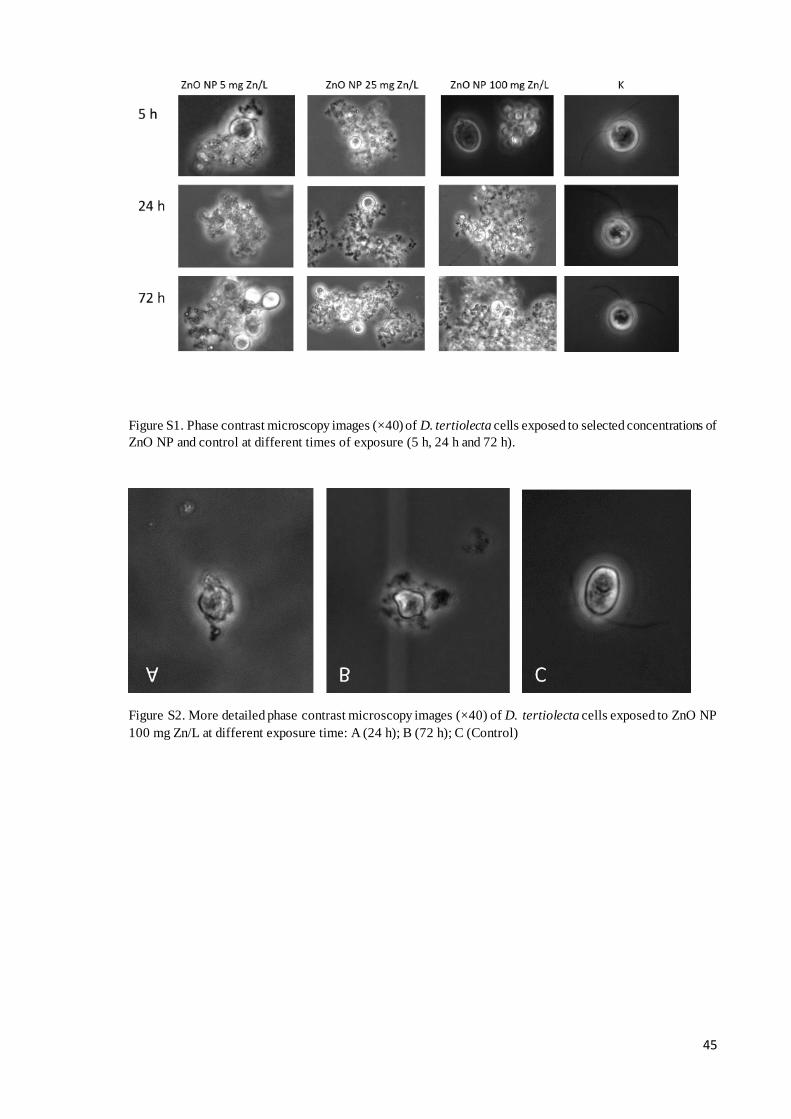
45
Figure S1. Phase contrast microscopy images (×40) of D. tertiolecta cells exposed to selected concentrations of
ZnO NP and control at different times of exposure (5 h, 24 h and 72 h).
Figure S2. More detailed phase contrast microscopy images (×40) of D. tertiolecta cells exposed to ZnO NP
100 mg Zn/L at different exposure time: A (24 h); B (72 h); C (Control)

46
Figure S3. FIB images of D.tertiolecta exposed to ZnO NP 100 mg Zn/L for 72 h.
Figure S4. Comet assay microscope images for D.tertiolecta exposed to ZnSO4

47
Figure S5. Additional algal cells comet resulting from the exposure (72 h) to ZnO NP 100mg Zn/L.

48
References
Abbott WS (1925) A method of computing the effectiveness of an insecticide. J Econ Entomol 18:265-267.
Akcha F, Arzul G, Rousseau S, Bardouil M (2008) Comet assay in phytoplankton as biomarker of genotoxic
effects of environmental pollution. Mar Environ Res 66:59-61.
Ali D, Alarifi S, Kumar S, Ahamed M, Siddiqui MA (2012) Oxidative stress and genotoxic effect of zinc oxide
nanoparticles in freshwater snail Lymnaea luteola. L Aquat Toxicol 124:83-90.
Apoorva G, Lavanya K, Vidisha, Pavani, Kumar RR, Hasan Q, Ramakrishna D (2013) Genotoxic effects of
silver and titanium dioxide nanoparticles. Proceedings of the "International Conference on Advanced
Nanomaterials & Emerging Engineering Technologies" (ICANMEET-2013), Sathyabama University, Chennai,
India in association with DRDO, New Delhi, India.
Aruoja V, Dubourguier HC, Kasemets K, Kahru A (2009) Toxicity of nanoparticles of CuO, ZnO and TiO2 to
microalgae Pseudokirchneriella subcapitata. Sci Total Environ 407:1461-1468
ASTM (1998) Standard guide for acute toxicity test with the rotifer Brachionus in: Annual Book of ASTM
Standards Philadelphia 1440-1491.
Bhattacharya K, Davoren M, Boertz J, Schins RP, Hoffmann E, Dopp E (2009) Titanium dioxide NPs induce
oxidative stress and DNA-adduct formation but not DNA breakage in human lung cells. Part Fibre Toxicol
6:17.
Bhuvaneshwari M, Iswarya V, Archanaa S, Madhu GM, Suraish Kumar GK, Nagarajan R, Chandrasekaran N,
Mukherjee A (2015) Cytotoxicity of ZnO NPs towards fresh water algae Scenedesmus obliquus at low exposure
concentrations in UV-C, visible and dark conditions. Aquat Toxicol 162:29-38.
Chen C, Zhang J, Ma P, Jin K, Li L, Luan J (2012) Spatial-temporal distribution of phytoplankton and safety
assessment of water quality in Xikeng reservoir. J Hydroecol 33(2):32- 38.
Chen X, Zhu X, Li R, Yao H, Lu Z, Yang X. (2012) Photosynthetic toxicity and oxidative damage induced by
nano-Fe3O4 on Chlorella vulgaris in the aquatic environment. Open J Ecol 1:21-28.
Choi HS, Kim YJ, Song M, Song MK, Ryu JC (2011) Genotoxicity of nano-silica in mammalian cell lines.
Toxicol Environ Heal Sci 3(1):7-13.
Clemente Z, Castro VL, Feitosa LO, Lima R, Jonsson CM, Maia AH, Fraceto LF (2013) Fish exposure to nano-
TiO2 under different experimental conditions: methodological aspects for nanoecotoxicology investigations.
Sci Total Environn 463-464:647-656.
Collins AR (2004) The comet assay for DNA damage and repair. Mol Biotechnol 26(3):249-261.
D’Agata A, Fasulo S, Dallas LJ, Fisher AS, Maisano M, Readman J, Jha AN (2014) Enhanced toxicity of ‘bulk’
titanium dioxide compared to ‘fresh’ and ‘aged’ nano-TiO2 in marine mussels (Mytilus galloprovincialis).
Nanotoxicology 8:549-558.
Dalai S, Pakrashi S, Nirmala MJ, Chaudhri A, Chandrasekaran N, Mandal AB, Mukherjee A (2013)
Cytotoxicity of TiO2 nanoparticles and their detoxification in a freshwater system. Aquat Toxicol 138-139:1-
11.
Demir E, Burgucu D, Turna F, Aksakal S, Kaya B (2013) Determination of TiO2, ZrO2, and Al2O3 nanoparticles
on genotoxic responses in human peripheral blood lymphocytes and cultured embryonic kidney cells. J Toxicol
Env Heal A Current Issues 76:990-1002.
Demir E, Kaya N, Kaya B (2014) Genotoxic effects of zinc oxide and titanium dioxide nanoparticles on root
meristem cells of Allium cepa by comet assay. Turk J Biol 38:31-39.
Downs TR, Crosby ME, Hu T, Kumar S, Sullivan A, Sarlo K, Reeder B, Lynch M, Wagner M, Mills T, Pfuhler
S. (2012) Silica nanoparticles administered at the maximum tolerated dose induce genotoxic effects through an
inflammatory reaction while gold nanoparticles do not. Mutat Res 745:38-50.

49
Falck GC, Lindberg HK, Suhonen S, Vippola M, Vanhala E, Catalán J, Savolainen K, Norppa H (2009)
Genotoxic effects of nanosized and fine TiO2. Hum Exp Toxicol 28:339-352.
Gerloff K, Albrecht C, Boots AW, Förster I, Schins RPF (2009) Cytotoxicity and oxidative DNA damage by
nanoparticles in human intestinal Caco-2 cells. Nanotoxicology 3(4):355-364.
Ghosh M, Chakraborty A, Mukherjee A (2013) Cytotoxic, genotoxic and the hemolytic effect of titanium
dioxide (TiO2) nanoparticles on human erythrocyte and lymphocyte cells in vitro. J Appl Toxicol 33:1097-110.
Golbamaki N, Rasulev B, Cassano A, Robinson RLM, Benfenati E, Leszczynski J, Cronin MTD (2015)
Genotoxicity of metal oxide nanomaterials: review of recent data and discussion of possible mechanisms.
Nanoscale 7(6):2154-2198.
Gomes T, Araújo O, Pereira R, Almeida AC, Cravo A, Bebianno MJ (2013) Genotoxicity of copper oxide and
silver nanoparticles in the mussel Mytilus galloprovincialis. Mar Environ Res 84:51-59.
Gonzalez L, Lison D, Kirsch-Volders M (2008) Genotoxicity of engineered nanomaterials: a critical review.
Nanotoxicology 2:252-273.
Gopalan RC, Osman IF, Amani A, De Matas M, Anderson D (2009) The effect of zinc oxide and titanium
dioxide nanoparticles in the comet assay with UVA photoactivation of human sperm and lymphocytes.
Nanotoxicology 3:33-9.
Guillard RRL (1975) Culture of phytoplankton for feeding marine invertebrates, in: Culture of Marine
Invertebrate Animals. Smith, W.L., Chanley, M.H. Eds.; Plenum Press, New York. pp. 26- 60.
Gunawan C, Sirimanoonphan A, Teoh WY, Marquis CP, Amal R (2013) Submicron and nano formulations of
titanium dioxide and zinc oxide stimulate unique cellular toxicological responses in the green microalga
Chlamydomonas reinhardtii. J Hazard Mater 260:984-992.
Guo YY, Zhang J, Zheng YF, Yang J, Zhu XQ (2011) Cytotoxic and genotoxic effects of multiwall carbon
nanotubes on human umbilical vein endothelial cells in vitro. Mutat Res DOI:10.1016/j.mrgentox.2011.01.014.
Hartmann NB, Von der Kammer F., Hofmann T, Baalousha M, Ottofuelling S, Baun A (2010) Algal testing of
titanium dioxide nanoparticles-Testing considerations, inhibitory effects and modification of cadmium
bioavailability. Toxicology 269:190-197.
Heim J, Felder E, Tahir MN, Kaltbeitzel A, Heinrich UR, Brochhausen C, Mailänderm V,Tremel W, Brieger J
(2015) Genotoxic effects of zinc oxide nanoparticles. Nanoscale 7(19): 8931-8938.
Hu CW, Li M, Cui YB, Li DS, Chen J, Yang LY (2010) Toxicological effects of TiO2 and ZnO nanoparticles
in soil on earthworm Eisenia fetida. Soil Biol Biochem 42(4):586-591.
Isani G, Falcioni ML, Barucca G, Sekar D, Andreani G, Carpenè E, Falcioni G (2013) Comparative toxicity of
CuO nanoparticles and CuSO4 in rainbow trout. Ecotox Environ Safe 97:40-46.
Ji J, Long Z, Lin D (2011) Toxicity of oxide nanoparticles to the green algae Chlorella sp. Chem. Eng. J.
170:525-530.
Jugan ML, Barillet S, Simon-Deckers A, Herlin-Boime N, Sauvaigo S, Douki T, Carriere M (2012) Titanium
dioxide nanoparticles exhibit genotoxicity and impair DNA repair activity in A549 cells. Nanotoxicology
6(5):501-513.
Kain J, Karlsson HL, Mӧller L (2012) DNA damage induced by micro and nanoparticles interaction with FPG
influences the detection of DNA oxidation in the comet assay. Mutagenesis 27(4):491-500.
Kalantari H, Rezaei M, Mahdavinia M, Kalantar M, Amanpour Z, Varnaseri G (2012) Determination of the
mutagenicity potential of supermint herbal medicine by single cell gel electrophoresis in rat hepatocytes. Adv
Pharm Bull 2(2):245-248.
Keller AA, Wang H, Zhou D, Lenihan HS, Cherr G, Cardinale BJ, Miller R (2010) Stability and aggregation
of metal oxide nanoparticles in natural aqueous matrices. Environ. Sci. Technol. 44:1962-1967.

50
Keller AA, McFerran S, Lazareva A, Suh S (2013) Global life cycle releases of engineered nanomaterials. J
Nanoparticle Res 15:1-17.
Keller AA, Vosti W, Wang H, Lazareva A (2014) Release of engineered nanomaterials from personal care
products throughout their life cycle. J Nanopart Res 16:2489.
Kitchin KT, Prasad RY, Wallace K (2011) Oxidative stress studies of six TiO2 and two CeO2 nanomaterials:
Immuno-spin trapping results with DNA. Nanotoxicology 5(4):546-556.
Kumar A, Pandey AK, Singh SS, Shanker R, Dhawan A (2011) Cellular uptake and mutagenic potential of
metal oxide nanoparticles in bacterial cells. Chemosphere 83:1124-1132.
Kumari M, Khan SS, Pakrashi S, Mukherjee A, Chandrasekaran N (2011) Cytogenetic and genotoxic effects
of zinc oxide nanoparticles on root cells of Allium cepa. J Hazard Mater 190:613-621.
Lankoff A, Arabski M, Wegierek-Ciuk A, Kruszewski M, Lisowska H, Banasik-Nowak A, Rozga-Wijas K,
Wojewodzka M, Slomkowski S (2013) Effect of surface modification of silica nanoparticles on toxicity and
cellular uptake by human peripheral blood lymphocytes in vitro.Nanotoxicology 7(3):235-250.
Lee SW, Kim SM, Choi J (2009) Genotoxicity and ecotoxicity assays using the freshwater crustacean Daphnia
magna and the larva of the aquatic midge Chironomus riparius to screen the ecological risks of nanoparticle
exposure. Environ. Toxicol Pharmacol 28(1):86-91.
Li F, Liang Z, Zheng X, Zhao W, Wu M, Wang Z (2015) Toxicity of nano-TiO2 on algae and the site of reactive
oxygen species production. Aquat Toxicol 158:1-13.
Lowry G, Gregory KB, Apte SC, Lead JR (2012) Transformations of nanomaterials in the environment. Environ
Sci Technol 46:6893-9.
Ma H, Williams PL, Diamond SA (2013). Ecotoxicity of manufactured ZnO nanoparticles-A review. Environ
Pollut 172:76-85.
Magdolenova Z, Collins A, Kumar A, Dhawan A, Stone V, Dusinska M (2013) Mechanisms of genotoxicity.
A review of in vitro and in vivo studies with engineered nanoparticles. Nanotoxicology
DOI:10.3109/17435390.2013.773464.
Manzo S, Miglietta ML, Rametta G, Buono S, Di Francia G (2013°) Toxic effects of ZnO nanoparticles towards
marine algae Dunaliella tertiolecta. Sci Total Environ 445-446:371-376.
Manzo S, Miglietta ML, Rametta G, Buono S, Di Francia G (2013b) Embryotoxicity and spermiotoxicity of
nanosized ZnO for mediterranean sea urchin Paracentrotus lividus. J Haz Mater 254:1-9.
Manzo S, Buono S, Rametta G, Miglietta M, Schiavo S, Di Francia G (2015) The diverse toxic effect of SiO2
and TiO2 nanoparticles toward the marine microalgae Dunaliella tertiolecta. Environ Sci Pollut Res DOI
10.1007/s11356-015-4790-2.
Matranga V, Corsi I, (2012) Toxic effects of engineered nanoparticles in the marine environment:model
organisms and molecular approaches. Mar Environ Res 76:32-40.
Misra SK, Dybowska A, Berhanu D, Luoma SN, Valsami-Jones E (2012) The complexity of nanoparticle
dissolution and its importance in nanotoxicological studies. Sci Total Environ 438:225-232.
Mu Q, David CA, Galceran J, Rey-Castro C, Krzemiński L, Wallace R, Bamiduro F, Milne SJ, Hondow NS,
Brydson R, Vizcay-Barrena G, Routledge MN, Jeuken LJ, Brown AP (2014) Systematic investigation of the
physicochemical factors that contribute to the toxicity of ZnO nanoparticles. Chem Res Toxicol 27:558-567.
Navarro E, Piccapietra F, Wagner B, Marconi F, Kaegi R, Odzak N, Sigg L, Behra R (2008) Toxicity of silver
nanoparticles to Chlamydomonas reinhardtii. Environ Sci Technol 42(23):8959-8964.
Pakrashi S, Dalai S, Prathna TC, Trivedi S, Myneni R, Raichur AM, Chandrasekaran N, Mukherjee A (2013)
Cytotoxicity of aluminium oxide nanoparticles towards fresh water algal isolate at low exposure concentrations.
Aquat Toxicol 132-133:34-45.

51
Pakrashi S, Jain N, Dalai S, Jayakumar J, Chandrasekaran PT, Raichur AM, Chandrasekaran N, Mukherjee A
(2014) In vivo genotoxicity assessment of titanium dioxide nanoparticles by Allium cepa root tip assay at high
exposure concentrations. PLoS One 9(2):e87789.
Prado R, García R, Rioboo C, Herrero C, Abalde J, Cid A (2009) Comparison of the sensitivity of different
toxicity test endpoints in a microalga exposed to the herbicide paraquat. Environ Int 35:240-247.
Reeves JF, Davies SJ, Dodd NJ, Jha AN (2008) Hydroxyl radicals (*OH) are associated with titanium dioxide
(TiO (2)) nanoparticle-induced cytotoxicity and oxidative DNA damage in fish cells. Mutat Res 640:113-22.
Rinna A, Magdolenova Z, Hudecova A, Kruszewski M, Refsnes M, Dusinska M (2015) Effect of silver
nanoparticles on mitogen-activated protein kinases activation: role of reactive oxygen species and implication
in DNA damage. Mutagenesis 30:59-66.
Rioboo C, Prado R, Herrero C, Cid A (2007) Population growth study of the rotifer Brachionus sp. fed with
triazine-exposed microalgae. Aquat Toxicol 83:247-253.
Rocco L, Santonastaso M, Mottola F, Costagliola D, Suero T, Pacifico S, Stingo V (2015) Genotoxicity
assessment of TiO2 nanoparticles in the teleost Danio rerio. Ecotoxicol Environ Saf 113:223-230.
Shukla RK, Sharma V, Pandey AK, Singh S, Sultana S, Dhawan A (2011) ROS-mediated genotoxicity induced
by titanium dioxide nanoparticles in human epidermal cells. Toxicol In Vitro 25:231-241.
Singh N, Manshian B, Jenkins GJS, Griffiths SM, Williams PM, Maffeis TGG, Wright CJ, Doak SH (2009)
NanoGenotoxicology: the DNA damaging potential of engineered nanomaterials. Biomaterials 30:3891-3914.
Suman TY, Radhika Rajasree SR, Kirubagaran R (2015) Evaluation of zinc oxide nanoparticles toxicity on
marine algae Chlorella vulgaris through flow cytometric, cytotoxicity and oxidative stress analysis. Ecotox
Environ Safe 113:23-30.
Thill A, Zeyons O, Spalla O, Chauvat F, Rose J, Auffan M, Flank AM (2006) Cytotoxicity of CeO2
nanoparticles for Escherichia coli. Physico-chemical insight of the cytotoxicity mechanism. Sci Total Environ
40:6151-6156.
Xia B, Chen B, Sun X, Qu K, Ma F, Du M (2015) Interaction of TiO2 nanoparticles with the marine microalga
Nitzschia closterium: Growth inhibition, oxidative stress and internalization. Sci Total Environ 508:525-533.
Yang H, Liu C, Yang D, Zhang H, Xi Z (2009) Comparative study of cytotoxicity, oxidative stress and
genotoxicity induced by four typical nanomaterials: the role of particle size, shape and composition. J Appl
Toxicol 29:69-78.

52
3.3 Growth inhibition of three species of marine microalgae exposed to different
sizes of Ag NPs and to coating agent PVP/PEI
This section has been published in: Simona Schiavo, Nerea Duroudier, Eider Bilbao, Mathilde Mikolaczyk,
Jorg Schafer, Miren Cajaraville, Sonia Manzo. Growth inhibition of three species of marine microalgae
exposed to different sizes of Ag NPs and to coating agent PVP/PEI. Aquatic toxicology. Under review.
Abstract
Microalgae are at the base of aquatic food chains, being starting points of biomagnificat ion processes that
transport increased amounts of accumulated contaminants into food chains. Ag NPs are increasingly used due
to their antimicrobial properties, therefore their presence in aquatic ecosystems is expected to grow. We
evaluated the potential toxic effects of PVP/PEI coated 5 nm Ag NPs and of uncoated 47 nm Ag NPs to three
species of marine microalgae with the aim of assessing their relative sensitivity. The selected species Isochrysis
galbana (Prymnesiophyceae), Tetraselmis suecica (Chlorophyceae) and Phaeodactilum tricornutum
(Bacillariophyceae) showed cell wall differences: the diatom P. tricornutum has an elaborate and thick cell
wall, T. suecica cell wall is composed of coalesced rigid carbohydrate scales while I. galbana has a relatively
soft cell coating. Microalgae were exposed for 72 h to PVP/PEI 5 nm Ag NPs (0.00001-100 mg Ag/L) or to 47
nm Ag NPs (1-10 mg Ag/L) following the OECD 201 guideline. In parallel, the toxicity of PVP/PEI was also
evaluated. Both types of NPs formed aggregates in seawater, larger for 47 nm Ag NPs than for 5 nm Ag NPs.
The release of Ag ions by PVP/PEI 5 nm Ag NPs was around 10 times higher than by 47 nm Ag NPs. Ag NPs
were able to interact with algal cells surface, as shown by microscope observations. There were clear differences
in sensitivity towards PVP/PEI 5 nm Ag NPs among species. T. suecica was about 10 times more sensitive
(EC50 0.0052 mg Ag/L) than I. galbana (EC50 0.039 mg Ag/L) and P. tricornutum (EC50 0.06 mg Ag/L).
PVP/PEI alone also showed effect to algae indicating that it contributed significantly to the toxicity of 5 nm Ag
NP suspensions. Ag NPs of 47 nm resulted less toxic than 5 nm Ag NPs, probably due to differences in size,
dispersant and consequent dissolution and aggregation behavior. P. tricornutum was slightly less sensitive
(EC50 4.72 mg Ag/L) than T. suecica (EC50 4.1 mg Ag/L) and I. galbana (EC50 3.30 mg Ag/L) which agrees
well with the presence of a resistant silicified cell wall in the diatom. The mechanisms leading to observed
inhibitory effects on algae growth need further studies.
Introduction
Silver nanoparticles (Ag NPs) are among the most used metallic nanoparticles in consumer products (Rejeski
et al. 2008; Marin et al. 2015) due to their unique physicochemical characteristics, such as high conductivity,
scattering, chemical stability and catalytic activity (Johari et al. 2013) as well as their antimicrobial properties
(Mohan et al. 2007; Wijnhoven et al. 2009). As the market for silver containing nano-functionalized products
is significantly increasing worldwide (Boxall et al. 2007; Blaser et al. 2008; Thio et al. 2012), release of Ag
NPs to the environment, including aquatic systems, is becoming relevant and is expected to grow up in the next
decades (Tiede et al. 2009; Fabrega et al. 2011; Moreno-Garrido et al. 2015). Once in the aquatic environment,
it is likely that Ag NPs become a source of (Ag NP) aggregates and of dissolved silver ions (Ag (I)), that could
produce adverse effects in aquatic organisms.
Toxicity of Ag NPs to different freshwater and saltwater organisms belonging to different trophic levels has
been widely reported (Fabrega et al. 2011; Wang et al. 2012; Ribeiro et al. 2014; Dorobantu et al. 2015;
Gambardella et al. 2015). However, as recently reviewed by Moreno-Garrido and co-workers (2015), data on
the toxicity on microalgae, and especially on microalgae from marine or estuarine environments, remain scarce
and scattered. Current literature shows a wide variation of results on the toxicity of Ag NPs to microalgae, that
vary in as much as five orders of magnitude of EC50 values (Lee et al. 2005; Navarro et al. 2008; Kennedy et
al. 2010; Burchardt et al. 2012; He et al. 2012; Ribeiro et al. 2014), possibly in relation to differential algae
sensitivity and to different factors including Ag NP size and presence of coating agents. Although the coating
agents used for NP stabilization are an integral part of the NPs, still scarce attention is devoted to their potential
contribution to the overall toxicity of NPs (El Badawy et al. 2011; Katsumiti et al. 2014a; Zhang et al. 2014;
Katsumiti et al. 2015; Navarro et al. 2015).

53
As microscopic primary producers, microalgae are the first target organisms for most of the pollutants present
in aquatic systems. They constitute the base of aquatic food chains, being potential starting points of
biomagnification processes. Therefore, marine microalgae, which are widely distributed in coastal ecosystems
(Behrenfeld et al. 2006) and are particularly susceptible to pollutants (Miglietta et al. 2011; Manzo et al. 2014)
can be regarded as suitable indicators for marine water pollution by NPs (Aruoja et al. 2009). The evaluation
of NP effects upon marine phytoplankton is indeed a necessary step to predict their potential impact on the
whole ecosystems they support. Different species of microalgae could show differences in sensitivity towards
NP toxicity depending on their structural and physiological characteristics. Phaeodactilum tricornutum
(Bacillariophyceae) is a widespread pennate diatom (Rushforth et al. 1988; Francius et al. 2008) commonly
used for assessing effects of NPs (Baker et al. 2014; Castro-Bugallo et al. 2014; Moreno Garrido et al. 2015).
The cell wall is formed by two valves overlapping in the girdle band region but in opposition to other diatoms,
Phaeodactylum is very poor in silica, being the cell wall essentially composed of organic compounds. This
diatom showed different sensitivities to Ag NPs and in particular a 50% growth inhibition (IC50) has been
recorded at 2380 ± 1880 and 3690 ± 2380 μg/L for citrate-capped (14 nm) and PVP-capped (15 nm) Ag NPs,
respectively (Angel et al. 2013) while a EC50 of around 930 μg/L for citrate Ag NPs has also been reported
(Moreno-Garrido et al. 2015).
Tetraselmis suecica is an elliptical microalgae of the Chlorophyceae (Prasinophyceae) class ranging up to 12
μm in length while Isochrysis galbana (Prymnesiophyceae) is the smallest (4-7 μm) among the three algae
tested in this study andis widely cultured to feed bivalves in the aquaculture industry. Both algae show a peculiar
cell wall structure: T. suecica is enclosed in a rigid polysaccharidic cell wall or theca (Becker et al. 1994) while
I. galbana has a relatively soft cell coating composed of coalesced carbohydrate scales (Zhu and Lee, 1997).
Although both algae are generally used as model organisms for toxicity assessment of NPs (Miller e t al. 2012;
Minetto et al. 2014), to the best of our knowledge no literature is available about Ag NPs effects on these
species.
In this study, the effects of PVP/PEI coated 5 nm Ag NPs and of uncoated 47 nm Ag NPs upon three marine
microalgae (P. tricornutum, T. suecica and I. galbana) were evaluated with the aim of assessing their relative
sensitivity. Population growth rate alterations as well as growth inhibition were evaluated and the No Observed
Effect Concentration (NOEC), the Lowest Observed Effect Concentration (LOEC) and EC50 values were
calculated. Interactions of algae cells surface with Ag NPs were also studied by microscopy analysis. Moreover,
the potential toxic effects caused by the PVP/PEI coating agent were determined in order to understand the
potential contribution of the coating agent to the overall toxicity of Ag NP suspensions.
Materials and methods
Obtainment and characterization of Ag NPs
Uncoated Ag NPs and Ag NPs coated with PVP/PEI (Poly N-vinyl-2-pirrolidone + Poly ethyleneimine;
77%:23%) were purchased as a stable aqueous suspension from Nanogap (Galicia, Spain). According to the
manufacturers’ information, PVP/PEI coated AgNPs showed an average size of 5.08 ± 2.03 nm and a zeta
potential of +18.6 ± 7.9 mV, while uncoated Ag NPs presented an average size of roughly 47.2±15.6 nm in
distilled water (DW). Particle size distribution of PVP/PEI coated 5 nm Ag NPs and of uncoated 47 nm Ag
NPs in artificial seawater (ASW, ASTM 1998) was analyzed daily by Dynamic Light Scattering (DLS) using a
Zetasizer Nano Z (Malvern Instruments Ltd., Worcestershire, UK) during the 72 h of the ecotoxicological assay.
Dissolution of both types of Ag NPs was assessed in ASW as described by Katsumiti et al. (2014b).
Briefly, 10 ml samples of the Ag NP suspensions were prepared at a concentration of 10 mg/L and filled into
dialyzer tubes (Spectra/ Por® Float-A-Lyzer; MWCO 0.1-0.5 kDa). The sample-filled dialyzers were then
immersed in 1 L ASW. Samples (3 replicates of 1 ml) were extracted at 24, 48 and 72 h from the solution and
analyzed for Ag ions using ICP-MS (Thermo X2 series) after 100- fold dilution using external calibration.
Differences obtained for replicate samples were consistently lower than 5 % (R.S.D).
Organisms
P. tricornutum (Bacillariophyceae: Naviculales), T. suecica (Chlorophyceae: volvocales) and I. galbana
(Prymnesiophyceae: Isochrysidales) were maintained in sterilized standard medium (Guillard, 1975) made with
ASW.
Microalgae were incubated under cool continuous white fluorescent lights until the log phase growth prevailed
(about 58 μmol photons m−2 s−1 at 24±1°C with aeration from 5 to 7 days) to provide enough inocula for
experiments. Cell density was measured by a hemocytometer.

54
Toxicity test
Algae growth inhibition test
Algae bioassays were performed according to IRSA-CNR (IRSA-CNR, 1978). All glassware was rinsed with
Milli-Q purified water and autoclaved before use. Microalgae (with a final density of 104 cells/ml) were first
filtered (0.22 μm) and rinsed three times with filtered autoclaved ASW.
Then, microalgae were added to each treatment and control (standard culture media, Guillard medium) together
with nutrients. Stock suspensions of PVP/PEI 5nm Ag NPs and 47 nm Ag NPs were prepared by dispersing the
primary stock solution into ASW to the final concentration of 100 mg/L. Stock dispersions were properly diluted
at concentrations ranging 0.00001-100 mg Ag/L for PVP/PEI 5nm Ag NPs and 1-10 mg Ag/L for 47nm Ag
NPs. Concentration ranges were selected based on the literature (Navarro et al. 2008; Oukarroum et al. 2012;
He et al. 2012) and on preliminary toxicity experiments performed with each NP type.
The PVP/PEI stock solution was diluted at concentrations ranging 0.0001-1040 mg/L (the same concentration
range present in the 5nm Ag NP suspensions). After dilution, all the suspensions were briefly vortexed before
the addition of micronutrients and test organisms.
Test plates (10 ml) were kept in a growth chamber constantly illuminated with a white fluorescent lamp
(enhanced irradiation between 400 and 500 nm), at a temperature of 24 ± 1 °C for 3 days.
Growth inhibition was expressed as percentage effect with respect to controls.
Growth rate determination
During the experiments, growth rate was determined daily by counting microalgae with a Burker chamber under
a light microscope. Growth rate was calculated according to the equation described by Xiong et al. (2005):
U = (ln Nt- ln N0) / (t - t0)
where U (cell number/h) is the growth rate; Nt and N0 mean the quantity of cells at times t and 0,
respectively,where t means any exposure time (h) and t0 means the initial time of the treatment (h).
Microscope observations
Observations on the interactions of Ag NPs with microalgae were performed through light microscope analysis
(ZEISS Axioskop 50) followed by more detailed observations through Focused Ion Beam (FIB) microscopy
(FEI QUANTA 2 D). Before FIB observations, microalgae were fixed as described in Li et al. (2015). Briefly,
after 72 h of exposure, microalgae were centrifuged (4000 rpm for 10min) and then samples were fixed with a
3% glutaraldehyde solution at 4°C for 2 h.
Samples were then washed three times with 0.1 M PBS (pH 7.8) by centrifugation (4000 rpm for 10 min).
Microalgae were fixed with 1% osmium tetroxide for 2 h at 4°C and finally 0.1 M PBS (pH 7.8) was added to
wash the cells by centrifugation (3800 rpm for 10 min, three times). Control and treated (10 mg Ag/L)
microalgae were placed on a thin glass slide, air dried and observed under the FIB.
Data analysis
Statistical analyses for growth inhibition as well as for NOEC and LOEC values were conducted using the
statistical package SPSS v.19 (SPSS Inc., IBM Company, Chicago, USA). Prior to the analysis, data were tested
for normality (Kolmogorov-Smirnov normality test) and homogeneity of variances (Levene’s test). Significant
differences (p<0.05) with respect to controls were set based on analysis of variance (ANOVA) and the multiple
comparisons Dunnett's test. EC50 values were calculated using the Linear Interpolation Method (Inhibition
Concentration procedure or ICp) (US EPA, 1993) and the bootstrap method was used to obtain 95% confidence
intervals

55
Results
Physicochemical characterization
PVP/PEI coated 5 nm Ag NPs dispersed in ASW at 10 mg/L immediately reached a mean size of around 100
nm according to DLS measurements (Figure 1).The mean size remained stable up to 24h (100 nm), while after
48-72 h aggregates were slightly smaller (around 90 nm, Figure 1). Zeta potential values for PVP/PEI 5 nm Ag
NPs in ASW ranged from -5 mV to +0.2 mV. Uncoated 47 nm Ag NPs tended to aggregate rapidly in ASW
reaching a mean size of about 880 nm that turned to be slightly smaller at 24 h (~600 nm) (Figure 2). The largest
size was achieved after 48 h (~3000 nm) and aggregates were smaller (around 1400 nm) after 72 h (Figure 2).
Zeta potential in ASW was around -8 mV.
Figure 1: Mean size (±SD) of aggregates of PVP/PEI 5 nm Ag NPs (10 mg/L) during the 3 days algae bioassay.
Figure 2: Mean size (±SD) of aggregates of 47 nm Ag NPs (10 mg/L) during the 3 days algae bioassay
Dissolution of both types of Ag NPs in ASW was detected along the 72 hours of experimentation (Table 1). In
general, the release of Ag ions by PVP/PEI coated 5 nm Ag NPs was around 10 times higher than that by
uncoated 47 nm Ag NPs (Table 1). After 24 hours, PVP/PEI coated 5 nm Ag NPs released around 20% of Ag
ions while only near 1.5% of the uncoated 47 nm Ag NPs was converted into ionic Ag (Table 1). At 72 hours,
dissolution of PVP/PEI coated 5 nm Ag NPs increased to 29.6% and for uncoated 47 nm Ag NPs to 3.4% (Table
1).

56
Table 1. Release of Ag ions as % of total starting material of Ag NPs (10 mg/L) in ASW.
Toxic effects of PVP/PEI 5nm Ag NPs and coating agent PVP/PEI
Figure 3 shows the effects of PVP/PEI 5 nm Ag NPs and of the coating agent PVP/PEI on the growth of selected
microalgae. Main ecotoxicological parameters evidenced clear differences insensitivity among studied species
(Table 2). For PVP/PEI 5 nm Ag NPs, the effects were significant at the lowest concentration for all three
microalgae. In fact, the NOEC was below 0.00001 mg Ag/L and the LOEC was always represented by the
lowest concentration tested (Table 2). Based on the EC50 values obtained, T. suecica was about 10 times more
sensitive (EC50 0.0052 mg Ag/L) than I. galbana (EC50 0.039 mg Ag/L) and P. tricornutum (EC50 0.06 mg
Ag/L). This greater sensitivity of T. suecica was also evidenced by the analysis of the growth rates reported in
Table 3. In fact, for T. suecica significant growth rate inhibition was measured starting from 0.01 mg Ag/L
whereas the growth rates for I. galbana and P. tricornutum were not significantly different from controls up to
0.1 mg Ag/L (Table 3).
Table 2. Ecotoxicological parameters (EC50; NOEC; LOEC) for the three microalgae exposed to PVP/PEI 5 nm Ag NPs
and PVP/PEI coating agent.
The coating agent PVP/PEI alone inhibited markedly the growth of the three microalgae (Figure 3 and Table
4). The highest EC50 value (0.83 mg/L corresponding to 0.083 mg Ag/L in PVP/PEI 5 nm Ag NP suspension)
was registered for P. tricornutum. T. suecica resulted the most sensitive with a EC50 value of 0.004 mg/L (=
0.0004 mg Ag/L in PVP-PEI 5 nm Ag NP suspension), while for I. galbana an intermediate EC50 value was
obtained (0.03 mg/L corresponding to 0.003 mg Ag/L in PVP/PEI 5 nm Ag NP suspension). Thus, PVP/PEI
alone was toxic to algae indicating that it contributed significantly to the toxicity of PVP/PEI 5 nm Ag NP
suspensions.

57
The observations by light microscopy and by FIB of the three microalgae exposed to PVP/PEI 5 nm Ag NPs
(10 mg Ag/L) are reported in Figure 5a and Figure 6a. PVP/PEI 5 nm Ag NPs seemed to interact with algae
mainly by entrapping them in a network of hetero-aggregates without changing their shape or morphology.
Similarly algae exposed to PVP/PEI alone appeared entirely covered by the polymeric matrix. Moreover, FIB
images allowed a more detailed observation of algae surface which resulted completely covered and surrounded
by PVP/PEI 5 nm Ag NPs aggregates. In the case of P. tricornutum and I. galbana this coverage did not result
in alterations of cell morphology, while for T. suecica a change in shape and a loss of cell turgor, with respect
to the control, could be observed.
Figure 3: Toxic effects of different concentrations of PVP/PEI 5 nm Ag NPs (red triangle) and PVP/PEI (blue triangle) upon
P. tricornutum, T. suecica and I. galbana. Asterisks indicate statistically significant differences with respect to controls
(p<0.05).
Table 4. Growth rate (mean ± SD) for the three microalgae exposed to PVP/PEI at the same concentration range present in
the 5 nm Ag NP suspensions. Asterisks indicate statistically significant differences with respect to controls (p<0.05).

58
Toxic effects of 47 nm Ag NPs
Overall, 47 nm Ag NPs were less toxic than PVP/PEI 5 nm Ag NPs for the three microalgae tested. P.
tricornutum was slightly less sensitive (EC50 4.72 mg Ag/L) than T. suecica (EC50 4.1 mg Ag/L) and I. galbana
(EC50 3.30 mg Ag/L) (Table 5). T. suecica showed a peculiar inverted U-shaped dose-response curve (Figure
4). For the three species tested, the decrease in growth rate was maximal at 5 mg Ag/L and then growth rate
values increased at 7.5 and 10 mg Ag/L (Figure 4, Table 6).
Figures 5b and 6b show light microscopy and FIB images obtained for the three microalgae exposed to 47 nm
Ag NPs (10 mg Ag/L). Light microscopy images showed that 47 nm Ag NPs interacted by surrounding algae
surface without entrapping them in hetero-aggregates. Algae surfaces were observed in greater detail by FIB
microscopy: algae cells resulted covered by 47 nm Ag NP aggregates but no signs of cell morphology alterations
and plasma membrane damages could be highlighted.
Figure 4: Toxic effects of different concentrations of 47 nm Ag NPs upon P. tricornutum, T. suecica and I. galbana.
Asterisks indicate statistically significant differences with respect to controls (p<0.05).
Table 5.Ecotoxicological parameters (EC50; NOEC; LOEC) for the three algae exposed to 47 nm Ag NPs
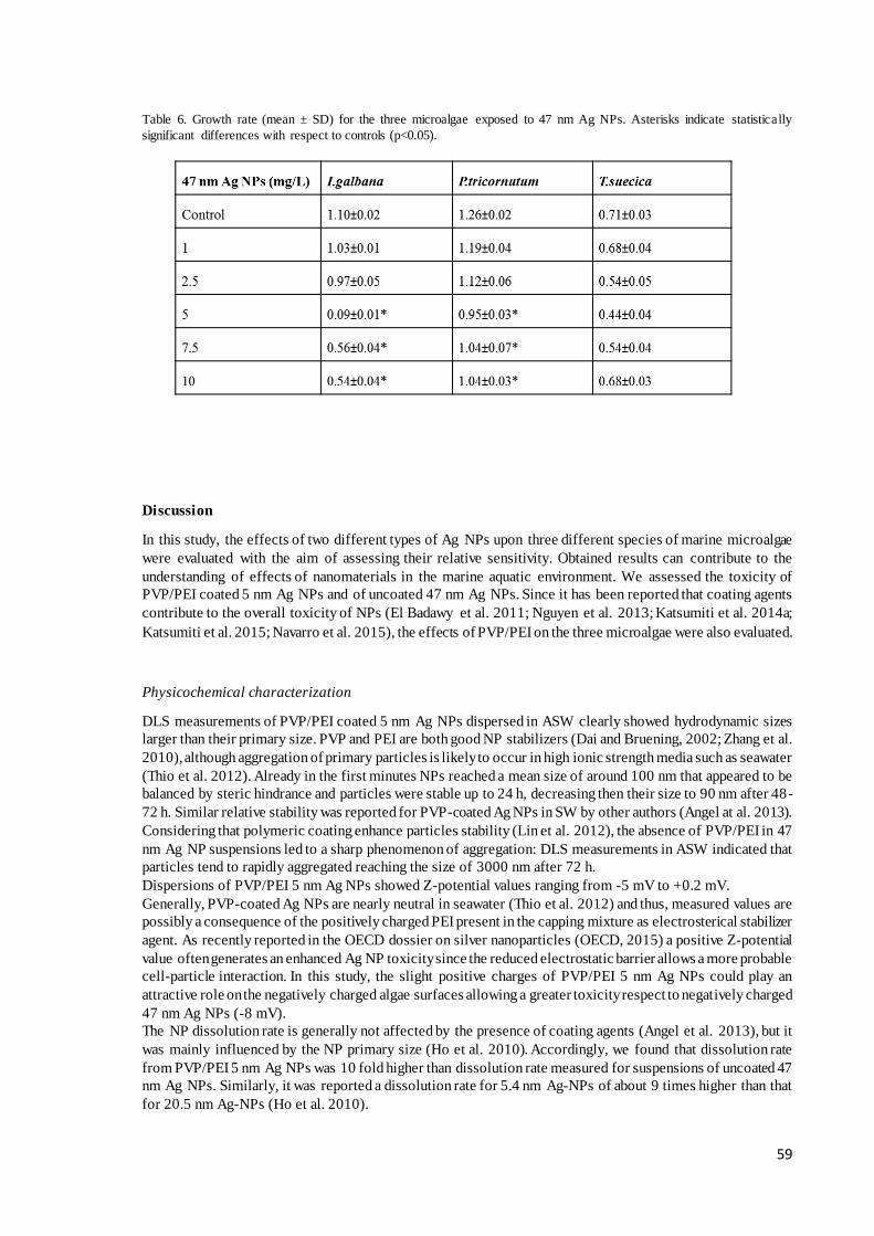
59
Table 6. Growth rate (mean ± SD) for the three microalgae exposed to 47 nm Ag NPs. Asterisks indicate statistically
significant differences with respect to controls (p<0.05).
Discussion
In this study, the effects of two different types of Ag NPs upon three different species of marine microalgae
were evaluated with the aim of assessing their relative sensitivity. Obtained results can contribute to the
understanding of effects of nanomaterials in the marine aquatic environment. We assessed the toxicity of
PVP/PEI coated 5 nm Ag NPs and of uncoated 47 nm Ag NPs. Since it has been reported that coating agents
contribute to the overall toxicity of NPs (El Badawy et al. 2011; Nguyen et al. 2013; Katsumiti et al. 2014a;
Katsumiti et al. 2015; Navarro et al. 2015), the effects of PVP/PEI on the three microalgae were also evaluated.
Physicochemical characterization
DLS measurements of PVP/PEI coated 5 nm Ag NPs dispersed in ASW clearly showed hydrodynamic sizes
larger than their primary size. PVP and PEI are both good NP stabilizers (Dai and Bruening, 2002; Zhang et al.
2010), although aggregation of primary particles is likely to occur in high ionic strength media such as seawater
(Thio et al. 2012). Already in the first minutes NPs reached a mean size of around 100 nm that appeared to be
balanced by steric hindrance and particles were stable up to 24 h, decreasing then their size to 90 nm after 48-
72 h. Similar relative stability was reported for PVP-coated Ag NPs in SW by other authors (Angel at al. 2013).
Considering that polymeric coating enhance particles stability (Lin et al. 2012), the absence of PVP/PEI in 47
nm Ag NP suspensions led to a sharp phenomenon of aggregation: DLS measurements in ASW indicated that
particles tend to rapidly aggregated reaching the size of 3000 nm after 72 h.
Dispersions of PVP/PEI 5 nm Ag NPs showed Z-potential values ranging from -5 mV to +0.2 mV.
Generally, PVP-coated Ag NPs are nearly neutral in seawater (Thio et al. 2012) and thus, measured values are
possibly a consequence of the positively charged PEI present in the capping mixture as electrosterical stabilizer
agent. As recently reported in the OECD dossier on silver nanoparticles (OECD, 2015) a positive Z-potential
value often generates an enhanced Ag NP toxicity since the reduced electrostatic barrier allows a more probable
cell-particle interaction. In this study, the slight positive charges of PVP/PEI 5 nm Ag NPs could play an
attractive role on the negatively charged algae surfaces allowing a greater toxicity respect to negatively charged
47 nm Ag NPs (-8 mV).
The NP dissolution rate is generally not affected by the presence of coating agents (Angel et al. 2013), but it
was mainly influenced by the NP primary size (Ho et al. 2010). Accordingly, we found that dissolution rate
from PVP/PEI 5 nm Ag NPs was 10 fold higher than dissolution rate measured for suspensions of uncoated 47
nm Ag NPs. Similarly, it was reported a dissolution rate for 5.4 nm Ag-NPs of about 9 times higher than that
for 20.5 nm Ag-NPs (Ho et al. 2010).

60
On the other hand, in environmental and biological solutions containing chloride ions, AgCl complexes (and
some times silver carbonate and silver phosphate) will form from the dissolved silver ions. Therefore, due to
the high salinity of seawater, dissolved silver is likely to be less toxic, respect to low salinity waters, because
silver speciation may have been dominated by AgCl complexes that are less toxic than free silver due to their
lower bioavailability (Lee et al. 2005). For example, Angel et al. (2013) reported that only 34% of the total
silver present at the start of the test dissolved in seawater. At the same way, we found that after 72 h the 29%
of total silver was released by PVP/PEI 5 nm Ag NP while only 3% of total silver was released by 47 nm Ag
NPs.
Toxic effects upon microalgae
By comparing the dose-response curves obtained for uncoated and coated Ag NPs, different levels of toxicity
were clearly detectable. Microalgae exposed to uncoated 47 nm Ag NPs showed EC50 values of several orders
of magnitude higher (3-4 mg Ag/L) than those obtained by the algae exposed to PVP/PEI coated 5 nm Ag NPs
(0.0052-0.06 mg Ag/L). The different algal sensitivity could be ascribable to a combined effect of particle size
and occurrence or not of coating agent PVP/ PEI once suspended in saline medium (Miao et al. 2009; He et al.
2012; Gambardella et al. 2015).
In the present study, the three algae, when exposed to PVP/PEI 5 nm Ag NP, were less sensitive respect to
Thalassiosira weissflogii (Miao et al. 2009) exposed to PVP Ag 10 nm NP (EC50 1-2 *10-12 M), but more
sensitive respect to D. tertiolecta (EC50 0.9 mg/L) and S. costatum (EC50 3.1 mg/L) treated with uncoated 10
nm Ag NP (Gambardella et al. 2015). Instead, when I. galbana, T. suecica and P. tricornutum were exposed to
40 nm Ag NPs, minor sensitivity respect to C. marina (EC50 0.2 mg/L, He et al. 2012). In the case of P.
tricornutum the obtained EC50 values were not directly comparable with that ones obtained for this species and
other diatoms in previous works (Angel et al. 2013; Moreno-Garrido et al. 2015) due to difference in NP sizes
used (14-15 nm) and in capping agent composition.
The nano aggregates of coated Ag NPs (less than 100 nm) resulted more available to microalgae in comparison
to the larger ones produced by uncoated NPs. This greater availability resulted in a closer interaction with
microalgae surfaces that appeared entrapped in a network of large heteroaggregates of PVP/PEI coated 5 nm
Ag NPs, as could be highlighted by light microscopy and FIB microscopy.
Figure 5: Micrographs obtained by light microscopy of P.tricornutum, T.suecica and I. galbana exposed to 10 mg/L
PVP/PEI 5 nm Ag NPs (A) and 47 nm Ag NPs (B). Control cells are shown in the insets

61
It seems that aggregates covering cell surfaces can induce a certain inhibition of the photosynthetic activity of
microalgae due to the reduction of light availability (Navarro et al. 2008; Wei et al. 2010; Manzo et al. 2015).
On the other hand, based on light microscope observations, aggregates of uncoated Ag NPs mainly entrapped
few algae cells at time resulting in generally small formations. This was also observable in FIB images in which
only few aggregates were found surrounding the algae cell surfaces.
Coating agents are chemicals (such as polymers and surfactants) used in the synthesis of Ag NPs to prevent
their aggregation through electrostatic repulsion, steric repulsion or both (Phenrat et al. 2008; Hotze et al. 2010).
In the case of silver, the most prevalent capping agents are citrate and polyvinylpyrrolidone (PVP) (El Badawy
et al. 2011). The PVP-AgNPs are stabilized through the steric repulsion caused by the adsorption of PVP on the
particle surface (Zhang and Zhang, 2014). The PEI-Ag NPs are electrosterically stabilized due to the adsorption
of the PEI on the particle surface (El Badawy et al. 2010). In general, no toxic effects have been previously
reported for PVP upon marine microalgae (Moreno-Garrido et al. 2015) while a clear toxic effect has been
reported for PEI for different organisms (El Badawy et al. 2011; Ivask et al. 2014). However, to the best of our
knowledge, there is no literature available upon the use of these two polymers together. In the present study,
algae exposed to the coating agent PVP/PEI alone showed an evident growth inhibition effect similar to that
observed for coated NPs, but a different behaviour could be distinguished for the three algae. In particular, in
the case of I. galbana and T. suecica, the coating agent alone resulted more toxic than PVP/PEI coated Ag NP
suspensions at PVP/PEI equivalent concentrations. We can speculate that once algae were added to the PVP/PEI
suspension, an immediate steric interaction occurred due to the stabilizing nature of the agent itself. This could
provoke the inclusion of microalgae in the polymeric matrices with a subsequent clear toxic effect, especially
in the case of microalgae without a resistant cell wall (I. galbana and T. suecica).
Similarly, a possible role of the coating agent in the overall toxicity to algae of different metal NPs as well as
some evidences for different organic coating agents has been previously reported by some authors (El Badawy
et al. 2011, Moreno-Garrido et al. 2015, Navarro et al. 2015).
Figure 6: FIB micrographs of P. tricornutum, T. suecica and I. galbana exposed to 10 mg/L PVP/PEI 5 nm Ag NPs (A) and
47 nm Ag NPs (B). Control cells are shown in the insets.
The three selected microalgae differed in sensitivity towards tested NPs; T. suecica and I. galbana were more
sensitive than P. tricornutum to both NPs as well as to the coating agent. The higher sensitivity of I. galbana
and T. suecica could be due to their cell wall characteristics. These algae have no distinct or mineralized cell
walls (Throndsen et al. 1993; Zhu et al. 1997) and therefore, they may be more vulnerable compared to other
taxa with different cell wall properties. Thus, the resistant cell wall of P. tricornutum seemed to limit the toxic
effects of coated and uncoated Ag NPs and of the coating agent. Additionally, small microalgae have been

62
found to be more sensitive than diatoms (Helbling et al. 1994; Karentz et al. 1994; Holm-Hansen et al. 1997).
This higher sensitivity has been related to their larger surface area to volume ratio compared to larger microalgae
(Quigg et al. 2006; Levy et al. 2007). Moreover, due to their dimensions, small microalgae may be covered
faster by NPs. Finally, algae motility is a key process that should be taken into account: it allows a greater
interaction between algae and NP aggregates but it could also be a very sensitive process that could be affected.
Mobile algae cells are widely distributed in culture medium, thus increasing their contact time with Ag NP
aggregates, with respect to immobile diatoms that tend to settle at the bottom of the well (Zhou et al. 2015).
Conclusions
Ag NPs of 47 nm resulted less toxic than PVP/PEI coated Ag NPs of 5 nm for the three microalgae species.
This is possibly related to the fact that 5 nm Ag NPs formed smaller aggregates that could be more available to
cells when compared to aggregates of 47 nm Ag NPs. An additional reason could be that PVP/PEI 5 nm Ag
NPs released around 10 times more Ag ions than 47 nm Ag NPs.
PVP/PEI alone was also toxic to algae indicating that it contributed significantly to the toxicity of PVP/PEI 5
nm Ag NP suspensions. The mechanisms leading to observed inhibitory effects on algae growth remain to be
explored and need further studies. Finally, of the three species tested, T. suecica was the most sensitive to
PVP/PEI 5 nm Ag NP exposure while the diatom P. tricornutum was most resistant, highlighting the influence
of algae cell surface characteristics on NP toxicity.

63
References
Angel BM, Batley GE, Jarolimek CV, Rogers NJ (2013) The impact of size on the fate and toxicity of
nanoparticulate silver in aquatic systems. Chemosphere 93:359-365.
Aruoja V, Dubourguier HC, Kasemets K (2009) Toxicity of nanoparticles of CuO, ZnO and TiO2 to microalgae
Pseudokirchneriella subcapitata. Sci Total Environ 407:1461-1468.
ASTM standard guide for acute toxicity test with the rotifer Brachionus. (1998) Annual Book of ASTM
Standards. Philadelphia, pp 1440-1491.
Baker TJ, Tyler CR, Galloway TS (2014) Impacts of metal and metal oxide nanoparticles on marine organisms.
Environmental Pollution 186:257-271.
Becker EW Microalgae. Biotechnology and Microbiology. (1994) Cambridge University Press, Cambridge.
ISBN 0-521-35020-4.
Behrenfeld MJ, O’Malley RT, Siegel DA, McClain CR, Sarmiento JL, Feldman GC, Milligan AJ, Falkowski
PG, Letelier RM, Boss ES (2006) Climate-driven trends in contemporary ocean productivity. Nature 444:752-
755.
Blaser SA, Scheringer M, MacLeod M, Hungerbühler K (2008) Estimation of cumulative aquatic exposure and
risk due to silver: Contribution of nano-functionalized plastics and textiles. Sci Total Enviro 390: 396-409.
Boxall ABA, Chaudhry Q, Jones A, Jefferson B, Watts CD (2007) Current future predicted environmental
exposure to engineered nanoparticles. Report Defra. Central Science Laboratory Sand Hutton York 3-89.
Burchardt A. D., Carvalho R. N., Valente A., Nativo P., Gilliland D., Garcìa C. P., Passarella R., Pedroni V.,
Rossi F., Lettieri T. 2012. Effects of silver nanoparticles in diatom Thalassiosira pseudonana and
Cyanobacterium synechococcus sp. Environ Sci Technol 46:11336-11344.
Castro-Bugallo A, González-Fernández Á, Guisande C, Barreiro A (2014) Comparative responses to metal
oxide nanoparticles in marine phytoplankton. Arch Environ Con Tox 67: 483-493.
Dai J, Bruening ML (2002) Catalytic nanoparticles formed by reduction of metal ions in multilayered
polyelectrolyte films. Nano Letters 2: 497-501.
Dorobantu LS, Fallone C, Noble AJ, Veinot J, Ma G, Goss GG, Burrell RE (2015) Toxicity of silver
nanoparticles against bacteria, yeast, and algae. J. Nanopart. Res. 17.
El Badawy AM, Silva RG, Morris B, Scheckel KG, Suidan MT, Tolaymat TM (2011) Surface charge-dependent
toxicity of silver nanoparticles. Environ Sci Technol 45: 283-287.
El Badawy A, Luxton T, Silva R, Scheckel K, Suidan M, Tolaymat T (2010) Impact of environmental conditions
(pH, ionic strength, and electrolyte type) on the surface charge and aggregation of silver nanoparticles
suspensions. Environ Sci Technol 44:1260-1266.
Fabrega J, Luoma SN, Tyler CR, Galloway TS, Lead JR (2011) Silver nanoparticles: Behaviour and effects in
the aquatic environment. Environ Int 37:517-531.
Francius G, Tesson B, Dague E, Martin-Jézéquel V, Dufrêne YF (2008) Nanostructure and nanomechanics of
live Phaeodactylum tricornutum morphotypes. Environ Microbiol 10:1344-1356.
Gambardella C, Costa E, Piazza V, Fabbrocini A, Magi E, Faimali M, Garaventa F (2015) Effect of silver
nanoparticles on marine organisms belonging to different trophic levels. Mar Environ Res 111: 41-49.
Guillard RL (1975) Culture of phytoplankton for feeding marine invertebrates. In: Culture of Marine
Invertebrate Animals: 29-60. W. L. Smith et al. (Eds). Plenum Press, New York.
He D, Dorantes-Aranda JJ, Waite DT (2012) Silver nanoparticle algae interactions: oxidative dissolution,
reactive oxygen species generation and synergistic toxic effects. Environ Sci Technol 46: 8731-8738.

64
Helbling EW, Villafañe V, Holm-Hansen O (1994) Effects of ultraviolet radiation on Antarctic marine
phytoplankton photosynthesis with particular attention to the influence of mixing. In: Ultraviolet Radiation in
Antarctica: Measurements and Biological Effects. DOI: 10.1029/AR062p0207.
Ho CM, Yau SKW, Lok CN, So MH, Che CM (2010) Oxidative Dissolution of SilverNanoparticles by
Biologically Relevant Oxidants: A Kinetic and Mechanistic Study. Chem-Asian J. DOI:
10.1002/asia.200900387.
Holm-Hansen O (1997) Short and long term effect of UVA and UVB on marine phytoplankton productivity.
Photochem Photol 65:267-268.
Hotze EM, Phenrat T, Lowry GV (2010) Nanoparticle aggregation: challenges to understanding transport and
reactivity in the environment. Journal of Environmental Quality 39:1909-1924.
IRSA-CNR (1978) Metodologia di saggio algale per lo studio della contaminazione delle acque marine. In:
Quaderni dell’Istituto di Ricerca sulle Acque. Milano: n. 39-IT ISNN 0390–6329; 116.
Ivask A, Kurvet I, Kasemets K, Blinova I, Aruoja V, Suppi S, Vija H, Käkinen A, Titma T, Heinlaan M,
Visnapuu M, Koller D, Kis V, Kahru A (2014) Size-dependent toxicity of silver nanoparticles to bacteria, yeast,
algae, crustaceans and mammalian cells in vitro. Plos One 9(7). e102108. DOI: 10.1371/journal.pone.0102108.
Johari SA, Kalbassi MR, Soltani M, Yu IJ (2013) Toxicity comparison of colloidal silver nanoparticles in
various life stages of rainbow trout (Oncorhynchus mykiss). Iranian Journal of Fisheries Sciences 12:76-95.
Karentz D (1994) Ultraviolet tolerance mechanisms in Antarctic marine organisms. In: Ultraviolet Radiation in
Antarctica: Measurements and Biological Effects. DOI: 10.1029/AR062p0093.
Katsumiti A, Arostegui I, Oron M, Gilliland D, Valsami-Jones E, Cajaraville MP (2015) Cytotoxicity of Au,
ZnO and SiO2 nanoparticles using in vitro assays with mussel hemocytes and gill cells: relevance of size, shape
and additives. Nanotoxicology. DOI:10.3109/17435390.2015.1039092.
Katsumiti A, Berhanu D, Howard KT, Arostegui I, Oron M, Reip P, Valsami-Jones E, Cajaraville MP (2014a)
Cytotoxicity of TiO2 nanoparticles to mussel hemocytes and gill cells in vitro: influence of synthesis method,
crystalline structure, size and additive. Nanotoxicology. DOI: 10.3109/17435390.2014.952362.
Katsumiti A, Gilliland D, Arostegui I, Cajaraville MP (2014b) Cytotoxicity and cellular mechanisms involved
in the toxicity of CdS quantum dots in hemocytes and gill cells of the mussel Mytilus galloprovincialis. Aquat
Toxicol 153: 39-52.
Kennedy AJ, Hull MS, Bednar AJ, Goss JD, Gunter JC, Bouldin JL, Vikesland PJ, Steevens JA (2010)
Fractionating nanosilver: Importance for determining toxicity to aquatic test organisms. Environ Sci Technol
44:9571-9577.
Lee DY, Fortin C., Campbell P. G. C. 2005. Contrasting effects of chloride on the toxicity of silver to two green
algae, Pseudokirchneriella subcapitata and Chlamydomonas reinhardtii. Aquat Toxicol 75:127-135.
Levy JL, Stauber JL, Jolley DF (2007) Sensitivity of marine microalgae to copper: The effect of biotic factors
on copper adsorption and toxicity. Sci Total Environ 38:141-154.
Li F, Liang Z, Zheng X, Zhao W, Wu M., Wang Z. 2015. Toxicity of nano-TiO2 on algae and the site of reactive
oxygen species production. Aquatic Toxicology 158:1-13.
Lin S, Cheng Y, Liu J, Wiesner MR (2012) Polymeric Coatings on Silver Nanoparticles Hinder
Autoaggregation but Enhance Attachment to Uncoated Surfaces. Langmuir, 28(9):4178–4186 DOI:
10.1021/la202884f.
Manzo S, Buono S, Rametta G, Miglietta M, Schiavo S, Di Francia G (2015) The diverse toxic effect of SiO2
and TiO2 nanoparticles toward the marine microalgae Dunaliella tertiolecta. Environ Sci Pollut Res 22:15941-
15951.
Manzo S, Schiavo S, Aleksi P, Tabaku A (2014) Application of a toxicity test battery integrated index for a first
screening of the ecotoxicological threat posed by ports and harbors in the southern Adriatic Sea (Italy). Environ
Monit Assess 86:7127-7139.

65
Marin S, Vlasceanu MG, Tiplea RE, Bucur IR, Lemnaru M, Marin MM, Grumezescu AM (2015) Applications
and toxicity of silver nanoparticles: A recent review. Current Topics in Medical Chemistry 15: 1596-604.
Miao AJ, Schwehr KA, Xu C, Zhang SJ, Luo Z, Quigg A, Santschi PH (2009) The algal toxicity of silver
engineered nanoparticles and detoxification by exopolymeric substances. Environ Pollut 157:3034–3041.
Miglietta ML, Rametta G, Di Francia G, Manzo S, Rocco A, Carotenuto R, De Luca Picione F, Buono S (2011)
Characterization of nanoparticles in seawater for toxicity assessment towards aquatic organisms. Sensors and
Microsystems. Lecture Notes in Electrical Engineering 91. DOI: 10.1007/978-94-007-1324-6_69425-429.
Miller RJ, Bennett S, Keller AA, Pease S, Lenihan HS (2012) TiO2 nanoparticles are phototoxic to marine
phytoplankton. Plos One 7: e30321.
Minetto D, Libralato G, Volpi Ghirardini A (2014) Ecotoxicity of engineered TiO2 nanoparticles to saltwater
organisms: an overview. Environ Intern 66:18-27.
Mohan YM, Lee K, Premkumar T, Geckeler KE (2007) Hydrogel networks as nanoreactors: A novel approach
to silver nanoparticles for antibacterial applications. Polymer 48:158-164.
Moreno-Garrido I, Pérez S, Blasco J (2015) Toxicity of silver and gold nanoparticles on marine microalgae.
Marine Environmental Research 111:60-73.
Navarro E, Piccapietra F, Wagner B, Marconi F, Kaegi R, Odzak N, Sigg L, Behra R (2008) Toxicity of silver
nanoparticles to Chlamydomonas reinhardtii. Environ Sci Technol42:8959-8964.
Navarro E, Wagner B, Odzak N, Sigg L, Behra R (2015) Effects of differently coated silver nanoparticles on
the photosynthesis of Chlamydomonas reinhardtii. Environ Sci Technol 49:8041-8047.
Nguyen KC, Seligy VL, Massarsky A, Moon TW, Rippstein P, Tan J, Tayabali AF (2013) Comparison of
toxicity of uncoated and coated silver nanoparticles. Journal of Physics, Conference Series 429 012025.
DOI:10.1088/1742-6596/429/1/012025.
OECD. (2015) Environment, Health and Safety Publications Series on the Safety of Manufactured
Nanomaterials No. 53 DOSSIER ON SILVER NANOPARTICLES - PART 7.
Oukarroum A, Polchtchikov S, Perreault F, Popovic R (2012) Temperature influence on silver nanoparticles
inhibitory effect on photosystem II photochemistry in two green algae, Chlorella vulgaris and Dunaliella
tertiolecta. Environ Sci Pollut Res 9:1755-1762.
Phenrat T, Saleh N, Sirk K, Kim HJ, Tilton RD, Lowry GV (2008) Stabilization of aqueous nanoscale zerovalent
iron dispersions by anionic polyelectrolytes: adsorbed anionic polyelectrolyte layer properties and their effect
on aggregation and sedimentation. J Nano Res 10:795-814.
Quigg A, Reinfelder JR, Fisher NS (2006) Copper uptake kinetics in diverse marine phytoplankton. Limn
Oceanography 51:893-899.
Rejeski D, Lekas D (2008) Nanotechnology field observations: scouting the new industrial west. J Clean Produc
16: 1014-1017.
Ribeiro F, Gallego-Urrea JA, Jurkschat K, Crossley A, Hassellöv M, Taylor C, Soares AMVM, Loureiro S
(2014) Silver nanoparticles and silver nitrate induce high toxicity to Pseudokirchneriella subcapitata, Daphnia
magna and Danio rerio. Sci Total Environ 466-467:232-241.
Rushforth SR, Johansen JR, Sorensen DL (1988) Occurrence of Phaeodactylum tricornutum in the Great Salt
Lake, Utah, USA. The Great Basin Naturalist 48:324-326.
Thio BJR, Montes MO, Mahmoud MA, Lee DW, Zhou D, Keller AA (2012) Mobility of capped silver
nanoparticles under environmentally relevant conditions. Environ Sci Techn 46:6985-6991.
Throndsen J (1993) The planktonic marine flagellates. C.R. Tomas (Ed.), Phytoplankton, A guide to naked
flagellates and coccolitophorids. Academic Press, San Diego 7-145.

66
Tiede K, Hassellövc M, Breitbarth E, Chaudhry Q, Boxall ABA (2009) Considerations for environmental fate
and ecotoxicity testing to support environmental risk assessments for engineered nanoparticles. J Chromat A
1216:503-509.
US EPA (1993) A linear interpolation method for sublethal toxicity: the inhibition concentration (ICp)
approach. National Effluent Toxicity Assessment Center Technical Report. Environmental Research
Laboratory, Duluth, Minnesota 03-93.
Wang Z, Li C, Shao J, Li X, Peijnenburg WJGM (2012) Aquatic toxicity of nanosilver colloids to different
trophic organisms: contributions of particles and free silver ion. Environ Toxicol Chem 31:2408-2413.
Wei C, Zhang Y, Guo J, Han B, Yang X, Yuan J (2010) Effects of silica nanoparticles on growth and
photosynthetic pigment contents of Scenedesmus obliquus. J Environ Sci 22:155-160.
Wijnhoven SWP, Peijnenburg WJGM, Herberts CA, Hagens WI, Oomen AG, Heugensd EHW (2009) Nano-
silver–a review of available data and knowledge gaps in human and environmental risk assessment.
Nanotoxicology 3:109-138.
Xiong L, Xie P, Sheng XM, Wu ZB, Xie LQ (2005) Toxicity of cypermethrin on growth, pigments, and
superoxide dismutase of Scenedesmus obliquus. Ecotoxicol Environ Saf 60:188-192.
Zhang C, Wu F, Hu P, Chen Z (2014) Interaction of polyethylenimine with model cell membranes studied by
linear and nonlinear spectroscopic techniques. The Journal of Physical Chemistry C 118:12195-12205.
Zhang H, Zhang C (2014) Transport of silver nanoparticles capped with different stabilizers in water saturated
porous media. J Mat Environ Sci 5:231-236.
Zhang Y, Liu JY, Ma S, Zhang YJ, Zhao X, Zhang XD, Zhang ZD (2010) Synthesis of PVP-coated ultra-small
Fe3O4 nanoparticles as a MRI contrast agent. J Mat Sci: Mat Med 21:1205-1210.
Zhou C, Vitiello V, Pellegrini D, Wu C, Morelli E, Buttino I (2015) Toxicological effects of CdSe/ZnS quantum
dots on marine planktonic organisms. Ecotoxicol Environ Saf 123:26-31.
Zhu CJ, Lee YK (1997) Determination of biomass dry weight of marine microalgae. J Appl Phycol 9:189-194.
Zhu CJ, Lee YK, Chao TM (1997) Effects of temperature and growth phase on lipid and biochemical
composition of Isochrysis galbana TK1. J Appl Phycoly 9:451-457.

67
4. General discussion In this work, the ecotoxicity of ZnO, SiO2 TiO2 (section 3.1-3.2), and of coated and uncoated Ag (Chapter 3.3)
NPs upon marine microalgae were studied using different approaches. Selected NPs varied in chemical
composition, size, shape, crystal structure and presence of coating agent. To evaluate the bioavailability to
microalgae, NPs were characterized in testing media (i.e. seawater) and all main results, such as size
distribution, Z-potential, ion release, sedimentation were summarized in Table 1.
Overall results of this work evidenced how the toxic effects were strictly related to the metal containing NPs
and then to their different physic-chemical behavior in a complex matrix like seawater.
In the main, these physicochemical features highlighted the instability of all tested nanomaterials in seawater,
which result in the formation of large, micrometric aggregates with specific sedimentation trend also in relation
to the initial particle concentrations. The Z-potential magnitudes indicated indeed that the repulsive energy
among the particles was smaller than van der Waals attraction energy, and so the particles showed a marked
tendency to flocculate. All these parameters influenced the, availability and then toxicity and the mode of action
of the different nanoparticles.
Therefore, it could be assumed that the Ag 5 nm is the most toxic metal followed by ZnO, TiO2 while SiO2 was
the least toxic metal. These results are in line with findings of other studies upon different organisms. Katsumiti
et al. (2014) founds the same rank of toxicity upon hemocytes and gill cells of Mytilus galloprovincialis. Other
authors found that ZnO was more toxic, respect to TiO2.and SiO2 upon 4 different microalgae species (Miller
et al. 2012) or upon two different bacteria (Adams et al., 2006)
The following section discusses the key findings of this thesis and aimed to make recommendations for future
investigations of nanomaterial effect in marine environment for a reliable hazard assessment.
4.1 The role of dissolution in NP toxic action for algae The dissolution is an important process to take into account in the NP toxicity as it largely influences the mode
of action and the impact on human and environmental health. The NP potential to dissolve influences their
persistence in the environment and acts as a critical factor that determines the diverse biological responses.
Dissolution can delivery highly toxic ions, as in the case of NPs composed of elements, which, in solution, are
known to be toxic (such as Zn2+, Cu2+, Cd2+ and Ag+ (Brunner et al., 2006; Xia et al., 2008).
The comparison among different metal bearing NPs in seawater, showed TiO2 and SiO2 NPs as almost insoluble,
while ZnO and Ag NPs were instead soluble. Dissolution of ions from metal bearing NPs such as ZnO and Ag
appears to be a major driver of toxicity for many aquatic organisms. The toxicity of ZnO NPs was previously
attributed to the dissolution of ionic Zn in freshwater (Aruoja et al., 2009; Franklin et al., 2007; Heinlaan et al.,
2008), seawater (Wong et al., 2010; Miller et al., 2010) and in cell culture media (Xia et al., 2008). Similarly in
many studies (Lok et al., 2007; Navarro et al., 2008) the dissolution of Ag ions was suggested as the responsible
for Ag NP toxicity.
In the present work, we confirmed that dissolution plays a role in NPs toxicity since the most toxic NPs were
those with the highest capability of toxic ion release: Ag NPs 5 nm and ZnO NPs (see table 1).
In the case of Ag NPs (Section 3.3), it has been found that algae population growth inhibition was positively
related to the Ag ion release. The higher was the dissolution rate measured (PVP/PEI 5 nm Ag NPs respect to
the uncoated 47 nm Ag NPs) the higher was the toxic effect observed.
For ZnO NPs (Section 3.2) the comparison among all the considered endpoint outcomes
(genotoxicity/cytotoxicity and growth inhibition) highlighted that a direct action of Zn ion release near algae
cellular membrane/wall could provoke Zn accumulation in the cell and consequently DNA damages (Heim et
al., 2015). In the case of the ZnO NPs the toxic effect is not solely ascribable to ion releasing but is also related
to the particles aggregation process in the medium.
4.2 NP Size dependent toxicity In this work we found that also other NP properties such as the pristine size could inf luence the toxicity to
microalgae cells. Comparing the two types of Ag NPs tested, smaller NPs (PVP/PEI 5nm) were more ecotoxic
than the larger ones (Ag 47 nm). Microalgae exposed to uncoated 47 nm Ag NPs showed EC50 values of several
orders of magnitude higher (3-4 mg Ag/L) than those obtained by the algae exposed to PVP/PEI coated 5 nm
Ag NPs (0.0052-0.06 mg Ag/L). The different algal sensitivity could be ascribable to a combined effect of
particle size and occurrence or not of coating agent PVP/PEI once suspended in saline medium (Miao et al.
2009; He et al. 2012; Gambardella et al. 2015).
68
Size dependent toxicity was also reported in other studies. For example Clement and co-authors reported that
TiO2 NPs (15 nm; 25 nm; 32 nm) were more toxic toward different organisms (microalgae, daphnia, rotifer and
plants) respect to microsized TiO2 (44 µm). For Ag NPs different researcher reported about silver size-
dependent toxicity (Jiang et al., 2008; Zhang et al., 2014; Gliga et al., 2014) in particular Carlson et al. (2008)
found that Ag NPs 15 nm and 30 nm were more toxic respect Ag NPs 55 nm. Passagne et al. (2012) reported
that toxicity was size- and time-dependent, with 20 nm SiO2 NPs being more cytotoxic than larger ones (50
nm).
Regarding ZnO NPs in different studies a higher toxicity of nano ZnO with respect to bulk was observed for
different organisms( Heinlaan et al., 2008, Ji et al., 2011, Jiang et al., 2009 and Wong et al., 2010). In particular
in Manzo et al 2013 bulk ZnO (>100nm) resulted less toxic than nano ZnO (<100nm) upon the green algae D.
tertiolecta.
In most of these studies the small size and the relatively large surface have been suggested to result in increased
toxicity when compared to particles in micrometer size. There is a general consensus on that ≤30 nm NPs are
more toxic than larger ones due to their dramatic changes in behavior that enhance their reactivity (Auffan et
al., 2009). Particles of 30 nm in size show less than 20% of the constituent atoms in their surface while particles
of 10 nm show approximately 35-40% of the atoms localized at their surface (Auffan et al., 2009). Therefore
the higher toxicity of NPs ≤ 30 nm respect larger ones was attributed to the considerable changes in NP
property that enhance their reactivity (Auffan et al., 2009).
4.3 The role of NP aggregation in toxic effect The aggregation status is a key factor affecting the NP availability, uptake and toxicity. Physicochemical
parameters of testing media such as pH and ionic strength can lead to a different precipitation of NPs and
consequently to lower bioavailability for biota (Baun et al., 2008). NPs are known to rapidly aggregate and
precipitate in high ionic strength media such as seawater (Canesi et al., 2010; Brunelli et al., 2013).
NP once in seawater gave place to two different aggregation phenomenon homoaggregation (aggregation
among NPs) and heteroaggregation (aggregation between NPs and organisms or natural organic matter (NOM).
In surface water, whether the NPs will undergo homoaggregation rather than heteroaggregation with inorganic
colloids depends on the respective kinetics of these competing scenarios. High NP concentration will increase
the homoaggregation rate, while high colloid concentration or presence of organisms will likely favor
heteroaggregation (Labille et al., 2015)
In the case of ZnO (470-1040 nm) the hetero-aggregation among algae and NP aggregates was favored at lowest
concentrations and algae were in fact rapidly surrounded by NP aggregates whereas, at highest concentrations
of NPs, the homo aggregation was prevalent and algae resulted not entrapped. The close interaction between
algal cells and aggregates provoked a noticeable effect on the cell morphology: cells loss a regular shape and
turgor. Regarding SiO2 NPs (1300-1800 nm), within 24 h from dispersion in seawater, mainly aggregates with
sizes around 600 nm were present in the suspension independently from the starting concentration. (Table 1).
This indicates that SiO2 formed a stable population of homoaggregates and that, by increasing the SiO2
concentration, only the overall number of these aggregates increased. Independently by the tested concentration,
the algae were covered by aggregates; however, no clear toxic effects upon algal cell number, viability, and
ROS production were evident. On the other hand, it is likely that the cell surfaces covering by the aggregates
can induce a certain inhibition of the photosynthetic activity due to the reduction of the light availability
(Navarro et al. 2008; Wei et al. 2010). This sharp tendency to a strong heteroagglomerations between SiO2
aggregates and algal cells was also reported, in experimental conditions (i.e., pH and IS) close to ours, in a
recent work by Ma et al. (2015).
The size of the TiO2 aggregates rapidly increased in seawater to a few microns. However, increasing the particle
concentrations increases the probability of collisions not only between particles (homoaggregation) affecting
the aggregation rate but also between aggregates and algal cells, thus increasing the number of potential cells
injured by the (nano)material. This heteroaggregation phenomenon was previously described (Wang et al. 2008,
Li et al. 2015, Ma et al. 2015, Xia et al. 2015), and the rate of agglomeration seemed to be faster in the presence
of algae (Sadiq et al. 2011). Since it was observed that the TiO2 aggregates were already few microns sized in
these first hours, it could be envisaged that the first step of the toxic action is the entrapment of the algal cells
by means of very large particle aggregates. Then, the close interaction between aggregates and cell membranes
induced an oxidative stress as ROS production (Thill et al. 2006; Hartmann et al. 2010). The NP aggregates of
coated Ag NPs (less than 100 nm) resulted more available to microalgae in comparison to the larger ones
produced by uncoated NPs. This greater availability resulted in a closer interaction with microalgae surfaces

69
that appeared entrapped in a network of large heteroaggregates of PVP/PEI coated 5 nm Ag NPs. It seems that
aggregates covering cell surfaces can induce a certain inhibition of the photosynthetic activity of microalgae
due to the reduction of light availability (Navarro et al. 2008; Wei et al. 2010). On the other hand, based on
light microscopy observations, aggregates of uncoated Ag NPs also entrapped few algae cells at a time, resulting
in generally small formations.
4.4 The presence of coating agent could influence the toxicity The modifications of NP surface chemistry (e.g. Coating agents) or the presence of additives in the NPs
preparation (e.g. surfactants) are used to increase the stability of NPs in suspension but may influence their
biological reactivity. The NP modifications can influence significantly NP dissolution and can promote toxicity
by its own chemistry (Falck et al., 2009; Mano et al., 2012) increasing the production of ROS and producing
oxidative stress (Mano et al., 2012). Although the coating agents used for NP stabilization (PEG, PVA, PVP,
PEI) are an integral part of the NPs, still scarce attention is devoted to their potential contribution to the overall
toxicity of NPs (El Badawy et al. 2011; Katsumiti et al. 2014a; Zhang et al. 2014; Katsumiti et al. 2015; Navarro
et al. 2015). Although various surfactants (Triton x, Tween) are commonly applied to stabilize metal NP their
toxicity upon different organisms still lacking (Kvıtek et al 2008) .
Coating agents are chemicals (such as polymers and surfactants) used in the synthesis of NPs to prevent their
aggregation through electrostatic repulsion, steric repulsion or both (Phenrat et al. 2008; Hotze et al. 2010). In
the case of silver, the most prevalent capping agents are citrate and polyvinylpyrrolidone (PVP) (El Badawy et
al. 2011). The PVP-AgNPs are stabilized through the steric repulsion caused by the adsorption of PVP on the
particle surface (Zhang and Zhang, 2014). The PEI-Ag NPs are electrosterically stabilized due to the adsorption
of the PEI on the particle surface (El Badawy et al. 2010). In general, no toxic effects have been previously
reported for PVP upon marine microalgae (Moreno-Garrido et al. 2015) while a clear toxic effect has been
reported for PEI for different organisms (El Badawy et al. 2011; Ivask et al. 2014). However, to the best of our
knowledge, there is no literature available upon the use of these two polymers together.
In the present study, algae exposed to the coating agent PVP/PEI alone showed an evident growth inhibition
effect similar to that observed for coated NPs, but a different behavior could be distinguished for the three algae.
We can speculate that once algae were added to the PVP/PEI suspension, an immediate steric interaction
occurred due to the stabilizing nature of the agent itself. This could provoke the inclusion of microalgae in the
polymeric matrices with a subsequent clear toxic effect, especially in the case of microalgae without a resistant
cell wall. So it could be concluded that the presence of the coating agents could influence the NP toxicity.
4.5 Different algae sensitivity In marine coastal ecosystems, microalgae play a key role as primary producers and, being at the base of the
aquatic food web, any modification of their growth could affect higher trophic levels (Rioboo et al., 2007).
Additionally, phytoplankton represents an excellent aquatic model for the study of the effects of pollutant
exposure at population level (C. Chen et al. 2012), due to a short generation time and high sensitivities . The
evaluation of NP effects upon marine phytoplankton is indeed a necessary step to predict their potential impact
on the whole ecosystems they support. Different species of microalgae could show differences in sensitivity
towards NP toxicity depending on their structural and physiological characteristics. The selected microalgae
belong to different classes Phaeodactilum tricornutum (Bacillariophyceae) is a widespread pennate diatom
(Rushforth et al. 1988; Francius et al. 2008) commonly used for assessing effects of NPs (Baker et al. 2014;
Castro-Bugallo et al. 2014; Moreno Garrido et al. 2015). The cell wall is formed by two valves overlapping in
the girdle band region but in opposition to other diatoms, Phaeodactylum is very poor in silica, being the cell
wall essentially composed of organic compounds. Tetraselmis suecica is an elliptical microalgae of the
Chlorophyceae (Prasinophyceae) class ranging up to 12 μm in length while Isochrysis galbana
(Prymnesiophyceae)is the smallest (4-7 μm) among the three algae tested in this study and is widely cultured
to feed bivalves in the aquaculture industry. Both algae show a peculiar cell wall structure: T. suecica is enclosed
in a rigid polysaccharidic cell wall or theca (Becker et al. 1994) while I. galbana has a relatively soft cell coating
composed of coalesced carbohydrate scales (Zhu and Lee, 1997). Although both algae are generally used as
model organisms for toxicity assessment of NPs (Miller et al. 2012; Minetto et al. 2014). The results of this
thesis (chapter 4) showed that these three microalgae differed in sensitivity toward tested NPs; T. suecica, I.
galbana and D. tertiolecta were more sensitive than P. tricornutum. The higher sensitivity of I. galbana, D.

70
tertiolecta and T. suecica could be due to their cell wall characteristics. These algae have no distinct or
mineralized cell walls (Throndsen et al. 1993; Zhu et al. 1997) and therefore, they may be more vulnerable
compared to other taxa with different cell wall properties. Thus, the resistant cell wall of P. tricornutum seemed
to limit the toxic effects of NPs and of the coating agent. see cap. Additionally, small microalgae have been
found to be more sensitive than diatoms (Helbling et al. 1994; Karentz et al. 1994; Holm-Hansen et al. 1997).
This higher sensitivity has been related to their larger surface area to volume ratio compared to larger microalgae
(Quigg et al. 2006; Levy et al. 2007). Moreover, due to their dimensions, small microalgae may be covered
faster by NPs. Finally, algae motility is a key process that should be taken into account: it allows a greater
interaction between algae and NP aggregates but it could also be a very sensitive process that could be affected.
Mobile algae cells are widely distributed in culture medium, thus increasing their contact time with NP
aggregates, with respect to immobile diatoms that tend to settle at the bottom of the well (Zhou et al. 2015).
4.6 Suitability of genotoxic and cytotoxic assays to evaluate NP toxicity
mechanisms The action of nanoparticles (NPs) upon microalgae is usually evaluated by parameters that integrate and reflect
sublethal effects at population level such as growth rate, biomass, chlorophyll fluorescence and primary
production (Aruoja et al., 2009; Ji et al., 2011; Chen X. et al. 2012; Ma et al., 2013). However to shed light on
the NP mode of toxic action it could be useful to combine the investigation of the cellular response, as cell
viability, ROS production, with genotoxicity as DNA damage (Dalai et al., 2013; Gunawan et al., 2013;
Bhuvaneshwari et al., 2015; Demir et al., 2014; Golbamaki et al., 2015; Suman et al., 2015). The results of this
thesis evidenced the usefulness of considering different parameters and endpoint (Cell viability, Oxidative
stress, DNA damage, algae population growth inhibition) to understand the mechanisms behind NP toxicity.
By carrying out different assays, it could be possible to observe not only the effects but even all the cascade
events that conduct to the toxicity.
In this thesis for the first time the evaluation of NP genotoxicity was performed using the comet assay upon
microalgae cells. Due to a high growth rate, phytoplankton offers the possibility to study the trans-generational
effects of NPs exposure. With a view to obtaining a better insight into the long-term consequences of
genotoxicity at population level, it appeared so valuable to evaluate the genotoxicity of different NPs on
microalgae through comet assay.
Despite other genotoxicity tests (e.g. micronucleus test), the comet assay is applicable to any kind of eukaryotic
cell and it is independent of cell proliferation or cell cycle status. This method, generally used to test NPs, as
well as other genotoxic agents (Kumar et al., 2011; Shukla et al., 2011) is a suitable tool for measuring primary
DNA damage also in microalgae (Akcha et al., 2008; Prado et al., 2009).
From the genotoxic evaluation herein performed it could be established that the Comet Assay is a useful method
to assess not only the NP genotoxicity but also to understand their mode of action.

71
Table 1. NPs physic-chemical characterization
NPs [NPs]
(mg/L)
Size (nm) Ζ-pot (mV) pH Ion
release
(mg/L)
SiO2 125 1300 ±100 -12.15±0.63 7.8 negligible
200 1800±180 -10.31±0.81 7.9 negligible
TiO2 7.5 1300±110 -10.7±0.28 8.0 negligible
20 1350±115 -9.4±0.35 8.0 negligible
ZnO 1 470±45 -10.35±0.83 8.0 --
5 1040±70 -10.51±1.43 8.0
10 3.5±0.5
PVP/PEI
Ag 5nm
10 100 -5 7.8 2
Ag 47 nm 10 600 -8 7.8 1.52

72
5. Conclusion In conclusion it could be assumed that many different factors should be taken into account in the understanding
the mechanisms underlying NP toxicity.
The NP ecotoxicity could not be only ascribed to ion release but it is also dependent to the NP behavior
(aggregation trends, sedimentation process) together with all the transformation that could have place in a
complex matrix like seawater.
In fig, 1 the mode of action of ZnO, SiO2 and TiO2 are graphically summarized on the bases of results presented
in this thesis. ZnO NP exerted its toxic action upon algae by a punctual and continuous ion release from
aggregates in proximity of algae cell wall. The first interference was at level of the regulation of cell division
then resulting in the inhibition of algae population growth while DNA molecule structure and vitality parameters
were compromised only at increasing concentration (5 mg Zn/L and 10 mg Zn/L respectively). The comparison
with SiO2 and TiO2 toxicity pattern allowed highlighting different pathways leading to the algae population
growth inhibition. For SiO2 a cascade of effects (ROS production–DNAdamages– growth inhibition) was
evidenced suggesting a toxicity starting from oxidative stress generation. TiO2 instead firstly act on DNA
structure and, being not soluble in seawater, an internalization during cell division or cell wall destruction could
occur together with the activation of cellular signals destabilizing DNA structure. In figure 2 the results obtained
for Ag NP are summarized In the case of the Ag NP Ag NPs of 47 nm resulted less toxic than PVP/PEI coated
Ag NPs of 5 nm for the three microalgae species. This is possibly related to the fact that 5 nm Ag NPs formed
smaller aggregates that could be more available to cells when compared to aggregates of 47 nm Ag NPs. An
additional reason could be that PVP/PEI 5 nm Ag NPs released around 10 times more Ag ions than 47 nm Ag
NPs. PVP/PEI alone was also toxic to algae indicating that it contributed significantly to the toxicity of PVP/PEI
5 nm Ag NP suspensions. It could be speculated that the aggregation and the dissolution play a key role in the
toxicity thus microalgae surfaces that appeared entrapped in a network of large heteroaggregates. It seems that
aggregates covering cell surfaces can induce a certain inhibition of the photosynthetic activity of microalgae
due to the reduction of light availability. However the mechanisms leading to observed inhibitory effects on
algae growth remain to be explored and need further studies.
Figure 1: The diverse mode of action of ZnO NPs, SiO2 and TiO2 upon microalgae

73
Figure 2: Toxic effect of PVP/PEI Ag NP 5 nm and Ag 47 nm upon microalgae

74
References
Adams LK, Lyon DY, McIntosh A, Alvarez PJJ (2006) Comparative toxicity of nano-scale TiO2, SiO2 and
ZnO water suspensions Water Sci Technol 54(11-12):327-334
Akcha F, Arzul G, Rousseau S, Bardouil M (2008) Comet assay in phytoplankton as biomarker of genotoxic
effects of environmental pollution. Mar Environ Res 66:59-61.
Aruoja V, Dubourguier HC, Kasemets K (2009) Toxicity of nanoparticles of CuO, ZnO and TiO2 to microalgae
Pseudokirchneriella subcapitata. Sci Total Environ 407:1461–1468
Auffan M, Rose J, Bottero JY, Lowry GV,Jolivet JP, Wiesner MR (2009) Towards a definition of inorganic
nanoparticles from an environmental, health and safety perspective. Nat Nanotechnol 4:634-641.
Baker TJ, Tyler CR, Galloway TS (2014) Impacts of metal and metal oxide nanoparticles on marine organisms.
Environ Pollut186:257-271
Baun A, Hartmann NB, Grieger K, Kusk KO (2008) Ecotoxicity of engineered nanoparticles to aquatic
invertebrates: a brief review and recommendations for future toxicity testing. Ecotoxicology 17:387 -395
Becker EW Microalgae. Biotechnology and Microbiology. (1994) Cambridge University Press, Cambridge.
ISBN 0-521-35020-4.
Bhuvaneshwari M, Iswarya V, Archanaa S, Madhu GM, Suraish Kumar GK, Nagarajan R, Chandrasekaran N,
Mukherjee A (2015) Cytotoxicity of ZnO NPs towards fresh water algae Scenedesmus obliquus at low exposure
concentrations in UV-C, visible and dark conditions. Aquat Toxicol 162:29-38.
Brunelli A, Pojana G, Callegaro S, Marcomini A (2013) Agglomeration and sedimentation of titanium dioxide
nanoparticles (n-TiO2) in synthetic and real waters. J Nanopart Res 15:1684-1694.
Brunner TJ, Wick P, Manser P, Spohn P, Grass RN, Limbach LK, Bruinink A, StarkWJ (2006) In vitro
cytotoxicity of oxide nanoparticles: comparison to asbestos, silica, and the effect of particle solubility. Environ
Sci Technol 15(40):4374–81
Canesi L, Fabbri R, Gallo G, Vallotto D, Marcomini A, Pojana G Biomarkers in Mytilus
galloprovincialis exposed to suspensions of selected nanoparticles (Nano carbon black, C60 fullerene, Nano-
TiO2, Nano-SiO2). Aquat Toxicol 100:168-177.
Carlson C, Hussain SM, Schrand AM, Braydich-Stolle LK, Hess KL, Jones RL, Schlager JJ ( 2008) Unique
Cellular Interaction of Silver Nanoparticles: Size-Dependent Generation of Reactive Oxygen Species. J phys
chem B 112:13608–13619
Castro-Bugallo A, González-Fernández Á, Guisande C, Barreiro A (2014) Comparative responses to metal
oxide nanoparticles in marine phytoplankton. Arch Environ Con Tox 67:483-493.
Chen C, Zhang J, Ma P, Jin K, Li L, Luan J (2012) Spatial-temporal distribution of phytoplankton and safety
assessment of water quality in Xikeng reservoir. J Hydroecol 33(2):32- 38.
Chen X, Zhu X, Li R, Yao H, Lu Z, Yang X. (2012) Photosynthetic toxicity and oxidative damage induced by
nano-Fe3O4 on Chlorella vulgaris in the aquatic environment. Open J Ecol 1:21-28.
Dalai S, Pakrashi S, Nirmala MJ, Chaudhri A, Chandrasekaran N, Mandal AB, Mukherjee A (2013)
Cytotoxicity of TiO2 nanoparticles and their detoxification in a freshwater system. Aquat Toxicol 138-139:1-
11
Demir E, Kaya N, Kaya B (2014) Genotoxic effects of zinc oxide and titanium dioxide nanopartic les on root
meristem cells of Allium cepa by comet assay. Turk J Biol 38:31-39.
El Badawy AM, Silva RG, Morris B, Scheckel KG, Suidan MT, Tolaymat TM (2011) Surface charge-dependent
toxicity of silver nanoparticles. Environ Sci Technol 45:283-287.

75
Falck GC, Lindberg HK, Suhonen S, Vippola M, Vanhala E, Catalán J, Savolainen K, Norppa H (2009)
Genotoxic effects of nanosized and fine TiO2. Hum Exp Toxicol 28:339-352.
Franklin NM, Rogers NJ, Apte SC, Batley GE, Gadd GE, Casey PS (2007) Comparative toxicity of
nanoparticulate ZnO, bulk ZnO and ZnCl2 to a freshwater microalga Pseudokirchneriella subcapitata:the
importance of particle solubility Environ Sci Technol 41:8484-8490.
Gambardella C, Costa E, Piazza V, Fabbrocini A, Magi E, Faimali M, Garaventa F (2015) Effect of silver
nanoparticles on marine organisms belonging to different trophic levels. Mar Environ Res 111:41-49.
Gliga AR, Skoglund S, Wallinder IO, Fadeel B, Karlsson HL (2014) Size R dependent cytotoxicity of silver
nanoparticles in human lung cells: the role of cellular uptake, agglomeration and Ag release. Part Fibre Toxicol
11:11.
Golbamaki N, Rasulev B, Cassano A, Robinson RLM, Benfenati E, Leszczynski J, Cronin MTD (2015)
Genotoxicity of metal oxide nanomaterials: review of recent data and discussion of possible mechanisms.
Nanoscale 7(6):2154-2198.
Gunawan C, Sirimanoonphan A, Teoh WY, Marquis CP, Amal R (2013) Submicron and nano formulations of
titanium dioxide and zinc oxide stimulate unique cellular toxicological responses in the green microalga
Chlamydomonas reinhardtii. J Hazard Mater 260:984-992
Hartmann NB, Von der Kammer F, Hofmann T, Baalousha M, Ottofuelling S, Baun A (2010) Algal testing of
titanium dioxide NPs-testing considerations, inhibitory effects and modification of cadmium bioavailability.
Toxicology 269:190–197.
He D, Dorantes-Aranda JJ, Waite DT (2012) Silver nanoparticle algae interactions: oxidative dissolution,
reactive oxygen species generation and synergistic toxic effects. Environ Sci Technol 46:8731-8738
Heim J, Felder E, Tahir MN, Kaltbeitzel A, Heinrich UR, Brochhausen C, Mailänderm V, Tremel W, Brieger
J (2015) Genotoxic effects of zinc oxide nanoparticles. Nanoscale 7(19): 8931-8938
Heinlaan M, Ivask A, Blinova I, Dubourguier HC, Kahru A (2008) Toxicity of nanosized and bulk ZnO, CuO
and TiO2 to bacteria Vibrio fischeri and crustaceans Daphnia magna and Thamnocephalus platyurus.
Chemosphere 71:1308-1316.
Helbling EW, Villafañe V, Holm-Hansen O (1994) Effects of ultraviolet radiation on Antarctic marine
phytoplankton photosynthesis with particular attention to the influence of mixing. In: Ultraviolet Radiation in
Antarctica: Measur Biol Eff. DOI: 10.1029/AR062p0207.
Holm-Hansen O (1997) Short and long term effect of UVA and UVB on marine phytoplankton productivity.
Photochem Photol 65:267-268.
Hotze EM, Phenrat T, Lowry GV (2010) Nanoparticle aggregation: challenges to understanding transport and
reactivity in the environment. J Environ Qual 39:1909-1924.
Isabelle Passagne, Marie Morille, Marine Rousset , Igor Pujalté, Béatrice L’Azou (2012) Implication of
oxidative stress in size-dependent toxicity of silica nanoparticles in kidney cells. Toxicology 299:112-124
Ivask A, Kurvet I, Kasemets K, Blinova I, Aruoja V, Suppi S, Vija H, Käkinen A, Titma T, Heinlaan M,
Visnapuu M, Koller D, Kis V, Kahru A (2014) Size-dependent toxicity of silver nanoparticles to bacteria, yeast,
algae, crustaceans and mammalian cells in vitro. Plos One 9(7). e102108. DOI: 10.1371/journal.pone.0102108
Jérôme Labille, Carrie Harns, Jean-Yves Bottero, and Jonathan Brant (2015) Heteroaggregation of Titanium
Dioxide Nanoparticles with Natural Clay Colloids. Environ Sci Technol 49:6608–6616
Ji J, Long Z, Lin D (2011) Toxicity of oxide nanoparticles to the green algae Chlorella sp. Chem Eng J 170:525–
30
Ji J, Long Z, Lin D (2011) Toxicity of oxide nanoparticles to the green algae Chlorella sp. Chem Eng J 170:525-
530
Jiang J, Oberdrster G, Elder A, Gelein R, Mercer P, Biswas P (2008) Does nanoparticle activity depend upon
size and crystal phase? Nanotoxicology 2:33-42

76
Karentz D (1994) Ultraviolet tolerance mechanisms in Antarctic marine organisms. In: Ultraviolet Radiation in
Antarctica: Measurs Biol Eff. DOI: 10.1029/AR062p0093
Katsumiti A, Arostegui I, Oron M, Gilliland D, Valsami-Jones E, Cajaraville MP (2015) Cytotoxicity of Au,
ZnO and SiO2 nanoparticles using in vitro assays with mussel hemocytes and gill cells: relevance of size, shape
and additives. Nanotoxicology. DOI:10.3109/17435390.2015.1039092.
Katsumiti A, Berhanu D, Howard KT, Arostegui I, Oron M, Reip P, Valsami-Jones E, Cajaraville MP (2014a)
Cytotoxicity of TiO2 nanoparticles to mussel hemocytes and gill cells in vitro: influence of synthesis method,
crystalline structure, size and additive. Nanotoxicology. DOI: 10.3109/17435390.2014.952362.
Kumar A, Pandey AK, Singh SS, Shanker R, Dhawan A (2011) Cellular uptake and mutagenic potential of
metal oxide nanoparticles in bacterial cells. Chemosphere 83:1124-1132.
Kvítek L, Panáček A, Soukupová J, Kolář M, Večeřová R, Prucek R, Holecová M, Zbořil R (2008) Effect
of Surfactants and Polymers on Stability and Antibacterial Activity of Silver Nanoparticles (NPs). J Phys Chem
C 112:5825–5834
Levy JL, Stauber JL, Jolley DF (2007) Sensitivity of marine microalgae to copper: The effect of biotic factors
on copper adsorption and toxicity. Sci Total Environ 38:141-154
Li F, Liang Z, Zheng X, Zhao W, Wu M, Wang Z (2015) Toxicity of nano-TiO2 on algae and the site of reactive
oxygen species production. Aquat Toxicol 158:1–13.
Lok CN,Ho C.M, Chen R, He QY, Yu WY,Sun H, Tam PK, Chiu JF, Che CM 2007 Silver nanoparticles: partial
oxidation and antibacterial activities. J Biol Inorg Chem 12:527–534
Ma S, Lin D (2013) The biophysicochemical interactions at the interfaces between nanoparticles and aquatic
organisms: adsorption and internalization. Environ Sci Processes Impacts 15:145–160.
Ma S, Zhou K, Yang K, Lin D (2015) Heteroagglomeration of oxide nanoparticles with algal cells: effects of
particle type, ionic strength and pH. Environ Sci Technol 49:932–939
Mano SSKK, Sonezaki S,Taniguchi A (2012) Effect of polyethylene glycol modification of TiO2 nanoparticles
on cytotoxicity and gene expressions in human cell lines. Int J Mol Sci 13:3703-3717
Miao AJ, Schwehr KA, Xu C, Zhang SJ, Luo Z, Quigg A, Santschi PH (2009) The algal toxicity of silver
engineered nanoparticles and detoxification by exopolymeric substances. Environ Pollut 157:3034–3041.
Miller RJ, Bennett S, Keller AA, Pease S, Lenihan HS (2012) TiO2 nanoparticles are phototoxic to marine
phytoplankton. PLoS One 7, e30321. doi:10.1371/journal.pone.0030321
Miller RJ, Lenihan HS, Muller EB, Tseng N, Hanna SK, Keller AA (2010) Impacts of metal oxide nanoparticles
on marine phytoplankton Environ Sci Technol 44:7329-7334.
Minetto D, Libralato G, VolpiGhirardini A (2014) Ecotoxicity of engineered TiO2 nanoparticles to saltwater
organisms: an overview. Environ Int 66:18–27.
Moreno-Garrido I, Pérez S, Blasco J (2015) Toxicity of silver and gold nanoparticles on marine microalgae.
Marine Environmental Research 111:60-73
Navarro E, Baun A, Behra R, Hartmann NB, Filser J, Miao AJ, Quigg A, Santschi PH, Sigg L (2008)
Environmental behavior and ecotoxicity of engineered nanoparticles to algae, plants, and fungi. Ecotoxicology
17:372–386.
Navarro E, Wagner B, Odzak N, Sigg L, Behra R (2015) Effects of differently coated silver nanoparticles on
the photosynthesis of Chlamydomonas reinhardtii. Environ Sci Technol 49:8041-8047
Phenrat T, Saleh N, Sirk K, Kim HJ, Tilton RD, Lowry GV (2008) Stabilization of aqueous nanoscale zerovalent
iron dispersions by anionic polyelectrolytes: adsorbed anionic polyelectrolyte layer properties and their effect
on aggregation and sedimentation. J Nano Res 10:795-814.
Prado R, García R, Rioboo C, Herrero C, Abalde J, Cid A (2009) Comparison of the sensitivity of different
toxicity test endpoints in a microalga exposed to the herbicide paraquat. Environ Int 35:240-247.

77
Quigg A, Reinfelder JR, Fisher NS (2006) Copper uptake kinetics in diverse marine phytoplankton. Limn
Oceanography 51:893-899.
Rioboo C, Prado R, Herrero C, Cid A (2007) Population growth study of the rotifer Brachionus sp. fed with
triazine-exposed microalgae. Aquat Toxicol 83:247-253
Sadiq IM, Dalai S, Chandrasekaran N, Mukherjee A (2011) Ecotoxicity study of titania (TiO2) NPs on
twomicroalgae species: Scenedesmus sp. and Chlorella sp. Ecotoxicol Environ Saf 74:1180–1187.
Shukla RK, Sharma V, Pandey AK, Singh S, Sultana S, Dhawan A (2011) ROS-mediated genotoxicity induced
by titanium dioxide nanoparticles in human epidermal cells. Toxicol In Vitro 25:231-241
Suman TY, Radhika Rajasree SR, Kirubagaran R (2015) Evaluation of zinc oxide nanoparticles toxicity on
marine algae Chlorella vulgaris through flow cytometric, cytotoxicity and oxidative stress analysis. Ecotox
Environ Safe 113:23-30.
Thill A, Zeyons O, Spalla O, Chauvat F, Rose J, Auffan M, Flank AM (2006) Cytotoxicity of CeO2
nanoparticles for Escherichia coli:physico-chemical insight of the toxicity mechanism. Environ Sci Technol
40:6151–6156
Throndsen J (1993) The planktonic marine flagellates. CR Tomas (Ed.), Phytoplankton, A guide to naked
flagellates and coccolitophorids. Academic Press, San Diego 7-145
Wang J, Zhang X, Chen Y, Sommerfeld M, Hu Q (2008) Toxicity assessment of manufactured nanomaterials
using the unicellular green alga Chlamydomonas reinhardtii. Chemosphere 73:1121–1128
Wei C, Zhang Y, Guo J, Han B, Yang X, Yuan J (2010) Effects of silica nanoparticles on growth and
photosynthetic pigment contents of Scenedesmus obliquus. J Environ Sci 22:155–160
Wong SWY, Leung PTY, Djurisic AB, Leung KMY (2010) Toxicities of nano zinc oxide to five marine
organisms: influences of aggregate size and ion solubility. Anal Bioanal Chem 396:609-618.
Xia B, Chen B, Sun X, Qu K, Ma F, Du M (2015) Interaction of TiO2 nanoparticles with the marine microalga
Nitzschia closterium: growth inhibition, oxidative stress and internalization. Sci Total Environ 508:525 –33.
Xia T, Kovochich M, Liong M, Madler L, Gilbert B, Shi H, Yeh JI, Zink JI, Ne AE (2008) Comparison of the
mechanism of toxicity of zinc oxide and cerium oxide nanoparticles based on dissolution and oxidative stress
properties. ACS Nano 2:2121-2134.
Zhang C, Wu F, Hu P, Chen Z (2014) Interaction of polyethylenimine with model cell membranes studied by
linear and nonlinear spectroscopic techniques. J Phys Chem C 118:12195-12205.
Zhang H, Zhang C (2014) Transport of silver nanoparticles capped with different stabilizers in water saturated
porous media. J Mat Environ Sci 5:231-236
Zhou C, Vitiello V, Pellegrini D, Wu C, Morelli E, Buttino I (2015) Toxicological effects of CdSe/ZnS quantum
dots on marine planktonic organisms. Ecotoxicol Environ Saf 123:26-31.
Zhu CJ, Lee YK (1997) Determination of biomass dry weight of marine microalgae. J Appl Phycol 9:189-194.
Zhu CJ, Lee YK, Chao TM (1997) Effects of temperature and growth phase on lipid and biochemical
composition of Isochrysis galbana TK1. J Appl Phycoly 9:451-457

78
Appendix 1 List of publications and participation in conferences
1. Graziani G, Schiavo S, Nicolai MA, Buono S, Fogliano V, Pinto G, Pollio A (2013) Microalgae as human
food: Chemical and nutritional characteristics of the thermo-acidophilic microalga Galdieria sulphuraria.
Food and function DOI: 10.1039/c2fo30198a
2. Manzo S, Schiavo S, Aleksi P Tabaku A (2014) Application of a toxicity test battery integrated index for
a first screening of the ecotoxicological threat posed by ports and harbors in the southern Adriatic Sea
(Italy). Environmental Monitoring and Assessment 186 (11). DOI: 10.1007/s10661-014-3915-2.
3. Manzo S, Ansanelli G, Parrella L, Di Landa G, Massanisso P, Schiavo S, Minopoli C, Lanza B, Boggia R,
Aleksie P and Tabaku A (2014) First evaluation of the threat posed by antifouling biocides in the Southern
Adriatic Sea. Environmental Science Processes & Impacts, 16, 1981
4. Manzo S, Ansanelli G, Monetti F, Parrella L, Di Landa G, Massanisso P, Schiavo S, Minopoli C, Lanza
B, Rimauro J, Salluzzo A, Aleksi P & Tabaku A (2014) CARISMA Project: an example of integrated
approach in the study of the adverse effects posed by antifouling agents in the Southern Adriatic Sea. ENEA
Journal Energia ambiente e innovazione. Pag 80-88. DOI: 10.12910/EAI2014-49.
5. Manzo S, Buono S, Rametta G, Miglietta M, Schiavo S, Di Francia G (2015) The diverse toxic effect of
SiO2 and TiO2 nanoparticles toward the marine microalgae Dunaliella tertiolecta. Environ. Sci. Pollut. R
DOI:10.1007/s11356-015-4790-2.
6. Tammaro M, Salluzzo A, Rimauro J, Schiavo S, Manzo S (2016) Experimental investigation to evaluate
the potential environmental hazards of photovoltaic panels. J Hazard Mater 306:395-405.
doi:10.1016/j.jhazmat.2015.12.018.
7. Schiavo S, Oliviero M, Miglietta M, Rametta G, Manzo S (2016) Genotoxic and cytotoxic effects of ZnO
nanoparticles for Dunaliella tertiolecta and comparison with SiO2 and TiO2 effects at population growth
inhibition levels. Sci Total Environ 550:619–627
8. Li J, Schiavo S, Rametta G, Miglietta ML, La Ferrara V, Wu C, Manzo S (2016) Comparative toxicity of
nano ZnO, bulk ZnO and Zn salt towards marine algae Tetraselmis suecica and Phaeodactylum
tricornutum. Environmental Science and Pollution Research (Under review).
9. Schiavo S, Duroudier N, Bilbao E, Mikolaczyk M, Schäfer J, Cajaraville MP & Manzo S. 2016. Growth
inhibition of three species of marine microalgae exposed to different sizes of Ag NPs and to coating agent
PVP/PEI. Aquat Toxicol. (Under review).
10. Li J, Schiavo S, Wu B, Yao W, Rametta G, Miglietta ML, Wu C, Manzo S (2016) Tissues zinc uptake
and histological damages of Mediterranean mussel exposed to ZnO nanoparticles. (Submission reparing).
11. Li J, Schiavo S, Dong X, Rametta G, Miglietta ML, Wu C, Manzo S (2016) Active expression of DNA
damage-responsive genes and antioxidant enzymes genes in Mytilus galloprovincialis exposed to ZnO
nanoparticles. (Submission preparing).
12. Li J, Schiavo S, Dong X, Wu C, Manzo S (2016) Active expression of DNA damage-responsive genes and
antioxidant enzymes genes in Mytilus galloprovincialis exposed to Zn salt. (Submission preparing).

79
1. Manzo S., Ansanelli G., Chiavarini S., Di Landa G., Lanza B., Massanisso P., Monopoli C., Parrella L.,
Salluzzo A., Schiavo S., Tabaku A., Aleski P., Lazo P. Caratterizzazione (chimico fisica ecotossicologica)
ed analisi rischio ecologico di biocidi antivegetativi nel sud del Mar Adriatico Progetto CARISMA.
Ricerca e applicazione di metodologie ecotossicologiche in ambienti acquatici e matrici contaminate -
Giornate di Studio - 5a edizione, Livorno; 11/2012
2. Manzo S., Ansanelli G., Di Landa G., Salluzzo A., Minopoli C., Lanza B., Rimauro J., Massanisso P.,
Parrella L., Schiavo S., Aleski P., Tabaku A., Monetti F. Il progetto carisma: un esempio di monitoraggio
ambientale integrato per la definizione del rischio ecologico da agenti antivegetativi. ISPRA - Giornate di
Studio 6° Edizione – Ricerca e applicazione di metodologie ecotossicologiche in ambienti acquatici e
matrici contaminate, Livorno (Italy); 11/2014
3. Manzo S., Cirino P., Schiavo S., Oliviero M., Ciaravolo M., Paglialonga A. I ricci di mare nella ricerca
ecotossicologica, possibili strategie per la disponibilità continua di gameti. Risultati preliminari. ISPRA -
Giornate di Studio 6° Edizione – Ricerca e applicazione di metodologie ecotossicologiche in ambienti
acquatici e matrici contaminate, Livorno (Italy); 11/2014
4. Schiavo S., Duroudier N., Bilbao E., Cajaraville M. P. and Manzo S. Conference paper and poster:
Growth inhibition of three species of microalgae exposed to different sizes of Ag NPs and to coating agent
PVP/PEI. 18 th International Symposium on Pollutant Responses In Marine Organisms (PRIMO)
Norway 24-27 May 2015.