the crustacean central nervous system in focus: subacute neurodegeneration induces a specific innate...
TRANSCRIPT
The Crustacean Central Nervous System in Focus:Subacute Neurodegeneration Induces a Specific InnateImmune ResponsePaula Grazielle Chaves da Silva1,2, Clynton Lourenco Correa3, Sergio Luiz de Carvalho1, Silvana Allodi1,2*
1 Programa de Neurobiologia, Instituto de Biofısica Carlos Chagas Filho, Universidade Federal do Rio de Janeiro, Rio de Janeiro, Rio de Janeiro, Brazil, 2 Programa de Pos-
Graduacao em Morfologia, Instituto de Ciencias Biomedicas, Universidade Federal do Rio de Janeiro, Rio de Janeiro, Rio de Janeiro, Brazil, 3 Faculdade de Medicina,
Universidade Federal do Rio de Janeiro, Rio de Janeiro, Rio de Janeiro, Brazil
Abstract
To date nothing is known about the subacute phase of neurodegeneration following injury in invertebrates. Among fewclues available are the results published by our group reporting hemocytes and activated glial cells at chronic and acutephases of the lesion. In vertebrates, glial activation and recruitment of immunological cells are crucial events duringneurodegeneration. Here, we aimed to study the subacute stage of neurodegeneration in the crab Ucides cordatus,investigating the cellular/molecular strategy employed 48 hours following ablation of the protocerebral tract (PCT). We alsoexplored the expression of nitric oxide (NO) and histamine in the PCT during this phase of neurodegeneration. Threeimmune cellular features which seem to characterize the subacute phase of neurodegeneration were revealed by: 1) therecruitment of granulocytes and secondarily of hyalinocytes to the lesion site (inducible NO synthase- and histamine-positive cells); 2) the attraction of a larger number of cells than observed in the acute phase; 3) the presence of activatedglial cells as shown by the round shaped nuclei and increased expression of glial fibrillary acidic protein. We suggest thatmolecules released from granulocytes in the acute phase attract the hyalinocytes thus moving the degeneration process tothe subacute phase. The importance of our study resides in the characterization of cellular and biochemical strategiespeculiar to the subacute stage of the neurodegeneration in invertebrates. Such events are worth studying in crustaceansbecause in invertebrates this issue may be addressed with less interference from complex strategies resulting from theacquired immune system.
Citation: Chaves da Silva PG, Correa CL, de Carvalho SL, Allodi S (2013) The Crustacean Central Nervous System in Focus: Subacute Neurodegeneration Induces aSpecific Innate Immune Response. PLoS ONE 8(11): e80896. doi:10.1371/journal.pone.0080896
Editor: Cristoforo Scavone, Universidade de Sao Paulo, Brazil
Received August 9, 2013; Accepted October 18, 2013; Published November 20, 2013
Copyright: � 2013 Chaves-da-Silva et al. This is an open-access article distributed under the terms of the Creative Commons Attribution License, which permitsunrestricted use, distribution, and reproduction in any medium, provided the original author and source are credited.
Funding: This work was supported by Conselho Nacional de Desenvolvimento Cientıfico e Tecnologico (CNPq) Grants numbers: 471250/2011-2 and 303629/2010-0. The funders had no role in study design, data collection and analysis, decision to publish, or preparation of the manuscript.
Competing Interests: Silvana Allodi is a PLOS ONE Editorial Board member. This does not alter the authors’ adherence to all the PLOS ONE policies on sharingdata and materials.
* E-mail: [email protected]
Introduction
Wallerian degeneration is an extensively studied phenomenon
which occurs a few days after nervous injury in vertebrates [1].
Although nervous degeneration has not been much studied in the
last years, it is known from papers published some decades ago,
that it is slower in invertebrates and lower vertebrates than in
higher vertebrates [2,3]. Indeed, long-term survival of axons for
many days and even for months has been shown in lower
vertebrates and invertebrates [2,3,4,5,6]. This makes sense if we
consider the entire animal kingdom: the rapid degeneration is
probably the exception, not the rule [2]. Two hypotheses related
to glial cells, based mainly on studies conducted in crustaceans,
may explain the long survival of axons in invertebrates after
degeneration. One presumes that glial cells surrounding the axons
produce the necessary proteins to maintain their function and
these proteins are transferred to the enucleated axons [7,8]. The
other presupposes that glial cells ‘‘donate’’ their nuclei to the
severed axons [9,10,11].
The activation of glia [12,13] and the recruitment of
immunological cells are events related to nervous degeneration
[14,15,16,17,18]. In addition, blood cells are important compo-
nents that integrate the nervous and immune systems during
inflammatory processes: after injuries to the nervous tissue [19,20],
blood cells are attracted to the injury site and produce different
substances, such as histamine and NO, which are common to both
innate and acquired immune systems (the immune system present
in vertebrates) [21,22,23,24,25].
It is known that in vertebrates the first cells of the immune
system that are attracted to the site of a nervous injury by trauma
during the acute phase are neutrophils [26,27,28] and, in the
subacute and chronic phases, lymphocytes among other cells
[27,29,30]. However, the cellular/molecular strategy employed in
invertebrates in response to nervous injury is still unknown. The
few morphological reports on crustacean degenerating nerve fibers
did not reveal the cellular immunological response for the acute,
subacute, and chronic phases [2,3,4,5,6]. In fact, the most relevant
available clues are the results published by our group reporting the
presence of hemocytes at the chronic [31] and acute phases of the
lesion [32].
Therefore, to further understand the events that occur in the
subacute stage of nervous degeneration in invertebrates, we
PLOS ONE | www.plosone.org 1 November 2013 | Volume 8 | Issue 11 | e80896

attempted to investigate the cellular/molecular strategy employed
by the crab Ucides cordatus at 48 hours following ablation of the
protocerebral tract (PCT). We also explored the expression of
inducible NO synthase (iNOS) and histamine in the PCT during
the subacute phase of nervous degeneration. The study of such
events in invertebrates is important because this issue may be
addressed with less interference from complex strategies resulting
from the acquired immune system. The PCT was chosen because
it is composed of axons and glial cells [33,34], and therefore
suitable for degeneration studies [31].
Materials and Methods
Animals and Ethics StatementAdult male crabs (Ucides cordatus) with carapace lengths between
6.5 and 8.0 cm were used in this study. These animals were
collected from Duque de Caxias, State of Rio de Janeiro, Brazil,
and maintained in the laboratory under standardized light
conditions (12 h/12 h light/dark cycle), temperature 24uC for
no longer than 10 days before they were killed.
All procedures adopted in this study, including the location
where the animals were captured, were performed after approval
by the National Environmental Committee (Certificate # 14689-
1/IBAMA/2008, permission to use the animals # 2440408), and
by the Ethics Commission on Research Animals of the Centro de
Ciencias da Saude, Universidade Federal do Rio de Janeiro
(protocol DHEICB 005).
The crabs were fed once a day with commercial pellet food
(Alcon Garden Basic Sticks). Each animal was anesthetized by
cooling for 30 min, before we produced a lesion in the PCT by
eyestalk ablation and collected the hemolymph as detailed below.
Only animals with carapace having no obvious signs of damage
were used in our study. The PCT was chosen given its simplicity
and easy accessibility for neurodegeneration study purposes.
Eyestalk ablationFive crabs had their eyestalks ablated unilaterally, as described
in Figure 1 of [31], in order to cause degeneration of the distal
stump of the PCT. After 48 h, the remaining part of the PCT was
dissected in 4% paraformaldehyde (PA) and 2.5% glutaraldehyde
in 0.1 M phosphate-buffered crustacean saline (crust-PBS),
pH 7.4, prepared according to [35]. The PCTs of the contralat-
eral eyestalks were used as controls.
Hemolymph collectionTen crabs had their hemolymph collected under normal
conditions (5 animals) and 48 h after ablation (5 animals). The
hemolymph was collected in an anticoagulant solution (1:3 v/v:
0.1 M glucose, 15 mM sodium citrate tribasic, 13 mM citric acid,
10 mM EDTA, 0.45 M sodium chloride, pH 7.4) by inserting a 5-
mL syringe into the articulation of the first pair of thoracic
appendages of each crab. The hemocytes were plaqued on glass
coverslips for 30 min (for cell adhesion), and fixed with 4% PA or
carbodiimide +4% PA (for histamine labeling) prepared in ideal
osmolality for another 30 min, followed by a rinse with crust-PBS.
This procedure was followed by immunohistochemistry and
histochemistry.
Histochemistry and routine light microscopyPlaqued hemocytes and PCT frozen sections were processed for
histochemistry in order to identify macrophages/microglia-like
cells (to be labeled with isolectin B4) among the circulating
hemocytes and the injured PCT. Fixed cells and tissues were
rinsed with crust-PBS. The slides with PCT or hemocytes were
incubated with Bandeiraea simplicifolia isolectin B4 (IB4, diluted to
5 mg/mL; Sigma) conjugated to fluorescein isothiocyanate pre-
pared in 1 mM crust-PBS-calcium, at 4uC, overnight. On the
following day, the material was washed with crust-PBS and
mounted with anti-fading mounting medium. The specificity of
the staining was checked by saturation of the lectin binding sites
with D-(1)-galactose (300 mg/mL). The negative control of the
reaction consisted of omitting the lectin. Some slides were used for
hematoxylin and eosin staining.
ImmunohistochemistryNormal and injured PCTs of five crabs were fixed in 4% PA for
4 h, cryoprotected in sucrose overnight, embedded in OCT
(Optimal Cutting Temperature; Tissue TekH), and sectioned in a
cryostat (Leica CM 1850). Five-micrometer sections were collected
on gelatinized slides, washed in crust-PBS, 3 times for 5 min each
time. For the immunohistochemical reactions, cryosections
obtained as described above were washed in distilled water,
permeabilized with 0.3% Triton X-100/crust-PBS for 15 min,
washed again with crust-PBS, and incubated overnight with the
primary antibody prepared in a blocking solution composed of 3%
bovine serum albumin in crust-PBS. The monoclonal antibody
used was against glial fibrillary acidic protein (GFAP – anti-rabbit
IgG; Sigma), diluted at 1:100, which labels glial cells, including in
invertebrates (Florim da Silva et al., 2004); iNOS (anti-rabbit;
Sigma), diluted at 1:100, histamine (anti-rabbit; Sigma) diluted
1:100, and phospho-histone 3 diluted at 1:50 (anti-rabbit; Sigma).
The secondary antibody was Alexa 546 (goat anti-rabbit) diluted at
1:600. DAPI (49,6-diamidino-2-phenylindole) was added as a
nuclear staining. The slides were mounted in crust-PBS/n-
propylgallate and observed under a Zeiss Axioskop 2 fluorescence
microscope equipped with a color camera (Media Cybernetics,
model EvolutionTM MP). The controls consisted of omitting the
incubation in the primary antibody.
Transmission electron microscopyUnilateral segments of the tracts from five crabs were removed
and used as controls. After 48 h the remaining tracts were
dissected and fixed in 4% PA and 2.5% glutaraldehyde in crust-
PBS, pH 7.4, prepared according to [35]. Then they were washed
in 0.1 M phosphate buffer (with adjusted salinity). The samples
were postfixed in 1% osmium tetroxide plus 0.8% potassium
ferrocyanide and 5 mM calcium chloride in 0.1 M cacodylate
buffer (pH 7.4) for 2 h (in the dark). After this procedure, the
samples were rinsed in 0.1 M phosphate buffer, and then block-
stained in 1% uranyl acetate overnight. The dehydration was
performed in a graded series of acetone up to 100%. The
specimens were then embedded in Polybed 812 (Polyscience) resin.
After polymerization for 48 h at 60uC, the resulting blocks were
sectioned using a RMC MT 6000 ultramicrotome. Semithin
sections (400–500 nm) were stained with 1% toluidine blue, and
observed by light microscopy to evaluate the orientation and the
quality of the sections. Ultrathin sections (60–70 nm) were
collected, stained with 2% uranyl acetate for 20 min and 1%
lead citrate for 3 min. The sections were analyzed by the
transmission electron microscope Jeol JEM-1011 belonging to
the Rudolf Barth Electron Microscopy Platform of the Oswaldo
Cruz Institute/Fiocruz, and by the transmission electron micro-
scope LM 906-Carl Zeiss belonging to the Laboratorio de
Microscopia Eletronica Prof. Luiz Henrique Monteiro Leal –
LABMEL/Instituto de Biologia Roberto Alcantara Gomes/
Universidade do Estado do Rio de Janeiro.
Crustacean Neuroimmune Response
PLOS ONE | www.plosone.org 2 November 2013 | Volume 8 | Issue 11 | e80896
Statistical analysisThe statistical analyses were performed to quantify cell nuclei in
the control and injured PCTs. We used paired analysis by
Student’s test (between two groups: normal and injured animals;
n = 5 crabs/group). We also used paired analysis by Student’s test
to compare numbers of cell nuclei in two different regions of the
same PCT 48 h after lesion (n = 5 crabs). The confidence interval
was 95%, with an accepted alpha value of 5% (p,0.05). The
analyses were carried out using the Prism Graph Pad 4.0 statistical
software.
Results
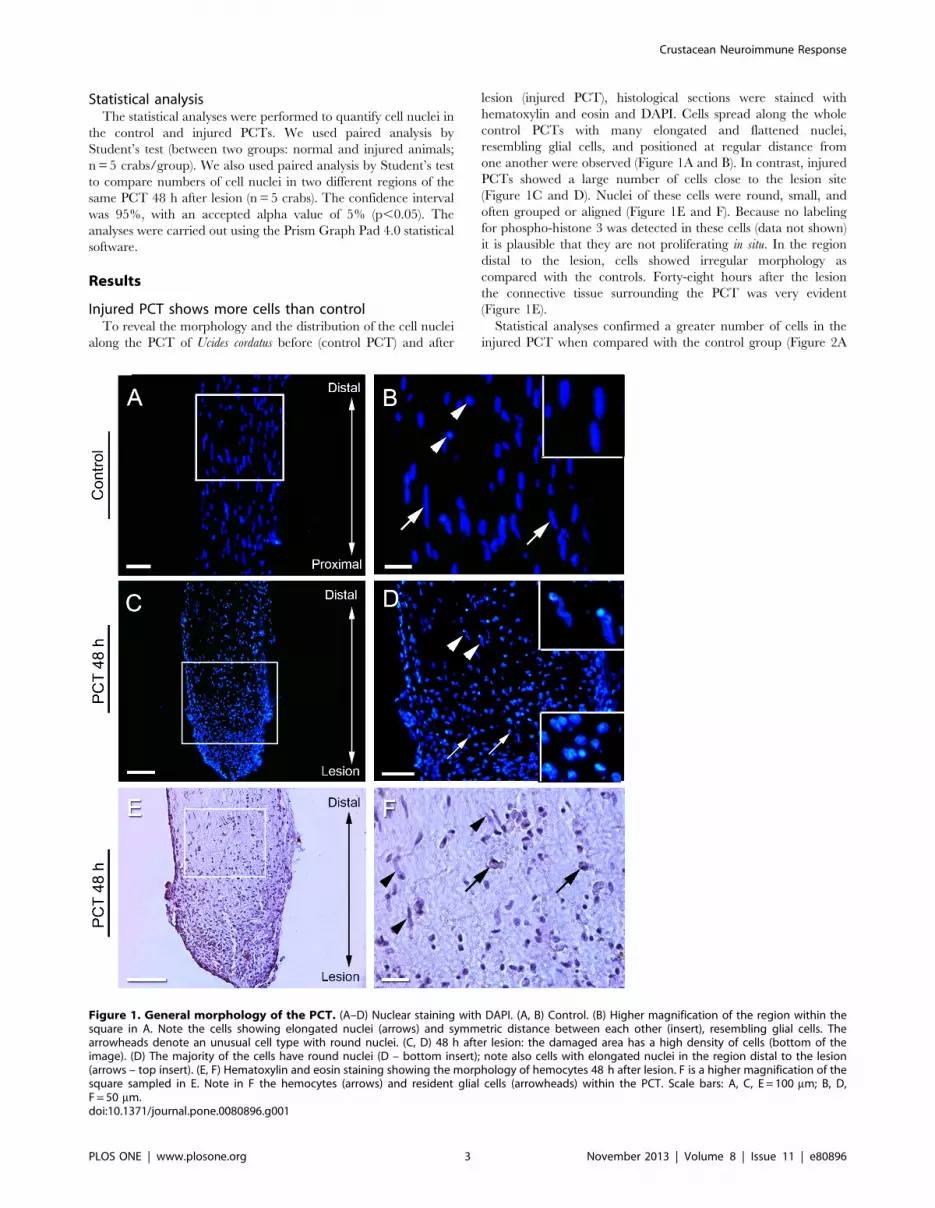
Injured PCT shows more cells than controlTo reveal the morphology and the distribution of the cell nuclei
along the PCT of Ucides cordatus before (control PCT) and after
lesion (injured PCT), histological sections were stained with
hematoxylin and eosin and DAPI. Cells spread along the whole
control PCTs with many elongated and flattened nuclei,
resembling glial cells, and positioned at regular distance from
one another were observed (Figure 1A and B). In contrast, injured
PCTs showed a large number of cells close to the lesion site
(Figure 1C and D). Nuclei of these cells were round, small, and
often grouped or aligned (Figure 1E and F). Because no labeling
for phospho-histone 3 was detected in these cells (data not shown)
it is plausible that they are not proliferating in situ. In the region
distal to the lesion, cells showed irregular morphology as
compared with the controls. Forty-eight hours after the lesion
the connective tissue surrounding the PCT was very evident
(Figure 1E).
Statistical analyses confirmed a greater number of cells in the
injured PCT when compared with the control group (Figure 2A
Figure 1. General morphology of the PCT. (A–D) Nuclear staining with DAPI. (A, B) Control. (B) Higher magnification of the region within thesquare in A. Note the cells showing elongated nuclei (arrows) and symmetric distance between each other (insert), resembling glial cells. Thearrowheads denote an unusual cell type with round nuclei. (C, D) 48 h after lesion: the damaged area has a high density of cells (bottom of theimage). (D) The majority of the cells have round nuclei (D – bottom insert); note also cells with elongated nuclei in the region distal to the lesion(arrows – top insert). (E, F) Hematoxylin and eosin staining showing the morphology of hemocytes 48 h after lesion. F is a higher magnification of thesquare sampled in E. Note in F the hemocytes (arrows) and resident glial cells (arrowheads) within the PCT. Scale bars: A, C, E = 100 mm; B, D,F = 50 mm.doi:10.1371/journal.pone.0080896.g001
Crustacean Neuroimmune Response
PLOS ONE | www.plosone.org 3 November 2013 | Volume 8 | Issue 11 | e80896

and A9). When we compared the region proximal to the injury
(region P) with the region distant to the lesion (region D), we
observed that the region P had almost twice as many cells as region
D (Figure 2B and B9).
Hyaline and granular hemocytes are attracted to thelesioned area
According to our previous study granular hemocytes are
attracted to the lesion site 24 h after PCT ablation [32]. Here
we investigated whether the same cells were recruited to the lesion
site in a subacute stage of degeneration. Semithin (Figure 3A and
B) and ultrathin (Figure 3C and D) sections showed two different
types of hemocytes in the PCT 48 h after lesion. Hyaline cells,
with no granules and a higher nucleus/cytoplasm ratio, as
classified by [32], were frequently found in region P, mainly in
the outer limit of the PCT. The majority of the cells displayed a
high amount of heterochromatin (Figure 3C).
Region D showed granular/semigranular hemocytes within the
tract (Figure 3B). These hemocytes were clearly distinguishable
from resident glial cells by the typical morphology and organiza-
tion of the glial cells in the PCT. Ultrathin sections revealed that
granulocytes have electron-dense granules with different sizes,
occupying almost the entire cytoplasm, and irregular nuclei with
peripheral euchromatin (Figure 3D). Different morphologies of the
semigranular hemocytes were also seen in region D after injury.
They were surrounded by the nervous fibers and had fewer
granules in the cytoplasm than the granulocytes (Figure 3E and F).
In addition, some hemocytes revealed many vesicles containing
membranes and electron-dense material resembling cellular debris
(Figure 3G and H), typical of phagocytosis.
Hemocytes attracted to the lesion site express histamineDuring neurodegenerative processes, the participation of mast
cells has been described due to production of pro-inflammatory
mediators, such as histamine [36,37]. In this paper, we observed
that histamine can be detected in the circulating hemocytes and in
cells recruited to the PCT 48 h after injury (Figure 4). No cell with
elongated nucleus (i.e., glial cell) showed histamine labeling.
iNOS expression is increased after injuryTo study different aspects of the inflammatory reaction, we
verified glial activation, microglia/microphage response, and
iNOS expression during the degeneration of the PCT. Immuno-
histochemistry for iNOS revealed that more cells were labeled
close to the injury site than in the control (Figure 5). Between cells
labeled for iNOS it was possible to identify a few resident cells
(with features of glial cells) which were seen distally to the lesion
(Figure 5C9 and C99). Apparently, no resident cells were labeled
with iNOS.
In vertebrates, macrophages and microglia are potentially able
to produce NO [38]. Considering that NO is increased in the
lesion site and that macrophages/microglia produce it, we wanted
to know whether the granular cells, which appear in high
quantities in the lesion, were analogous to vertebrate macrophag-
Figure 2. Density of cells in the PCT increases after injury. (A) Schematic illustration of both normal and injured PCT showing the cellulararrangement along the tract, including the quantified area (1 mm). The anatomical position of the tract is represented by a double-sensed arrow onthe top (retina – brain). (A9) 48 h after injury, the distal part of the tract had approximately three times as many cells as the control group. Student ttest revealed a significant difference between the two groups (n = 5 crabs/group; p,0.005). (B) Schematic illustration of the injured tract divided intotwo regions (500 mm each): proximal (P) and distal (D) to the lesion. Most cells are grouped in the damaged area 500 mm from the injury. In contrast,region D maintains the same cellular density as seen in the control. (B9) Statistical analysis shows twice as many cells in region P as in D. Student t testrevealed a significant difference between the two groups (n = 5 crabs/group; p,0.05). Asterisks denote significant differences between groups.doi:10.1371/journal.pone.0080896.g002
Crustacean Neuroimmune Response
PLOS ONE | www.plosone.org 4 November 2013 | Volume 8 | Issue 11 | e80896

es/microglia (revealed by the labeling with IB4). Our data show
some circulating hemocytes with granules co-labeled for IB4 and
iNOS (Figure 6). iNOS production was not observed in PCT cells
from crabs that were not submitted to ablation.
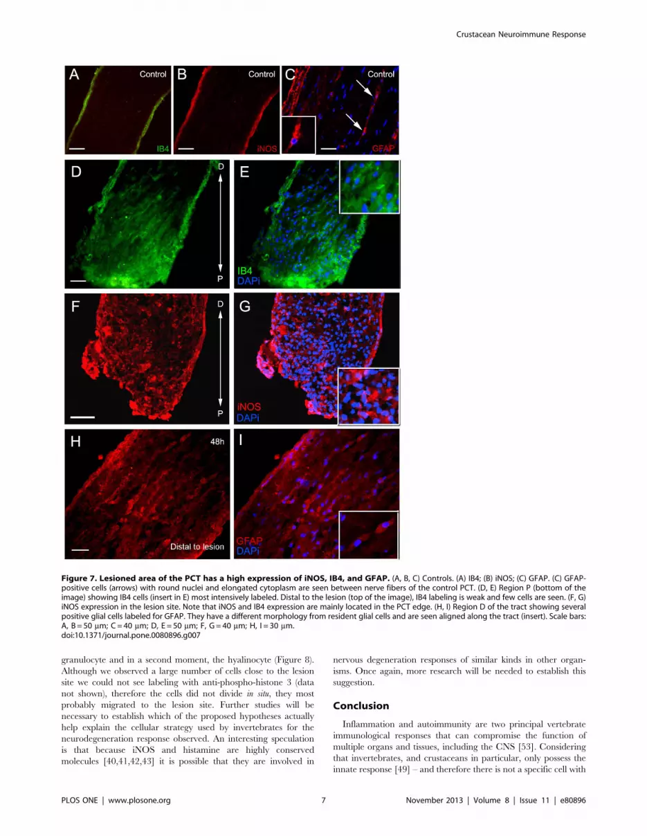
Because the inflammatory response during nervous system
injuries triggers immune responses [14,17,18] and glial activation
[12,13], we wanted to investigate how glial cells were disposed in
the PCT together with recruited hemocytes, and the nature of
their morphology in the subacute phase (or 48 h after the lesion).
We observed more GFAP-, IB4-, and iNOS-positive cells
concentrated in the lesion site when compared with the controls.
Additionally, GFAP-positive cells showed different nuclear mor-
phology from resident glial cells. They showed round nuclei and
were aligned along the tract (Figure 7).
Discussion
The subacute phase of nervous degeneration following injury in
invertebrates has not been a much explored subject, particularly
whether such a stage would involve immune cellular events. In this
study we revealed three immune cellular features which seem to
characterize the subacute phase of nervous degeneration in the
invertebrate studied: 1) The recruitment of mainly granular or
semigranular hemocytes and, secondarily, hyalinocytes to the
lesion site. Using iNOS and histamine labeling we were able to
confirm that the recruited hemocytes were active. 2) The
recruitment of a larger number of cells than observed in the
acute phase, also to lesion site [32]. 3) The presence of activated
glial cells revealed by the round shape of their nuclei and increased
expression of GFAP.
In vertebrates, after acute central nervous system injuries there
is an inflammatory response involving microglial activation and
infiltration of granular cells such as macrophages, and neutrophils
[27,29,36]. It is known that these cells may produce both NO and
histamine [39,40]. Despite lacking acquired immunity, inverte-
brates use the same molecules (iNOS and histamine) produced by
defense mechanisms triggered by vertebrate macrophages, mast
cells, and neutrophils: hemocytes from other invertebrates have
been shown to produce histamine (ascidians in [41]) and NO
(ascidians in [42], horseshoe crab in [43]). Because expression of
histamine and iNOS was seen in both the circulating hemocytes
and in the PCT 48 h after eyestalk ablation, we suggest that the
circulating hemocytes are the cells recruited to the PCT.
Moreover, we identified by specific markers that the recruited
hemocytes producing iNOS are macrophage/microglia-like cells
Figure 3. PCT attracts two different types of cells to the lesionsite. (A, B) Semithin sections of injured PCT stained with toluidine blue.(A) Region P (proximal) is surrounded by cells with typical morphology:round shape, high nucleus/cytoplasm ratio, and no granules within thecytoplasm (arrows), suggesting hyaline cells. (B) Granular hemocytes(arrows) infiltrating into the nerve fibers of the tract after eyestalkablation – region D (distal to the lesion). Only granular cells areobserved in this region intermingled with nerve fibers. (C, D) Ultrathinsections of regions P and D, respectively. (C) Hemocyte, resembling ahyaline cell, with round shape and no granules in the scarce cytoplasm.The eccentric nucleus (n) has abundant euchromatin displaying thesame features as the cells shown in (A). The asterisk indicates a nervefiber. (D) Typical granular hemocyte with numerous electron-densegranules occupying the whole cytoplasm. The nucleus (n) shows anirregularly shaped membrane and heterochromatin in the periphery. (E,F) Semigranular hemocytes infiltrated into the injured tract – region D(distal to the lesion). A small number of electron-dense granules(asterisks) occupy the whole cytoplasm; the eccentric nuclei (n) haveirregular/flattened shape with abundant heterochromatin in theperiphery, which is not seen in hyaline hemocytes. (G, H) Granularhemocytes are easily seen in region D and most of them show double-membrane vesicles (square) within the cytoplasm suggesting phago-cytosis. This is better seen in D (higher magnification). Electron-densegranules (asterisks) with different shapes and sizes surround thesevesicles. Scale bars: A, B = 10 mm; C–G = 2 mm; H = 1 mm.doi:10.1371/journal.pone.0080896.g003
Figure 4. Circulating hemocytes and PCT cells are labeled forhistamine. (A) Histamine-positive cells are red in the hemolymph; andthe nuclei, DAPI stained, are in blue. (B) Injured PCT (48 h after eyestalkablation) showing cells labeled for histamine (red) and DAPI (blue). Themajority of histamine-positive cells contain granules (arrowheads) andround nuclei. No labeling was seen in the elongated nuclei (arrow) ofthe cells with morphology of glial cells. Scale bars: A = 20 mm;B = 10 mm.doi:10.1371/journal.pone.0080896.g004
Crustacean Neuroimmune Response
PLOS ONE | www.plosone.org 5 November 2013 | Volume 8 | Issue 11 | e80896

and those producing histamine are another subtype of granule
cells present in the lesion site.
Because iNOS and histamine were more expressed in the lesion
than in the controls we may speculate that they participate in the
tissue strategy used for attracting circulating hemocytes and
activation of glial cells as observed herein. Since only granulocytes
(granular/semigranular cells), and not hyalinocytes, are present in
the acute phase [32] we suggest that histamine and NO released
from granulocytes in the acute phase attract the hyalinocytes, thus
moving the degeneration process to the subacute phase.
Hyalinocytes are considered less differentiated than granulo-
cytes [44] and as such they could replenish the lesion site,
differentiate, and perform defensive roles, such as phagocytosis
[45,46,47] of cellular debris as observed here. This should be the
case if hemocytes are all differentiated from a same progenitor cell
type [48,49].We may suggest this case to be one hypothesis for the
source of hemocytes to the injured PCT (Figure 8). In this context
semigranular cells may constitute an intermediate stage of
maturation, since less mature hemocytes are acquiring granules,
or they may constitute a more mature stage undergoing
degranulation in response to a challenge [50].
However, because other authors have suggested that hemocytes
are originated by diverse precursor cell types [51,52] - and this
may be another hypothesis for the source of hemocytes to the
injured PCT - they would arrive at different times, characterizing
the acute and subacute stages: the first to appear being the
Figure 5. Expression of iNOS in the PCT 48 h after lesion. (A, B, C) Control animals. (A, D, G) DAPI stained cells; (B, E, H) iNOS immunoreacted(red); (C, F, I) Merge. No cells from control PCTs were labeled for iNOS (C – insert). (D–F) Region P (proximal) of the tract observed after DAPI nuclearstaining (D), after iNOS reaction (E), and the merged image (F). The insert shows a higher magnification of the double-labeled cells. (G–I) Region D(distal) of the tract observed after DAPI staining (G), after iNOS labeling (H), and the merged image (I) revealing numerous hemocytes containinggranules (arrowhead), infiltrating into the tissue. No labeling was seen in the elongated nuclei (arrows) of the cells with morphology of glial cells. Theinsert for I shows a higher magnification of the labeled hemocytes. Scale bars: A–I = 100 mm.doi:10.1371/journal.pone.0080896.g005
Figure 6. iNOS and IB4 expression are colocalized in circulating hemocytes. Hemolymph was collected from five crabs, 48 h after eyestalkablation. Hemocytes were labeled for IB4 (A –green) and iNOS (B – red). Only hemocytes with granules, semigranular/granular cells, showed double-labeling (insert). Scale bar: A–C = 10 mm.doi:10.1371/journal.pone.0080896.g006
Crustacean Neuroimmune Response
PLOS ONE | www.plosone.org 6 November 2013 | Volume 8 | Issue 11 | e80896
granulocyte and in a second moment, the hyalinocyte (Figure 8).
Although we observed a large number of cells close to the lesion
site we could not see labeling with anti-phospho-histone 3 (data
not shown), therefore the cells did not divide in situ, they most
probably migrated to the lesion site. Further studies will be
necessary to establish which of the proposed hypotheses actually
help explain the cellular strategy used by invertebrates for the
neurodegeneration response observed. An interesting speculation
is that because iNOS and histamine are highly conserved
molecules [40,41,42,43] it is possible that they are involved in
nervous degeneration responses of similar kinds in other organ-
isms. Once again, more research will be needed to establish this
suggestion.
Conclusion
Inflammation and autoimmunity are two principal vertebrate
immunological responses that can compromise the function of
multiple organs and tissues, including the CNS [53]. Considering
that invertebrates, and crustaceans in particular, only possess the
innate response [49] – and therefore there is not a specific cell with
Figure 7. Lesioned area of the PCT has a high expression of iNOS, IB4, and GFAP. (A, B, C) Controls. (A) IB4; (B) iNOS; (C) GFAP. (C) GFAP-positive cells (arrows) with round nuclei and elongated cytoplasm are seen between nerve fibers of the control PCT. (D, E) Region P (bottom of theimage) showing IB4 cells (insert in E) most intensively labeled. Distal to the lesion (top of the image), IB4 labeling is weak and few cells are seen. (F, G)iNOS expression in the lesion site. Note that iNOS and IB4 expression are mainly located in the PCT edge. (H, I) Region D of the tract showing severalpositive glial cells labeled for GFAP. They have a different morphology from resident glial cells and are seen aligned along the tract (insert). Scale bars:A, B = 50 mm; C = 40 mm; D, E = 50 mm; F, G = 40 mm; H, I = 30 mm.doi:10.1371/journal.pone.0080896.g007
Crustacean Neuroimmune Response
PLOS ONE | www.plosone.org 7 November 2013 | Volume 8 | Issue 11 | e80896

immune memory – perhaps this is the mechanism they developed
allowing neurodegeneration to occur in such a delayed period
when compared with vertebrates [4,5,31]. To conclude, the
importance of our study resides in the characterization of the
cellular and biochemical strategies peculiar to the subacute stage of
the neurodegeneration response in invertebrates.
Acknowledgments
S.A. is a CNPq research fellow. P.G.C.S. is recipient of a scholarship from
CNPq. We are indebted to the Rudolf Barth Electron Microscopy Platform
of the Oswaldo Cruz Institute/Fiocruz and to the Laboratorio de
Microscopia Eletronica Prof. Luiz Henrique Monteiro Leal, Instituto de
Biologia Roberto Alcantara Gomes, Universidade do Estado do Rio de
Janeiro, for the electron microscopy facilities.
Author Contributions
Conceived and designed the experiments: PGCS CLC SA. Performed the
experiments: PGCS CLC SLC SA. Analyzed the data: PGCS CLC SLC
SA. Contributed reagents/materials/analysis tools: PGCS CLC SLC SA.
Wrote the paper: PGCS CLC SLC SA.
References
1. Waller A (1850) Experiments on the section of glossopharyngeal and hypoglossal
nerves of the frog and observations of the alteration produced thereby in the
structure of primitive fibers. Phil Trans R Soc Lond 140.
2. Bittner GD, Schallert T, Peduzzi JD (2000) Degeneration, Trophic Interactions,
and Repair of Severed Axons: A Reconsideration of Some Common
Assumptions. The Neuroscientist6.
3. Blundon JA, Sheller RA, Moehlenbruck JW, Bittner GD (1990) Effect of
temperature on long-term survival of anucleate giant axons in crayfish and
goldfish. J Comp Neurol 297: 377–391.
4. Bittner GD (1991) Long-term survival of anucleate axons and its implications for
nerve regeneration. Trends Neurosci 14: 188–193.
5. Parnas I, Shahrabany-Baranes O, Feinstein N, Grant P, Adelsberger H, et al.
(1998) Changes in the ultrastructure of surviving distal segments of severed axons
of the rock lobster. J Exp Biol 201: 779–791.
6. Tanner SL, Storm EE, Bittner GD (1995) Maintenance and degradation of
proteins in intact and severed axons: implications for the mechanisms of long-
term survival of anucleate crayfish axons. J Neurosci 15: 540–548.
7. Bittner GD (1988) Long Term Survival of Severed Distal Axonal Stumps in
Vertebrates and Invertebrates. American Society of Zoologists 28: 1165–1179.
8. Lieberman EM, Hargittai PT, Grossfeld RM (1994) Electrophysiological and
metabolic interactions between axons and glia in crayfish and squid. Prog
Neurobiol 44: 333–376.
9. Atwood HL, Dudel J, Feinstein N, Parnas I (1989) Long-term survival of
decentralized axons and incorporation of satellite cells in motor neurons of rock
lobsters. Neurosci Lett 101: 121–126.
10. Govind CK, Blundon JA, Kirk MD (1992) Functional degeneration of isolated
central stumps of crayfish sensory axons. J Comp Neurol 322: 111–120.
11. Sheller RA, Ballinger ML, Bittner GD (1991) Long-term survival of severed
crayfish giant axons is not associated with an incorporation of glial nuclei into
axoplasm. Neurosci Lett 133: 113–116.
12. Fawcett JW, Asher RA (1999) The glial scar and central nervous system repair.
Brain Res Bull 49: 377–391.
13. Ladeby R, Wirenfeldt M, Garcia-Ovejero D, Fenger C, Dissing-Olesen L, et al.
(2005) Microglial cell population dynamics in the injured adult central nervous
system. Brain Res Brain Res Rev 48: 196–206.
14. Clark RS, Schiding JK, Kaczorowski SL, Marion DW, Kochanek PM (1994)
Neutrophil accumulation after traumatic brain injury in rats: comparison of
weight drop and controlled cortical impact models. J Neurotrauma 11: 499–506.
15. Hawkes CA, McLaurin J (2009) Selective targeting of perivascular macrophages
for clearance of beta-amyloid in cerebral amyloid angiopathy. Proc Natl Acad
Sci U S A 106: 1261–1266.
16. Majumdar A, Chung H, Dolios G, Wang R, Asamoah N, et al. (2008)
Degradation of fibrillar forms of Alzheimer’s amyloid beta-peptide by
macrophages. Neurobiol Aging 29: 707–715.
17. Ransohoff RM, Brown MA (2012) Innate immunity in the central nervous
system. J Clin Invest 122: 1164–1171.
18. Soares HD, Hicks RR, Smith D, McIntosh TK (1995) Inflammatory leukocytic
recruitment and diffuse neuronal degeneration are separate pathological
processes resulting from traumatic brain injury. J Neurosci 15: 8223–8233.
19. Clatworthy AL, Grose E (1999) Immune-mediated alterations in nociceptive
sensory function in Aplysia californica. J Exp Biol 202: 623–630.
20. Raivich G, Jones LL, Kloss CU, Werner A, Neumann H, et al. (1998) Immune
surveillance in the injured nervous system: T-lymphocytes invade the
axotomized mouse facial motor nucleus and aggregate around sites of neuronal
degeneration. J Neurosci 18: 5804–5816.
21. Ibiza S, Serrador J.M. (2008) The role of nitric oxide in the regulation of
adaptive immune responses Inmunologia 27: 103–117.
22. Wink DA, Hines HB, Cheng RY, Switzer CH, Flores-Santana W, et al. (2011)
Nitric oxide and redox mechanisms in the immune response. J Leukoc Biol 89:
873–891.
23. Bogdan C, Rollinghoff M, Diefenbach A (2000) The role of nitric oxide in innate
immunity. Immunol Rev 173: 17–26.
24. Ferstl R, Akdis CA, O’Mahony L (2012) Histamine regulation of innate and
adaptive immunity. Front Biosci (Landmark Ed) 17: 40–53.
Figure 8. Schematic diagram of the hypotheses for the source of hemocytes to the injured PCT. Hypothesis 1: Granular/semigranular(dark blue) are the first cell type to be attracted to the injured tract. In sequence, hyaline cells (light blue) are also attracted and can differentiate ingranular hemocytes as they infiltrate into the nerve fibers (due to several stimuli, such as histamine and iNOS). Hypothesis 2: Granular/semigranularhemocytes (blue) are attracted to the lesion site. Following the time course of the lesion, they infiltrate into the tract. Then, in a subacute stage,hyaline cells (red) are also attracted to the injury. In both hypotheses, we can identify two different types of cells in the PCT.doi:10.1371/journal.pone.0080896.g008
Crustacean Neuroimmune Response
PLOS ONE | www.plosone.org 8 November 2013 | Volume 8 | Issue 11 | e80896

25. Fernandez-Novoa L, Cacabelos R (2001) Histamine function in brain disorders.
Behav Brain Res 124: 213–233.26. Andersson PB, Perry VH, Gordon S (1992) The acute inflammatory response to
lipopolysaccharide in CNS parenchyma differs from that in other body tissues.
Neuroscience 48: 169–186.27. Beck KD, Nguyen HX, Galvan MD, Salazar DL, Woodruff TM, et al. (2010)
Quantitative analysis of cellular inflammation after traumatic spinal cord injury:evidence for a multiphasic inflammatory response in the acute to chronic
environment. Brain 133: 433–447.
28. Bell MD, Perry VH (1995) Adhesion molecule expression on murine cerebralendothelium following the injection of a proinflammagen or during acute
neuronal degeneration. J Neurocytol 24: 695–710.29. Carlson SL, Parrish ME, Springer JE, Doty K, Dossett L (1998) Acute
inflammatory response in spinal cord following impact injury. Exp Neurol 151:77–88.
30. Yong VW (1996) Cytokines, astrogliosis, and neutrophis following CNS trauma.
Cytokines and the CNS: Development, Defense and Disease: CRC Press, BocaRaton, FL., USA. pp. 309–322.
31. Correa CL, Allodi S, Martinez AM (2005) Ultrastructural study of normal anddegenerating nerve fibers in the protocerebral tract of the crab Ucides cordatus.
Brain Behav Evol 66: 145–157.
32. Chaves-da-Silva PG, de Barros CM, Lima FR, Biancalana A, Martinez AM, etal. (2010) Identity of the cells recruited to a lesion in the central nervous system
of a decapod crustacean. Cell Tissue Res 342: 179–189.33. Allodi S, Silva SF, Taffarel M (1999) Glial cells of the central nervous system in
the crab Ucides cordatus. Invertebrate Biology 118: 175–183.34. Allodi S, Taffarel M (1999) Electron microscopy of glial cells of the central
nervous system in the crab Ucides cordatus. Braz J Med Biol Res 32: 327–331.
35. Lockwood APM (1961) ‘‘Ringer’’, solutions and some notes on the physiologicalbasis of their ionic composition. Comparative Biochemistry and Physiology 2.
36. Esposito P, Chandler N, Kandere K, Basu S, Jacobson S, et al. (2002)Corticotropin-releasing hormone and brain mast cells regulate blood-brain-
barrier permeability induced by acute stress. J Pharmacol Exp Ther 303: 1061–
1066.37. Wilhelm M, King B, Silverman AJ, Silver R (2000) Gonadal steroids regulate the
number and activational state of mast cells in the medial habenula.Endocrinology 141: 1178–1186.
38. Kroncke KD, Fehsel K, Kolb-Bachofen V (1997) Nitric oxide: cytotoxicityversus cytoprotection—how, why, when, and where? Nitric Oxide 1: 107–120.
39. Hibbs JB Jr, Taintor RR, Vavrin Z, Rachlin EM (1988) Nitric oxide: a cytotoxic
activated macrophage effector molecule. Biochem Biophys Res Commun 157:87–94.
40. Radomski MW, Jenkins DC, Holmes L, Moncada S (1991) Human colorectal
adenocarcinoma cells: differential nitric oxide synthesis determines their ability
to aggregate platelets. Cancer Res 51: 6073–6078.
41. de Barros CM, Andrade LR, Allodi S, Viskov C, Mourier PA, et al. (2007) The
Hemolymph of the ascidian Styela plicata (Chordata-Tunicata) contains heparin
inside basophil-like cells and a unique sulfated galactoglucan in the plasma. J Biol
Chem 282: 1615–1626.
42. de Barros CM, de Carvalho DR, Andrade LR, Pavao MS, Allodi S (2009) Nitric
oxide production by hemocytes of the ascidian Styela plicata. Cell Tissue Res
338: 117–128.
43. Crivellato E, Ribatti D (2010) The mast cell: an evolutionary perspective. Biol
Rev Camb Philos Soc 85: 347–360.
44. Cochennec-Laureau N, Auffret M, Renault T, Langlade A (2003) Changes in
circulating and tissue-infiltrating hemocyte parameters of European flat oysters,
Ostrea edulis, naturally infected with Bonamia ostreae. J Invertebr Pathol 83:
23–30.
45. Smith VJ, Soderhall K (1983) Induction of degranulation and lysis of
haemocytes in the freshwater crayfish, Astacus astacus by components of the
prophenoloxidase activating system in vitro. Cell Tissue Res 233: 295–303.
46. Soderhall K, Smith VJ, Johansson MW (1986) Exocytosis and uptake of bacteria
by isolated haemocyte populations of two crustaceans: evidence for cellular co-
operation in the defence reactions of arthropods. Cell and Tissue Research 245.
47. Thornqvist PO, Johansson MW, Soderhall K (1994) Opsonic activity of cell
adhesion proteins and beta-1,3-glucan binding proteins from two crustaceans.
Dev Comp Immunol 18: 3–12.
48. Ghiretti-Magaldi A, Milanesi C, Tognon G (1977) Hemopoiesis in crustacea
decapoda: origin and evolution of hemocytes and cyanocytes of Carcinus
maenas. Cell Differentiation 6: 167–186.
49. Saha S (2011) Innate Immune Source and Functional Machinery in Decapods of
Crustacea. Indian Journal of Fundamental and Applied Life Sciences 1.
50. Rebelo Mde F, Figueiredo Ede S, Mariante RM, Nobrega A, de Barros CM, et
al. (2013) New insights from the oyster Crassostrea rhizophorae on bivalve
circulating hemocytes. PLoS One 8: e57384.
51. Jiravanichpaisal P, Lee BL, Soderhall K (2006) Cell-mediated immunity in
arthropods: hematopoiesis, coagulation, melanization and opsonization. Immu-
nobiology 211: 213–236.
52. Zhang ZF, Shao M, Ho Kang K (2006) Classification of haematopoietic cells
and haemocytes in Chinese prawn Fenneropenaeus chinensis. Fish Shellfish
Immunol 21: 159–169.
53. Kapadia M, Sakic B (2011) Autoimmune and inflammatory mechanisms of CNS
damage. Prog Neurobiol 95: 301–333.
Crustacean Neuroimmune Response
PLOS ONE | www.plosone.org 9 November 2013 | Volume 8 | Issue 11 | e80896