nanofiber-based integrative repair of orthopedic soft tissues
TRANSCRIPT
2-1
2.1 Introduction
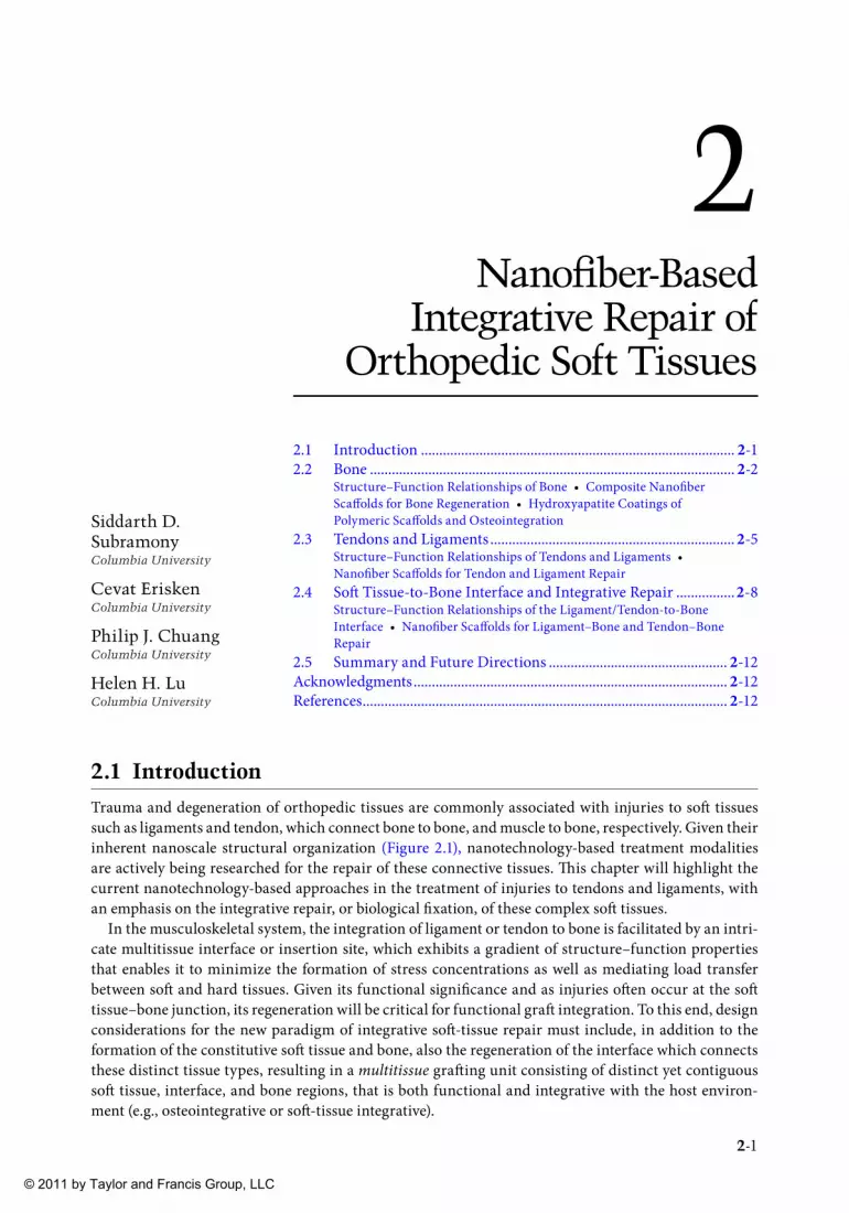
Trauma and degeneration of orthopedic tissues are commonly associated with injuries to soft tissues such as ligaments and tendon, which connect bone to bone, and muscle to bone, respectively. Given their inherent nanoscale structural organization (Figure 2.1), nanotechnology-based treatment modalities are actively being researched for the repair of these connective tissues. Th is chapter will highlight the current nanotechnology-based approaches in the treatment of injuries to tendons and ligaments, with an emphasis on the integrative repair, or biological fi xation, of these complex soft tissues.
In the musculoskeletal system, the integration of ligament or tendon to bone is facilitated by an intri-cate multitissue interface or insertion site, which exhibits a gradient of structure–function properties that enables it to minimize the formation of stress concentrations as well as mediating load transfer between soft and hard tissues. Given its functional signifi cance and as injuries oft en occur at the soft tissue–bone junction, its regeneration will be critical for functional graft integration. To this end, design considerations for the new paradigm of integrative soft -tissue repair must include, in addition to the formation of the constitutive soft tissue and bone, also the regeneration of the interface which connects these distinct tissue types, resulting in a multitissue graft ing unit consisting of distinct yet contiguous soft tissue, interface, and bone regions, that is both functional and integrative with the host environ-ment (e.g., osteointegrative or soft -tissue integrative).
2Nanofi ber-Based
Integrative Repair of Orthopedic Soft Tissues
2.1 Introduction ...................................................................................... 2-12.2 Bone .................................................................................................... 2-2
Structure–Function Relationships of Bone • Composite Nanofi ber Scaff olds for Bone Regeneration • Hydroxyapatite Coatings of Polymeric Scaff olds and Osteointegration
2.3 Tendons and Ligaments ................................................................... 2-5Structure–Function Relationships of Tendons and Ligaments • Nanofi ber Scaff olds for Tendon and Ligament Repair
2.4 Soft Tissue-to-Bone Interface and Integrative Repair ................2-8Structure–Function Relationships of the Ligament/Tendon-to-Bone Interface • Nanofi ber Scaff olds for Ligament–Bone and Tendon–Bone Repair
2.5 Summary and Future Directions ................................................. 2-12Acknowledgments ...................................................................................... 2-12References .................................................................................................... 2-12
Siddarth D. SubramonyColumbia University
Cevat EriskenColumbia University
Philip J. ChuangColumbia University
Helen H. LuColumbia University
© 2011 by Taylor and Francis Group, LLC

2-2 Nanotechnology in Tissue Engineering and Regenerative Medicine
Focusing on these design considerations, the objective of this chapter is to provide an overview of current approaches to integrative soft tissue repair, beginning with a brief description of the application of nanotechnology in bone repair, with an emphasis on osteointegration. Th is section is followed by a review of nanofi ber-based approaches to tendon and ligament repair, while the fi nal section of this chapter highlights the exciting research into interface regeneration or interface tissue engineering, with an emphasis on the concurrent formation of multitissue systems for integrative soft -tissue repair. Each of these sections will begin with a summary of the structure–function relationship of the tissue at the nanoscale, which provides the biomimetic design inspiration for the scaff old systems intended for the regeneration of each tissue type. Th e chapter will conclude with a summary and future direction section which will outline the challenges in this exciting fi eld.
2.2 Bone
2.2.1 Structure–Function Relationships of Bone
Bone serves as the main structural unit of the human body and is the most commonly replaced organ with over 800,000 graft ing procedures performed annually in the United States alone (Langer and Vacanti, 1993). It is composed of approximately 25% extracellular matrix (ECM), 50% mineral, and 25% water (Ozawa et al., 2008) and is populated largely by three cell types: osteoblasts, osteocytes, and osteo-clasts. Osteoblasts, which form new bone, and osteoclasts, which resorb bone, serve to continuously remodel and rebuild the skeleton. Th is cycle is mediated by osteocytes, the most abundant cell type pres-ent in bone that acts as mechanosensors.
Th e organic component of bone ECM consists predominantly of type-I collagen fi brils. Th e collagen molecule is typically built on the repetition of the sequence glycine-proline-4-hydroxyproline with slight variations (Mann, 2001). Individual collagen fi laments are composed of three molecules of this sequence wrapped around each other, resulting in a superhelical structure called tropocollagen. For type-I collagen, the tropocollagen fi brils are organized at the nanoscale into the revised quarter stagger
Core proteinHyaluronic acidLink protein
Proteoglycan
Ligament
Bone
TropocollagenTriple-helix
Ligament–boneinsertion
Collagen (type I)Collagen (type III)HydroxyapatiteProteoglycans
~280
nm
FIGURE 2.1 (See color insert following page 10-24.) Overview of the nanoscale structural organization of bone, ligament/tendon, and direct ligament/tendon-to-bone insertion site.
© 2011 by Taylor and Francis Group, LLC

Nanofi ber-Based Integrative Repair of Orthopedic Soft Tissues 2-3
model (Figure 2.1). Th e adjacent parallel fi ber rows are spaced by 64 nm longitudinally, with each tropo-collagen strand being about 280 nm long, and 1.5 nm in width (Mann, 2001). Th is arrangement allows for maximum crosslinking of hydroxylysine and lysine residues for maximum stability of the matrix (Knott and Bailey, 1998). Th e natural spacing formed between the ends of the collagen fi bers are 40 × 5 nm in size (Mann, 2001). It is proposed that these vacancies between the collagen fi bers serve as nucleation sites for the mineral component of bone (Landis and Silver, 2008).
Hydroxyapatite crystals constitute the inorganic phase of bone, with the mineralized collagen fi bers exhibiting greater mechanical properties than unmineralized collagen fi bers (Buehler, 2007). Buehler et al. investigated the diff erence in tensile mechanical properties between mineralized and unmineral-ized collagen fi bers and found that uncalcifi ed collagen behaves mechanically like an elastic polymer with a Young’s modulus of 4.59 GPa and a maximum stress of 0.3 GPa, while the mineralized collagen fi bers were reported to have a Young’s modulus of 6.23 GPa and a maximum stress of 0.6 GPa. Th is observed increase in mechanical properties is attributed to the hydroxyapatite crystals, which act as strengthening precipitates that impart resistance to plastic deformation and fracture. Interestingly, the weak adhesion between collagen and hydroxyapatite also allows for slip deformation to occur at these interfaces, which prevents the presence of mineral from making the overall fi ber too brittle. Furthermore, the nanoscale organization of the collagen fi brils and hydroxyapatite crystals is theorized to increase the strength of the overall fi ber by maximizing intermolecular forces.
Given the complex nanoscale organization of bone, the ideal scaff old must be biomimetic with nanoscale organization, able to support bone growth (osteoconductive), induce bone formation (osteoinductive), and structurally integrate with the host bone tissue (osteointegrative). Many such biomimetic systems have been actively researched which combine collagenous or synthetic nanofi brils and hydroxyapatite nano-particles, with the ultimate goal of engineering a bone graft ing system that can meet all of these afore-mentioned design criteria. Examples discussed below include composite nanofi ber scaff olds (Table 2.1) and hydroxyapatite coatings on biodegradable polymeric scaff olds for osteointegration (Table 2.2).
2.2.2 Composite Nanofi ber Scaffolds for Bone Regeneration
Th e natural fi bril morphology of the collagen in the ECM can be readily mimicked with nanofi bers in order to optimize osteoblast response on polymeric biomaterials. Th e most common polymers used include polylactide-co-glycolide acid (PLGA) and polycaprolactone (PCL) which are selected for their favorable biodegradation profi les and mechanical properties (Agrawal and Ray, 2001).
In general, nanofi bers are fabricated via the classical method of electrospinning (Reneker and Chun, 1996). For example, electrospinning was used to generate unaligned PLGA (Li et al., 2001) as well as PCL nanofi ber scaff olds (Yoshimoto et al., 2003). In each case, the polymer is dissolved in a solvent and dispensed by a syringe pump, and drawn out into nanofi bers under the application of high voltage, and a ground collector is used to capture the polymer fi bers formed. It was reported that when mesenchymal stem cells (MSCs) were seeded on the PCL scaff olds (Yoshimoto et al., 2003), they diff erentiated into
TABLE 2.1 Nanofi ber Scaff olds for Bone Formation
Study Scaff old Design Application Study Model
Li et al., 2002 Poly(d,l-lactide-co-glycolide) nanofi ber (500–800 nm)
Biocompatibility In vitro/Mouse fi broblasts
Yoshimoto et al., 2003 Poly(ε-caprolactone) nanofi bers (400 nm)
Bone In vitro/Mouse MSCs
Venugopal et al., 2008 Poly(ε-caprolactone) nanofi bers(411 nm), hydroxyapatite, gelatin (856 nm)
Bone In vitro/Human fetal osteoblasts
Song et al., 2008 Collagen type-I nanofi bers (70–170 nm), hydroxyapatite
Bone In vitro/MC3T3-E1 osteoblastic cells
© 2011 by Taylor and Francis Group, LLC

2-4 Nanotechnology in Tissue Engineering and Regenerative Medicine
osteoblasts under rotational culture in osteogenic diff erentiation medium, with abundant type-I collagen matrix formation and mineralization observed throughout the scaff old. In addition, aligned polymer scaff olds have been formed using the same technique with a rotating collecting target in order to not only replicate the fi brous structure of collagen but also the natural alignment of the native collagen matrix (Moff at et al., 2009). Th e versatility of the electrospinning process also enables the incorporation of other biomaterials, both organic (e.g., collagen, chitosan) and synthetic, into these nanofi ber meshes (Reneker and Chun, 1996; Matthews et al., 2002; Shin et al., 2005) which will further promote bone formation.
Moreover, the fabrication of composite nanofi bers or incorporation of organic–inorganic nanocom-posite complexes into nanofi ber scaff olds has been of signifi cant interest. For example, Song et al. elec-trospun nanofi ber scaff olds from hydroxyapatite–collagen nanocomposites, whereby a coprecipitation method was fi rst used to form the nanocomposites, which involved the addition of two emulsions (hydroxyapatite and collagen) drop-wise into a reaction vessel while the pH is fi xed with a reaction time on the order of 48 h (Song et al., 2008). Th e resulting nanocomposites were dried and dissolved in 1,1,1,3,3,3-hexafl uoro-2-propanol and electrospun into unaligned nanofi ber meshes. When MC3T3-E1 preosteoblasts were seeded onto these substrates, these cells measured a signifi cant increase in alkaline phosphatase activity over time, demonstrating the osteoconductive potential of these novel nanofi ber composites. Recently, using the same fabrication system, Venugopal et al. substituted gelatin for collagen in order to reduce cost and further enhance the osteoconductivity of these nanocomposite scaff olds (Venugopal et al., 2008). Specifi cally, hydroxyapatite, PCL, and gelatin were electrospun together to form composite nanofi bers. While the mechanical properties of these composite nanofi bers were several orders of magnitudes lower than those of bone in terms of both Young’s modulus and ultimate tensile strength (UTS), the scaff olds supported osteoblast proliferation, higher ALP activity, and mineralization.
For composite nanofi ber scaff olds, the primary challenge is in how to successfully match scaff old mechanical properties (such as Young’s modulus, UTS) with those of native bone, as well as optimizing the induction of mineralization and addresses the need for osteointegration. Introduction of additional elements into the nanofi ber systems, such as peptides and growth factors, as well as the incorporation of the other nanofi bers into polymeric or titanium implants are examples of how these scaff olds can be further tailored to improve osteoconductivity and osteoinductivity (Khang et al., 2006; Horii et al., 2007; Sargeant et al., 2008).
2.2.3 Hydroxyapatite Coatings of Polymeric Scaffolds and Osteointegration
A common method that has been researched extensively for improving osteointegration involves the formation of a calcium phosphate layer directly onto the scaff old surface (Table 2.2), specifi cally by incubating the scaff olds in a simulated body fl uid (SBF) with physiologic or supra-physiologic calcium
TABLE 2.2 Simulated Body Fluid Coating Methods to Improve Osteointegration
Study Material Immersion Time Mineral Function
Murphy et al., 2002 Poly(lactide-co-glycolide), SBF
16-day immersion Carbonated apatite Shown to improve bone regeneration in a rat cranial defect model
Yang et al., 2008 PCL, SBF, and 10× SBF
7 day immersion in 1× SBF; two 10 h immersion in 10× SBF
Brushite and apatite Uses a plasma discharge treatment prior to SBF treatment; compared normal SBF with 10× SBF
Mavis et al., 2009 PCL, 10× SBF 6 h immersion Brushite, monetite, and apatite
Evaluated response of osteoblasts on coated PCL scaff olds, and demonstrate superior osteoconductivity in vitro over uncoated scaff olds
© 2011 by Taylor and Francis Group, LLC

Nanofi ber-Based Integrative Repair of Orthopedic Soft Tissues 2-5
and/or phosphate concentrations. Th ese ions promote the nucleation of mineral crystals on the polymeric substrate, and it is anticipated that this newly formed calcium phosphate layer can thereby lead to improved osteointegration in vivo. Incubation in the SBF off ers a relatively low temperature processing technique, ideal for substrates such as polymers that cannot sustain the high temperature treatments necessary for the well established plasma spraying of metallic or ceramic implants (Du et al., 2002). Another advantage of this method is the ability to form a relatively uniform coating on surfaces with complex topography and predesigned geometries.
Modifi cation of the polymeric substrate has been reported to improve the effi ciency of forming a calcium phosphate layer on polymer scaff olds. Murphy and Mooney showed that carbonate-apatite can be deposited onto PLGA surfaces aft er pretreating the polymer surface in a NaOH solution (Murphy and Mooney, 2002). In this study, polymeric fi lms were immersed in a SBF solution for 16 days at 37°C, with the solution replaced daily. Moreover, it was observed that when the PLGA fi lms were presoaked in 0.5 M NaOH, a hydrolyzed, carboxylic acid-rich surface was formed which resulted in improved mineral deposition. A follow-up study elegantly demonstrated that a macroporous PLGA scaff old coated with a layer of carbonate-apatite formed through this method reported a 53% increase in bone forma-tion when evaluated in a rat cranial defect model (Murphy et al., 2005).
Working with PCL nanofi bers, Yang et al. as well as Mavis et al. demonstrated that the SBF soaking method can be applied to form calcium phosphate coatings on these scaff olds (Yang et al., 2008; Mavis et al., 2009). In contrast to Murphy et al., a concentrated SBF solution was used to expedite the coat-ing process, reducing week-long immersion times to just a couple of hours. Specifi cally, Yang et al. pre- exposed the PCL scaff old to a plasma discharge treatment and then immersed the nanofi bers in two types of solution: a SBF solution with calcium and phosphate ion concentrations matching those found in human blood plasma and a 10× concentrated SBF solution, and the scaff olds were incubated in each for 7 days or up to 6 h, respectively. Both methods resulted in the deposition of a calcium phosphate layer on the PCL nanofi bers. Interestingly, the scaff olds immersed in the physiologic SBF exhibited a crystalline structure more closely resembling natural apatite whereas the scaff olds immersed in the concentrated SBF solution had a mixture of apatite and dicalcium phosphate dihy-drate. Th e coating resulting from scaff old immersion in the normal SBF solution was also found to be porous, which may be benefi cial for cell migration and nutrient transport. Later using a 10× concen-trated SBF, Mavis et al. immersed the PCL scaff olds for 6 h and subsequently evaluated murine osteo-blast response on these coated scaff olds. It was observed that the cells exhibited elevated ALP activity and expressed osteocalcin, indicating that the coated scaff olds enhance osteoblastic diff erentiation over PCL controls.
Collectively, these studies and others demonstrate that surface calcium phosphate coatings have the potential to increase osteconductivity of polymeric scaff olds and enable them to be osteointegrative in vivo. Challenges to be addressed include matching the degradation and mechanical properties to enable functional repair, as well as controlling the stability of the preformed calcium phosphate coating and establishing long term osteointegration in vivo.
2.3 Tendons and Ligaments
2.3.1 Structure–Function Relationships of Tendons and Ligaments
Tendons and ligaments are fi brous tissues that connect muscle to bone and bone to bone, respectively. Similar in structure, they are primarily composed of fi broblasts that secrete a dense collagenous ECM (Amiel et al., 1984). Th e primary constituent of tendons and ligaments is type-I collagen which is responsible for resisting tensile deformation and sustain physiological loading (Woo et al., 1993). Also present is type-III collagen, distributed within the ECM in the form of loosely packed thin fi brils, which plays a role in wound healing and tendon–bone/ligament–bone attachment. Additionally, type-V collagen functions to regulate the assembly of collagen fi brils and ultimately controls fi ber diameter
© 2011 by Taylor and Francis Group, LLC

2-6 Nanotechnology in Tissue Engineering and Regenerative Medicine
(Liu et al., 1995). Small amounts of proteoglycans and elastin are also present. While this overall simi-larity between tendon and bone has resulted in the two tissue types being largely regarded as identical structures suited for diff erent functions, biochemical analyses of tendons and ligaments have revealed certain diff erences. Specifi cally, Amiel et al. compared rabbit tendons and ligaments and found that the tendons contain more type-I collagen, fewer cells, more proteoglycans, and exhibit a diff erent collagen crosslinking pattern from ligaments. Th ese studies also detected variations between diff erent types of tendon (e.g., the patellar tendon and Achilles tendon), indicating that the observed tissue heterogeneity is site-specifi c and may depend on the specifi c physiological function of the soft tissue in the body (Amiel et al., 1983).
Th e highly organized nano-level structure of tendons and ligaments is characterized by closely packed parallel collagen fi ber bundles, varying in diameter and are composed of bundles of individual collagen fi brils approximately 1–2 nm in diameter (Woo et al., 1995). Th is structural arrangement is critical in allowing tendons and ligaments to perform their physiological functions, including the stabilization and guidance of joint motion, transmission of physiological loads, and the maintenance of the anato-mical alignment of the skeleton. Furthermore, this arrangement of collagen fi bers, parallel to the direction of applied loads, results in one of the strongest tissues in the body (Jung et al., 2009). For example, the Young’s modulus of the human patellar tendon and anterior cruciate ligament (ACL) is approximately 650 and 350 MPa, respectively (Butler, 1989). Th e collagen fi bers of tendons and ligaments typically exhibit a bimodal diameter distribution in the nanometer range (approximately 40–400 nm) that varies according to the specifi c tissue type, as well as between individuals and may also be altered during the formation of scar tissue post injury (Liang et al., 2006).
Tendon and ligament injuries are among the most common trauma affl icting the young and physi-cally active population (Kumbar et al., 2008). For example, over 100,000 ACL reconstructions and more than 75,000 rotator cuff tendon repairs are performed annually in the United States, many of which are accompanied by injuries to the surrounding ligaments and other tissues (Griffi n et al., 2000; Vitale et al., 2007). Soft tissue tears are oft en susceptible to incomplete healing and recurrent injury even aft er treat-ment has been administered, with failure rates in excess of 90% in some instances (Galatz et al., 2004). Furthermore, the limited healing potential of the injured tissue, relative scarcity of autograft s, and the inherent risks associated with allograft s leave current repair strategies largely unsatisfactory. While previously evaluated synthetic approaches, such as the Kennedy Ligament Augmentation Device and the Profl ex poly(ethylene terephthalate) graft , have proven to be insuffi cient, tissue engineering has emerged as a promising approach by which to form functional tissue replacements with material prop-erties similar to that of native tissue (Ventura et al., 2009). Several research groups have explored tissue engineering approaches to tendon and ligament repair (Dunn et al., 1995; Altman et al., 2002b; Lu et al., 2005; Cooper et al., 2007; Kimura et al., 2008). Synthetic as well as biologically derived graft s have shown favorable results during in vitro culture trials as well as in relevant in vivo models. One of the most common approaches is the use of scaff olds composed of fi bers that are several tens of micrometers in diameter. Th ese include the use of a variety of synthetic polymers, such as poly-l-lactic acid, polylactide-co-glycolide, and polyurethane (Lu et al., 2005; Cooper et al., 2007), as well as biological materials, such as collagen (Dunn et al., 1995) and silk (Altman et al., 2002a; Horan et al., 2009). While these approaches have shown promising results, the scaff old architecture diff ers signifi cantly from that of the native nanoscale organization of tendons and ligaments. Given that scaff old fi ber diameters have been shown to directly aff ect fi broblast phenotype and matrix production (Bashur et al., 2006), there is signifi cant interest in enhancing physio logically relevant ligament or tendon regeneration utilizing scaff olds that more closely mimics the native tissue nanostructure and mechanics.
2.3.2 Nanofi ber Scaffolds for Tendon and Ligament Repair
When aiming to regenerate tendons/ligaments, the ideal tissue engineering scaff old must be able to mimic the native structure–function relation of the tissue to be replaced, and promote neo-tissue
© 2011 by Taylor and Francis Group, LLC

Nanofi ber-Based Integrative Repair of Orthopedic Soft Tissues 2-7
formation. Specifi cally, it must be able to support the growth of relevant cell populations and enable the deposition of a tendon- or ligament-like ECM. In addition, it should be able to support physiological loads while degrading at a rate that coincides with neo-tissue formation (Lu et al., 2005). It is anticipated that these characteristics would result in the eventual regeneration of graft s with properties equaling those of native tendon or ligaments, accompanied by the complete elimination of implanted materials.
Th e nanoscale architecture of the underlying collagen matrix of tendons and ligaments may be read-ily recapitulated with nanofi ber scaff olds, which exhibits high surface to volume ratio, low density, high porosity, variable pore size, and mechanical properties approaching those of the native tissues. Similar to the bone-tissue engineering applications described above, these nanofi bers can be fabricated using a variety of methods, such as drawing, template synthesis, temperature-induced phase separation, molecular self-assembly, and, most frequently, electrospinning (Kumbar et al., 2008). Currently, a number of polymer-based and naturally derived nanofi ber scaff olds (Table 2.3) have been evaluated for tendon and ligament repair, predominantly for rotator cuff and ACL reconstruction, and these studies are highlighted below.
Designed for rotator cuff repair, Moff at et al. recently reported on the fabrication of polylactide-co-glycolide (PLGA) nanofi ber scaff olds with physiologically relevant structural and mechanical proper-ties (Moff at et al., 2009). It was observed that human rotator cuff fi broblast morphology and growth on aligned (615 nm mean fi ber diameter) and unaligned (568 nm mean fi ber diameter) fi ber matrices was dictated by the substrate fi ber alignment, with distinct cell morphology (Figure 2.2a and b) and integrin expression profi les. Upregulation of the α2 integrin, a key mediator of cellular attachment to collage-nous matrices, was observed when the fi broblasts were cultured on aligned fi bers, and upon which the cells deposited a collagen-rich matrix containing both type-I and type-III collagen. Bashur et al., also working with PLGA nanofi ber scaff olds, subsequently reported that fi broblast morphology is modu-lated by increasing or decreasing fi ber diameter below a specifi c threshold. Th ese interesting fi ndings indicate that cellular response on these biomimetic scaff olds can be modulated by varying nanofi ber alignment and organization.
Additionally, Lee et al. evaluated polyurethane (PU) nanofi bers for ACL tissue engineering and examined the impact of cyclic uniaxial tensile strain on human ligament fi broblasts seeded on these scaff olds (Lee et al., 2005). It was observed that these nanofi ber scaff olds with physiologically relevant mechanical properties supported ligament fi broblast proliferation and matrix deposition. Th e study also found that uniaxial tensile strain applied parallel to the direction of fi ber alignment increases the amount of collagen deposited by the ligament fi broblasts. Recently, using a high throughput bioreactor system, Subramony et al. evaluated the eff ect of mechanical strain on pluripotent MSCs seeded on PLGA nanofi bers (Subramony et al., 2010). Stem cells have been shown to diff erentiate toward a fi broblastic
TABLE 2.3 Nanofi ber Scaff olds for Ligament and Tendon Tissue Engineering
Study Scaff old Design Application Study Model
Moff at et al., 2009 Aligned (615 nm) and unaligned (568 nm) electrospun PLGA (85:15) nanofi bers
Rotator cuff repair In vitro/Human tendon fi broblasts
Bashur et al., 2006 Aligned (140–760 nm) and unaligned (130–660 nm) PLGA (75:25) nanofi bers
Ligament repair In vitro/NIH 3T3 fi broblasts
Rho et al., 2006 Electrospun collagen (type-I) nanofi bers (460 nm) Wound healing In vivo/Rat modelLee et al., 2005 Aligned electrospun polyurethane nanofi bers
(657 nm)Ligament repair In vitro/Human ligament
fi broblastsSubramony et al.,
2010Aligned (615 nm) electrospun PLGA (85:15)
nanofi bersLigament repair In vitro/Human MSCs
Sahoo et al., 2006 PLGA (10:90) knitted scaff old with electrospun PLGA (65:35) nanofi bers (300–900 nm) on surface
Ligament repair In vitro/Porcine bone marrow stromal cells
© 2011 by Taylor and Francis Group, LLC

2-8 Nanotechnology in Tissue Engineering and Regenerative Medicine
lineage when mechanically stimulated in a collagen hydrogel (Altman et al., 2002b). Subramony et al. observed that MSC attachment was guided by the nanofi ber substrate, and the expression of ligament fi broblast markers and key integrins (e.g., α2, β1, and α5) were signifi cantly upregulated. Th ese promis-ing results suggest that nanofi bers coupled with mechanical loading can be used to promote MSC-mediated ligament or tendon regeneration.
Biological response to polymeric nanofi bers may also be enhanced by additional surface modifi ca-tions. For example, when Rho et al. electrospun aligned type-I collagen nanofi ber scaff olds with a mean fi ber diameter of 460 nm and evaluated the response of human epidermal cells aft er coating the scaff olds with various adhesion proteins (Rho et al., 2006), it was found that proliferation was enhanced by coating the scaff olds with an additional layer of type-I collagen and laminin, two ECM proteins that modulate keratinocyte adhesion. It is likely that the polymeric scaff old described here may be similarly modifi ed to promote tendon or ligament fi broblast attachment and biosynthesis.
Nanofi bers have also been used to improve existing scaff old design, resulting in a graft with a more biomimetic surface for eliciting desired cell response. For example, Sahoo et al. electrospun PLGA nanofi bers directly onto a woven microfi ber PLGA scaff old in order to increase cell seeding effi ciency while maintaining a scaff old that was mechanically competent for ACL reconstruction (Sahoo et al., 2006). Th e attachment, proliferation, and diff erentiation of porcine bone marrow stromal cells was evaluated on these scaff olds and when compared to scaff olds seeded using a fi brin gel delivery system, it was found that seeding the cells onto nanofi ber-coated scaff olds enhanced proliferation, collagen production, and also increased the gene expression of several ligament/tendon-related markers, namely decorin, biglycan, and collagen I.
2.4 Soft Tissue-to-Bone Interface and Integrative Repair
2.4.1 Structure–Function Relationships of the Ligament/Tendon-to-Bone Interface
Structural integration between soft and hard tissues is crucial for physiological function of the muscu-loskeletal system. Soft tissues, such as the ACL of the knee or the supraspinatus tendon (ST) of the rotator cuff connect to bone via the direct insertion site, a complex enthesis consisting of three distinct
PLGA-Unaligned fibers(a) (b)
PLGA-Aligned fibers
FIGURE 2.2 (See color insert following page 10-24.) (a) Aligned and unaligned nanofi ber scaff olds evaluated with scanning electron microscopy (SEM) (4000× , bar = 10 μm); (b) Human rotator cuff tendon fi broblasts cul-tured on aligned and unaligned PLGA (85:15) nanofi ber scaff olds (day 14, 20× , bar = 100 μm).
© 2011 by Taylor and Francis Group, LLC

Nanofi ber-Based Integrative Repair of Orthopedic Soft Tissues 2-9
yet continuous regions of soft tissue, fi brocartilage, and bone (Cooper and Misol, 1970; Benjamin et al., 1986; Wang et al., 2006). Th e fi brocartilage region is further divided into noncalcifi ed and calcifi ed interface regions. Th e insertion site serves several functions, including enabling the transfer of loads between distinct tissues (Benjamin et al., 1986; Woo et al., 1988), minimizing the formation of stress concentrations (Woo et al., 1983; Benjamin et al., 1986; Moff at et al., 2008a) and supporting the communication between multiple cell types necessary for interface function and homeostasis (Lu and Jiang, 2006). Th erefore, regeneration of this multi-tissue transition is a prerequisite for biological fi xation. To this end, an in-depth understanding of interface structure–function relationship will be essential for developing eff ective strategies for integrative soft -tissue repair.
As noted above, direct insertion sites found at the ACL-bone or supraspinatus tendon–bone interface are anatomically divided into several distinct yet continuous tissue regions (Figure 2.3) with character-istic spatial variations in cell type and matrix composition (Cooper and Misol, 1970; Benjamin et al., 1986; Woo and Buckwalter, 1988; Kumagai et al., 1994; Niyibizi et al., 1995, 1996; Wei and Messner, 1996; Blevins et al., 1997; Messner, 1997; Petersen and Tillmann, 1999; Th omopoulos et al., 2003; Wang et al., 2006). Th e fi rst region is the soft -tissue proper that contains tendon or ligament fi broblasts within a matrix rich in collagen types-I and III. Directly adjacent to this is the fi brocartilage interface region that is further divided into nonmineralized and mineralized regions. Th e nonmineralized fi brocartilage (NFC) region is populated by fi brochondrocytes in a matrix containing proteoglycans and types-I and II collagen fi brils. Th e mineralized fi brocartilage (MFC) region contains hypertrophic chondrocytes situated within a type-X collagen matrix with mineralized collagen fi brils containing hydroxyapatite nanoparticles. Th e MFC then connects directly to the bone in a parallel and oblique fashion (Woo and Buckwalter, 1988) whereby fi brils from the MFC blend with those of bone (Cooper and Misol, 1970; Raspanti et al., 1996; Oguma et al., 2001).
From a structure–function perspective, the matrix heterogeneity evident at the multiple-tissue interface is known to allow for a gradual increase in stiff ness through the interface, thus minimizing stress concentrations and also enabling eff ective load transfer from soft tissue to bone (Woo and Buckwalter, 1988; Matyas et al., 1995; Moff at et al., 2008a). It has been reported that matrix organization at the tendon-to-bone transition is optimized to sustain both tensile and compressive stresses (Woo et al., 1988; Matyas et al., 1995; Benjamin and Ralphs, 1998). Using ultrasound elastography, Spalazzi et al. experimentally determined the strain distribution at the ACL-to-bone interface (Spalazzi et al., 2006a). Th e deformation across the insertion site is observed to be region-dependent, with the highest dis placement found at the ACL and then decreasing in magnitude from the fi brocartilage interface to bone when the joint is loaded in tension. Th ese regional diff erences suggest an increase in tissue stiff ness from ligament to bone. Recently, Moff at et al. determined the compressive mechanical properties of the ACL-bone interface by combining microscopic mechanical testing with optimized digital image correlation methods (Moff at et al., 2008a). Similarly, it was observed that deformation decreased
Ligament
Tissue
Ligament/tendon
Non-mineralizedfibrocartilage
Mineralizedfibrocartilage
Fibrolasts
Ovoidchondrocytes
Hypertrophicchondrocytes
Osteoblast,osteocytes,osteoclasts
Bone
Bone
FibrocartilageCollagen type X
Collagen type I
Collagen types I, III
Collagen types I, IIproteoglycans
Cell type Major matrixcomposition
FIGURE 2.3 (See color insert following page 10-24.) Structure and composition of direct insertion site (ACL-to-bone interface, FTIR-I).
© 2011 by Taylor and Francis Group, LLC

2-10 Nanotechnology in Tissue Engineering and Regenerative Medicine
gradually from the fi brocartilage interface to bone. Moreover, these region-dependent changes in strain were accompanied by a gradual increase in compressive modulus. A signifi cantly higher elastic modulus was found for the MFC (Moff at et al., 2008a), with the compressive modulus increasing more than 50% from the nonmineralized to the MFC region.
Th e presence of the noncalcifi ed and calcifi ed fi brocartilage regions at the interface is of functional signifi cance, as higher matrix mineral content have been associated with greater mechanical properties in connective tissues (Currey, 1988; Ferguson et al., 2003; Radhakrishnan et al., 2004). Moff at et al. correlated the aforementioned increase in compressive modulus across the interface to the onset of mineral presence in the calcifi ed fi brocartilage region (Moff at et al., 2008a). Characterization of the ACL-bone insertion site using Fourier Transform Infrared Imaging (FTIR-I, Figure 2.3) further revealed an exponential increase in calcium and phosphorous content progressing from ligament, interface, and then to bone (Spalazzi et al., 2007). Recently, Genin et al. carried out a Raman spectroscopic study of the rat tendon-to-bone transition and identifi ed the presence of apatite crystals with increasing concentra-tions in regions approaching the bone (Genin et al., 2009).
On the basis of the analyses of the direct insertion site detailed above, any attempt to regenerate the soft tissue-to-bone interface must take into consideration the complex structure of the insertion and how to strategically mimic the native structure–function of the interface. Th e multi-tissue interface represents a signifi cant challenge as several distinct types of tissue are observed. Th e ideal scaff old for interface tissue engineering should therefore exhibit a gradient of structural and mechanical proper-ties mimicking those of the multi-tissue insertion. Compared to a homogenous structure, a stratifi ed scaff old with predesigned tissue-specifi c matrix inhomogeneity can better sustain and transmit the distribution of complex loads inherent at the direct insertion site. A key criterion in stratifi ed scaff old design is that the phases must be interconnected and preintegrated with each other, thereby supporting the formation of distinct yet continuous multi-tissue regions. In other words, the scaff old would exhibit a gradient of physical properties in order to allow for the recapitulation of interface-like heterogeneity throughout the scaff old. It should also support the growth and diff erentiation, as well as the interac-tions between heterotypic and homotypic cell populations, thereby promoting the formation and maintenance of multitissue interface. In addition, the scaff old phases should be biodegradable so it is gradually replaced by living tissue, and the degradation process must be balanced with respect to mechanical properties in order to permit physiological loading and neo-interface function. Finally, the interface scaff old must be compatible with existing ligament or tendon reconstruction graft s or prein-corporated into tissue engineered graft design in order to achieve integrative and functional soft -tissue repair.
To this end, a scaff old recapturing the nanoscale interface organization, and particularly with preferentially aligned nanofi ber organization would be highly advantageous, and current eff orts in this exciting area are highlighted below.
2.4.2 Nanofi ber Scaffolds for Ligament–Bone and Tendon–Bone Repair
Scaff olds with micro- and/or nano-scale structural features have been investigated for interface tissue engineering (Table 2.4). Modeled aft er the multi-tissue ACL-bone insertion, Spalazzi et al. designed a stratifi ed scaff old consisting of three distinct yet continuous phases, with each engineered for a particular cell type and tissue region found at the interface (Spalazzi et al., 2006b): Phase I was designed with a PLGA (10:90) mesh for fi broblast culture and ligament formation, Phase II consists of PLGA (85:15) microspheres and is the interface region intended for fi brochondrocyte culture, and Phase III is comprised of sintered PLGA (85:15) and 45S5 bioactive glass composite microspheres for osteoblast culture and bone formation. To control the spatial distribution of the diff erent cell types, a nanofi ber-based barrier was incorporated between the three phases. Th is novel stratifi ed design resulted in essence a “single” scaff old system with three distinct yet continuous phases mimicking the organization of the native insertion site.
© 2011 by Taylor and Francis Group, LLC

Nanofi ber-Based Integrative Repair of Orthopedic Soft Tissues 2-11
Th rough in vitro (Spalazzi et al., 2006b) and in vivo (Spalazzi et al., 2008a) evaluations, it was reported that the triphasic scaff old supported multilineage interactions between interface-relevant cell popula-tions, as well as tissue infi ltration and abundant matrix production. In addition, controlled phase- specifi c matrix heterogeneity was established on the multiphased scaff old, with distinct yet continuous ligament-, fi brocartilage-, and bone-like tissue regions formed on the scaff old. In other words, the neo-fi brocartilage formed was continuous with the ligament-like tissue observed in Phase I as well as the bone-like tissue found in Phase III. In a related study, Spalazzi et al. designed a mechanoactive scaf-fold system for ligament-to-bone interface regeneration based on a composite of poly-α-hydroxyester nanofi bers and sintered microspheres to be used as tendon graft collar (Spalazzi et al., 2008b). Taking advantage of the inherent contractile property of aligned PLGA nanofi bers, compression was applied directly to tendon graft s using the novel graft collar. It was found that the nanofi ber scaff old-mediated compression led to signifi cant remodeling of the tendon matrix as well as the expression of fi bro cartilage interface-related markers including type-II collagen, aggrecan, and transforming growth factor-β3. Th ese promising results suggest that nanofi ber scaff olds may be directly employed to induce the forma-tion of fi brocartilage interface on biological graft s or incorporated into a tissue engineered scaff old system designed for interface regeneration.
Th ese composite nano-/micro-scale structures have yielded promising results for ligament-to-bone applications, and scaff olds made entirely from nanofi brous structures could be advantageous for tendon-to-bone integration, especially when fabricated with aligned organization mimicking that of the native insertion. To this end, it is important that the fi ber size, alignment, and the overall scaff old struc-ture meet the requirements of the interface of interest. Building on the PLGA nanofi ber scaff old described earlier, Moff at et al. subsequently designed a composite nanofi ber system of PLGA and hydroxyapatite nanoparticles aimed at regenerating both the nonmineralized and mineralized fi brocar-tilage regions of the tendon-to-bone insertion site (Moff at et al., 2008b). Th e response of tendon fi bro-blasts, osteoblasts, and chondrocytes were evaluated on these nanocomposite scaff olds with promising results demonstrating the potential of a biodegradable nanofi ber-based scaff old system for integrative tendon-to-bone repair.
Controlling scaff old mineral distribution may be another promising approach for repairing the soft tissue-to-bone insertion site. Working with PCL nanofi bers and utilizing a novel extrusion system,
TABLE 2.4 Nanofi ber Scaff olds for Interface Tissue Engineering
Study Scaff old Design Application Study Model
Spalazzi et al., 2008a TriphasicPhase I: PLGA meshPhase II: PLGA (85:15)
microspheresPhase III: PLGA-45S5 composite
Ligament–bone In vivo/Rat model with bovine fi broblasts, chondrocytes, osteoblasts
Spalazzi et al., 2008b AlignedPLGA (85:15) nanofi bers (900 nm)
PLGA(85:15) microspheres
Ligament–bone In vitro/Bovine patellar tendon graft
Moff at et al., 2008b BiphasicPhase I: PLGA nanofi bers (615 nm)Phase II: PLGA-HA composite
nanofi ber
Tendon–bone In vitro/Human fi broblasts, osteoblasts, chondrocytes
Erisken et al., 2008a Continuously gradedPCL nanofi bers (200–2000 nm)
incorporated with calcium phosphate nanoparticles
Cartilage–bone In vitro/Mouse preosteoblasts
Li et al., 2009 Continuously gradedCalcium phosphate on gelatin-
coated PCL nanofi bers
Tendon–bone In vitro/Mouse preosteoblasts
© 2011 by Taylor and Francis Group, LLC

2-12 Nanotechnology in Tissue Engineering and Regenerative Medicine
Erisken et al. incorporated calcium phosphate nanoparticles into nonwoven nanofi ber meshes, resulting in a gradient of mineral distribution across the depth of the PCL scaff old (Erisken et al., 2008a,b). Within 4 weeks, culturing of MC3T3 cells on these nanofi ber constructs led to the formation of a gradient of calcifi ed matrix. Recently, using the SBF immersion method discussed earlier in this chapter, Li et al. formed a calcium phosphate coating on a nonwoven mat of gelatin-coated PCL nanofi bers in a graded manner (Li et al., 2009). It was observed that the gradient in mineral content resulted in spatial variations in the stiff ness and aff ected the number of mouse preosteoblast MC3T3 cells adhered to the substrate.
2.5 Summary and Future Directions
Nanotechnology-based approaches to connective tissue repair have several distinct advantages. Specifi cally, nanofi brous substrates are advantageous for tissue engineering due to their potential to directly mimic the native collagenous tendon/ligament matrix and ultimately direct cellular response. In addition, nanofi bers can be fabricated from a variety of polymers, as well as natural materials, with relative ease and reproducibility. Furthermore, the versatility of the fabrication process allows for the construction of scaff olds that possesses tunable geometry, mechanical properties, porosity, permeabil-ity, degradation kinetics, and fi ber diameter. Th e studies described in this chapter and many others collectively illustrate the promise and the excitement in the fi eld regarding nanotechnology-based scaff olds for guided orthopedic tissue engineering and integrative repair, from nano-coatings for promoting osteointegration to nanofi ber and composite nanofi ber scaff olds for tissue engineering, be it for the regeneration of bone, ligaments, tendons, or the critical junction that connect soft and hard tis-sues together to enable musculoskeletal function and joint motion.
Several challenges remain to be overcome for the widespread clinical utilization of nanofi ber scaf-folds for orthopedic tissue engineering and integrative repair. One of the challenges is that the electro-spinning process generally utilizes a variety of toxic solvents to dissolve polymers which may have undesired eff ects on biomolecules or cells. Additionally, high throughput fabrication and delivery pro-cesses need to be developed for nanofi ber scaff olds in order to augment their commercial applicability. Furthermore, optimization of scaff old design strategies will be critical for eff ective clinical translation. To this end, biomimetic design of nanofi ber-based scaff olds will be aided by continued elucidation of the structure–function relationship of the native tissue and increased understanding of the mechanisms governing its regeneration. Moreover, recent advances in nanotechnology and in the delivery of bio-active agents that are immobilized within the carriers would provide additional methods to control the formation of single or complex tissues. Finally, the establishment of physiologically relevant in vivo models to evaluate the clinical effi cacy of these biomimetic scaff olds will validate their potential for regenerative medicine and advance these scaff old for integrative soft -tissue repair.
Acknowledgments
Th e authors gratefully acknowledge the National Institutes of Health (NIH/NIAMS AR052402; AR056459; and AR055280), the Wallace H. Coulter Foundation and the National Science Foundation Graduate Fellowship (SDS), and GK-12 Fellowship (GK-12 0338329, PJC) for funding support.
References
Agrawal, C. M. and R. B. Ray. 2001. Biodegradable polymeric scaff olds for musculoskeletal tissue engineering. Journal of Biomedical Materials Research Part B: Applied Biomaterials 55:141–150.
Altman, G. H., R. L. Horan, H. H. Lu, J. Moreau, I. Martin, J. C. Richmond, and D. L. Kaplan. 2002a. Silk matrix for tissue engineering anterior cruciate ligaments. Biomaterials 23(20):4131–4141.
© 2011 by Taylor and Francis Group, LLC

Nanofi ber-Based Integrative Repair of Orthopedic Soft Tissues 2-13
Altman, G. H., R. L. Horan, I. Martin, J. Farhadi, P. R. Stark, V. Volloch, J. C. Richmond, G. Vunjak-Novakovic, and D. L. Kaplan. 2002b. Cell diff erentiation by mechanical stress. Th e FASEB Journal 16(2):270–272.
Amiel, D., C. Frank, F. Harwood, J. Fronek, and W. Akeson. 1984. Tendons and ligaments: A morpho-logical and biomechanical comparison. Journal of Orthopaedic Research 1(3):257–365.
Bashur, C. A., L. A. Dahlgren, and A. S. Goldstein. 2006. Eff ect of fi ber diameter and orientation on fi broblast morphology and proliferation on electrospun poly(d, l-lactic-co-glycolic acid) meshes. Biomaterials 27(33):5681–5688.
Benjamin, M. and J. R. Ralphs. 1998. Fibrocartilage in tendons and ligaments—an adaptation to com-pressive load. Journal of Anatomy 193:481–494.
Benjamin, M., E. J. Evans, and L. Copp. 1986. Th e histology of tendon attachments to bone in man. Journal of Anatomy 149:89–100.
Blevins, F. T., M. Djurasovic, E. L. Flatow, and K. G. Vogel. 1997. Biology of the rotator cuff tendon. Orthopedic Clinics in North America 28:1–16.
Buehler, M. J. 2007. Molecular nanomechanics of nascent bone: Fibrillar toughening by mineralization. Nanotechnology 18:1–9.
Butler, D. L. 1989. Anterior cruciate ligament: Its normal response and replacement. Journal of Orthopaedic Research 7:910–921.
Cooper Jr, J. A., J. S. Sahota, W. J. Gorum 2nd, J. Carter, S. B. Doty, and C. T. Laurencin. 2007. Biomimetic tissue-engineering anterior cruciate ligament replacement. Proceedings of the National Academy of Sciences 104(9):3049–3054.
Cooper, R. R., and S. Misol. 1970. Tendon and ligament insertion. A light and electron microscopic study. Journal of Bone and Joint Surgery of America 52:1–20.
Currey, J. D. 1988. Th e eff ect of porosity and mineral content on the Young’s modulus of elasticity of compact bone. Journal of Biomechanics 21:131–139.
Du, C., P. Klasens, R. E. de Haan, et al. 2002. Biomimetic calcium phosphate coatings on PolyActive 1000/70/30. Journal of Biomedical Materials Research 59:535–546.
Dunn, M. G., J. B. Liesch, M. L. Tiku, and J. P. Zawadsky. 1995. Development of fi broblast-seeded liga-ment analogs for ACL reconstruction. Journal of Biomedical Materials Research 29(11):1363–1371.
Erisken, C., D. M. Kalyon, and H. Wang. 2008a. Functionally graded electrospun polycaprolactone and beta-tricalcium phosphate nanocomposites for tissue engineering applications. Biomaterials 29:4065–4073.
Erisken, C., D. M. Kalyon, and H. J. Wang. 2008b. A hybrid twin screw extrusion/electrospinning method to process nanoparticle-incorporated electrospun nanofi bres. Nanotechnology 19 (16): 165–302.
Ferguson, V. L., A. J. Bushby, and A. Boyde. 2003. Nanomechanical properties and mineral concentration in articular calcifi ed cartilage and subchondral bone. Journal of Anatomy 203:191–202.
Galatz, L. M., C. M. Ball, S. A. Teefey, W. D. Middleton, and K. Yamaguchi. 2004. Th e outcome and repair integrity of completely arthroscopically repaired large and massive rotator cuff repairs. Journal of Bone and Joint Surgery 86A:219–224.
Genin, G. M., A. Kent, V. Birman, B. Wopenka, J. D. Pasteris, P. J. Marquez, and S. Th omopoulos. 2009. Functional grading of mineral and collagen in the attachment of tendon to bone. Biophysical Journal 97:976–985.
Griffi n, L. Y., J. Agel, M. J. Albohm, E. A. Arendt, R. W. Dick, W. E. Garrett, J. G. Garrick et al. 2000. Noncontact anterior cruciate ligament injuries: Risk factors and prevention strategies. Journal of the American Academy of Orthopaedic Surgeons 8:141–150.
Horan, R. L., I. Toponarski, H. E. Boepple, P. P. Weitzel, J. C. Richmond, and G. H. Altman. 2009. Design and characterization of a scaff old for anterior cruciate ligament engineering. Journal of Knee Surgery 22(1):82–92.
© 2011 by Taylor and Francis Group, LLC

2-14 Nanotechnology in Tissue Engineering and Regenerative Medicine
Horii, A., X. Wang, F. Gelain, and S. Zhang. 2007. Biological designer self-assembling peptide nanofi ber scaff olds signifi cantly enhance osteoblast proliferation, diff erentiation and 3-D migration. PLoS ONE 2:e190.
Jung, H-J., M. B. Fisher, and S. L-Y. Woo. 2009. Role of biomechanics in the understanding of normal, injured, and healing ligaments and tendons. Sports Medicine, Arthroscopy, Rehabilitation Th erapy & Technology 1(1):9.
Kay, M. I., R. A. Young, and A. S. Posner. 1964. Crystal structure of hydroxyapatite. Nature 204:1050–1052.Khang, D., M. Sato, R. L. Price, A. E. Ribbe, and T. J. Webster. 2006. Selective adhesion and mineral
deposition by osteoblasts on carbon nanofi ber patterns. International Journal of Nanomedicine 1:65–72.
Kimura, Y., A. Hokugo, T. Takamoto, Y. Tabata, and H. Kurosawa. 2008. Regeneration of anterior cruciate ligament by biodegradable scaff old combined with local controlled release of basic fi broblast growth factor and collagen wrapping. Tissue Engineering Part C 14(1):47–57.
Knott, L. and A. J. Bailey. 1998. Collagen cross-links in mineralizing tissues: A review of their chemistry, function, and clinical relevance. Bone 22:181–187.
Kumagai, J., K. Sarkar, H. K. Uhthoff , Y. Okawara, and A. Ooshima. 1994. Immunohistochemical distribu-tion of type I, II and III collagens in the rabbit supraspinatus tendon insertion. Journal of Anatomy 185:279–284.
Kumbar, S. G., R. James, S. U. Nukavarapu, and C. T. Laurencin. 2008. Electrospun nanofi ber scaff olds: Engineering soft tissues. Biomedical Materials 3(3):034002.
Landis, W. J. and F. H. Silver. 2008. Mineral deposition in the extracellular matrices of vertebrate tissues: Identifi cation of possible apatite nucleation sites on type I collagen. Cells Tissues Organs 189:20–24.
Langer, R. and J. P. Vacanti. 1993. Tissue engineering. Science 260:920–926.Lee, C. H., H. J. Shin, I. H. Cho, Y. M. Kang, I. A. Kim, K. D. Park, and J. W. Shin. 2005. Nanofi ber align-
ment and direction of mechanical strain aff ect the ECM production of human ACL fi broblast. Biomaterials 26(11):1261–1270.
Li, W. J., C. T. Laurencin, E. J. Caterson, R. S. Tuan, and F. K. Ko. 2002. Electrospun nanofi brous structure: A novel scaff old for tissue engineering. Journal of Biomedical Materials Research 60:613–621.
Li, X. R., J. W. Xie, J. Lipner, X. Y. Yuan, S. Th omopoulos, and Y. N. Xia. 2009. Nanofi ber scaff olds with gradations in mineral content for mimicking the tendon-to-bone insertion site. Nano Letters 9:2763–2768.
Liang, R., S. L-Y. Woo, Y. Takakura, D. K. Moon, F. Jia, and S. Abramowitch. 2006. Long-term eff ects of porcine small intestinal submucosa on the healing of the medial collateral ligament: A functional tissue engineering study. Journal of Orthopaedic Research 24(4):811–819.
Liu, S. H., R-S. Yang, R. Al-Shaikh, and J. M. Lane. 1995. Collagen in tendon, ligament, and bone healing. Clinical Orthopaedics and Related Research 318:265–278.
Lu, H. H. and J. Jiang. 2006. Interface tissue engineering and the formulation of multiple-tissue systems. Advances in Biochemical Engineering and Biotechnology 102:91–111.
Lu, H. H., J. A. Cooper Jr., S. Manuel, J. W. Freeman, M.A. Attawia, F. K. Ko, and C. T. Laurencin. 2005. Anterior cruciate ligament regeneration using braided biodegradable scaff olds: In vitro optimization studies. Biomaterials 26(23):4805–4816.
Mann, S. 2001. Biomineralization: Principles and Concepts in Bioinorganic Materials Chemistry. New York: Oxford University Press.
Matthews, J. A., G. E. Wnek, D. G. Simpson, and G. L. Bowlin. 2002. Electrospinning of collagen nano-fi bers. Biomacromolecules 3:232–238.
Matyas, J. R., M. G. Anton, N. G. Shrive, and C. B. Frank. 1995. Stress governs tissue phenotype at the femoral insertion of the rabbit MCL. Journal of Biomechanics 28:147–157.
Mavis, B., T. T. Demirtas, M. Gumusderelioglu, G. Gunduz, and U. Colak. 2009. Synthesis, characteriza-tion and osteoblastic activity of polycaprolactone nanofi bers coated with biomimetic calcium phos-phate. Acta Biomaterialia 5: 3098–3111.
© 2011 by Taylor and Francis Group, LLC

Nanofi ber-Based Integrative Repair of Orthopedic Soft Tissues 2-15
Messner, K. 1997. Postnatal development of the cruciate ligament insertions in the rat knee. Morpho-logical evaluation and immunohistochemical study of collagens types I and II. Acta Anatomica 160:261–268.
Moff at, K. L., W. H. Sun, P. E. Pena, N. O. Chahine, S. B. Doty, G. A. Ateshian, C. T. Hung, and H. H. Lu. 2008a. Characterization of the structure–function relationship at the ligament-to-bone interface. Proceedings of the National Academy of Sciences USA 105:7947–7952.
Moff at, K. L., W. N. Levine, and H. H. Lu. 2008b. In vitro evaluation of rotator cuff tendon fi broblasts on aligned composite scaff old of polymer nanofi bers and hydroxyapatite nanoparticles. Transactions of the 54th Annual Meeting of the Orthopaedic Research Society.
Moff at, K. L., S. P. Kwei, J. P. Spalazzi, S. B. Doty, W. N. Levine, and H. H. Lu. 2009. Novel nanofi ber-based scaff old for rotator cuff repair and augmentation. Tissue Engineering Part A 15:115–126.
Murphy, W. L., S. Hsiong, T. P. Richardson, C. A. Simmons, and D. J. Mooney. 2005. Eff ects of a bone-like mineral fi lm on phenotype of adult human mesenchymal stem cells in vitro. Biomaterials 26:303–310.
Murphy, W. L. and D. J. Mooney. 2002. Bioinspired growth of crystalline carbonate apatite on biodegrad-able polymer substrata. Journal of the American Chemical Society 124:1910–1917.
Niyibizi, C., C. S. Visconti, K. Kavalkovich, and S. L. Woo. 1995. Collagens in an adult bovine medial collateral ligament: immunofl uorescence localization by confocal microscopy reveals that type XIV collagen predominates at the ligament–bone junction. Matrix Biology 14:743–751.
Niyibizi, C., V. C. Sagarrigo, G. Gibson, and K. Kavalkovich. 1996. Identifi cation and immunolocaliza-tion of type X collagen at the ligament-bone interface. Biochemistry and Biophysics Research Communication 222:584–589.
Oguma, H., G. Murakami, H. Takahashi-Iwanaga, M. Aoki, and S. Ishii. 2001. Early anchoring collagen fi bers at the bone–tendon interface are conducted by woven bone formation: Light microscope and scanning electron microscope observation using a canine model. Journal of Orthopedic Research 19:873–880.
Ozawa, H., K. Hoshi, and N. Amizuka. 2008. Current concepts of bone biomineralization. Journal of Oral Bioscience 50:1–14.
Petersen, W. and B. Tillmann. 1999. Structure and vascularization of the cruciate ligaments of the human knee joint. Anatomy and Embryology 200:325–334.
Radhakrishnan, P., N. T. Lewis, and J. J. Mao. 2004. Zone-specifi c micromechanical properties of the extracellular matrices of growth plate cartilage. Annales in Biomedical Engineering 32:284–291.
Raspanti, M., R. Strocchi, V. De Pasquale, D. Martini, C. Montanari, and A. Ruggeri. 1996. Structure and ultrastructure of the bone/ligament junction. Italian Journal of Anatomy and Embryology 101:97–105.
Reneker, D. H., and I. Chun. 1996. Nanometre diameter fi bres of polymer, produced by electrospinning. Nanotechnology 7:216–233.
Rho, K. S., L. Jeong, G. Lee, B. M. Seo, Y. J. Park, S. D. Hong, S. Roh, J. J. Cho, W. H. Park, and B. M. Min. 2006. Electrospinning of collagen nanofi bers: Eff ects on the behavior of normal human keratino-cytes and early-stage wound healing. Biomaterials 27(8):1452–1461.
Sahoo, S., H. Ouyang, J. C. Goh, T. E. Tay, and S. L. Toh. 2006. Characterization of a novel polymeric scaff old for potential application in tendon/ligament tissue engineering. Tissue Engineering 12(1):91–99.
Sargeant, T. D., M. O. Guler, S. M. Oppenheimer, et al. 2008. Hybrid bone implants: Self-assembly of peptide amphiphile nanofi bers within porous titanium. Biomaterials 29:161–171.
Shin, S. Y., H. N. Park, K. H. Kim, et al. 2005. Biological evaluation of chitosan nanofi ber membrane for guided bone regeneration. Journal of Periodontology 76:1778–1784.
Song, J., H. Kim, and H. Kim. 2008. Electrospun fi brous web of collagen-apatite precipitated nano composite for bone regeneration. Journal of Material Science: Material and Medicine 19:2925–2932.
Spalazzi, J. P., J. Gallina, S. D. Fung-Kee-Fung, E. E. Konofagou, and H. H. Lu. 2006a. Elastographic imaging of strain distribution in the anterior cruciate ligament and at the ligament-bone insertions. Journal of Orthopedic Research 24:2001–2010.
© 2011 by Taylor and Francis Group, LLC

2-16 Nanotechnology in Tissue Engineering and Regenerative Medicine
Spalazzi, J. P., S. B. Doty, K. L. Moff at, W. N. Levine, and H. H. Lu. 2006b. Development of controlled matrix heterogeneity on a triphasic scaff old for orthopedic interface tissue engineering. Tissue Engineering 12:3497–3508.
Spalazzi, J. P., A. L. Boskey, and H. H. Lu. 2007. Region-dependent variations in matrix collagen and mineral distribution across the femoral and tibial anterior cruciate ligament-to-bone insertion sites. Transactions of the 53rd Annual Meeting of the Orthopeadic Research Society.
Spalazzi, J. P., E. Dagher, S. B. Doty, X. E. Guo, S. A. Rodeo, and H. H. Lu. 2008a. In vivo evaluation of a multiphased scaff old designed for orthopaedic interface tissue engineering and soft tissue-to-bone integration. Journal of Biomedical Material Research A 86:1–12.
Spalazzi, J. P., M. C. Vyner, M. T. Jacobs, K. L. Moff at, and H. H. Lu. 2008b. Mechanoactive scaff old induces tendon remodeling and expression of fi brocartilage markers. Clinical Orthopaedics and Related Research 466:1938–1948.
Subramony, S. D., B. R. Dargis, M. Castillo, E. U. Azeloglu, M. S. Tracey, and H. H. Lu. 2010. Mechanical stimulation of hMSC diff erentiation on nanofi ber scaff olds for ligament tissue engineering. Transactions of the 56th Annual Meeting of the Orthopaedic Research Society.
Th omopoulos, S., G. R. Williams, J. A. Gimbel, M. Favata, and L. J. Soslowsky. 2003. Variations of bio mechanical, structural, and compositional properties along the tendon to bone insertion site. Journal of Orthopedic Research 21:413–419.
Ventura, A., C. Terzaghi, C. Legnani, E. Borgo, and W. Albisetti. 2010. Synthetic graft s for anterior cruciate ligament rupture: 19-year outcome study. Th e Knee 17(2): 108–113.
Venugopal, J. R., S. Low, A. T. Choon, A. B. Kumar, and S. Ramakrishna. 2008. Nanobioengineered electrospun composite nanofi bers and osteoblasts for bone regeneration. Artifi cial Organs 32:388–397.
Vitale, M. A., M. G. Vitale, J. G. Zivin, J. P. Braman, L. U. Bigliani, and E. L. Flatow. 2007. Rotator cuff repair: An analysis of utility scores and cost-eff ectiveness. Journal of Shoulder and Elbow Surgery 16:181–187.
Wang, I. E., S. Mitroo, F. H. Chen, H. H. Lu, and S. B. Doty. 2006. Age-dependent changes in matrix composition and organization at the ligament-to-bone insertion. Journal of Orthopedic Research 24:1745–1755.
Wei, X. and K. Messner. 1996. Th e postnatal development of the insertions of the medial collateral ligament in the rat knee. Anatomy and Embryology 193:53–59.
Woo, S. L. and J. A. Buckwalter. 1988. AAOS/NIH/ORS workshop. Injury and repair of the musculos-keletal soft tissues. Savannah, GA, June 18–20, 1987. Journal of Orthopedic Research 6:907–931.
Woo, S. L., M. A. Gomez, Y. Seguchi, C. M. Endo, and W. H. Akeson. 1983. Measurement of mechanical properties of ligament substance from a bone-ligament-bone preparation. Journal of Orthopedic Research 1:22–29.
Woo, S. L., J. Maynard, D. L. Butler, R. M. Lyon, P. A. Torzilli, W. H. Akeson, R. R. Cooper, and B. Oakes. 1988. Ligament, tendon, and joint capsule insertions to bone. In Woo, S. L., and Bulkwater, J. A. (Eds.), Injury and Repair of the Musculosketal Soft Tissues. American Academy of Orthopaedic Surgeons, Savannah, GA, pp. 133–166.
Woo, S. L-Y., G. A. Johnson, and B. A. Smith. 1993. Mathematical modeling of tendons and ligaments. Transactions of the ASME 115:468–473.
Yang, F., J. G. C. Wolke, and J. A. Jansen. 2008. Biomimetic calcium phosphate coating on electrospun poly(e-caprolactone) scaff olds for bone tissue engineering. Chemical Engineering Journal 137:154–161.
Yoshimoto, H., Y. M. Shin, H. Terai, and J. P. Vacanti. 2003. A biodegradable nanofi ber scaff old by electro-spinning and its potential for bone tissue engineering. Biomaterials 24:2077–2082.
© 2011 by Taylor and Francis Group, LLC