journal of orthopedic research and therapy - gavin publishers
TRANSCRIPT
J Orthop Ther, an open access journalISSN: 2575-8241
1 Volume 2018; Issue 02
Journal of Orthopedic Research and TherapyResearch Article
Hackla S, et al. J Orthop Ther: JORT-163.
Correlation of Canal Dimension with Neurological Status and Sur-gical Outcome in Lumbar PIVDShafiq Hackla*, Farid Hussain Malik, Saumyjit Basu, Anil Kumar Gupta
Department of Orthopaedics, GMC Jammu, India
*Corresponding author: Shafiq Hackla, R/O vill. Post. Office Rahya Teh. Vijaypur Distt. Samba, Jammu and Kashmir, India. Tel: +919596634180; +917006937699; Email: [email protected]
Citation: Hackla S, Malik FH, Basu S, Gupta AK (2018) Correlation of Canal Dimension with Neurological Status and Surgical Outcome in Lumbar PIVD. J Orthop Ther: JORT-163. DOI: 10.29011/2575-8241. 000063
Received Date: 10 November, 2018; Accepted Date: 02 February, 2018; Published Date: 09 February, 2018
DOI: 10.29011/2575-8241. 000063
AbstractIntroduction: Symptomatic lumbar PIVD is major of disability and absteenism from work. The symptom of disc herniation depends upon multiple factors like level of disc, stage of disc, percent canal compromise by the disc etc. We report a prospec-tive study which studies the correlation of spinal canal dimension with neurological status and its surgical outcome in lumbar PIVD.
Methods and Material: Forty-one patients from May 2011 to Dec 2015 with mean follow up of one year were included in the study. The patients with cauda equina syndrome, persistent symptoms of back or leg pain for more than 6 weeks despite con-servative treatment and the patients with progressive motor weakness, and leg symptoms were included in the study. Patients with age > 60 years, traumatic disc prolaspe.and spondylolethesis with disc prolapse were excluded from the study. Spinal canal dimension after prolapsed disc in AP and transverse dimension were noted on MRI scan. Symptoms were evaluated according JOA SCORE and ODI SCORE at preoperative, 1, 3, 6, and 12 months follow up. The correlation was made using Pearson cor-relation co-efficient.
Results: The average age of the patients was 37.62 years. The mean anteroposterior canal dimension was 6.72mm while in transverse, it was 14.20mm in AP, mean preoperative JOA score was (7.46±3.45) and (10.75±4.26) in group 1 and 2 respectively with the p-value of 0.068 but the postoperative JOA score was almost the same (27.09±3.4) and (27.37±5.09) in both the groups with the p-value of 0.855. In transverse group, mean preoperative JOA score was (6.45±4.05) and (11.2±2.14) in group 1 and 2 respectively with the p-value of 0.004 and the postoperative JOA score was (27.09±4.78) and (28.1±3.1) in two groups with the p-value of -5.78. The p-value of 0.004 was significant in only preoperative transverse group. Mean preoperative ODI score in AP group was higher (37±7.11) in group 1 as compared to group 2(30.62±9.13) with p-value of 0.39 but the final postoperative ODI score (9.3±10.8) and (8.87±8.9) was almost the same in both groups with p-value of 0.926. Mean preoperative ODI score in transverse group was (37.27±7.55) and (31.66±8.52) in group 1 and group 2 respectively with p-value of 0.122 but the final postoperative ODI score (11.36±12.85) and (6.7±4.76) was almost the same in both groups with p-value of 0.294. The p-value is insignificant in both the groups, so it can safely have concluded that decrease in canal dimension is not related to the symptoma-tology of the patients.
Conclusion: Keeping in view the above findings, it can be safely concluded the canal compromise by the prolapsed disc alone is not related to the patient’s symptoms. There are other factors like position of prolapsed disc in relation to nerve root, stage of disc herniation, etc. which are important in overall symptomatology of prolapsed disc.
Citation: Hackla S, Malik FH, Basu S, Gupta AK (2018) Correlation of Canal Dimension with Neurological Status and Surgical Outcome in Lumbar PIVD. J Orthop Ther: JORT-163. DOI: 10.29011/2575-8241. 000063
2 Volume 2018; Issue 02
J Orthop Ther, an open access journalISSN: 2575-8241
AbbreviationsAP : Anteroposterior
MRI : Magnetic Resonance Imaging
PIVD : Prolasped Intervertebral Disc
JOA : Japanese Orthopaedic Association Score
ODI : Oswestry Disability Index
Pre-op : Preoperative
Post-op : Postoperative
L : Lumbar
S: Sacral
IntroductionBack pain, the ancient curse is now appearing as a modern
epidemic. Humans have been plagued by back and leg pain since the beginning of recorded history [1]. Hult estimates that upto 80% of the population is affected by this symptom at sometime of their life. Impairments of the back and spine are ranked as the most frequent cause of limitation of activities in people of all age groups. Lumbar discs are responsible for well over 90% of all organic symptoms attributable to low backache. Svenson and Anderson noted that the incidence and prevalence of low back pain was about 61% and 31% respectively in a random sample of 40 to 47 years old men. In women between 38 to 64 years of age, the incidence was 66% and prevalence was 35% [2]. Spangfort (1973) in a review of 2504 operations done between 1951-1966 stated that the proportion of L4-L5 herniations increased and L5-S1 herniation decreased during last 30 years. The average age of patients undergoing lumbar discectomy is 42 years. The lifetime prevalence of sciatica is 40%, but only 3 percent of patients with acute back pain have nerve root symptoms. Horal noted that 35 percent of patients with low back pain will at some time develop sciatica. Nachemson in his review indicated that 4.8 percent of male population and 2.5 percent of female population beyond the age of 35 years will at some time in their life experience sciatica.
Hakelius reported that 75 percent of patients with acute lumbar radiculopathy will experience improvement within 10 to 30 days of onset of their symptoms and less than 20 percent of these will eventually become surgical candidates. Whereas lumbar disc herniation in adult is largely secondary to degenerative disc disease as evidenced by operative findings and by routine pathological examination of specimens removed, often with large sequestrated fragments, disc herniation in adolescents is usually seen after severe injury [3]. The operative findings in most of the adolescents - tightly bulging intact annulus and gelatinous core - suggested absence of degeneration. Histological examination of excised material however did not reveal any difference from
material removed from adults. The symptom of disc herniation depends upon multiple factors like level of disc, stage of disc, percent canal compromise by the disc etc. We report a prospective study which studies the correlation of spinal canal dimension with neurological status and its surgical outcome in lumbar PIVD.
Methods and MaterialA prospective study was conducted on patients of lumbar
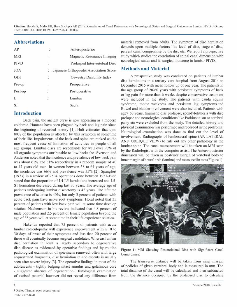
disc herniations in a tertiary care hospital from August 2014 to December 2015 with mean follow up of one year. The patients in the age group of 20-60 years with persistent symptoms of back or leg pain for more than 6 weeks despite conservative treatment were included in the study. The patients with cauda equina syndrome, motor weakness and persistant leg symptoms.and Bowel and bladder involvement were also included. Patients with age> 60 years, traumatic disc prolapse, spondylolithesis with disc prolapse and neurological conditions like Parkinsonism or cerebral palsy etc were excluded from the study. The detailed history and physical examination was performed and recorded in the profroma. Neurological examination was done to find out the level of involvement. Radiographs of lumbosacral spine (AP, LATERAL AND OBLIQUE VIEW) to rule out any other pathology in the lumbar spine. The canal measurement will be taken on MRI scan by the Radiologist with the computer assist. The Antero-posterior dimension will be taken as posterior margin of vertebral body to inner margin of neural arch (lamina) and measured in mm (Figure 1).
Figure 1: MRI Showing Posterolateral Disc with Significant Canal Compromise.
The transverse distance will be taken from inner margin of pedicles of given vertebral body and is measured in mm. The total distance of the canal will be calculated and then subtracted from the distance occupied by the prolapsed disc to calculate
Citation: Hackla S, Malik FH, Basu S, Gupta AK (2018) Correlation of Canal Dimension with Neurological Status and Surgical Outcome in Lumbar PIVD. J Orthop Ther: JORT-163. DOI: 10.29011/2575-8241. 000063
3 Volume 2018; Issue 02
J Orthop Ther, an open access journalISSN: 2575-8241
the final distance occupied by the prolapsed disc. The calculated canal compromise by the prolapsed disc will be graded into three grades to calculate the final outcome on degree of improvement after discectomy. The grade 1 will include distance from 7-10 mm, grade 2 from 5-7mm and grade 3 with the distance less than 5mm. The transverse dimension will be graded into four grades. Grade I include distance from 15-20 mm, grade II will include distance from 10-15 mm, grade III includes distance from 5-10mm and grade IV includes distances <5 mm. The pre-op JOA Score will be calculated and compared with post op JOA Score after standard lumbar discectomy. The correlation then will be calculated.
The assesment of the patient was done according to Japanese Orthopaedic Association (1996) clinical symptom score for a patient with lumbar herniated disc. This can help determine the degree of improvement following surgical intervention. OSWESTRY DISABILITY INDEX will be used to calculate the pain and disability of the patient first pre-operatively and then at 1, 3, 6 and 12 months. The data shall be analysed with the help of computer software, Microsoft excel and SPSS Version 15 for windows. The outcome shall be reported as mean and standard deviation or in percentage as deemed appropriate. The correlation shall be made using paired t-test or chi- square test and measure of correlation such as Pearson product moment correlation co-efficient. P value <0.05 will be statistically significant.
ResultsForty-one patients were included in the study There were
25 males and 16 females. Mean age of the patients is 37.62 years. 20(48%) patients are in 31-50 age group. L4-S1 is the level where most of the patients present. About 2/3rd of the patients 31(75%) are present with disc at this level. Extruded disc constitutes maximum number of the patients 26(63%) (Table 1).
GRADE AP DIMENSION(MM)
NO. OF PATIENTS
PERCENTAGE (%)
0 >10 7 181 7-9.99 10 242 5-6.99 10 243 <5 14 34
TOTAL 41 100
Table 1: Grading of canal dimension based on AP measurement.
17 (41%) patients were having canal measurement greater than 7 while 24 (59%) patients were having canal measurement less than 7. The mean canal measurement is 6.78mm (Table 2).
GRADE TRANSVERSE DIMENSION (MM)
NO. OF PATIENTS
PERCENTAGE (%)
0 >20 1 21 15-19.99 14 342 10-14.99 14 343 5-9.99 12 30
4 <5 0 0TOTAL 41 100
Table 2: Grading of canal dimension based on transverse measurement.
Grade 1 and 2 constituted 14(34%) patients each while 12(30%) patients were in grade 3. No patient was having canal dimension less than 5mm. Mean canal measurement was 14.20mm 27 patients present with cauda equina syndrome. Nearly 2/3rd of the patients were having excised disc weight 2 or less than 2 gm. The average weight of the excised disc is 2.09gm. Dural tear occurred in 3(7%) patients while 1 patient each was having infection, postoperative discitis. The Oswestry disability index shows that the preoperative disability was greater than 40% in all patients which was less than 40% after surgery in all patients except one (Table 3,4).
GRADE PREOP CASES POSTOP CASESMinimal (0-20%) 0 33
Moderate (21-40%) 0 6Severe (41-60%) 9 2
Crippled (61-80%) 10 081-100% 22 0TOTAL 41 41
Table 3: Oswestry disability index.
DIMENSION (mm)
PREOP JOA SCORE
POSTOP JOA SCORE
AP .264 .221TRANSVERSE .603 .129
Table 4: Correlation of canal dimension with JOA score.
shows the correlation of AP and Transverse canal dimension with JOA score. There is weak correlation of AP canal dimension with JOA score. The transverse canal dimension shows significant correlation with preoperative JOA score. This implies that AP and transverse canal dimensions are weakly correlated with patients’ clinical symptoms in all except preoperative JOA in transverse dimension. In the (Table 5),
DIMENSION (mm)
PREOP ODI SCORE
POSTOP ODI SCORE
AP 0.162 -0.137TRANSVERSE -0.348 -0.240
Table 5: Correlation of canal dimension with ODI score.
there negative correlation of AP and transverse canal dimension with ODI score in all except AP in preoperative ODI score. This implies that the patient’s clinical disability resulting from PIVD is not related to the canal compromise observed on MRI.
Mean Score and t-testWe had divided the patients with AP canal dimension into
Citation: Hackla S, Malik FH, Basu S, Gupta AK (2018) Correlation of Canal Dimension with Neurological Status and Surgical Outcome in Lumbar PIVD. J Orthop Ther: JORT-163. DOI: 10.29011/2575-8241. 000063
4 Volume 2018; Issue 02
J Orthop Ther, an open access journalISSN: 2575-8241
two groups, one with canal measurement < 7mm in group 1 and > 7mm into group 2. Similarly, patients with transverse dimension were divided into group 1 having canal dimension <13mm and group 2 with canal dimension >13mm. We had added the constant [4,5] to nullify the negative score in both preoperative and postoperative JOA score. From this (Table 6),
S No. Scoring System
Canal Measurement Groups
Pre Post Pre Post
Mean Score SD Mean Score SD t p t p
1 JOA AP 1 7.46 3.45 27.09 3.4 -1.939 0.068 0.172 0.855
2 10.75 4.26 27.37 5.09
Trans 1 6.45 4.05 27.09 4.78 -3.29 0.004 -0.566 -5.78
2 11.2 2.14 28.1 3.1
2 ODI AP 1 37 7.11 9.3 10.8 1.791 0.39 0.095 0.926
2 30.62 9.13 8.87 8.9
Trans 1 37.27 7.55 11.36 12.85 1.612 0.122 1.08 0.294
2 31.66 8.52 6.7 4.76
Table 6: Mean score and t-test.
mean score was lower in group 1 in JOA but the final score was almost same in both the groups. The p-value of 0.004 was significant in only preoperative transverse group. In short, we can conclude that JOA score was correlated only to transverse group but the number of patients is small, so any definite conclusion can’t be derived at the isolated significant p-value. In the second group, mean score was higher in group 1 but the final score again almost the same in both groups. The mean difference in score and associated decrease is not significant in both AP and transverse group. The p-value is insignificant in both the groups.
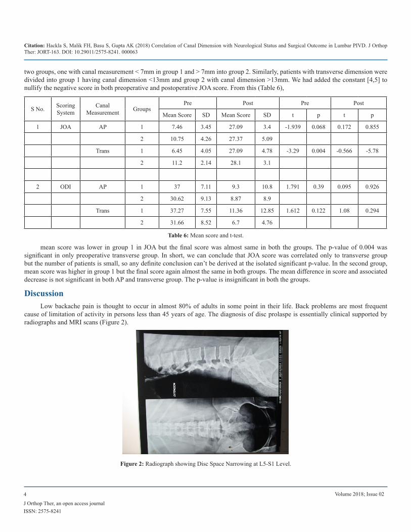
DiscussionLow backache pain is thought to occur in almost 80% of adults in some point in their life. Back problems are most frequent
cause of limitation of activity in persons less than 45 years of age. The diagnosis of disc prolaspe is essentially clinical supported by radiographs and MRI scans (Figure 2).
Figure 2: Radiograph showing Disc Space Narrowing at L5-S1 Level.

Citation: Hackla S, Malik FH, Basu S, Gupta AK (2018) Correlation of Canal Dimension with Neurological Status and Surgical Outcome in Lumbar PIVD. J Orthop Ther: JORT-163. DOI: 10.29011/2575-8241. 000063
5 Volume 2018; Issue 02
J Orthop Ther, an open access journalISSN: 2575-8241
MRI scan gives valuable information about level of disc, type of disc herniation, affection of nerve root etc. The treatment of disc prolaspe is non-operative in all cases except patients with Cauda equina syndrome. Conservative measures include back school therapy, epidural injections, and selective nerve blocks [6-9]. Discectomy is the standard of care in patients with failed conservative treatment which can standard discectomy, microdiscectomy or endoscopic discectomy. The degree of canal compromise still remains an area where not much work had been done. The effect of canal compromise by the prolapsed disc on patients’ neurological status and disability resulting from the herniation had been studied on 41 patients at Govt. Medical College, Jammu. All patients were followed up for one-year [5] finds significant correlation between patient reports of symptoms and anatomical impairment visible on lumbar MRI scan. described that the degree of annular competence after discectomy and type of herniation appear to have value for prediction of recurrence of sciatica, reoperation and clinical outcome after lumbar discectomy [10-13]. had done retrospective analysis to study on long term outcome of standard lumbar discectomy to address postoperative problems including residual low back pain and recurrent herniation for 10 years after lumbar discectomy (Figure 3).
Figure 3: Intraoperative Picture.
They reported favorable outcome. conducted study on 115 monozygotic twins’ pair of 36 to 69 years of age. They concluded that the sensitivity of only significant MRI parameters of disc height narrowing, and annular tears is poor and alone of limited clinical importance [14-17]. Study done consisted of 283 patients who had had severe sciatica for 6 to 12 weeks were subjected to early surgery or to prolonged conservative treatment. The 1-year outcomes were similar for patients assigned to early surgery and those assigned to conservative treatment with eventual surgery if needed, but the rates of pain relief and of perceived recovery were faster for those assigned to early surgery [18-22]. Presented a review of 553 patients who underwent surgery for lumbar intervertebral disc prolapse out of which 42 patients subsequently required a second operation for recurrent sciatica (7.9% revision rate). They concluded that a contained disc protrusion was almost three times more likely to need revision surgery compared with extruded or
sequestrated discs. Also, they had a significantly greater straight leg raise and reduced incidence of positive neurological findings. Therefore, a more enthusiastic conservative treatment program should be implemented in treating these patients. Prospective cohort study on 400 patients with 217 treated surgically and 183 non-surgically (Figure 4).
Figure 4: Preoperative MRI Scan showing Extrusion of the Disc at L5-S1 Level Picture.
They concluded that the surgically treated patients with herniated lumbar disc had more relief of leg pain compared with non-surgically treated patients over 10 years. Prospective observational study in 154 patients with variables of internal disc contour and nerve root compromise at symptomatic disc level. MRI outcome was generally good for disc herniation and nerve root compromise. Nerve root compromise had best MRI prognosis if disc was extruded at the baseline. Concluded that in patients with PIVD, those with thecal sac compression of one/third or more had greater surgical treatment effect than those with small disc herniations and modic type 1 changes. In addition, patients with nerve root compression and displacement benefit more from surgery than those with minimal nerve root impingement. Prospective randomized (501participants) and observational cohorts (743 participants) at 13 spine clinics. Comparison between standard open dissectomy versus usual non-operative care was done. They concluded that carefully selected patients who underwent surgery for lumbar disc herniation achieved greater improvement than non-surgically treated patients. There was little or no degradation of outcomes in either group from 4 to 8 years study [23-25].
Japanese Orthopaedic Association (JOA) ScoreThe mean score preoperatively in anteroposterior group is 9.10
and score of 27.23 postoperatively. There is increase in mean JOA score of 18.13. In transverse group, mean JOA score preoperatively is 8.8 and 27.6 postoperatively. There is also increase in mean
Citation: Hackla S, Malik FH, Basu S, Gupta AK (2018) Correlation of Canal Dimension with Neurological Status and Surgical Outcome in Lumbar PIVD. J Orthop Ther: JORT-163. DOI: 10.29011/2575-8241. 000063
6 Volume 2018; Issue 02
J Orthop Ther, an open access journalISSN: 2575-8241
score of 18.8. There is definite improvement in mean JOA score in two groups in our study with dissectomy. This in comparison to the Azimi et al who suggested that it is reliable and valid measure of functionality and pain among lumbar disc hernaiation patients. The cronbach alpha score preoperatively and postoperatively was 0.64 and 0.81 respectively in the said study. The mean Oswestry Disability Index (ODI) score preoperatively and postoperatively anteroposterior group is 38.81(77%) and 9.05(18%) respectively in our study. There is decrease in mean score of 29.76(59.52%) in our study. In transverse group, mean ODI score is 39.5(79%) and 9.03 (18%) respectively in preoperative and postoperative group with decrease in mean score of 30.47(60%). There is decrease in mean score of almost 30 (60%) in both the groups, so there is definite benefit of doing dissectomy in these patients.
In preoperative AP group, mean JOA score and standard deviation in group 1 and 2 is (7.46±3.45) and (10.75±4.26) respectively while mean score and standard deviation in postoperative group is (27.09±3.4) and (27.37±5.09). The difference in mean score in preoperative group is 3.29 while in postoperative group is 0.28. The difference in mean score is not significant. The p-value is 0.06 in first group while it is 0.85 in second group which is again not significant to have any statistical value. In postoperative transverse group, mean JOA score and standard deviation respectively are (6.45±4.05) and (11.2±2.14). There is difference in mean score of 4.75 between two groups. The p-value 0.004 is significantly correlated in this group. Mean score and standard deviation in postoperative transverse group is (27.09±4.78) and (28.1±3.1). The difference in mean score is 1.01. The p-value is -5.78 which is statistically not significant. The canal compromise by prolasped disc is thus related to patient symptoms in preoperative transverse group but the definitive conclusion can’t be derived at this isolated significant value (Figure 5).
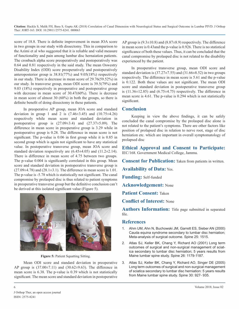
Figure 5: Patient Squatting Sitting.
Mean ODI score and standard deviation in preoperative AP group is (37.00±7.11) and (30.62±9.63). The difference in mean score is 6.38. The p-value is 0.39 which is not statistically significant. The mean score and standard deviation in postoperative
AP group is (9.3±10.8) and (8.87±8.9) respectively. The difference in mean score is 0.43and the p-value is 0.926. There is no statistical significance of both these values. Thus, it can be concluded that the canal compromise by prolasped disc is not related to the disability experienced by the patient.
In preoperative transverse group, mean ODI score and standard deviation is (37.27±7.55) and (31.66±8.52) in two groups respectively. The difference in mean score is 5.61 and the p-value is 0.122. Both these values are not significant. The mean ODI score and standard deviation in postoperative transverse group is (11.36±12.85) and (6.75±4.75) respectively. The difference in mean score is 4.61. The p-value is 0.294 which is not statistically significant.
ConclusionKeeping in view the above findings, it can be safely
concluded the canal compromise by the prolasped disc alone is not related to the patient’s symptoms. There are other factors like position of prolapsed disc in relation to nerve root, stage of disc herniation etc. which are important in overall symptomatology of prolapsed disc
Ethical Approval and Consent to Participate: IEC/168. Government Medical College, Jammu.
Consent for Publication: Taken from patients in written.
Availability of Data: Yes.
Funding: Self-funded
Acknowledgement: None
Patient Consent: Taken
Conflict of Interest: None
Authors Information: Title page submitted in separated file.
ReferencesAhm UM, Ahn N, Buchowski JM, Garrett ES, Sieber AN (2000) 1. Cauda equina syndrome secondary to lumbar disc herniation; Meta-analysis of surgical outcome. Spine 25: 1515.
Atlas SJ, Keller BK, Chang Y, Richard AO (2001) Long term 2. outcomes of surgical and non-surgical management of sciat-ica secondary to lumbar disc herniation; 5 years results from Maine lumbar spine study. Spine 26: 1179-1187.
Atlas SJ, Keller BK, Chang Y, Richard AO, Singer DE (2005) 3. Long term outcomes of surgical and non-surgical management of sciatica secondary to lumbar disc herniation: 5 years results from Maine lumbar spine study. Spine 30: 927- 935.
Citation: Hackla S, Malik FH, Basu S, Gupta AK (2018) Correlation of Canal Dimension with Neurological Status and Surgical Outcome in Lumbar PIVD. J Orthop Ther: JORT-163. DOI: 10.29011/2575-8241. 000063
7 Volume 2018; Issue 02
J Orthop Ther, an open access journalISSN: 2575-8241
Awad JN and Moskovich R (2008) Lumbar disc herniations: 4. Surgical versus non-surgical treatment. Clin Orthop Relat Res 443: 183-197.
Beattie PF, Meyers SP, Start-ford P (2000) Association be-5. tween patient reports of symptoms and anatomical impairment visible on lumbar magnetic resonance imaging. Spine 25: 82.
Cribb GL, Jaffray DC, Cassar-pulticino YN (2007) Observa-6. tions on natural history of massive lumbar disc herniation. J Bone Joint Surg 89B: 782.
Devi R and Rajagopalan N (2003) Dimensions of lumbar ver-7. tebral canal 37: 13.
Fairbank JC and Pynsent PB (2000) The Oswestry disability 8. index. Spine 23: 2940-2952.
Gotfryd A and Avanzio O (2009) A systemic review of ran-9. domised clinical trials using posterior dissectomy to treat lum-bar disc herniations. Int Orthop 33: 11.
Hadjipavlou AG, Tzermiadianos MN, Bogduk N, Zindrick MR 10. (2008) The pathology of disc degeneration: critical review. J Bone Joint Surg 50B: 1261.
Harold LA, Lewis PJ, Douglas B, Egnatchik JG, Yu JY, et al. 11. (2002) Prospective multiple outcome study of outpatient lum-bar microdissectomy: Should 75 to 80% success be the norm. J Neurosurg (spine 1) 96: 34-44.
Haughton V (2006) Imaging inter-vertebral disc degeneration. 12. J Bone Joint Surg 88: 15.
Hueme M and Alaranta H (1987) Factors predicting the results 13. of surgery for lumbar intervertebral disc herniation. Spine 12: 933-938.
Katz JN (2006) Lumbar disc disorders and low back pain: so-14. cio-economic factors and consequences. J Bone Joint Surg 88: 21.
Kim SW, Yeom JS, Park SK, Chang BS, Lee DH, et al. (2009) 15. Inter- and intra-observer reliability of MRI for lumbar lateral disc herniation. Clin Orthop Surg 1: 34-39.
Kohtes SS, Kohies DA, Karp AP, Erlich VM, Polissar NL (2004) 16. Time dependent surgical outcome following cauda equina syn-drome diagnosis; comments on meta-analysis. Spine 29: 1281.
Lurie JD, Faucett SC, Hanscome B, Tosteson TD, Ball PA, et al. 17. (2008) Lumbar dissectomy outcome vary by herniation level in spine patient outcomes research trial. J Bone Joint Surg 90A: 181.
Matsui T, Yukawa Y, Nakamura S, Kajino G, Matsubara Y 18. (2005) Natural history of patients with lumbar disc herniations observed by magnetic resonance imaging for minimum of 7 years. J Spinal Disord Tech 18: 121.
Miyamoto H, Doita M, Nishida K, Yamamoto T, Sumi M, et al. 19. (2006) Effects of cyclic mechanical stress on production of in-flammatory agents by nucleus pulposus and annulus fibrosus cells in vitro. Spine 31: 4-9.
Turek SL (2005) Turek’s orthopaedics. edition 420. th 2: p1485-1488.
Videman T, Battie MC, Gibbons LE, Maravilla K, Manninen H, 21. et al. (2003) Association between back pain history and lumbar MRI findings. Spine 28: 582.
Videman T, Battie MG, Laura E, Maravilla K (2003) association 22. between back pain history lumbar MRI findings studied 115 monozygotic twins qualitatively assessed disc height, bulging herniation, annular tears, osteophytes, spinal stenosis and endplate changes. Spine 28: 582-585.
Weinstein JN and Lurie JD (2014) Surgical versus nonopera-23. tive treatment for lumbar disc herniation: eight years results for the spine patient outcome research trial. Spine 39: 3-16.
Yorimitse E, Chiba K, Tojama Y, Hirabayashi K (2001) Long 24. term outcomes of standard dissectomy for lumbar disc her-niation: A follow up study of more than 10 years. Spine 26: 1652-1657.
Yoshito K, Matsuyama Y, Yoshihara H, Sakai Y, Nakamura H, et 25. al. (2006) Comparison of surgical outcomes between macrodis-sectomy and microdissectomy for lumbar disc herniation: A pro-spective randomized study. J Spinal Disord Tech 19: 344-347.