gold-decorated block copolymer microspheres with controlled surface nanostructures
TRANSCRIPT
KIM ET AL . VOL. 6 ’ NO. 3 ’ 2750–2757 ’ 2012
www.acsnano.org
2750
February 21, 2012
C 2012 American Chemical Society
Gold-Decorated Block CopolymerMicrospheres with Controlled SurfaceNanostructuresMinsoo P. Kim,† Dong Jin Kang,† Dae-Woong Jung,‡ Aravindaraj G. Kannan,† Ki-Hyun Kim,† Kang Hee Ku,†
Se Gyu Jang,§ Weon-Sik Chae,^ Gi-Ra Yi, ),* and Bumjoon J. Kim†,*
†Department of Chemical and Biomolecular Engineering, Korea Advanced Institute of Science and Technology (KAIST), Daejeon 305-701, Republic of Korea,‡Department of Engineering Chemistry, Chungbuk National University, Cheongju, Chungbuk 361-763, Republic of Korea, §Materials Research Laboratory,University of California, Santa Barbara, California 93106, United States, ^Gangneung Center, Korea Basic Science Institute, Gangneung 210-702, Republic of Korea,and )Department of Polymer Science and Engineering, Sungkyunkwan University, Suwon, 440-746 Republic of Korea
Using emulsion droplets to confineblock copolymer (BCP) solutions inwater, BCP colloidal particles can be
produced as the solvent in the emulsiondroplets evaporates. The nanostructures ofthese particles are unique and can be con-trolled by changing the volume fraction ofblocks, the particle size, and the interfacialtension between each of the blocks and theparticle interface.1�3 For example, whileonion-like particles were prepared usingsymmetric diblock copolymers, other nano-structured particles were prepared sim-ply by adding low-molecular-weight homo-polymers in different ratios.1 In addition,it has been reported that the internal nano-morphology of BCPs in particles is signifi-cantly affected by block interactions withthe particle surfaces (i.e., controlling surfac-tants), and the overall colloidal particleshape changes during structural evolutionas evaporation takes place.3
Functional nanoparticles (NPs) can beincorporated into such colloidal particles,using methods previously demonstrated inthin films,4�7 to produce colloidal compo-site particles with complex internal nano-phases. Several experimental methods havebeen developed to incorporate inorganicNPs into polymeric nanostructures, whichcan be categorized into so-called ex situ andin situ methods.5,8�21 The ex situ approachexploits the cooperative self-organization ofpreformed NPs and BCPs. The strategy forincorporating and controlling the locationof NPs in a BCP or polymer blend involvestuning the surface properties of the NPs byend-attaching ligands such as organicmole-cules,5,22�24 homopolymers,10,25�30 mixturesof homopolymers,4,8 or copolymers16,21,31�33
to theNP surfaces. In addition, a variety of NPs
can be incorporated into a polymer matrixwith a controlled positioning once they arecoated with suitable ligands. Another pop-ular approach involves the in situ synthesisof NPs within a BCP template using pre-formed micelles of BCPs that contain metalprecursors.9,11�14,17,34 In this method, oneof the blocks should retain a strong affinityfor metallic precursors. Such blocks in-clude poly(2-vinylpyridine) (P2VP) and poly-(4-vinylpyridine) (P4VP),whosenitrogenatoms
* Address correspondence [email protected] (B.J.K.);[email protected] (G.-R.Y.).
Received for review January 14, 2012and accepted February 21, 2012.
Published online10.1021/nn300194z
ABSTRACT Gold-decorated block copoly-
mer microspheres (BCP-microspheres) display-
ing various surface morphologies were pre-
pared by the infiltration of Au precursors into
polystyrene-b-poly(4-vinylpyridine) (PS-b-P4VP)
microspheres. The microspheres were fabricated by emulsifying the PS-b-P4VP polymers in
chloroform into a surfactant solution in water, followed by the evaporation of chloroform. The
selective swelling of the P4VP domains in the microspheres by the Au precursor under acidic
conditions resulted in the formation of Au-decorated BCP-microspheres with various surface
nanostructures. As evidenced by transmission electron microscopy (TEM) and scanning electron
microscopy (SEM) measurements, dotted surface patterns were formed when microspheres
smaller than 800 nm were synthesized, whereas fingerprint-like surface patterns were
observed with microspheres larger than 800 nm. Au nanoparticles (NPs) were located inside
P4VP domains near the surfaces of the prepared microspheres, as confirmed by TEM. The
optical properties of the BCP-microspheres were characterized using UV�vis absorption
spectroscopy and fluorescence lifetime measurements. A maximum absorption peak was
observed at approximately 580 nm, indicating that Au NPs are densely packed into P4VP
domains on the microspheres. Our approach for creating Au-NP-hybrid BCP-microspheres can
be extended to other NP systems such as iron-oxide or platinum NPs. These precursors can also
be selectively incorporated into P4VP domains and induce the formation of hybrid BCP-
microspheres with controlled surface nanostructures.
KEYWORDS: block copolymer . colloid particle . block copolymermicrosphere .raspberry particle . surface nanostructure . Au nanoparticles
ARTIC
LE
KIM ET AL . VOL. 6 ’ NO. 3 ’ 2750–2757 ’ 2012
www.acsnano.org
2751
interact strongly with metal precursors.6,7 Metal�polymer composite particles can be produced by thedirect reduction of metallic precursors inside BCPmicelles, e.g., using a reducing agent or plasmatreatment.34�37 However, the in situ formation ofmetallic particles within submicrometer BCP colloidalparticles has not been reported.In this study, we have demonstrated a simple and
facile method to produce metal hybrid “BCP-micro-spheres” with controlled surface nanostructures. First,microspheres of polystyrene-b-poly(4-vinylpyridine)(PS-b-P4VP) polymeric micelles were prepared by anemulsion encapsulation and shrinkage method. Then,upon the addition of Au precursors in acidic water,Au NPs formed only near the surfaces of these BCP-microspheres. Surprisingly, unique dot or fingerprint-like patterns on their surfaces evolved during metalli-zation because P4VP domains were selectively swollenand migrated to the surfaces. The surface structurescan be controlled by tuning the diameter of the BCP-microspheres. Thus, we successfully synthesized Au-NP-decorated BCP-microspheres and demonstrated thesimultaneous control of their surface morphologies.
RESULTS AND DISCUSSION
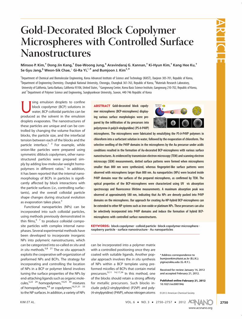
For BCP-microspheres, a 1 wt % chloroform solutionof PS-b-P4VP diblock copolymer was emulsified inwater containing 1 wt % Pluronic F108 surfactant viahigh-intensity ultrasonic irradiation. Subsequently,through the evaporation of the chloroform, BCP-microspheres were produced, which were well dis-persed in water, as shown in the optical micrographof Figure 1a. The SEM image in Figure 1c shows BCP-microspheres with smooth surfaces. In contrast, afterthe microspheres were treated with the Au precursor(HAuCl4) solution in water, they exhibited very roughsurfaces covered with small hemispheres, as shown inFigure 1d. These surface structures were found to beuniform in size and ordered regularly. In addition, thesetransformed microspheres were well dispersed inwater, as illustrated in the optical micrograph ofFigure 1b.To gain deeper insight into the morphological
changes at the surface of the BCP-microspheres, themicrospheres were characterized by surface and cross-sectional TEM images. For cross-sectional TEM mea-surements, the microspheres were first dropped ontoan epoxy film and dried. Then, the samples were
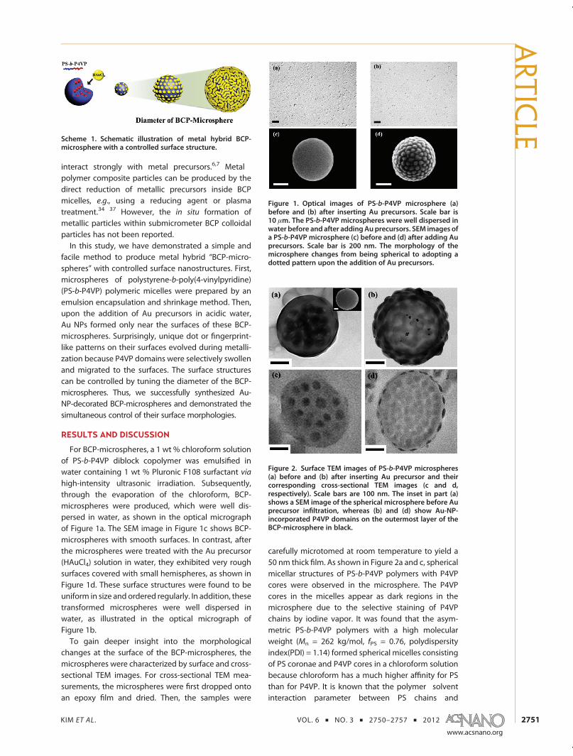
carefully microtomed at room temperature to yield a50 nm thick film. As shown in Figure 2a and c, sphericalmicellar structures of PS-b-P4VP polymers with P4VPcores were observed in the microsphere. The P4VPcores in the micelles appear as dark regions in themicrosphere due to the selective staining of P4VPchains by iodine vapor. It was found that the asym-metric PS-b-P4VP polymers with a high molecularweight (Mn = 262 kg/mol, fPS = 0.76, polydispersityindex(PDI) = 1.14) formed spherical micelles consistingof PS coronae and P4VP cores in a chloroform solutionbecause chloroform has a much higher affinity for PSthan for P4VP. It is known that the polymer�solventinteraction parameter between PS chains and
Figure 1. Optical images of PS-b-P4VP microsphere (a)before and (b) after inserting Au precursors. Scale bar is10 μm. The PS-b-P4VP microspheres were well dispersed inwater before and after addingAuprecursors. SEM images ofa PS-b-P4VP microsphere (c) before and (d) after adding Auprecursors. Scale bar is 200 nm. The morphology of themicrosphere changes from being spherical to adopting adotted pattern upon the addition of Au precursors.
Scheme 1. Schematic illustration of metal hybrid BCP-microsphere with a controlled surface structure.
Figure 2. Surface TEM images of PS-b-P4VP microspheres(a) before and (b) after inserting Au precursor and theircorresponding cross-sectional TEM images (c and d,respectively). Scale bars are 100 nm. The inset in part (a)shows a SEM image of the spherical microsphere before Auprecursor infiltration, whereas (b) and (d) show Au-NP-incorporated P4VP domains on the outermost layer of theBCP-microsphere in black.
ARTIC
LE
KIM ET AL . VOL. 6 ’ NO. 3 ’ 2750–2757 ’ 2012
www.acsnano.org
2752
chloroform is 0.34, which is much lower than thatbetween P4VP chains and chloroform (1.55).38,39 Theaverage P4VP core size of PS-b-P4VP micelles in themicrospheres was estimated to be 47.5( 9.2 nm fromthe TEM image. This value agrees with the averageP4VP core size (43.7( 3.1 nm) from the AFMmeasure-ment, where a PS-b-P4VPmicellar filmwas prepared onSi substrate from 0.5 wt % PS-b-P4VP solution inchloroform. The AFM measurement (Figure S2) alsoprovides an estimate of the average PS-b-P4VP micellesize from the center-to-center distance between twodifferent P4VP micelle cores, which was found to be59.5 ( 5.8 nm.After the infiltration of the Auprecursor into the BCP-
microspheres, the surfaces of the BCP-microsphereswere dramatically transformed, changing from smoothto dot-patterned surfaces. Figure 2b and d showsurface and cross-sectional TEM images of the BCP-microspheres with dot-patterned surfaces, which wereobserved without iodine staining. The surface TEMimage in Figure 2b shows a uniform arrangement ofsmall hemispheres containing Au NPs on the micro-sphere surfaces, with structures resembling raspber-ries. This structure corresponds to the SEM imageshown in Figure 1d. To examine the internal structuresof the BCP-microspheres, cross-sectional TEM imagesof themicrospheres were taken aftermicrotoming. TheTEM image in Figure 2d clearly demonstrates that theAu precursor infiltrated the P4VP domains only nearthe surfaces of the microspheres but not the P4VPdomains inside the microspheres. The Au precursorscould not reach the P4VP domain inside, because theP4VP domains inside were isolated by the PS matrix inthe microspheres. The P4VP domains appear slightlydarker than the PS matrix in Figure 2d because theelectron scattering from P4VP polymers is strongerthan that of PS polymers.40
In previous reports, raspberry-like particles wereprepared by attaching premade NPs on spheres, whichwas typically based on chemical interactions betweenNPs and organic molecules on the sphere surfaces.41,42
For instance, amino-functionalized ligands43,44 onspheres were used to attach iron or silica NPs, whilethiol-functionalized ligands45 were used to interactwith gold, silver, or copper NPs. However, in thisapproach, specific types of NPs and organic moleculeson the sphere are required, which limits the types ofraspberry-structured particles that are formed. Re-cently, a few researchers have prepared raspberry-likeparticles using self-assembled structures of BCP micelleswith cross-linkable core units.46,47 Similarly, our BCP-microspheres, which show dotted surface patterns,resemble raspberry-like structures. However, the mech-anism of the formation of such raspberry particles isvery different from that of the particles mentionedabove. Our BCP-microspheres were formed based onthe swelling and deswelling processes that occurred
upon the selective incorporation of Au NPs with acidicwater into P4VP domains.To characterize the Au NPs formed within the P4VP
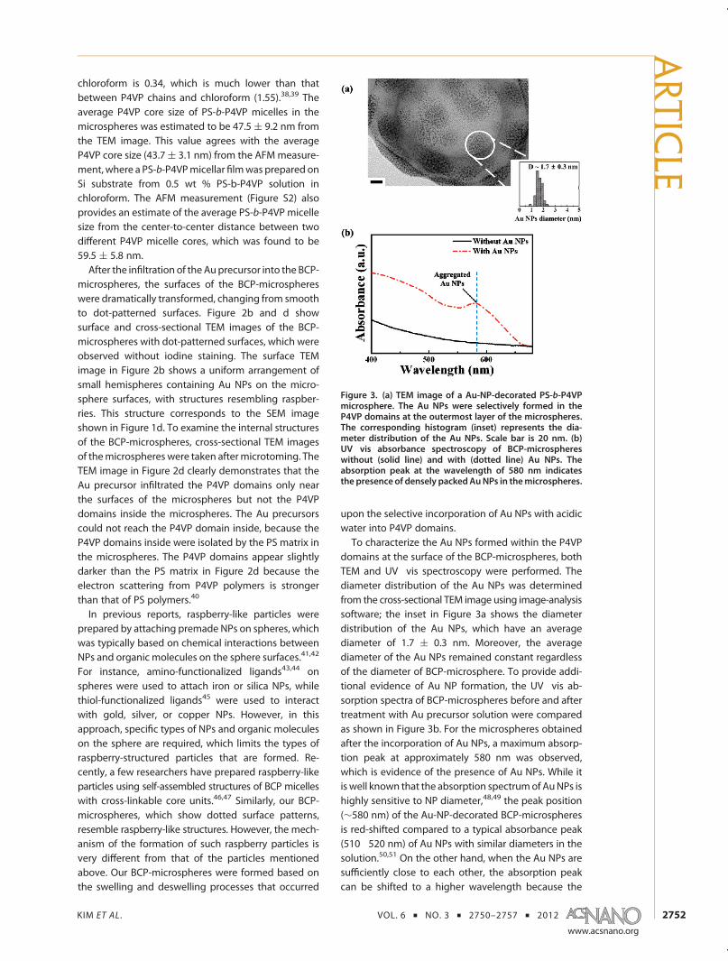
domains at the surface of the BCP-microspheres, bothTEM and UV�vis spectroscopy were performed. Thediameter distribution of the Au NPs was determinedfrom the cross-sectional TEM image using image-analysissoftware; the inset in Figure 3a shows the diameterdistribution of the Au NPs, which have an averagediameter of 1.7 ( 0.3 nm. Moreover, the averagediameter of the Au NPs remained constant regardlessof the diameter of BCP-microsphere. To provide addi-tional evidence of Au NP formation, the UV�vis ab-sorption spectra of BCP-microspheres before and aftertreatment with Au precursor solution were comparedas shown in Figure 3b. For the microspheres obtainedafter the incorporation of Au NPs, a maximum absorp-tion peak at approximately 580 nm was observed,which is evidence of the presence of Au NPs. While itis well known that the absorption spectrumof AuNPs ishighly sensitive to NP diameter,48,49 the peak position(∼580 nm) of the Au-NP-decorated BCP-microspheresis red-shifted compared to a typical absorbance peak(510�520 nm) of Au NPs with similar diameters in thesolution.50,51 On the other hand, when the Au NPs aresufficiently close to each other, the absorption peakcan be shifted to a higher wavelength because the
Figure 3. (a) TEM image of a Au-NP-decorated PS-b-P4VPmicrosphere. The Au NPs were selectively formed in theP4VP domains at the outermost layer of the microspheres.The corresponding histogram (inset) represents the dia-meter distribution of the Au NPs. Scale bar is 20 nm. (b)UV�vis absorbance spectroscopy of BCP-microsphereswithout (solid line) and with (dotted line) Au NPs. Theabsorption peak at the wavelength of 580 nm indicatesthe presence of densely packedAuNPs in themicrospheres.
ARTIC
LE
KIM ET AL . VOL. 6 ’ NO. 3 ’ 2750–2757 ’ 2012
www.acsnano.org
2753
dipole plasmonmodes of the AuNPs can be coupled.52
For example, the UV�vis absorption peak of Au clus-ters consisting of Au NPs less than 2 nm was observedat a higher wavelength than that of individual AuNPs.53 Therefore, the red-shifted UV�vis absorptionpeak indicates that the Au NPs were densely packedin the P4VP domains near the microsphere surfaces.In addition, the presence of Au NPs at the outermostlayer of the microspheres was confirmed by energydispersive X-ray spectrometry (EDX) measurementusing a cross-sectioned TEM sample, as shown inFigure S3.Deeper insight into the morphological transition
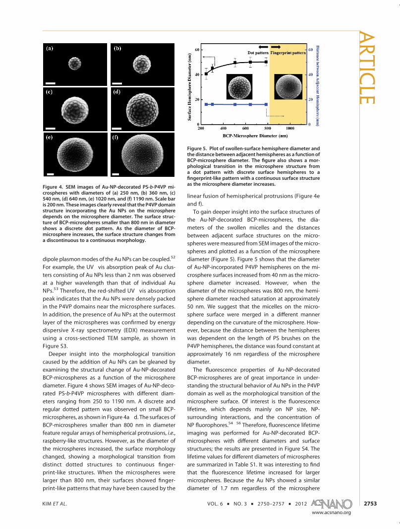
caused by the addition of Au NPs can be gleaned byexamining the structural change of Au-NP-decoratedBCP-microspheres as a function of the microspherediameter. Figure 4 shows SEM images of Au-NP-deco-rated PS-b-P4VP microspheres with different diam-eters ranging from 250 to 1190 nm. A discrete andregular dotted pattern was observed on small BCP-microspheres, as shown in Figure 4a�d. The surfaces ofBCP-microspheres smaller than 800 nm in diameterfeature regular arrays of hemispherical protrusions, i.e.,raspberry-like structures. However, as the diameter ofthe microspheres increased, the surface morphologychanged, showing a morphological transition fromdistinct dotted structures to continuous finger-print-like structures. When the microspheres werelarger than 800 nm, their surfaces showed finger-print-like patterns that may have been caused by the
linear fusion of hemispherical protrusions (Figure 4eand f).To gain deeper insight into the surface structures of
the Au-NP-decorated BCP-microspheres, the dia-meters of the swollen micelles and the distancesbetween adjacent surface structures on the micro-spheres weremeasured from SEM images of themicro-spheres and plotted as a function of the microspherediameter (Figure 5). Figure 5 shows that the diameterof Au-NP-incorporated P4VP hemispheres on the mi-crosphere surfaces increased from 40 nm as the micro-sphere diameter increased. However, when thediameter of the microspheres was 800 nm, the hemi-sphere diameter reached saturation at approximately50 nm. We suggest that the micelles on the micro-sphere surface were merged in a different mannerdepending on the curvature of the microsphere. How-ever, because the distance between the hemisphereswas dependent on the length of PS brushes on theP4VP hemispheres, the distance was found constant atapproximately 16 nm regardless of the microspherediameter.The fluorescence properties of Au-NP-decorated
BCP-microspheres are of great importance in under-standing the structural behavior of Au NPs in the P4VPdomain as well as the morphological transition of themicrosphere surface. Of interest is the fluorescencelifetime, which depends mainly on NP size, NP-surrounding interactions, and the concentration ofNP fluorophores.54�56 Therefore, fluorescence lifetimeimaging was performed for Au-NP-decorated BCP-microspheres with different diameters and surfacestructures; the results are presented in Figure S4. Thelifetime values for different diameters of microspheresare summarized in Table S1. It was interesting to findthat the fluorescence lifetime increased for largermicrospheres. Because the Au NPs showed a similardiameter of 1.7 nm regardless of the microsphere
Figure 4. SEM images of Au-NP-decorated PS-b-P4VP mi-crospheres with diameters of (a) 250 nm, (b) 360 nm, (c)540 nm, (d) 640 nm, (e) 1020 nm, and (f) 1190 nm. Scale baris 200 nm. These images clearly reveal that the P4VPdomainstructure incorporating the Au NPs on the microspheredepends on the microsphere diameter. The surface struc-ture of BCP-microspheres smaller than 800 nm in diametershows a discrete dot pattern. As the diameter of BCP-microsphere increases, the surface structure changes froma discontinuous to a continuous morphology.
Figure 5. Plot of swollen-surface hemisphere diameter andthe distance between adjacent hemispheres as a function ofBCP-microsphere diameter. The figure also shows a mor-phological transition in the microsphere structure froma dot pattern with discrete surface hemispheres to afingerprint-like pattern with a continuous surface structureas the microsphere diameter increases.
ARTIC
LE
KIM ET AL . VOL. 6 ’ NO. 3 ’ 2750–2757 ’ 2012
www.acsnano.org
2754
diameter, the increase in the fluorescence lifetime wasnot due to the changes in the diameter of individual AuNPs. On the contrary, it is well known that fluorophoreconcentration affects lifetime measurements, and ahigh concentration of fluorophore typically increasesthe lifetime through trivial reabsorption processes dueto the increased reabsorption probability in the high-concentration region of bulk solution, although theintrinsic lifetime remains unchanged.55 Therefore, wesuggest that fluorescence lifetime increases with in-creasing microsphere diameter because a larger num-ber of Au NPs can participate in these reabsorptionprocesses within the microsphere.57 For example, asthemicrosphere diameter increased from 380 to 810 nm,the P4VP hemisphere on the microsphere surface in-creased from 40 to 50 nm and thus caused a correspond-ing increase in the number of Au NPs per P4VP domain,which resulted in the change of the fluorescence lifetimefrom 2.1 ( 0.2 to 3.3 ( 0.2 ns. Interestingly, the fluores-cence lifetime further increased to 3.7 nswith the increasein the microsphere diameter from 810 to 1380 nm.We speculate that this was caused by the change in thesurface structure from a dotted to a continuous finger-print pattern, which eventually produced larger numbersof Au NPs per P4VP domain on the surface.Morphological changes of the sphere surface during
swelling and deswelling processes have been investi-gated in previous studies using buckling or wrinklingtheory.58�60 Diverse patterns are formed by surfacewrinkling on a core�shell sphere because the sphericalsurface buckles as a consequence of energy minimiza-tion, and this configuration is energetically favorable.With increasing film stress on the spheres, the protru-sions coalesce to reduce the stretching energy andgradually evolve into labyrinth-like patterns. In ourexperiment, the observed morphologies were quitesimilar to those of buckled spheres. Moreoever, ourresults show the formation of BCP-microspheres withdifferent surface structures as a function of the micro-sphere diameter and thus the surface curvatures.Scheme 2 presents the illustration of the surface-structure evolution on the microspheres as a functionof themicrosphere diameter. Before the addition of Auprecursors, the spherical micelles of PS-b-P4VP were
present within the microspheres, in which the PS blockis highly swollen with solvent (chloroform) and theP4VP in contrast is collapsed. Therefore, that sphericalmorphology is retained in the microsphere. Near thesurface when the BCP-microspheres is treated withacidic water the P4VP domains swell and burst throughthe surface coating of PS brushes, forming amushroomof highly swollen P4VP. We believe that the surfacereconstruction of PS-b-P4VP structures by Au precur-sors in acidic water resembles that in thin film geome-try by an alcohol reported by Russell et al.61�63 If thecurvature of the surface is high enough, the P4VPmushrooms do not touch, thus forming the dot patternof P4VP structures. However, as the surface curvaturedecreases, the swollen mushrooms can touch eachother and will tend to form continuous half-cylinders.Therefore, the final size of the P4VP surface structureon the microsphere shown in Figure 5 could representthe considerably shrunkone after thedeswellingprocess.Similar curvature-dependentbehaviors on the surfaces ofemulsions have been reported in a previous study.64
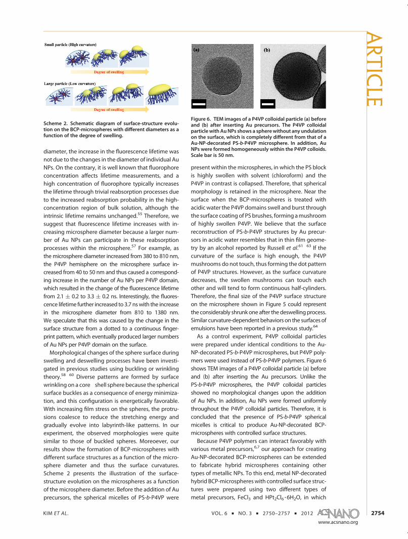
As a control experiment, P4VP colloidal particleswere prepared under identical conditions to the Au-NP-decorated PS-b-P4VPmicrospheres, but P4VP poly-mers were used instead of PS-b-P4VP polymers. Figure 6shows TEM images of a P4VP colloidal particle (a) beforeand (b) after inserting the Au precursors. Unlike thePS-b-P4VP microspheres, the P4VP colloidal particlesshowed no morphological changes upon the additionof Au NPs. In addition, Au NPs were formed uniformlythroughout the P4VP colloidal particles. Therefore, it isconcluded that the presence of PS-b-P4VP sphericalmicelles is critical to produce Au-NP-decorated BCP-microspheres with controlled surface structures.Because P4VP polymers can interact favorably with
various metal precursors,6,7 our approach for creatingAu-NP-decorated BCP-microspheres can be extendedto fabricate hybrid microspheres containing othertypes of metallic NPs. To this end, metal NP-decoratedhybrid BCP-microsphereswith controlled surface struc-tures were prepared using two different types ofmetal precursors, FeCl3 and HPt2Cl6 3 6H2O, in which
Scheme 2. Schematic diagram of surface-structure evolu-tion on the BCP-microspheres with different diameters as afunction of the degree of swelling.
Figure 6. TEM images of a P4VP colloidal particle (a) beforeand (b) after inserting Au precursors. The P4VP colloidalparticlewith AuNPs shows a spherewithout any undulationon the surface, which is completely different from that of aAu-NP-decorated PS-b-P4VP microsphere. In addition, AuNPs were formed homogeneously within the P4VP colloids.Scale bar is 50 nm.
ARTIC
LE
KIM ET AL . VOL. 6 ’ NO. 3 ’ 2750–2757 ’ 2012
www.acsnano.org
2755
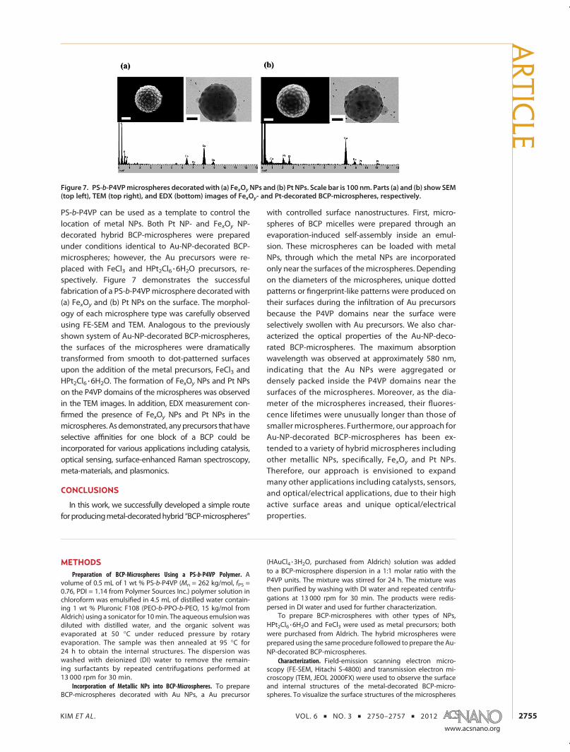
PS-b-P4VP can be used as a template to control thelocation of metal NPs. Both Pt NP- and FexOy NP-decorated hybrid BCP-microspheres were preparedunder conditions identical to Au-NP-decorated BCP-microspheres; however, the Au precursors were re-placed with FeCl3 and HPt2Cl6 3 6H2O precursors, re-spectively. Figure 7 demonstrates the successfulfabrication of a PS-b-P4VPmicrosphere decorated with(a) FexOy and (b) Pt NPs on the surface. The morphol-ogy of each microsphere type was carefully observedusing FE-SEM and TEM. Analogous to the previouslyshown system of Au-NP-decorated BCP-microspheres,the surfaces of the microspheres were dramaticallytransformed from smooth to dot-patterned surfacesupon the addition of the metal precursors, FeCl3 andHPt2Cl6 3 6H2O. The formation of FexOy NPs and Pt NPson the P4VP domains of the microspheres was observedin the TEM images. In addition, EDX measurement con-firmed the presence of FexOy NPs and Pt NPs in themicrospheres. Asdemonstrated, anyprecursors thathaveselective affinities for one block of a BCP could beincorporated for various applications including catalysis,optical sensing, surface-enhanced Raman spectroscopy,meta-materials, and plasmonics.
CONCLUSIONS
In this work, we successfully developed a simple routeforproducingmetal-decoratedhybrid “BCP-microspheres”
with controlled surface nanostructures. First, micro-spheres of BCP micelles were prepared through anevaporation-induced self-assembly inside an emul-sion. These microspheres can be loaded with metalNPs, through which the metal NPs are incorporatedonly near the surfaces of themicrospheres. Dependingon the diameters of the microspheres, unique dottedpatterns or fingerprint-like patterns were produced ontheir surfaces during the infiltration of Au precursorsbecause the P4VP domains near the surface wereselectively swollen with Au precursors. We also char-acterized the optical properties of the Au-NP-deco-rated BCP-microspheres. The maximum absorptionwavelength was observed at approximately 580 nm,indicating that the Au NPs were aggregated ordensely packed inside the P4VP domains near thesurfaces of the microspheres. Moreover, as the dia-meter of the microspheres increased, their fluores-cence lifetimes were unusually longer than those ofsmaller microspheres. Furthermore, our approach forAu-NP-decorated BCP-microspheres has been ex-tended to a variety of hybrid microspheres includingother metallic NPs, specifically, FexOy and Pt NPs.Therefore, our approach is envisioned to expandmany other applications including catalysts, sensors,and optical/electrical applications, due to their highactive surface areas and unique optical/electricalproperties.
METHODSPreparation of BCP-Microspheres Using a PS-b-P4VP Polymer. A
volume of 0.5 mL of 1 wt % PS-b-P4VP (Mn = 262 kg/mol, fPS =0.76, PDI = 1.14 from Polymer Sources Inc.) polymer solution inchloroform was emulsified in 4.5 mL of distilled water contain-ing 1 wt % Pluronic F108 (PEO-b-PPO-b-PEO, 15 kg/mol fromAldrich) using a sonicator for 10min. The aqueous emulsionwasdiluted with distilled water, and the organic solvent wasevaporated at 50 �C under reduced pressure by rotaryevaporation. The sample was then annealed at 95 �C for24 h to obtain the internal structures. The dispersion waswashed with deionized (DI) water to remove the remain-ing surfactants by repeated centrifugations performed at13 000 rpm for 30 min.
Incorporation of Metallic NPs into BCP-Microspheres. To prepareBCP-microspheres decorated with Au NPs, a Au precursor
(HAuCl4 3 3H2O, purchased from Aldrich) solution was addedto a BCP-microsphere dispersion in a 1:1 molar ratio with theP4VP units. The mixture was stirred for 24 h. The mixture wasthen purified by washing with DI water and repeated centrifu-gations at 13 000 rpm for 30 min. The products were redis-persed in DI water and used for further characterization.
To prepare BCP-microspheres with other types of NPs,HPt2Cl6 3 6H2O and FeCl3 were used as metal precursors; bothwere purchased from Aldrich. The hybrid microspheres wereprepared using the same procedure followed to prepare the Au-NP-decorated BCP-microspheres.
Characterization. Field-emission scanning electron micro-scopy (FE-SEM, Hitachi S-4800) and transmission electron mi-croscopy (TEM, JEOL 2000FX) were used to observe the surfaceand internal structures of the metal-decorated BCP-micro-spheres. To visualize the surface structures of the microspheres
Figure 7. PS-b-P4VPmicrospheres decoratedwith (a) FexOyNPs and (b) Pt NPs. Scale bar is 100 nm. Parts (a) and (b) show SEM(top left), TEM (top right), and EDX (bottom) images of FexOy- and Pt-decorated BCP-microspheres, respectively.
ARTIC
LE
KIM ET AL . VOL. 6 ’ NO. 3 ’ 2750–2757 ’ 2012
www.acsnano.org
2756
using FE-SEM, the samples were prepared by drop-castingmicrosphere suspensions onto silicon wafers, which were thenwashed and sputtered with gold. To investigate the surfacestructures of the microspheres by TEM, the samples wereprepared by dipping TEM grids coated with a 20�30 nm thickcarbon film into the microsphere suspensions, followed bydrying in air. The prepared samples were exposed to I2 vaporto selectively stain the P4VP domains of PS-b-P4VP. However,after inserting the Au precursor into the PS-b-P4VP micro-spheres, the samples were characterized without staining dueto the strong contrast between the Au NPs and polymericdomains. The sizes and distributions of the PS-b-P4VP micellesand the Au NPs in the microspheres were determined byanalyzing the FE-SEM, TEM, and AFM images. To investigatethe internal structures of the microspheres by cross-sectionalTEM, the samples were prepared by drop-casting microspheresuspensions onto an epoxy film and allowing the solvent to dry.Then, the epoxy-supported films were cured in an oven at 60 �Cfor 24 h. The epoxy-supported filmswere thenmicrotomedwitha diamond knife at room temperature into 50 nm slices.
To characterize the optical properties of the Au-NP-deco-rated BCP-microspheres, UV�vis absorption spectroscopy (Cary50 Conc UV�vis spectrophotometer) and fluorescence lifetimeimaging FLIM were performed. FLIM was performed using aninverted-type scanning confocal microscope (MicroTime-200,Picoquant, Germany) with a 100� objective. A single-modepulsed diode laser (with a 470 nm output and an instrumentalresponse function of∼96 ps in full-width at half-maximum, a 40MHz repetition rate, and an average power of less than 1 μW)was used as an excitation source. A dichroic mirror (490 DCXR,AHF), a long-pass filter (HQ500lp, AHF), a 50 μm pinhole, and asingle-photon avalanche diode were used to collect the emis-sions (λ > 500 nm) from Au-NP-decorated samples that werespin-coated onto glass coverslips. Data acquisition was basedon a time-correlated single-photon counting technique. Time-resolved fluorescence decay curves were obtained from FLIMimages, and fluorescence lifetimes were evaluated according tononlinear least-squares iterative curve fitting using the Sym-PhoTime software (ver. 5.1.3).
The presence of Au, FexOy, and Pt in the hybrid BCP-micro-spheres was confirmed by EDX measurement (JEOL). Thesamples were prepared using the same procedure as that usedto prepare the samples for TEM but were not stained.
Conflict of Interest: The authors declare no competingfinancial interest.
Acknowledgment. This research was supported by the KoreaResearch Foundation Grant, funded by the Korean Government(2011-0017943, 2011-0027240, 2011-0027518, 2009-0082451).The authors thank Prof. Edward J. Kramer for valuable discussions.
Supporting Information Available: Additional NMR, AFM,EDX, and FILM data. This material is available free of chargevia the Internet at http://pubs.acs.org.
REFERENCES AND NOTES1. Jeon, S. J.; Yi, G. R.; Yang, S. M. Cooperative Assembly of
Block Copolymers with Deformable Interfaces: TowardNanostructured Particles. Adv. Mater. 2008, 20, 4103–4108.
2. Yabu, H.; Higuchi, T.; Shimomura, M. Unique Phase-Separation Structures of Block-Copolymer Nanoparticles.Adv. Mater. 2005, 17, 2062–2065.
3. Jeon, S. J.; Yi, G. R.; Koo, C. M.; Yang, S. M. Nanostructuresinside Colloidal Particles of Block Copolymer/Homopoly-mer Blends. Macromolecules 2007, 40, 8430–8439.
4. Kim, B. J.; Bang, J.; Hawker, C. J.; Chiu, J. J.; Pine, D. J.; Jang,S. G.; Yang, S. M.; Kramer, E. J. Creating Surfactant Nano-particles for Block Copolymer Composites through SurfaceChemistry. Langmuir 2007, 23, 12693–12703.
5. Bockstaller, M. R.; Lapetnikov, Y.; Margel, S.; Thomas, E. L.Size-Selective Organization of Enthalpic CompatibilizedNanocrystals in Ternary Block Copolymer/Particle Mix-tures. J. Am. Chem. Soc. 2003, 125, 5276–5277.
6. Hayward, R. C.; Chmelka, B. F.; Kramer, E. J. CrosslinkedPoly(styrene)-Block-Poly(2-vinylpyridine) Thin Films asSwellable Templates for Mesostructured Silica and Titania.Adv. Mater. 2005, 17, 2591–2595.
7. Chai, J.; Wang, D.; Fan, X. N.; Buriak, J. M. Assembly ofAligned Linear Metallic Patterns on Silicon. Nat. Nanotech-nol. 2007, 2, 500–506.
8. Chiu, J. J.; Kim, B. J.; Kramer, E. J.; Pine, D. J. Control ofNanoparticle Location in Block Copolymers. J. Am. Chem.Soc. 2005, 127, 5036–5037.
9. Boontongkong, Y.; Cohen, R. E. Cavitated Block CopolymerMicellar Thin Films: Lateral Arrays of Open Nanoreactors.Macromolecules 2002, 35, 3647–3652.
10. Kim, B. J.; Chiu, J. J.; Yi, G. R.; Pine, D. J.; Kramer, E. J.Nanoparticle-Induced Phase Transitions in Diblock-Copolymer Films. Adv. Mater. 2005, 17, 2618–2622.
11. Kane, R. S.; Cohen, R. E.; Silbey, R. Synthesis of Doped ZnSNanoclusters within Block Copolymer Nanoreactors.Chem. Mater. 1999, 11, 90–93.
12. Misner, M. J.; Skaff, H.; Emrick, T.; Russell, T. P. DirectedDeposition of Nanoparticles Using Diblock CopolymerTemplates. Adv. Mater. 2003, 15, 221–224.
13. Sankaran, V.; Yue, J.; Cohen, R. E.; Schrock, R. R.; Silbey,R. J. Synthesis of Zinc-Sulfide Clusters and Zinc Particleswithin Microphase-Separated Domains of Organome-tallic Block-Copolymers. Chem. Mater. 1993, 5, 1133–1142.
14. Spatz, J.; Mossmer, S.; Moller, M.; Kocher, M.; Neher, D.;Wegner, G. Controlled Mineralization and Assembly ofHydrolysis-Based Nanoparticles in Organic Solvents Com-bining Polymer Micelles and Microwave Techniques. Adv.Mater. 1998, 10, 473–475.
15. Lopes, W. A.; Jaeger, H. M. Hierarchical Self-Assembly ofMetal Nanostructures on Diblock Copolymer Scaffolds.Nature 2001, 414, 735–738.
16. Tsutsumi, K.; Funaki, Y.; Hirokawa, Y.; Hashimoto, T. Selec-tive Incorporation of Palladium Nanoparticles into Micro-phase-Separated Domains of Poly(2-vinylpyridine)-Block-Polyisoprene. Langmuir 1999, 15, 5200–5203.
17. Sohn, B. H.; Seo, B. H. Fabrication of the MultilayeredNanostructure of Alternating Polymers and Gold Nano-particles with Thin Films of Self-Assembling Diblock Co-polymers. Chem. Mater. 2001, 13, 1752–1757.
18. Paek, K.; Chung, S.; Cho, C. H.; Kim, B. J. Fluorescent and pH-responsive Diblock Copolymer-Coated Core-Shell CdSe/ZnS Particles for a Color-Displaying, Ratiometric pH Sen-sor. Chem. Commun. 2011, 47, 10272–10274.
19. Lim, J.; Yang, H.; Paek, K.; Cho, C. H.; Kim, S.; Bang, J.; Kim,B. J. “Click” Synthesis of Thermally Stable Au Nanoparticleswith Highly Grafted Polymer Shell and Control of TheirBehavior in Polymer Matrix. J. Polym. Sci. A: Polym. Chem.2011, 49, 3464–3474.
20. Yoo, M.; Kim, S.; Lim, J.; Kramer, E. J.; Hawker, C. J.; Kim, B. J.;Bang, J. Facile Synthesis of Thermally Stable Core-ShellGold Nanoparticles via Photo-Cross-Linkable PolymericLigands. Macromolecules 2010, 43, 3570–3575.
21. Jang, S. G.; Khan, A.; Dimitriou, M. D.; Kim, B. J.; Lynd, N. A.;Kramer, E. J.; Hawker, C. J. Synthesis of Thermally StableAu-Core/Pt-Shell Nanoparticles and Their SegregationBehavior in Diblock Copolymer Mixtures. Soft Matter2011, 7, 6255–6263.
22. Zhao, Y.; Thorkelsson, K.; Mastroianni, A. J.; Schilling, T.;Luther, J. M.; Rancatore, B. J.; Matsunaga, K.; Jinnai, H.; Wu,Y.; Poulsen, D.; et al. Small-Molecule-Directed NanoparticleAssembly towards Stimuli-Responsive Nanocomposites.Nat. Mater. 2009, 8, 979–985.
23. Lin, Y.; Daga, V. K.; Anderson, E. R.; Gido, S. P.; Watkins, J. J.Nanoparticle-Driven Assembly of Block Copolymers: ASimple Route to Ordered Hybrid Materials. J. Am. Chem.Soc. 2011, 133, 6513–6516.
24. Li, L.; Miesch, C.; Sudeep, P. K.; Balazs, A. C.; Emrick, T.;Russell, T. P.; Hayward, R. C. Kinetically Trapped Co-Continuous Polymer Morphologies through IntraphaseGelation of Nanoparticles. Nano Lett. 2011, 11, 1997–2003.
ARTIC
LE
KIM ET AL . VOL. 6 ’ NO. 3 ’ 2750–2757 ’ 2012
www.acsnano.org
2757
25. Listak, J.; Bockstaller, M. R. Stabilization of Grain BoundaryMorphologies in Lamellar Block Copolymer/NanoparticleBlends. Macromolecules 2006, 39, 5820–5825.
26. Spontak, R. J.; Shankar, R.; Bowman, M. K.; Krishnan, A. S.;Hamersky, M. W.; Samseth, J.; Bockstaller, M. R.; Rasmussen,K. O. Selectivity- and Size-Induced Segregation of Molecularand Nanoscale Species in Microphase-Ordered TriblockCopolymers. Nano Lett. 2006, 6, 2115–2120.
27. Chen, X. C.; Green, P. F. Structure of Thin Film Polymer/Nanoparticle Systems: Polystyrene (PS) Coated-Au Nano-particle/Tetramethyl Bisphenol-A Polycarbonate Mixtures(TMPC). Soft Matter 2011, 7, 1192–1198.
28. Chung, H.; Ohno, K.; Fukuda, T.; Composto, R. J. Self-Regulated Structures in Nanocomposites by DirectedNanoparticle Assembly. Nano Lett. 2005, 5, 1878–1882.
29. Kim, B. J.; Bang, J.; Hawker, C. J.; Kramer, E. J. Effect of ArealChain Density on the Location of Polymer-Modified GoldNanoparticles in a Block Copolymer Template. Macromo-lecules 2006, 39, 4108–4114.
30. Jeon, S. J.; Kim, B. J.; Petrie, J. D.; Jang, S. G.; Kramer, E. J.;Pine, D. J.; Yi, G. R.; Yang, S. M. Hierarchically StructuredColloids of Diblock Copolymers and Au Nanoparticles.Chem. Mater. 2009, 21, 3739–3741.
31. Kang, D. J.; Kwon, T.; Kim, M. P.; Cho, C.-H.; Jung, H.; Bang, J.;Kim, B. J. Creating Opal-Templated Continuous ConductingPolymer Films with Ultralow Percolation Thresholds UsingThermally StableNanoparticles.ACSNano2011, 5, 9017–9027.
32. Kwon, T.; Kim, T.; Fathilah, A.; Kang, D. J.; Bang, J.; Lee, W.;Kim, B. J. Size Controlled Polymer Coated Nanoparticles asEfficient Compatibilizers for Polymer Blends. Macromole-cules 2011, 44, 9852–9862.
33. Jang, S. G.; Kramer, E. J.; Hawker, C. J. Controlled Supra-molecular Assembly of Micelle-like Gold Nanoparticles inPS-b-P2VP Diblock Copolymers via Hydrogen Bonding.J. Am. Chem. Soc. 2011, 133, 16986–16996.
34. Koh, H. D.; Park, S.; Russell, T. P. Fabrication of Pt/AuConcentric Spheres from Triblock Copolymer. ACS Nano2010, 4, 1124–1130.
35. Koh, H. D.; Kang, N. G.; Lee, J. S. Fabrication of an Open Au/Nanoporous Film by Water-in-Oil Emulsion-Induced BlockCopolymer Micelles. Langmuir 2007, 23, 12817–12820.
36. Li, B.; Lu, G.; Zhou, X.; Cao, X.; Boey, F.; Zhang, H. ControlledAssembly of Gold Nanoparticles and Graphene OxideSheets on Dip Pen Nanolithography-Generated Tem-plates. Langmuir 2009, 25, 10455–10458.
37. Yoo, H.; Park, S. The Fabrication of Highly Ordered BlockCopolymer Micellar Arrays: Control of the SeparationDistances of Silicon Oxide Dots. Nanotechnology 2010,21, 245304.
38. Cong, Y.; Zhang, Z.; Fu, J.; Li, J.; Han, Y. Water-InducedMorphology Evolution of Block Copolymer Micellar ThinFilms. Polymer 2005, 46, 5377–5384.
39. Ningrum, E. O.; Lin, W. T.; Lo, C. T. The Nanostructure andDewetting of Block Copolymer Thin Films Annealed inDifferent Neutral Solvents. Polym. Eng. Sci. 2011, 51, 1339–1346.
40. Maki-Ontto, R.; de Moel, K.; de Odorico, W.; Ruokolainen, J.;Stamm, M.; ten Brinke, G.; Ikkala, O. “Hairy Tubes”: Meso-porous Materials Containing Hollow Self-Organized Cylin-ders with Polymer Brushes at the Walls. Adv. Mater. 2001,13, 117–121.
41. Agrawal, M.; Rubio-Retama, J.; Zafeiropoulos, N. E.;Gaponik, N.; Gupta, S.; Cimrova, V.; Lesnyak, V.; Lopez-Cabarcos, E.; Tzavalas, S.; Rojas-Reyna, R.; et al. SwitchablePhotoluminescence of CdTe Nanocrystals by Temperature-Responsive Microgels. Langmuir 2008, 24, 9820–9824.
42. Ming, W.; Wu, D.; van Benthem, R.; de With, G. Super-hydrophobic Films from Raspberry-like Particles. NanoLett. 2005, 5, 2298–2301.
43. Guan, N.; Xu, J.; Wang, L.; Sun, D. One-Step Synthesisof Amine-Functionalized Thermo-Responsive MagnetiteNanoparticles and Single-Crystal Hollow Structures.Colloids Surf., A 2009, 346, 221–228.
44. Isenbugel, K.; Gehrke, Y.; Ritter, H. Evaporation-Driven Self-Assembly of Colloidal Silica Dispersion: New Insights on
Janus Particles. Macromol. Rapid Commun. 2012, 33,41–46.
45. Love, J. C.; Estroff, L. A.; Kriebel, J. K.; Nuzzo, R. G.; Whitesides,G. M. Self-Assembled Monolayers of Thiolates on Metals asa Form of Nanotechnology. Chem. Rev. 2005, 105, 1103–1169.
46. Cheng, F.; Zhang, K.; Chen, D.; Zhu, L.; Jiang, M. Self-Assembly of Heteroarms Core-Shell Polymeric Nanoparti-cles (HCPNs) and Templated Synthesis of Gold Nanopar-ticles within HCPNs and the Superparticles. Macromolecules2009, 42, 7108–7113.
47. Huang, R.; Chen, D.; Jiang, M. Polymeric Core-Shell Starswith a Novel Fluorescent, Cross-Linked and SwollenCore: Their Efficient One-Step Preparation, Further Self-Assembly into Superparticles and Application as aChemosensor. J. Mater. Chem. 2010, 20, 9988–9994.
48. Link, S.; El-Sayed, M. A. Size and Temperature Dependenceof the Plasmon Absorption of Colloidal Gold Nanoparti-cles. J. Phys. Chem. B 1999, 103, 4212–4217.
49. Liz-Marzan, L. M.; Giersig, M.; Mulvaney, P. Synthesis ofNanosized Gold-Silica Core-Shell Particles. Langmuir 1996,12, 4329–4335.
50. Alvarez, M. M.; Khoury, J. T.; Schaaff, T. G.; Shafigullin, M. N.;Vezmar, I.; Whetten, R. L. Optical Absorption Spectra ofNanocrystal Gold Molecules. J. Phys. Chem. B 1997, 101,3706–3712.
51. Liu, Y. C.; Lin, L. H.; Chiu, W. H. Size-Controlled Synthesis ofGold Nanoparticles from Bulk Gold Substrates by Sonoe-lectrochemical Methods. J. Phys. Chem. B 2004, 108,19237–19240.
52. Ung, T.; Liz-Marzan, L. M.; Mulvaney, P. Optical Propertiesof Thin Films of Au@SiO2 Particles. J. Phys. Chem. B 2001,105, 3441–3452.
53. Polavarapu, L.; Manna, M.; Xu, Q. H. Biocompatible Glu-tathione Capped Gold Clusters as One- and Two-PhotonExcitation Fluorescence Contrast Agents for Live CellsImaging. Nanoscale 2011, 3, 429–434.
54. Seelig, J.; Leslie, K.; Renn, A.; Kuhn, S.; Jacobsen, V.; van deCorput, M.; Wyman, C.; Sandoghdar, V. Nanoparticle-Induced Fluorescence Lifetime Modification as Nano-scopic Ruler: Demonstration at the Single Molecule Level.Nano Lett. 2007, 7, 685–689.
55. Selanger, K. A.; Falnes, J.; Sikkeland, T. Fluorescence Life-time Studies of Rhodamine 6g in Methanol. J. Phys. Chem.1977, 81, 1960–1963.
56. Chhabra, R.; Sharma, J.; Wang, H. N.; Zou, S. L.; Lin, S.; Yan, H.;Lindsay, S.; Liu, Y. Distance-Dependent Interactions betweenGold Nanoparticles and Fluorescent Molecules with DNA asTunable Spacers. Nanotechnology 2009, 20, 485201.
57. Berezin, M. Y.; Achilefu, S. Fluorescence Lifetime Measure-ments and Biological Imaging. Chem. Rev. 2010, 110,2641–2684.
58. Yin, J.; Cao, Z.; Li, C.; Sheinman, I.; Chen, X. Stress-DrivenBuckling Patterns in Spheroidal Core/Shell Structures.Proc. Natl. Acad. Sci. U. S. A. 2008, 105, 19132–19135.
59. Cao, G.; Chen, X.; Li, C.; Ji, A.; Cao, Z. Self-AssembledTriangular and Labyrinth Buckling Patterns of Thin Filmson Spherical Substrates. Phys. Rev. Lett. 2008, 100, 036102.
60. Li, B.; Jia, F.; Cao, Y. P.; Feng, X. Q.; Gao, H. J. SurfaceWrinkling Patterns on a Core-Shell Soft Sphere. Phys. Rev.Lett. 2011, 106, 234301.
61. Cho, H.; Park, H.; Russell, T. P.; Park, S. Precise Placements ofMetal Nanoparticles from Reversible Block CopolymerNanostructures. J. Mater. Chem. 2010, 20, 5047–5051.
62. Park, S.; Wang, J. Y.; Kim, B.; Xu, J.; Russell, T. P. A SimpleRoute to Highly Oriented and Ordered Nanoporous BlockCopolymer Templates. ACS Nano 2008, 2, 766–772.
63. Park, S.; Kim, B.; Wang, J. Y.; Russell, T. P. Fabrication ofHighly Ordered Silicon Oxide Dots and Stripes from BlockCopolymer Thin Films. Adv. Mater. 2008, 20, 681–685.
64. Chang, C. B.; Knobler, C. M.; Gelbart, W. M.; Mason, T. G.Curvature Dependence of Viral Protein Structures onEncapsidated Nanoemulsion Droplets. ACS Nano 2008,2, 281–286.
ARTIC
LE