development of pentablock copolymer based formulations
TRANSCRIPT
DEVELOPMENT OF PENTABLOCK COPOLYMER BASED FORMULATIONS FOR
THE SUSTAINED DELIVERY OF PROTEIN THERAPEUTICS IN THE TREATMENT
OF POSTERIOR SEGMENT OCULAR DISEASES
A DISSERTATION IN
Pharmaceutical Sciences
and
Chemistry
Presented to the Faculty of University
of Missouri - Kansas City in partial fulfillment of
the requirements for the degree
DOCTOR OF PHILOSOPHY
by
SULABH P. PATEL
M.Sc., National Institute of Pharmaceutical Education and Research (NIPER), India, 2006
Kansas City, Missouri
2013

iii
DEVELOPMENT OF PENTABLOCK COPOLYMER BASED FORMULATIONS FOR
THE SUSTAINED DELIVERY OF PROTEIN THERAPEUTICS IN THE TREATMENT
OF POSTERIOR SEGMENT OCULAR DISEASES
Sulabh P. Patel, Candidate for the Doctor of Philosophy degree,
University of Missouri-Kansas City, 2011
ABSTRACT
We have successfully synthesized pentablock (PB) copolymers comprised of various
FDA approved polymer blocks such as polyethylene glycol (PEG), polycaprolactone (PCL),
polylactic acid (PLA) and polyglycolic acid (PGA). PB copolymers with different
composition, molecular weights and block arrangements were utilized to develop protein-
embedded thermosensitive gels or nanoparticles (NPs) for sustained delivery in the treatment
of posterior segment ocular diseases. In order to eliminate the burst release effect, we have
studied PB composite formulation comprised of protein-encapsulated PB NPs dispersed in PB
thermosensitive gel. The composite formulation eliminated burst release effect and exhibited
nearly zero-order protein release for significantly longer durations. In this research work, we
have utilized various model proteins (lysozyme, IgG-Fab, IgG, BSA, and catalase) and
therapeutic proteins (octreotide, insulin and bevacizumab) to optimize the formulation.
We have synthesized various triblock (TB) (PCL-PEG-PCL, B-A-B) and PB (PLA-
PCL-PEG-PCL-PLA (C-B-A-B-C) and PEG-PCL-PLA-PCL-PEG (A-B-C-B-A)) copolymers
based thermosensitive gelling polymers. We have observed distinct effect of block
arrangement and molecular weights of block copolymers on the sol-gel transition and on the
kinematic viscosity of aqueous solutions. PB copolymers with A-B-C-B-A block arrangement
exhibited significantly lower viscosity relative to TB copolymers or other types of PB
copolymers (C-B-A-B-C). The difference in viscosity and sol-gel transition behavior has been

iv
explained by two different processes of micellization for A-B-C-B-A and B-A-B, or C-B-A-
B-C types of copolymers. Moreover, a PB copolymer based formulation sustained the release
of IgG up to ~20 days, which is significantly longer relative to TB copolymers based
formulations.
In order to sustain release for longer duration, we have synthesized various PB
copolymers (PLA-PCL-PEG-PCL-PLA and PGA-PCL-PEG-PCL-PGA) with high molecular
weight and utilized them for the fabrication of protein-encapsulated NPs. We observed a
significant effect of the presence of PLA or PGA on entrapment efficiency (EE), drug loading
(DL) and in vitro release behavior. This may be due to the fact that PB copolymers exhibited
significantly reduced crystallinity relative to TB copolymers. In addition, we have successfully
optimized NP preparation methods to achieve maximum possible DL. This achievement
allowed the loading of a large amount of drug which can last for ~6 month in a limited injection
volume (100 µL). The optimized methods were successfully utilized to encapsulate a wide
variety of peptides and proteins with molecular weights ranging from 1 - 237 kDa in PB NPs.
PB NPs alone exhibited significant burst release in the first few days of release study. However,
a composite formulation comprised of protein-encapsulated PB-NPs prepared with optimized
method and optimized PB copolymers (PB copolymers for NPs and thermosensitive gel)
exhibited protein release for significantly longer duration of time (~6 months) with nearly zero-
order release rate.
We have evaluated the structural integrity of released protein at different time intervals
by CD spectroscopy. Moreover, biological activity of bevacizumab was evaluated by cell
proliferation and cell migration assays. Enzymatic activity of lysozyme and catalase were
confirmed with their respective enzymatic assays. Our results indicated that proteins retained
v
their structural integrity and bioactivity during the preparation of formulation and also during
the release process. In vitro cell culture studies such as cell viability, cytotoxicity and
biocompatibility studies performed on various ocular cell lines confirmed the safety of PB
copolymers for ocular applications. Further, we have performed in vivo ocular tolerability
studies with optimized PB formulations which demonstrated no inflammation, retinal toxicity,
change in intraocular pressure or cataract even after 16 week of exposure. Moreover, in vivo
studies further revealed that PB copolymers based formulations were slowly degraded and
dissolved in vitreous humor confirming biodegradability of polymers.
Our studies indicated that PB copolymer based composite formulation can serve as a
platform technology for the development of sustained release therapy in the treatment of
posterior segment ocular diseases such as wet age-related macular degeneration (wet-AMD),
diabetic macular edema (DME) and diabetic retinopathy (DR). This technology has a scope
beyond ocular treatments and can also be used for the treatment of other chronic diseases.

vi
APPROVAL PAGE
The faculty listed below, appointed by the Dean of the School of Graduate Studies have
examined a dissertation titled “Development of Pentablock Copolymer Based Formulations
for the Sustained Delivery of Protein Therapeutics in the Treatment of Posterior Segment
Ocular Diseases” presented by Sulabh P. Patel, candidate for the Doctor of Philosophy degree,
and certify that in their opinion it is worthy of acceptance.
Supervisory Committee
Ashim K. Mitra, Ph.D., Committee Chair
Department of Pharmaceutical Sciences
Kun Cheng, Ph.D.
Department of Pharmaceutical Sciences
J. David Van Horn, Ph.D.
Department of Chemistry
Zhonghua Peng, Ph.D.
Department of Chemistry
Jacob Marszalek, Ph.D.
School of Education
vii
CONTENTS
ABSTRACT ............................................................................................................................. iii
LIST OF ILLUSTRATIONS ................................................................................................... ix
LIST OF TABLES .................................................................................................................. xii
ACKNOWLEDGEMENTS ................................................................................................... xiii
CHAPTERS
1. LITERATURE REVIEW ....................................................................................................1
Ocular barriers, routes of administration and their significance in drug delivery ............. 1
Role of cell membrane transporters and melanin in drug ocular bioavailability ................ 7
Various strategies for ocular drug delivery ......................................................................... 8
Biodegradable polymers ................................................................................................... 33
2. INTRODUCTION ............................................................................................................40
Statement of the problem .................................................................................................. 40
Hypothesis......................................................................................................................... 41
Objectives ......................................................................................................................... 44
3. NOVEL THERMOSENSITIVE PENTABLOCK (PB) COPOLYMERS FOR
SUSTAINED DELIVERY OF PROTEINS IN THE TREATMENT OF POSTERIOR
SEGMENT DISEASES ....................................................................................................45
Rationale ........................................................................................................................... 45
Materials and methods ...................................................................................................... 47
Results and discussion ...................................................................................................... 58
Conclusion ........................................................................................................................ 87
4. SUSTAINED DELIVERY OF PROTEINS EMPLOYING NOVEL PENTABLOCK
COPOLYMER BASED NANOPARTICLULATE SYSTEN FOR THE TREATMENT
OF POSTERIOR SEGMENT OCULAR DISEASES .......................................................88
Rationale ........................................................................................................................... 88
Materials and methods ...................................................................................................... 89
viii
Results and discussion ...................................................................................................... 98
Conclusion ...................................................................................................................... 123
5. TAILOR-MADE PENTABLOCK COPOLYMER BASED COMPOSITE
FORMULATION FOR SUSTAINED OCULAR DELIVERY OF PROTEIN
THERAPEUTICS ............................................................................................................125
Rationale ......................................................................................................................... 125
Materials and methods .................................................................................................... 126
Results and discussion .................................................................................................... 138
Conclusion ...................................................................................................................... 165
6. OPTIMIZATION OF NOVEL PENTABLOCK COPOLYMER BASED COMPOSITE
FORMULATION FOR SUSTAINED DELIVERY OF PROTEIN THERAPEUTICS IN
THE TREATMENT OF OCULAR DISEASES .............................................................166
Rationale ......................................................................................................................... 166
Materials and methods .................................................................................................... 167
Results and discussion .................................................................................................... 175
Conclusion ...................................................................................................................... 195
7. IN VIVO OCULAR TOLERABILITY STUDIES OF VARIOUS PENTABLOCK
COPOLYMER BASED FORMULATIONS: DELIVERED TOPICALLY OR
INTRAVITREALLY .......................................................................................................197
Rationale ......................................................................................................................... 197
Materials and methods .................................................................................................... 198
Results and discussion .................................................................................................... 208
Conclusion ...................................................................................................................... 221
8. SUMMARY AND RECOMMENDATIONS..................................................................222
Summary ......................................................................................................................... 222
Recommendations ........................................................................................................... 228
APPENDIX ............................................................................................................................231
LIST OF REFERENCES .......................................................................................................235
VITA ......................................................................................................................................249
ix
LIST OF ILLUSTRATIONS
1.1: Anatomical sites for various routes of ocular drug delivery. ............................................ 2
1.2: Corneal barriers ................................................................................................................. 3
1.3: Blood retinal-barriers ......................................................................................................... 6
1.4: Structure of biodegradable polymers ............................................................................... 39
3.1a: Synthesis scheme for the TB-1, TB-2, TB-3, TB-4, TB-5, PB-1 and PB-2 copolymers
................................................................................................................................................. 49
3.1b: Synthesis scheme for the PB-4 and PB-5 copolymers .................................................. 50
3.2: FTIR spectrum of PB-4 (PEG550-PCL550-PLA1100-PCL550-PEG550). ............................... 61
3.3: 1H NMR of (a) TB-1 (PCL-PEG-PCL), (b) PB-1 (PLA-PCL-PEG-PCL-PLA) and (c)
PB-4 (PEG-PCL-PLA-PCL-PEG). ......................................................................................... 62
3.4: GPC chromatograms for (a) TB-1 (PCL-PEG-PCL), (b) PB-1 (PLA-PCL-PEG-PCL-
PLA) and (c) PB-4 (PEG-PCL-PLA-PCL-PEG). ................................................................... 63
3.5: XRD patterns of block copolymers. ................................................................................ 64
3.6: Sol-gel transition curves of TB-1 and TB-2. ................................................................... 67
3.7: Sol-gel transition curves (a) PB-1/PB-2, (b) PB-4/PB-5, and (c) TB-2/PB-1. ................ 68
3.8: Sol-gel transition curves of PB-1 and PB-5..................................................................... 70
3.9: In vitro cytotoxicity assay (LDH) of various block copolymers at different
concentrations were performed on (a) ARPE-19 and (b) RAW 264.7 cells. .......................... 71
3.10: In vitro cell viability assay (MTS) of various block copolymers at different
concentrations were performed on (a) ARPE-19 and (b) RAW 264.7 cells. .......................... 72
3.11: In vitro release of (a) TNF-α, (b) IL-6 and (c) IL-1β from RAW 264.7 cells upon
exposure to various concentrations of block copolymers. ...................................................... 73
3.12: Effect of block copolymer composition on in vitro release of IgG from thermosensitive
gels (20 wt%). ......................................................................................................................... 78
3.13: Effect of polymer concentrations (15, 20 and 25 wt%) on in vitro release of IgG from
PB-1 thermosensitive gels....................................................................................................... 79
3.14: 1H-NMR of PB-1 and coumarin-6 in (a) CDCl3 and (b) D2O, and PB-5 and coumarin-6
in (c) CDCl3 and (d) D2O. ....................................................................................................... 81
3.15: Estimation of PB-1 copolymer micelles at various concentrations i.e., 0.1, 0.5, 2 and 5
wt%. ........................................................................................................................................ 84
3.16: Possible micellar gelation schemes for B-A-B, C-B-A-B-C types of copolymers in
water during the thermoreversible phase transition. ............................................................... 86
4.1: Synthesis scheme for TB-A, TB-B, PB-A, PB-B, PB-D and PB-E. ............................... 91
4.2: Synthesis scheme for PB-C and PB-F. Note: For synthesis of PB-C and PB-F.............. 92
4.3: 1H-NMR of TB-B (PCL7000-PEG4000-PCL7000). ............................................................. 100
4.4: 1H-NMR of PB-A (PL(L)A2000-PCL5000-PEG4000-PCL5000-PL(L)A2000). ...................... 101
4.5: Gel permeation chromatogram of PB-A copolymer.. .................................................... 102
4.6: XRD patterns of various TB and PB copolymers where (a) TB-A, PB-A, PB-B, and PB-
C, (b) TB-B, PB-D, PB-E, and PB-F. ................................................................................... 103
x
4.7: In vitro cytotoxicity (LDH) assay of various block copolymers at different
concentrations were performed on (a) RAW 264.7 and (b) ARPE-19 cells. ........................ 107
4.8: In vitro cell viability (MTS) assay of various block copolymers at different
concentrations were performed on (a) RAW 264.7 and (b) ARPE-19 cells. ........................ 108
4.9: In vitro biocompatibility of block copolymers were evaluated by estimating the levels of
(a) TNF-α, (b) IL-6 and (c) IL-1β in the supernatants of polymer treated RAW 264.7 cells.
............................................................................................................................................... 109
4.10: In vitro release of IgG from NPs prepared with TB-A and TB-B block copolymers. 118
4.11: In vitro release of IgG from NPs prepared with TB-A, PB-A, PB-B, and PB-C block
copolymers. ........................................................................................................................... 119
4.12: In vitro release of IgG from NPs prepared with TB-B, PB-D, PB-E, and PB-F block
copolymers. ........................................................................................................................... 120
4.13: Effect of molecular weights of block copolymers on in vitro release of IgG (a) PB-A
and PB-D NPs, (b) PB-B and PB-E NPs, and (c) PB-C and PB-F NPs. .............................. 121
4.14: CD spectrum of released IgG from PB-E NPs after 15 days....................................... 122
5.1: Synthesis scheme for PB-A (PGA-PCL-PEG-PCL-PGA) copolymer. ......................... 128
5.2: Synthesis scheme for PB-B (PLA-PCL-PEG-PCL-PLA) copolymer. .......................... 128
5.3: Synthesis scheme for PB-C (PEG-PCL-PLA-PCL-PEG) copolymer. .......................... 129
5.4: 1H-NMR spectrum of PB-A copolymer in CDCl3. ....................................................... 140
5.5: 1H-NMR spectrum of PB-B copolymer in CDCl3. ........................................................ 141
5.6: 1H-NMR spectrum of PB-C copolymer in CDCl3. ........................................................ 142
5.7: In vitro cytotoxicity assay (LDH) of PB-A and PB-B copolymers at the concentration of
10 mg/mL was performed on ARPE-19, SIRC, HCEC and RAW-264.7 cell lines. ............ 144
5.8: In vitro cell viability assay (MTS) of PB-A and PB-B copolymers at the concentration of
10 mg/mL was performed on ARPE-19, SIRC, HCEC and RAW-264.7 cell lines. ............ 145
5.9: In vitro biocompatibility of PB-A and PB-B copolymers was evaluated by estimating the
levels of TNF-α, IL-6 and IL-1β in the supernatants of polymer treated RAW 264.7 cells. 146
5.10: In vitro release of FITC-BSA from NPs prepared with PB-A and PB-B copolymers. 152
5.11: In vitro release of IgG from NPs prepared with PB-A and PB-B copolymers. ........... 153
5.12: In vitro release of FITC-BSA and IgG from PB-B NPs .............................................. 154
5.13: In vitro release of IgG and bevacizumab from PB-B NPs .......................................... 155
5.14: In vitro release of FITC-BSA from PB-B NPs and PB-B NPs suspended in PB-C
gelling polymer ..................................................................................................................... 156
5.15: In vitro release of IgG from PB-B NPs and PB-B NPs suspended in PB-C gelling
polymer (20 wt%). ................................................................................................................ 157
5.16: Stability of released IgG confirmed by CD spectroscopy. .......................................... 162
5.17: Cell proliferation assay performed on RF/6A cells to evaluate the biological activity of
bevacizumab ......................................................................................................................... 163
5.18: Cell migration assay performed on RF/6A cells to evaluate the biological activity of
bevacizumab ......................................................................................................................... 164
6.1b: Synthesis scheme for PB-C and PB-D (PGA-PCL-PEG-PCL-PGA). ........................ 169
6.1a: Synthesis scheme for PB-A and PB-B (PLA-PCL-PEG-PCL-PLA) .......................... 169
6.2: Synthesis scheme for PB-E (PEG-PCL-PLA-PCL-PEG). ............................................ 170
xi
6.3: 1H-NMR spectrum of PB-A copolymer in CDCl3. ....................................................... 177
6.4: 1H-NMR spectrum of PB-D copolymer in CDCl3. ....................................................... 178
6.5: 1H-NMR spectrum of PB-E copolymer in CDCl3. ........................................................ 179
6.6: In vitro release of protein/peptide from PB-A NPs suspended in PB-E thermosensitive
gel (20 wt%). ......................................................................................................................... 189
7.1: Preparation of low endotoxin PB NPs for ocular tolerability studies............................ 200
7.2: Representative images of rabbit eyes, 6 h post-topical application. .............................. 209
7.3: Mean +/-SD Cumulative Hackett-McDonald Irritation Scores. .................................... 213
7.4: Images of rabbit eyes taken after single intravitreal injection (100 µL) of PB
thermosensitive gel, (a) day 1, (b) day 21, (c) day 42, (d) day 49, and (e) day 98. .............. 214
7.5: Images of rabbit eyes taken after single intravitreal injection (100 µL) of PB NPs
dispersed in PBS, (a) day 1, (b) day 21, (c) day 35, (d) day 42, and (e) day 77. .................. 215
7.6: Images of rabbit eyes taken after single intravitreal injection (100 µL) of PB NPs
dispersed in PB thermosensitive gel, (a) day 1, (b) day 21, (c) day 35, (d) day 42, and (e) day
77........................................................................................................................................... 216
7.7: Intraocular pressure (IOP) estimated after single intravitreal injection. ....................... 217
7.8: Electroretinography performed after single intravitreal injection. ................................ 218
xii
LIST OF TABLES
3.1: List of TB and PB copolymers studied ............................................................................ 51
3.2: Coefficient of determination (R2) for various kinetic models for in vitro release of IgG.
................................................................................................................................................. 80
3.3: Viscosity of thermosensitive gelling solutions (15 wt%) at various temperatures. ......... 85
4.1: List of TB and PB copolymers studied .......................................................................... 104
4.2: Characterization of NPs prepared TB-A and TB-B. ...................................................... 112
4.3: Characterization of NPs prepared PB-A, PB-B and PB-C. ........................................... 113
4.4: Characterization of NPs prepared PB-D, PB-E and PB-F. ............................................ 114
5.1: Characterization of PB copolymers. .............................................................................. 143
5.2: Characterization of FITC-BSA and IgG-loaded NPs. ................................................... 151
5.3: Coefficient of determination (R2) for various kinetic models for in vitro release of FITC-
BSA, IgG and bevacizumab. ................................................................................................. 161
6.1: Characterization of polymers. ........................................................................................ 180
6.2: Optimization of process parameters for the preparation of IgG-Fab-loaded PB NPs. .. 181
6.3: Encapsulation of various proteins with optimized methods A and B (PB-A). .............. 186
6.4: Encapsulation of various proteins with optimized methods A and B (PB-C). .............. 187
6.5: Encapsulation of various proteins with optimized methods A and B (PB-D). .............. 188
6.6: Coefficient of determination (R2) for various kinetic models for in vitro release of
octreotide, insulin, lysozyme, IgG-Fab, IgG and catalase. ................................................... 192
6.7: Enzymatic activity of lysozyme estimated in the released samples and controls. ........ 193
6.8: Enzymatic activity of catalase estimated in the released samples and controls. ........... 194
7.1: Animals, housing and environmental conditions. ......................................................... 202
7.2: Animals assigned per group for topical tolerability study. ............................................ 204
7.3: Animals assigned per group for tolerability study of intravitreal injection. .................. 205
7.4: Mean cumulative Hackett-McDonald Ocular Irritation Scores after topical treatment. 210
7.5: Ocular histopathology of rabbit eyes after single intravitreal injection of various PB
copolymer based formulations. ............................................................................................. 219
xiii
ACKNOWLEDGEMENTS
It is with immense gratitude that I acknowledge exceptional guidance and support of
my mentor Dr. Ashim K. Mitra. His constant motivation was extremely helpful in solving
scientific challenges encountered throughout my graduate studies. I thank him for inspiring
and believing in me which helped me to navigate through the rough waters of my graduate
studies. It gives me great pleasure in acknowledging the support, and valuable critics of my
dissertation supervisory committee members Drs. Kun Cheng, J. David Van Horn, Zhonghua
Peng and Jacob Marszalek. I feel privileged to have such extraordinary researchers in my
supervisory committee. I am greatly thankful to Dr. Dhananjay Pal for teaching me cell culture
techniques and valuable scientific discussions which helped me to guide my research project.
I am thankful to our collaborator Dr. Brian Gilger from North Carolina State University for
performing in vivo tolerability studies of our PB copolymer based formulations. I am also
thankful to Dr. Poonam, Dr. Grau and Dr. Weiss for their valuable suggestions. I would like to
extend my appreciation to Mrs. Ranjana Mitra for her constant support, encouragement and
timely help during my stay at UMKC.
I am greatly thankful to Ravi Vaishya, Varun Khurana and Mitan Gokulgandhi for
standing beside me during the tough time of graduate study. I feel privileged to have such good
friends. I am also thankful to Vibhuti Agrahari and Mary Joseph for helping me in my research
experiments. I am thankful to Drs. Gyan Prakash Mishra and Viral Tamboli for their help in
many experiments and scientific discussions. I thank my fellow lab mates Mitesh Patel, Megha
Barot, Sujay Shah, Asha Patel, Animikh Ray, Ashwani Dutt Vadlapudi, and Kishore Cholkar
for their timely help and creating such a cheerful environment in the lab throughout my studies.
xiv
My special thanks to Drs. Deep Kwatra, Sriram Gunda and Nanda Kishore Mandava for their
valuable suggestions in my research project.
I sincerely thank Joyce Johnson and Sharon Self for their constant support in
administrative assistance throughout my stay at UMKC. I appreciate support from Nancy
Hoover and Connie Mahone from School of Graduate Studies. I am thankful to National
Institute of Health and School of Graduate Studies for constant financial support. I would also
express my sincere thanks to Dr. James B. Murowchick (School of Geological Sciences) and
Dr. Natalya Shipulina (School of Biological Sciences, UMKC) for helping in XRD and CD
spectroscopy.
I cannot find words to express my gratitude to my parents Pravin Patel and Ranjan
Patel, my brother Tejas Patel and sister-in-law Dipali Patel for their invaluable sacrifices and
love. I am greatly thankful to my beloved wife, Unnati for her selfless love, immense faith and
moral support. Finally, I would thank to my spiritual Guru, Dhruv Vyas and the God Almighty
for their mercy and grace.
1
CHAPTER 1
LITERATURE REVIEW
Development of controlled and sustained ophthalmic delivery systems still remains a
major area for pharmaceutical research. It is even more relevant today because of the
emergence of new and potent drugs, especially biological modifiers. Recent advances in the
field of nanotechnology have led to the development of novel drug delivery systems for ocular
complications.
Ocular barriers, routes of administration and their significance in drug delivery [1]
Various routes of administration have been explored for the local and targeted delivery of the
therapeutics in the treatment of ocular diseases (Figure 1.1, anatomical locations for various
routes of administration denoted in bold & italic). An understanding of various ocular barriers
will be necessary to design novel drug delivery systems for the treatment of ocular diseases.
Topical ophthalmic delivery systems are indicated for the treatment of anterior chamber
diseases including glaucoma, allergic conjunctivitis, corneal epithelial keratitis, stromal
keratitis and dry eye. For most topical administration, the sites of action are different anterior
chamber tissues such as the layers of cornea, lacrimal glands, sclera, conjunctiva, iris and
ciliary body. Topical formulations are also prescribed for the treatment of back of the eye
diseases such as diabetic retinopathy (DR), bacterial endophthalmitis, age-related macular
degeneration (AMD) and retinitis. However, the physical barriers imposed by various
structural defensive mechanisms of the eye lead to poor absorption of the therapeutics after
topical administration [2].

2
Figure 1.1: Anatomical sites for various routes of ocular drug delivery [1].
3
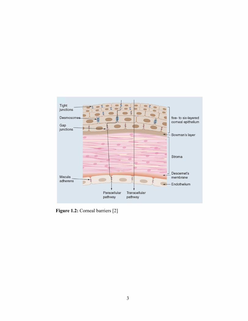
Figure 1.2: Corneal barriers [2]
4
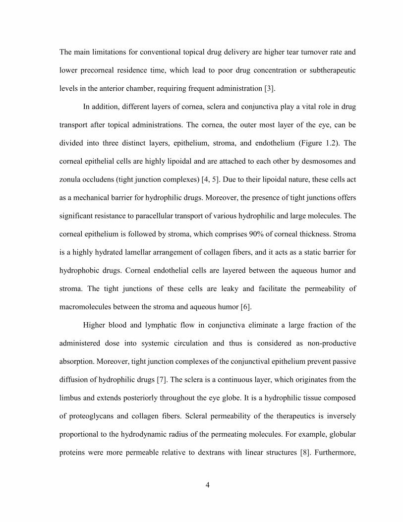
The main limitations for conventional topical drug delivery are higher tear turnover rate and
lower precorneal residence time, which lead to poor drug concentration or subtherapeutic
levels in the anterior chamber, requiring frequent administration [3].
In addition, different layers of cornea, sclera and conjunctiva play a vital role in drug
transport after topical administrations. The cornea, the outer most layer of the eye, can be
divided into three distinct layers, epithelium, stroma, and endothelium (Figure 1.2). The
corneal epithelial cells are highly lipoidal and are attached to each other by desmosomes and
zonula occludens (tight junction complexes) [4, 5]. Due to their lipoidal nature, these cells act
as a mechanical barrier for hydrophilic drugs. Moreover, the presence of tight junctions offers
significant resistance to paracellular transport of various hydrophilic and large molecules. The
corneal epithelium is followed by stroma, which comprises 90% of corneal thickness. Stroma
is a highly hydrated lamellar arrangement of collagen fibers, and it acts as a static barrier for
hydrophobic drugs. Corneal endothelial cells are layered between the aqueous humor and
stroma. The tight junctions of these cells are leaky and facilitate the permeability of
macromolecules between the stroma and aqueous humor [6].
Higher blood and lymphatic flow in conjunctiva eliminate a large fraction of the
administered dose into systemic circulation and thus is considered as non-productive
absorption. Moreover, tight junction complexes of the conjunctival epithelium prevent passive
diffusion of hydrophilic drugs [7]. The sclera is a continuous layer, which originates from the
limbus and extends posteriorly throughout the eye globe. It is a hydrophilic tissue composed
of proteoglycans and collagen fibers. Scleral permeability of the therapeutics is inversely
proportional to the hydrodynamic radius of the permeating molecules. For example, globular
proteins were more permeable relative to dextrans with linear structures [8]. Furthermore,
5
permeability across the sclera is also affected by charge. A recent report suggests that
permeability of anionic molecules is higher compared to cationic molecules, and it might be
due to the interaction between positively charged molecules with the negatively charged
proteoglycans [9].
Although intravitreal and periocular administrations are not patient compliant, these
routes are employed to deliver therapeutics for the treatment of posterior segment diseases.
The periocular route is relatively less invasive than the intravitreal route, which includes
subtenon, subconjunctival, peribulbar and retrobulbar administrations (Figure 1.1). After
periocular administrations, drug molecules can reach back of the eye by three different routes:
systemic circulation through the choroid, transscleral pathway, and the anterior route (tear film,
cornea, aqueous humor and vitreous humor) [10]. Subconjunctival administration can bypass
conjunctival epithelium, which is the rate limiting factor for the absorption of hydrophilic
drugs. However, many metabolic, static and dynamic barriers (conjunctival blood and
lymphatic circulations) increase the elimination of the drug and eventually, reduce drug
transport to the posterior section [11, 12]. Molecules, which escape the conjunctival
vasculature, eventually reach to the photoreceptor cells and the retina via sclera and choroid.
Permeability across the sclera is not a major constraint because it is highly dependent upon the
molecular radius and not the hydrophobicity of drug molecule [13, 14]. However, choroidal
blood circulation does act as a dynamic barrier for the drug permeability and eliminates a
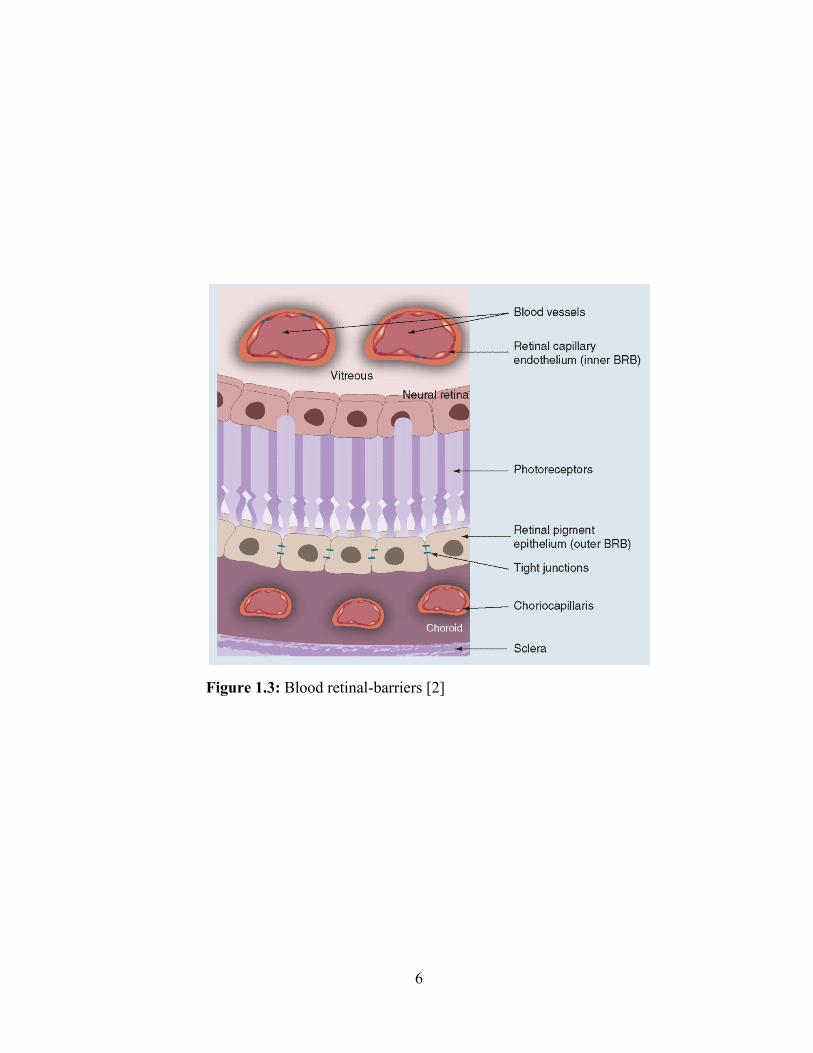
considerable amount of the dose. Nevertheless, the blood-retinal barrier (BRB) also plays a
critical role for permeability of active molecules to the photoreceptor cells (Figure 1.3). Direct
delivery of therapeutics to the vitreous humor can offer enormous advantage over the
subconjunctival administration.
6
Figure 1.3: Blood retinal-barriers [2]
7
However, intravitreal injection has the least patient compliance and it also leads to non-uniform
drug distribution in the vitreous humor. The vitreous allows rapid diffusion to small molecule,
while it restricts the diffusion of macromolecules. Furthermore, pathophysiological conditions
also have a significant effect on the distribution of therapeutics [15].
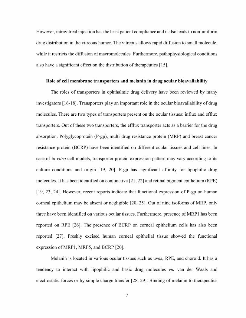
Role of cell membrane transporters and melanin in drug ocular bioavailability
The roles of transporters in ophthalmic drug delivery have been reviewed by many
investigators [16-18]. Transporters play an important role in the ocular bioavailability of drug
molecules. There are two types of transporters present on the ocular tissues: influx and efflux
transporters. Out of these two transporters, the efflux transporter acts as a barrier for the drug
absorption. Polyglycoprotein (P-gp), multi drug resistance protein (MRP) and breast cancer
resistance protein (BCRP) have been identified on different ocular tissues and cell lines. In
case of in vitro cell models, transporter protein expression pattern may vary according to its
culture conditions and origin [19, 20]. P-gp has significant affinity for lipophilic drug
molecules. It has been identified on conjunctiva [21, 22] and retinal pigment epithelium (RPE)
[19, 23, 24]. However, recent reports indicate that functional expression of P-gp on human
corneal epithelium may be absent or negligible [20, 25]. Out of nine isoforms of MRP, only
three have been identified on various ocular tissues. Furthermore, presence of MRP1 has been
reported on RPE [26]. The presence of BCRP on corneal epithelium cells has also been
reported [27]. Freshly excised human corneal epithelial tissue showed the functional
expression of MRP1, MRP5, and BCRP [20].
Melanin is located in various ocular tissues such as uvea, RPE, and choroid. It has a
tendency to interact with lipophilic and basic drug molecules via van der Waals and
electrostatic forces or by simple charge transfer [28, 29]. Binding of melanin to therapeutics
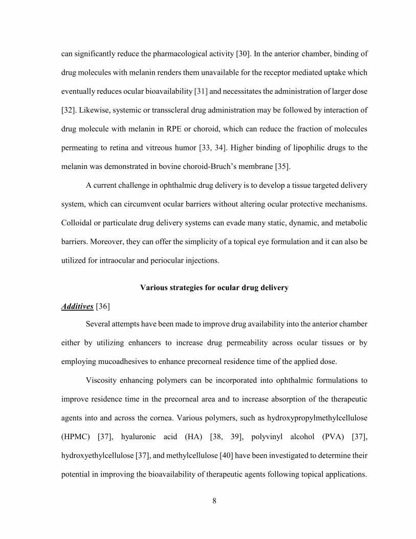
8
can significantly reduce the pharmacological activity [30]. In the anterior chamber, binding of
drug molecules with melanin renders them unavailable for the receptor mediated uptake which
eventually reduces ocular bioavailability [31] and necessitates the administration of larger dose
[32]. Likewise, systemic or transscleral drug administration may be followed by interaction of
drug molecule with melanin in RPE or choroid, which can reduce the fraction of molecules
permeating to retina and vitreous humor [33, 34]. Higher binding of lipophilic drugs to the
melanin was demonstrated in bovine choroid-Bruch’s membrane [35].
A current challenge in ophthalmic drug delivery is to develop a tissue targeted delivery
system, which can circumvent ocular barriers without altering ocular protective mechanisms.
Colloidal or particulate drug delivery systems can evade many static, dynamic, and metabolic
barriers. Moreover, they can offer the simplicity of a topical eye formulation and it can also be
utilized for intraocular and periocular injections.
Various strategies for ocular drug delivery
Additives [36]
Several attempts have been made to improve drug availability into the anterior chamber
either by utilizing enhancers to increase drug permeability across ocular tissues or by
employing mucoadhesives to enhance precorneal residence time of the applied dose.
Viscosity enhancing polymers can be incorporated into ophthalmic formulations to
improve residence time in the precorneal area and to increase absorption of the therapeutic
agents into and across the cornea. Various polymers, such as hydroxypropylmethylcellulose
(HPMC) [37], hyaluronic acid (HA) [38, 39], polyvinyl alcohol (PVA) [37],
hydroxyethylcellulose [37], and methylcellulose [40] have been investigated to determine their
potential in improving the bioavailability of therapeutic agents following topical applications.
9
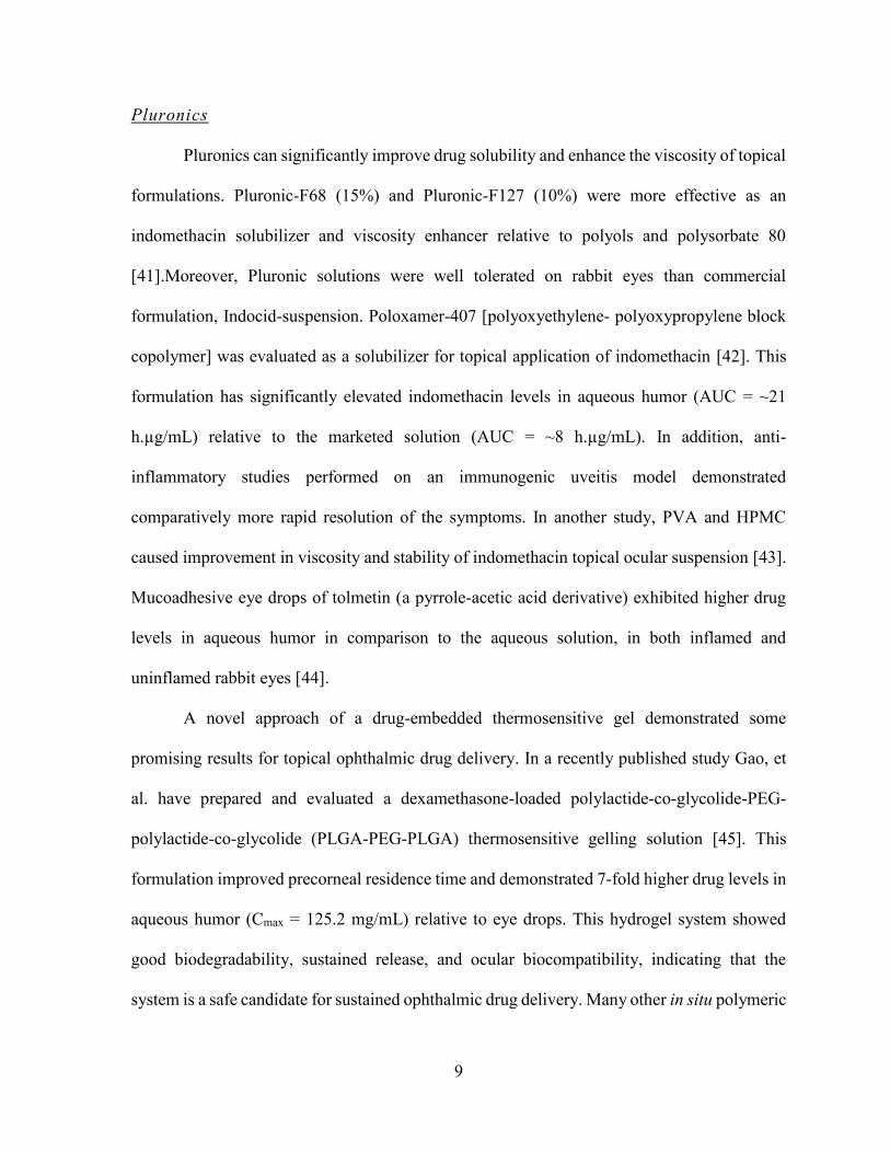
Pluronics
Pluronics can significantly improve drug solubility and enhance the viscosity of topical
formulations. Pluronic-F68 (15%) and Pluronic-F127 (10%) were more effective as an
indomethacin solubilizer and viscosity enhancer relative to polyols and polysorbate 80
[41].Moreover, Pluronic solutions were well tolerated on rabbit eyes than commercial
formulation, Indocid-suspension. Poloxamer-407 [polyoxyethylene- polyoxypropylene block
copolymer] was evaluated as a solubilizer for topical application of indomethacin [42]. This
formulation has significantly elevated indomethacin levels in aqueous humor (AUC = ~21
h.µg/mL) relative to the marketed solution (AUC = ~8 h.µg/mL). In addition, anti-
inflammatory studies performed on an immunogenic uveitis model demonstrated
comparatively more rapid resolution of the symptoms. In another study, PVA and HPMC
caused improvement in viscosity and stability of indomethacin topical ocular suspension [43].
Mucoadhesive eye drops of tolmetin (a pyrrole-acetic acid derivative) exhibited higher drug
levels in aqueous humor in comparison to the aqueous solution, in both inflamed and
uninflamed rabbit eyes [44].
A novel approach of a drug-embedded thermosensitive gel demonstrated some
promising results for topical ophthalmic drug delivery. In a recently published study Gao, et
al. have prepared and evaluated a dexamethasone-loaded polylactide-co-glycolide-PEG-
polylactide-co-glycolide (PLGA-PEG-PLGA) thermosensitive gelling solution [45]. This
formulation improved precorneal residence time and demonstrated 7-fold higher drug levels in
aqueous humor (Cmax = 125.2 mg/mL) relative to eye drops. This hydrogel system showed
good biodegradability, sustained release, and ocular biocompatibility, indicating that the
system is a safe candidate for sustained ophthalmic drug delivery. Many other in situ polymeric
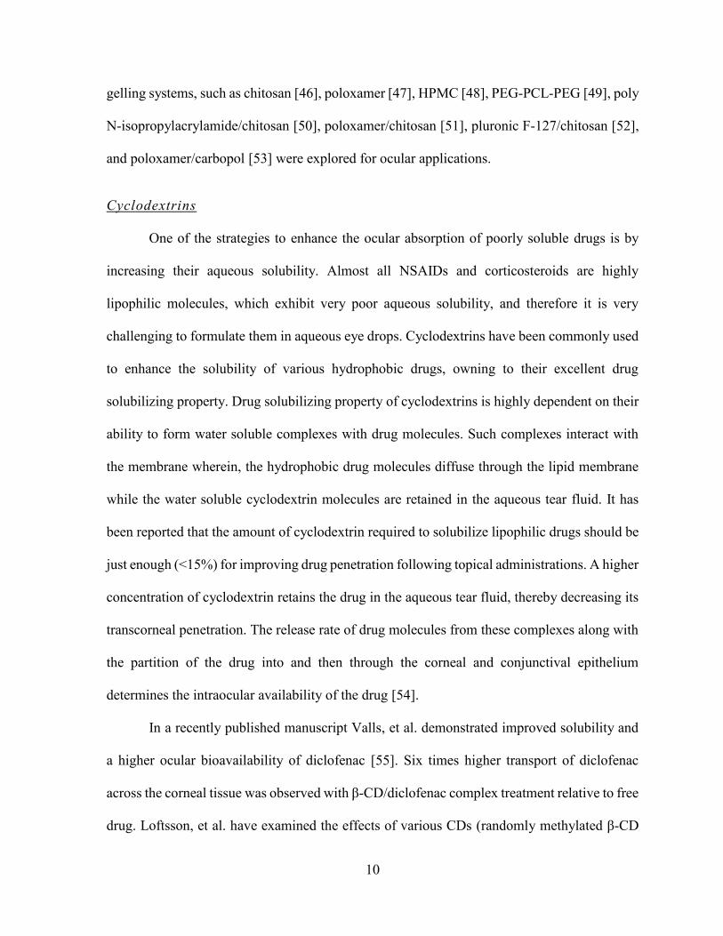
10
gelling systems, such as chitosan [46], poloxamer [47], HPMC [48], PEG-PCL-PEG [49], poly
N-isopropylacrylamide/chitosan [50], poloxamer/chitosan [51], pluronic F-127/chitosan [52],
and poloxamer/carbopol [53] were explored for ocular applications.
Cyclodextrins
One of the strategies to enhance the ocular absorption of poorly soluble drugs is by
increasing their aqueous solubility. Almost all NSAIDs and corticosteroids are highly
lipophilic molecules, which exhibit very poor aqueous solubility, and therefore it is very
challenging to formulate them in aqueous eye drops. Cyclodextrins have been commonly used
to enhance the solubility of various hydrophobic drugs, owning to their excellent drug
solubilizing property. Drug solubilizing property of cyclodextrins is highly dependent on their
ability to form water soluble complexes with drug molecules. Such complexes interact with
the membrane wherein, the hydrophobic drug molecules diffuse through the lipid membrane
while the water soluble cyclodextrin molecules are retained in the aqueous tear fluid. It has
been reported that the amount of cyclodextrin required to solubilize lipophilic drugs should be
just enough (<15%) for improving drug penetration following topical administrations. A higher
concentration of cyclodextrin retains the drug in the aqueous tear fluid, thereby decreasing its
transcorneal penetration. The release rate of drug molecules from these complexes along with
the partition of the drug into and then through the corneal and conjunctival epithelium
determines the intraocular availability of the drug [54].
In a recently published manuscript Valls, et al. demonstrated improved solubility and
a higher ocular bioavailability of diclofenac [55]. Six times higher transport of diclofenac
across the corneal tissue was observed with β-CD/diclofenac complex treatment relative to free
drug. Loftsson, et al. have examined the effects of various CDs (randomly methylated β-CD
11
(RMβCD) and 2-hydroxypropyl-β CD (HPβCD)) on ocular delivery of dexamethasone [56].
Results from in vivo ocular tissue distribution studies illustrated that both lipophilic RMβCD
and hydrophilic HPβCD have improved dexamethasone levels in rabbit eyes. However,
RMβCD delivered higher amounts of dexamethasone relative to other CDs.
The same investigators have recently published a patent, demonstrating the role of CDs
(RMβCD and γ-cyclodextrin (γ-CD)) in the delivery of corticosteroids to various ocular tissues
[57]. Nanoparticulate formulation of γ-CD-drug conjugates were able to deliver corticosteroids
more efficiently to the back of the eye, contrary to the RMβCD drug solution, which causes
localization of more drug into the anterior chamber of the rabbit eyes. Cyclooxygenase-2
(COX-2) inhibitors are also indicated in the treatment of ocular inflammations. However, poor
aqueous solubility of these agents limits their topical application. In an attempt to improve
ocular bioavailability, a nanoparticulate formulation of valdecoxib with HPβCD was evaluated
[58]. As anticipated, levels of valdecoxib in the cornea and conjunctiva were significantly
higher in NP-treated rabbit eyes relative to control.
In vivo ocular bioavailability of three different hydrocortisone (HC) formulations (1%
HC solution with HPβCD, 1% HC solution with HPβCD along with sodium hyaluronate or
carbopol 934P, and 1% HC suspension) was evaluated in New Zealand White rabbit [59].
Incorporation of HPβCD in formulation improved HC solubility and bioavailability in the
cornea and aqueous humor by 75% and 55%, respectively, relative to aqueous suspension.
Interestingly, inclusion of hyaluronate or carbopol in the HPβCD solution did not alter ocular
bioavailability.
In clinical trials, topical delivery of the dexamethasone/HPβCD solution exhibited a
significantly higher aqueous humor concentration (2.6-fold higher AUC) in human subjects
12
relative to suspension [60]. Currently, several CD containing formulations are marketed in
Europe, such as Indocid (indomethacin with HPβCD) and Voltaren (diclofenac-Na with
HPβCD), for the treatment of anterior chamber inflammations. It will not be surprising if CD
containing ophthalmic topical formulations enter the US market in the near future.
Colloidal dosage forms for drug delivery to anterior segment of the eye
In the last decade, many novel strategies including liposomes, polymeric NPs,
dendrimers, nanoemulsions, micelles, nanosuspensions, and combination approaches have
been investigated for the development of sustained ocular drug delivery systems. These
colloidal dosage forms offer numerous advantages over conventional dosage forms, such as
higher drug solubility, enhanced bioavailability, improved physical and chemical stability, and
sustained drug delivery. Furthermore, nanocarriers can lower toxicity and irritability concerns
related to drug and/or formulation, and eventually these systems can improve in vivo
performance and patient compliance. Such delivery systems can significantly bypass the
blood-ocular barriers, overcome the efflux related issues of parent drugs, and reduce frequency
of administration [61]. However, a clear understanding of the size, charge, and affinity of drug
molecules towards various ocular tissues and pigments are crucial for the development of
effective ocular formulations. For example, for transcorneal delivery, it is important to
understand the structure and properties of each layer of the cornea. Corneal epithelium
facilitates the transport of hydrophobic drugs, while the stroma acts as a barrier for
hydrophobic drugs and allows only hydrophilic drugs to enter into the anterior chamber.
Corneal mucosa is negatively charged, and therefore it increases the permeability of positively
charged drugs and prolongs retention of positively charged nanocarriers [62]. Other than the
transcorneal route, conjunctival and transscleral routes play a vital role for the treatment of
13
posterior segment diseases. Permeability of a drug molecule across the sclera depends upon
various physicochemical properties such as hydrodynamic radius, molecular weight, surface
charge, and hydrophilicity. Studies revealed that 200 nm particles could not cross the sclera,
while 20 nm particles crossed the scleral tissue at very low extent [63, 64]. Similarly, the
performance of nanocarriers is also affected by choroidal circulations (dynamic barrier: blood
and lymphatic flow), surface modifications with endogenous molecules, size, and abundance
of various enzymes particularly for biodegradable nanocarriers.
Liposomes
Research on liposomes has expanded considerably in the last thirty years. Liposomes
are spherical biphasic vesicular system where the internal aqueous phase is surrounded by
phospholipid bilayer membrane [65]. Hydrophilic drugs can be encapsulated in the inner
aqueous core while the hydrophobic drugs tend to stay in the lipid bilayer [66]. Depending on
the size, liposomes can be classified into small unilamellar vesicles (SUV) (10-100 nm), large
unilamellar vesicles (LUV) (100-300 nm) and multilamellar vesicles (contains more than one
bilayer). Liposomes can be formulated from sphingolipids, long chain fatty acids, cholesterols,
glycolipids, membrane proteins, and non-toxic surfactants [67]. Therapeutic molecules such
as proteins, nucleotides, small molecules, and even plasmids can be delivered with liposomes
[68]. These delivery systems can be employed to control and sustain the release of therapeutic
molecules, and more importantly, they can be used to protect the therapeutic agent from
metabolic degradation [69]. Due to numerous advantages, liposomes have been extensively
investigated in ophthalmic treatments.
Investigators have studied transcorneal permeation of neutral, anionic and cationic
liposomes. Cationic liposomes interact more efficiently with negatively charged corneal
14
epithelial membrane relative to the anionic or neutral liposomes, and eventually provide higher
drug (tropicamide, penicillin G and acetazolamide) transport across the cornea [70-72]. Along
with small molecule drug delivery, liposomes have also been investigated as non-viral vectors
for gene transfections [73] and as a carrier for the delivery of monoclonal antibodies [74].
Recently, a focal laser was employed to manage the release of drug and dyes from liposomes
at the target site [75]. In the future, laser-targeted delivery system may be employed for the
treatment of neovascular vessel occlusion, choroidal and retinal blood vessel stasis,
angiography, and selective tumors.
Liposomes prepared with naturally derived phospholipids such as egg phosphatidyl
ethanolamine or dioleoyl phosphatidyl ethanolamine (DOPE) are more suitable for the
ophthalmic drug delivery purposes [76]. Liposomes have been successfully utilized for drug
delivery to the anterior segment of the eye. The therapeutic efficiency of liposomes depends
on various factors, including size and charge, encapsulation efficiency, retention and stability
in conjunctival sac and ocular tissues and affinity towards corneal surface. The surface charge
of liposomes plays a key role in determining their affinity towards the corneal surface.
According to Felt, et al. negatively charged corneal surface has higher affinity towards
positively charged liposomes. They also showed that the drug elimination due to lachrymal
flow was reduced as the cationic liposomes increased the viscocity and interacted with the
negatively charged mucus [77]. Liposomes-loaded with pilocarpine hydrochloride were
prepared by Monem, et al. and researchers observed that neutral MLVs showed the most
prolonged effect when compared to negatively charged MLVs and free drug [78]. Acyclovir
(ACV) liposomes were formulated and evaluated for their in vitro permeation and in vivo
absorption across the cornea in rabbits. Experiments showed that positively charged liposomes
15
formed a coating on the corneal surface. The morphology depicted that liposomes bound
tightly to the corneal tissue and increased the residence time and thus improved the ACV
absorption [79]. Ocular pharmacokinetics of ganciclovir (GCV) encapsulated in liposomes was
studied in albino rats and compared with GCV solution. Transcorneal permeability of GCV
liposomes was 3.9 fold higher when compared to GCV solution. The AUC of GCV in aqueous
humor was found to be 1.7 fold higher in case of GCV liposomes. Ocular tissue distribution of
GCV from liposomes demonstrated 2-10 times higher concentration in sclera, cornea, iris, lens
and vitreous humor when compared to solution treated groups. These results suggest that
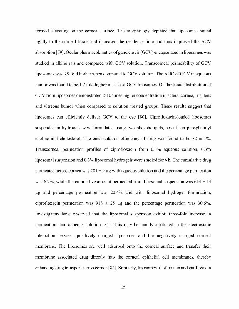
liposomes can efficiently deliver GCV to the eye [80]. Ciprofloxacin-loaded liposomes
suspended in hydrogels were formulated using two phospholipids, soya bean phosphatidyl
choline and cholesterol. The encapsulation efficiency of drug was found to be 82 ± 1%.
Transcorneal permeation profiles of ciprofloxacin from 0.3% aqueous solution, 0.3%
liposomal suspension and 0.3% liposomal hydrogels were studied for 6 h. The cumulative drug
permeated across cornea was 201 ± 9 µg with aqueous solution and the percentage permeation
was 6.7%; while the cumulative amount permeated from liposomal suspension was 614 ± 14
µg and percentage permeation was 20.4% and with liposomal hydrogel formulation,
ciprofloxacin permeation was 918 ± 25 µg and the percentage permeation was 30.6%.
Investigators have observed that the liposomal suspension exhibit three-fold increase in
permeation than aqueous solution [81]. This may be mainly attributed to the electrostatic
interaction between positively charged liposomes and the negatively charged corneal
membrane. The liposomes are well adsorbed onto the corneal surface and transfer their
membrane associated drug directly into the corneal epithelial cell membranes, thereby
enhancing drug transport across cornea [82]. Similarly, liposomes of ofloxacin and gatifloxacin

16
were prepared and studied for determining their efficiency in increasing the drug ocular
bioavailability [83]. In case of ofloxacin liposomal hydrogel the permeation was seven fold
higher than that of aqueous solution. Hence liposomal hydrogels can overcome all the
precorneal barriers and ensures steady and prolonged transcorneal permeation of the drug.
Like other delivery systems, liposomal treatment is also associated with few
drawbacks, such as possible toxicity and irritability [84-87]. Lipid components of the
liposomes are believed to be a primary source of toxicity, while charge of the liposomes is a
main reason for irritability. These constraints may restrict their chances for becoming popular
ophthalmic dosage form of the future. Moreover, commercial success of liposomes is also
limited because of the difficulties in sterilization and their relatively short shelf life.
Polymeric NPs
In order to address the problems of possible toxicity and irritability associated with
liposomes, drug-loaded nanometer sized polymeric particles may be considered a viable
alternative for the development of sustained release ophthalmic formulation. Nanoparticulate
systems comprise of particles with less than one micron particle size in which therapeutically
active agent is entrapped, absorbed, encapsulated, attached or adsorbed [67]. Aqueous or non-
aqueous suspension of drug-loaded polymeric NPs can either be delivered as topical drops in
the cul-de-sac or can be administered via transscleral, intravitreal or intraperitoneal routes.
Development of sustained release biodegradable dosage form for intravitreal delivery may
evade the limitation of frequent administration and it may also improve patient compliance.
Polymeric NPs can sustain drug release by diffusion, dissolution, or mechanical disintegration
and/or erosion of the polymer matrix [88]. NPs can circumvent the limitation of poor solubility
of therapeutics. Moreover, the NP can also protect the drug (e.g. peptides and proteins) from
17
enzymatic degradation, and eventually, improves an ocular bioavailability. Thus, NPs are a
better candidate for both posterior and anterior segment delivery.
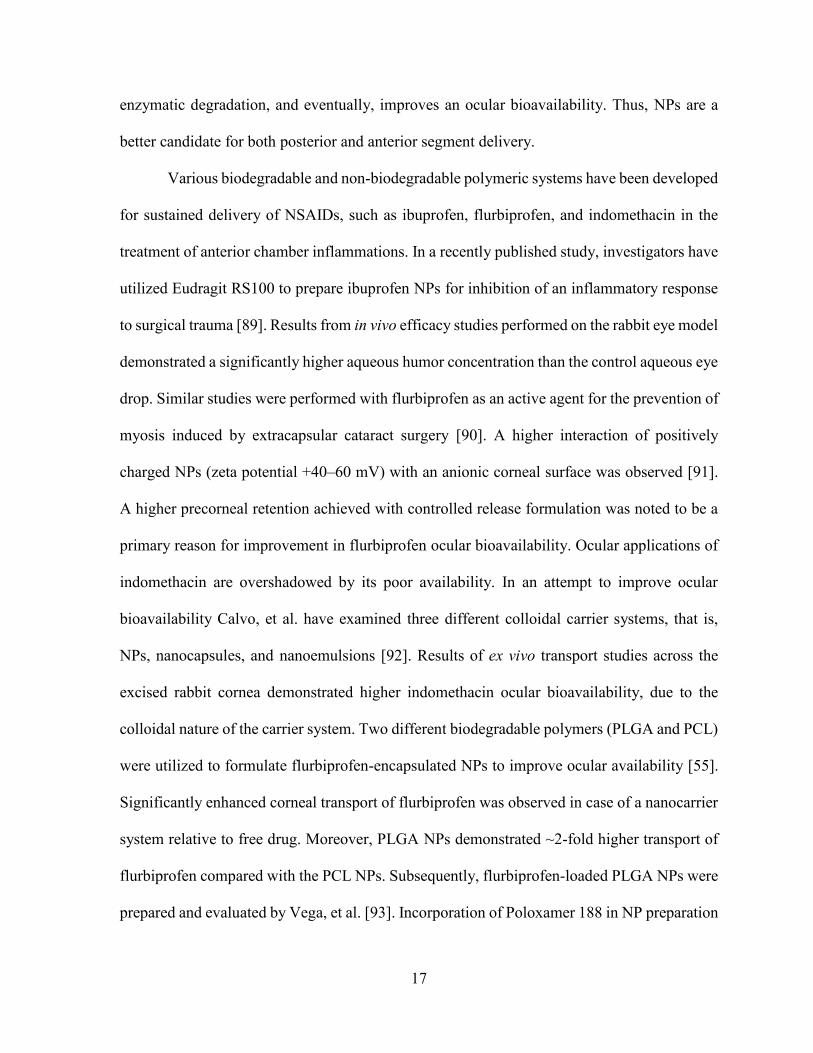
Various biodegradable and non-biodegradable polymeric systems have been developed
for sustained delivery of NSAIDs, such as ibuprofen, flurbiprofen, and indomethacin in the
treatment of anterior chamber inflammations. In a recently published study, investigators have
utilized Eudragit RS100 to prepare ibuprofen NPs for inhibition of an inflammatory response
to surgical trauma [89]. Results from in vivo efficacy studies performed on the rabbit eye model
demonstrated a significantly higher aqueous humor concentration than the control aqueous eye
drop. Similar studies were performed with flurbiprofen as an active agent for the prevention of
myosis induced by extracapsular cataract surgery [90]. A higher interaction of positively
charged NPs (zeta potential +40–60 mV) with an anionic corneal surface was observed [91].
A higher precorneal retention achieved with controlled release formulation was noted to be a
primary reason for improvement in flurbiprofen ocular bioavailability. Ocular applications of
indomethacin are overshadowed by its poor availability. In an attempt to improve ocular
bioavailability Calvo, et al. have examined three different colloidal carrier systems, that is,
NPs, nanocapsules, and nanoemulsions [92]. Results of ex vivo transport studies across the
excised rabbit cornea demonstrated higher indomethacin ocular bioavailability, due to the
colloidal nature of the carrier system. Two different biodegradable polymers (PLGA and PCL)
were utilized to formulate flurbiprofen-encapsulated NPs to improve ocular availability [55].
Significantly enhanced corneal transport of flurbiprofen was observed in case of a nanocarrier
system relative to free drug. Moreover, PLGA NPs demonstrated ~2-fold higher transport of
flurbiprofen compared with the PCL NPs. Subsequently, flurbiprofen-loaded PLGA NPs were
prepared and evaluated by Vega, et al. [93]. Incorporation of Poloxamer 188 in NP preparation
18
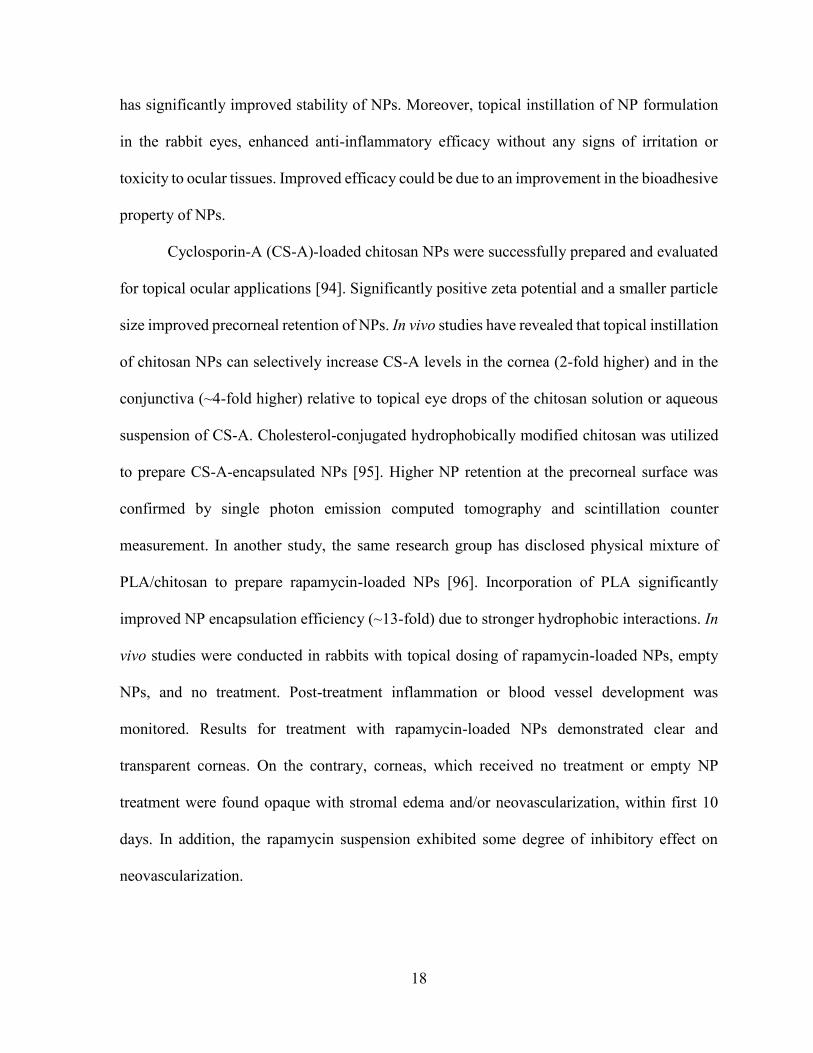
has significantly improved stability of NPs. Moreover, topical instillation of NP formulation
in the rabbit eyes, enhanced anti-inflammatory efficacy without any signs of irritation or
toxicity to ocular tissues. Improved efficacy could be due to an improvement in the bioadhesive
property of NPs.
Cyclosporin-A (CS-A)-loaded chitosan NPs were successfully prepared and evaluated
for topical ocular applications [94]. Significantly positive zeta potential and a smaller particle
size improved precorneal retention of NPs. In vivo studies have revealed that topical instillation
of chitosan NPs can selectively increase CS-A levels in the cornea (2-fold higher) and in the
conjunctiva (~4-fold higher) relative to topical eye drops of the chitosan solution or aqueous
suspension of CS-A. Cholesterol-conjugated hydrophobically modified chitosan was utilized
to prepare CS-A-encapsulated NPs [95]. Higher NP retention at the precorneal surface was
confirmed by single photon emission computed tomography and scintillation counter
measurement. In another study, the same research group has disclosed physical mixture of
PLA/chitosan to prepare rapamycin-loaded NPs [96]. Incorporation of PLA significantly
improved NP encapsulation efficiency (~13-fold) due to stronger hydrophobic interactions. In
vivo studies were conducted in rabbits with topical dosing of rapamycin-loaded NPs, empty
NPs, and no treatment. Post-treatment inflammation or blood vessel development was
monitored. Results for treatment with rapamycin-loaded NPs demonstrated clear and
transparent corneas. On the contrary, corneas, which received no treatment or empty NP
treatment were found opaque with stromal edema and/or neovascularization, within first 10
days. In addition, the rapamycin suspension exhibited some degree of inhibitory effect on
neovascularization.
19
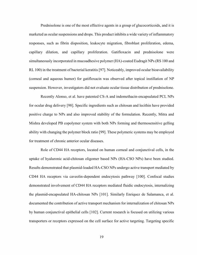
Prednisolone is one of the most effective agents in a group of glucocorticoids, and it is
marketed as ocular suspensions and drops. This product inhibits a wide variety of inflammatory
responses, such as fibrin disposition, leukocyte migration, fibroblast proliferation, edema,
capillary dilation, and capillary proliferation. Gatifloxacin and prednisolone were
simultaneously incorporated in mucoadhesive polymer (HA)-coated Eudragit NPs (RS 100 and
RL 100) in the treatment of bacterial keratitis [97]. Noticeably, improved ocular bioavailability
(corneal and aqueous humor) for gatifloxacin was observed after topical instillation of NP
suspension. However, investigators did not evaluate ocular tissue distribution of prednisolone.
Recently Alonso, et al. have patented CS-A and indomethacin-encapsulated PCL NPs
for ocular drug delivery [98]. Specific ingredients such as chitosan and lecithin have provided
positive charge to NPs and also improved stability of the formulation. Recently, Mitra and
Mishra developed PB copolymer system with both NPs forming and thermosensitive gelling
ability with changing the polymer block ratio [99]. These polymeric systems may be employed
for treatment of chronic anterior ocular diseases.
Role of CD44 HA receptors, located on human corneal and conjunctival cells, in the
uptake of hyaluronic acid-chitosan oligomer based NPs (HA-CSO NPs) have been studied.
Results demonstrated that plasmid-loaded HA-CSO NPs undergo active transport mediated by
CD44 HA receptors via caveolin-dependent endocytosis pathway [100]. Confocal studies
demonstrated involvement of CD44 HA receptors mediated fluidic endocytosis, internalizing
the plasmid-encapsulated HA-chitosan NPs [101]. Similarly Enriquez de Salamanca, et al.
documented the contribution of active transport mechanism for internalization of chitosan NPs
by human conjunctival epithelial cells [102]. Current research is focused on utilizing various
transporters or receptors expressed on the cell surface for active targeting. Targeting specific
20
transporters or receptors with functionalized NPs may facilitate enhanced uptake into ocular
tissues. Kompella, et al. studied the effect of surface functionalization on the uptake of NPs
employing ex vivo bovine eye model [103]. NPs surface functionalized with deslorelin, a
luteinizing hormone-releasing hormone agonist, or transferrin demonstrated 64% and 74%
higher transport respectively, relative to non-functionalized NPs.
Nanosuspension
It is very difficult to formulate a poorly soluble drug in conventional ophthalmic dosage
forms. Many approaches have been explored to enhance the solubility of such drugs to make
them suitable for the preparation of ophthalmic formulations. A general approach for
enhancing the solubility of drugs is micronization wherein the drug particle size is reduced to
approximately 0.1 mm to 25 µm. However, this size range is not sufficient to increase the
saturation solubility of the drug and hence its ocular bioavailability. Use of co-solvents can
also improve drug solubility but they are not devoid of toxic effects. The most commonly used
approach to trim down drug solubility issues is to formulate the drug into nanosuspension.
Nanosuspension is a colloidal dispersion of nanosized particles. Nanosuspensions can be
prepared by precipitation, pearl milling and high pressure homogenization techniques.
Moreover, the solid state of the drug in nanosuspensions minimizes the problem of chemical
stability of the drug as well as the physical stability of the formulation [104]. For ocular drug
delivery, nanosuspension provides numerous advantages such as dose reduction, ease of eye
drop formulation, increased bioadhesion and corneal penetration, reduced ocular irritation and
enhanced bioavailability.
Nanosuspension is stabilized by other excipients, such as surfactants, viscosity
enhancers, or charge modifiers. Topical delivery of 1% aqueous suspension and 1% oil
21
suspension in human subjects demonstrated higher levels of indomethacin in aqueous humor
relative to oral delivery [105]. Notably, oil suspension exhibited higher aqueous humor levels
(429 ng/mL) relative to aqueous suspension (198 ng/mL).
Glucocorticoids are widely prescribed in the treatment of ophthalmic inflammations.
However, poor aqueous solubility poses a challenge to ophthalmic formulation development.
In a recently published report Kassem, et al. have prepared and evaluated nanosuspension
formulation of prednisolone, hydrocortisone, and dexamethasone for topical ocular delivery
[106]. In vivo tissue distribution studies of the glucocorticoids nanosuspensions demonstrated
significantly higher levels in anterior chamber tissues relative to solution and microcrystalline
suspension of similar compounds. Moreover, investigators also reported a direct relationship
between nanosuspension viscosity and ocular bioavailability. Recently, a randomized double-
blind clinical study of Sophisen derivatives, 3A Ofteno (1.0% diclofenac sodium w/v), and
Modusik-A Ofteno (0.1% CS-A w/v) were performed in 120 healthy volunteers [107]. Topical
instillation of 3A Ofteno-diclofenac nanosuspension remained on the ocular surface for longer
period with less annoying sensation and irritation. Also, Modusik-A Ofteno-CS-A
nanosuspension caused significant improvement in the tear production from baseline 5 to 11
mm.
In a recent patent disclosure (WO 2006/062875) entitled, ‘‘Ophthalmic nanoparticulate
formulation of a COX-2 selective inhibitor,’’ investigators have incorporated rofecoxib (COX-
2 inhibitor) in the polystyrene nanoparticulate system for ophthalmic applications [108].
Formulation prepared with Poloxamer-407 (0.05% w/w) and HPMC remained physically
stable, without any change in particle size up to 4 weeks. Drug levels in anterior chamber
ocular tissues, such as the cornea (~6,610 ng·gram/tissue) and aqueous humor (~251
22
ng·gram/tissue) were significantly higher after topical instillation of nanosuspension. Readers
are advised to read the patent for more detailed information of in vivo tissue distribution
studies.
The efficiency of nanosuspensions in increasing the drug ocular bioavailability depends
upon the intrinsic solubility of drugs in the lachrymal fluid, this intern is governed by the
intrinsic dissolution rate of drugs in the lachrymal fluid which can vary due to constant inflow
and outflow of lachrymal fluid. Nanosuspensions may therefore fail to give a consistent
performance. However, nanosuspension formulation of drugs represents an ideal approach for
ocular delivery of poorly soluble drug due to their inherent ability to increase a drug’s
saturation solubility [109].
Nanoemulsion
The only difference between conventional emulsion and nanoemulsion is the globule
size of an internal phase. Nanoemulsion offers several advantages in ocular drug delivery, such
as high capacity to dissolve both hydrophilic and lipophilic drugs, stability, improved
bioavailability, and good spreadability [16]. In addition, surfactants used in formulating
emulsions can also act as penetration enhancers, thereby improving drug permeability across
the cornea. Emulsion based delivery of hydrophobic drugs has always remained a primary
choice for the formulation researchers.
Chitosan, a cationic polymer, finds application in the field of ocular drug delivery due
to its potential ability to enhance corneal drug permeability by opening tight junctions. It
strongly interacts with negatively charged mucin and improves residence time on the
precorneal surface. An indomethacin-embedded chitosan nanoemulsion was evaluated for its
residence time and ability to deliver therapeutics into the anterior chamber of the eye [110].

23
Topical application of their nanoemulsion has significantly improved indomethacin levels in
the cornea and aqueous humor of rabbit eyes relative to the indomethacin solution. Drug levels
in the cornea and aqueous humor were about 12-fold higher for the nanoemulsion relative to
solution-treated eyes.
A CS-A-loaded microemulsion, in situ electrolyte-triggered gelling system was
developed by Gan, et al. [111] for the treatment of corneal allograft rejection. The
microemulsion was dispersed in the Kelcogel (deacetylated gellan gum) solution, which
provided the in situ gelling property when applied to the corneal surface. In vivo studies
suggested that the microemulsion-Kelcogel system can generate ~3-fold higher levels of CS-
A relative to CS-A microemulsion even at 32 h post-dosing. Moreover, concentration of CS-
A in Kelco gel system treated corneas were maintained at therapeutic levels with no ocular
irritation, even after 32 h. In another study, n-octenyl succinate starch was utilized to prepare
the diclofenac solution and emulsion, and these formulations exhibit improved permeability
across the excised porcine cornea compared to the commercial product Voltaren Ophtha [112].
An indomethacin nanoemulsion prepared with amphoteric surfactant (lauroamphodiacetate)
improved corneal permeability by 3.8 times relative to a marketed product (Indocollyre) [113].
In a recently published patent, CS-A was successfully incorporated in a nanoemulsion,
utilizing a positively charged polar lipid, such as stearylamine [114]. Mean droplet size of the
formulation was within the range of 150-250 nm, with zeta potentials of 34-45 mV. Gan, et al.
have recently patented a nanoemulsion-based in situ gelling system for topical ocular delivery
of flurbiprofen [115]. A CS-A-loaded nanoemulsion (NOVA22007) containing cationic lipid
formulation has just completed Phase III studies for dry eye [116]. The same emulsion was
24
applied for vernal keratoconjunctivitis treatment and this study has recently completed phase
II/III studies [117].
Nanomicelles
Polymeric micelles are self-assembling colloidal systems comprised of block or graft
amphiphilic copolymers and surface-active agents [118]. Development of ophthalmic drug
delivery systems with micelles is very promising, particularly because of their advantages such
as high thermodynamic and kinetic stability, ability to sustain the release, and the improvement
of drug solubility and permeability across the ocular tissues [119]. Moreover, micelles can be
functionalized with endogenous molecules for targeted ocular delivery. Normally, particle size
of the micelles ranges from 5 to 50 nm [118].
Recently, a rapamycin and corticosteroid-loaded aqueous nanomicellar formulation
was developed and patented (WO2010/144194) by Mitra, et al. [120]. Nanomicelles were
prepared with 1 to 7% w/v of Vitamin E tocopherol PEG succinate (vitamin E TPGS, HLB-
10) and 1 to 3% w/v of octoxynol-40 (HLB-13). Vitamin-E TPGS helped to increase the
solubility of the poorly soluble drugs, while octoxynol-40 reduced ocular discomfort and also
provided extra stability with higher optical clarity to the nanomicellar formulation.
Investigators suggested that any buffer system with adjusted osmolality and physiological pH
could be used to prepare an external aqueous phase. Rapamycin has very low aqueous
solubility (2.6 µg/mL). However, after preparation of nanomicelles, its solubility improved to
2 mg/mL (~1000 fold). An average diameter of the nanomicelles was around 25 nm.
Nanomicellar formulation of 14C rapamycin was instilled in rabbit eyes, and 60 min post-
dosing ocular distribution of the drug was determined by liquid scintillation counter.
Noticeably, a higher concentration of 14C rapamycin was observed in the choroid/retina (~360
25
ng/g), while very negligible radioactivity was found in the lens, aqueous humor and vitreous
humor. Higher accumulations of rapamycin in the posterior segment of the eye was believed
to be a result of the smaller mean diameter of nanomicelles. Optically clear and
thermodynamically stable nanomicellar formulation could be a promising innovation for the
non-invasive treatment of the posterior segment diseases.
In another disclosure (US 2009/0092665), Mitra and co-inventors developed mixed
micellar formulation of calcineurin inhibitors (voclosporin) and mTOR inhibitors for
ophthalmic applications [121]. Various concentrations of Vitamin-E TPGS and octoxynol-40
were used to prepare nanomicelles. Dilution studies were performed to evaluate the stability
of voclosporin-loaded mixed micelles (0.2 wt%), and micellar stability was confirmed up to a
20 fold dilution in saline. In addition, results have demonstrated that micelles dissociated at
around 44 °C and the formulation was re-stabilized well within 8 min. Moreover, formulation
remained stable in LDPE, polypropylene and polyvinylchloride containers at room
temperature for 48 h. The particle size of the voclosporin-loaded (0.2 wt%) nanomicelles was
between 13-33 nm. In vivo ocular tissue distribution studies were performed in two groups
(single dose and 7 days repeat dose) of rabbits after topical application of mixed micellar
formulation-loaded with 14C-radio labeled voclosporin. Almost 1.5-2.5 fold higher Cmax were
observed in all ocular tissues (cornea, aqueous humor, sclera, upper eyelid, lower eyelid,
retina/choroid, lacrimal gland, optic nerve and lower bulbar conjunctiva) of a group with 7
days repeated dosing over to the single dose group. No signs or serious symptoms of irritation
were observed in any rabbits after a tolerability and tissue irritability study. According to the
results discussed in this disclosure, nanomicellar formulation seems to be very promising in
the treatment of anterior and/or posterior segment ocular diseases including AMD and DME.
26
Results discussed in these patent applications explained that the nanomicellar drug
delivery systems can effectively change the course of ocular treatment.
Cubosomes
Novel dexamethasone-embedded self-assembled liquid crystalline particles
(cubosomes) were developed and investigated for precorneal retention and ocular tissue
distribution [122]. The apparent permeability coefficient of dexamethasone, delivered in
cubosomes was 3.5–4.5 times higher than dexamethasone eye drops. In addition, precorneal
retention of cubosomes was significantly longer than carbopol gel or solution. In vivo
microdialysis studies were performed to evaluate pharmacokinetics of dexamethasone in
aqueous humor. Delivery of cubosomes exhibited 1.8-fold and 8-fold higher AUC 0/240min
of dexamethasone in aqueous humor relative to eye drops and suspension, respectively.
Moreover, tissue integrity and corneal structure indicated good biocompatibility with the
cubosome formulation.
Ocular implants and inserts
Intraocular implants have been studied in order to control and sustain the drug delivery
in the treatment of proliferative vitreoretinopathy (PVR), cytomegalovirus (CMV) retinitis,
glaucoma, endophtalmitis, and posterior capsule opacification. Understanding of
physicochemical properties of the active compound and release kinetics helps to design
specific ocular implantable drug delivery systems. Ocular implants can be classified in two
categories according to their in vivo degradability i.e., non-biodegradable implants and
biodegradable implants.
27
Non-biodegradable ocular implants
Since the early in 70s, non-biodegradable implants have offered the advantages of
controlled and sustained release of active drugs with minimal host responses. Nowadays, new
non-biodegradable polymers (e.g., polyvinyl alcohol (PVA), ethylene vinyl acetate (EVA), and
polysulfone capillary fiber (PCF)) have been designed to ensure sustained release for longer
period of time with higher biocompatibility [123]. Intraocular drug release kinetics is
controlled by the rate of erosion and spontaneous degradation of these polymers. Diffusion of
fluid (water) inside the polymer structure dissolves the drug pellet and generates a saturated
drug solution inside the implant. Saturated drug solution diffuses out from the implant and
provides nearly zero-order drug release [124]. Lack of initial burst release makes non-
biodegradable implants superior to implants constructed from biodegradable polymers.
Complications such as retinal detachment, endophthalmitis, vitreous hemorrhage, formation of
tenacious epiretinal membranes, and cystoids macular edema have been observed with the use
of non-biodegradable polymeric devices [125]. Moreover, implantation of this device requires
surgical treatment and necessitates surgery to remove the empty device.
Sanborn and collogues have employed non-biodegradable implants for the ocular
delivery of ganciclovir in the treatment of CMV retinitis [126]. These implants were reservoir
type devices, composed of drug and coating polymers such as PVA and EVA. PVA is a
permeable polymer, which acts as a framework and controls release of ganciclovir. In contrast,
EVA acts as an impermeable polymer, which limits the practical surface area of the device and
hence, sustains drug release. Implication of both polymers in structure design enables zero-
order controlled drug release.
28
Vitrasert, a commercially available implant of ganciclovir, is relatively large and
requires 4-5 mm of sclerotomy at pars plana for implantation [127]. Moreover, the device is
made from non-biodegradable polymers and, needs to be removed after 5-8 months for the
implantation of another device, or because of unwanted complications such as retinal
detachment related to the implant, or severe inflammation. Almost 12% of eye complications
(endophthalmitis, retinal detachment, cystoids macular edema, formation of tenacious
epiretinal membranes and vitreous hemorrhage) have been observed after implantation of these
devices.
To overcome these problems, a betamethasone-loaded intrascleral implant (4 mm in
diameter, 1 mm in thickness and 4 mg in weight) composed of PVA and EVA has been
developed and evaluated [128]. Results demonstrated that the disc was able to maintain
therapeutic levels of betamethasone up to 4 weeks without an adverse burst release of drug.
No substantial adverse reactions were observed after histological and electroretinographical
evaluations. Intrascleral implantation does not require perforation of the eye wall, hence this
system may reduce complications associated with other implanted devices.
The FDA has approved fluocinolone-loaded Retisert in April 2005 in the treatment of
chronic non-infectious uveitis [127]. A total of 36 eyes with non-infectious posterior uveitis,
which had an average of 2.5 episodes of recurrence annually, were treated with these implants.
After implantation of fluocinolone-loaded implants, no recurrence was observed up to two
years [129]. Pearson, et al. have prepared Cs-A-loaded PVA-EVA device for the controlled
intravitreal delivery of Cs-A [130]. A 500 ng/mL of Cs-A concentrations was observed for
more than 6 months in rabbit and cynomolgus monkey eyes. However, complications such as
opacity of lens were observed in the implanted rabbit eyes. A PVA-EVA device loaded with
29
both Cs-A and dexamethasone was prepared, and examined in the treatment of PVR and uveitis
[131].
Biodegradable ocular implants
Biodegradable polymers generate non-toxic degradation products after in vivo
enzymatic degradation that can easily eliminate from the body without any side effects. Hence,
biodegradable implants are not required to be removed after implantation. It is more difficult
to achieve optimal drug release through biodegradable implants when compared to the
reservoir type of non-biodegradable implants. The main two categories of biodegradable
implants are monolithic and binding types. Monolithic types of implants are made by
solidifying the homogenous physical mixture of the drug and polymer. The procedure to
produce this kind of implant involves heating of drug, and hence thermolabile drugs such as
proteins, polypeptides or nucleic acids are difficult to incorporate in monolithic implants.
However, binding type of implants have showed suitability for the delivery of biologically
active molecules. Biologically active molecules make chemical bond or a polyion complex
with polymers, which finally undergoes enzymatic degradation and gets released from the
polymer matrix. Biodegradable polymers can be made into sheets, plugs, rods, pellets and
discs, and can be implanted into intrascleral or peribulbar, anterior chamber, vitreous cavity,
or through the pars plana [132]. An undesirable final burst release is the major drawback of
the controlled release system composed of biodegradable polymers.
Scleral implants of ganciclovir were successfully made using various blends of PLA
(DL isomer) and evaluated in the treatment of CMV retinitis in pigmented rabbit eyes. Implants
made up from physical mixture of 80% of PLA (70 kDa) and 20% of PLA (5 kDa) were able
to maintain therapeutic levels of ganciclovir up to 6 months without any significant burst

30
release [133]. Fluorouracil-loaded biodegradable intravitreal implants were prepared from
PLGA and evaluated in the treatment of tractional retinal detachment due to PVR [134]. The
sustained therapeutic concentration of drug was observed to be above 0.3 μg/mL up to 21 days
however, 89% of the rabbit eyes were suffered retinal detachment after treatment. Zhou, et al.
have investigated multiple drug delivery through implant in the treatment of PVR [135]. Three
cylindrical parts (triamcinolone, human recombinant tissue plasminogen activator (tPA) and
5-fluorouridine) containing PLGA implant (0.8 mm in diameter, 7 mm long) have been
prepared and evaluated. This implant exhibited release of 5-fluorouridine and triamcinolone at
the maximal rate of 1 μg/day over 4 weeks and 10 - 190 μg/day over 2 weeks, respectively.
Moreover, the portion of PLGA coated tPA started release after 2 days at the rate of 0.2 - 0.5
μg/day for 2 weeks, which may minimize the risk of post-operative bleeding. These types of
implants unwrap the feasibility of co-delivery of three different drugs or drugs with different
release profiles.
Xie, et al. have examined heparin incorporated PLGA implants (HPI) in the treatment
of posterior capsular opacification (PCO) [136]. Heparin levels in blood and aqueous humor
were investigated after subconjunctival implantation of HPI, posterior chamber implantation
of HPI and instillation of 5% topical heparin drops. Implantation of heparin delivery systems
in posterior chamber demonstrated significantly higher heparin levels in aqueous humor for
longer period of time (12 weeks). A proline analog (cis-4-hydroxyproline (CHP)), inhibits
collagen secretion in proliferative vitreoretinopathy (PVR). Yasukawa and collogues have
incorporated CHP in different blends of PLGA (65/35 and 50/50) for the sustained delivery to
rabbit eyes [137]. The PLGA 65/35 implants have decreased the incidence of PVR from 89%
31
to 56% but the PLGA 50/50 implant did not show any significant effect. On the other side, the
application of both implants together revealed synergistic inhibition of PVR.
Natural polymers such as gelatin, collagen and alginates have also been studied for the
preparation of ocular implants. Koelwel, et al. have prepared and investigated epidermal
growth factor (EGF) incorporated alginate inserts in 18 test volunteers in the treatment of
keratoconjunctivitissicca (KCS) [138]. Alginate inserts with higher amount of guluronic acid
(G block) were able to sustain the release of EGF for much longer period of time. Dried gelatin
hydrogel prepared by cross-linking of acidic gelatin was socked in basic fibroblast growth
factor (bFGF) solution with different concentrations and implanted into rabbit corneal pocket
[139]. The inserts with 50 ng or more doses exhibited dose dependent corneal angiogenesis
started from day 3 to day 4, while inserts with 20 ng or below were devoid of this side effect.
Moreover, effective sustained delivery of antimetabolic [140, 141], antifungal [142],
immunosuppressive agents [143] and steroids [144, 145] have been studied using
biodegradable ocular implants. Hence, the employment of implants for the ocular delivery of
small molecules (hydrophilic and hydrophobic) and macromolecules is a viable approach.
Microneedles
The concept of microneedles was proposed in early 1970s but practically this method
not demonstrated until 1990s when the microelectronic industries provided tools to fabricate
such small devices. Application of an array of microneedles creates large transport pathways,
larger than the molecular dimensions, and thus facilitates the transport of macromolecules
(peptides such as insulin, desmopressin and human growth factors), supramolecular
complexes, NPs, microparticles and liposomes [146]. Although micro-scale holes in the skin
are safer than intraocular injections, safety studies are needed to be performed. Microneedle
32
based drug delivery is widely studied in transdermal applications, but recently, several studies
have been performed to evaluate its application in ocular drug delivery. Mainly, two
approaches have been studied for the ocular application of microneedles, solid coated
microneedles [147] and hollow microneedles [148]. In the first approach, Jiang, et al. have
coated microneedle with five different model drugs (sulforhodamine, sodium fluorescein,
bovine serum albumin (BSA), plasmid DNA, and pilocarpine) for intrascleral and intracorneal
application to evaluate different model drugs with different physicochemical properties [147].
Microneedles coated with sulforhodamine and BSA demonstrated excellent penetration into
human cadaveric sclera and rapid dissolution of coated model drugs. Moreover, intracorneal
delivery of sodium fluorescein exhibited higher in vivo levels when compared to the levels
achieved after topical application of same dose. The same study using pilocarpine in rabbit
eyes has also revealed some promising results.
Moreover, researchers have evaluated the applicability of hollow microneedles for the
delivery of solutions containing soluble molecules, NPs, and microparticles into sclera in a
minimally invasive manner [148]. The results described successful intrascleral delivery of
solutions and fluorescence tagged NPs with insignificant effects of scleral thickness and
infusion pressure. However, investigators have reported that the presence of scleral
glycosaminoglycans and collagen fibers are the rate limiting factors for the microneedle based
intrascleral microparticles delivery.
These studies exhibited the versatility of microneedles’ applications in terms of drug
selection (coated microneedles) as well as formulation delivery tool (hollow microneedles).
Moreover, these targeted delivery systems are less invasive, less painful and more patient
compliant.
33
Biodegradable polymers [149]
Biodegradable polymers have numerous applications in the field of ocular drug
delivery and can be classified as natural or synthetic. Applications of PGA as suture material
in the late 1960 provided an impetus for the design and development of novel synthetic
biodegradable polymers. Synthetic biodegradable polymers can be tailored in various
compositions and molecular weights. The molecular weight or composition regulates the
degradation of polymer where the main weight loss takes place due to chain cleavage. Many
researchers have investigated several types of biodegradable polymers and studied their
applicability for the development sustained ocular formulations. Structures of various
biodegradable polymers are depicted in Figure 1.4.
Polyalkylcyanoacrylates (PACA)
Polyalkylcyanoacrylates (PACA) belong to a class of acrylate polymers synthesized
from alkylcyanoacrylic monomers through anionic polymerization. The faster degradation rate
of PACA was attributed to the unique instability of a carbon-carbon sigma bond and presence
of electron withdrawing neighboring groups. It has shown remarkable applicability as surgical
glue and skin adhesive. PACA was also explored for the development of NPs. The PACA
degradation rate varies from hours to days depends on the length of alkyl side chain. For
example, PACA having shorter alkyl chain length such as polymethylcyanoacrylate degrades
in few hours whereas higher alkyl chain length derivatives such as octyl and isobutyl
cyanoacrylates degrade slowly. NPs composed of PACA are advantageous in terms of
fabrication and application in drug delivery [150].
34
Polyanhydrides
Numerous investigations have elucidated the role of polyanhydrides (POA) for ocular
drug delivery applications. These polymers exhibit faster degradation and limited mechanical
strength, which make them an ideal candidate for fabrication of sustained release devices. Low
molecular weight polyanhydrides were synthesized through dehydrative coupling and
dehydrochlorination whereas melt polycondensation polymerization was employed for
synthesis of high molecular weight polymers. The degradation rate of polyanhydrides can be
easily modulated by changing the polymer composition and depends on the crystallinity and
hydrophilicity of the final polymer. These polymers undergo surface erosion and generate
monomeric acids that are non-toxic. Homo-polyanhydrides have limited application in
controlled drug delivery due to their crystalline nature. In contrast, copolymers such as poly
(carboxyphenoxy)propane–sebacic acid (PCPP-SA) demonstrated controlled degradation rates
[151]. This polymer was approved by the FDA for human applications for the delivery of
carmustine in the treatment of brain cancer [152]. Other approaches based on aromatic co-
monomers composed of hydrophobic aliphatic linear fatty acids were also investigated for drug
delivery applications [153].
Polyesters
Polyesters are biodegradable polymers having short aliphatic, ester-linked backbones.
These classes of polymers are generally produced by either ring-opening or condensation
polymerization. Ring-opening polymerization is preferred over condensation reaction to
produce high molecular weight polyesters. Homo- or co-polymers of cyclic lactones and
anhydrides having narrow molecular weight distribution can be produced via ring-opening
polymerization. The molecular weight of the final polymer can be controlled by varying the
35
ratio of monomers. The molecular weight of the polyesters regulates the hydrolytic cleavage
that follows bulk erosion kinetics to produce metabolic products which are eliminated through
normal metabolic pathways [154]. The hydrolytic degradation rate of the polymers can be
altered by varying the molecular weight, crystallinity and structure of the polymeric chain.
Among polyesters, poly-α-hydroxyesters are the most broadly investigated class of polymers
for ocular drug delivery applications; this includes PGA, PLA and their copolymers.
Polycaprolactone (PCL)
PCL is a semicyrstalline polymer synthesized by ring-opening polymerization of ε-
caprolactone. It has a glass transition temperature of -60 °C and a melting temperature in the
range of 59 to 64 °C. PCL was investigated for long term delivery due to higher permeability
to many drugs, excellent biocompatibility and extremely slow hydrolytic cleavage of polyester
backbone. The PCL based Capronor® implant was developed for controlled delivery of
levonorgestrel. It has a low tensile strength of 23 MPa and an extremely high elongation
factors, more than 700%. Numerous investigations were attempted to improve the slower
degradation of PCL. Copolymers of ε-caprolactone with lactide or glycolide exhibits
remarkably better degradation profile.
Polyglycolide (PGA)
PGA is a relatively hydrophilic polymer compared to other polyesters with high
crystallinity and low solubility in organic solvents. The low solubility in organic solvent is
attributed to its higher tensile modulus. It has a high melting point of 225 °C and glass transition
temperature of 36 °C. It has a comparatively faster degradation rate than other polyesters and
generates glycine upon degradation, which eventually eliminates through the citric acid cycle.
Major losses in the mechanical strength of PGA usually take place in one to two months and
36
it completely degrades in vivo within six to twelve months. PGA was initially explored for
developing sutures because of their fiber-forming properties and excellent mechanical strength.
However, it has limited role in ocular drug delivery due to its faster degradation rate and higher
crystallinity. PGA implants can be easily fabricated by widely applicable processing
techniques such as solvent casting, compression and extrusion techniques. Processing
technique utilized for the production of implant regulates the degradation properties of implant
[155].
Polylactic acid (PLA)
PLA is comparatively more hydrophobic than PGA due to the presence of an additional
methyl group. It is chiral in nature because of the structure of lactic acid and commonly exists
in three isomeric forms the D (-), L (+) and racemic (D, L) lactide. The crystalline nature of
PLA depends upon the isomeric forms and molecular weight of the polymer. PLA (L) is
crystalline in nature and hydrolyzed through normal metabolic pathway due to presence of
naturally occurring isomer (L-lactide). It has a melting point of 175 °C and glass transition
temperature of 60 to 65 °C. It also possesses a good tensile strength of 50-70 MPa and high
modulus of 4.8 GPa. On the other hand, PLA (DL) is amorphous in nature due to presence of
the racemic mixture. It has glass transition temperature of 55 to 60 °C and comparatively faster
degradation rate than PLLA. All isomeric forms of PLA follow bulk erosion kinetics and
generate lactic acid upon hydrolytic cleavage [116]. Researchers have often utilized PLA for
ocular drug delivery applications.
Poly (lactide-co-glycolide) (PLGA)
Poly (lactide-co-glycolide), commonly known as PLGA is obtained by the
copolymerization of lactide and glycolide. This copolymer is hydrolytically less stable than
37
the homopolymers, PLA or PGA. Extensive research on a full range of these copolymers
suggests their implication in drug delivery. These copolymers are divided into two main
compositions, comprised of lactide and glycolide. PLGA follows bulk erosion kinetics, and its
degradation depends on molecular weight and lactide to glycolide ratio. As the ratio of
glycolide in copolymer decreases, the hydrolytic degradation rate decreases. The intermediate
PLGA, i.e. 50:50, hydrolyses faster than PLGA 75:25 and PLGA 85:15. PLGA copolymers
are FDA approved for human applications because of the excellent biocompatibility and
controlled degradation profiles [117]. PLGA is negatively charged and thus has a non-
mucoadhesive nature.
Polyorthoesters
Polyorthoesters (POE) are hydrophobic polymers composed of a hydrolytically
unstable polyester linkage. However, they exhibit slower degradation due to surface erosion.
This degradation characteristic is ideal for designing sustained release devices. In this polymer
the degradation profile can be easily adjusted by employing different diols for polymerization
[156]. The POE group is hydrolytically unstable in acidic conditions and requires basic
additives to inhibit autocatalysis. The first generation of POE was developed by the ALZA
Corporation. It was synthesized by the transesterification reaction of diol with diethoxy
tetrahydrofuran [157]. Degradation profile of POE II can be easily altered by incorporation of
acidic additives such as adipic acid. POE III upon hydrolysis generates diol and pentaerythriol
dipropionate, which subsequently generate propionic acid and pentaerythriol. This class of
polymers is biocompatible and follows pH dependent degradation behavior. They do not
require organic solvents for the incorporation of drugs due to their semisolid nature. However,
there are difficulties in scale up processes that limit their application in drug delivery.
38
Modification of second generation polyester (POE II) with smaller lactic or glycolic acid
chains led to the development of POE IV. This polymer upon hydrolysis liberates acids which
further promotes the polymer degradation. The physical form and degradation rate of the
polymer can be easily varied by changing diols and acid segment respectively. POE IV has
demonstrated good biocompatibility for controlled delivery applications [158].
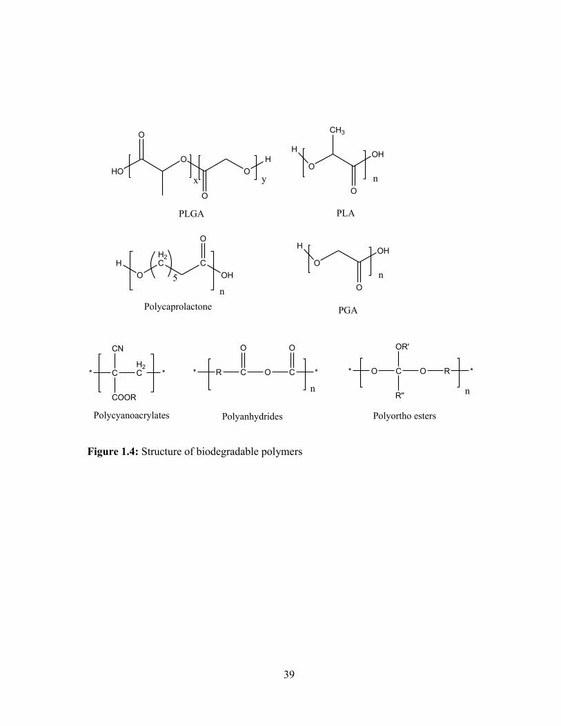
39
HO
O
O
O
O
H
x y
PLGA
O
H
CH3
OH
O
n
PLA
O
H2CH C
OH
O
5
n
Polycaprolactone
O
HOH
O
n
PGA
* CH2C
COOR
CN
*
Polycyanoacrylates
* R C
O
O C *
O
n
Polyanhydrides
* O C O R
OR'
R''
*
n
Polyortho esters
Figure 1.4: Structure of biodegradable polymers
40
CHAPTER 2
INTRODUCTION
Statement of the problem
The retinal pigment epithelium (RPE), macular region, choriocapillaries and Bruch’s
membrane are the primary target sites of vision threatening diseases such as dry and wet AMD,
proliferative vitreoretinopathy, DR and DME [159]. Out of all these diseases, wet AMD is
believed to be a major cause for severe central vision loss or legal blindness in the individuals
65 years or older [160]. Elevated levels of vascular endothelial growth factor (VEGF) are
reported in choroidal neovascularization (CNV), and it is a primary reason for the development
of wet AMD. Occurrence of CNV is followed by leakage of fluid and blood into the subretinal
space and subsequent scar formation, which eventually leads to irreversible vision loss [161].
Therefore, anti-VEGF antibodies such as bevacizumab and ranibizumab are recommended in
the treatment of wet AMD [162]. Bevacizumab (149 kDa) is a full-length recombinant
humanized murine monoclonal antibody (rhum-anti-VEGF antibody) specific to all isoforms
of VEGF [163]. It specifically binds to the extracellular VEGF and blocks the angiogenic
action of VEGF. At a dose of 1 mg or 1.25 mg [164], it has shown reduction in the macular
thickness, reduction in angiogenic leakage, and improvement in visual acuity of neovascular
AMD patients [165, 166]. Ranibizumab is a 48 kDa Fab-fragment of a humanized murine anti-
VEGF antibody active against all isomers of VEGF. It is one-third the size of bevacizumab
and use the same molecular mechanism to block the VEGF [167]. Due to short half-life, current
anti-VEGF therapy with bevacizumab or ranibizumab requires frequent intravitreal injection
to maintain therapeutic levels at retina/choroid. However, treatment with frequent intravitreal

41
administrations is associated with many potential complications like endophthalmitis, retinal
detachment, retinal hemorrhage, and patient non-compliance [168].
Therefore, there is a need to develop a novel formulation for the sustained delivery of
macromolecules (e.g., antibodies). Sustained delivery systems such as NPs may offer an
advantage of higher residence time for macromolecules at the site of absorption. Drug
encapsulation within NPs (1-1000 nm) can be achieved with most commonly used
biodegradable and biocompatible polymers such as PLA, PCL, and PLGA. However, in vivo
degradation of PLA and PLGA NPs produces high molar masses of lactic acid and glycolic
acid, which reduces the pH in microenvironment. This pH change leads to the higher hydrolytic
degradation of protein therapeutics, and also causes tissue irritation and toxicity [169]. In
addition, rapid degradation of PGA or PLGA based formulation results in significantly higher
burst release followed by faster rate of release [170, 171]. On the other hand, the rate of
degradation for PLA or PCL based copolymers are very slow. Poor degradation of PCL based
formulation leads to accumulation of formulation (without drug) in limited vitreous space,
which is highly unacceptable.
Hypothesis
There is a need to develop novel polymeric system which can sustain the release of
macromolecules and also improves the stability of the same. Therefore, preparation of novel
biodegradable PB copolymers, and their applications for the development of macromolecules-
loaded NPs and thermosensitive gel have been proposed in this research protocol. Various
macromolecules such as catalase (237 kDa), IgG (150 kDa), IgG-Fab (48 kDa), bovine serum
albumin (BSA) (66 kDa), lysozyme (14.7 kDa), insulin (5.8 kDa), octreotide (1 kDa), and
bevacizumab (149 kDa) loaded formulations have been investigated in this research work.
42
The PB copolymers are comprised of FDA approved polymer blocks such as PEG, PCL
and a small molar mass of PLA/PGA. Tailoring of the molecular weight or interplay of the
sequence of blocks can provide different hydrophilic or hydrophobic polymers. The presence
of PEG in the PB copolymers will allow improving the physical stability of NP formulation.
In addition, it will also help to enhance the stability of peptide/protein molecules by avoiding
the direct interaction between hydrophobic segments of polymers with peptide/protein. PEG
will also decrease the direct contact of protein therapeutics with the organic solvents during
the NP preparation. PCL is the second important block of PB copolymers. In corporation of
highly crystalline PCL block in the backbone of PB copolymers will significantly improve DL
and drug EE of PB NPs. Furthermore, slower degradation of PCL blocks will sustain the drug
release for significant period of time. PB copolymers are also composed of small molar mass
of PLA/PGA. Incorporation of PLA/PGA in PB copolymers significantly reduces crystallinity
of PCL which eventually accelerates hydrolytic degradation of PCL. Alteration of polymer
degradation rate will shift the drug release kinetics from diffusion controlled to degradation
controlled. Hence, by these structural modifications in PB copolymers, nearly zero-order drug
release can be easily achieved. The second important reason behind designing of PB copolymer
is to minimize the amount of lactic acid produced upon in vivo degradation. It is anticipated
that reduction of the PLA molar mass in PB copolymers will improve the stability of
encapsulated protein molecules, and it will also reduce possible tissue irritation and toxicity.
This novel approach will provide the entire range of polymers with different
hydrophilicity-hydrophobicity index. This enormous advantage will allow us to select a unique
composition of polymer which will be best suited for a respective therapeutic agent.
Furthermore, reduction in the molecular weight and/or change in the polymer block
43
arrangement will allow us to prepare PB copolymer with totally different and unique
physicochemical property i.e., temperature sensitivity. These new compositions will be water
soluble at room temperature but upon increase in temperature, they will transform to solid
hydrogel.
Catalase (237 kDa), IgG (150 kDa), IgG-Fab (48 kDa), BSA (66 kDa), lysozyme (14.7
kDa), insulin (5.8 kDa), and octreotide (1 kDa) will be used as model proteins for the
optimization of formulation components and processes. PB copolymers will be utilized for the
preparation of NPs and thermosensitive gel. Many investigators have reported significant burst
release of therapeutic agents (peptides/proteins) when delivered from NPs or hydrogel systems
[170, 171]. This problem may be eliminated by introducing dual approach or composite
formulations, in which drug-loaded NPs are suspended in thermosensitive gel. This composite
approach may minimize the burst release effect and provide nearly zero-order drug release by
offering longer drug diffusion pathway across the system.
Subconjunctival/intravitreal administration of such a novel formulation may result in a
prolong duration of action (approximately 6 months) and thereby eliminate the need for
repeated administration. It is anticipated that this formulation will provide higher patient
compliance with reduced side effects. This approach may act as a platform for ocular delivery
of other therapeutic macromolecules such as siRNA, aptamers, peptides and large proteins.
Therefore, the broad objective of this research is to develop novel PB copolymer based
sustained delivery systems of various peptide/protein therapeutics for the treatment of posterior
segment diseases.
44
Objectives
Objectives of this research are as follow,
1) To synthesize and characterize various triblock (TB) and PB copolymers for the
preparation of thermosensitive hydrogels. In addition, the effects of various parameters
including molecular weight, arrangement of blocks and hydrophobicity on sol-gel
transition, viscosity and in vitro drug release will be investigated. The possible
mechanism of sol-gel transition will also be evaluated.
2) To synthesize and characterize various TB and PB copolymers for the preparation of
IgG-loaded PB NPs. The effect of molecular weight, hydrophobicity and isomerism on
EE, loading and in vitro release of IgG will be investigated.
3) To synthesize and characterize various PB copolymers for the preparation of protein-
encapsulated NPs. More importantly, the effect of hydrophobicity of PB copolymer on
the encapsulation of different proteins (IgG, BSA and Bevacizumab) will be
investigated. The effect of size and hydrodynamic diameter of protein on various
formulation parameters will also be studied. The stability of released protein will be
investigated by chemical and biological assays. Approaches to composite formulations
(NP suspended in thermosensitive gel) to achieve zero order drug release will be
investigated.
4) In order to achieve maximum DL and zero-order drug release, various formulation
process parameters including addition of NaCl will be optimized.
5) In vitro and in vivo polymer tolerability studies will be performed.
45
CHAPTER 3
NOVEL THERMOSENSITIVE PENTABLOCK (PB) COPOLYMERS FOR
SUSTAINED DELIVERY OF PROTEINS IN THE TREATMENT OF POSTERIOR
SEGMENT DISEASES
Rationale
Bevacizumab, a chimeric anti-VEGF antibody (149 kDa) approved for treatment of
colon cancer is being used off-label for the treatment of ocular neovascularization. Intravitreal
injection of bevacizumab has caused reduction in macular thickness, angiogenic leakage and
improvement of visual acuity in neovascular AMD patients [165, 166]. However, due to the
chronic nature of ocular diseases and shorter intravitreal half-life of this anti-VEGF antibody,
frequent intravitreal injections are indicated to maintain therapeutic activity in the retina and
choroid. Frequent administrations are inconvenient and cause potential complications such as
endophthalmitis, retinal detachment, retinal hemorrhage, and more importantly, patient non-
compliance [172-174]. Since the wide application of protein therapeutics in many treatments
including ophthalmology, tumor biology and immunology, pharmaceutical scientists are more
focused towards the development of novel delivery strategies utilizing current therapeutics
rather than on development of new drugs. Development of sustained release formulations of
protein therapeutics can reduce the frequency of intravitreal injections and eventually eliminate
potential complications. This approach may lower cost of treatment and improve patient
compliance.
Recently, various gel forming polymers, sensitive to external environmental stimuli
such as pH, temperature, electric field and ionic concentrations have been investigated as
sustained delivery systems [175]. In particular, temperature sensitive biodegradable polymers
46
composed of hydrophilic and hydrophobic blocks have drawn more attention. Injectable
thermosensitive hydrogel remains in solution phase during injection and as soon as it is
exposed to body temperature, it immediately phase transforms into a solid hydrogel polymer
matrix. The hydrogel matrix protects protein therapeutics from enzymatic degradation and
provides sustained release for a longer period of time, eliminating repeated monthly injections.
Various biodegradable copolymers composed of hydrophobic polymer blocks including PCL,
PLA, polyglycolide (PGA) and PLGA, and hydrophilic polymer blocks, in particular PEG,
have been investigated for their thermosensitive behavior [176-178]. Adjustment of the
hydrophilic-hydrophobic balance in the block copolymer backbone allows manipulation of the
sol-gel transition curve.
Many researchers have synthesized and investigated thermosensitive TB copolymers
(A-B-A or B-A-B) composed of PLA/PCL/PLGA blocks (A) and PEG block (B) for their
applicability in the development of sustained delivery formulations [176, 179]. However,
applicability of PCL based TB copolymers (A-B-A or B-A-B) as thermosensitive gels is not
very suitable due to their poor biodegradability and faster drug release [180]. Slow degradation
of PCL is attributed to its highly crystalline nature [181]. Degradation of PLGA/PLA/PGA
produces lactic acid and glycolic acid, and significantly reduces pH in the microenvironment
and hence triggers the degradation of protein therapeutics [182]. Moreover, PLGA also induces
structural changes in protein molecules by the process of acylation [182, 183]. Therefore, an
alternative development of novel polymeric system which reduces the molar mass of lactic
acid/glycolic acid upon degradation becomes pivotal. It is also crucial to establish coordination
between degradation profile and drug release profile. To date, very limited studies utilizing
47
thermosensitive hydrogels have been established for the treatment of posterior segment
neovascular diseases.
In the present study, we have synthesized and evaluated various novel PB copolymer
based thermosensitive biodegradable hydrogels. PB copolymers are composed of FDA
approved polymer blocks including PEG, PCL and PLA. This study has addressed four
important aspects ocular sustained release formulation i.e., synthesis and structural
characterization of PCL-PEG-PCL, PLA-PCL-PEG-PCL-PLA, and PEG-PCL-PLA-PCL-
PEG block copolymers, the effect of molecular weight and block arrangements of polymers on
sol-gel transition behavior, the in vitro cytotoxicity/biocompatibility and the in vitro release of
IgG (a model full length antibody similar to bevacizumab, 150 kDa). Moreover, a possible
gelation mechanism of PB copolymers has been hypothesized and supportive studies are
discussed.
Materials and methods
Materials
PEG (1000, 1500, 2000 and 4000), monomethoxy PEG (550), L-lactide, ε-
caprolactone, stannous octoate, coumarin-6 and lipopolysaccharide were procured from
Sigma-Aldrich (St. Louis). Hexamethylene diisocyanate (HMDI) and Micro-BCATM were
obtained from Fisher scientific. Mouse TNF-α, IL-6 and IL-1β (Ready-Set-Go) ELISA kits
were purchased from eBioscience Inc. Lactate dehydrogenase estimation kit and CellTiter 96®
AQueous non-radioactive cell proliferation assay (MTS) kit were obtained from Takara Bio Inc.
and Promega Corp., respectively. All other reagents utilized in this study were of analytical
grade.
48
Methods
Synthesis of TB copolymers with B-A-B (PCL-PEG-PCL) and PB copolymers
with C-B-A-B-C (PLA-PCL-PEG-PCL-PLA) block arrangements
The PCL-PEG-PCL TB copolymers were synthesized by ring-opening bulk
copolymerization of ε-caprolactone [176]. PEG was utilized as macroinitiator and stannous
octoate as a catalyst. Briefly, before polymerization, PEG (1, 1.5, 2 and 4 kDa) were vacuum
dried for 4 h. A predetermined amount of PEG (4.0 g) and ε-caprolactone (8.0 g) were added
in the round bottom flask. The polymer melt was degassed under vacuum for 30 min at 130
°C. The flask was then purged with nitrogen gas, followed by addition of stannous octoate (0.5
wt%). The reaction was carried out for 24 h at 130 °C. The resulting polymer was then
dissolved in dichloromethane and precipitated by addition of cold diethyl ether. Precipitate was
centrifuged and vacuum-dried to remove residual solvents. Purified polymers were stored at -
20 °C. A schematic synthesis scheme is presented in Figure 3.1a.
Synthesis of PB copolymers with C-B-A-B-C (PLA-PCL-PEG-PCL-PLA) block arrangements
TB copolymer (PCL1250-PEG1500-PCL1250, TB-5) was synthesized as per method described in
earlier section for preparation of PB copolymers A (PB-1), B (PB-2) and C (PB-3) (Table 3.1).
To synthesize PB copolymers, predetermined amount of TB-5 and L-lactide were added in a
round bottom flask and degassed under vacuum for 30 min at 130 °C. The flask was then
purged with nitrogen gas and followed by addition of stannous octoate (0.5 wt%). The reaction
was carried out at 130 °C for 24 h. The resulting polymers were purified and stored in a similar
manner as described earlier.
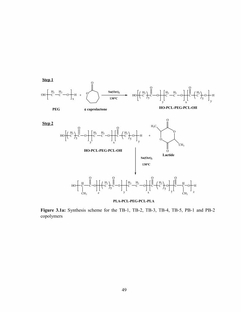
49
Figure 3.1a: Synthesis scheme for the TB-1, TB-2, TB-3, TB-4, TB-5, PB-1 and PB-2
copolymers
OHH2C
H2C O H
x
O
O
+ HOH2C C
O
OH2C
H2C O C
O
H2C O
5 5H
y yx
Sn(Oct)2
130°C
caprolactonePEG HO-PCL-PEG-PCL-OH
HOH2C C
O
OH2C
H2C O C
O
H2C O
5 5H
y yx
HO-PCL-PEG-PCL-OH
Sn(Oct)2
130°C
OH2C C
O
OH2C
H2C O C
O
H2C O
5 5C
y yx
C
O
HC
CH3
HO
O
HC
CH3
O
z z
+
O
O
O
O
H3C
CH3
PLA-PCL-PEG-PCL-PLA
Step 1
Step 2
Lactide
H
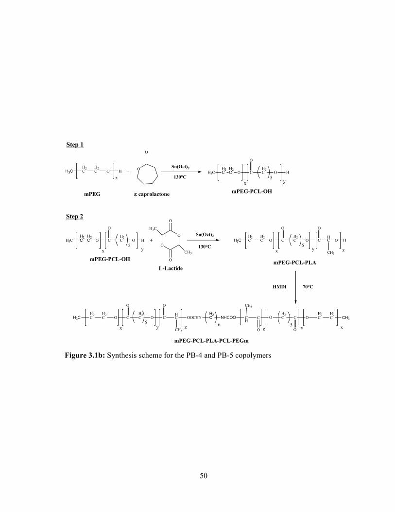
50
Figure 3.1b: Synthesis scheme for the PB-4 and PB-5 copolymers
H3CH2C
H2C O H
x
O
O
+ H3C O C
O
H2C O
5H
yx
Sn(Oct)2
130°C
caprolactonemPEGmPEG-PCL-OH
Sn(Oct)2
130°C
H3CH2C
H2C O C
O
H2C O
5C
yx
O
HC
CH3
O
z
+O
O
O
O
H3C
CH3
mPEG-PCL-PLA
Step 1
Step 2
L-Lactide
70°CHMDI
H3CH2C
H2C O C
O
H2C O
5C
yx
O
HC
CH3
OOCHN
z
H2C NHCOO CH3
H2C
H2COC
O
H2COC
O
CH
CH3
xy5
z6
mPEG-PCL-PLA-PCL-PEGm
H2C
H2C
H3C O C
O
H2C O
5H
yx
mPEG-PCL-OH
H2C
H2C H
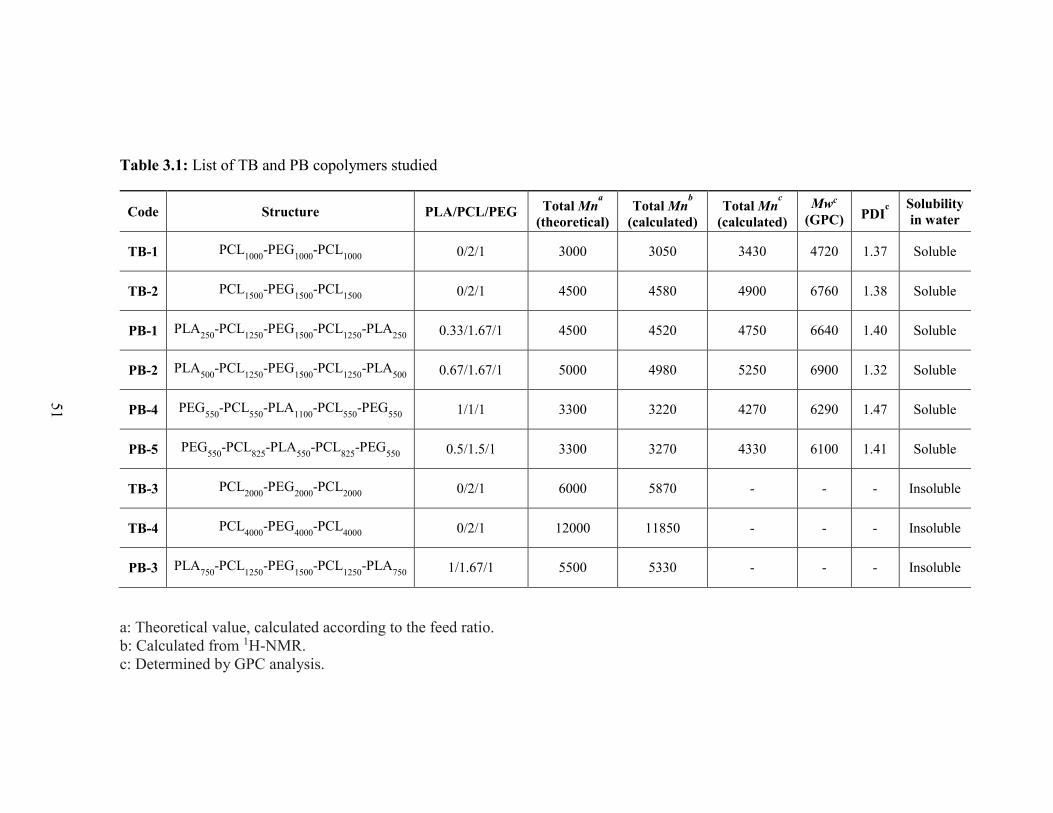
51
Table 3.1: List of TB and PB copolymers studied
Code Structure PLA/PCL/PEG Total Mna
(theoretical)
Total Mnb
(calculated)
Total Mnc
(calculated)
Mwc
(GPC) PDIc
Solubility
in water
TB-1 PCL1000
-PEG1000
-PCL1000
0/2/1 3000 3050 3430 4720 1.37 Soluble
TB-2 PCL1500
-PEG1500
-PCL1500
0/2/1 4500 4580 4900 6760 1.38 Soluble
PB-1 PLA250
-PCL1250
-PEG1500
-PCL1250
-PLA250
0.33/1.67/1 4500 4520 4750 6640 1.40 Soluble
PB-2 PLA500
-PCL1250
-PEG1500
-PCL1250
-PLA500
0.67/1.67/1 5000 4980 5250 6900 1.32 Soluble
PB-4 PEG550
-PCL550
-PLA1100
-PCL550
-PEG550
1/1/1 3300 3220 4270 6290 1.47 Soluble
PB-5 PEG550
-PCL825
-PLA550
-PCL825
-PEG550
0.5/1.5/1 3300 3270 4330 6100 1.41 Soluble
TB-3 PCL2000
-PEG2000
-PCL2000
0/2/1 6000 5870 - - - Insoluble
TB-4 PCL4000
-PEG4000
-PCL4000
0/2/1 12000 11850 - - - Insoluble
PB-3 PLA750
-PCL1250
-PEG1500
-PCL1250
-PLA750
1/1.67/1 5500 5330 - - - Insoluble
a: Theoretical value, calculated according to the feed ratio.
b: Calculated from 1H-NMR.
c: Determined by GPC analysis.
52
Synthesis of PB copolymers with A-B-C-B-A (PEG-PCL-PLA-PCL-PEG) block
arrangements
PB copolymers, PB-4 and PB-5 were synthesized by ring-opening copolymerization
where mPEG (550) was utilized as macroinitiator and stannous octoate (0.5 wt%) as a catalyst
[176]. Firstly, TB copolymers, mPEG-PCL-PLA were synthesized by ring-opening
copolymerization in a manner described earlier. The resulting TB copolymers were coupled
utilizing hexamethylene diisocyanate (HMDI) as a linker to prepare PEG-PCL-PLA-PCL-PEG
PB copolymers. Coupling reaction was carried out at 70 °C for 8 h. Polymers were purified by
cold ether precipitation and stored at -20 °C. A brief synthetic scheme is depicted in Figure
3.1b.
Characterization of PB copolymers
FTIR analysis
Fourier transform infrared spectroscopy (FTIR) spectra were recorded with a Perkin
Elmer SpectrumOne infrared spectrophotometer at a resolution of 4 cm-1 with scan number
of 16. FTIR scan was carried out in a range of 4000-650 cm-1. The resulting IR spectra were
analyzed with spectrum-v5.3.1 software.
1H-NMR analysis
Purity, molecular structure and molecular weight (Mn) of the block copolymers were
estimated utilizing a Varian 400-MHz NMR spectrometer. NMR spectra were recorded by
dissolving block copolymers in CDCl3.
Gel Permeation Chromatography (GPC) analysis
Molecular weights (Mn and Mw) and polydispersity of polymers were examined by
GPC analysis. Briefly, 5 mg of polymer was dissolved in 1.5 mL of tetrahydrofuran (THF).
53
Polymer samples were separated on a Styragel HR-3 column maintained at 35 °C. THF at the
rate of 1 mL/min was utilized as eluting solvent. Samples were analyzed by refractive index
detector (Shimadzu).
X-ray diffraction (XRD) analysis
Physical states of all the synthesized polymers were determined by XRD analysis. TB
and PB copolymers were analyzed at room temperature by MiniFlex automated X-ray
diffractometer (Rigaku, The Woodlands, Texas, USA) equipped with Ni-filtered Cu-kα
radiation (30 kV and 15 mA). The diffraction angle ranged from 5 to 45° using a 1° per min
increment. Jade 8+ (Material Data, Inc, Livermore, CA) was employed to process the
diffraction patterns.
Sol-Gel transition
The sol (flow)-gel (no flow) transition of block copolymers was examined by following
a previously published protocol with minor modifications [184]. Briefly, block copolymers
ranging from 15-30 wt% were dissolved in distilled deionized water followed by 12 h
incubation at 4 °C. After equilibration, 1 mL of aqueous polymeric solution was transferred in
4 mL glass vial and placed in water bath. The temperature of water bath was raised gradually
from 10 to 60 °C at an increment of 1 °C. Vials were kept for 5 min at each temperature. The
gel formation was observed visually by inverting the tubes. A physical state with no fluidity
for 1 min was considered as gel phase. The temperature at which solution transforms to gel
phase was considered as critical gelling temperature (CGT) and the temperature where a
polymer starts to precipitate (phase separation) was described as critical precipitation
temperature (CPT).

54
Cytotoxicity
Cell culture
Human retinal pigment epithelial cell line (ARPE-19) were cultured and maintained
according to a protocol provided by ATCC. In brief, ARPE-19 cells were cultured in
Dubelcco's modified Eagle medium (DMEM)/F-12 containing 10% heat-inactivated fetal
bovine serum (FBS), sodium bicarbonate (2 mM), HEPES (15 mM), streptomycin (100 mg/L)
and penicillin (100 U/L). Mouse macrophage (RAW-264.7) cells were procured from ATCC.
RAW-264.7 cells were cultured and maintained in DMEM supplemented with 10% FBS,
streptomycin (100 mg/L) and penicillin (100 U/L). Both cell lines were maintained in
humidified atmosphere at 37 °C and 5% CO2.
Lactate Dehydrogenase (LDH) assay
In order to evaluate cytotoxicity of polymeric materials, various concentrations of
block copolymers were exposed to the ARPE-19 cells or RAW-264.7 cells at density of 1.0 x
104 per well. Cells were incubated at 37 °C and 5% CO2 in humidified atmospherefor 48 h.
After incubation, levels of LDH in cell supernatant were estimated by LDH detection kit.
Samples were analyzed at 450 nm by 96-well plate reader. The amount of released LDH is
directly proportional to the cytotoxicity of the polymers. In this study, more than 10% of LDH
release was considered as cytotoxic. LDH release (%) was calculated according to the
following equation,
𝐿𝐷𝐻 𝑟𝑒𝑙𝑒𝑎𝑠𝑒(%) =𝐴𝑏𝑠. 𝑜𝑓 𝑆𝑎𝑚𝑝𝑙𝑒 − 𝐴𝑏𝑠. 𝑜𝑓 𝑛𝑒𝑔𝑎𝑡𝑖𝑣𝑒 𝑐𝑜𝑛𝑡𝑟𝑜𝑙
𝐴𝑏𝑠. 𝑜𝑓 𝑝𝑜𝑠𝑖𝑡𝑖𝑣𝑒 𝑐𝑜𝑛𝑡𝑟𝑜𝑙 − 𝐴𝑏𝑠. 𝑜𝑓 𝑛𝑒𝑔𝑎𝑡𝑖𝑣𝑒 𝑐𝑜𝑛𝑡𝑟𝑜𝑙∗ 100 ...Eq. 3.1
55
MTS assay
MTS assay was performed according to a previously published protocol with minor
modifications [185]. Briefly, ARPE-19 and RAW-264.7 cells at a density of 1.0 x 10⁴ cells per
well were seeded in 96-well plate. Cells were incubated for 24 h at 37 °C and 5% CO2 in
humidified atmosphere. After incubation, the culture medium was replaced with fresh medium
containing various concentrations of block copolymers. Cells were further incubated for 48 h.
At the end of incubation period, culture medium was substituted with 100µL of serum free
medium containing 20 µL of MTS solution. Cells were then incubated for 4 h at 37 °C and 5%
CO2. After 4 h, absorbance of each well was estimated at 450 nm by 96-well plate reader.
Polymer concentrations which exhibited more than 90% cell viability were considered as non-
toxic. Percent cell viability was estimated by following equation.
𝐶𝑒𝑙𝑙 𝑣𝑖𝑎𝑏𝑖𝑙𝑖𝑡𝑦 (%) =𝐴𝑏𝑠. 𝑜𝑓 𝑆𝑎𝑚𝑝𝑙𝑒−𝐴𝑏𝑠. 𝑜𝑓 𝑛𝑒𝑔𝑎𝑡𝑖𝑣𝑒 𝑐𝑜𝑛𝑡𝑟𝑜𝑙
𝐴𝑏𝑠. 𝑜𝑓 𝑝𝑜𝑠𝑖𝑡𝑖𝑣𝑒 𝑐𝑜𝑛𝑡𝑟𝑜𝑙−𝐴𝑏𝑠. 𝑜𝑓 𝑛𝑒𝑔𝑎𝑡𝑖𝑣𝑒 𝑐𝑜𝑛𝑡𝑟𝑜𝑙∗ 100 …Eq. 3.2
Biocompatibility
RAW-264.7 cells were cultured and maintained according to a protocol described in
previous section. In order to evaluate in vitro biocompatibility of gelling polymers, a
previously published protocol was followed with minor modifications [185]. Briefly, 5.0 x 10⁴
cells were seeded per well of 48-well plates and incubated for 24 h. After incubation, the cell
culture medium was replaced with fresh medium containing various concentrations of block
copolymers. After 24 h of incubation at 37 °C and 5% CO2, the supernatant of each well was
analyzed by ELISA for quantitative estimation of various cytokines (TNF-α, IL-6 and IL-1β).
ELISA was performed according to the manufacturer’s protocol. Calibration curves for TNF-
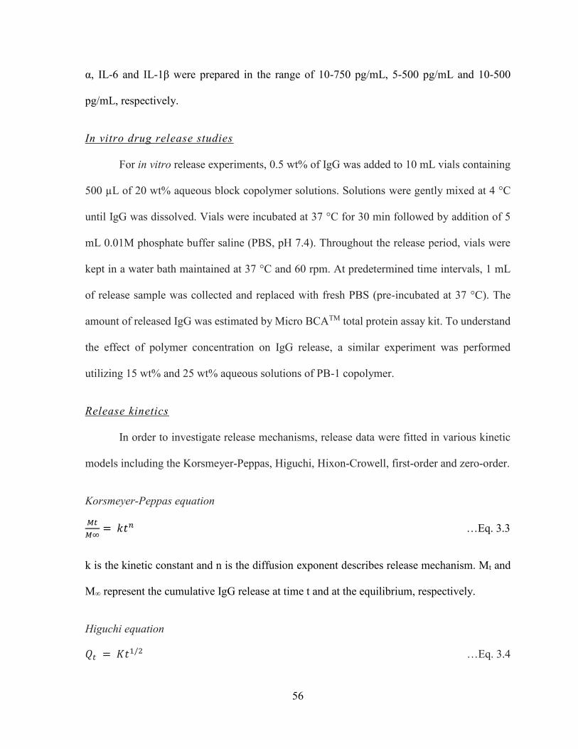
56
α, IL-6 and IL-1β were prepared in the range of 10-750 pg/mL, 5-500 pg/mL and 10-500
pg/mL, respectively.
In vitro drug release studies
For in vitro release experiments, 0.5 wt% of IgG was added to 10 mL vials containing
500 µL of 20 wt% aqueous block copolymer solutions. Solutions were gently mixed at 4 °C
until IgG was dissolved. Vials were incubated at 37 °C for 30 min followed by addition of 5
mL 0.01M phosphate buffer saline (PBS, pH 7.4). Throughout the release period, vials were
kept in a water bath maintained at 37 °C and 60 rpm. At predetermined time intervals, 1 mL
of release sample was collected and replaced with fresh PBS (pre-incubated at 37 °C). The
amount of released IgG was estimated by Micro BCATM total protein assay kit. To understand
the effect of polymer concentration on IgG release, a similar experiment was performed
utilizing 15 wt% and 25 wt% aqueous solutions of PB-1 copolymer.
Release kinetics
In order to investigate release mechanisms, release data were fitted in various kinetic
models including the Korsmeyer-Peppas, Higuchi, Hixon-Crowell, first-order and zero-order.
Korsmeyer-Peppas equation
𝑀𝑡
𝑀∞= 𝑘𝑡𝑛 …Eq. 3.3
k is the kinetic constant and n is the diffusion exponent describes release mechanism. Mt and
M∞ represent the cumulative IgG release at time t and at the equilibrium, respectively.
Higuchi equation
𝑄𝑡 = 𝐾𝑡1/2 …Eq. 3.4

57
K denotes the Higuchi rate kinetic constant, Qt is the amount of released IgG at time t, and t is
time in hours.
Hixon-Crowell equation
𝐶01/3 − 𝐶𝑡
1/3 = 𝑘𝑡 …Eq. 3.5
C0 and Ct represents the initial amount and remaining amount of IgG in gel, respectively. k is
the constant incorporating surface-volume relation and t is time in hours.
First order equation
𝐿𝑜𝑔𝐶 = 𝐿𝑜𝑔𝐶0 − 𝐾𝑡
2.303 …Eq. 3.6
K denotes the first order rate constant, C0 is the initial IgG concentration and t represents time
in hours.
Zero order equation
𝐶 = 𝐾0𝑡 …Eq. 3.7
K0 is the zero-order rate constant and t is time in hours.
1H-NMR for coumarin-6-loaded gel
Five mg of PB-1 copolymer was dissolved in either CDCl3 or D2O followed by addition
of 0.5 mg of coumarin-6 a (hydrophobic dye). Samples containing both polymer and
hydrophobic dye were subjected to 1H-NMR analysis. A similar study was performed utilizing
PB-5 copolymer.
58
Micelle size analysis
Aqueous solutions of PB-1 copolymer were subjected to micelle size analysis at room
temperature utilizing a particle size analyzer (ZetasizerNano ZS, Malvern Instruments Ltd,
Worcestershire, UK). PB-1 copolymer concentrations ranging from 0.1 to 5 wt% were
investigated without any further dilution.
Viscosity measurements
Rheological properties of 15 wt% aqueous solution of block copolymers were
estimated with an Ubbelohde capillary viscometer at temperatures ranging from 5 ± 1 °C to 25
± 1 °C. Temperature of the viscometer was maintained with a temperature controlled water
bath. Viscosity values are represented as an average of triplicates (kinematic viscosity, cP ±
standard deviation).
Results and discussion
Synthesis and characterization of block copolymers
FTIR spectrum of PB-4 is reported in Figure 3.2. Absorption band at 1725 cm-1 and
multiple bands ranging 1000-1300 cm-1 established the presence of ester linkages in PB
copolymer. C-H stretching bands at 2936 and 2865 cm-1 depicted presence of PCL blocks.
Absorption band at 1534 cm-1 (N-H banding) and 3344 cm-1 (N-H stretching) exhibited the
formation of urethane group in PB-4 copolymer.
1H-NMR was employed to characterize PCL-PEG-PCL, PLA-PCL-PEG-PCL-PLA
and PEG-PCL-PLA-PCL-PEG copolymers. Figure 3.3 depicts the 1H-NMR spectra of TB-1,
PB-1 and PB-4 block copolymers in deuterated chloroform. As described in Figures 3.3a, 3.2b
and 3.2c, characteristic 1H-NMR peaks were observed at 1.40, 1.65, 2.30 and 4.06 ppm
59
corresponding to the methylene protons of -(CH2)3-, -OCO-CH2-, and -CH2OOC- of PCL units,
respectively. A sharp peak at 3.65 ppm was attributed to the methylene protons (-CH2CH2O-)
of PEG. Typical signals (Figures 3.3b and 3.3c) at 1.50 (-CH3) and 5.17 (-CH-) ppm were
assigned for PLA blocks. Whereas, a peak (Figure 3.3c) at 3.38 ppm was denoted to terminal
methyl of (-OCH3-) of PEG.
The [EO]-[CL]-[LA] molar ratios of final products were calculated from integration of
PEG signal at 3.65 ppm, PCL signal at 2.30 ppm and PLA signal at 5.17 ppm. In case of PB-4
copolymer, the PEG signal at 3.38 ppm was applied for the calculation of molar ratio.
The molecular weight (Mw and Mn) and polydispersity of polymers were determined
by GPC. Typical GPC curves of TB-1, PB-1 and PB-4 are shown in Figure 3.4. A single peak
for each polymer was observed describing unimodel distribution of molecular weight and
absence of any other homopolymer block such as PEG, PCL or PLA. Moreover, molecular
weights of block copolymers were very close to feed ratio. Polydispersity (PD) was also below
1.47, describing a narrow distribution of molecular weights. 1H-NMR and GPC were applied
to calculate molecular weight of block copolymers (Table 3.1). As summarized in Table 3.1,
experimental values were consistent with theoretical values derived from feed ratios. Hence
for simplicity, theoretical values are mentioned in the following text.
In order to evaluate crystallinity and phase composition, all the block copolymers were
analyzed for XRD patterns (Figure 3.5). Two sharp peaks were observed at 2θ = 21.5° and
23.9° which belong to PCL blocks. No peaks for PEG or PLA were observed. Interestingly,
only TB-1, TB-2, PB-1 and PB-2 exhibited crystalline peaks of PCL, whereas PB-4 and PB-5
were devoid of any such peaks. XRD patterns of TB-1 and TB-2 indicated that PCL blocks
retained a semi-crystalline structure even after covalent conjugation with PEG blocks.

60
Interestingly, conjugation of PLA blocks at the terminals of TB copolymers exhibited
significant reduction in crystalline peak indicating semi-crystalline structures of PB-1 and PB-
2. However, PB-4 and PB-5 were devoid of any crystalline peak suggesting amorphous nature
of copolymers with A-B-C-B-A block arrangements. Thus, crystallinity of polymer can be
easily controlled by the arrangement of polymer blocks in structural backbone. Moreover,
previously published reports suggest that decrease in crystallinity significantly enhanced
degradation of block polymer [181]. Hence, it is anticipated that PB-4 and PB-5 might cause
faster rate of degradation relative to TB-1, TB-2, PB-1 and PB-2 copolymers.
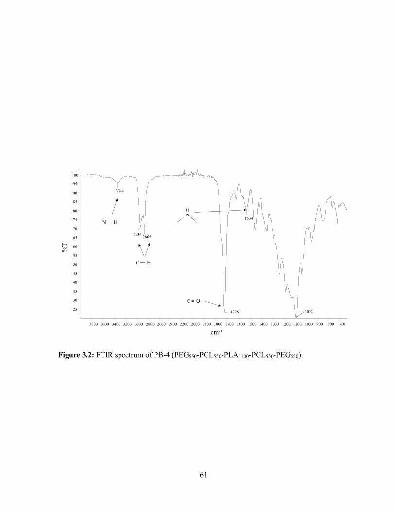
61
Figure 3.2: FTIR spectrum of PB-4 (PEG550-PCL550-PLA1100-PCL550-PEG550).
3800 3600 3400 3200 3000 2800 2600 2400 2200 2000 1900 1800 1700 1600 1500 1400 1300 1200 1100 1000 900 800 700
25
30
35
40
45
50
55
60
65
70
75
80
85
90
95
100
cm-1
%T
3344
29362865
1725
1534
1092
H
N
C H
N H
C O=
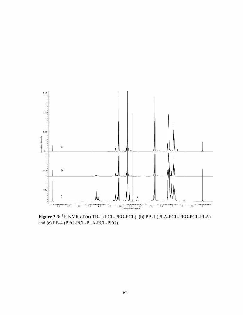
62
Figure 3.3: 1H NMR of (a) TB-1 (PCL-PEG-PCL), (b) PB-1 (PLA-PCL-PEG-PCL-PLA)
and (c) PB-4 (PEG-PCL-PLA-PCL-PEG).
a
b
c
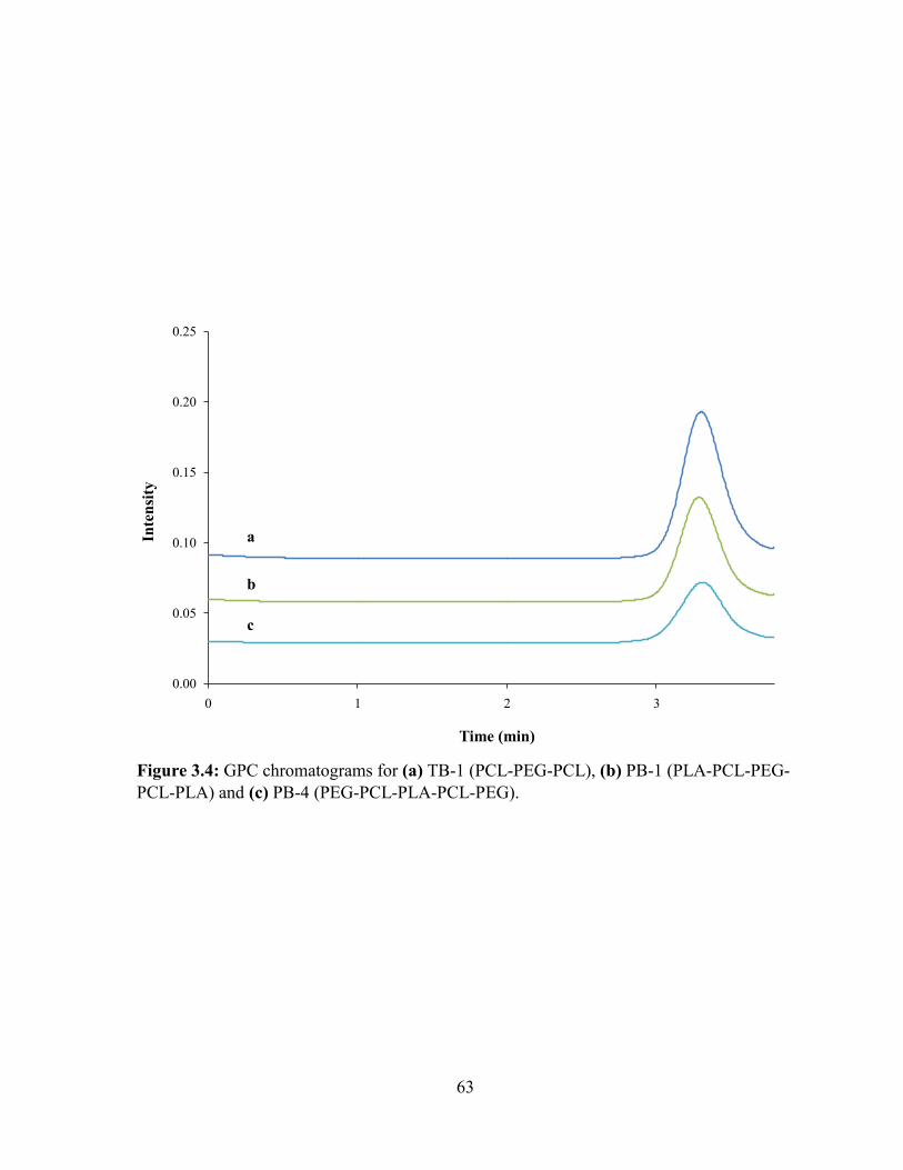
63
Figure 3.4: GPC chromatograms for (a) TB-1 (PCL-PEG-PCL), (b) PB-1 (PLA-PCL-PEG-
PCL-PLA) and (c) PB-4 (PEG-PCL-PLA-PCL-PEG).
0.00
0.05
0.10
0.15
0.20
0.25
0 1 2 3
Inte
nsi
ty
Time (min)
c
b
a
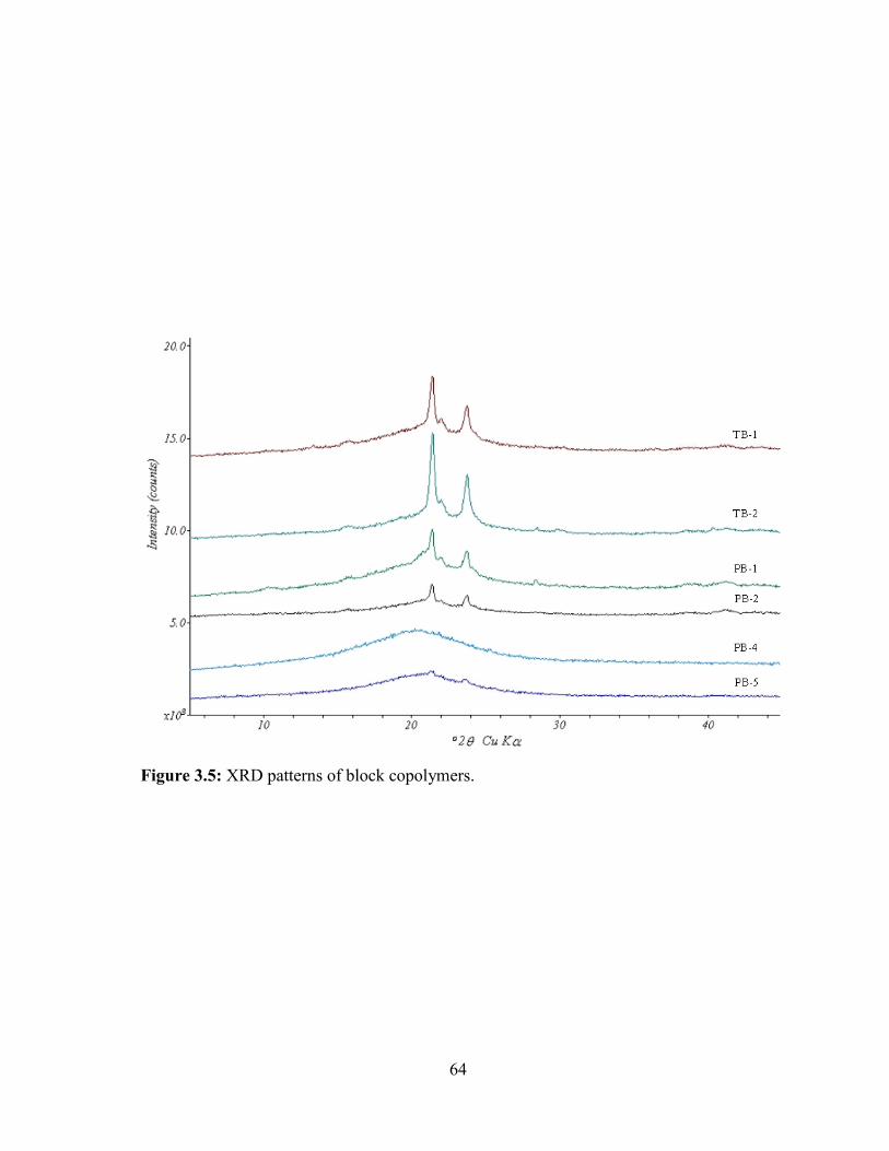
64
Figure 3.5: XRD patterns of block copolymers.

65
Sol-gel transition
The block copolymers reported in this study are aphiphilic in nature containing
hydrophilic block (PEG) and hydrophobic block(s) (PCL and/or PLA). In the case of TB
copolymers (TB-1 and TB-2), increased total molecular weight, but keeping molecular weight
ratio constant i.e., PCL/PEG (2:1), did not alter their thermosensitive behavior. However,
further increase in molecular weights (TB-3 and TB-4) with the same hydrophobic-hydrophilic
block ratio (2:1) reduced aqueous solubility. This may be attributed to longer PCL chains,
which may significantly enhance intermolecular and intramolecular hydrophobic interactions
of polymer and outweigh the ability of PEG to solubilize polymer. PB-1 and PB-2 copolymers
are easily soluble in water and exhibit sol-gel transition behavior. However, the increase in
hydrophobicity by increasing molecular weight of PLA chain (PB-3) exhibited poor aqueous
solubility, which might be due to limitation of PEG to solubilize polymer. For all block
copolymers, an increase in aqueous polymer concentration from 15 to 30 wt% significantly
shifted CGT to lower and CPT to higher values.
Effect of molecular weight of block copolymer
In order to understand the effect of molecular weight of the block copolymers, sol-gel
transition curves of TB-1 and TB-2 were compared (Figure 3.6). With an increase in total
molecular weight of block copolymers from 3000 (TB-1) to 4500 (TB-2), CGT decreased
whereas the CPT increased to higher values. The temperature range for the gel region or area
between CGT and CPT at any given concentration was also significantly enhanced. A similar
trend was observed for PB-1 and PB-2 polymers, where molecular weight of PLA was raised
in PB-2.

66
Effect of hydrophobicity of block copolymer
Hydrophibicity of block copolymers can be increased by enhancing the molecular
weight of the whole while keeping the molecular weight of PEG constant (PB-1 and PB-2) or
by substituting the molecular weight of PLA by PCL (TB-2 and PB-1, PB-4 and PB-5). PB-2
has larger chains of PLA relative to PB-1 copolymer suggesting a greater hydrophobicity of
PB-2 copolymer. It is important to note that a PLA block is less hydrophobic relative to a PCL
block of similar molecular weight. Therefore, TB-2 and PB-5 are more hydrophobic compared
to PB-1 and PB-4, respectively. Sol-gel transition curves of PB-1 and PB-2 (Figure 3.7a), PB-
4 and PB-5 (Figure 3.7b), and TB-2 and PB-1 (Figure 3.7c) were compared to understand the
effect of hydrophobicity on the sol-gel behavior of block copolymers. As described in Figure
3.7a, increased hydrophobicity of PB-2 has significantly reduced the CGT and shifted the value
of CPT to higher temperature. A similar behavior was observed when the sol-gel transition
curves of PB-4/PB-5, and TB-2/PB-1 were compared.
Higher hydrophobicity polymers may enhance intramolecular and intermolecular
hydrophobic interactions even at lower temperatures compared to hydrophilic copolymers
which lead to lower CGT. Additionally, these hydrophobic interactions allow for a more rigid
gel matrix and hence delay polymer precipitation at higher temperature (CPT).
Effect of block arrangement
In order to understand the effect of block arrangement on thermogelling behavior, sol-
gel transition curves of PB-1 (C-B-A-B-C) and PB-5 (A-B-C-B-A) were compared (Figure
3.8). Interestingly, CGT and CPT for PB-1 copolymer were significantly lower than PB-5
copolymer at any respective concentration. This behavior may be attributed to the different
mechanism of gelation of these block copolymers.
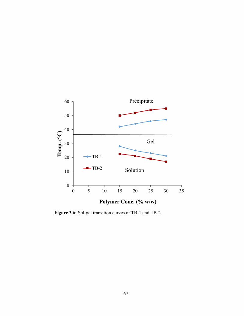
67
Figure 3.6: Sol-gel transition curves of TB-1 and TB-2.
0
10
20
30
40
50
60
0 5 10 15 20 25 30 35
Tem
p.
( C
)
Polymer Conc. (% w/w)
TB-1
TB-2
Precipitate
Gel
Solution
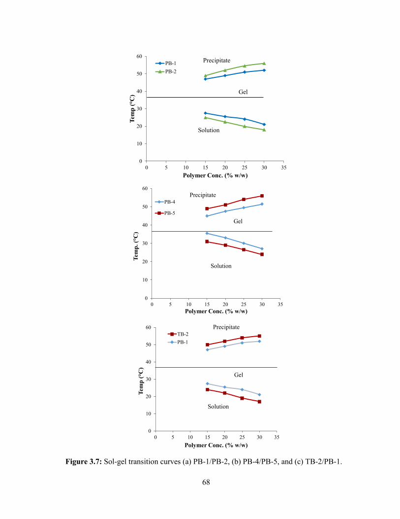
68
Figure 3.7: Sol-gel transition curves (a) PB-1/PB-2, (b) PB-4/PB-5, and (c) TB-2/PB-1.
0
10
20
30
40
50
60
0 5 10 15 20 25 30 35
Tem
p ( C
)
Polymer Conc. (% w/w)
PB-1
PB-2
Precipitate
Gel
Solution
0
10
20
30
40
50
60
0 5 10 15 20 25 30 35
Tem
p.
( C
)
Polymer Conc. (% w/w)
PB-4
PB-5
Precipitate
Gel
Solution
0
10
20
30
40
50
60
0 5 10 15 20 25 30 35
Tem
p ( C
)
Polymer Conc. (% w/w)
TB-2
PB-1
Precipitate
Gel
Solution
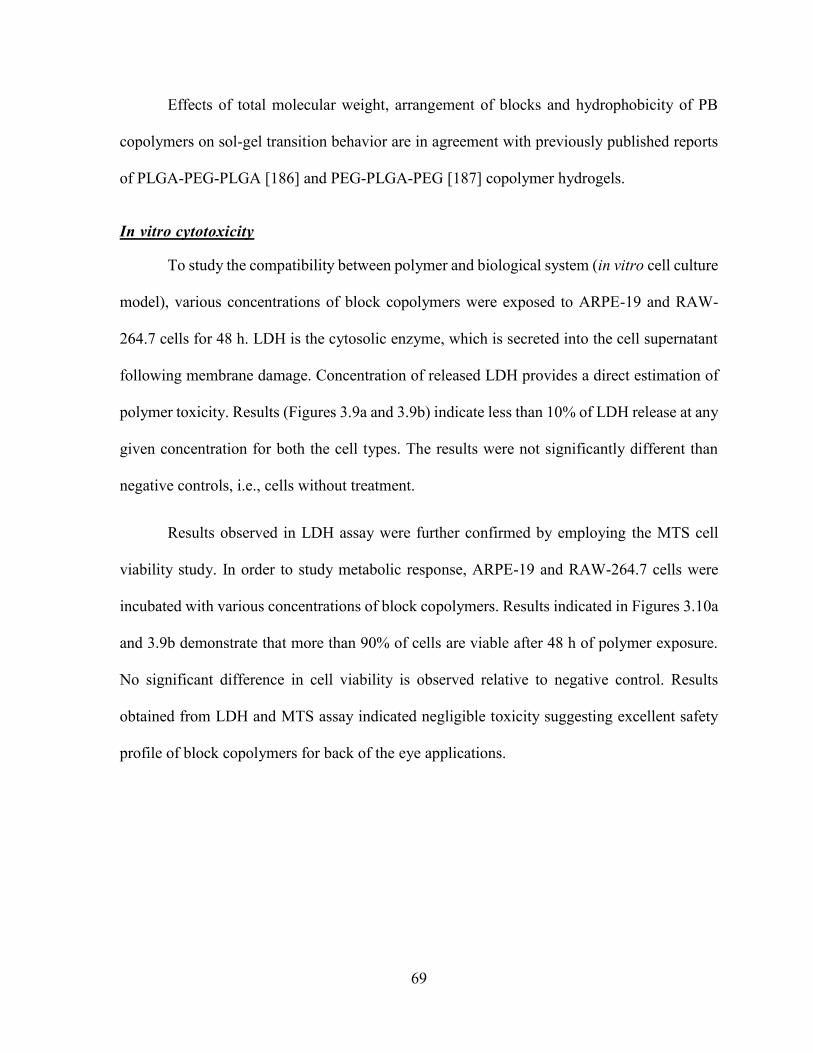
69
Effects of total molecular weight, arrangement of blocks and hydrophobicity of PB
copolymers on sol-gel transition behavior are in agreement with previously published reports
of PLGA-PEG-PLGA [186] and PEG-PLGA-PEG [187] copolymer hydrogels.
In vitro cytotoxicity
To study the compatibility between polymer and biological system (in vitro cell culture
model), various concentrations of block copolymers were exposed to ARPE-19 and RAW-
264.7 cells for 48 h. LDH is the cytosolic enzyme, which is secreted into the cell supernatant
following membrane damage. Concentration of released LDH provides a direct estimation of
polymer toxicity. Results (Figures 3.9a and 3.9b) indicate less than 10% of LDH release at any
given concentration for both the cell types. The results were not significantly different than
negative controls, i.e., cells without treatment.
Results observed in LDH assay were further confirmed by employing the MTS cell
viability study. In order to study metabolic response, ARPE-19 and RAW-264.7 cells were
incubated with various concentrations of block copolymers. Results indicated in Figures 3.10a
and 3.9b demonstrate that more than 90% of cells are viable after 48 h of polymer exposure.
No significant difference in cell viability is observed relative to negative control. Results
obtained from LDH and MTS assay indicated negligible toxicity suggesting excellent safety
profile of block copolymers for back of the eye applications.
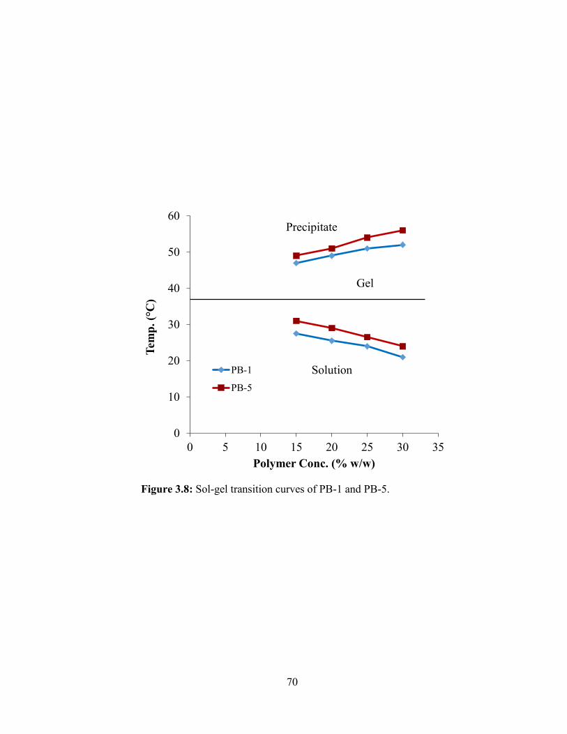
70
Figure 3.8: Sol-gel transition curves of PB-1 and PB-5.
0
10
20
30
40
50
60
0 5 10 15 20 25 30 35
Tem
p.
( C
)
Polymer Conc. (% w/w)
PB-1
PB-5
Precipitate
Gel
Solution
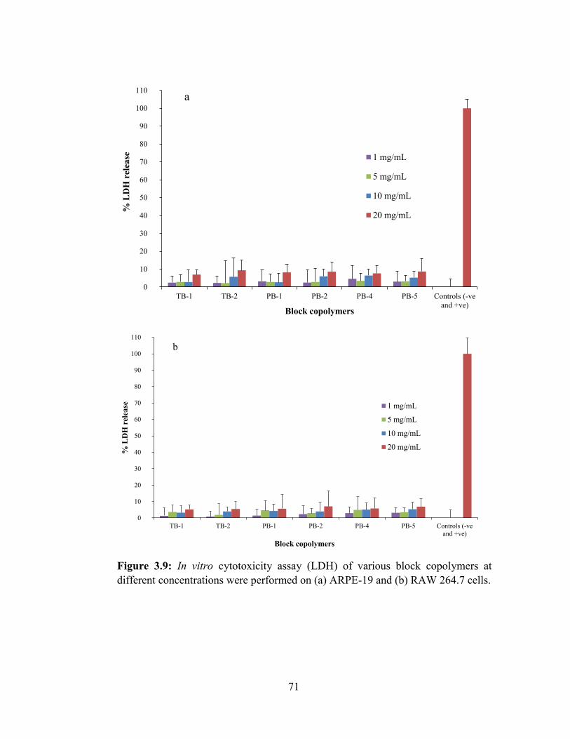
71
Figure 3.9: In vitro cytotoxicity assay (LDH) of various block copolymers at
different concentrations were performed on (a) ARPE-19 and (b) RAW 264.7 cells.
0
10
20
30
40
50
60
70
80
90
100
110
TB-1 TB-2 PB-1 PB-2 PB-4 PB-5 Controls (-ve
and +ve)
% L
DH
rel
ease
Block copolymers
1 mg/mL
5 mg/mL
10 mg/mL
20 mg/mL
a
0
10
20
30
40
50
60
70
80
90
100
110
TB-1 TB-2 PB-1 PB-2 PB-4 PB-5 Controls (-ve
and +ve)
% L
DH
rel
ease
Block copolymers
1 mg/mL
5 mg/mL
10 mg/mL
20 mg/mL
b
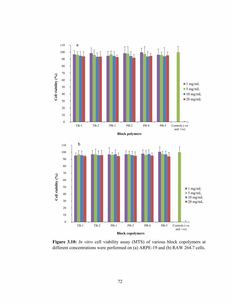
72
Figure 3.10: In vitro cell viability assay (MTS) of various block copolymers at
different concentrations were performed on (a) ARPE-19 and (b) RAW 264.7 cells.
0
10
20
30
40
50
60
70
80
90
100
110
TB-1 TB-2 PB-1 PB-2 PB-4 PB-5 Controls (-ve
and +ve)
Cel
l v
iab
ilit
y (
%)
Block polymers
1 mg/mL
5 mg/mL
10 mg/mL
20 mg/mL
a
0
10
20
30
40
50
60
70
80
90
100
110
TB-1 TB-2 PB-1 PB-2 PB-4 PB-5 Controls (-ve
and +ve)
Cel
l v
iab
ilit
y (
%)
Block copolymers
1 mg/mL
5 mg/mL
10 mg/mL
20 mg/mL
b

73
Figure 3.11: In vitro release of (a) TNF-α, (b) IL-6 and (c) IL-1β from RAW
264.7 cells upon exposure to various concentrations of block copolymers.
0
200
400
600
800
1000
1200
TB-1 TB-2 PB-1 PB-2 PB-4 PB-5 Controls (-
ve and +ve)
Co
nce
ntr
ati
on
of
TN
F-α
(p
g/m
L)
Block copolymers
20 mg/mL
10 mg/mL
5 mg/mL
1 mg/mL
a
0
100
200
300
400
500
600
700
800
900
TB-1 TB-2 PB-1 PB-2 PB-4 PB-5 Controls (-ve
and +ve)
Co
nce
ntr
ati
on
of
IL-6
(pg
/mL
)
Block copolymers
20 mg/mL
10 mg/mL
5 mg/mL
1 mg/mL
b
0
50
100
150
200
250
300
350
400
TB-1 TB-2 PB-1 PB-2 PB-4 PB-5 Controls (-ve
and +ve)
Co
nce
ntr
ati
on
of
IL-1
β (
pg
/mL
)
Block copolymers
20 mg/mL
10 mg/mL
5 mg/mL
1 mg/mL
c

74
In vitro biocompatibility study
Estimation of inflammatory mediators such as TNF-α, IL-6 and IL-1β secreted in cell
supernatant is a good measure to confirm biocompatibility of block copolymers. These
cytokines were estimated in the supernatant of RAW-264.7 cells after 24 h of polymer
exposure. Levels of cytokine were estimated by sandwich ELISA method according to
manufacturer’s protocol. Our results (Figures 3.11a, 3.11b, and 3.11c) indicate that at 20
mg/mL polymer concentration, release of cytokines was negligible with no significant
difference to negative control. Negligible release of TNF-α, IL-6 and IL-1β suggest excellent
biocompatibility of PB copolymers with low toxicity.
In vitro release study
IgG was selected as a model protein to evaluate the suitability of block copolymers as
controlled release delivery systems for ocular indications. In vitro release studies were
performed by dissolving IgG in 20 wt% aqueous solution of respective block copolymers. To
estimate the concentration of IgG, release samples were analyzed by Micro BCATM. In vitro
release behavior of IgG from various thermosensitive gels is compared in Figure 3.12. Release
of IgG was noticeably affected by the chemical composition of block copolymers. TB
copolymers (TB-1 and TB-2) exhibited significantly higher burst release (~40-45% of initial
dose) relative to PB copolymers (PB-1, PB-2, PB-4 and PB-5) which showed burst release
between ~24-31% of initial dose. Moreover, release of IgG from PB copolymer based
thermosensitive gels were sustained for more than 20 days, whereas TB copolymers prolonged
the release for only ~12-14 days. Partial replacement of PCL with PLA block significantly
reduces the crystallinity (XRD data, Figure 3.5) and hydrophobicity of copolymers. This
75
reduction in hydrophobicity of PB copolymers may have increased the affinity with IgG
resulting in prolonged release of IgG from PB copolymers relative to TB copolymers.
In addition, the effect of polymer concentration on the rate of IgG release was also
studied. As depicted in Figure 3.13, 25 wt% of PB-1 copolymer solution exhibited 74.92% of
IgG release within 20 days, whereas 20 wt% and 15 wt% gel showed 85.32% and 96.23% of
IgG release during the same time period. It appears that an increase in polymer concentration
from 15 wt% to 25 wt% significantly retards drug release rate, indicating direct relationship of
polymer concentration on drug release. At higher polymer concentration, the gels may form
compact structures with a smaller porosity in the gel matrix relative to lower polymer
concentration. This structural change may lower the diffusion of IgG across gel matrix
resulting in lower initial burst release and prolonged duration of release.
Release kinetics
In vitro release data were fitted to zero and first order, Korsmeyer-Peppas, Higuchi and
Hixson-Crowell models to delineate kinetics of IgG release (Table 3.2). The Korsmeyer-
Peppas model was found to be the best fit model based on R2 value. The diffusion exponent,
n, ranged from 0.272 - 0.386 for all gelling polymers. The n-values below 0.43 indicates a
diffusion controlled mechanism of IgG release.
1H-NMR of Coumarin-6-loaded polymer solutions
In order to understand the process of polymer solubilization and behavior of sol-gel
transition, 1H-NMR of PB-1 copolymer was carried out in CDCl3 (Figure 3.14a) and D2O
(Figure 3.14b) spiked with coumarin-6. Coumarin-6 is insoluble in water however aqueous
solubility is significantly enhanced in the presence of PB-1 copolymer, which may be due to
76
micellization. A sample containing PB-1 copolymer and coumarin-6 in CDCl3 exhibited sharp
peaks representing various protons of PEG, PCL, PLA and coumarin-6. Interestingly, a sample
containing PB-1 copolymer and coumarin-6 in D2O was devoid of signals of coumarin-6
suggesting that coumarin-6 is inside the miceller core. Broad peaks observed at 3.65 ppm and
2.30 ppm were attributed to the protons of PEG (-CH2-CH2-) and PCL (-OCO-CH2-),
respectively. Proton peaks for PLA and coumarin-6 were absent. Exactly, similar behavior was
also observed with the sample containing PB-5 copolymer and coumarin-6 simultaneously
dissolved in CDCl3 (Figure 3.14c) or D2O (Figure 3.14d). NMR analysis carried out in CDCl3
exhibited sharp peaks for all the protons indicating free movement of polymer chains and
coumarin-6 in organic solvent. However, NMR spectra carried out in D2O exhibited very few
broad peaks representing PEG and PCL blocks only. These results suggest that coumarin-6
was located in the core of the micelles along with PCL and PLA blocks. Hence, we were able
to see very weak NMR signals for PCL blocks due to the restricted molecular movement, and
no signals for PLA and coumarin-6. Strong peak of PEG in D2O was observed indicating that
the location of PEG is in the corona of the micelles. Results from this study suggest that PB
copolymers are solubilized in water via micellization process where core is composed of PCL-
PLA and shell is of PEG.
Micelle size analysis
Micelle size and its distribution of PB-1 copolymer were evaluated in water by DLS at
room temperature as a function of concentration (Figure 3.15). As the aqueous concentration
of polymer was raised from 0.1 wt% to 5 wt%, two peaks (22 nm and 150 nm) were
continuously shifted toward higher particle size. Moreover, a broader size distribution with the
increase of polymer concentration was observed. Interestingly, at 5 wt% polymer
77
concentration, emergence of a third peak with very large micelle size (~5 µm) was also
revealed. Increase in particle size and polydispersity as a function of concentration clearly
indicate the involvement of a micellar aggregation mechanism.
Viscosity measurement
Table 3.3 describes the kinematic viscosity of 15 wt% aqueous gelling solutions of
different block copolymers at various temperatures ranging from 5 to 25 °C. Kinematic
viscosities of polymer solutions accelerated with rise in temperature. Interestingly, at any given
temperature, an increase in molecular weight of the block copolymers exhibited higher
viscosity (TB-1 and TB-2 or PB-1 and PB-2).
Also, the viscosities of TB-2 and PB-5 (hydrophobic polymers) were considerably
higher relative to PB-1 and PB-4 (hydrophilic polymers), respectively. we speculate that the
hydrophobic interactions exerted by PCL or PCL-PLA blocks with large molecular weight and
hydrophobic copolymers are significantly stronger at any given temperature relative to small
molecular weight and hydrophilic copolymers, respectively. Subsequently, this phenomenon
may have elevated the viscosity of aqueous solutions. In addition, as temperature of the
solution rises, these hydrophobic interactions also begin to dominate which eventually improve
the viscosity of aqueous polymer solution. Interestingly, polymer structure (arrangement of
blocks) also exhibited a noticeable effect on viscosity. Hence, kinematic viscosity of PB-1
solution was significantly higher compared to PB-5 aqueous solution.
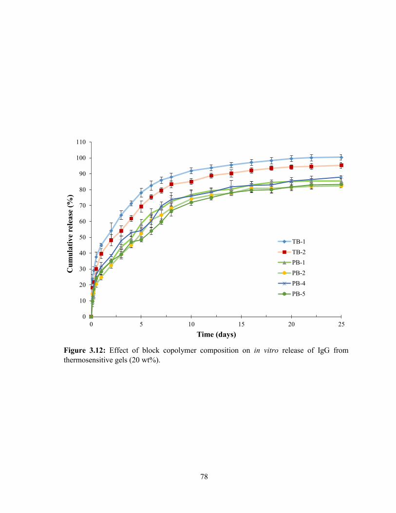
78
Figure 3.12: Effect of block copolymer composition on in vitro release of IgG from
thermosensitive gels (20 wt%).
0
10
20
30
40
50
60
70
80
90
100
110
0 5 10 15 20 25
Cu
mu
lati
ve
rele
ase
(%
)
Time (days)
TB-1
TB-2
PB-1
PB-2
PB-4
PB-5
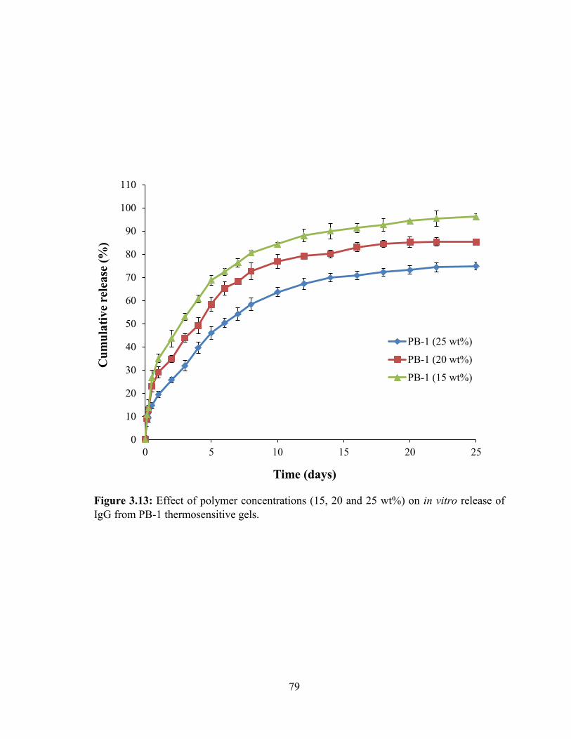
79
Figure 3.13: Effect of polymer concentrations (15, 20 and 25 wt%) on in vitro release of
IgG from PB-1 thermosensitive gels.
0
10
20
30
40
50
60
70
80
90
100
110
0 5 10 15 20 25
Cu
mu
lati
ve
rele
ase
(%
)
Time (days)
PB-1 (25 wt%)
PB-1 (20 wt%)
PB-1 (15 wt%)
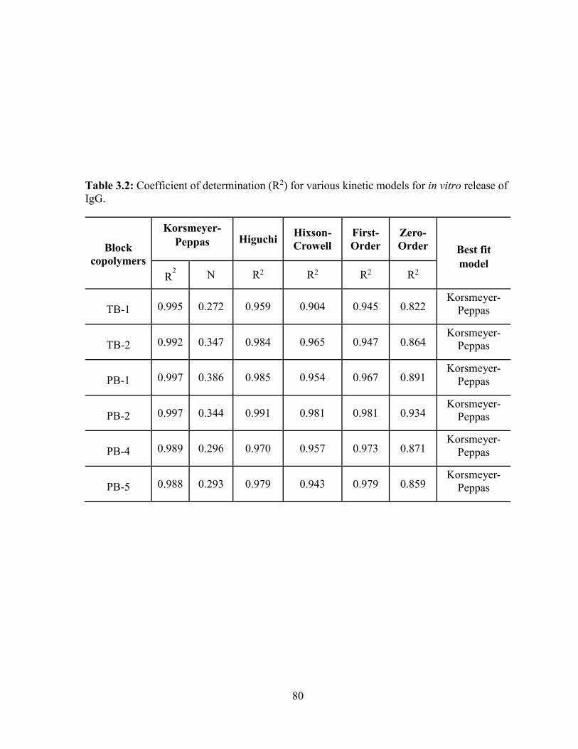
80
Table 3.2: Coefficient of determination (R2) for various kinetic models for in vitro release of
IgG.
Block
copolymers
Korsmeyer-
Peppas Higuchi Hixson-
Crowell
First-
Order
Zero-
Order Best fit
model
R2 N R2 R2 R2 R2
TB-1 0.995 0.272 0.959 0.904 0.945 0.822 Korsmeyer-
Peppas
TB-2 0.992 0.347 0.984 0.965 0.947 0.864 Korsmeyer-
Peppas
PB-1 0.997 0.386 0.985 0.954 0.967 0.891 Korsmeyer-
Peppas
PB-2 0.997 0.344 0.991 0.981 0.981 0.934 Korsmeyer-
Peppas
PB-4 0.989 0.296 0.970 0.957 0.973 0.871 Korsmeyer-
Peppas
PB-5 0.988 0.293 0.979 0.943 0.979 0.859 Korsmeyer-
Peppas
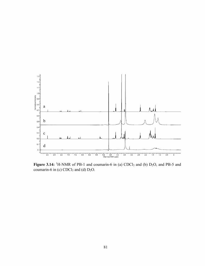
81
Figure 3.14: 1H-NMR of PB-1 and coumarin-6 in (a) CDCl3 and (b) D2O, and PB-5 and
coumarin-6 in (c) CDCl3 and (d) D2O.
a
b
c
d

82
Hypothesis for gelation mechanisms
From the observed results of 1H-NMR, viscosity and micelle size analysis, we propose
two different mechanisms of sol-gel transition for different types of block copolymers related
to, (i) the hydrophobic segments at the terminals (B-A-B (TB-1 and TB-2) or C-B-A-B-C (PB-
1 and PB-2)) and (ii) the hydrophilic segments at the termini (A-B-C-B-A (PB-4 and PB-5)).
According to our hypothesis, both types of copolymers can be dissolved via micellization
where the core is composed of hydrophobic segments and shell is made up of hydrophilic
segments. Therefore, as described in Figure 3.16, interaction of PEG with water molecules
dominated at low temperature (4 °C) allowing the polymer to solubilize (Figure 3.16a). In
contrast, elevation in temperature causes polymer aggregation and initiates the process of
micellization. Therefore, it is possible to solubilize a hydrophobic dye (coumarin-6) in an
aqueous solution of PB-1 copolymers. B-A-B or C-B-A-B-C copolymers possess hydrophobic
termini and hence behave differently in aqueous solution relative to A-B-C-B-A types of
copolymers. In the case of B-A-B or C-B-A-B-C copolymers, during micellization polymer
molecules can serve as intermicellar bridges, where the hydrophobic ends of the polymer are
diffused in the core of different micelles (Figure 3.16b). With a rise in temperature (up to
CGT), progression of intermiceller bridges may initiate miceller aggregation and eventually
enhance the viscosity of solution. Therefore, we have observed significant enhancement in
viscosity of each block copolymer solution as a function of temperature (Table 3.3). Moreover,
the numbers of intermicellar bridges are also proportional to the concentration of block
copolymers at any given temperature. Micellar aggregation and larger micelle size were
observed with an increase in polymer concentration (Figure 3.15). At CGT, intermicellar
bridges and micellar aggregation may be sufficient enough to form the opaque hydrogel
(Figure 3.16c). Further heating of the heterogeneous opaque gel results in polymer
83
precipitation. This may be due to strong hydrophobic interactions between PCL or PCL-PLA
chains that can significantly overcome weaker hydrophilic interactions (hydrogen bonds)
between PEG and water. This phenomenon may eventually dehydrate the PCL or PCL-PLA
core leading to collapse of hydrogel structure (Figure 3.16d).
A-B-C-B-A (PB-4 and PB-5) types of copolymers may also be solubilized by
micellization. However, in case of A-B-C-B-A types of copolymers, micellar arrangements
may lack intermicellar bridges. Therefore, such copolymers demonstrated lower viscosity
relative to B-A-B or C-B-A-B-C types of copolymers at any given temperature. At CGT, A-
B-C-B-A copolymers may aggregate in compact micellar structure resulting in the opaque
hydrogel. Further heating of the gelling solution (above CPT) may lead to dominate
hydrophobic interactions causing dehydrated micelle cores which eventually result in polymer
precipitation.
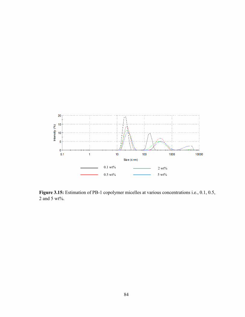
84
Figure 3.15: Estimation of PB-1 copolymer micelles at various concentrations i.e., 0.1, 0.5,
2 and 5 wt%.
0.1 wt%
0.5 wt%
2 wt%
5 wt%
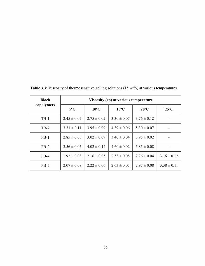
85
Table 3.3: Viscosity of thermosensitive gelling solutions (15 wt%) at various temperatures.
Block
copolymers
Viscosity (cp) at various temperature
5ºC 10ºC 15ºC 20ºC 25ºC
TB-1 2.45 ± 0.07 2.75 ± 0.02 3.30 ± 0.07 3.76 ± 0.12 -
TB-2 3.31 ± 0.11 3.95 ± 0.09 4.39 ± 0.06 5.30 ± 0.07 -
PB-1 2.85 ± 0.05 3.02 ± 0.09 3.40 ± 0.04 3.95 ± 0.02 -
PB-2 3.56 ± 0.05 4.02 ± 0.14 4.60 ± 0.02 5.85 ± 0.08 -
PB-4 1.92 ± 0.03 2.16 ± 0.05 2.53 ± 0.08 2.76 ± 0.04 3.16 ± 0.12
PB-5 2.07 ± 0.08 2.22 ± 0.06 2.63 ± 0.05 2.97 ± 0.08 3.38 ± 0.11
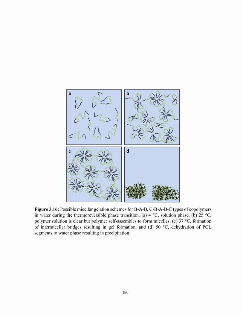
86
Figure 3.16: Possible micellar gelation schemes for B-A-B, C-B-A-B-C types of copolymers
in water during the thermoreversible phase transition. (a) 4 °C, solution phase, (b) 25 °C,
polymer solution is clear but polymer self-assembles to form micelles, (c) 37 °C, formation
of intermicellar bridges resulting in gel formation, and (d) 50 °C, dehydration of PCL
segments to water phase resulting in precipitation.
87
Conclusion
Compositions of PCL-PEG-PCL (B-A-B), PLA-PCL-PEG-PCL-PLA (C-B-A-B-C)
and PEG-PCL-PLA-PCL-PEG (A-B-C-B-A) block copolymers were successfully synthesized
and evaluated for their utility as an injectable in situ hydrogel forming depot for controlled
ocular protein delivery. PB copolymers exhibit significantly reduced crystallinity. It is
anticipated that biodegradability of these novel copolymers is significantly improved relative
to TB copolymers. PB copolymers with A-B-C-B-A block arrangements are easy to handle at
room temperature. Cell viability and biocompatibility studies confirmed that PB copolymers
may be considered safe biomaterials for ocular delivery. More importantly, PB copolymers
also exhibit significant sustained release of IgG relative to TB copolymers. These outcomes
clearly suggest that a PB copolymer based controlled drug delivery system may serve as a
promising platform for back of the eye complications.
88
CHAPTER 4
SUSTAINED DELIVERY OF PROTEINS EMPLOYING NOVEL PENTABLOCK
COPOLYMER BASED NANOPARTICLULATE SYSTEN FOR THE TREATMENT OF
POSTERIOR SEGMENT OCULAR DISEASES
Rationale
A considerable amount of research has been carried out for the development of peptide-
or protein-encapsulated, sustained delivery formulations for the treatment of posterior segment
diseases such as wet AMD and DR. However, physical and chemical instability, and rapid
enzymatic degradation of protein therapeutics are few of the many challenges encountered by
formulation scientists during the development of controlled release formulation. To date,
biodegradable micro- and nanoparticles (NPs) are proven to be the most promising carrier
systems that provide peptide/protein asylum against catalytic enzymes, allowing improvement
in their biological half-lives.
Among all biomaterials, biodegradable polymers such as PLA [188], PCL-PEG-PCL
[189], and PLGA [190] have been extensively investigated for the development of peptide- or
protein-encapsulated NPs. It is widely reported that protein/peptide can undergo a loss of
biological activity during formulation, storage and/or release [191-194]. Presence of
hydrophobic/hydrophilic interface [195], reduction in pH during polymer degradation [196],
and polymer degradation product-induced acylation processes [183, 197] are potential sources
of irreversible aggregation or inactivation of therapeutic proteins inside the PLA, PLGA, PCL-
PLA-PCL and PLA-PEG-PLA based delivery systems. It is imperative to note that any change
in protein/peptide structure, either physically or chemically, may cause toxicity or
immunogenicity and inactivity (loss of pharmacological effect). The consecutive response to
89
antibody directs safety concerns and thus restricts efficacy of subsequent applications [198].
Therefore, there is an urgent need to develop a biodegradable sustained-release system, which
does not compromise protein stability during release. In addition, sustained release of
therapeutic agents for longer durations can eliminate repeated intravitreal injections.
PB copolymers with thermosensitive properties, studied in chapter 3 exhibited
sustained release of IgG up to only ~15-20 days. Sustained release for lower duration and
significantly higher burst release exhibited by thermosensitive gels are unfavorable. Hence in
this study, we have investigated PB copolymers with block arrangements of PLA-PCL-PEG-
PCL-PLA and PGA-PCL-PEG-PCL-PGA for the development of protein-loaded NPs which
can sustain drug release for significantly longer duration of time (~60 days). We have
synthesized and characterized PB copolymer variants to investigate effects of molecular
weight, isomerism and polymer composition on various parameters such as entrapment
efficiency, drug loading, and in vitro drug release profiling. Human immunoglobulin G (IgG,
150 kDa), a full length antibody was utilized as model protein which is very similar to
bevacizumab.
Materials and methods
Materials
PEG (4 kDa), stannous octoate, ε-caprolactone, polyvinylalcohol (PVA),
lipopolysaccharide were procured from Sigma-Aldarich (St. Louis, MO; USA). L-lactide and
D,L-lactide were purchased from Acros organics (Morris Plains, NJ; USA). Micro-BCATM
was obtained from Fisher Scientific. Mouse TNF-α, IL-6 and IL-1β (Ready-Set-Go) ELISA
kits were purchased from eBioscience Inc. Lactate dehydrogenase estimation kit and CellTiter
96® AQueous non-radioactive cell proliferation assay (MTS) kit were obtained from Takara Bio
90
Inc. and Promega Corp., respectively. All other reagents utilized in this study were of analytical
grade. ARPE-19 and RAW-264.7 cells were procured from the American type culture
collection (ATCC).
Methods
Synthesis of TB and PB copolymers
The TB and PB copolymers were synthesized by ring-opening bulk copolymerization
[176]. In brief, PCL-PEG-PCL copolymers were synthesized by copolymerization of ε-
caprolactone on the hydroxyl ends of PEG (4 kDa). In this reaction, PEG was utilized as
macroinitiator and stannous octoate (0.5 wt%) as a catalyst. To synthesize PCL-PEG-PCL, a
pre-determined amount of PEG was vacuum-dried for 4 h followed by addition of ε-
caprolactone and catalyst. Reaction was carried out in a closed vessel under nitrogen, at 130
°C for 36 h. The reaction mixture was solubilized in DCM followed by precipitation in ice-
cold diethyl ether. The precipitated polymer was vacuum-dried to remove residual solvent and
characterized to evaluate the reaction yield. The structure and molecular weight of TB
copolymers were confirmed by 1H-NMR and GPC.
In order to prepare PB copolymers, a predetermined amount of TB copolymer was
utilized as macroinitiator and stannous octoate (0.5 wt%) as catalyst. For the synthesis of PB-
A and PB-D, L-lactide was polymerized on the hydroxyl ends of TB copolymer. Similarly,
PB-B/PB-E, and PB-C/PB-F were synthesized by polymerizing D,L-lactide and glycolide,
respectively. For the synthesis of PB-A, PB-B, PB-D and PB-E, the reaction was carried out
at 130 °C for 36 h. However, to synthesis of PB-C and PB-F, reaction was performed at 200
°C for 24 h.
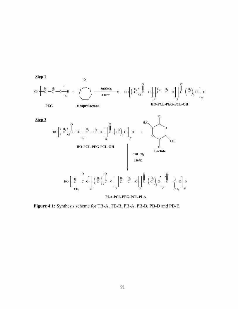
91
Figure 4.1: Synthesis scheme for TB-A, TB-B, PB-A, PB-B, PB-D and PB-E.
OHH2C
H2C O H
x
O
O
+ HOH2C C
O
OH2C
H2C O C
O
H2C O
5 5H
y yx
Sn(Oct)2
130°C
caprolactonePEG HO-PCL-PEG-PCL-OH
HOH2C C
O
OH2C
H2C O C
O
H2C O
5 5H
y yx
HO-PCL-PEG-PCL-OH
Sn(Oct)2
130°C
OH2C C
O
OH2C
H2C O C
O
H2C O
5 5C
y yx
C
O
HC
CH3
HO
O
HC
CH3
O
z z
+
O
O
O
O
H3C
CH3
PLA-PCL-PEG-PCL-PLA
Step 1
Step 2
Lactide
H
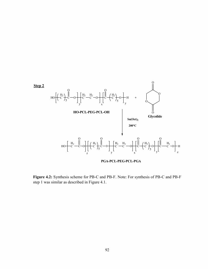
92
Figure 4.2: Synthesis scheme for PB-C and PB-F. Note: For synthesis of PB-C and PB-F
step 1 was similar as described in Figure 4.1.
HOH2C C
O
OH2C
H2C O C
O
H2C O
5 5H
y yx
HO-PCL-PEG-PCL-OH
Sn(Oct)2
200°C
OH2C C
O
OH2C
H2C O C
O
H2C O
5 5C
y yx
C
O
H2CHO
O
H2C O
z z
+
O
O
O
O
PGA-PCL-PEG-PCL-PGA
Step 2
Glycolide
H
93
In order to remove catalyst and unreacted monomers, the reaction mixture was
dissolved in DCM and precipitated by addition of ice-cold diethyl ether. Polymers were
vacuum-dried and characterized for their structure and polydispersity by employing 1H-NMR
and GPC as analytical techniques. Purified polymers were stored at -20 °C until further use.
Reaction scheme for the synthesis of TB-A, TB-B, PB-A, PB-B, PB-D and PB-E are described
in Figure 4.1 whereas for the synthesis of PB-C/PB-F are described in Figure 4.2.
Characterization of polymers
Polymers were characterized for purity, molecular weight and polydispersity by 1H-
NMR and GPC. Polymers were further evaluated by powder XRD to examine their crystalline
nature.
1H-NMR
To perform 1H-NMR spectroscopy, polymeric material was dissolved in CDCl3 and
analyzed on a Varian-400 MHz NMR spectrometer. Purity and molecular weight (Mn) of the
polymers were confirmed from the 1H-NMR spectrum.
GPC
To further confirm purity, molecular weight and polydispersity, polymeric samples
were analyzed via GPC equipped with refractive index detector (Waters 410). Briefly, 5 mg of
polymeric material was dissolved in tetrahydrofuran (THF). THF was utilized as eluting
solvent at the flow rate of 1 mL/min whereas separation was carried out on Styragel HR-3
column. Polystyrene samples with narrow molecular weight distribution were utilized as
standards.
94
X-ray diffraction (XRD) analysis of copolymers
Samples were analyzed for diffraction patterns to understand the effects of polymer
composition (TB vs PB), molecular weight, and block types (P(L)LA, P(DL)LA and PGA) on
the crystallinity of copolymers. MiniFlex automated X-ray diffractometer (Rigaku, The
Woodlands, Texas) with Ni-filtered Cu-kα radiation (30 kV and 15 mA) was employed to
study the diffraction patterns. XRD analysis was carried out at room temperature.
In vitro cytotoxicity studies
Cell culture
RAW-264.7 cells were cultured and maintained in Dubelcco's modified Eagle medium
(DMEM) supplemented with 10% FBS, 100 mg/L of streptomycin and 100 U/L of penicillin.
Human retinal pigment epithelial cells (ARPE-19) were cultured and maintained according to
the protocol reported from our lab [199]. In brief, ARPE-19 cells were cultured in DMEM/F-
12 medium containing 10% heat-inactivated fetal bovine serum (FBS), 29 mM of sodium
bicarbonate, 15 mM of HEPES, 100 mg/L of streptomycin and 100 U/L of penicillin. Both cell
lines were maintained in humidified atmosphere at 37 °C and 5% CO2.
Lactate Dehydrogenase (LDH) assay
The cytotoxicity of block copolymers were evaluated according to previously
published protocol with minor modifications [200]. Briefly, different concentrations (1-20
mg/mL) of TB and PB copolymers were prepared in acetonitrile (ACN). A 100 µL of solutions
was aliquoted in each well of 96-well cell culture plates. In order to evaporate ACN and to
sterilize block copolymers, cell culture plates were exposed overnight under UV light (laminar
flow). Once the ACN is evaporated, 1.0 x 10⁴ of RAW 264.7 cells were seeded in each well
and incubated for 48 h at 37 °C and 5% CO2 in humidified atmosphere. Followed by
95
appropriate incubation, cell supernatant were analyzed for the levels of LDH by LDH detection
kit. LDH assay was performed according to the supplier’s protocol. Samples were analyzed at
450 nm by 96-well plate reader. Amount of released LDH is directly proportional to
cytotoxicity of the polymers. In this study, more than 10% of LDH release was considered as
cytotoxic. To evaluate toxicity of block copolymers on retinal cell line, the similar experiment
was performed with ARPE-19 cells. LDH release (%) was calculated by employing following
equation,
𝐿𝐷𝐻 𝑟𝑒𝑙𝑒𝑎𝑠𝑒(%) =𝐴𝑏𝑠. 𝑜𝑓 𝑆𝑎𝑚𝑝𝑙𝑒 − 𝐴𝑏𝑠. 𝑜𝑓 𝑛𝑒𝑔𝑎𝑡𝑖𝑣𝑒 𝑐𝑜𝑛𝑡𝑟𝑜𝑙
𝐴𝑏𝑠. 𝑜𝑓 𝑝𝑜𝑠𝑖𝑡𝑖𝑣𝑒 𝑐𝑜𝑛𝑡𝑟𝑜𝑙 − 𝐴𝑏𝑠. 𝑜𝑓 𝑛𝑒𝑔𝑎𝑡𝑖𝑣𝑒 𝑐𝑜𝑛𝑡𝑟𝑜𝑙∗ 100 … Eq. 4.1
MTS assay
In order to confirm safety of PB copolymers, in vitro cell viability (MTS) assay was
performed. MTS assay was carried out according to previously published protocol with minor
modifications [185]. Different concentrations of block copolymer solutions were prepared,
aliquoted and sterilized according the procedure mentioned in previous section. After
sterilization, RAW-264.7 cells at the density of 1.0 x 10⁴ cells per well were seeded in 96-well
plate. Cells were incubated for 48 h at 37 °C and 5% CO2 in humidified atmosphere. After
incubation, cell culture medium was replaced with 100 µL of serum free medium containing
20 µL of MTS solution. Cells were further incubated at 37 °C and 5% CO2 for 4 h. Absorbance
of each well was determined at 450 nm. Polymer concentrations at which more than 90% of
cell viability was observed, were considered as non-toxic. Percent cell viability was estimated
by following equation. The similar experiment was repeated with ARPE-19 cells.
𝐶𝑒𝑙𝑙 𝑣𝑖𝑎𝑏𝑖𝑙𝑖𝑡𝑦 (%) =𝐴𝑏𝑠. 𝑜𝑓 𝑆𝑎𝑚𝑝𝑙𝑒 − 𝐴𝑏𝑠. 𝑜𝑓 𝑛𝑒𝑔𝑎𝑡𝑖𝑣𝑒 𝑐𝑜𝑛𝑡𝑟𝑜𝑙
𝐴𝑏𝑠. 𝑜𝑓 𝑝𝑜𝑠𝑖𝑡𝑖𝑣𝑒 𝑐𝑜𝑛𝑡𝑟𝑜𝑙 − 𝐴𝑏𝑠. 𝑜𝑓 𝑛𝑒𝑔𝑎𝑡𝑖𝑣𝑒 𝑐𝑜𝑛𝑡𝑟𝑜𝑙∗ 100 … Eq. 4.2
96
In vitro biocompatibility studies
As described in previous section, different concentrations (1-20 mg/mL) of block
copolymer solutions were prepared in acetonitrile. A 200 µL aliquot of polymer solution was
added in each well of 48-well cell culture plate. These plates were incubated overnight under
UV light (laminar flow) for sterilization as well as for evaporation of ACN. After sterilization,
RAW-264.7 cells were plated at the cell density of 5.0 x 10⁴ per well. Cells were exposed to
polymer for 48 h at 37 °C and 5% CO2. After incubation, cell supernatant was analyzed for the
quantification of three different cytokines, i.e., TNF-α, IL-6 and IL-1β. Levels of cytokines
were estimated by ELISA method. ELISA was performed according to the manufacturer’s
instruction. Calibration curves for TNF-α, IL-6 and IL-1β were prepared in the range of 10-
750 pg/mL, 5-500 pg/mL and 10-500 pg/mL, respectively.
Preparation of NPs
NPs were prepared by W1/O/W2 double emulsion solvent evaporation method
according to previously published protocol with minor modifications [201]. Briefly,
predetermined amount of IgG was dissolved in water (1 mL) containing 50 µL of Tween-80
(W1 phase) and 100 mg of respective block copolymer was dissolved in 4 mL of
dichloromethane (DCM) comprising 50 µL of Span-20 (organic phase). W1/O primary
emulsion was prepared by drop-wise addition of W1 phase in organic phase under constant
sonication for 1 min at 4 W output. Immediately after sonication, W1/O primary emulsion was
added (drop-wise) in 20 mL of 2% PVA solution (W2) and sonication was applied for 4 min at
5 W output. W1/O/W2 double emulsion was stirred for 30 min at room temperature followed
by evaporation of DCM under vacuum. The resulting NPs were centrifuged at 20000 rpm for
30 min at 4 °C followed by two washing cycles with distilled deionized water. NPs were freeze-
97
dried in 5% mannitol solution and stored at -20 °C until further characterization. NPs were
prepared utilizing three different drug to polymer ratios i.e., 1:10, 1:15 and 1:20. Freeze-dried
NPs were characterized for particle size, EE, DL and in vitro drug release behavior.
Characterization of NPs
Particle size
NPs (1 mg/mL) were suspended in DDW and subjected to particle size analysis.
Particle size was evaluated at room temperature and 90° scattering angel utilizing Zeta sizer
(Zetasizer Nano ZS, Malvern Instruments Ltd, Worcestershire, UK). All the samples were
analyzed in triplicate and average particle size were reported in ± SD.
Entrapment efficiency (EE %) and drug loading (DL %)
IgG-loaded freeze-dried NPs were examined for the estimation of DL and EE. EE was
calculated by analyzing supernatants (collected during NP preparation) with Micro BCATM
protein estimation kit. To evaluate DL, 2 mg equivalent drug-encapsulated NPs were dissolved
in 200 µL of DMSO. The resulting solution was subjected for protein estimation via UV
absorbance spectroscopy. A standard curve of IgG ranging from 31.25 to 2000 µg/mL was
prepared in DMSO. EE (%) and DL (%) were estimated according to the following equations.
EE (%) was calculated with eq. 4.3
%𝐸𝐸 = (1 −𝐴𝑚𝑜𝑢𝑛𝑡 𝑜𝑓 𝑑𝑟𝑢𝑔 𝑖𝑛 𝑠𝑢𝑝𝑒𝑟𝑛𝑎𝑡𝑎𝑛𝑡
𝑇𝑜𝑡𝑎𝑙 𝑎𝑚𝑜𝑢𝑛𝑡 𝑜𝑓 𝑑𝑟𝑢𝑔) ∗ 100 …Eq. 4.3
DL (%) was calculated by eq. 4.4.
%𝐷𝐿 = (𝐴𝑚𝑜𝑢𝑛𝑡 𝑜𝑓 𝑑𝑟𝑢𝑔 𝑖𝑛 𝑛𝑎𝑛𝑜𝑝𝑎𝑟𝑡𝑖𝑐𝑙𝑒𝑠
𝑇𝑜𝑡𝑎𝑙 𝑎𝑚𝑜𝑢𝑛𝑡 𝑜𝑓 𝑑𝑟𝑢𝑔 𝑎𝑛𝑑 𝑝𝑜𝑙𝑦𝑚𝑒𝑟) ∗ 100 …Eq. 4.4
98
In vitro release studies
For in vitro drug release studies, 1 mg protein equivalent IgG-encapsulated NPs were
dispersed in 1 mL of 0.1 M phosphate buffer saline (pH - 7.4). The NPs suspension was
incubated in water bath maintained at 37 °C. In order to collect release samples, at pre-
determined time intervals NPs suspension were centrifuged at 13000 rpm for 30 min and 4 °C.
A 200 µL of clear supernatant was withdrawn for protein estimation and replaced with same
volume of 0.1 M PBS. NPs where then resuspended and release was continued at 37 °C. In
vitro release experiments were repeated in triplicates and mean value ± SD was expressed as
cumulative % drug released with time.
Stability estimation of IgG
Secondary structure analysis by circular dichroism (CD) spectroscopy
To confirm the stability of secondary structure of released IgG, CD spectroscopy was
employed as an analytical technique. CD analysis was performed using Jasco 720
spectropolarimeter at 25 °C. CD spectra were recorded at scanning speed of 5 nm/min between
the range of 200 to 250 nm utilizing 1 cm cell, and 1 nm band width. CD measurements were
described as molar elipticity [θ]. A CD spectrum of PBS was used as blank.
Results and discussion
In this study, we have prepared novel PB copolymers which are composed of FDA
approved polymer blocks such as PEG, PCL, and PLA/PGA. Each block plays an important
role such as presence of PEG helps to improve stability of NPs by reducing NP aggregation,
and also prevents phagocytosis by macrophages resulting in improved half-life. PCL is a
slowly degrading, semi-crystalline polymer which improves protein loading in NPs and also
sustains drug release for longer duration of time. Such a slow degradation is also not
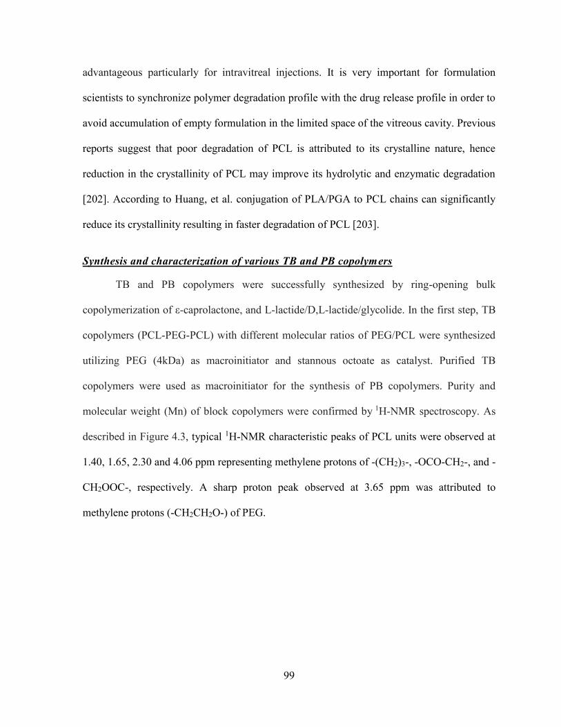
99
advantageous particularly for intravitreal injections. It is very important for formulation
scientists to synchronize polymer degradation profile with the drug release profile in order to
avoid accumulation of empty formulation in the limited space of the vitreous cavity. Previous
reports suggest that poor degradation of PCL is attributed to its crystalline nature, hence
reduction in the crystallinity of PCL may improve its hydrolytic and enzymatic degradation
[202]. According to Huang, et al. conjugation of PLA/PGA to PCL chains can significantly
reduce its crystallinity resulting in faster degradation of PCL [203].
Synthesis and characterization of various TB and PB copolymers
TB and PB copolymers were successfully synthesized by ring-opening bulk
copolymerization of ε-caprolactone, and L-lactide/D,L-lactide/glycolide. In the first step, TB
copolymers (PCL-PEG-PCL) with different molecular ratios of PEG/PCL were synthesized
utilizing PEG (4kDa) as macroinitiator and stannous octoate as catalyst. Purified TB
copolymers were used as macroinitiator for the synthesis of PB copolymers. Purity and
molecular weight (Mn) of block copolymers were confirmed by 1H-NMR spectroscopy. As
described in Figure 4.3, typical 1H-NMR characteristic peaks of PCL units were observed at
1.40, 1.65, 2.30 and 4.06 ppm representing methylene protons of -(CH2)3-, -OCO-CH2-, and -
CH2OOC-, respectively. A sharp proton peak observed at 3.65 ppm was attributed to
methylene protons (-CH2CH2O-) of PEG.
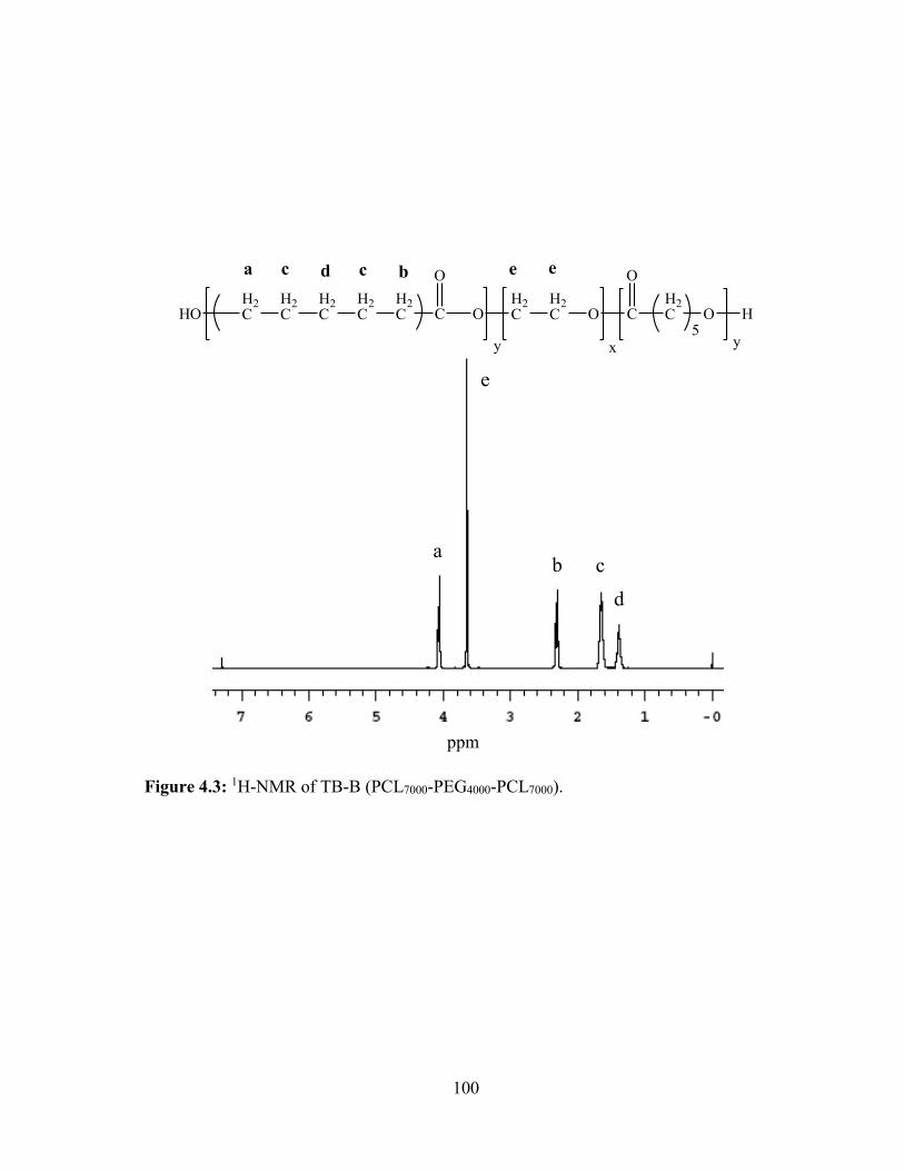
100
Figure 4.3: 1H-NMR of TB-B (PCL7000-PEG4000-PCL7000).
HO C
O
OH2C
H2C O C
O
H2C O
5H
y yx
H2C
H2C
H2C
H2C
H2C
a bc cd e e
a
e
b c
d
ppm
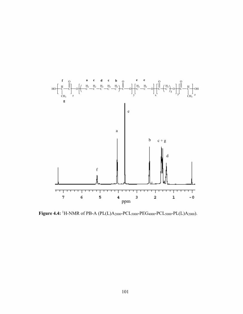
101
Figure 4.4: 1H-NMR of PB-A (PL(L)A2000-PCL5000-PEG4000-PCL5000-PL(L)A2000).
O C
O
OH2C
H2C O C
O
H2C O
5y yx
H2C
H2C
H2C
H2C
H2C
a bc cd e e
C
O
HC
CH3
HO
z
f
g
C
O
HC
CH3
OH
z
f
a
e
b c + g
d
ppm
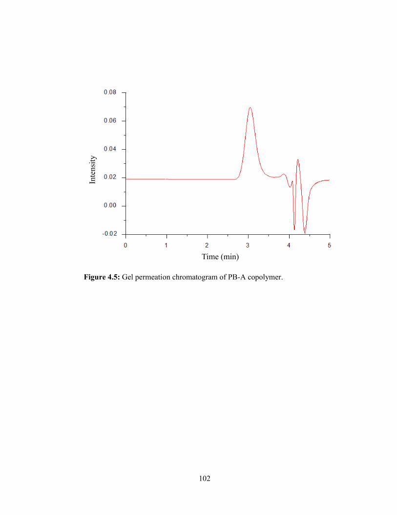
102
Figure 4.5: Gel permeation chromatogram of PB-A copolymer.
Inte
nsi
ty
Time (min)
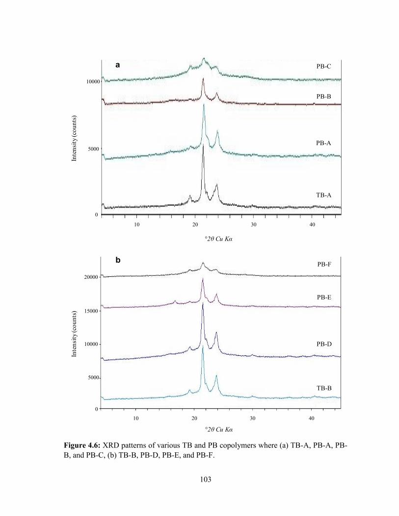
103
Figure 4.6: XRD patterns of various TB and PB copolymers where (a) TB-A, PB-A, PB-
B, and PB-C, (b) TB-B, PB-D, PB-E, and PB-F.
10000
5000
0
10 20 30 40
Inte
nsi
ty (co
un
ts)
2θ Cu Kα
TB-A
PB-A
PB-B
PB-Ca
10000
5000
0
10 20 30 40
Inte
nsi
ty (co
un
ts)
2θ Cu Kα
TB-B
PB-D
PB-E
PB-Fb
15000
20000

104
Table 4.1: List of TB and PB copolymers studied
a. Theoretical value, calculated according to the feed ratio
b. Calculated from 1H-NMR results
c. Determined by GPC analysis
Code Structures
PLA/PCL/PEG
or
PGA/PCL/PEG
Total Mna
(theoretical)
Total Mnb
(calculated)
Total Mnc
(calculated)
Mwc
(GPC) PDc
TB-A PCL5000-PEG4000-PCL5000 2.5/1 14000 13350 10990 14520 1.32
TB-B PCL7000-PEG4000-PCL7000 3.5/1 18000 16650 14560 18800 1.29
PB-A PL(L)A2000-PCL5000-PEG4000-PCL5000-PL(L)A2000 1/2.5/1 18000 16950 14990 20800 1.39
PB-B PL(DL)A2000-PCL5000-PEG4000-PCL5000-PL(DL)A2000 1/2.5/1 18000 16880 13810 19560 1.42
PB-C PGA2000-PCL5000-PEG4000-PCL5000-PGA2000 1/2.5/1 18000 17210 13450 19210 1.43
PB-D PL(L)A3000-PCL7000-PEG4000-PCL7000-PL(L)A3000 1.5/3.5/1 24000 21350 17640 23830 1.35
PB-E PL(DL)A3000-PCL7000-PEG4000-PCL7000-PL(DL)A3000 1.5/3.5/1 24000 21330 18210 24940 1.37
PB-F PGA3000-PCL7000-PEG4000-PCL7000-PGA3000 1.5/3.5/1 24000 21290 16180 23210 1.43
105
1H-NMR spectra of PB copolymers with PLA as terminals (PB-A, PB-B, PB-D and
PB-E) exhibited two additional peaks at 1.50 (-CH3) and 5.17 (-CH-) ppm (Figure 4.4).
However, PB-C and PB-E exhibited cluster of singlets between 4.6 to 4.9 ppm representing
methylene protons (-CH2-) of PGA units (data not shown). The [EO]-[CL]-[LA] molar ratio of
the final products were calculated from the integration values of PEG signal at 3.65 ppm, PCL
signal at 2.30 ppm and PLA signal at 5.17 ppm. In case of PB-C and PB-F copolymers, proton
signals between 4.6 to 4.9 ppm were utilized for the calculation of molar ratio.
In order to confirm the molecular weight and purity, all the block copolymers were
further analyzed by GPC. Block copolymers exhibited a monodistributed molecular weight
and absence of any other homopolymers such as PEG, PCL, PLA or PGA. A typical GPC
chromatogram of PB-A copolymer is depicted in Figure 4.5. Moreover, calculated molecular
weights were very close to the feed ratio, and polydispersity of block copolymers were well
below 1.45 indicating narrow distribution of molecular weights. Table 4.1 represents
theoretical molecular weights, calculated molecular weights (1H-NMR and GPC) and
polydispersity. The Mn values obtained from 1H-NMR were noticeably higher relative to the
Mn values observed from GPC analysis. This observation may attributed to the difference in
hydrodynamic diameter of block copolymers relative to parent homopolymers. As reported in
Table 4.1, observed molecular weights were very similar to theoretical molecular weights. For
the ease, in the following text, theoretical molecular weights will be mentioned instead of
calculated molecular weights.
Reports published elsewhere described that covalent conjugation between PLA and
PCL, significantly reduces the crystallinity of PCL [176, 204-207]. However, effects of various
isomers of PLA (L or D,L) and also an effect of PGA on the crystallinity of PCL-PEG-PCL
106
tri-block copolymers are not reported, yet. Hence, we have analyzed TB and PB copolymers
for their XRD patterns. PLA with D,L-lactide has amorphous structure whereas PLA with L-
lactide is a semi-crystalline [202]. Both polymers may act differently to reduce the crystallinity
of PCL. This may have direct effect on various formulation parameters such as EE, DL and in
vitro release. Moreover, we have also evaluated the effect of PGA to reduce the crystallinity
of PCL-PEG-PCL TB copolymers.
Figure 4.6 describes the XRD patterns of various TB and PB copolymers. As reported
in Figure 4.6a, TB-A exhibited two strong characteristic crystalline peaks of PCL blocks at
diffraction angles (2θ) of 21.5° and 23.8°. L-lactide containing PB copolymer (PB-A) exhibited
reduced intensity of PCL peaks, which suggest that conjugation of PLA (L-lactide) has
significantly diminished the crystallinity of TB-A. Noticeably, conjugation of D,L-lactide has
further reduced the crystalline peaks of PCL blocks. These results may be attributed to the fact
that PLA with L-lactide is more crystalline than PLA with D,L-lactide. Moreover, PLA with
D,L-lactide have a random arrangement of L and D-lactide in their polymer chain which does
not allow systemic arrangement of polymer chains, resulting in reduced crystallinity.
Surprisingly, PB-C demonstrated amorphous structure suggesting that conjugation of PGA at
the termini of TB-A have totally eliminated crystallinity of PCL. To further confirm this
behavior, we have evaluated the solid states of TB-B, PB-D, PB-E and PB-F by XRD. Results
described in Figure 4.6b demonstrated similar pattern, where PB-F with PGA a block was
amorphous in nature. PLA with L-lactide reduced peak intensity of PCL up to some extent,
while PLA with D,L-lactide has significantly diminished crystalline peaks.
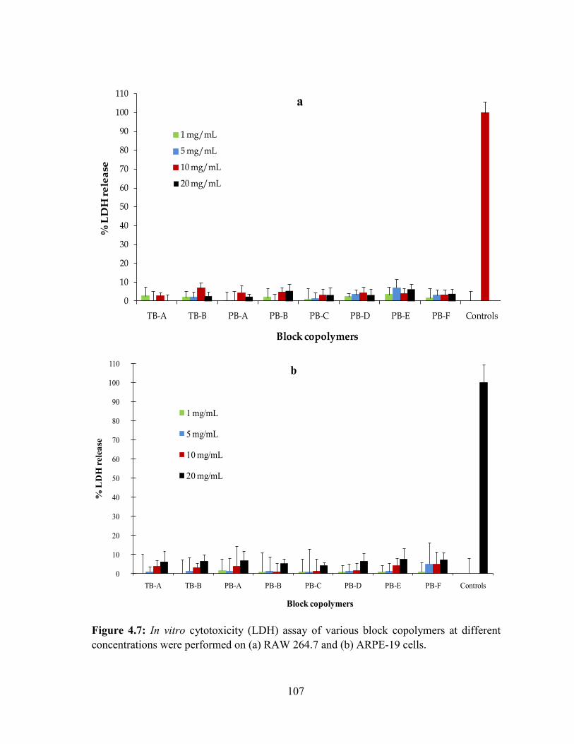
107
Figure 4.7: In vitro cytotoxicity (LDH) assay of various block copolymers at different
concentrations were performed on (a) RAW 264.7 and (b) ARPE-19 cells.
0
10
20
30
40
50
60
70
80
90
100
110
TB-A TB-B PB-A PB-B PB-C PB-D PB-E PB-F Controls
% L
DH
re
lea
se
Block copolymers
1 mg/mL
5 mg/mL
10 mg/mL
20 mg/mL
a
0
10
20
30
40
50
60
70
80
90
100
110
TB-A TB-B PB-A PB-B PB-C PB-D PB-E PB-F Controls
% L
DH
rele
ase
Block copolymers
1 mg/mL
5 mg/mL
10 mg/mL
20 mg/mL
b
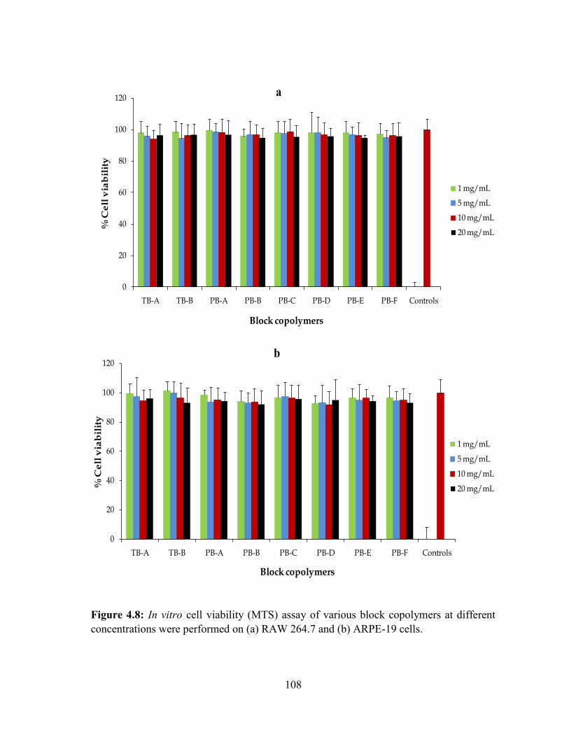
108
Figure 4.8: In vitro cell viability (MTS) assay of various block copolymers at different
concentrations were performed on (a) RAW 264.7 and (b) ARPE-19 cells.
0
20
40
60
80
100
120
TB-A TB-B PB-A PB-B PB-C PB-D PB-E PB-F Controls
% C
ell
via
bil
ity
Block copolymers
1 mg/mL
5 mg/mL
10 mg/mL
20 mg/mL
a
0
20
40
60
80
100
120
TB-A TB-B PB-A PB-B PB-C PB-D PB-E PB-F Controls
% C
ell
via
bil
ity
Block copolymers
1 mg/mL
5 mg/mL
10 mg/mL
20 mg/mL
b
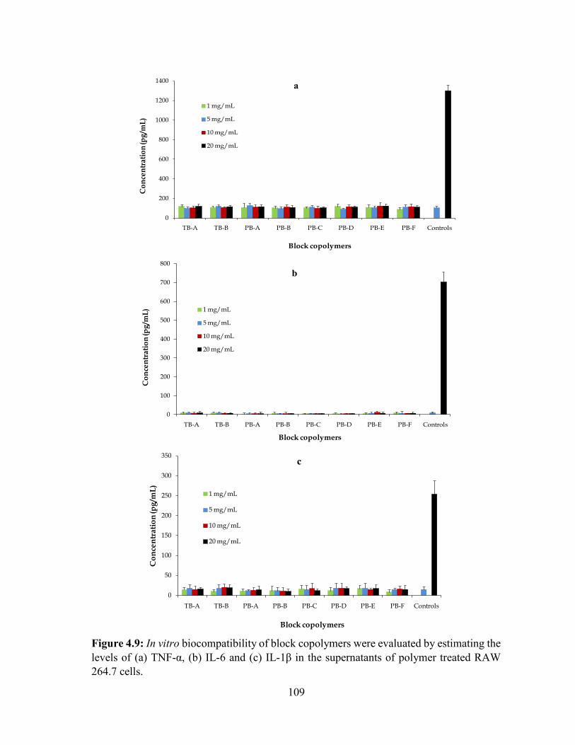
109
Figure 4.9: In vitro biocompatibility of block copolymers were evaluated by estimating the
levels of (a) TNF-α, (b) IL-6 and (c) IL-1β in the supernatants of polymer treated RAW
264.7 cells.
0
200
400
600
800
1000
1200
1400
TB-A TB-B PB-A PB-B PB-C PB-D PB-E PB-F Controls
Co
nce
ntr
ati
on
(pg
/mL
)
Block copolymers
1 mg/mL
5 mg/mL
10 mg/mL
20 mg/mL
a
0
100
200
300
400
500
600
700
800
TB-A TB-B PB-A PB-B PB-C PB-D PB-E PB-F Controls
Co
nce
ntr
ati
on
(pg
/ mL
)
Block copolymers
1 mg/mL
5 mg/mL
10 mg/mL
20 mg/mL
b
0
50
100
150
200
250
300
350
TB-A TB-B PB-A PB-B PB-C PB-D PB-E PB-F Controls
Co
nce
ntr
ati
on
(pg
/ mL
)
Block copolymers
1 mg/mL
5 mg/mL
10 mg/mL
20 mg/mL
c
110
In vitro cytotoxicity studies
LDH assay
Lactate dehydrogenase (LDH) is the cytosolic enzyme, secreted in the culture medium
upon the rupture of cell membrane. As the cell membrane is the potential site for polymer-cell
interaction, measuring the amount of LDH release has been the preferred way to estimate the
membrane damage and hence the cytotoxicity of polymer. As described in Figure 4.7a (RAW-
264.7 cells) and Figure 4.7b (ARPE-19), LDH release upon the exposure of block copolymers
were less than 10% at any given concentrations and also comparable with negative control
indicating negligible or no toxicity.
MTS assay
In order to confirm the results observed in cytotoxicity (LDH assay) studies, the safety
of block copolymers were further evaluated by MTS assay. Block copolymers exhibited more
than 90% cell viability upon exposure to various concentrations ranging from 1-20 mg/mL
(Figures 4.8a and 4.8b). Moreover, results were comparable to the negative control indicating
no toxicity of PB copolymers. Results observed in LDH and MTS assays clearly indicated that
block copolymers are very safe for the intravitreal applications.
In vitro biocompatibility studies
It is also important to confirm that newly synthesized PB copolymers are not producing
inflammatory responses upon intravitreal administration. In vitro assessment for release of
cytokines upon exposure to polymer is a quick, cost effective and reliable technique to examine
biocompatibility of polymers. RAW-264.7 cells, is a well-established in vitro cell culture
model to study inflammatory responses of polymers intended for human applications. Results
described in Figures 4.9a, 4.9b and 4.9c have demonstrated negligible release of TNFα, IL-6
111
and IL-1β upon 24 h exposure to different concentrations of block copolymers ranging from
1-20 mg/mL. Release of any cytokines was not significantly different to their respective
negative controls indicating excellent biocompatibility of block copolymers for ocular
applications.
Characterization of NPs
Particle size
IgG-loaded TB and PB NPs were prepared by the W1/O/W2 double emulsion solvent
evaporation method. As described in Tables 4.2, 4.3 and 4.4, the size of NPs were between 285
nm to 330 nm. We did not observe any significant effect of polymer composition on the particle
size. Moreover, in order to understand the effect of drug to polymer ratio on particle size, we
have prepared NPs with three distinct drug to polymer ratios i.e., 1:10, 1:15 and 1:20.
Interestingly, a change in drug to polymer ratio did not exhibit any noticeable effect on particle
size. These results suggest that particle size is highly influenced by process parameters and not
by polymer:drug ratio.
Entrapment efficiency (EE) and drug loading (DL)
EE and DL are influenced by many parameters including copolymer composition
(isomerism, molecular weight and polymer structure) and volumes of various phases (W1, O
and W2). To understand the effect of copolymer compositions on DL and EE, we kept volumes
of W1, O and W2 phases constant. In addition, we have also studied the effect of drug to
polymer ratio on DL and EE. As described in Table 4.2, at respective drug to polymer ratios,
TB-B NPs exhibited significantly higher DL and EE relative to TB-A NPs. PB-D, PB-E and
PB-F (Table 4.3) with significantly higher molecular weight relative to PB-A, PB-B and PB-
C (Table 4.4), respectively exhibited improved EE and DL.
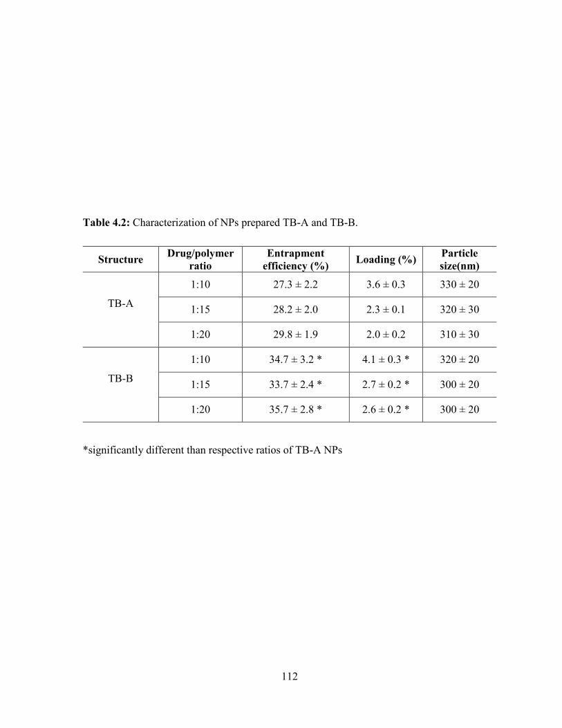
112
Table 4.2: Characterization of NPs prepared TB-A and TB-B.
*significantly different than respective ratios of TB-A NPs
Structure Drug/polymer
ratio
Entrapment
efficiency (%) Loading (%)
Particle
size(nm)
TB-A
1:10 27.3 ± 2.2 3.6 ± 0.3 330 ± 20
1:15 28.2 ± 2.0 2.3 ± 0.1 320 ± 30
1:20 29.8 ± 1.9 2.0 ± 0.2 310 ± 30
TB-B
1:10 34.7 ± 3.2 * 4.1 ± 0.3 * 320 ± 20
1:15 33.7 ± 2.4 * 2.7 ± 0.2 * 300 ± 20
1:20 35.7 ± 2.8 * 2.6 ± 0.2 * 300 ± 20
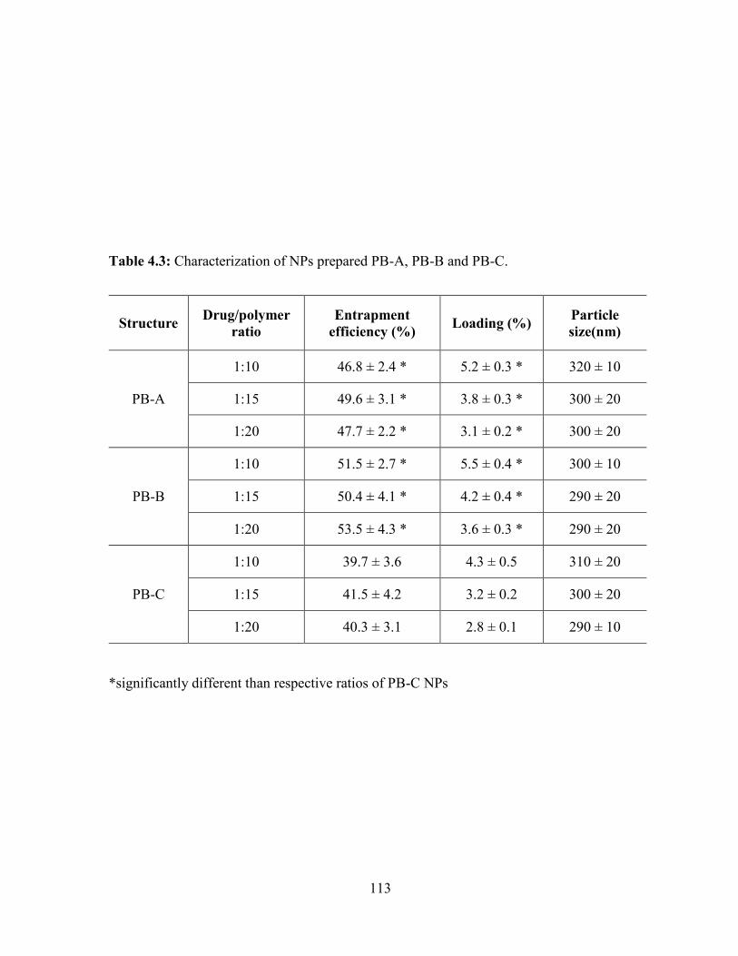
113
Table 4.3: Characterization of NPs prepared PB-A, PB-B and PB-C.
*significantly different than respective ratios of PB-C NPs
Structure Drug/polymer
ratio
Entrapment
efficiency (%) Loading (%)
Particle
size(nm)
PB-A
1:10 46.8 ± 2.4 * 5.2 ± 0.3 * 320 ± 10
1:15 49.6 ± 3.1 * 3.8 ± 0.3 * 300 ± 20
1:20 47.7 ± 2.2 * 3.1 ± 0.2 * 300 ± 20
PB-B
1:10 51.5 ± 2.7 * 5.5 ± 0.4 * 300 ± 10
1:15 50.4 ± 4.1 * 4.2 ± 0.4 * 290 ± 20
1:20 53.5 ± 4.3 * 3.6 ± 0.3 * 290 ± 20
PB-C
1:10 39.7 ± 3.6 4.3 ± 0.5 310 ± 20
1:15 41.5 ± 4.2 3.2 ± 0.2 300 ± 20
1:20 40.3 ± 3.1 2.8 ± 0.1 290 ± 10
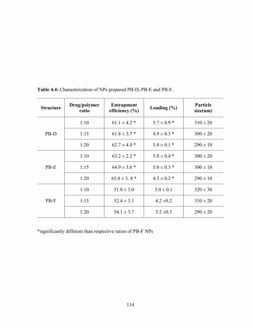
114
Table 4.4: Characterization of NPs prepared PB-D, PB-E and PB-F.
*significantly different than respective ratios of PB-F NPs
Structure Drug/polymer
ratio
Entrapment
efficiency (%) Loading (%)
Particle
size(nm)
PB-D
1:10 61.1 ± 4.2 * 5.7 ± 0.9 * 310 ± 20
1:15 61.8 ± 3.7 * 4.9 ± 0.3 * 300 ± 20
1:20 62.7 ± 4.0 * 3.8 ± 0.1 * 290 ± 10
PB-E
1:10 63.2 ± 2.2 * 5.8 ± 0.4 * 300 ± 20
1:15 64.9 ± 3.6 * 5.0 ± 0.3 * 300 ± 10
1:20 65.8 ± 3. 8 * 4.3 ± 0.2 * 290 ± 10
PB-F
1:10 51.0 ± 3.0 5.0 ± 0.1 320 ± 30
1:15 52.4 ± 3.1 4.2 ±0.2 310 ± 20
1:20 54.1 ± 3.7 3.2 ±0.3 290 ± 20
115
It is important to note that higher molecular weights of block copolymers acquire longer
polymer chain with significantly higher hydrophobicity relative to lower molecular weight
block copolymers. Additionally, during the step of solvent evaporation, high hydrophobicity
(PB-D, PB-E and PB-F) may allow faster polymer precipitation to form NPs preventing
diffusion of IgG in the external aqueous (W2) phase.
Interestingly, we have also observed significantly higher entrapment and loading
efficiency in PLA based PB copolymers (PB-A, PB-B, PB-D and PB-E) relative to PGA based
PB copolymers (PB-C and PB-F). These results may be attributed to the fact that PLA is more
hydrophobic compare to PGA, hence its copolymers were also hydrophobic than that of PGA
based copolymers. Due to higher hydrophilicity, PB-C and PB-F copolymers may have higher
interaction with W1 and W2 phase which may allow IgG to escape from W1 to W2 phase (during
NP preparation) resulting in lower EE and DL. In contrast, PLA based PB copolymers may
have inhibited or reduced the diffusion of IgG from W1 to W2 phase ensuring improved DL
and EE.
In vitro release studies
In vitro drug release studies were performed to understand the effects of isomerism
(P(L)LA vs P(D,L)LA), molecular weight and polymer composition. IgG-loaded NPs with
drug to polymer ratio of 1:10 were utilized for release study for all polymer compositions. In
vitro release profiles of IgG from TB-A and TB-B NPs are compared in Figure 4.10. Both NPs
demonstrated bi-phasic release profile i.e., initial burst release phase followed by a phase of
sustained release. Noticeably, TB-A NPs exhibited significantly higher burst release (~64%)
relative to TB-B NPs (~55%). In later stage, TB-A NPs displayed ~95% of IgG release in first
15 days whereas TB-B NPs released ~87% of IgG during the duration.
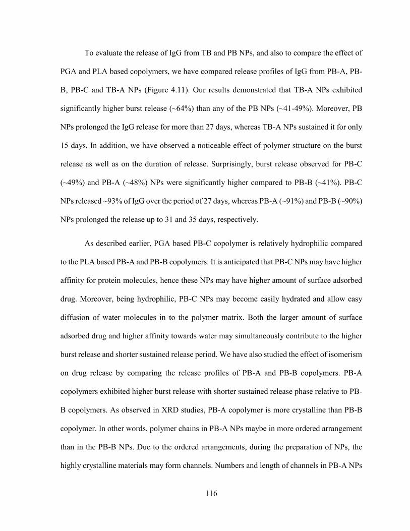
116
To evaluate the release of IgG from TB and PB NPs, and also to compare the effect of
PGA and PLA based copolymers, we have compared release profiles of IgG from PB-A, PB-
B, PB-C and TB-A NPs (Figure 4.11). Our results demonstrated that TB-A NPs exhibited
significantly higher burst release (~64%) than any of the PB NPs (~41-49%). Moreover, PB
NPs prolonged the IgG release for more than 27 days, whereas TB-A NPs sustained it for only
15 days. In addition, we have observed a noticeable effect of polymer structure on the burst
release as well as on the duration of release. Surprisingly, burst release observed for PB-C
(~49%) and PB-A (~48%) NPs were significantly higher compared to PB-B (~41%). PB-C
NPs released ~93% of IgG over the period of 27 days, whereas PB-A (~91%) and PB-B (~90%)
NPs prolonged the release up to 31 and 35 days, respectively.
As described earlier, PGA based PB-C copolymer is relatively hydrophilic compared
to the PLA based PB-A and PB-B copolymers. It is anticipated that PB-C NPs may have higher
affinity for protein molecules, hence these NPs may have higher amount of surface adsorbed
drug. Moreover, being hydrophilic, PB-C NPs may become easily hydrated and allow easy
diffusion of water molecules in to the polymer matrix. Both the larger amount of surface
adsorbed drug and higher affinity towards water may simultaneously contribute to the higher
burst release and shorter sustained release period. We have also studied the effect of isomerism
on drug release by comparing the release profiles of PB-A and PB-B copolymers. PB-A
copolymers exhibited higher burst release with shorter sustained release phase relative to PB-
B copolymers. As observed in XRD studies, PB-A copolymer is more crystalline than PB-B
copolymer. In other words, polymer chains in PB-A NPs maybe in more ordered arrangement
than in the PB-B NPs. Due to the ordered arrangements, during the preparation of NPs, the
highly crystalline materials may form channels. Numbers and length of channels in PB-A NPs
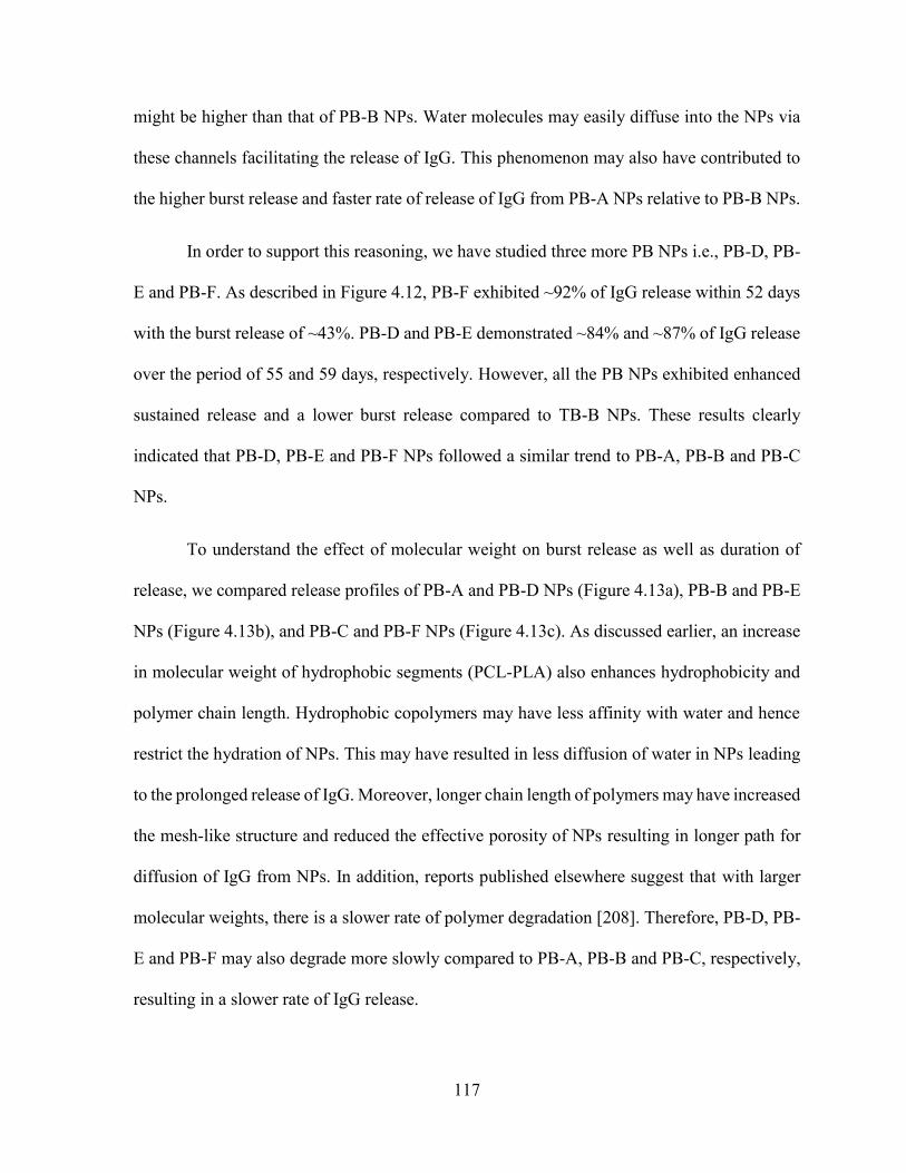
117
might be higher than that of PB-B NPs. Water molecules may easily diffuse into the NPs via
these channels facilitating the release of IgG. This phenomenon may also have contributed to
the higher burst release and faster rate of release of IgG from PB-A NPs relative to PB-B NPs.
In order to support this reasoning, we have studied three more PB NPs i.e., PB-D, PB-
E and PB-F. As described in Figure 4.12, PB-F exhibited ~92% of IgG release within 52 days
with the burst release of ~43%. PB-D and PB-E demonstrated ~84% and ~87% of IgG release
over the period of 55 and 59 days, respectively. However, all the PB NPs exhibited enhanced
sustained release and a lower burst release compared to TB-B NPs. These results clearly
indicated that PB-D, PB-E and PB-F NPs followed a similar trend to PB-A, PB-B and PB-C
NPs.
To understand the effect of molecular weight on burst release as well as duration of
release, we compared release profiles of PB-A and PB-D NPs (Figure 4.13a), PB-B and PB-E
NPs (Figure 4.13b), and PB-C and PB-F NPs (Figure 4.13c). As discussed earlier, an increase
in molecular weight of hydrophobic segments (PCL-PLA) also enhances hydrophobicity and
polymer chain length. Hydrophobic copolymers may have less affinity with water and hence
restrict the hydration of NPs. This may have resulted in less diffusion of water in NPs leading
to the prolonged release of IgG. Moreover, longer chain length of polymers may have increased
the mesh-like structure and reduced the effective porosity of NPs resulting in longer path for
diffusion of IgG from NPs. In addition, reports published elsewhere suggest that with larger
molecular weights, there is a slower rate of polymer degradation [208]. Therefore, PB-D, PB-
E and PB-F may also degrade more slowly compared to PB-A, PB-B and PB-C, respectively,
resulting in a slower rate of IgG release.

118
Figure 4.10: In vitro release of IgG from NPs prepared with TB-A and TB-B block
copolymers.
0
10
20
30
40
50
60
70
80
90
100
110
0 4 8 12 16 20 24 28
Cu
mu
lati
ve
IgG
rele
ase
(%
)
Time (days)
TB-A [PCL-PEG-PCL (5000-4000-5000)]
TB-B [PCL-PEG-PCL (7000-4000-7000)]
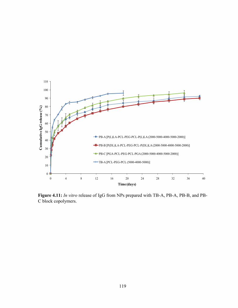
119
Figure 4.11: In vitro release of IgG from NPs prepared with TB-A, PB-A, PB-B, and PB-
C block copolymers.
0
10
20
30
40
50
60
70
80
90
100
110
0 4 8 12 16 20 24 28 32 36 40
Cu
mu
lati
ve
IgG
rel
ease
(%
)
Time (days)
PB-A [P(L)LA-PCL-PEG-PCL-P(L)LA (2000-5000-4000-5000-2000)]
PB-B [P(DL)LA-PCL-PEG-PCL-P(DL)LA (2000-5000-4000-5000-2000)]
PB-C [PGA-PCL-PEG-PCL-PGA (2000-5000-4000-5000-2000)]
TB-A [PCL-PEG-PCL (5000-4000-5000)]

120
Figure 4.12: In vitro release of IgG from NPs prepared with TB-B, PB-D, PB-E, and PB-
F block copolymers.
0
10
20
30
40
50
60
70
80
90
100
110
0 4 8 12 16 20 24 28 32 36 40 44 48 52 56 60
Cu
mu
lati
ve
IgG
rel
ease
(%
)
Time (days)
PB-D [P(L)LA-PCL-PEG-PCL-P(L)LA (3000-7000-4000-7000-3000)]
PB-E [P(DL)LA-PCL-PEG-PCL-P(DL)LA (3000-7000-4000-7000-3000)]
PB-F [PGA-PCL-PEG-PCL-PGA (3000-7000-4000-7000-3000)]
TB-B [PCL-PEG-PCL (7000-4000-7000)]
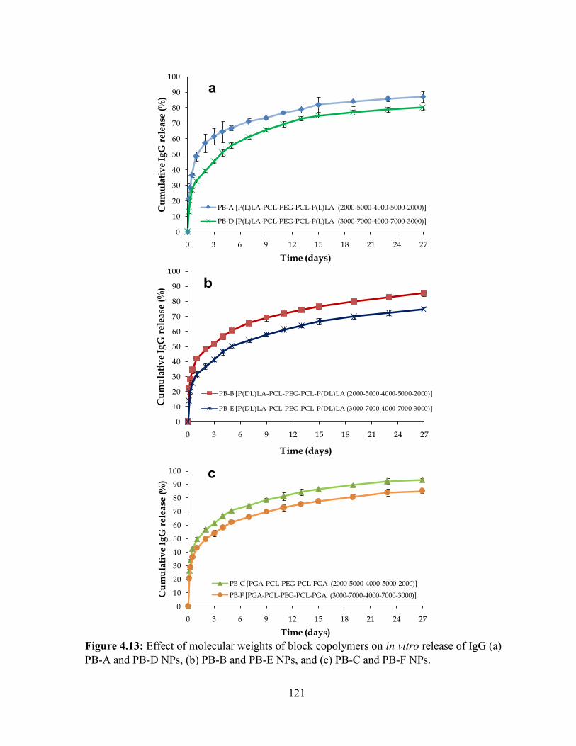
121
Figure 4.13: Effect of molecular weights of block copolymers on in vitro release of IgG (a)
PB-A and PB-D NPs, (b) PB-B and PB-E NPs, and (c) PB-C and PB-F NPs.
0
10
20
30
40
50
60
70
80
90
100
0 3 6 9 12 15 18 21 24 27
Cu
mu
lati
ve
IgG
rele
ase
(%)
Time (days)
PB-A [P(L)LA-PCL-PEG-PCL-P(L)LA (2000-5000-4000-5000-2000)]
PB-D [P(L)LA-PCL-PEG-PCL-P(L)LA (3000-7000-4000-7000-3000)]
a
0
10
20
30
40
50
60
70
80
90
100
0 3 6 9 12 15 18 21 24 27
Cu
mu
lati
ve
IgG
rele
ase
(%)
Time (days)
PB-B [P(DL)LA-PCL-PEG-PCL-P(DL)LA (2000-5000-4000-5000-2000)]
PB-E [P(DL)LA-PCL-PEG-PCL-P(DL)LA (3000-7000-4000-7000-3000)]
b
0
10
20
30
40
50
60
70
80
90
100
0 3 6 9 12 15 18 21 24 27
Cu
mu
lati
ve
Ig
Gre
lea
se (
%)
Time (days)
PB-C [PGA-PCL-PEG-PCL-PGA (2000-5000-4000-5000-2000)]
PB-F [PGA-PCL-PEG-PCL-PGA (3000-7000-4000-7000-3000)]
c
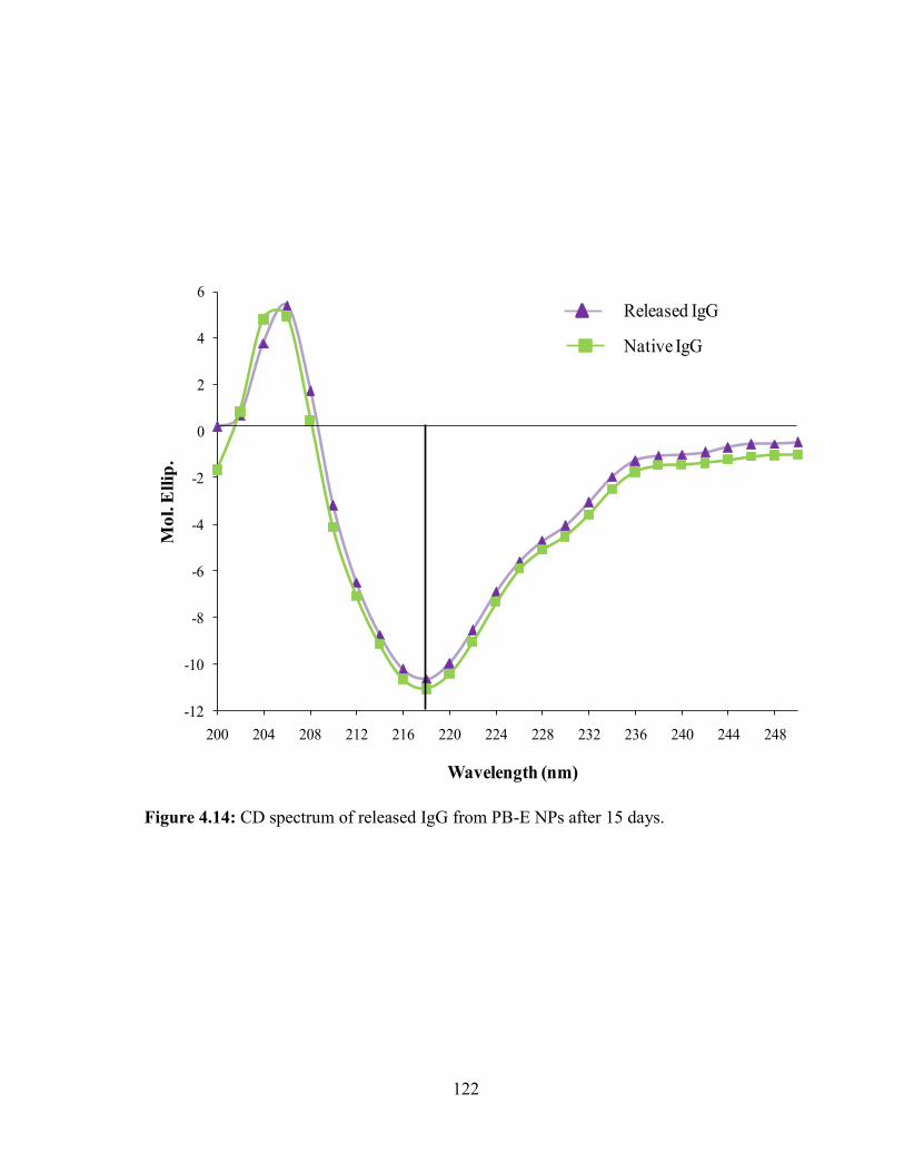
122
Figure 4.14: CD spectrum of released IgG from PB-E NPs after 15 days.
-12
-10
-8
-6
-4
-2
0
2
4
6
200 204 208 212 216 220 224 228 232 236 240 244 248
Mo
l. E
llip
.
Wavelength (nm)
Released IgG
Native IgG

123
Secondary structure stability estimation of IgG
It is very important for protein therapeutics to maintain their structural conformation to
exert pharmacological actions. CD spectroscopy is a very sensitive analytical technique
utilized for the estimation of secondary, and to some extent tertiary, structures of proteins. The
method provides α-helix and β-sheet conformational information and also able to detect minor
changes in protein conformation. Therefore, we have employed CD spectroscopy to study the
conformational stability of released IgG and compared it with the CD spectrum of native IgG.
Results reported in Figure 4.14 exhibited λ minima of released IgG at 218 nm which is similar
to the λ minima of native IgG. Moreover, the entire spectrum of released IgG ranging from
200 nm to 250 nm was identical to native IgG suggesting retention of conformation of IgG
during the NPs preparation and even after the release from NPs. Previous reports suggest that
PLA and/or PGA based copolymer produce large molar mass of lactic acid and/or glycolic acid
[183, 197]. These degradation products can stimulate hydrolytic degradation of protein
therapeutics. The retention of protein stability in PB NPs may be attributed to lower molar
masses of PLA or PGA blocks which produce lower amounts of lactic or glycolic acid,
reducing the possibility of protein degradation.
Conclusion
We have successfully synthesized and characterized novel PB copolymers with
different block ratios of PEG/PCL/PLA or PEG/PCL/PGA. These PB copolymers were studied
for the development of protein-loaded NPs in the treatment of posterior segment diseases such
as wet AMD and DR. Our results demonstrated that crystallinity of PB copolymers can be
easily modulated by changing the ratios of PLA/PCL or PGA/PCL blocks and also by utilizing
different isomers of PLA (L or D,L). Moreover, results exhibited that molecular weight,

124
crystallinity and copolymer composition have significant effects on EE, DL and in vitro release
kinetics. PB copolymers composed of PLA with D,L-lactide exhibited higher EE and slower
release relative to PB copolymers comprising PLA (L-lactide) or PGA. The stability of
released IgG was established by CD spectroscopy. In addition, cytotoxicity, cell viability and
in vitro inflammatory studies confirmed that PB copolymers are excellent biomaterials for the
development of protein-loaded, sustained delivery formulations for the treatment of posterior
segment ocular diseases.

125
CHAPTER 5
TAILOR-MADE PENTABLOCK COPOLYMER BASED COMPOSITE FORMULATION
FOR SUSTAINED OCULAR DELIVERY OF PROTEIN THERAPEUTICS
Rationale
Various biodegradable polymeric NP formulations have been extensively investigated
for controlled delivery of protein therapeutics. Biodegradable polymers such as PCL, PLA,
PGA and PEG have been comprehensively studied for the preparation of protein-encapsulated
NPs. Current challenges in the development of protein-encapsulated NPs are to ensure protein
stability during the NP preparation and drug release. In addition, the second major problem
with any NP formulation is higher burst release (initial rapid drug release phase).
The objective of this research is to synthesize and evaluate novel tailor-made PB
copolymers for the controlled and non-invasive delivery of macromolecules in the treatment
of posterior segment diseases. We have synthesized three PB copolymers by sequential ring-
opening polymerization either for the preparation of protein-loaded NPs (two PB copolymers)
or thermosensitive gel (one PB copolymer). Different ratios and molecular weights of each
block (PGA, PEG, PLA and PCL) were selected for synthesis to optimize the release profile
of FITC-BSA, IgG and bevacizumab from NPs. We have hypothesized that release of protein
therapeutics from NPs is affected by their hydrodynamic diameter. Therefore, in this study we
have examined effect of hydrodynamic diameter of proteins on EE and in vitro drug release.
We have characterized NPs for particle size, polydispersity, EE, DL and in vitro release studies.
As described earlier, PB thermosensitive gel (chapter 3) and PB NPs (chapter 4)
exhibited significantly higher burst release and relatively shorter duration of sustained release.
In many of the cases, burst release (~25-45%) can exhibit dose dependent toxicity. Moreover,
126
loss of ~25-45% of dose in first two days can significantly reduce the duration of release. So
in order to eliminate burst release phase and to achieve continuous zero-order drug release, we
have prepared and characterized a novel composite formulation comprised of protein-loaded
NPs suspended thermosensitive gelling solution. A composite formulation also offers an
additional advantage to eliminate any possibility of floater effect which may arise during the
intravitreal injection of NPs alone. Stability of released protein was also confirmed by CD
spectroscopy and in vitro biological assays.
Materials and methods
Materials
PEG (1 kDa and 4 kDa), methoxy-PEG (550 Da), stannous octoate, ε-caprolactone,
poly (vinyl alcohol) (PVA), lipopolysaccharide were procured from Sigma-Aldarich (St.
Louis, MO; USA). L-lactide and hexamethylene diisocyanate (HMDI) were purchased from
Acros organics (Morris Plains, NJ; USA). Micro-BCATM was obtained from Fisher scientific.
Mouse TNF-α, IL-6 and IL-1β (Ready-Set-Go) ELISA kits were purchased from eBioscience
Inc. Lactate dehydrogenase estimation kit and CellTiter 96® AQueous non-radioactive cell
proliferation assay (MTS) kit were obtained from Takara Bio Inc. and Promega Corp.,
respectively. All other reagents utilized in this study were of analytical grade.
Methods
Synthesis of PB copolymers
Novel PB copolymers, PGA-PCL-PEG-PCL-PGA (PB-A), PLA-PCL-PEG-PCL-PLA
(PB-B) and PEG-PCL-PLA-PCL-PEG (PB-C) were synthesized by ring-opening bulk
polymerization method [176]. PB copolymers for the preparation of NPs i.e., PB-A and PB-B
were synthesized in two steps by sequential ring-opening polymerization. PEG (1 kDa and 4
127
kDa) was utilized as macroinitiator and stannous octoate as a catalyst. In the first step, TB
copolymer PCL-PEG-PCL (Figure 5.1, step 1) was synthesized by polymerization of ε-
caprolactone on two open hydroxyl ends of PEG. In brief, PEG was dissolved in anhydrous
toluene followed by distillation to remove residual moisture. ε-caprolactone and stannous
octoate (0.5% w/w) were added to anhydrous PEG and temperature was raised to 130 °C. After
24 h, reaction mixture was dissolved in methylene chloride followed by precipitation in cold
petroleum ether. The precipitated polymer was filtered and dried for 24 h under vacuum at
room temperature. In the second step, the PCL-PEG-PCL TB copolymer was reacted with
glycolide and L-lactide to prepare the PB-A (Figure 5.1, step 2) and PB-B (Figure 5.2)
copolymers, respectively. The TB copolymer and glycolide/L-lactide were added in round
bottom flask and temperature was raised to 130 °C under inert atmosphere. To this, stannous
octoate (0.5% w/w) was added and reaction was allowed to run for 24 h. PB copolymer was
purified by cold ether precipitation method described in the first step. The polymer was dried
under vacuum and stored at -20 °C until further use.
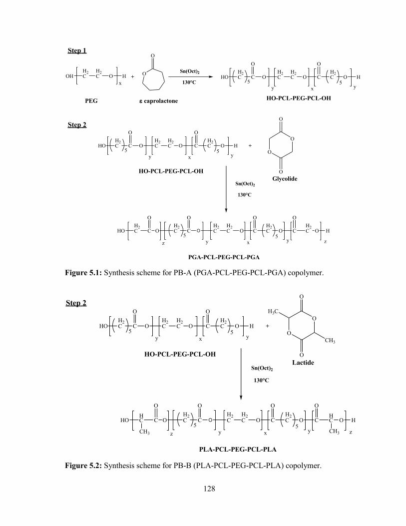
128
Figure 5.1: Synthesis scheme for PB-A (PGA-PCL-PEG-PCL-PGA) copolymer.
OHH2C
H2C O H
x
O
O
+ HOH2C C
O
OH2C
H2C O C
O
H2C O
5 5H
y yx
Sn(Oct)2
130°C
caprolactonePEG HO-PCL-PEG-PCL-OH
HOH2C C
O
OH2C
H2C O C
O
H2C O
5 5H
y yx
HO-PCL-PEG-PCL-OH
Sn(Oct)2
130°C
OH2C C
O
OH2C
H2C O C
O
H2C O
5 5C
y yx
C
O
H2CHO
O
H2C O
z z
+
O
O
O
O
PGA-PCL-PEG-PCL-PGA
Step 1
Step 2
Glycolide
H
Figure 5.2: Synthesis scheme for PB-B (PLA-PCL-PEG-PCL-PLA) copolymer.
HOH2C C
O
OH2C
H2C O C
O
H2C O
5 5H
y yx
HO-PCL-PEG-PCL-OH
Sn(Oct)2
130°C
OH2C C
O
OH2C
H2C O C
O
H2C O
5 5C
y yx
C
O
HCHO
O
HC O
z z
+
O
O
O
O
PLA-PCL-PEG-PCL-PLA
Step 2
Lactide
H
CH3 CH3
CH3
H3C
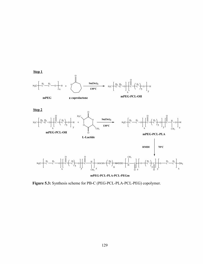
129
Figure 5.3: Synthesis scheme for PB-C (PEG-PCL-PLA-PCL-PEG) copolymer.
H3CH2C
H2C O H
x
O
O
+ H3C O C
O
H2C O
5H
yx
Sn(Oct)2
130°C
caprolactonemPEGmPEG-PCL-OH
Sn(Oct)2
130°C
H3CH2C
H2C O C
O
H2C O
5C
yx
O
HC
CH3
O
z
+O
O
O
O
H3C
CH3
mPEG-PCL-PLA
Step 1
Step 2
L-Lactide
70°CHMDI
H3CH2C
H2C O C
O
H2C O
5C
yx
O
HC
CH3
OOCHN
z
H2C NHCOO CH3
H2C
H2COC
O
H2COC
O
CH
CH3
xy5
z6
mPEG-PCL-PLA-PCL-PEGm
H2C
H2C
H3C O C
O
H2C O
5H
yx
mPEG-PCL-OH
H2C
H2C H

130
For the synthesis of thermosensitive gelling polymer, TB copolymer (mPEG-PCL-
PLA) was synthesized by ring-opening bulk copolymerization as described above (Figure 5.3).
Firstly, ε-caprolactone was polymerized at the hydroxyl terminal of mPEG (550 Da) followed
by polymerization of L-lactide. Resulting TB copolymers were coupled utilizing HMDI as a
linker. Coupling reaction was carried out at 70 °C for 8 h. Polymers were purified by cold ether
precipitation followed by drying under vacuum and stored at -20 °C.
Characterization of polymers
The resulting polymers were characterized for molecular weight and purity by 1H NMR
spectroscopy and gel permeation chromatography (GPC).
1H-NMR analysis
To perform 1H-NMR spectroscopy, polymeric materials were dissolved in CDCI3 and
spectra were recorded with Varian-400 NMR instrument. Purity and molecular weight (Mn)
were calculated from the 1H-NMR spectra.
GPC analysis
Purity, molecular weights and polydispersity of PB copolymers were further confirmed
by GPC analysis. Polymeric samples were analyzed with refractive index detector (Waters
410). Briefly, samples were prepared by dissolving 5 mg of polymeric material in
tetrahydrofuran (THF) whereas THF was utilized as eluting agent at the flow rate of 1 mL/min.
Separation was carried out on Styragel HR-3 column and polystyrene samples with narrow
molecular weight distribution were utilized as standards.
131
In vitro cytotoxicity studies
Cell culture
Human retinal pigment epithelial cells (ARPE-19) were cultured and maintained
according to the previously published protocol from our lab [199]. In brief, cells were cultured
in Dubelcco's modified Eagle medium (DMEM)/F-12 medium containing 10% fetal bovine
serum (FBS), 15 mM of HEPES, 29 mM of sodium bicarbonate, 100 U/L of penicillin and 100
mg/L of streptomycin. A mouse macrophage cell line, RAW-264.7 was procured from ATCC.
RAW-264.7 cells were cultured and maintained in DMEM supplemented with 10% FBS, 100
U/L of penicillin and 100 mg/L of streptomycin. The Statens Seruminstitut rabbit corneal
(SIRC) cell line was purchased from ATCC at passage 400 and used between passages 410 -
425. During the study and maintenance period cells were supplemented with cell culture media
composed of MEM containing 10% FBS, lactalbumin, HEPES, sodium bicarbonate, 100 U/L
of penicillin and 100 mg/L of streptomycin. Human conjunctival epithelial cells (HCEC) were
maintained in cell culture flask containing MEM Earle’s BSS medium supplemented by 10%
FBS, 100 U/L of penicillin, 100 mg/L of streptomycin, 29 mM of sodium bicarbonate, and 2
mM L-glutamine. Choroid-retinal endothelial cells (RF/6A cells) were procured from ATCC.
Briefly, cells were cultured and maintained in cell culture medium composed of RPMI-1640
comprising 10% FBS, 100 U/L of penicillin and 100 mg/L of streptomycin [209]. All five cell
lines were maintained in humidified atmosphere at 37 °C and 5% CO2.
Lactate Dehydrogenase (LDH) assay
Previously published protocol with minor modification was employed to evaluate the
cytotoxicity of PB copolymers [210]. Briefly, 10 mg/mL of PB copolymers (PB-A and PB-B)
were dissolved in ACN and 100 µL of these solutions were aliquoted in each well of 96-well
132
cell culture plates. Plates were exposed overnight under UV light (laminar flow) for the
sterilization of polymer and evaporation of ACN. ARPE-19 cells at the density of 1.0 x 10⁴
were seeded in each well and incubated at 37 °C, 5% CO2 in humidified atmosphere for 48 h.
After completion of incubation period, cell supernatant were analyzed for the quantification of
LDH. According to supplier’s protocol, levels of LDH were analyzed utilizing a LDH detection
kit. Absorbance of each well was estimated at 450 nm by 96-well plate reader. More than 10%
of LDH release was considered as cytotoxic. To evaluate the cytotoxicity of block copolymers
on cunjunctiva, cornea and macrophages, the similar experiment was performed with HCEC,
SIRC and RAW-264.7 cells. LDH release (%) was calculated with following equation,
𝐿𝐷𝐻𝑟𝑒𝑙𝑒𝑎𝑠𝑒(%) =𝐴𝑏𝑠. 𝑜𝑓 𝑆𝑎𝑚𝑝𝑙𝑒 − 𝐴𝑏𝑠. 𝑜𝑓 𝑛𝑒𝑔𝑎𝑡𝑖𝑣𝑒 𝑐𝑜𝑛𝑡𝑟𝑜𝑙
𝐴𝑏𝑠. 𝑜𝑓 𝑝𝑜𝑠𝑖𝑡𝑖𝑣𝑒 𝑐𝑜𝑛𝑡𝑟𝑜𝑙 − 𝐴𝑏𝑠. 𝑜𝑓 𝑛𝑒𝑔𝑎𝑡𝑖𝑣𝑒 𝑐𝑜𝑛𝑡𝑟𝑜𝑙∗ 100 … Eq. 5.1
MTS assay
Safety of PB copolymers was further established by performing in vitro cell viability
assay (MTS assay) [211]. MTS assay was performed according to previously reported protocol
with minor changes. As described earlier, PB copolymer solutions at the concentration of 10
mg/mL were prepared, aliquoted and sterilized. After sterilization, ARPE-19 cells were seeded
in each well of 96-well plate at the cell density of 1.0 x 10⁴, and incubated at 37 °C and 5%
CO2 in humidified atmosphere for 48 h. At the end of incubation period, cell culture medium
was aspirated and cells were incubated for 4 h (37 °C and 5% CO2) in presence of 100 µL of
serum free medium containing 20 µL of MTS solution. Absorbance of each well was estimated
at 450 nm. The similar experiment was repeated with other ocular and macrophage cell line
such as HCEC, SIRC and RAW-264.7 cells. Percent cell viability was calculated by the
following equation.
133
𝐶𝑒𝑙𝑙𝑣𝑖𝑎𝑏𝑖𝑙𝑖𝑡𝑦(%) =𝐴𝑏𝑠. 𝑜𝑓 𝑆𝑎𝑚𝑝𝑙𝑒 − 𝐴𝑏𝑠. 𝑜𝑓 𝑛𝑒𝑔𝑎𝑡𝑖𝑣𝑒 𝑐𝑜𝑛𝑡𝑟𝑜𝑙
𝐴𝑏𝑠. 𝑜𝑓 𝑝𝑜𝑠𝑖𝑡𝑖𝑣𝑒 𝑐𝑜𝑛𝑡𝑟𝑜𝑙 − 𝐴𝑏𝑠. 𝑜𝑓 𝑛𝑒𝑔𝑎𝑡𝑖𝑣𝑒 𝑐𝑜𝑛𝑡𝑟𝑜𝑙∗ 100 … Eq. 5.2
In this study, PB copolymers which exhibited more than 90% of cell viability were considered
non-toxic for ocular applications.
In vitro biocompatibility studies
PB copolymers were dissolved in ACN at the concentration of 10 mg/mL and 200 µL
of these solutions were aliquoted in each well of 48-well cell culture plates. Cell culture plates
were incubated overnight under UV lights (laminar flow) for the evaporation of ACN and
sterilization of resulting polymer film. After sterilization, RAW-264.7 cells (5.0 x 10⁴) were
seeded in each well of cell culture plate and incubated for 24 h at 37 °C and 5% CO2. After 24
h, cell supernatants were analyzed for the presence of cytokines such as TNF-α, IL-6 and IL-
1β. Lipopolysaccharide (LPS) was utilized as positive control whereas cells without treatment
were considered as negative control. Levels of cytokines were quantified by ELISA method
which was performed according to supplier’s instructions. Standard calibration curves for
TNF-α, IL-6 and IL-1β were prepared in the range of 10-750 pg/mL, 5-500 pg/mL and 10-500
pg/mL, respectively.
Preparation of NPs
IgG-loaded PB NPs were prepared by W1/O/W2 double emulsion solvent evaporation
method [212]. Briefly, predetermined quantity of IgG (10 mg) was dissolved in 0.1 M
phosphate buffer saline (PBS, pH 7.4) (1 mL) containing 50 µL of Tween-80 (W1 phase). In
order to prepare organic phase, 100 mg of respective PB copolymers were solubilized in 4 mL
of dichloromethane (DCM) with 50 µL of Span-20 (organic phase). Primary emulsion (W1/O)
was prepared by drop-wise addition of W1 phase in organic phase under constant sonication.
134
Sonication was applied with probe-sonicator for 1 min at 4 W output. To avoid excessive
heating and subsequent degradation of protein, the preparation of emulsion was carried out in
ice-bath. Resulting W1/O primary emulsion was then added drop-wise in 20 mL of 2%
polyvinyl alcohol (PVA) solution (W2 phase) under constant sonication for 4 min at 5 W
output. Double emulsion (W1/O/W2) was stirred at room temperature for 30 min followed by
evaporation of DCM under low pressure. Once the DCM was evaporated, NPs were
centrifuged for 30 min at 20000 rpm and 4 °C followed by two washing cycles with distilled
deionized water (DDW). Finally, IgG-loaded NPs were freeze-dried in presence of 5%
mannitol (cryprotectant) and stored at -20 °C until further use. NPs were prepared with two PB
copolymers i.e., PB-A and PB-B. The similar protocol was utilized to prepare FITC-BSA and
bevacizumab-loaded NPs. Freeze-dried NPs were evaluated for particle size, EE (%), DL (%)
and in vitro drug release behavior.
Characterization of NPs
Particle size and polydispersity
Freeze-dried NPs were dispersed in DDW (1 mg/mL) and analyzed for their size and
its distribution. Particle size was evaluated by particle size analyzer (Zetasizer Nano ZS,
Malvern Instruments Ltd, Worcestershire, UK) at 90° scattering angel. All the NP samples
were analyzed in triplicate.
Entrapment efficiency (EE) and drug loading (DL)
Protein-encapsulated freeze-dried NPs were evaluated for the estimation of EE and DL.
EE of the NPs was estimated by quantifying the amount of protein in the supernatants which
were obtained during the NP preparation. Micro BCATM protein estimation kit was employed
for the quantification of total protein. In order to evaluated DL, 2 mg equivalent protein-loaded
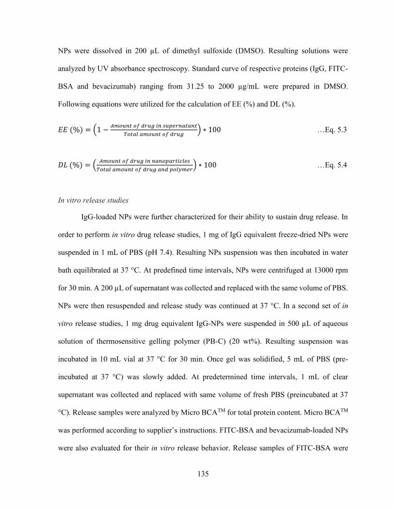
135
NPs were dissolved in 200 µL of dimethyl sulfoxide (DMSO). Resulting solutions were
analyzed by UV absorbance spectroscopy. Standard curve of respective proteins (IgG, FITC-
BSA and bevacizumab) ranging from 31.25 to 2000 µg/mL were prepared in DMSO.
Following equations were utilized for the calculation of EE (%) and DL (%).
𝐸𝐸 (%) = (1 −𝐴𝑚𝑜𝑢𝑛𝑡 𝑜𝑓 𝑑𝑟𝑢𝑔 𝑖𝑛 𝑠𝑢𝑝𝑒𝑟𝑛𝑎𝑡𝑎𝑛𝑡
𝑇𝑜𝑡𝑎𝑙 𝑎𝑚𝑜𝑢𝑛𝑡 𝑜𝑓 𝑑𝑟𝑢𝑔) ∗ 100 …Eq. 5.3
𝐷𝐿 (%) = (𝐴𝑚𝑜𝑢𝑛𝑡 𝑜𝑓 𝑑𝑟𝑢𝑔 𝑖𝑛 𝑛𝑎𝑛𝑜𝑝𝑎𝑟𝑡𝑖𝑐𝑙𝑒𝑠
𝑇𝑜𝑡𝑎𝑙 𝑎𝑚𝑜𝑢𝑛𝑡 𝑜𝑓 𝑑𝑟𝑢𝑔 𝑎𝑛𝑑 𝑝𝑜𝑙𝑦𝑚𝑒𝑟) ∗ 100 …Eq. 5.4
In vitro release studies
IgG-loaded NPs were further characterized for their ability to sustain drug release. In
order to perform in vitro drug release studies, 1 mg of IgG equivalent freeze-dried NPs were
suspended in 1 mL of PBS (pH 7.4). Resulting NPs suspension was then incubated in water
bath equilibrated at 37 °C. At predefined time intervals, NPs were centrifuged at 13000 rpm
for 30 min. A 200 µL of supernatant was collected and replaced with the same volume of PBS.
NPs were then resuspended and release study was continued at 37 °C. In a second set of in
vitro release studies, 1 mg drug equivalent IgG-NPs were suspended in 500 µL of aqueous
solution of thermosensitive gelling polymer (PB-C) (20 wt%). Resulting suspension was
incubated in 10 mL vial at 37 °C for 30 min. Once gel was solidified, 5 mL of PBS (pre-
incubated at 37 °C) was slowly added. At predetermined time intervals, 1 mL of clear
supernatant was collected and replaced with same volume of fresh PBS (preincubated at 37
°C). Release samples were analyzed by Micro BCATM for total protein content. Micro BCATM
was performed according to supplier’s instructions. FITC-BSA and bevacizumab-loaded NPs
were also evaluated for their in vitro release behavior. Release samples of FITC-BSA were
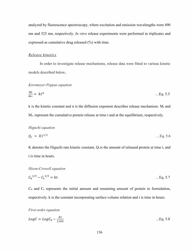
136
analyzed by fluorescence spectroscopy, where excitation and emission wavelengths were 490
nm and 525 nm, respectively. In vitro release experiments were performed in triplicates and
expressed as cumulative drug released (%) with time.
Release kinetics
In order to investigate release mechanisms, release data were fitted to various kinetic
models described below,
Korsmeyer-Peppas equation
𝑀𝑡
𝑀∞= 𝑘𝑡𝑛 …Eq. 5.5
k is the kinetic constant and n is the diffusion exponent describes release mechanism. Mt and
M∞ represent the cumulative protein release at time t and at the equilibrium, respectively.
Higuchi equation
𝑄𝑡 = 𝐾𝑡1/2 …Eq. 5.6
K denotes the Higuchi rate kinetic constant, Qt is the amount of released protein at time t, and
t is time in hours.
Hixon-Crowell equation
𝐶01/3 − 𝐶𝑡
1/3 = 𝑘𝑡 …Eq. 5.7
C0 and Ct represents the initial amount and remaining amount of protein in formulation,
respectively. k is the constant incorporating surface-volume relation and t is time in hours.
First-order equation
𝐿𝑜𝑔𝐶 = 𝐿𝑜𝑔𝐶0 −𝐾𝑡
2.303 ...Eq. 5.8

137
K denotes the first-order rate constant, C0 is the initial protein concentration and t represents
time in hours.
Zero-order equation
𝐶 = 𝐾0𝑡 …Eq. 5.9
K0 is the zero-order rate constant and t is time in hours.
Stability estimation of IgG
Released IgG was analyzed by CD spectroscopy for the estimation of secondary
structure. CD analysis was carried at room temperature with Jasco 720 spectropolarimeter. CD
spectra were recorded between the wavelengths of 200 to 250 nm at scanning speed of 5
nm/min utilizing 1 cm cell. CD measurements were reported as molar elipticity [θ]. CD
spectrum of PBS was utilized as blank.
Stability estimation of bevacizumab by in vitro biological assays
Cell proliferation assay
A cell proliferation assay was performed according previously published protocols with
minor modifications [213-215]. Concentrations of live cells were quantified by MTS assay,
which was performed according to manufacturer’s instructions. Briefly, cells were seeded at
the density of 5 x 10³ cells/well of 96-well cell culture plate. After 24 h of incubation, cells
were serum-starved overnight followed by addition of serum free medium containing 100
ng/mL of VEGF and 0.25 mg/mL of released bevacizumab (test samples) or 0.25 mg/mL of
native bevacizumab. Cells without VEGF or bevacizumab were considered as negative control
or cells exposed only to VEGF (100 ng/mL) as positive control. Cells were further incubated
for 24 h at 37 °C and 5% CO2. After completion of treatment, cells were exposed to 100 µL of
138
serum free medium containing 20 µL of MTS solution and incubated for 4 h. The absorbance
was recorded at 450 nm using a microplate reader.
Cell migration assay
A cell migration assay was performed as described elsewhere with few modifications
[216]. RF/6A cells were starved overnight (by exposing to serum free medium), trypsinized
and suspended in serum free medium containing 0.25 mg/mL of bevacizumab (native or
released from NPs). The 5 x 10³cells were seeded in upper chamber of Transwell (8.0 μm pore
size, 10 mm diameter; Corning Inc.), pre-incubated with cell culture medium. VEGF (100
ng/mL) was placed into the lower chamber and the cells were incubated for 24 h at 37 °C and
5% CO2. Non-migrated cells were removed from upper chamber by cotton swab and
concentration of migrated cells was estimated by AlamarBlue® assay. AlamarBlue® assay was
performed according to supplier’s protocol. Moreover, for visual evidence, migrated cells
were also stained with methylene blue and images were taken with Leica DMI3000B inverted
microscope (Germany). Each experiment was repeated three times.
Results and discussion
Synthesis and characterization of PB copolymers
PB copolymers (PB-A and PB-B) were successfully synthesized by ring-opening bulk
copolymerization of ε-caprolactone, and L-lactide/glycolide. Firstly, TB copolymers (PCL-
PEG-PCL) were synthesized, purified and characterized. Purified TB copolymers were then
utilized for the synthesis of respective PB copolymers i.e., PB-A (PGA-PCL-PEG-PCL-PGA)
and PB-B (PLA-PCL-PEG-PCL-PLA). Purity and molecular weights (Mn) of PB copolymers
were calculated by 1H-NMR spectroscopy. As described in Figures 5.4 and 5.5, typical 1H-
NMR signals of PCL blocks were observed at 1.40, 1.65, 2.30 and 4.06 ppm depicting
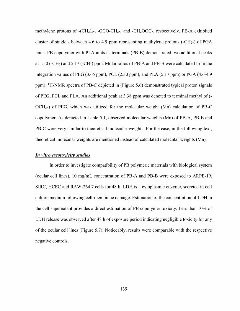
139
methylene protons of -(CH2)3-, -OCO-CH2-, and -CH2OOC-, respectively. PB-A exhibited
cluster of singlets between 4.6 to 4.9 ppm representing methylene protons (-CH2-) of PGA
units. PB copolymer with PLA units as terminals (PB-B) demonstrated two additional peaks
at 1.50 (-CH3) and 5.17 (-CH-) ppm. Molar ratios of PB-A and PB-B were calculated from the
integration values of PEG (3.65 ppm), PCL (2.30 ppm), and PLA (5.17 ppm) or PGA (4.6-4.9
ppm). 1H-NMR spectra of PB-C depicted in (Figure 5.6) demonstrated typical proton signals
of PEG, PCL and PLA. An additional peak at 3.38 ppm was denoted to terminal methyl of (-
OCH3-) of PEG, which was utilized for the molecular weight (Mn) calculation of PB-C
copolymer. As depicted in Table 5.1, observed molecular weights (Mn) of PB-A, PB-B and
PB-C were very similar to theoretical molecular weights. For the ease, in the following text,
theoretical molecular weights are mentioned instead of calculated molecular weights (Mn).
In vitro cytotoxicity studies
In order to investigate compatibility of PB polymeric materials with biological system
(ocular cell lines), 10 mg/mL concentration of PB-A and PB-B were exposed to ARPE-19,
SIRC, HCEC and RAW-264.7 cells for 48 h. LDH is a cytoplasmic enzyme, secreted in cell
culture medium following cell-membrane damage. Estimation of the concentration of LDH in
the cell supernatant provides a direct estimation of PB copolymer toxicity. Less than 10% of
LDH release was observed after 48 h of exposure period indicating negligible toxicity for any
of the ocular cell lines (Figure 5.7). Noticeably, results were comparable with the respective
negative controls.
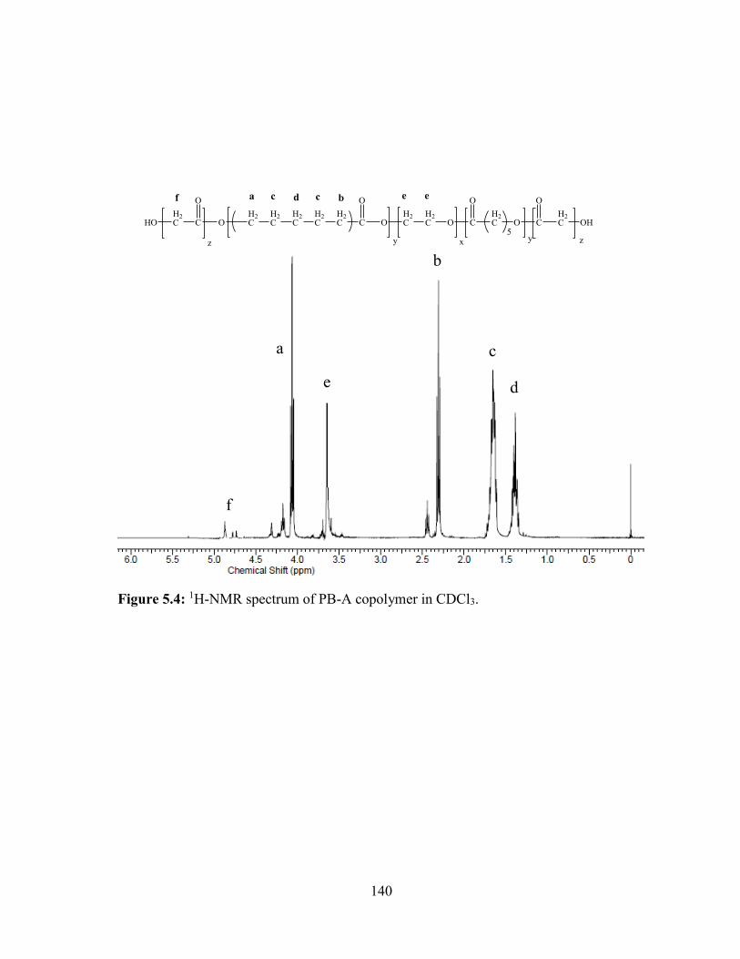
140
Figure 5.4: 1H-NMR spectrum of PB-A copolymer in CDCl3.
b
a c
de
f
O C
O
OH2C
H2C O C
O
H2C O
5y yx
H2C
H2C
H2C
H2C
H2C
a bc cd e e
C
O
H2CHO
z
f
C
O
H2C OH
z
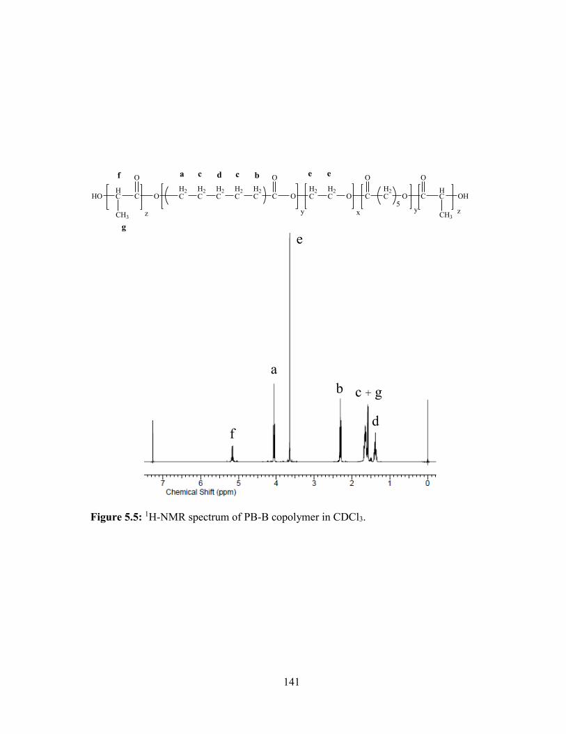
141
Figure 5.5: 1H-NMR spectrum of PB-B copolymer in CDCl3.
b
a
c + g
d
e
f
O C
O
OH2C
H2C O C
O
H2C O
5y yx
H2C
H2C
H2C
H2C
H2C
a bc cd e e
C
O
HC
CH3
HO
z
f
g
C
O
HC
CH3
OH
z
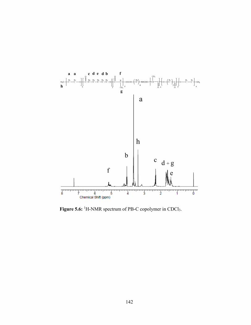
142
Figure 5.6: 1H-NMR spectrum of PB-C copolymer in CDCl3.
bc
d + g
ef
a
h
H3CH2C
H2C O C
O
C
yx
O
HC
CH3
OOCHN
z
H2C NHCOO CH3
H2C
H2COC
O
H2COC
O
CH
CH3
xy5
z6
OH2C
H2C
H2C
H2C
H2C
a a c d bde f
g
h
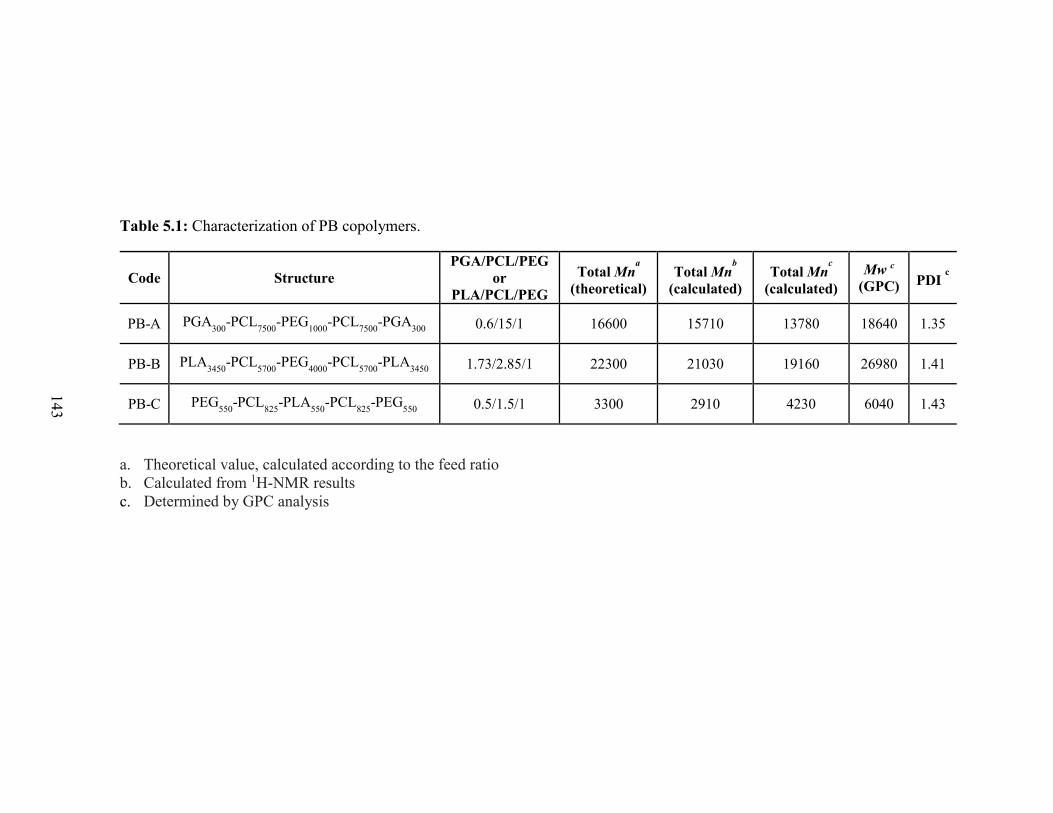
143
Table 5.1: Characterization of PB copolymers.
Code Structure
PGA/PCL/PEG
or
PLA/PCL/PEG
Total Mna
(theoretical)
Total Mnb
(calculated)
Total Mnc
(calculated)
Mw c
(GPC) PDI c
PB-A PGA300
-PCL7500
-PEG1000
-PCL7500
-PGA300
0.6/15/1 16600 15710 13780 18640 1.35
PB-B PLA3450
-PCL5700
-PEG4000
-PCL5700
-PLA3450
1.73/2.85/1 22300 21030 19160 26980 1.41
PB-C PEG550
-PCL825
-PLA550
-PCL825
-PEG550
0.5/1.5/1 3300 2910 4230 6040 1.43
a. Theoretical value, calculated according to the feed ratio
b. Calculated from 1H-NMR results
c. Determined by GPC analysis
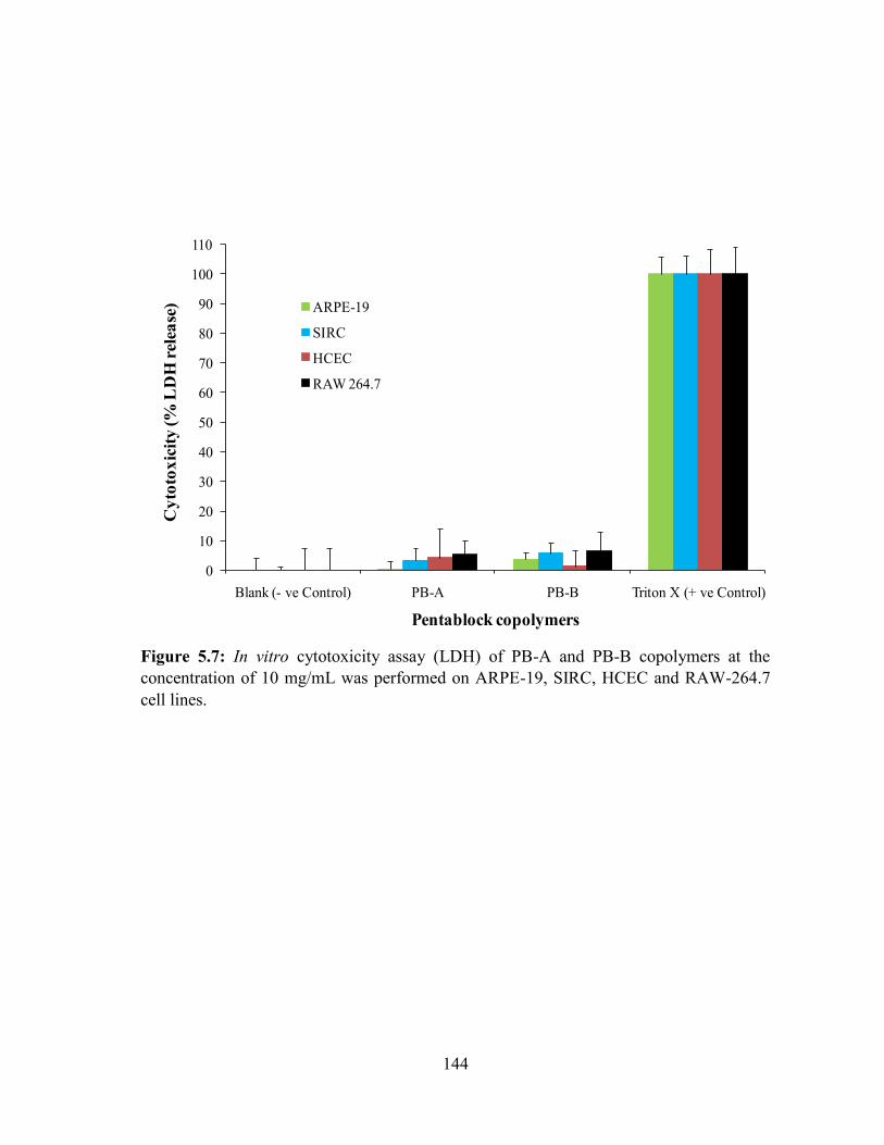
144
Figure 5.7: In vitro cytotoxicity assay (LDH) of PB-A and PB-B copolymers at the
concentration of 10 mg/mL was performed on ARPE-19, SIRC, HCEC and RAW-264.7
cell lines.
0
10
20
30
40
50
60
70
80
90
100
110
Blank (- ve Control) PB-A PB-B Triton X (+ ve Control)
Cy
toto
xic
ity
(%
LD
H r
elea
se)
Pentablock copolymers
ARPE-19
SIRC
HCEC
RAW 264.7

145
Figure 5.8: In vitro cell viability assay (MTS) of PB-A and PB-B copolymers at the
concentration of 10 mg/mL was performed on ARPE-19, SIRC, HCEC and RAW-264.7
cell lines.
0
10
20
30
40
50
60
70
80
90
100
110
Blank (- ve Control) PB-A PB-B Triton X (+ ve Control)
Cel
l v
iab
ilit
y (%
)
Pentablock copolymers
ARPE-19
SIRC
HCEC
RAW 264.7
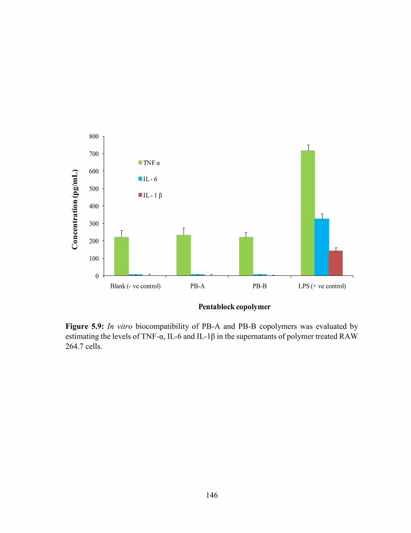
146
Figure 5.9: In vitro biocompatibility of PB-A and PB-B copolymers was evaluated by
estimating the levels of TNF-α, IL-6 and IL-1β in the supernatants of polymer treated RAW
264.7 cells.
0
100
200
300
400
500
600
700
800
Blank (- ve control) PB-A PB-B LPS (+ ve control)
Co
ncen
tra
tio
n (p
g/m
L)
Pentablock copolymer
TNF α
IL - 6
IL - 1 β
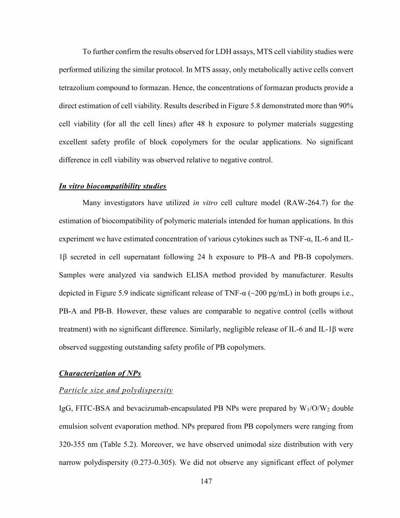
147
To further confirm the results observed for LDH assays, MTS cell viability studies were
performed utilizing the similar protocol. In MTS assay, only metabolically active cells convert
tetrazolium compound to formazan. Hence, the concentrations of formazan products provide a
direct estimation of cell viability. Results described in Figure 5.8 demonstrated more than 90%
cell viability (for all the cell lines) after 48 h exposure to polymer materials suggesting
excellent safety profile of block copolymers for the ocular applications. No significant
difference in cell viability was observed relative to negative control.
In vitro biocompatibility studies
Many investigators have utilized in vitro cell culture model (RAW-264.7) for the
estimation of biocompatibility of polymeric materials intended for human applications. In this
experiment we have estimated concentration of various cytokines such as TNF-α, IL-6 and IL-
1β secreted in cell supernatant following 24 h exposure to PB-A and PB-B copolymers.
Samples were analyzed via sandwich ELISA method provided by manufacturer. Results
depicted in Figure 5.9 indicate significant release of TNF-α (~200 pg/mL) in both groups i.e.,
PB-A and PB-B. However, these values are comparable to negative control (cells without
treatment) with no significant difference. Similarly, negligible release of IL-6 and IL-1β were
observed suggesting outstanding safety profile of PB copolymers.
Characterization of NPs
Particle size and polydispersity
IgG, FITC-BSA and bevacizumab-encapsulated PB NPs were prepared by W1/O/W2 double
emulsion solvent evaporation method. NPs prepared from PB copolymers were ranging from
320-355 nm (Table 5.2). Moreover, we have observed unimodal size distribution with very
narrow polydispersity (0.273-0.305). We did not observe any significant effect of polymer
148
composition (PB-A/PB-B) or type of protein molecule (IgG/BSA/bevacizumab) on particle
size. These results suggest that hydrodynamic diameter of protein therapeutics or polymer
composition has little or no effect on particle size or its distribution.
Entrapment efficiency (EE) and drug loading (DL)
EE and DL are significantly influenced by various parameters including copolymer
composition (hydrophobicity of polymer) and volumes of various phases (W1, O and W2). In
order to understand the effect of hydrophobicity of copolymers on EE and DL, we have
prepared and evaluated IgG and FITC-BSA-loaded NPs with PB-A and PB-B. It is important
to note that PB-A copolymer comprising PGA (hydrophilic block) is relatively hydrophilic
copolymer than PB-B copolymer comprising PLA (hydrophobic block). As described in Table
5.2, encapsulation of IgG or FITC-BSA in PB-A NPs were ~40% and ~35%, respectively.
However, PB-B NPs exhibited significantly higher encapsulation for IgG (~70%) and FITC-
BSA (~69%) relative to PB-A NPs. This may attributed to the fact that during the preparation
of NPs (solvent evaporation), high hydrophobicity may allow faster precipitation of PB-B
copolymer to form NPs preventing diffusion of IgG/FITC-BSA from W1 phase to external
aqueous (W2) phase. This phenomenon possibly ensured higher EE of protein therapeutics in
PB-B NPs. Due to higher hydrophilicity, PB-A copolymer may remain hydrated with W1 and
W2 phase (during NPs preparation) allowing escape of IgG/FITC-BSA in external phase
resulting poor EE. However, we did not observe any effect of hydrodynamic diameter of IgG
or FITC-BSA on EE or DL. It may possible that both the proteins are too large (≥ 66 kDa) to
show any significant effect of hydrodynamic diameter on EE or DL. Bevacizumab-loaded PB-
B NPs exhibited ~67% of EE and ~6% of DL, very similar to IgG-loaded PB-B NPs (Table
5.2) suggested that bevacizumab may behaved very similar to IgG during NP preparation.
149
In vitro release studies
In order to evaluate the effect of polymer hydrophoicity, release of FITC-BSA and IgG
from PB-A and PB-B NPs were evaluated. As described in Figure 5.10, both NPs (PB-A and
PB-B) demonstrated biphasic release profile i.e., initial burst release followed by sustained
release. PB-A NPs exhibited significantly higher burst release (~56%) of FITC-BSA relative
to PB-B NPs (~48%). In a second phase of release, PB-B NPs sustained release of BSA for
~36 days whereas PB-A NPs prolonged the release for only ~27 days. Similarly, effects of
polymer hydrophobicity were observed with IgG-encapsulated NPs where PB-B NPs
displayed prolonged release (~44 days) than PB-A NPs (~30 days) (Figure 5.11). As described
earlier, PGA based PB-A copolymer is hydrophilic than the PLA based PB-B copolymer.
Therefore, it is anticipated that PB-A NPs possibly have higher affinity for the protein
molecules which may allow higher amount of surface adsorbed drug. Moreover, being
hydrophilic, PB-A NPs may get easily hydrated and allow easy diffusion of water molecules
through the polymer matrix. Both, higher amount of surface adsorbed proteins and higher
affinity towards water molecules may have simultaneously contributed to the higher burst
release and shorter duration of release period.
We have hypothesized that hydrodynamic diameter of protein therapeutics has
significant effect on drug release pattern. In order to confirm that we have compared in vitro
release profiles of FITC-BSA (66 kDa) and IgG (150 kDa) (Figure 5.12) from PB-B NPs.
Results clearly indicated significantly higher burst release and shorter release duration for
FITC-BSA relative to IgG from their respective NPs. It may be attributed to the fact that FITC-
BSA has smaller hydrodynamic diameter compared to IgG which may lead to faster diffusion
through the polymer matrix of NPs. To further confirm this hypothesis, we have compared in
150
vitro release profile of IgG and bevacizumab (149 kDa) from their respective PB-B NPs.
Results reported in Figure 5.13 described no significant difference between the release profile
of IgG and bevacizumab. This may be due to the fact that IgG and bevacizumab are full length
antibodies with similar hydrophilicity and molecular weight (hydrodynamic diameter). Hence,
both protein molecules might behave alike during NPs preparation and also during release
study. These results indicate that there is a little or no effect of the type of protein (IgG or
bevacizumab) on drug release pattern but there is a significant effect of hydrodynamic diameter
(FITC-BSA or IgG).
Protein therapeutics possess very high specificity hence require very low dose to assert
their therapeutic action. Any nanoparticulate or microparticulate system have certain amount
of surface adsorbed drug, which releases within first 24 h giving burst effect. Being very potent
by nature, burst release of protein therapeutics may produce serious side effects. Therefore,
pharmaceutical scientists are focused to develop a formulation which can eliminate burst effect
and offer zero-order drug release throughout a release period. In order to achieve zero-order
drug release profile, we have suspended protein-encapsulated PB-B NPs in an aqueous solution
of thermosensitive gelling polymer (PB-C). Thermosensitive gelling solution was composed
of 20 wt% PB-C copolymer in DDW. Aqueous solution of PB-C copolymer remains liquid at
room temperature or below and immediately transforms to hydrogel at body temperature (sol-
gel transition curve is not shown).
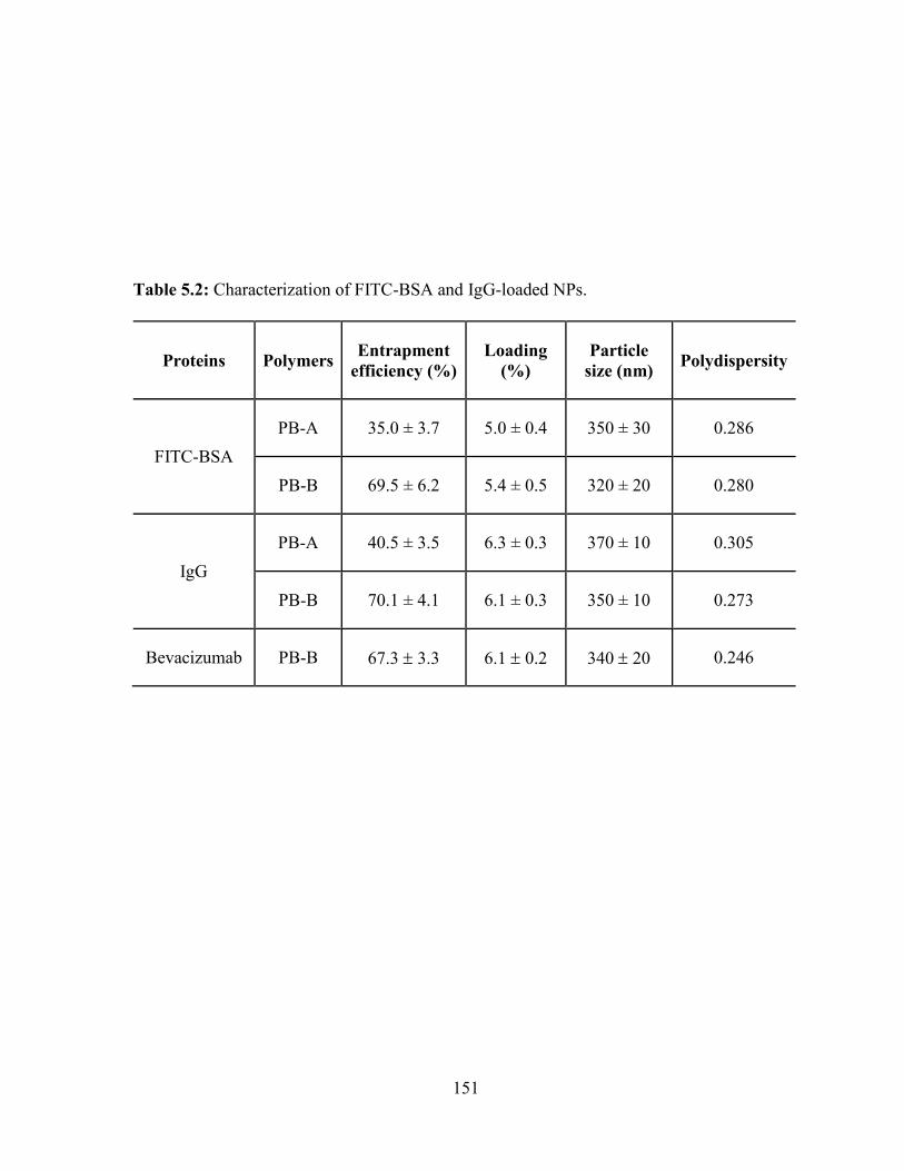
151
Table 5.2: Characterization of FITC-BSA and IgG-loaded NPs.
Proteins Polymers Entrapment
efficiency (%)
Loading
(%)
Particle
size (nm) Polydispersity
FITC-BSA
PB-A 35.0 ± 3.7 5.0 ± 0.4 350 ± 30 0.286
PB-B 69.5 ± 6.2 5.4 ± 0.5 320 ± 20 0.280
IgG
PB-A 40.5 ± 3.5 6.3 ± 0.3 370 ± 10 0.305
PB-B 70.1 ± 4.1 6.1 ± 0.3 350 ± 10 0.273
Bevacizumab PB-B 67.3 3.3 6.1 0.2 340 20 0.246
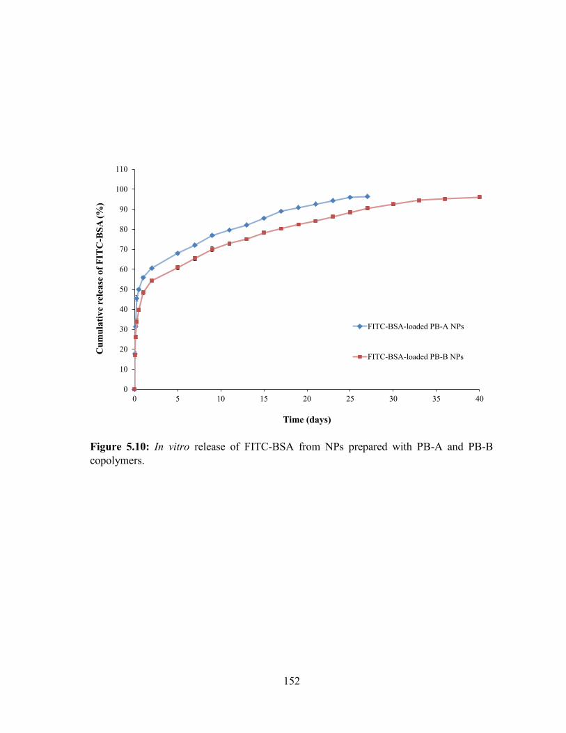
152
Figure 5.10: In vitro release of FITC-BSA from NPs prepared with PB-A and PB-B
copolymers.
0
10
20
30
40
50
60
70
80
90
100
110
0 5 10 15 20 25 30 35 40
Cu
mu
lati
ve
rele
ase
of
FIT
C-B
SA
(%
)
Time (days)
FITC-BSA-loaded PB-A NPs
FITC-BSA-loaded PB-B NPs
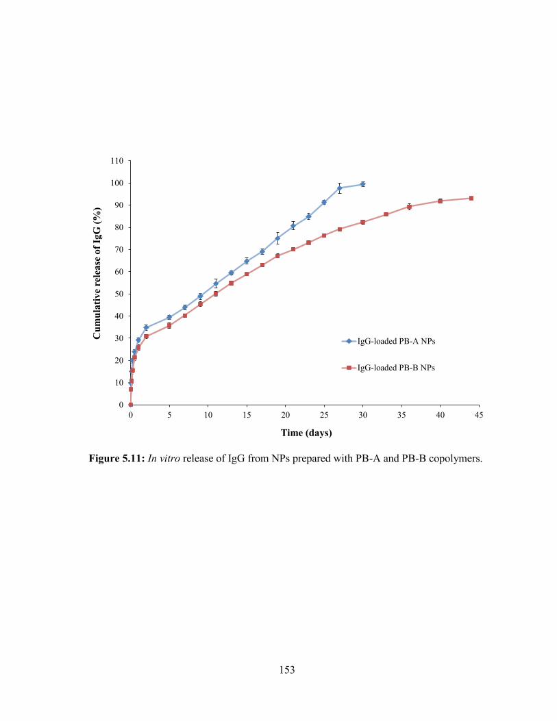
153
Figure 5.11: In vitro release of IgG from NPs prepared with PB-A and PB-B copolymers.
0
10
20
30
40
50
60
70
80
90
100
110
0 5 10 15 20 25 30 35 40 45
Cu
mu
lati
ve
rele
ase
of
IgG
(%
)
Time (days)
IgG-loaded PB-A NPs
IgG-loaded PB-B NPs
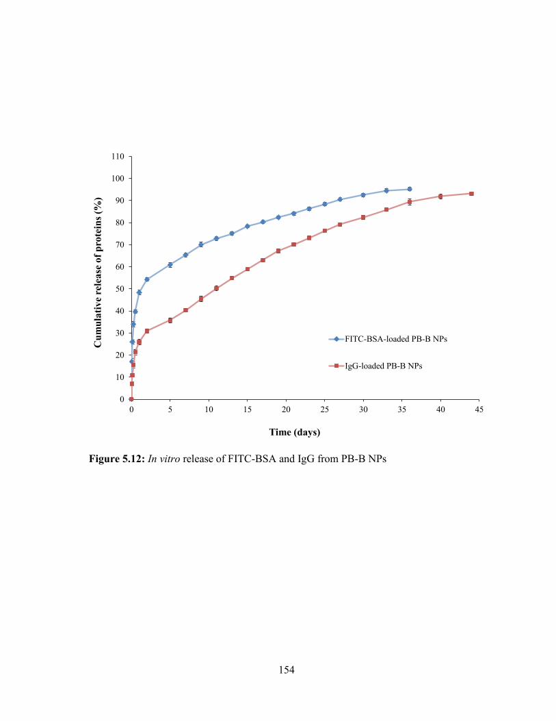
154
Figure 5.12: In vitro release of FITC-BSA and IgG from PB-B NPs
0
10
20
30
40
50
60
70
80
90
100
110
0 5 10 15 20 25 30 35 40 45
Cu
mu
lati
ve
rele
ase
of
pro
tein
s (%
)
Time (days)
FITC-BSA-loaded PB-B NPs
IgG-loaded PB-B NPs

155
Figure 5.13: In vitro release of IgG and bevacizumab from PB-B NPs
0
10
20
30
40
50
60
70
80
90
100
110
0 5 10 15 20 25 30 35 40 45
Cu
mu
lati
ve
rele
ase
of
pro
tein
s (%
)
Time (days)
Bevacizumab-loaded PB-B NPs
IgG-loaded PB-B NPs
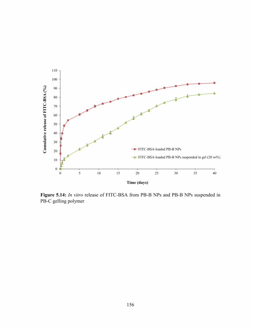
156
Figure 5.14: In vitro release of FITC-BSA from PB-B NPs and PB-B NPs suspended in
PB-C gelling polymer
0
10
20
30
40
50
60
70
80
90
100
110
0 5 10 15 20 25 30 35 40
Cu
mu
lati
ve
rele
ase
of
FIT
C-B
SA
(%
)
Time (days)
FITC-BSA-loaded PB-B NPs
FITC-BSA-loaded PB-B NPs suspended in gel (20 wt%)
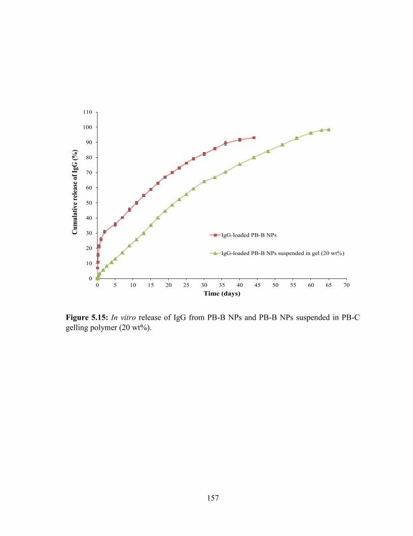
157
Figure 5.15: In vitro release of IgG from PB-B NPs and PB-B NPs suspended in PB-C
gelling polymer (20 wt%).
0
10
20
30
40
50
60
70
80
90
100
110
0 5 10 15 20 25 30 35 40 45 50 55 60 65 70
Cu
mu
lati
ve
rele
ase
of
IgG
(%
)
Time (days)
IgG-loaded PB-B NPs
IgG-loaded PB-B NPs suspended in gel (20 wt%)
158
Protein-loaded NPs were suspended in 20 wt% gelling solution and exposed to 37 °C
which immediately transformed to solid hydrogel entrapping NPs throughout the polymer
matrix. These composite formulations comprised of protein-loaded PB-B NPs (FITC-BSA and
IgG) suspended in thermosensitive gel were evaluated for their release behavior. As described
in Figure 5.14, burst release of FITC-BSA from composite formulation was negligible (~10%)
relative to burst release observed from PB-B NPs (~48%) alone. In addition, release of FITC-
BSA was prolonged for ~40 days. The similar behavior was observed for a composite
formulation comprising IgG-loaded PB-B NPs suspended in 20 wt% thermosensitive gelling
solution (Figure 5.15). Composite formulation of IgG exhibited negligible burst release
followed zero-order release up to ~60 days. This behavior may be due to the fact that when the
NPs were suspended into the gel matrix, gel matrix might serve as an additional diffusion
barrier for the surface adsorbed drug. The gel matrix might deter the dumping of surface
adsorbed dose eliminating burst effect which eventually provided nearly zero-order drug
release throughout the release period.
Release kinetics
In order to evaluate drug release mechanism, we have fitted in vitro drug release data
in five different release kinetic models i.e., Korsmeyer-Peppas, Higuchi, Hixon-Crowell, zero-
order and first-order. Results showed in Table 5.3, indicated that Korsmeyer-Peppas was the
best fit model for all the formulations with R2 values ranging between 0.977-0.997. Moreover,
n values in Korsmeyer-Peppas model for release of FITC-BSA, IgG and bevacizumab from
PB-B NPs were below 0.43 indicating diffusion controlled release. Interestingly, n values for
composite formulation of FITC-BSA (0.549) and IgG (0.818) (NPs suspended in
thermosensitive gel) were between 0.43-0.89 suggesting anomalous diffusion. In other words,
159
release of protein therapeutics from composite formulation is controlled by diffusion as well
as degradation of polymer.
Stability of secondary structure of IgG confirmed by CD spectroscopy
It is obvious that protein therapeutics need to maintain their three dimensional structure
to exert pharmacological action. CD spectroscopy is a sensitive and robust analytical technique
exploited for the investigation of secondary and at some extent tertiary conformation of
proteins. It is sensitive enough to detect minor conformational changes in α-helix and β-sheets
of the protein. Therefore, we have utilized this technique to confirm the conformational
stability of released IgG and compared with CD spectrum of native IgG (Figure 5.16). CD
spectrum of released IgG demonstrated λ minima of 218 nm similar to the native IgG. In
addition, the CD spectra of native IgG and released IgG ranging from 200 nm to 250 nm were
identical to each other indicating retention of structural conformation during NP preparation.
Cell proliferation assay
A cell proliferation assay was performed as described previously [215]. RF/6A cells
proliferated rapidly in presence of VEGF (100 ng/mL, +ve control) whereas its proliferation
was inhibited in absence of VEGF indicating sensitivity of endothelial cells toward growth
factor particularly, VEGF (Figure 5.17). Native bevacizumab (standard group) has strongly
inhibited the VEGF-induced cell proliferation at the concentration of 0.25 mg/mL. The similar
level of inhibition was observed when released (released from NPs) bevacizumab (test/sample
group) was exposed for 24h to VEGF treated cells. Moreover, inhibitory effects of released
and native bevacizumab were not significantly different than -ve control.
160
Cell migration assay
Chemo-attractant property of VEGF stimulates RF/6A cell migration of across a porous
membrane toward a VEGF stimulus. Biological activity of released bevacizumab was further
evaluated by VEGF-induced cell migration assay. As described in Figure 5.18, a test/sample
group (released bevacizumab) exhibited significant inhibition of cell migration across the
transwell membrane relative to VEGF group (+ve control). Results showed that this inhibitory
effect was not significantly different than -ve control or a standard group (cells treated with
native bevacizumab). For the visual evidence, migrated cells were stained with methylene blue
and images were taken. Data depicted in Figure 5.18b also supported the results described
earlier.
Data observed in cell proliferation and cell migration assays clearly pointed out that
bevacizumab has retained its biological activity during the process of NP preparation. Previous
reports suggest that PLA and/or PGA based copolymer produce large molar mass of lactic acid
and/or glycolic acid [183, 197]. These degradation products stimulate hydrolytic degradation
of protein therapeutics. Retention of protein stability (IgG and bevacizumab) in PB NPs may
attributed to lower molar mass of PLA or PGA blocks which produce very low amounts of
lactic acid or glycolic acid, eliminating or reducing protein degradation.
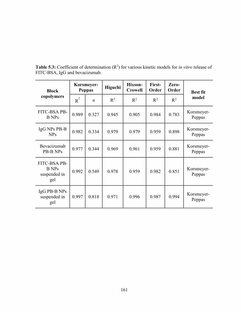
161
Table 5.3: Coefficient of determination (R2) for various kinetic models for in vitro release of
FITC-BSA, IgG and bevacizumab.
Block
copolymers
Korsmeyer-
Peppas Higuchi
Hixson-
Crowell
First-
Order
Zero-
Order Best fit
model R
2 n R2 R2 R2 R2
FITC-BSA PB-
B NPs 0.989 0.327 0.945 0.905 0.984 0.783
Korsmeyer-
Peppas
IgG NPs PB-B
NPs 0.982 0.334 0.979 0.979 0.959 0.898
Korsmeyer-
Peppas
Bevacizumab
PB-B NPs 0.977 0.344 0.969 0.961 0.959 0.881
Korsmeyer-
Peppas
FITC-BSA PB-
B NPs
suspended in
gel
0.992 0.549 0.978 0.959 0.982 0.851 Korsmeyer-
Peppas
IgG PB-B NPs
suspended in
gel
0.997 0.818 0.971 0.996 0.987 0.994 Korsmeyer-
Peppas
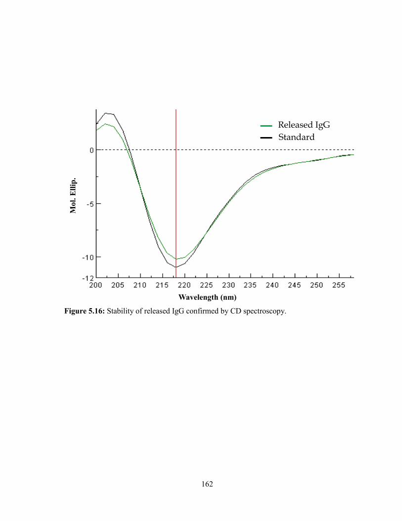
162
Figure 5.16: Stability of released IgG confirmed by CD spectroscopy.
Standard
Released IgG
Mo
l. E
llip
.
Wavelength (nm)

163
Figure 5.17: Cell proliferation assay performed on RF/6A cells to evaluate the biological
activity of bevacizumab (a) absorbance produced by live cells (b) images of live cells where
(1) negative control (untreated cells), (2) standard (cells exposed to 100 ng/mL of VEGF
and 0.25 mg/mL of bevacizumab), (3) sample (cells exposed to 100 ng/mL of VEGF and
0.25 mg/mL of released bevacizumab), and (4) positive control (cell exposed to 100 ng/mL
VEGF).
0.500
0.600
0.700
0.800
0.900
1.000
1.100
Negative control Standard Sample Positive control
Ab
sorb
an
ce
a
(1) (2)
(3) (4)
b
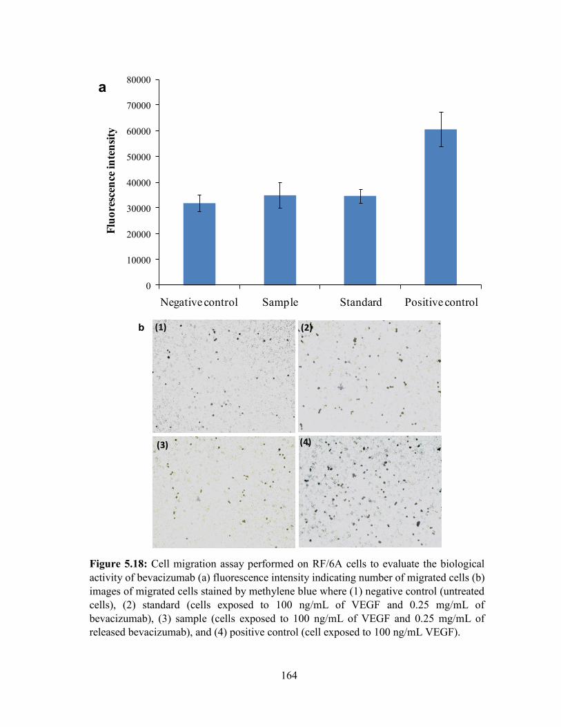
164
Figure 5.18: Cell migration assay performed on RF/6A cells to evaluate the biological
activity of bevacizumab (a) fluorescence intensity indicating number of migrated cells (b)
images of migrated cells stained by methylene blue where (1) negative control (untreated
cells), (2) standard (cells exposed to 100 ng/mL of VEGF and 0.25 mg/mL of
bevacizumab), (3) sample (cells exposed to 100 ng/mL of VEGF and 0.25 mg/mL of
released bevacizumab), and (4) positive control (cell exposed to 100 ng/mL VEGF).
0
10000
20000
30000
40000
50000
60000
70000
80000
Negative control Sample Standard Positive control
Flu
ore
scen
ce in
ten
sity
a
(1) (2)
(3) (4)
b
165
Conclusion
We have successfully synthesized and characterized novel PB copolymers for the
preparation of NPs and thermosensitive gel. In order to eliminate burst release phase, a novel
composite formulation comprised of protein-loaded PB NPs suspended in a PB
thermosensitive gel was successfully formulated and evaluated. FITC-BSA and IgG-
encapsulated NPs suspended in thermosensitive gel demonstrated continuous zero-order
release, avoiding the possibility of dose dependent toxicity that may occur due to an initial
burst release. Moreover, CD spectroscopy demonstrated the retention of structural
conformation of released IgG. Bevacizumab-loaded NPs demonstrated similar patterns of burst
release and sustained release as of IgG-embedded NPs. In vitro cell proliferation and cell
migration assay confirmed retention of biological activity of the bevacizumab. This approach
can act as a platform for the ocular delivery of therapeutic macromolecules, and can minimize
the side effects associated with frequent intravitreal injections.
166
CHAPTER 6
OPTIMIZATION OF NOVEL PENTABLOCK COPOLYMER BASED COMPOSITE
FORMULATION FOR SUSTAINED DELIVERY OF PROTEIN THERAPEUTICS IN
THE TREATMENT OF OCULAR DISEASES
Rationale
The ideal formulation for the intravitreal delivery should possess following
characteristics, (a) high drug loading in small volume (≤ 100 µL) that lasts up to 6 months or
more, (b) provide constant release (zero-order release) throughout the release period without
any burst effect, (c) easy to administered such as injectable system and not implants, (d) ensure
stability of protein/peptide, (e) biodegradable and biocompatible, and (f) the time required for
biodegradation of formulation should not be more than 1.5 times the release period.
Many researchers are investigating protein-encapsulated NPs for the sustained delivery
in the treatment of back of the eye diseases. It is easy to achieve high drug loading in NPs for
hydrophobic therapeutic agents. In contrast, it is very difficult to achieve high drug loading of
protein therapeutics (hydrophilic agents). One more limitation for intravitreal injection is small
injection volume (≤100 µL). Therefore, one of the important challenges is to develop a
formulation which can carry large amount of dose in limited injection volume and provide
constant release up to 6 months or more.
In order to address mentioned issues, we have synthesized novel biodegradable and
biocompatible PB copolymers composed of PEG, PCL, and PLA/PGA. In this section, we
study the effects of various formulation parameters on EE, DL and in vitro release profile. In
order to incorporate a large amount of protein therapeutic in small volume of formulation, we
have optimized NP preparation methods to attain maximum possible DL. To achieve constant
167
(zero-order) release, we have utilized a novel concept of composite formulation (discussed in
chapter 5) where protein/peptide-loaded PB NPs were suspended in a PB thermosensitive gel.
Moreover, the optimized methods for NP preparation were also applied to encapsulate proteins
or peptides with different molecular weights ranging from 1-237 kDa. Biological activity of
released proteins or peptides was also determined utilizing enzymatic activity assays.
Materials and methods
Materials
PEG (2 kDa and 4 kDa), methoxy-PEG (550 Da), ε-caprolactone, poly (vinyl alcohol)
(PVA), stannous octoate, lysozyme from chicken egg white and Micrococcus luteus were
procured from Sigma-Aldarich (St. Louis, MO; USA). Hexamethylene diisocyanate (HMDI),
glycolide and L-lactide were obtained from Acros organics (Morris Plains, NJ; USA). Catalase
was purchased from Worthington Biochemical Corp. IgG-Fab and IgG were purchased from
Athens research technology Inc., and Lee Biosolutions, respectively. Octreotide and insulin
were procured from China Peptides Co. ltd. and MP Biomedicals LLC., respectively. Micro-
BCATM and catalase colorimetric assay kits were obtained from Fisher Scientific and Arbor
Assays Inc., respectively. All other reagents utilized in this study were of analytical grade.
Methods
Synthesis of PB copolymers
Novel PB copolymers, PLA-PCL-PEG-PCL-PLA (PB-A/PB-B), PGA-PCL-PEG-
PCL-PGA (PB-C/PB-D), and PEG-PCL-PLA-PCL-PEG (PB-E) were synthesized by ring-
opening bulk copolymerization [217]. Briefly, ε-caprolactone was polymerized on two open
hydroxyl ends of PEG (2 kDa or 4 kDa) utilizing stannous octoate as catalyst (0.5% w/w). The
reaction was carried out for 24 h at 130 C in inert environment. TB (TB) copolymer (PCL-
168
PEG-PCL) was purified by dissolving in dichloromethane (DCM) followed by cold-ether
precipitation. Purified TB copolymer was then utilized for the preparation of PB copolymer.
Predetermined quantities of TB copolymer and L-lactide (PB-A/PB-B) or glycolide (PB-C/PB-
D) were added in round bottom flask. Stannous octoate (0.5% w/w) was added in reaction
mixture as a catalyst. Reaction for the synthesis of PB-A/PB-B were carried out at 130 C for
24 h whereas for PB-C/PB-D at 200 C for 24 h. At the end, reaction mixture was purified for
PB copolymers as described earlier. Purified PB copolymers were vacuum-dried and stored at
-20 C until further characterization. PB copolymer with thermosensitive properties (PB-E)
was synthesized, purified and characterized according to previously published protocol with
minor modifications [217]. For the synthesis of PB-E, TB copolymer (mPEG-PCL-PLA) was
synthesized by ring-opening bulk copolymerization. Firstly, ε-caprolactone was polymerized
at the hydroxyl terminal of mPEG (550 Da) followed by polymerization of L-lactide. Resulting
TB copolymers were coupled with HMDI as a linker. Coupling reaction was carried out for 8
h at 70 °C. Resulting polymer was purified by cold-ether precipitation followed by drying
under vacuum. Reaction schemes for the synthesis of PB copolymers are depicted in Figures
6.1 and 6.2.

169
Figure 6.1a: Synthesis scheme for PB-A and PB-B (PLA-PCL-PEG-PCL-PLA)
OHH2C
H2C O H
x
O
O
+ HOH2C C
O
OH2C
H2C O C
O
H2C O
5 5H
y yx
Sn(Oct)2
130°C
caprolactonePEG HO-PCL-PEG-PCL-OH
HOH2C C
O
OH2C
H2C O C
O
H2C O
5 5H
y yx
HO-PCL-PEG-PCL-OH
Sn(Oct)2
130°C
OH2C C
O
OH2C
H2C O C
O
H2C O
5 5C
y yx
C
O
HC
CH3
HO
O
HC
CH3
O
z z
+
O
O
O
O
H3C
CH3
PLA-PCL-PEG-PCL-PLA
Step 1
Step 2
Lactide
H
Figure 6.1b: Synthesis scheme for PB-C and PB-D (PGA-PCL-PEG-PCL-PGA).
HOH2C C
O
OH2C
H2C O C
O
H2C O
5 5H
y yx
HO-PCL-PEG-PCL-OH
Sn(Oct)2
200°C
OH2C C
O
OH2C
H2C O C
O
H2C O
5 5C
y yx
C
O
H2CHO
O
H2C O
z z
+
O
O
O
O
PGA-PCL-PEG-PCL-PGA
Step 2
Glycolide
H
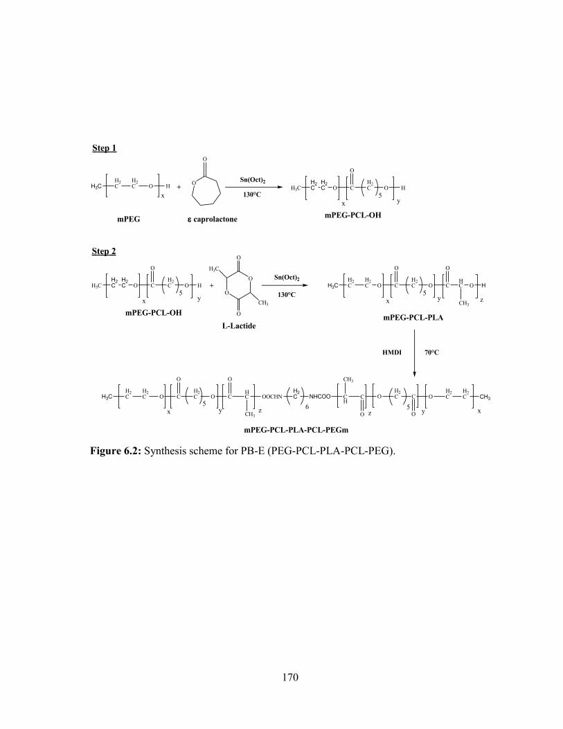
170
Figure 6.2: Synthesis scheme for PB-E (PEG-PCL-PLA-PCL-PEG).
H3CH2C
H2C O H
x
O
O
+ H3C O C
O
H2C O
5H
yx
Sn(Oct)2
130°C
caprolactonemPEGmPEG-PCL-OH
Sn(Oct)2
130°C
H3CH2C
H2C O C
O
H2C O
5C
yx
O
HC
CH3
O
z
+O
O
O
O
H3C
CH3
mPEG-PCL-PLA
Step 1
Step 2
L-Lactide
70°CHMDI
H3CH2C
H2C O C
O
H2C O
5C
yx
O
HC
CH3
OOCHN
z
H2C NHCOO CH3
H2C
H2COC
O
H2COC
O
CH
CH3
xy5
z6
mPEG-PCL-PLA-PCL-PEGm
H2C
H2C
H3C O C
O
H2C O
5H
yx
mPEG-PCL-OH
H2C
H2C H
171
Characterization of polymers
PB copolymers were characterized for their molecular weight and purity by 1H-NMR
spectroscopy and gel permeation chromatography (GPC).
1H-NMR
Polymeric materials (5 mg) were dissolved in 600 µL of CDCl3 and were analyzed on
Varian-400 MHz NMR instrument. Molecular weight (Mn) and purity of the polymers were
evaluated from the 1H-NMR spectrum.
GPC
Molecular weights (Mn and Mw), purity and polydispersity (PD) were further evaluated
by Ecosec HLC 8320 gel permeation chromatograph connected with differential refractometer.
Briefly, 5 mg of polymeric materials were dissolved in tetrahydrofuran (THF) and separated
on Styragel HR-3 column. THF was used as eluting solvent and polystyrene samples with
narrow molecular weight distribution were utilized as standards.
Preparation of thermosensitive gelling solution
To prepare 20 wt% aqueous gelling solution, 200 mg of PB-E copolymer was dissolved
in 800 mg of phosphate buffer saline (PBS, pH 7.4) by keeping overnight at 4 °C. Sol-gel
behavior of aqueous solution was confirmed by vial inverting method reported earlier [179].
Aqueous solution was exposed to 37 °C for 5 min followed by inversion of vial for 1 min, state
of no flow was considered as hydrogel.
Preparation of NPs
Protein/peptide-loaded PB-NPs were prepared by W1/O/W2 double emulsion solvent
evaporation method [201]. Briefly, a peptide or protein aqueous solution (W1 phase) was
172
emulsified in organic phase (dichloromethane (DCM) comprising PB copolymers) using probe
sonication to form W1/O primary emulsion. The primary emulsion was further emulsified in
aqueous phase containing 2% polyvinyl alcohol (PVA) using probe sonication to prepare
W1/O/W2 double emulsion. Resulting emulsion was diluted with 2% PVA (W3) under
continuous stirring. DCM of organic phase was then evaporated under vacuum using rotavap
to obtain NPs. NPs were separated by ultracentrifugation at 20,000 rpm for 30 min (4 C).
Particles were washed twice with distilled deionized water (DDW), and centrifuged to remove
traces of PVA and unentrapped peptide/protein. Purified NPs were freeze-dried with mannitol
(5% w/v) and stored at -20 °C until further use. Freeze-dried NPs were evaluated for EE, DL
and in vitro drug release behavior. Process parameters such as phase volume ratio,
drug/polymer ratio, and types of polymer were optimized to achieve higher DL. The detailed
process parameters are reported in Table 6.2.
Characterization of NPs
Entrapment efficiency (EE) and drug loading (DL)
Protein/peptide-encapsulated freeze-dried NPs were evaluated for the estimation of EE
and DL. Amount of unentrapped protein/peptide in supernatant was determined by micro
BCA™ protein assay kit following manufacturer’s protocol. Standard curve of respective
proteins/peptides (octreotide, insulin, lysozyme, IgG-Fab, IgG and catalase) were prepared in
the range of 3-200 µg/mL. Following equations were utilized for the calculation of EE (%) and
DL (%).
EE (%) was calculated with eq. 6.1
𝐸𝐸 (%) = (1 −𝐴𝑚𝑜𝑢𝑛𝑡 𝑜𝑓 𝑑𝑟𝑢𝑔 𝑖𝑛 𝑠𝑢𝑝𝑒𝑟𝑛𝑎𝑡𝑎𝑛𝑡
𝑇𝑜𝑡𝑎𝑙 𝑎𝑚𝑜𝑢𝑛𝑡 𝑜𝑓 𝑑𝑟𝑢𝑔) ∗ 100 …Eq. 6.1
173
DL (%) was calculated by eq. 6.2
𝐷𝐿 (%) = (𝐴𝑚𝑜𝑢𝑛𝑡 𝑜𝑓 𝑑𝑟𝑢𝑔 𝑖𝑛 𝑛𝑎𝑛𝑜𝑝𝑎𝑟𝑡𝑖𝑐𝑙𝑒𝑠
𝑇𝑜𝑡𝑎𝑙 𝑎𝑚𝑜𝑢𝑛𝑡 𝑜𝑓 𝑑𝑟𝑢𝑔 𝑎𝑛𝑑 𝑝𝑜𝑙𝑦𝑚𝑒𝑟) ∗ 100 …Eq. 6.2
In vitro release studies
Protein/peptide-loaded freeze-dried NPs were characterized for their release behavior.
To perform in vitro drug release from composite formulation, precalculated amount of NPs
were suspended in 100 µL of PB-E thermosensitive gelling solution (20 wt%) maintained at 4
°C. Resulting PB NP suspension was then incubated at 37 °C for 30 min followed by slow
addition of 1 mL of PBS (pH 7.4) preincubated at 37 °C. At predefined time intervals, 200 µL
of clear supernatant was collected and replaced with fresh PBS (37 °C). Release samples were
evaluated for protein content by Micro BCATM total protein assay which was performed
according to supplier’s instructions. Release experiments were carried out in triplicates and
depicted as cumulative drug release (%) against time. Biological activity of lysozyme and
catalase were confirmed by their enzymatic assays.
Release kinetics
In order to investigate release mechanisms, release data were fitted to various kinetic
models i.e., Korsmeyer-Peppas (Mt/M∞ = ktn), Higuchi (Qt = Kt1/2), Hixon-Crowell (C01/3 –
Ct1/3 = kt), First-order (LogC = LogC0 – Kt/2.303), and Zero-order (C = K0t). Based on the R2
value, best fit model was identified. Diffusion exponent (n) of Korsmeyer-Peppas equation
was utilized to understand the mechanism of release.
174
Stability evaluation of lysozyme and catalase
Estimation of enzymatic activity of lysozyme
Enzymatic activity of lysozyme in the released samples was estimated by comparing
with freshly prepared lysozyme solutions and/or controlled samples. Controls were comprised
of lysozyme solution incubated at 37 °C in PBS (pH 7.4) which kept parallel to the in vitro
release study from composite formulation. In order to determine enzymatic activity of
lysozyme, a stock solution of Micrococcus luteus (0.01% w/v) was prepared with phosphate
buffer (66 mM, pH 6.15) and diluted to achieve absorbance between 0.2 - 0.6 at 450 nm. A
100 µL of samples, standards or controls were mixed with 2.5 mL of Micrococcus luteus
suspension. A rate of deceleration of absorbance at 450 nm was determined over a period of 4
min at room temperature. Data was plotted for absorbance against time and slop was utilized
for the quantification of lysozyme in enzyme unit (EU). The units of lysozyme (active) per
milligram of protein were calculated from the following equations [218].
Units of lysozyme in one mL sample =(∆A 450 nm min Test⁄ − ∆A 450 nm min Blank⁄ )(df)
(0.001)(0.1) …Eq. 6.3
Units of lysozyme
mg of sample=
Units of lysozyme mL of sample⁄
mg of lysozyme mL of sample⁄ …Eq. 6.4
Where, 0.001 was obtained from the definition of lysozyme as one unit of enzyme is able to
produce ΔAbs450nm of 0.001 per minute at pH 6.15 and 25 °C utilizing Micrococcus luteus
suspension, 0.1 represent the volume of release samples, standards or controls and df depicts
dilution factor. Biological activity observed for release samples were compared with the
respective controls of the same time points.

175
Estimation of enzymatic activity of catalase
Enzymatic activity of catalase was estimated for in vitro release samples. Control
samples with known concentration of catalase were prepared in PBS (pH 7.4) and exposed at
37 °C along with in vitro release samples. Control, test and standard samples were analyzed
for catalase activity with catalase colorimetric assay kit. Assay was performed according to
supplier’s protocol. Briefly, standards with known concentrations were prepared in assay
buffer. A 25 µL of standard, control or sample was added in 96-well plate containing 25 µL of
hydrogen peroxide solution. The resulting mixture was then incubated for 30 min at room
temperature. After incubation, 25 µL of colorimetric detection reagent was added in each well
followed by addition of 25 µL horseradish peroxidase (HRP) reagent. Plate was incubated for
15 min at room temperature and then analyzed by UV spectrophotometer at 570 nm. According
to catalase activity assay, reduction in the absorbance is directly proportional to the catalase
activity.
Results and discussion
Synthesis and characterization of PB copolymers
PB copolymers designed for the preparation of NPs and thermosensitive gel were
successfully synthesized by ring-opening bulk copolymerization. Firstly, TB copolymers
(PCL-PEG-PCL or mPEG-PCL-PLA) were synthesized, purified and characterized. Resulting
TB copolymers were utilized for the preparation of PB copolymers i.e., PB-A/PB-B (PLA-
PCL-PEG-PCL-PLA), PB-C/PB-D (PGA-PCL-PEG-PCL-PGA) and PB-E (PEG-PCL-PLA-
PCL-PEG). Molecular weights (Mn) and purity of the PB copolymers were examined by 1H-
NMR spectroscopy. As depicted in Figures 6.3, 6.4 and 6.5, PCL blocks exhibited typical 1H-
NMR peaks at 1.40, 1.65, 2.30 and 4.06 ppm attributed to methylene protons of -(CH2)3-, -
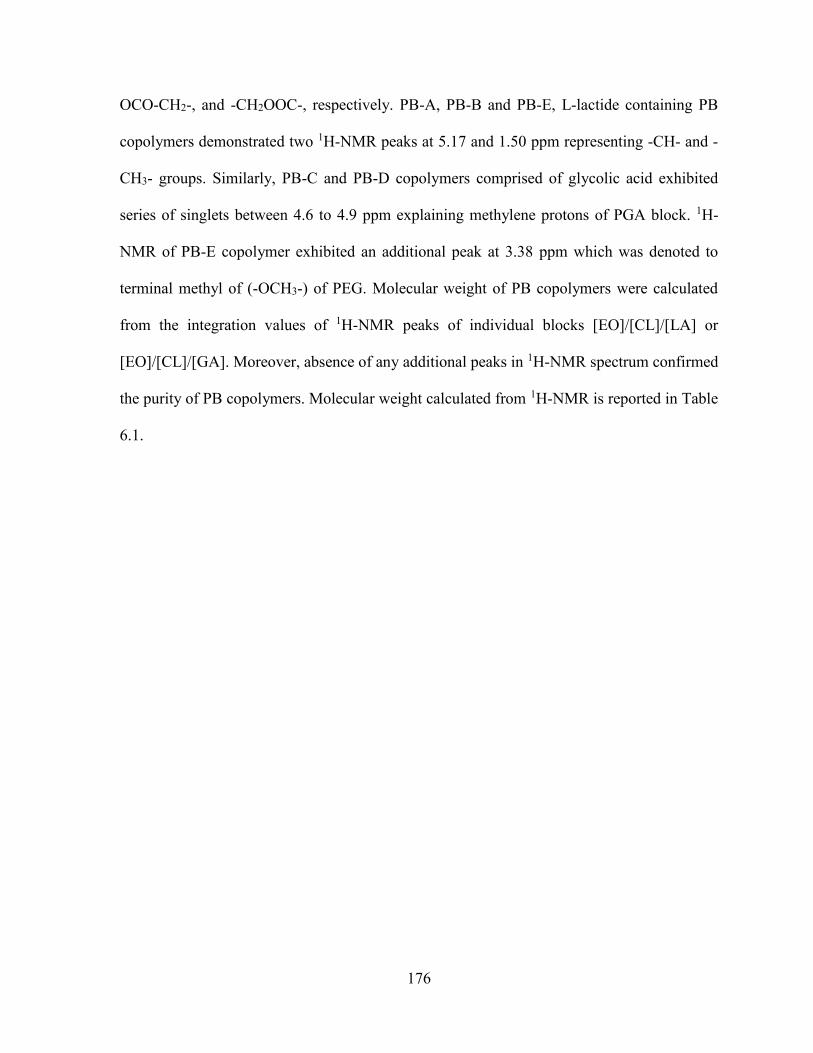
176
OCO-CH2-, and -CH2OOC-, respectively. PB-A, PB-B and PB-E, L-lactide containing PB
copolymers demonstrated two 1H-NMR peaks at 5.17 and 1.50 ppm representing -CH- and -
CH3- groups. Similarly, PB-C and PB-D copolymers comprised of glycolic acid exhibited
series of singlets between 4.6 to 4.9 ppm explaining methylene protons of PGA block. 1H-
NMR of PB-E copolymer exhibited an additional peak at 3.38 ppm which was denoted to
terminal methyl of (-OCH3-) of PEG. Molecular weight of PB copolymers were calculated
from the integration values of 1H-NMR peaks of individual blocks [EO]/[CL]/[LA] or
[EO]/[CL]/[GA]. Moreover, absence of any additional peaks in 1H-NMR spectrum confirmed
the purity of PB copolymers. Molecular weight calculated from 1H-NMR is reported in Table
6.1.
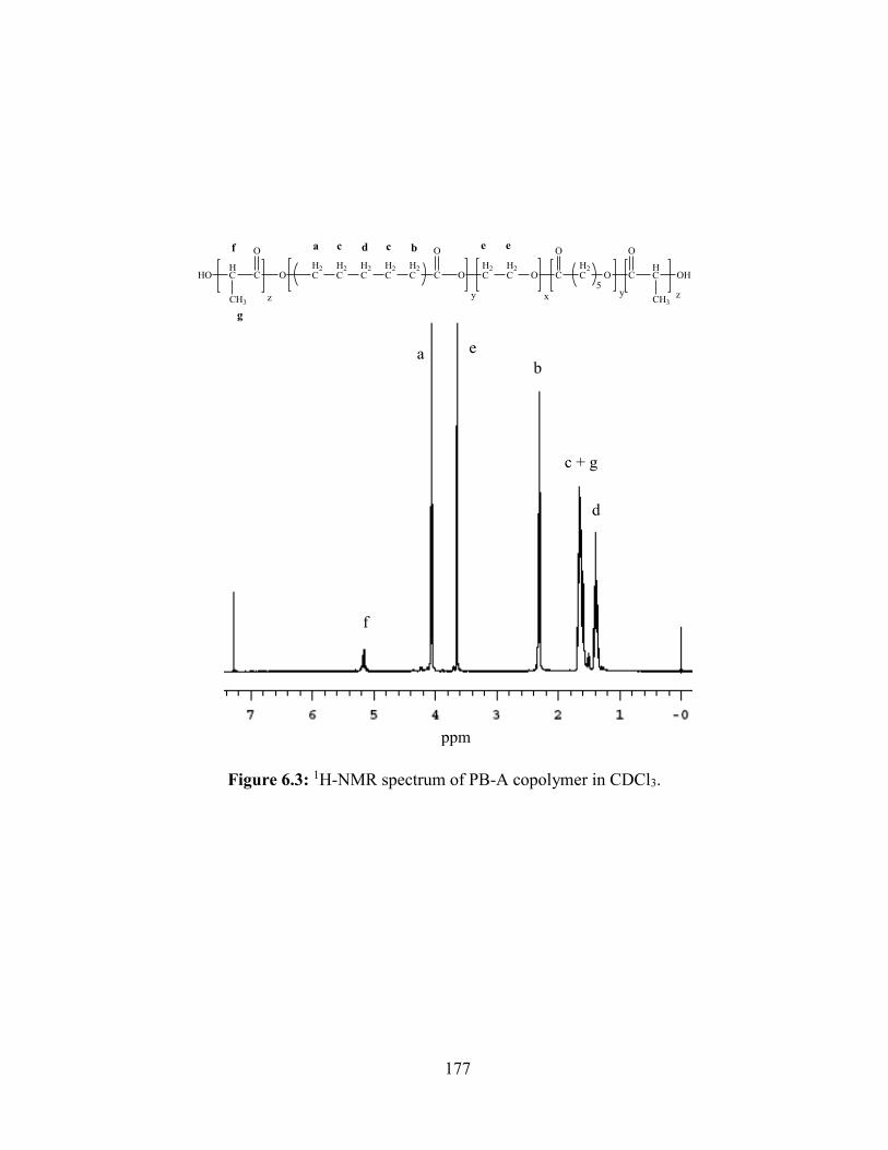
177
Figure 6.3: 1H-NMR spectrum of PB-A copolymer in CDCl3.
O C
O
OH2C
H2C O C
O
H2C O
5y yx
H2C
H2C
H2C
H2C
H2C
a bc cd e e
C
O
HC
CH3
HO
z
f
g
C
O
HC
CH3
OH
z
f
eab
c + g
d
ppm
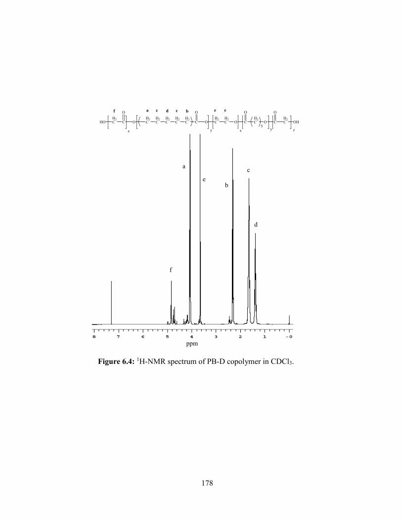
178
Figure 6.4: 1H-NMR spectrum of PB-D copolymer in CDCl3.
O C
O
OH2C
H2C O C
O
H2C O
5y yx
H2C
H2C
H2C
H2C
H2C
a bc cd e e
C
O
H2CHO
z
f
C
O
H2C OH
z
f
e
a
b
c
d
ppm
179
Figure 6.5: 1H-NMR spectrum of PB-E copolymer in CDCl3.
bc
d + g
ef
a
h
H3CH2C
H2C O C
O
C
yx
O
HC
CH3
OOCHN
z
H2C NHCOO CH3
H2C
H2COC
O
H2COC
O
CH
CH3
xy5
z6
OH2C
H2C
H2C
H2C
H2C
a a c d bde f
g
h
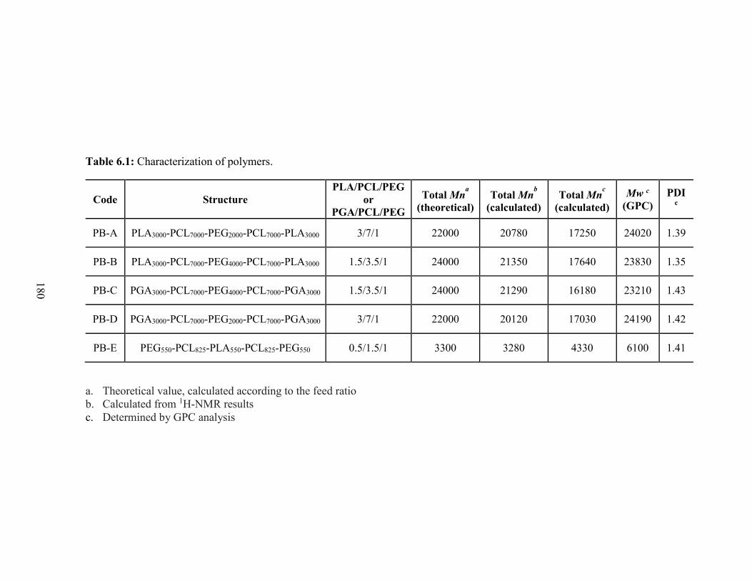
180
Table 6.1: Characterization of polymers.
Code Structure
PLA/PCL/PEG
or
PGA/PCL/PEG
Total Mna
(theoretical)
Total Mnb
(calculated)
Total Mnc
(calculated)
Mw c
(GPC)
PDI c
PB-A PLA3000-PCL7000-PEG2000-PCL7000-PLA3000 3/7/1 22000 20780 17250 24020 1.39
PB-B PLA3000-PCL7000-PEG4000-PCL7000-PLA3000 1.5/3.5/1 24000 21350 17640 23830 1.35
PB-C PGA3000-PCL7000-PEG4000-PCL7000-PGA3000 1.5/3.5/1 24000 21290 16180 23210 1.43
PB-D PGA3000-PCL7000-PEG2000-PCL7000-PGA3000 3/7/1 22000 20120 17030 24190 1.42
PB-E PEG550-PCL825-PLA550-PCL825-PEG550 0.5/1.5/1 3300 3280 4330 6100 1.41
a. Theoretical value, calculated according to the feed ratio
b. Calculated from 1H-NMR results
c. Determined by GPC analysis

181
Table 6.2: Optimization of process parameters for the preparation of IgG-Fab-loaded PB NPs.
a. W2 phase was comprised of 2% PVA
b. W3 phase was comprised of 2% PVA
Batch #
Process parameters
EE (%)
Drug
loading
(%) Polymer
types
Polymer
(mg)
IgG-Fab
(mg)
Volume
of W1
(mL)
Volume of
organic
phase (mL)
Volume
of W2
(mL)a
Volume
of W3
(mL)b
Sonication
time W1/O
Sonication
time
W1/O/W2
Batch 1 PB-A 100 10 1 4 20 - 45 Sec
(4 Output)
3 min
(7 Output) 54.0 ± 1.8 5.6 ± 0.2
Batch 2 PB-B 100 10 1 4 20 - 45 Sec
(4 Output)
3 min
(7 Output) 45.7 ± 2.7 5.0 ± 0.3
Batch 3 PB-A 100 10 1 +
1% NaCl 4
20 +
10% NaCl -
45 Sec
(4 Output)
3 min
(7 Output) 54.8 ± 2.3 6.7 ± 0.2
Batch 4 PB-B 100 10 1 +
1% NaCl 4
20 +
10% NaCl -
45 Sec
(4 Output)
3 min
(7 Output) 49.5 ± 1.3 5.9 ± 0.2
Batch 5 PB-A 10 2.5 0.25 1 8.8 - 30 Sec
(1 Output)
45 Sec
(2 Output) 25.9 ± 2.3 6.4 ± 1.0
Batch 6 PB-A 10 2.5 0.25 1 2 3 30 Sec
(1 Output)
45 Sec
(2 Output) 45.2 ± 1.6 11.7 ± 1.3
Batch 7
(Method A) PB-A 10 2.5 0.167 0.667 1.333 3.667
30 Sec
(1 Output)
45 Sec
(2 Output) 69.6 ± 9.0 15.6 ± 2.0
Batch 8
(Method B) PB-A 10 2.5 0.1 0.4 0.8 4.2
30 Sec
(1 Output)
45 Sec
(2 Output) 73.2 ± 0.2 15.5 ± 0.1
182
Purity, molecular weight (Mn and Mw) and polydispersity were further evaluated by
GPC. As described in Table 6.1, polydispersity of all the polymers were below 1.45 suggesting
narrow distribution of molecular weights. Molecular weights obtained for GPC analysis were
very close to the feed ratio. Moreover, block copolymers depicted a single peak in GPC
chromatogram (data not shown) indicating monodistribution of molecular weight and absence
of any homopolymers such as PLA, PGA, PCL and PEG. The Mn values obtained from GPC
analysis were noticeably smaller than Mn values observed from 1H-NMR spectroscopy. This
result was attributed to the difference in hydrodynamic diameter of block copolymers relative
to parent homopolymers [219]. Calculated molecular weights were very similar to the
theoretical molecular weights obtained from feed ratio. Hence, in the following text theoretical
molecular weights are mentioned instead of calculated molecular weights.
Preparation and characterization of NPs
PB NPs were successfully prepared with double emulsion solvent evaporation method
(W1/O/W2). We have attempted to optimize NP preparation method to achieve maximum
possible DL. In this section we have studied the effect of polymer hydrophobicity, salt (NaCl),
drug to polymer ratio, concentration and phase volumes, and types of protein/peptide on EE
and DL.
Entrapment efficiency (EE) and Drug loading (DL)
IgG-Fab-loaded NPs were prepared utilizing two PB copolymers (PB-A and PB-B) to
determine the effect of polymer type or hydrophobicity on DL where PB-A copolymer is more
hydrophobic than PB-B copolymer. As represented in Table 6.2, EE and DL for PB-A
copolymers (Batch 1) were ~54% and 5.62%, respectively whereas PB-B copolymer (Batch 2)
exhibited ~46% of EE and 4.99% of DL. NPs composed of PB-A copolymer demonstrated
183
higher DL relative to PB-B NPs, which may be attributed to relatively high hydrophobicity of
PB-A copolymer. During solvent evaporation, high hydrophobicity may allow faster polymer
precipitation to form NPs preventing diffusion of IgG-Fab in external aqueous (W2) phase.
Reports published elsewhere suggested significant effect of NaCl on EE and DL in
nanoparticulate system [220]. As described in Batches 3 and 4, incorporation of NaCl in W1
(1%) and W2 (10%) phase exhibited higher EE and loading of IgG-Fab in both PB-A (EE =
~55% and DL = 6.68%) and PB-B (EE = ~49% and DL = 5.88%) NPs relative to the respective
PB NPs prepared without NaCl. An important prerequisite for higher DL is separation of
droplets during emulsification process where organic phase act as a diffusion barrier between
the W1 and W2 phase. Higher concentration of drug in W1 phase may enhance osmotic pressure
in the internal phase which facilitates diffusion of water from external (W2) phase. Process of
diffusion may result in thinning of organic phase and eventually in lower EE and DL. Addition
of salt in external phase may helped to balance osmotic pressure between W1 and W2 phases
which resulted in lower diffusion of external phase and higher DL. However, addition of salt
did not exhibit significant effect on IgG-Fab loading. This may be due to the colligative
characteristic of osmotic pressure, hence, this effect may enhance the loading of lower
molecular weight drug drastically relative to higher molecular weight proteins. Based on these
results, PB-A copolymer was utilized for further optimization of NP preparation.
In order to study the effect of drug to polymer ratio, PB-A NPs with two different
drug/polymer ratios (1/10 and 1/4) were prepared and evaluated. Results depicted in Batch 5
suggest that as drug/polymer ratio was increased from 1/10 to 1/4, EE was significantly
reduced which is in accordance to the previously published results [221]. This might have
resulted from the fact that in case of 1/10 (drug/polymer) ratio, more polymer (10 mg) was
184
available to entrap 1 mg of IgG-Fab relative to a ratio of 1/4 (drug/polymer). Despite the high
drug/polymer ratio with Batch 5, there was no significant difference observed in DL compared
to earlier PB-A NPs (Batch 1). The poor DL could be attributed to high volume of external
aqueous (W2) phase. Hence, in further preparation the drug/polymer ratio was kept constant at
1/4 and effect of external phase (W2) volume was evaluated. For instance, in Batch 6 total
external phase (W2) volume was lowered to 5 mL. In addition, double emulsion was prepared
with only 2 mL of external aqueous (W2) phase to reduce partition of protein in water (W2
phase). The resulting multiple emulsion was stabilized by dilution with 3 ml of 2% PVA (W3
phase). Interestingly, 11.7% of DL was observed with Batch 6 which was significantly higher
compared to all earlier batches. Reduction in external phase volume has significantly improved
EE relative to Batch 5.
Based on results from Batch 6, it can be inferred that reduction in external phase (W2)
volume along with high drug/polymer ratio have significant effect on DL. Hence, volumes of
all the phases were further reduced while keeping the volume ratio constant. It was
hypothesized that the lower volume of W2 would diminish partition of protein in aqueous phase
improving loading efficiency. In addition, a reduction in organic phase volume would increase
polymer concentration that may lead to faster polymer precipitation and NP formation. A DL
of 15.61% was observed with NPs in Batch 7 as expected with improved EE (~70%). However,
further reduction in volumes (Batch 8) did not result in any improvement of DL (15.47%) or
EE (~73%). Optimized process parameters utilized for the preparation of Batch 7 and Batch 8
were denoted as method-A and method-B, respectively.
Process parameters optimized in Batches 7 and 8 for the preparation of IgG-Fab-loaded
PB-A NPs were utilized to encapsulate various protein/peptide molecules with different
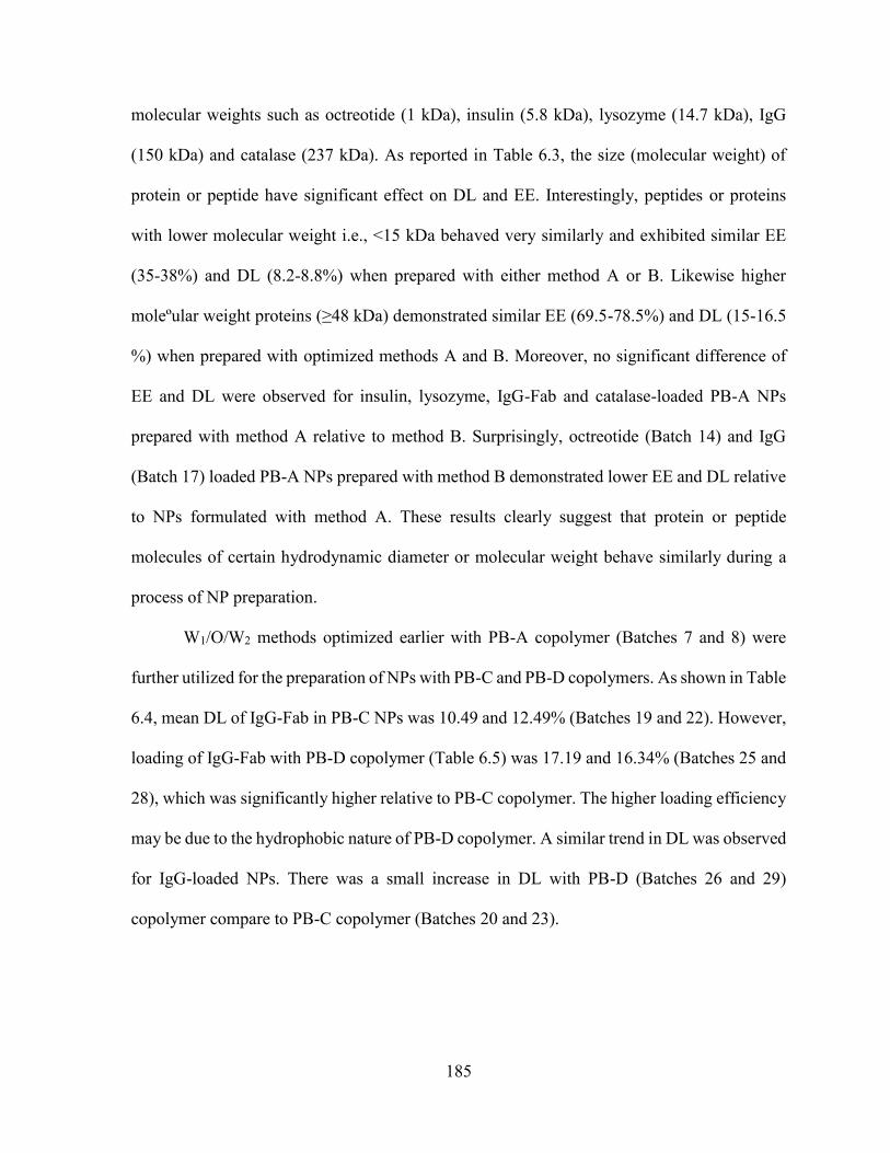
185
molecular weights such as octreotide (1 kDa), insulin (5.8 kDa), lysozyme (14.7 kDa), IgG
(150 kDa) and catalase (237 kDa). As reported in Table 6.3, the size (molecular weight) of
protein or peptide have significant effect on DL and EE. Interestingly, peptides or proteins
with lower molecular weight i.e., <15 kDa behaved very similarly and exhibited similar EE
(35-38%) and DL (8.2-8.8%) when prepared with either method A or B. Likewise higher
moleºular weight proteins (≥48 kDa) demonstrated similar EE (69.5-78.5%) and DL (15-16.5
%) when prepared with optimized methods A and B. Moreover, no significant difference of
EE and DL were observed for insulin, lysozyme, IgG-Fab and catalase-loaded PB-A NPs
prepared with method A relative to method B. Surprisingly, octreotide (Batch 14) and IgG
(Batch 17) loaded PB-A NPs prepared with method B demonstrated lower EE and DL relative
to NPs formulated with method A. These results clearly suggest that protein or peptide
molecules of certain hydrodynamic diameter or molecular weight behave similarly during a
process of NP preparation.
W1/O/W2 methods optimized earlier with PB-A copolymer (Batches 7 and 8) were
further utilized for the preparation of NPs with PB-C and PB-D copolymers. As shown in Table
6.4, mean DL of IgG-Fab in PB-C NPs was 10.49 and 12.49% (Batches 19 and 22). However,
loading of IgG-Fab with PB-D copolymer (Table 6.5) was 17.19 and 16.34% (Batches 25 and
28), which was significantly higher relative to PB-C copolymer. The higher loading efficiency
may be due to the hydrophobic nature of PB-D copolymer. A similar trend in DL was observed
for IgG-loaded NPs. There was a small increase in DL with PB-D (Batches 26 and 29)
copolymer compare to PB-C copolymer (Batches 20 and 23).
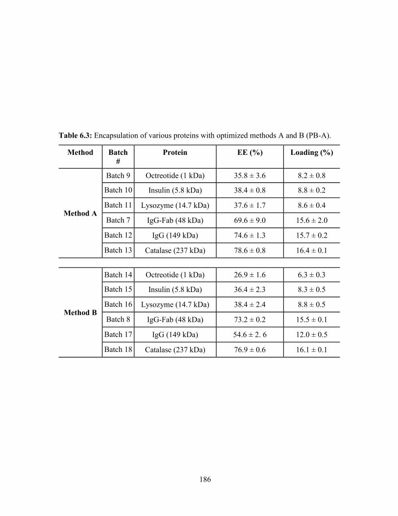
186
Table 6.3: Encapsulation of various proteins with optimized methods A and B (PB-A).
Method Batch
#
Protein EE (%) Loading (%)
Method A
Batch 9 Octreotide (1 kDa) 35.8 ± 3.6 8.2 ± 0.8
Batch 10 Insulin (5.8 kDa) 38.4 ± 0.8 8.8 ± 0.2
Batch 11 Lysozyme (14.7 kDa) 37.6 ± 1.7 8.6 ± 0.4
Batch 7 IgG-Fab (48 kDa) 69.6 ± 9.0 15.6 ± 2.0
Batch 12 IgG (149 kDa) 74.6 ± 1.3 15.7 ± 0.2
Batch 13 Catalase (237 kDa) 78.6 ± 0.8 16.4 ± 0.1
Method B
Batch 14 Octreotide (1 kDa) 26.9 ± 1.6 6.3 ± 0.3
Batch 15 Insulin (5.8 kDa) 36.4 ± 2.3 8.3 ± 0.5
Batch 16 Lysozyme (14.7 kDa) 38.4 ± 2.4 8.8 ± 0.5
Batch 8 IgG-Fab (48 kDa) 73.2 ± 0.2 15.5 ± 0.1
Batch 17 IgG (149 kDa) 54.6 ± 2. 6 12.0 ± 0.5
Batch 18 Catalase (237 kDa) 76.9 ± 0.6 16.1 ± 0.1
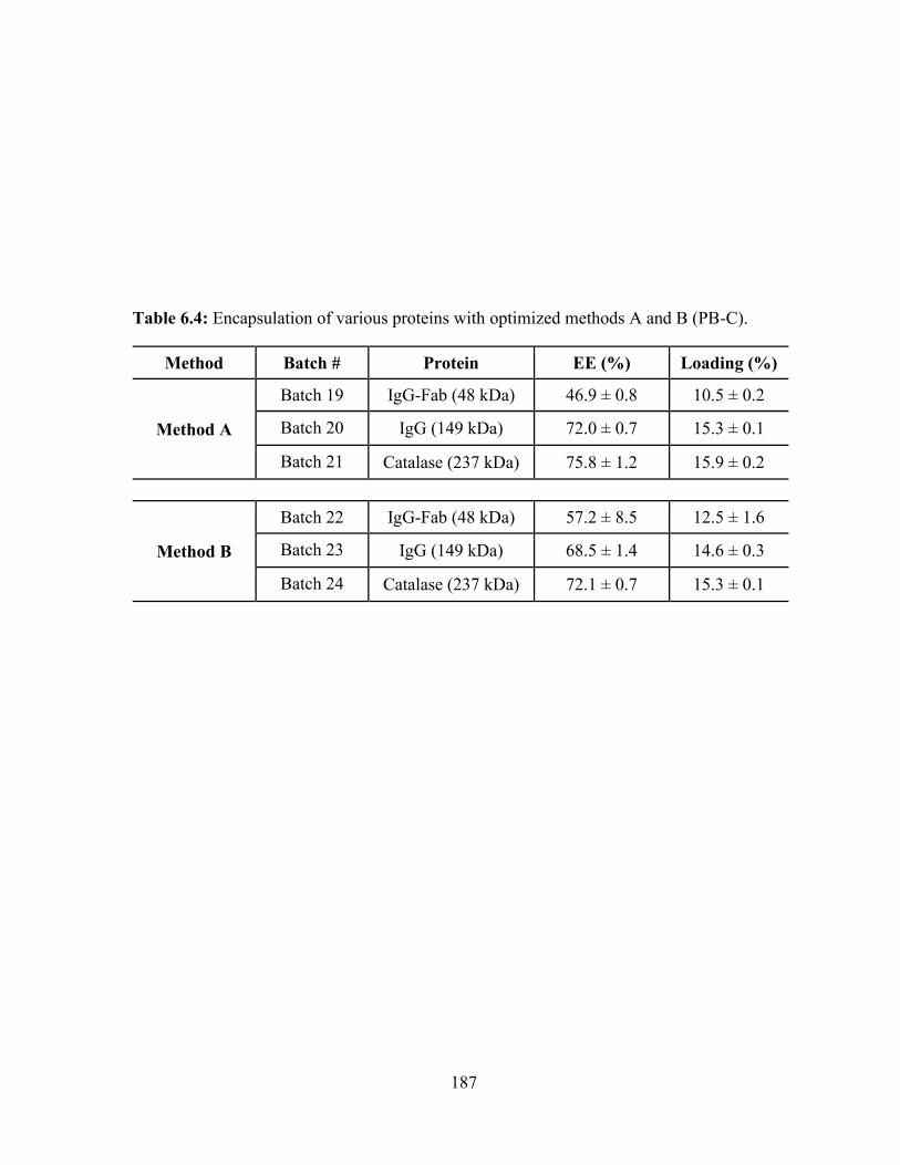
187
Table 6.4: Encapsulation of various proteins with optimized methods A and B (PB-C).
Method Batch # Protein EE (%) Loading (%)
Method A
Batch 19 IgG-Fab (48 kDa) 46.9 ± 0.8 10.5 ± 0.2
Batch 20 IgG (149 kDa) 72.0 ± 0.7 15.3 ± 0.1
Batch 21 Catalase (237 kDa) 75.8 ± 1.2 15.9 ± 0.2
Method B
Batch 22 IgG-Fab (48 kDa) 57.2 ± 8.5 12.5 ± 1.6
Batch 23 IgG (149 kDa) 68.5 ± 1.4 14.6 ± 0.3
Batch 24 Catalase (237 kDa) 72.1 ± 0.7 15.3 ± 0.1
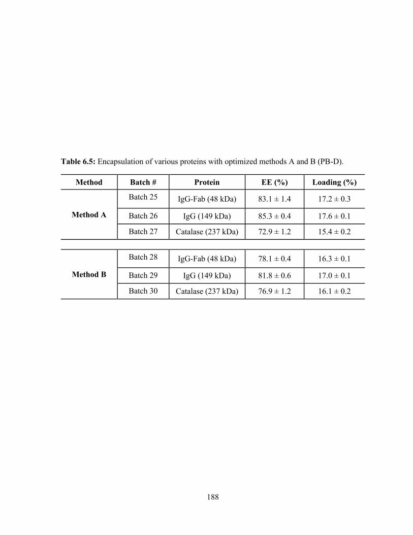
188
Table 6.5: Encapsulation of various proteins with optimized methods A and B (PB-D).
Method Batch # Protein EE (%) Loading (%)
Method A
Batch 25 IgG-Fab (48 kDa) 83.1 ± 1.4 17.2 ± 0.3
Batch 26 IgG (149 kDa) 85.3 ± 0.4 17.6 ± 0.1
Batch 27 Catalase (237 kDa) 72.9 ± 1.2 15.4 ± 0.2
Method B
Batch 28 IgG-Fab (48 kDa) 78.1 ± 0.4 16.3 ± 0.1
Batch 29 IgG (149 kDa) 81.8 ± 0.6 17.0 ± 0.1
Batch 30 Catalase (237 kDa) 76.9 ± 1.2 16.1 ± 0.2
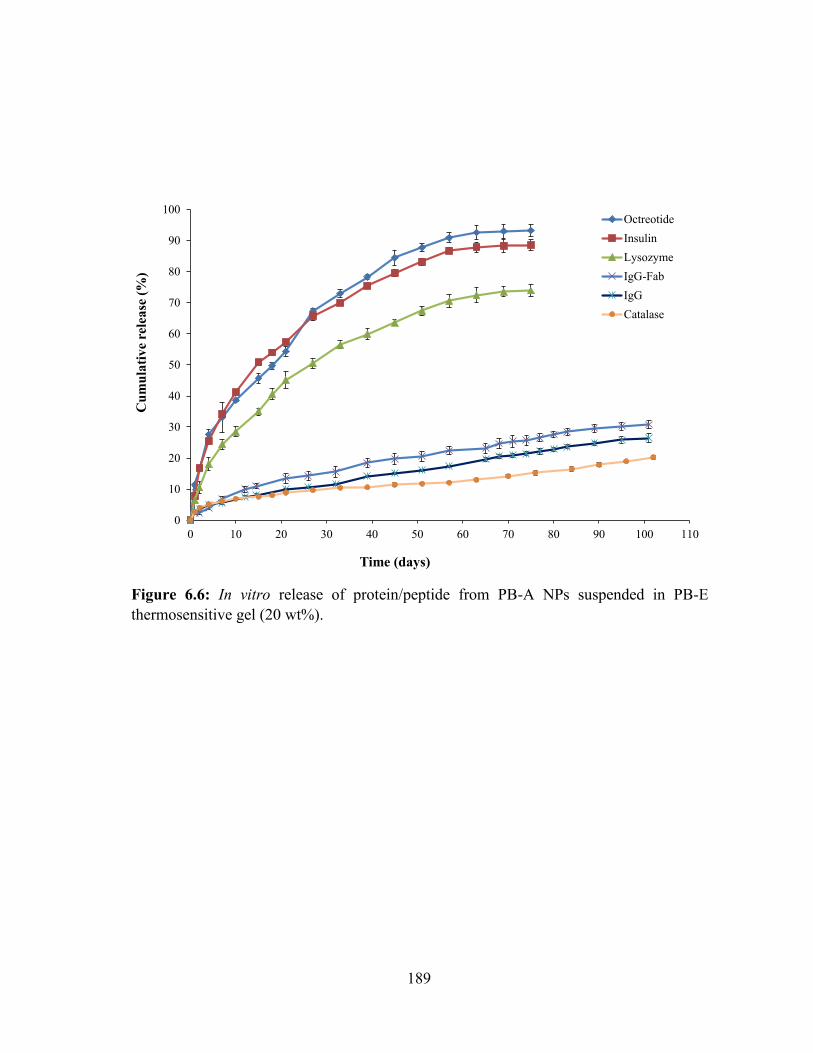
189
Figure 6.6: In vitro release of protein/peptide from PB-A NPs suspended in PB-E
thermosensitive gel (20 wt%).
0
10
20
30
40
50
60
70
80
90
100
0 10 20 30 40 50 60 70 80 90 100 110
Cu
mu
lati
ve
rele
ase
(%
)
Time (days)
Octreotide
Insulin
Lysozyme
IgG-Fab
IgG
Catalase
190
With catalase, a 237 kDa protein, DL efficiency remained relatively similar for both PB-C
(Batches 21 and 24) and PB-D NPs (Batches 27 and 30) prepared by either method.
Overall, as the molecular weight of protein was enhanced from 48 kDa to 237 kDa, the
difference in DL for PB-C and PB-D was diminished. This may be due to the reduced
diffusivity of protein with large molecular weight.
In vitro release studies
In order to study the release behavior of various proteins, we have performed in vitro
release studies from a composite formulation comprised of PB-A NPs suspended in
thermosensitive gel (20 wt%). Figure 6.6 describes in vitro release profiles of octreotide,
insulin, lysozyme, IgG-Fab, IgG and catalase from a composite formulation. All the release
profiles depicted negligible or no burst release phase. It may be due the presence of
thermosensitive gel which may act as additional diffusion layer for the NP-surface adsorbed
protein. Interestingly, we have observed significantly rapid release of octreotide (~93% in 63
days) from composite formulation relative to the release of lysozyme (~72% in 63 days). It can
be explained due to the fact that octreotide has smaller hydrodynamic radius than lysozyme
that may have facilitated rapid diffusion of octreotide from composite formulation. The similar
trend was observed when we compared the release profiles of lysozyme and IgG-Fab (~24%
in 65 days). These results clearly suggest that the hydrodynamic radius of protein molecules
plays a crucial role in defining of their release profiles from composite formulations.
However, we have not observed any significant difference between the release profile
of octreotide and insulin (~89% in 63 days). It may possible that the difference in molecular
weights between octreotide and insulin were not sufficient enough to exert any difference of
diffusivity through formulation that led to the similar release profiles. This fact may be true
191
with smaller molecular weight proteins. There were no noticeable difference between the
release profiles of IgG-Fab, IgG (~20% in 65 days) and catalase (~13% in 63 days) from PB-
A NPs suspended in thermosensitive gel. It may possible that IgG-Fab, IgG and catalase have
much less diffusivity through the polymer matrix (due to large molecular weight), thus their
release was mainly controlled by polymer degradation. As published in previous reports,
degradation of PCL is very slow [204-207, 217], so it was anticipated that PB copolymers
composed of PCL blocks may also degrade very slowly. Due to the slower degradation of PB
copolymers, release of IgG-Fab, IgG and catalase may be sustained for significantly longer
durations.
Release kinetics
In vitro release data were fitted to zero and first order, Korsmeyer-Peppas, Higuchi and
Hixson-Crowell models to delineate the kinetics of IgG release (Table 6.6). The Korsmeyer-
Peppas model had the best fit based on R2 value. The diffusion exponent, n value ranged from
0.513-0.642 for the tested composite formulations. The n-values between 0.43-0.89 indicate
anomalous transport (diffusion and degradation controlled) mechanism of protein release.
Enzymatic activity of lysozyme and catalase
Specific enzymatic activity of lysozyme is reported in Table 6.7. Lysozyme activity of
freshly prepared solution was found to be 61.4 ± 3.8 (U/mg of protein) ×103. As described in
the Table 6.7, enzymatic activity estimated for released samples were relatively higher than
the respective controls. PB copolymers are composed of PEG, a hydrophilic block.
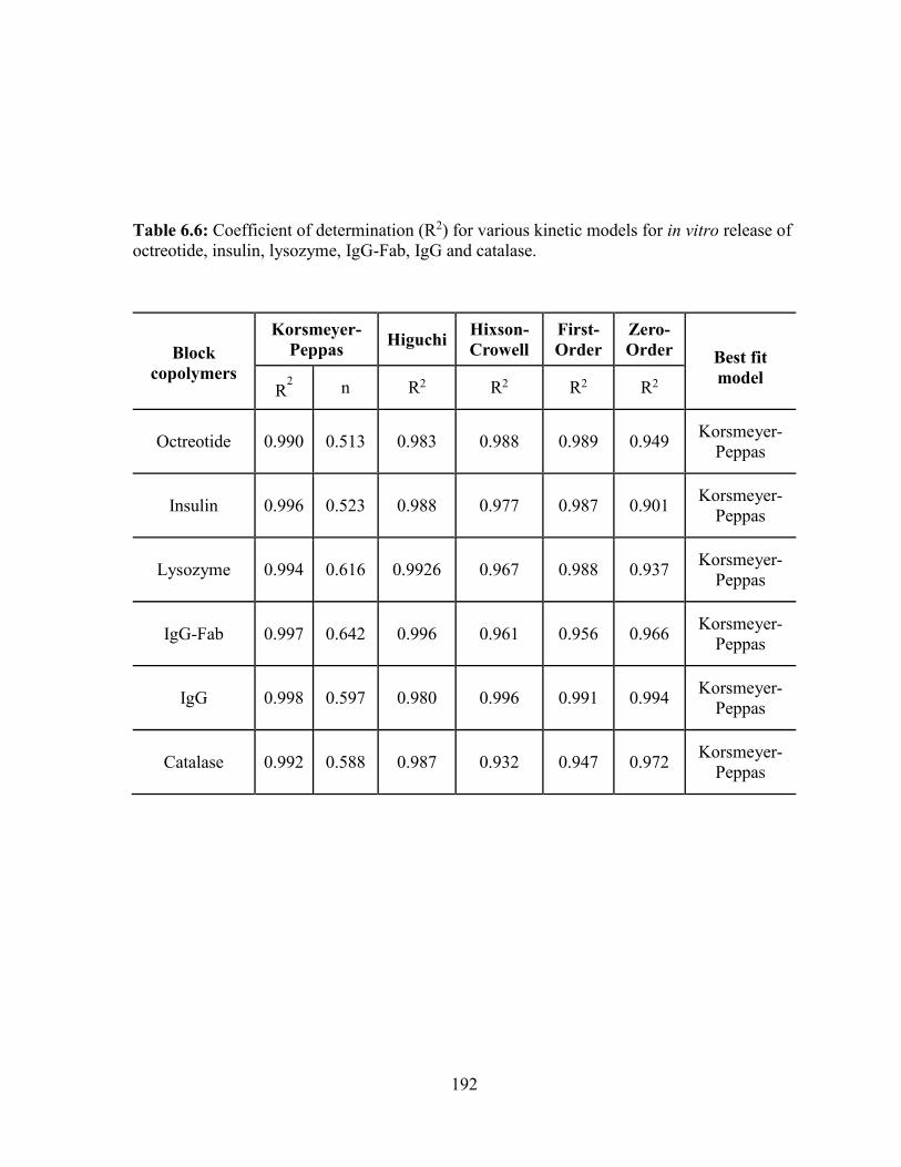
192
Table 6.6: Coefficient of determination (R2) for various kinetic models for in vitro release of
octreotide, insulin, lysozyme, IgG-Fab, IgG and catalase.
Block
copolymers
Korsmeyer-
Peppas Higuchi
Hixson-
Crowell
First-
Order
Zero-
Order Best fit
model R
2 n R2 R2 R2 R2
Octreotide 0.990 0.513 0.983 0.988 0.989 0.949 Korsmeyer-
Peppas
Insulin 0.996 0.523 0.988 0.977 0.987 0.901 Korsmeyer-
Peppas
Lysozyme 0.994 0.616 0.9926 0.967 0.988 0.937 Korsmeyer-
Peppas
IgG-Fab 0.997 0.642 0.996 0.961 0.956 0.966 Korsmeyer-
Peppas
IgG 0.998 0.597 0.980 0.996 0.991 0.994 Korsmeyer-
Peppas
Catalase 0.992 0.588 0.987 0.932 0.947 0.972 Korsmeyer-
Peppas
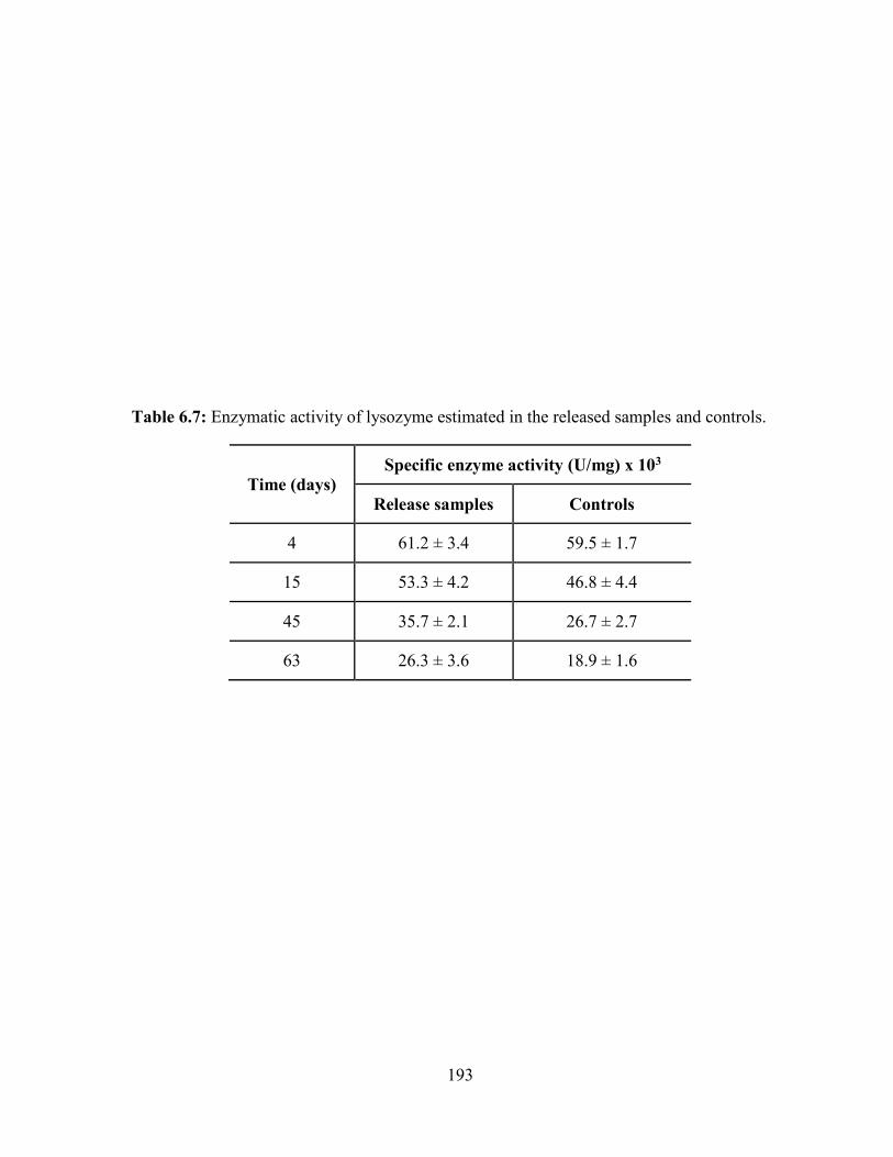
193
Table 6.7: Enzymatic activity of lysozyme estimated in the released samples and controls.
Time (days)
Specific enzyme activity (U/mg) x 103
Release samples Controls
4 61.2 ± 3.4 59.5 ± 1.7
15 53.3 ± 4.2 46.8 ± 4.4
45 35.7 ± 2.1 26.7 ± 2.7
63 26.3 ± 3.6 18.9 ± 1.6
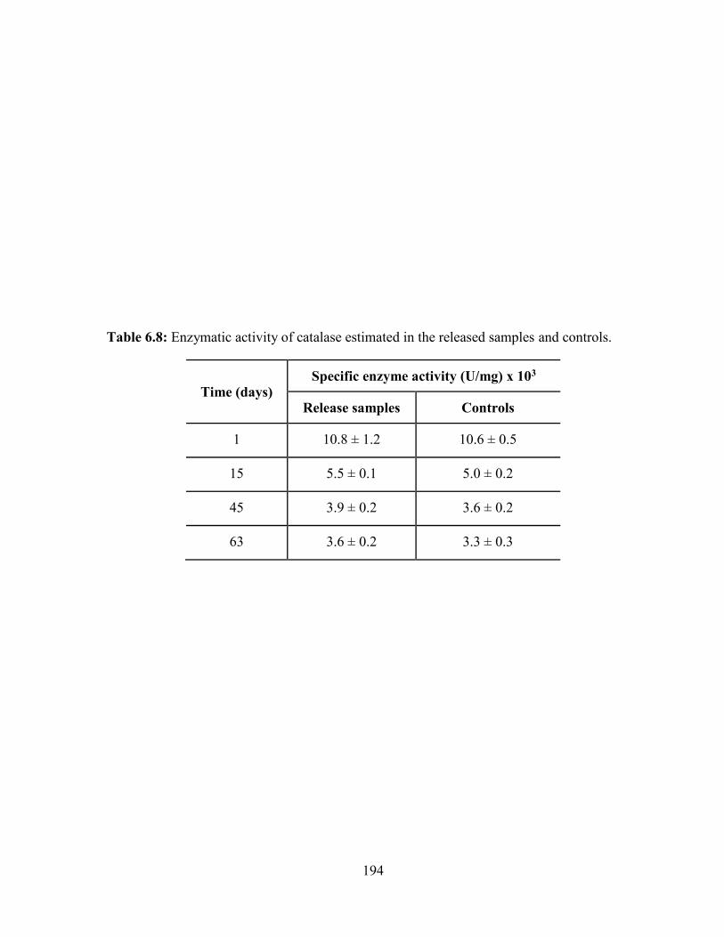
194
Table 6.8: Enzymatic activity of catalase estimated in the released samples and controls.
Time (days)
Specific enzyme activity (U/mg) x 103
Release samples Controls
1 10.8 ± 1.2 10.6 ± 0.5
15 5.5 ± 0.1 5.0 ± 0.2
45 3.9 ± 0.2 3.6 ± 0.2
63 3.6 ± 0.2 3.3 ± 0.3
195
It is anticipated that during NP preparation (W1/O/W2 double emulsion), PEG may have been
located on the aqueous-organic interface facing towards W1 and W2 phase. Hence, PEG may
have reduced the interaction of lysozyme with the hydrophobic polymer segments (PCL and
PLA) as well as with organic solvent (DCM) which may have reduced the denaturation of
protein. However, the biological activity for lysozyme was reduced with the respect to time,
as were the controls. This may be due to the fact that protein remained in the release medium
for a significant time during the release study. Therefore, storage conditions may have affected
the stability of proteins, which will not be the situation at in vivo conditions. During in vivo
conditions, proteins released from composite formulation will be immediately absorbed from
the vitreous cavity.
According to the results depicted in Table 6.8, enzymatic activity of freshly prepared
catalase solution was estimated to be 10.8 ± 0.2 × 10³. Similar to the lysozyme release samples,
enzymatic activity estimated for catalase control samples were relatively lower than the
respective release samples. It may be due to the fact that catalase-encapsulated in the
formulation may have been protected from hydrolytic degradation. Moreover, the biological
activity of the catalase release samples and controls were reduced with the time which may be
attributed to the storage conditions. The polymer matrix of NPs and thermosensitive gel may
have reduced the exposure of proteins to the water molecules and hence protected the
lysozymes/catalase from hydrolytic degradation [178].
Conclusion
We have successfully synthesized and characterized novel PB copolymers with
different block ratios and block arrangements of PEG/PCL/PLA or PEG/PCL/PGA. These PB
copolymers (PLA-PCL-PEG-PCL-PLA and PGA-PCL-PEG-PCL-PGA) were studied for the
196
preparation of protein/peptide-encapsulated NPs. NP preparation methods were successfully
optimized with respect to polymer hydrophobicity, presence of salt (NaCl), drug to polymer
ratio, and phase volumes to achieve maximum possible DL. In addition, effect of
hydrodynamic radius of protein/peptides on DL and in vitro release behavior were studied.
With different arrangement of polymer blocks i.e., PEG-PCL-PLA-PCL-PEG, we have
synthesized PB thermosensitive gel which remained in solution phase at room temperature and
transformed to gel at body temperature. PB copolymer based composite formulation (PB NPs
suspended in PB thermosensitive gel) exhibited constant release for significantly longer
duration of time without showing any burst release effect. Moreover, retention of enzymatic
activity of lysozyme and catalase in release samples were also confirmed. A biodegradable and
biocompatible PB composite formulation comprising protein-encapsulated NPs dispersed in
thermosensitive gel can be easily injected intravitreally. This formulation can carry
significantly high doses in very limited volume (<100 µL) and can sustain the release of
protein/peptides for significantly longer durations without exhibiting burst release. These
results clearly suggest that PB copolymer-based protein-encapsulated, formulations can act as
a platform therapy for the treatment of posterior segment chronic diseases such as wet-AMD,
DR and DME.

197
CHAPTER 7
IN VIVO OCULAR TOLERABILITY STUDIES OF VARIOUS PENTABLOCK
COPOLYMER BASED FORMULATIONS DELIVERED TOPICALLY OR
INTRAVITREALLY
Rationale
Vision-threatening disorders such as age-related macular degeneration (wet-AMD),
diabetic retinopathy (DR) and diabetic macular edema (DME) are commonly treated with
frequent intravitreal injections of anti-VEGF antibodies or fragments thereof. Intravitreal
injections are associated with complications such as secondary infection (endophthalmitis),
retinal hemorrhage, retinal detachment and more importantly patient non-compliance.
Therefore, sustained release therapeutic formulations for ocular posterior segment diseases that
can reduce the frequency of intravitreal injection are highly desirable.
This need for sustained-release of anti VEGF therapy has stimulated research on use of
thermosensitive in situ hydrogels composed of biodegradable and biocompatible polymers and
tissue scaffolds [222-227]. In situ gelling systems offer several advantages over current
formulations, including minimal invasiveness (no surgery required) and injectability through
small (27 gauge) needles. However, thermosensitive gels alone do not extend drug release
sufficiently to reduce the need for frequent injections. Nanoparticulate systems (NPs) have
been investigated for the long-term controlled release of anti-VEGF medications. However,
NPs alone typically exhibit burst release (initial rapid drug release phase) which can result in
dose dependent toxicity. This can be overcome by composite formulations of nanoparticles
(NPs) dispersed in a hydrogel, which can provide ocular delivery of therapeutic drug molecules
over a longer period of time with a reduced burst effect.

198
In vitro drug release assays provide limited information regarding in vivo drug release.
Moreover, in vitro cell culture based assays only provide toxicity information regarding short-
term exposure only. Therefore, in vivo, long-term biocompatibility testing is necessary to
establish safety of controlled delivery systems.
In this study, we report the development of novel PB copolymers based controlled
release systems and the evaluation of their ocular tolerability. This study provides ocular
tolerability of thermosensitive PBC gel was evaluated in rabbits after topical instillation and
after a single intravitreal injection of gel, NPs and a composite formulation comprising NPs
dispersed in gel. PB copolymer based formulations are under development for the sustained
release of biologicals (proteins, antibodies/fragments, RNA) and small molecules (hydrophilic
and hydrophobic) for the treatment of ocular diseases.
Materials and methods
Materials
PB copolymers, PB-D and PB-E described in chapter 6, were utilized for the
preparation of NPs and thermosensitive gel, respectively. Endotoxin-free water was purchased
from Fisher Scientific. Endotoxin estimation kit was procured from GenScript USA Inc.
Polyvinyl alcohol (PVA) was obtained from Sigma Aldrich.
Methods
Preparation of sterile, low endotoxin PB thermosensitive gel
Thermosensitive gels were prepared in sterile glassware pre-rinsed with endotoxin free
water. In order to prepare low endotoxin PB thermosensitive gel, 20 wt% of PB copolymer
was dissolved in endotoxin free (EF) water. Once the polymer was solubilized, 20 wt%
aqueous solutions was filtered through 0.2 µm (pore size) filter in aseptic conditions under
199
laminar flow. The filtrate was analyzed for its gelling behavior and endotoxin levels. Gelling
behavior of aqueous solution was confirmed by exposing the solution to 37 °C for 5 min.
Preparation of low endotoxin PB NPs
All aqueous solutions utilized during NP preparation were prepared with EF water
followed by filter sterilization. NPs were prepared in sterile glassware pre-rinsed with
endotoxin free water. PB-D NPs were prepared by W1/O/W2 double emulsion solvent
evaporation method. Briefly, syringe filtered EF water (W1 phase) was added drop-wise in
organic phase (PB-D copolymer dissolved in dichloromethane) under constant sonication. The
resulting W1/O primary emulsion was then added drop-wise in the W2 phase (2% PVA) under
constant sonication. The W1/O/W2 double emulsion was diluted with a W3 phase (2% PVA)
and vortexed for 5 min. Dichloromethane was then evaporated from multiple emulsions under
low pressure. The resulting NPs were centrifuged at 20000 rpm for 30 min at 4 °C. In order to
remove residual PVA, NPs were washed two times with EF water followed by centrifugation.
Finally, NPs were suspended in 2 mL of EF water and freeze-dried overnight. After freeze-
drying, NPs were stored at -20 °C until further analysis. NPs were tested for endotoxin levels.
A schematic diagram of NP preparation is illustrated in Figure 7.1.
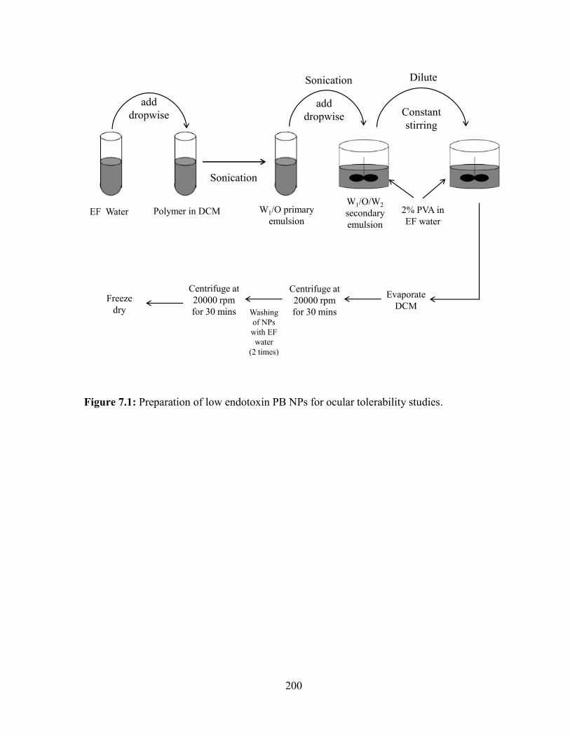
200
Figure 7.1: Preparation of low endotoxin PB NPs for ocular tolerability studies.
EF Water Polymer in DCM
Sonication
add
dropwise
W1/O primary
emulsion
add
dropwise
W1/O/W2
secondary
emulsion
Sonication
2% PVA in
EF water
Evaporate
DCM
Centrifuge at
20000 rpm
for 30 mins
Centrifuge at
20000 rpm
for 30 mins
Freeze
dry Washing
of NPs
with EF
water
(2 times)
Dilute
Constant
stirring
201
All the aqueous solutions were prepared with EF water followed by filter sterilization.
Thermosensitive gel and NPs were prepared in sterile glassware pre-rinsed with endotoxin free
water.
Evaluation of endotoxin levels
Levels of endotoxins in various formulations were evaluated utilizing an endotoxin
assay kit. Limulus Amebocyte Lysate (LAL) endotoxin assay was performed according to the
supplier’s protocol under aseptic conditions (laminar flow). Briefly, three test samples were
prepared, i.e. thermosenstive gel solution (20 wt%), NP suspension in EF water (5.5 mg/100
µL) and NP suspension in thermosensitive gel solution (20 wt%). A 100 µL aliquot of test or
standard solution/suspensions were transferred in vials followed by addition of 100 µL LAL
reagent. Mixture was gently stirred for 30 sec, and followed by incubation at 37 °C for 45 min.
After incubation, 100 µL of chromogenic reagent was added and gently stirred for 30 sec
followed by 6 min incubation at 37 °C. At the end of incubation, 500 µL of color-stabilizer 1,
2 and 3 were added in a defined order. In order to remove NPs from the samples, samples
containing NPs were syringe-filtered. Absorbance of samples and standards were estimated at
570 nm utilizing 96-well plate reader.
In vivo tolerability studies
Use of animals in this study adhered to the ARVO Statement for the Use of Animals in
Ophthalmic and Vision Research and was approved and monitored by the North Carolina State
University Institutional Animal Care and Use Committee. Details about animal, housing and
environment are described in Table 7.1. Methods along with evaluation parameters for topical
drops and intravitreal injections are described below.
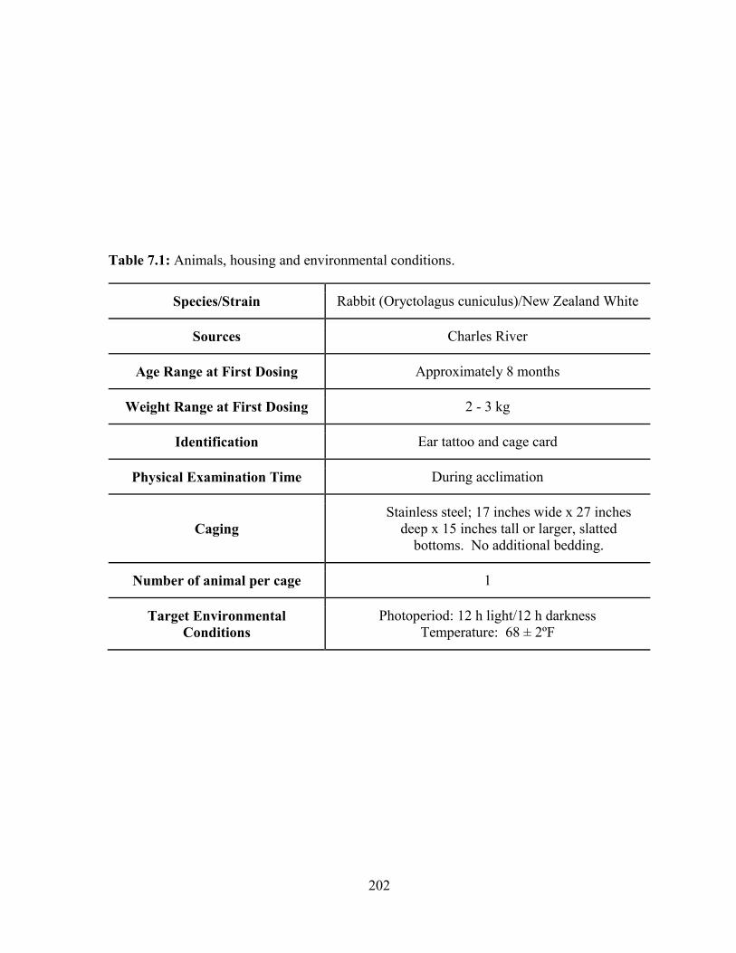
202
Table 7.1: Animals, housing and environmental conditions.
Species/Strain Rabbit (Oryctolagus cuniculus)/New Zealand White
Sources Charles River
Age Range at First Dosing Approximately 8 months
Weight Range at First Dosing 2 - 3 kg
Identification Ear tattoo and cage card
Physical Examination Time During acclimation
Caging
Stainless steel; 17 inches wide x 27 inches
deep x 15 inches tall or larger, slatted
bottoms. No additional bedding.
Number of animal per cage 1
Target Environmental
Conditions
Photoperiod: 12 h light/12 h darkness
Temperature: 68 ± 2ºF
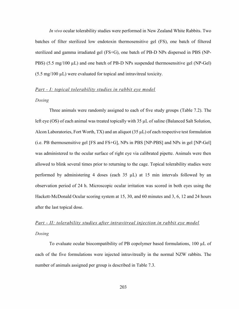
203
In vivo ocular tolerability studies were performed in New Zealand White Rabbits. Two
batches of filter sterilized low endotoxin thermosensitive gel (FS), one batch of filtered
sterilized and gamma irradiated gel (FS+G), one batch of PB-D NPs dispersed in PBS (NP-
PBS) (5.5 mg/100 µL) and one batch of PB-D NPs suspended thermosensitive gel (NP-Gel)
(5.5 mg/100 µL) were evaluated for topical and intravitreal toxicity.
Part - I: topical tolerability studies in rabbit eye model
Dosing
Three animals were randomly assigned to each of five study groups (Table 7.2). The
left eye (OS) of each animal was treated topically with 35 µL of saline (Balanced Salt Solution,
Alcon Laboratories, Fort Worth, TX) and an aliquot (35 µL) of each respective test formulation
(i.e. PB thermosensitive gel [FS and FS+G], NPs in PBS [NP-PBS] and NPs in gel [NP-Gel]
was administered to the ocular surface of right eye via calibrated pipette. Animals were then
allowed to blink several times prior to returning to the cage. Topical tolerability studies were
performed by administering 4 doses (each 35 µL) at 15 min intervals followed by an
observation period of 24 h. Microscopic ocular irritation was scored in both eyes using the
Hackett-McDonald Ocular scoring system at 15, 30, and 60 minutes and 3, 6, 12 and 24 hours
after the last topical dose.
Part - II: tolerability studies after intravitreal injection in rabbit eye model
Dosing
To evaluate ocular biocompatibility of PB copolymer based formulations, 100 µL of
each of the five formulations were injected intravitreally in the normal NZW rabbits. The
number of animals assigned per group is described in Table 7.3.
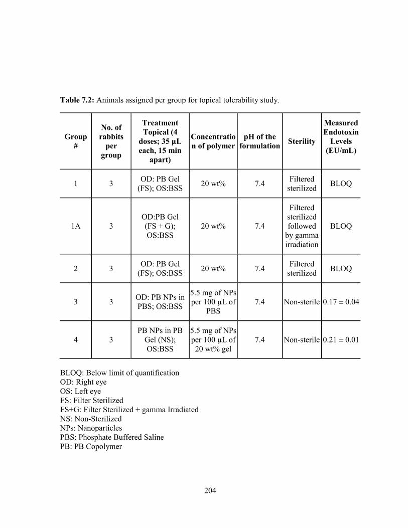
204
Table 7.2: Animals assigned per group for topical tolerability study.
Group
#
No. of
rabbits
per
group
Treatment
Topical (4
doses; 35 µL
each, 15 min
apart)
Concentratio
n of polymer
pH of the
formulation Sterility
Measured
Endotoxin
Levels
(EU/mL)
1 3 OD: PB Gel
(FS); OS:BSS 20 wt% 7.4
Filtered
sterilized BLOQ
1A 3
OD:PB Gel
(FS + G);
OS:BSS
20 wt% 7.4
Filtered
sterilized
followed
by gamma
irradiation
BLOQ
2 3 OD: PB Gel
(FS); OS:BSS 20 wt% 7.4
Filtered
sterilized BLOQ
3 3 OD: PB NPs in
PBS; OS:BSS
5.5 mg of NPs
per 100 µL of
PBS
7.4 Non-sterile 0.17 ± 0.04
4 3
PB NPs in PB
Gel (NS);
OS:BSS
5.5 mg of NPs
per 100 µL of
20 wt% gel
7.4 Non-sterile 0.21 ± 0.01
BLOQ: Below limit of quantification
OD: Right eye
OS: Left eye
FS: Filter Sterilized
FS+G: Filter Sterilized + gamma Irradiated
NS: Non-Sterilized
NPs: Nanoparticles
PBS: Phosphate Buffered Saline
PB: PB Copolymer
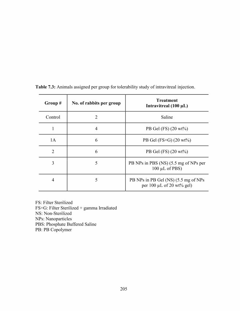
205
Table 7.3: Animals assigned per group for tolerability study of intravitreal injection.
Group # No. of rabbits per group Treatment
Intravitreal (100 µL)
Control 2 Saline
1 4 PB Gel (FS) (20 wt%)
1A 6 PB Gel (FS+G) (20 wt%)
2 6 PB Gel (FS) (20 wt%)
3 5 PB NPs in PBS (NS) (5.5 mg of NPs per
100 µL of PBS)
4 5 PB NPs in PB Gel (NS) (5.5 mg of NPs
per 100 µL of 20 wt% gel)
FS: Filter Sterilized
FS+G: Filter Sterilized + gamma Irradiated
NS: Non-Sterilized
NPs: Nanoparticles
PBS: Phosphate Buffered Saline
PB: PB Copolymer
206
Rabbits were tranquilized with ketamine/dexmeditomidine intramuscularly and were prepared
by applying 5% betadine solution topically followed by rinsing with sterile saline and
application of 0.5% proparacaine HCl. Intravitreal injections (100 µL) of test formulation or
saline was done 2 mm posterior to the superior limbus (through the pars plana) using a 25
gauge needle. Special care was taken during the injection to avoid needle contact with the lens.
Following the injection, one drop of 0.5% moxifloxacin (Alcon Laboratories, Fort Worth, TX)
was instilled topically to the ocular surface.
Parameters to be evaluated
Ocular Examination
Ocular surface morphology, posterior and anterior segment inflammation, retinal
changes and cataract formation were evaluated by ocular examination using a slit lamp and
indirect ophthalmoscope. Modified Hackett-McDonald ocular scoring (Microscopic Ocular
Grading System without fluorescein application) of inflammation was recorded. Parameters
such as congestion (0-3), swelling (0-4), discharge (0-3), aqueous flare (0-3), light reflex of
iris (0-4), corneal cloudiness affected area (0-4), corneal cloudiness severity (0-4) and pannus
and vitreal cells (0-2) were considered to calculate cumulative mean Hackett-McDonald ocular
irritation scoring. Ocular examination was carried out for each rabbit to determine pre-dose
(pre-study) baseline data, followed by examination during predetermined time intervals.
Formulations were evaluated for tolerability studies after topical and intravitreal injections.
Intraocular Pressure
Intraocular pressure (IOP) was measured in both eyes for control and treated groups.
IOP was measured at least twice during the acclimation, then immediately following ocular
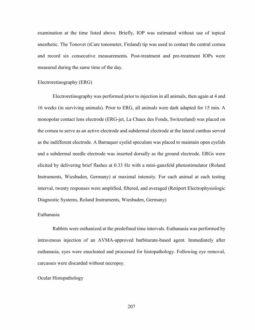
207
examination at the time listed above. Briefly, IOP was estimated without use of topical
anesthetic. The Tonovet (iCare tonometer, Finland) tip was used to contact the central cornea
and record six consecutive measurements. Post-treatment and pre-treatment IOPs were
measured during the same time of the day.
Electroretinography (ERG)
Electroretinography was performed prior to injection in all animals, then again at 4 and
16 weeks (in surviving animals). Prior to ERG, all animals were dark adapted for 15 min. A
monopolar contact lens electrode (ERG-jet, La Chaux des Fonds, Switzerland) was placed on
the cornea to serve as an active electrode and subdermal electrode at the lateral canthus served
as the indifferent electrode. A Barraquer eyelid speculum was placed to maintain open eyelids
and a subdermal needle electrode was inserted dorsally as the ground electrode. ERGs were
elicited by delivering brief flashes at 0.33 Hz with a mini-ganzfeld photostimulator (Roland
Instruments, Wiesbaden, Germany) at maximal intensity. For each animal at each testing
interval, twenty responses were amplified, filtered, and averaged (Retiport Electrophysiologic
Diagnostic Systems, Roland Instruments, Wiesbaden, Germany)
Euthanasia
Rabbits were euthanized at the predefined time intervals. Euthanasia was performed by
intravenous injection of an AVMA-approved barbiturate-based agent. Immediately after
euthanasia, eyes were enucleated and processed for histopathology. Following eye removal,
carcasses were discarded without necropsy.
Ocular Histopathology
208
Both eyes of each animal were fixed in Davidson’s solution for 24 h, followed by
alcohol. Central sections of each globe, including the optic nerve were stained with
hematoxylin and eosin stain (H&E stain), and examined by light microscopy.
Results and discussion
Estimation of endotoxin levels in PB thermosensitive gel and PB NPs
Levels of endotoxins in various formulations were evaluated utilizing Limulus
Amebocyte Lysate (LAL) endotoxin assay kit. Both batches of PB thermosensitive gelling
solution depicted negligible endotoxin levels (below the limit of quantification) (Table 7.2).
PB NPs alone (Group 3), and PB NPs (Group 3) dispersed in PB thermosensitive gel (Group
2) exhibited endotoxin levels of 0.17 ± 0.04 EU/mL and 0.21 ± 0.01 EU/mL, respectively.
However, these levels of endotoxins for Groups 3 and 4 are well within the acceptable limit (2
EU/mL) for ocular applications.
Tolerability study after topical application
Topical ocular tolerability studies were performed on two batches of thermosensitive
gel (20 wt%), PB NPs in PBS buffer, and PB NPs dispersed in thermosensitive gel (20 wt%
filter sterilized). These formulations were instilled directly on the ocular surface in right eye
of each animal, while left eyes were treated with balanced salt solution (BSS).
General ocular observation
No adverse reactions (blepharospasm or evasive action) by the animals were observed
during or after the administration of test samples or BSS. Immediately after the application to
the left eye, BSS rapidly flowed to the ventral conjunctival cul-de-sacs and disappeared from
view.
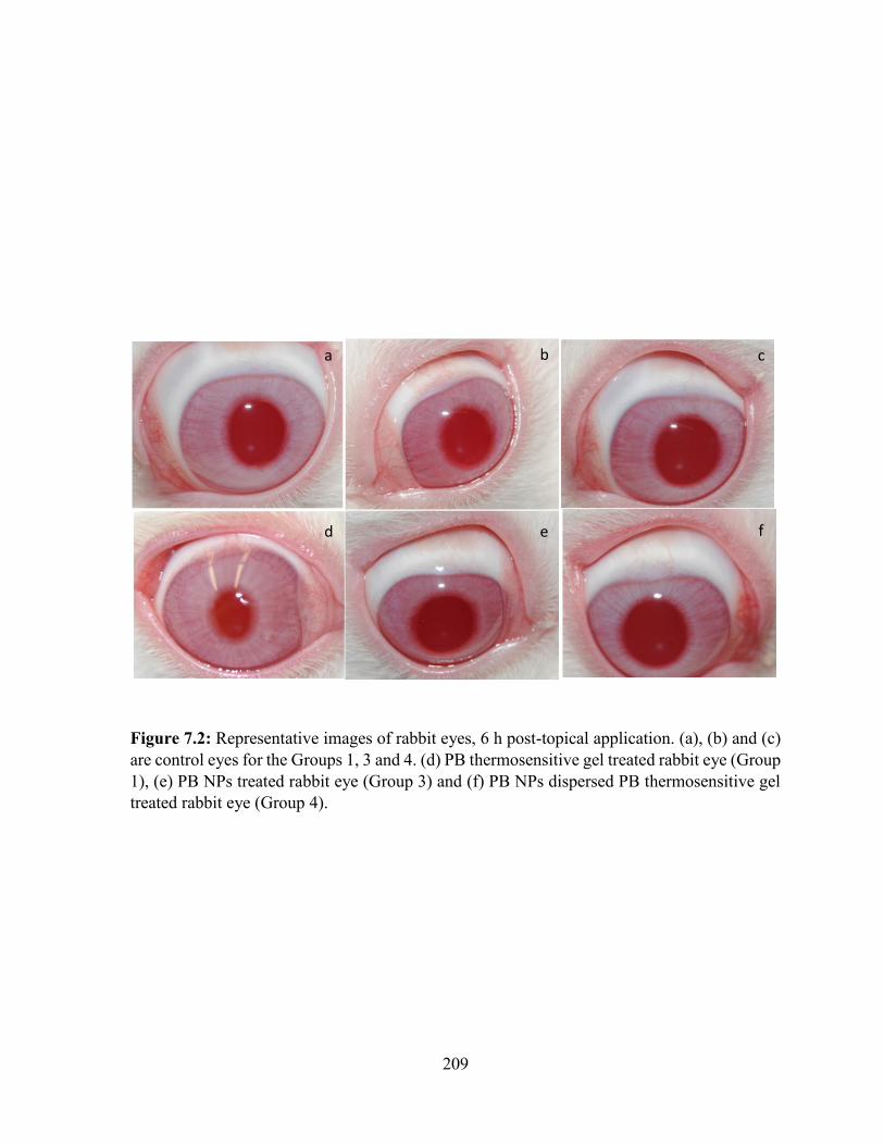
209
Figure 7.2: Representative images of rabbit eyes, 6 h post-topical application. (a), (b) and (c)
are control eyes for the Groups 1, 3 and 4. (d) PB thermosensitive gel treated rabbit eye (Group
1), (e) PB NPs treated rabbit eye (Group 3) and (f) PB NPs dispersed PB thermosensitive gel
treated rabbit eye (Group 4).
a b c
d e f
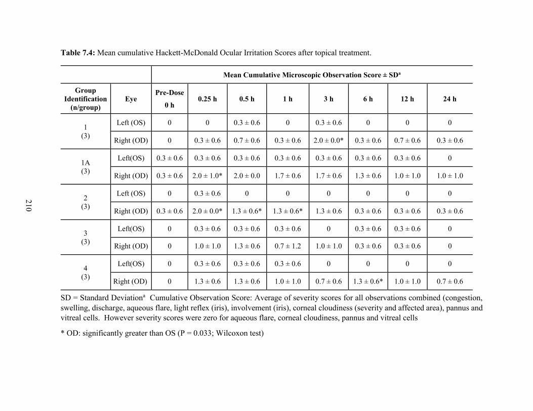
210
Table 7.4: Mean cumulative Hackett-McDonald Ocular Irritation Scores after topical treatment.
Mean Cumulative Microscopic Observation Score ± SDa
Group
Identification
(n/group)
Eye Pre-Dose
0 h 0.25 h 0.5 h 1 h 3 h 6 h 12 h 24 h
1
(3)
Left (OS) 0 0 0.3 ± 0.6 0 0.3 ± 0.6 0 0 0
Right (OD) 0 0.3 ± 0.6 0.7 ± 0.6 0.3 ± 0.6 2.0 ± 0.0* 0.3 ± 0.6 0.7 ± 0.6 0.3 ± 0.6
1A
(3)
Left(OS) 0.3 ± 0.6 0.3 ± 0.6 0.3 ± 0.6 0.3 ± 0.6 0.3 ± 0.6 0.3 ± 0.6 0.3 ± 0.6 0
Right (OD) 0.3 ± 0.6 2.0 ± 1.0* 2.0 ± 0.0 1.7 ± 0.6 1.7 ± 0.6 1.3 ± 0.6 1.0 ± 1.0 1.0 ± 1.0
2
(3)
Left (OS) 0 0.3 ± 0.6 0 0 0 0 0 0
Right (OD) 0.3 ± 0.6 2.0 ± 0.0* 1.3 ± 0.6* 1.3 ± 0.6* 1.3 ± 0.6 0.3 ± 0.6 0.3 ± 0.6 0.3 ± 0.6
3
(3)
Left(OS) 0 0.3 ± 0.6 0.3 ± 0.6 0.3 ± 0.6 0 0.3 ± 0.6 0.3 ± 0.6 0
Right (OD) 0 1.0 ± 1.0 1.3 ± 0.6 0.7 ± 1.2 1.0 ± 1.0 0.3 ± 0.6 0.3 ± 0.6 0
4
(3)
Left(OS) 0 0.3 ± 0.6 0.3 ± 0.6 0.3 ± 0.6 0 0 0 0
Right (OD) 0 1.3 ± 0.6 1.3 ± 0.6 1.0 ± 1.0 0.7 ± 0.6 1.3 ± 0.6* 1.0 ± 1.0 0.7 ± 0.6
SD = Standard Deviationa Cumulative Observation Score: Average of severity scores for all observations combined (congestion,
swelling, discharge, aqueous flare, light reflex (iris), involvement (iris), corneal cloudiness (severity and affected area), pannus and
vitreal cells. However severity scores were zero for aqueous flare, corneal cloudiness, pannus and vitreal cells
* OD: significantly greater than OS (P = 0.033; Wilcoxon test)
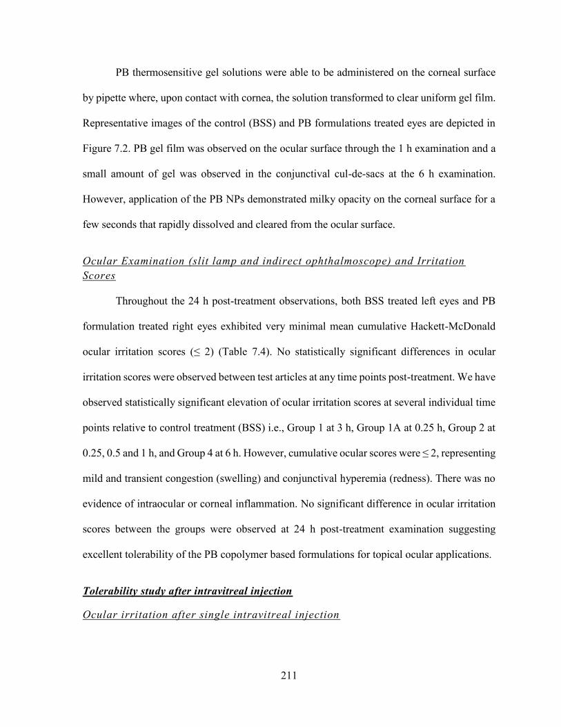
211
PB thermosensitive gel solutions were able to be administered on the corneal surface
by pipette where, upon contact with cornea, the solution transformed to clear uniform gel film.
Representative images of the control (BSS) and PB formulations treated eyes are depicted in
Figure 7.2. PB gel film was observed on the ocular surface through the 1 h examination and a
small amount of gel was observed in the conjunctival cul-de-sacs at the 6 h examination.
However, application of the PB NPs demonstrated milky opacity on the corneal surface for a
few seconds that rapidly dissolved and cleared from the ocular surface.
Ocular Examination (slit lamp and indirect ophthalmoscope) and Irritation
Scores
Throughout the 24 h post-treatment observations, both BSS treated left eyes and PB
formulation treated right eyes exhibited very minimal mean cumulative Hackett-McDonald
ocular irritation scores (≤ 2) (Table 7.4). No statistically significant differences in ocular
irritation scores were observed between test articles at any time points post-treatment. We have
observed statistically significant elevation of ocular irritation scores at several individual time
points relative to control treatment (BSS) i.e., Group 1 at 3 h, Group 1A at 0.25 h, Group 2 at
0.25, 0.5 and 1 h, and Group 4 at 6 h. However, cumulative ocular scores were ≤ 2, representing
mild and transient congestion (swelling) and conjunctival hyperemia (redness). There was no
evidence of intraocular or corneal inflammation. No significant difference in ocular irritation
scores between the groups were observed at 24 h post-treatment examination suggesting
excellent tolerability of the PB copolymer based formulations for topical ocular applications.
Tolerability study after intravitreal injection
Ocular irritation after single intravitreal injection
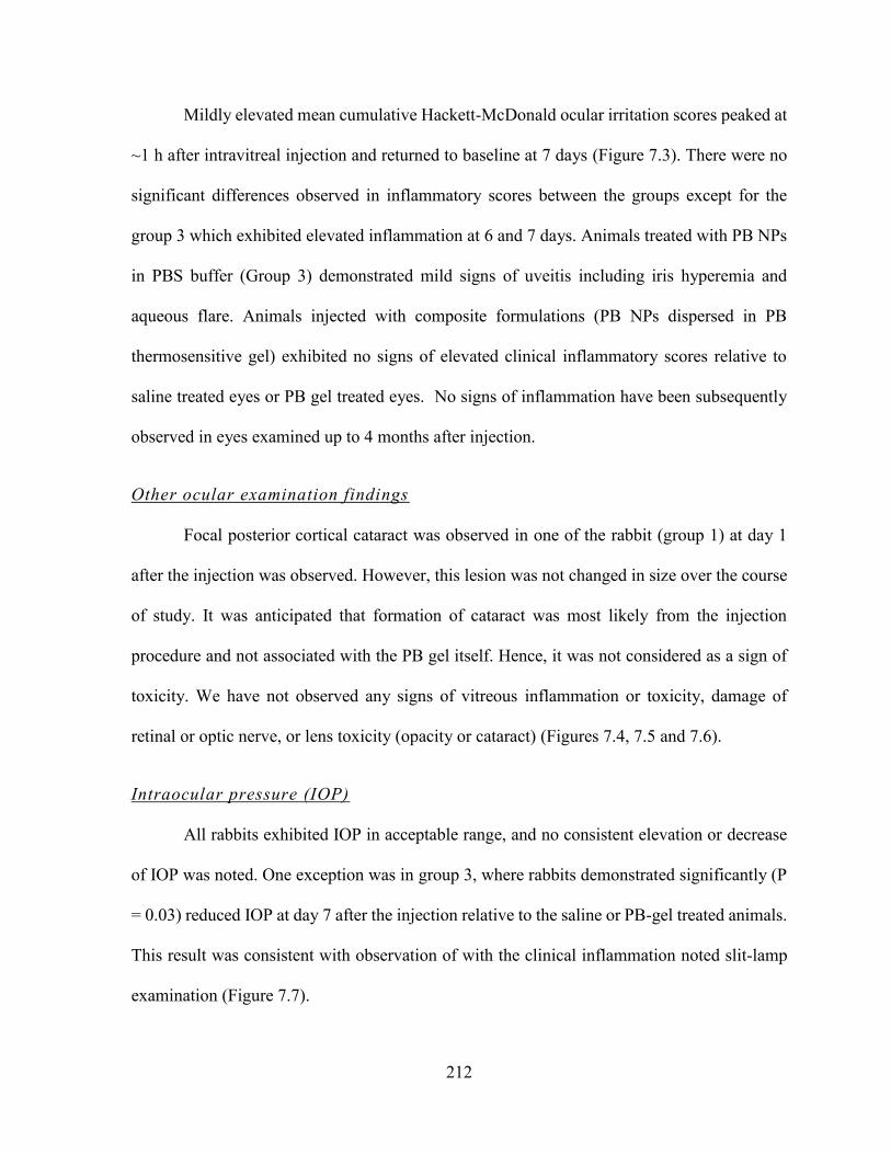
212
Mildly elevated mean cumulative Hackett-McDonald ocular irritation scores peaked at
~1 h after intravitreal injection and returned to baseline at 7 days (Figure 7.3). There were no
significant differences observed in inflammatory scores between the groups except for the
group 3 which exhibited elevated inflammation at 6 and 7 days. Animals treated with PB NPs
in PBS buffer (Group 3) demonstrated mild signs of uveitis including iris hyperemia and
aqueous flare. Animals injected with composite formulations (PB NPs dispersed in PB
thermosensitive gel) exhibited no signs of elevated clinical inflammatory scores relative to
saline treated eyes or PB gel treated eyes. No signs of inflammation have been subsequently
observed in eyes examined up to 4 months after injection.
Other ocular examination findings
Focal posterior cortical cataract was observed in one of the rabbit (group 1) at day 1
after the injection was observed. However, this lesion was not changed in size over the course
of study. It was anticipated that formation of cataract was most likely from the injection
procedure and not associated with the PB gel itself. Hence, it was not considered as a sign of
toxicity. We have not observed any signs of vitreous inflammation or toxicity, damage of
retinal or optic nerve, or lens toxicity (opacity or cataract) (Figures 7.4, 7.5 and 7.6).
Intraocular pressure (IOP)
All rabbits exhibited IOP in acceptable range, and no consistent elevation or decrease
of IOP was noted. One exception was in group 3, where rabbits demonstrated significantly (P
= 0.03) reduced IOP at day 7 after the injection relative to the saline or PB-gel treated animals.
This result was consistent with observation of with the clinical inflammation noted slit-lamp
examination (Figure 7.7).
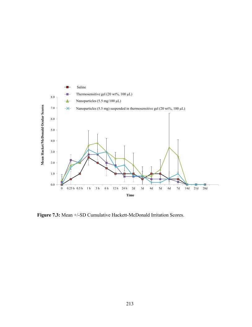
213
Figure 7.3: Mean +/-SD Cumulative Hackett-McDonald Irritation Scores.
0.0
1.0
2.0
3.0
4.0
5.0
6.0
7.0
8.0
0 0.25 h 0.5 h 1 h 3 h 6 h 12 h 24 h 2d 3d 4d 5d 6d 7d 14d 21d 28d
Mea
n H
ack
et/M
cDo
na
ld O
cula
r S
core
s
Time
Saline
Thermosensitive gel (20 wt%, 100 µL)
Nanoparticles (5.5 mg/100 µL)
Nanoparticles (5.5 mg) suspended in thermosensitive gel (20 wt%, 100 µL)
214
Figure 7.4: Images of rabbit eyes taken after single intravitreal injection (100 µL) of PB
thermosensitive gel, (a) day 1, (b) day 21, (c) day 42, (d) day 49, and (e) day 98.
a b c
d e
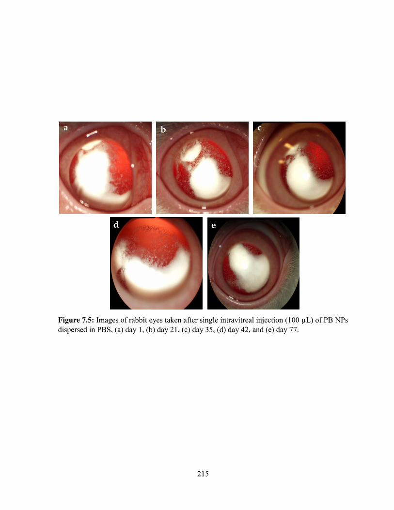
215
Figure 7.5: Images of rabbit eyes taken after single intravitreal injection (100 µL) of PB NPs
dispersed in PBS, (a) day 1, (b) day 21, (c) day 35, (d) day 42, and (e) day 77.
a b c
d e
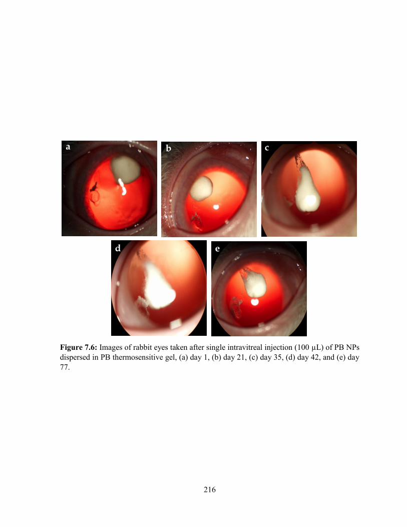
216
Figure 7.6: Images of rabbit eyes taken after single intravitreal injection (100 µL) of PB NPs
dispersed in PB thermosensitive gel, (a) day 1, (b) day 21, (c) day 35, (d) day 42, and (e) day
77.
a b c
d e
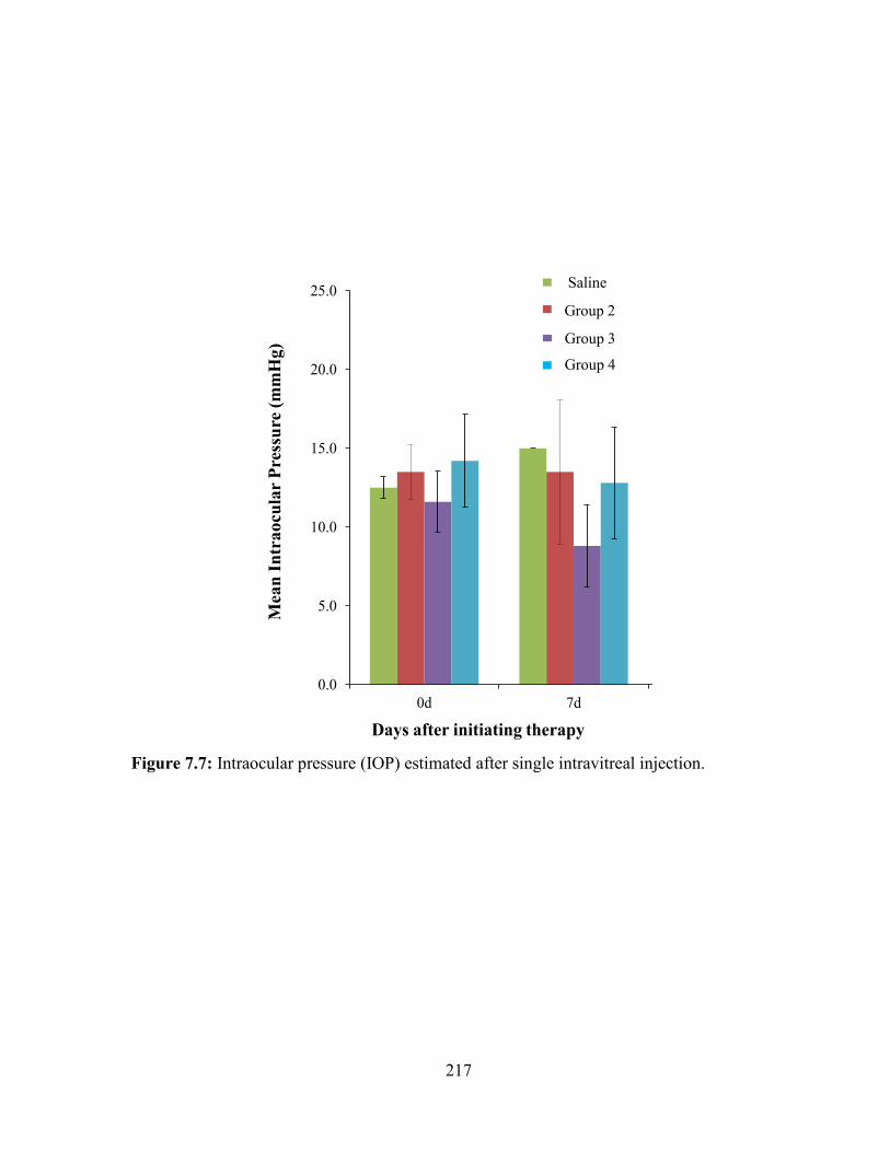
217
Figure 7.7: Intraocular pressure (IOP) estimated after single intravitreal injection.
Saline
Group 2
Group 3
Group 4
0.0
5.0
10.0
15.0
20.0
25.0
0d 7d 14d 21d
Mea
n I
ntr
ao
cu
lar
Pre
ssu
re (
mm
Hg
)
Days after initiating therapy
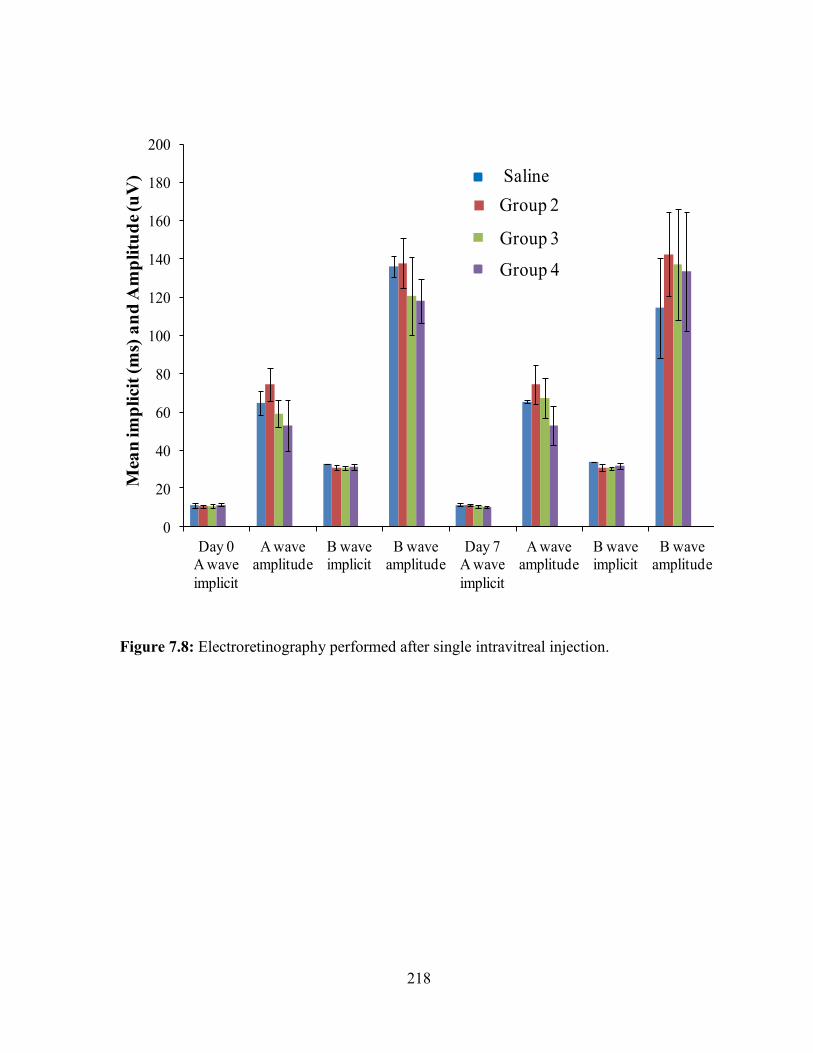
218
Figure 7.8: Electroretinography performed after single intravitreal injection.
0
20
40
60
80
100
120
140
160
180
200
Day 0 A wave
implicit
A wave amplitude
B wave implicit
B wave amplitude
Day 7 A wave
implicit
A wave amplitude
B wave implicit
B wave amplitude
Day 28 A wave
implicit
A wave amplitude
B wave implicit
B wave amplitude
Mea
n im
pli
cit
(ms)
an
d A
mp
litu
de
(uV
) Saline
Group 2
Group 3
Group 4
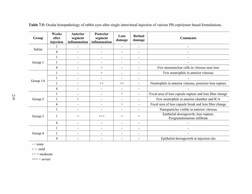
219
Table 7.5: Ocular histopathology of rabbit eyes after single intravitreal injection of various PB copolymer based formulations.
- = none
+ = mild
++ = moderate
+++ = severe
Group
Weeks
after
injection
Anterior
segment
inflammation
Posterior
segment
inflammation
Lens
damage
Retinal
damage Comments
Saline 1 - - - - -
4 - - - - -
Group 1
1 - - - - -
1 - - - - -
4 - + - - Few mononuclear cells in vitreous near lens
Group 1A
1 - + - - Few neutrophils in anterior vitreous
1 - - - - -
1 - ++ ++ - Neutrophils in anterior vitreous, posterior lens rupture
4 - - - - -
Group 2
1 - - + - Focal area of lens capsule rupture and lens fiber change
1 + - - - Few neutrophils in anterior chamber and ICA
4 - - + - Focal area of lens capsule break and lens fiber change
Group 3
1 - - - - Nanoparticles visible in anterior vitreous
1 + +++ + + Epithelial downgrowth, lens rupture,
Pyogranulomatous infiltrate
4 - - - - -
Group 4
1 - - - - -
1 - - - - -
4 - - - - Epithelial downgrowth at injection site
220
Electroretinography
The pre-study and weeks 1, 4, add 16 post injection ERGs were normal in each rabbit.
No significant differences in a-wave and b-wave implicit times or amplitudes before and after
treatment (Figure 7.8) were observed, suggesting a lack of retinal toxicity with the injected
polymers up to 4 months.
Ocular histopathology
Ocular histopathology was performed at weeks 1, 4 and 16 after intravitreal injection.
Results reported in Table 7.5 indicated that in most eyes no evidence of inflammation or
toxicity associated with the injection procedure or test articles were observed. Two eyes
exhibited mild anterior segment/anterior vitreous inflammation. However, we have observed
injection procedure associated damage and inflammation in a few eyes, including lens trauma
and wound epithelial downgrowth. These changes appear to be associated with the injection
procedure in sensitive rabbit eyes rather than the test articles. Only one animal of group 3 (PB
NPs in PBS) exhibited severe posterior segment inflammation at week 1 after injection. These
results are consistent with the results of ocular irritation score and reduced IOP observed in the
same group of animals. Based on these findings, it appears that the test articles are well
tolerated by the rabbit eyes, without evidence of overt inflammation or histopathologic
evidence of tissue damage indicating excellent biocompatibility of PB copolymer based
formulations. However, changes associated with injection procedure are relatively common
and may cause most of the inflammatory and tissue damage observed (both histologic and
clinical).
221
Conclusion
We have prepared endotoxin-free thermogelling polymer solutions and have tested
these with topical application to rabbit eyes. In addition we have tested thermogelling solution
with and without PB NPs. Topical application of 4 consecutive doses (35 µL each) at intervals
of 15 min produced no signs of irritation. No adverse reactions, such as evasive action or
blepharospasm were observed during or after the application suggesting adequate tolerability
for topical ocular applications. Minimal mean cumulative Hackett-McDonald ocular irritation
scores of ≤2 were observed for all the formulations indicating mild and transient conjunctival
hyperemia (redness) and congestion (swelling). Low ocular irritation scores up to 4 weeks
demonstrate that PB copolymer NPs dispersed in thermogelling solution are well tolerated. No
significant difference in IOP was observed between saline treated or PB copolymer
formulation treated eyes. ERG results showed no changes in retinal functions in either control
or test groups. Moreover, no sign of inflammation or cataract formation during the treatment
period was observed. These results indicate that PB copolymer based formulations are
biodegradable and biocompatible, and well suited for ocular application, both topically or by
intravitreal injection. These polymers can be successfully used for the development of
controlled release systems for ocular delivery of therapeutic proteins.
222
CHAPTER 8
SUMMARY AND RECOMMENDATIONS
Summary
Development of sustained release formulation for the protein therapeutics in the
treatment of posterior segment diseases such as wet-AMD, DR and DME, is a very challenging
task for the formulation scientists. The ideal formulation for the intravitreal delivery should
possess following characteristics, (a) high drug loading in small volume (≤ 100 µL) that lasts
up to 6 months or more, (b) provide constant release (zero-order release) throughout the release
period without any burst effect, (c) easy to administered such as injectable system and not
implants, (d) ensure stability of protein/peptide, (e) biodegradable and biocompatible, and (f)
the time required for biodegradation of formulation should not be more than 1.5 times the
release period.
In order to achieve this goal, we have synthesized novel biodegradable and
biocompatible PB copolymers. PB copolymers are composed of various FDA approved
polymeric blocks such as PEG, PCL, and PLA/PGA. Each block plays an important role such
as presence of PEG helps to improve stability of NPs by reducing NP aggregation, and also
helps to escape phagocytosis by macrophages resulting in improved half-life. PCL is a slow
degrading semi-crystalline polymer which improves protein loading in NPs and also sustains
drug release for longer duration of time. Such a slow degradation is also not advantageous
particularly for intravitreal injections. It is important for formulation scientists to synchronize
polymer degradation profiles with the drug release profiles in order to avoid accumulation of
empty formulation in the limited vitreous cavity. Previous reports suggest that poor
degradation of PCL is attributed to its crystalline nature, hence reduction in the crystallinity of
PCL may improve its hydrolytic and enzymatic degradation. Covalent conjugation of

223
PLA/PGA to PCL chains can significantly reduce crystallinity resulting in faster degradation
of PCL [203, 219]. PB copolymers were utilized to prepare thermosensitive gel or NPs.
Firstly, we have synthesized and characterized various TB (PCL-PEG-PCL, B-A-B)
and PB (PLA-PCL-PEG-PCL-PLA (C-B-A-B-C) and PEG-PCL-PLA-PCL-PEG (A-B-C-B-
A)) copolymer based thermosensitive gelling polymers. Arrangement of polymer blocks,
molecular weight and presence of PLA exhibited significant effect on sol-gel transition curve
and kinematic viscosity of respective aqueous solutions. IgG-loaded PB thermosensitive gels
exhibited sustained release up to ~20 days. Results of release kinetics suggested Korsmeyer-
Peppas as a best fit model and diffusion exponent values (n value) were ranging from 0.272-
0.386 for all gelling polymers indicating diffusion controlled release of IgG. Moreover,
secondary stability of released IgG was confirmed by CD spectroscopy. PB copolymers with
A-B-C-B-A block arrangements demonstrated noticeably lower kinematic viscosity of
aqueous solution at 25 °C than the TB (B-A-B) copolymers and other PB copolymers with C-
B-A-B-C block arrangements suggesting easy to handle during injection. Presence of PLA
block has significantly reduced crystallinity of PB copolymers (C-B-A-B-C and A-B-C-B-A).
Hence, it was anticipated that PB copolymers will have faster rate of degradation relative to
PCL-PEG-PCL based TB copolymers. The difference in results of viscosity and sol-gel
transition has been explained by two different processes of micellization for A-B-C-B-A and
B-A-B or C-B-A-B-C types of copolymers. Cell viability and biocompatibility studies
suggested that PCL, PLA and PEG based block copolymers are compatible with various ocular
and macrophage cell lines. In our first approach, we were able to develop sustained release,
biocompatible and biodegradable polymeric formulation which can be administered with semi-
invasive technique. However, thermogelling system exhibited sustained release up to only ~20
224
days, which was shorter than our target profile (4-6 months). Moreover, it also demonstrated
significant burst release.
Therefore in our second approach, we have utilized PB copolymers for the preparation
of protein-encapsulated NPs. We have synthesized and evaluated series of PB copolymers with
significantly high molecular weights and utilized them for the preparation of NPs. PB
copolymers with different block arrangement and molecular weights were successfully
synthesized by ring-opening bulk polymerization and characterized by 1H-NMR, gel
permeation chromatography (GPC) and X-ray diffraction (XRD) spectroscopy. We have
prepared IgG-loaded (a model protein) PB NPs and studied the effects of molecular weight,
polymer composition (PEG/PCL/PLA or PEG/PCL/PGA) and isomerism (PLA with L or DL
isomers) on various formulation parameters such as EE, DL and in vitro release profile. Results
demonstrated that crystallinity of PB copolymers can be easily modulated by altering the ratio
of PLA/PCL or PGA/PCL. Moreover, molecular weight, crystallinity and copolymer
composition exhibited significant effect on EE, DL and in vitro release kinetics. PB
copolymers composed of PLA with D,L-lactide demonstrated EE (~63%), and DL (~5.8%)
with sustained release up to ~60 days which were significantly higher than PB copolymers
comprising PLA (L-lactide) or PGA. Secondary structure of released IgG was confirmed by
CD spectroscopy. In vitro cytotoxicity, cell viability and inflammatory studies performed on
human retinal pigment epithelial cells (ARPE-19) and/or macrophage cell line (RAW-264.7)
established biocompatibility of PB copolymers for ocular applications. However, PB NPs
exhibited significant burst release within first two days which may be attributed to surface
adsorbed protein. Moreover, poor DL was also an issue which was needed to be resolved. In
225
addition, upon intravitreal injection, NPs may distribute throughout the vitreous (floater effect)
and obstruct patient’s vision which may eventually leads to patient’s non-compliance.
In order to resolve the problem of burst release and floater effect, we designed a
composite formulation. The composite formulation was comprised of protein-encapsulated PB
NPs suspended in PB thermosensitive gel. A novel approach had successfully diminished burst
release effect and exhibited nearly zero-order release throughout the release period. This
behavior may be due to the fact that when NPs were suspended in gel matrix, entangled
polymer chains of thermosensitive gel may serve as an additional diffusion barrier for the
surface adsorbed drug. The polymer matrix might deter the dumping of surface adsorbed dose
eliminating burst effect and avoiding any possibility of dose dependent toxicity which may
occur due to the initial burst release. Simultaneously, we have also hypothesized that proteins
with similar molecular weights behave very identical in same polymer matrix. Hence, we have
prepared and characterized PB NPs loaded with BSA (66 kDa), IgG (150 kDa) and
bevacizumab (150 kDa). Our results demonstrated not significant difference in EE, DL and in
vitro release profile of IgG or bevacizumab from PB NPs. However, these results were
significantly different than BSA-encapsulated PB NPs. Moreover, cell viability, cytotoxicity
and biocompatibility studies performed with PB copolymers revealed non-toxic and
biocompatible nature of PB copolymers. Results of CD spectroscopy, cell migration and cell
proliferation assays confirmed structural and biological activity of released proteins. In situ
thermosensitive gelling solution immediately transforms to gel at body temperature entrapping
protein-loaded PB NPs that inhibits PB NP dispersion in the vitreous fluid and localizes them
in the gel matrix. Hence, by developing a composite formulation, we were able to eliminate
burst release, achieved nearly zero-order drug release and also eliminated possibility of the
226
floater effect. However, in order to deliver large dose in very limited injection volume, the
most important challenge was to improve DL. We were also not able to sustain the release for
more than ~60 days. From our previous results we learnt that hydrophobicity of polymer have
significant effect on sustaining the release of proteins.
In our last approach we have synthesized most hydrophobic PB copolymers to sustain
the release for significantly longer duration and we have optimized NP preparation parameters
to achieve maximum possible DL. Most hydrophobic PB copolymers were synthesized with
two different block arrangements i.e., PLA/PCL/PEG and PGA/PCL/PEG with very high
molecular weight. NP preparation method was successfully optimized with respect to polymer
hydrophobicity, presence of salt (NaCl), drug to polymer ratio, and phase volumes to achieve
high DL. With optimized NP preparation methods, we were able to achieve more than 15% of
loading for IgG-Fab, IgG and catalase, and ~8% of loading for octreotide, insulin and lysozyme
in PB NPs. PB NPs encapsulating different proteins were also evaluated for their sustain
release behavior. All the NPs depicted negligible or no burst release phase. We have observed
significantly rapid release of octreotide (~93% in 63 days) from composite formulation relative
to the release of lysozyme (~72% in 63 days). It can be explained due to the fact that octreotide
has smaller hydrodynamic diameter than lysozyme which may have facilitated rapid diffusion
of octreotide from composite formulation. The similar trend was observed when we compared
the release profiles of lysozyme and IgG-Fab (~24% in 65 days). These results clearly suggest
that the hydrodynamic radius of protein molecules plays a crucial role in defining of their
release profiles from composite formulation. However, we have not observed any significant
difference between the release profile of octreotide and insulin (~89% in 63 days). It may
possible that difference in molecular weights between octreotide and insulin was not sufficient
227
enough to exert any difference of diffusivity through formulation which may eventually lead
to similar release profiles. This fact may be true with smaller molecular weight proteins.
However, we observed no significant difference between the release profiles of IgG-Fab, IgG
(~20% in 65 days) and catalase (~13% in 63 days) from PB NPs dispersed in thermosensitive
gel. It may possible that IgG-Fab, IgG and catalase have very less diffusivity through polymer
matrix (due to large molecular weight) and hence their release was mainly controlled by
degradation of polymers. With the projected release profiles for IgG-Fab, IgG and catalse, we
can easily achieve sustain release for more than 6 months. We have also confirmed enzymatic
activity of lysozyme and catalse in the release samples with their respective enzymatic activity
assays.
Finally, we have evaluated in vivo tolerability of in situ thermosensitive gel alone, PB
NPs alone and a composite formulation in rabbit eye model. Intravitreal injection of in situ
thermosensitive gel and composite formulation demonstrated immediate transformation from
solution to hydrogel. All the formulations were easily injectable through a 27 gauge needle
without showing any back pressure. Moreover, as anticipated, PB NPs were dispersed
throughout the vitreous immediately after intravitreal injection and remained dispersed even
after 56 days. However, intravitreal injection of composite formulation formed a depot
entrapping PB NPs inhibiting NP dispersion in vitreous fluid. In situ thermosensitive gel
(alone) disintegrated and dissolved in vitreous fluid within 56 days after intravitreal delivery
indicating the biodegradable nature of PB copolymers. Rabbit eyes exhibited no signs of
toxicity (inflammation, cataract or change in ocular pressure) against any of the PB copolymer-
based formulations. Similar formulations were also investigated for their topical applicability.
228
Even after 4 doses of topical instillation, no signs of irritation or inflammation were observed
in rabbit eyes.
In this project, we were successful to develop novel PB based composite formulation
for the intravitreal delivery of protein therapeutics. This formulation exhibited all the desired
properties of ideal formulation such as, biodegradability, biocompatibility, ability to carry
large dose in limited volume, depicted more than 6 month of release, exhibited nearly zero-
order release without burst effect, easy injectability, and also ensured stability of
proteins/peptides.
Recommendations
According to our results, proteins with similar molecular weights behave very similarly
during NP preparation with same PB copolymers. Hence, they exhibit very similar EE, DL and
in vitro release profile. Our optimized sustained delivery formulation can be utilized with fine
tuning to develop a formulation with same molecular weight proteins. PB copolymers utilized
for NP preparation are composed of PLA/PCL/PEG and PGA/PCL/PEG with block
arrangements of PLA-PCL-PEG-PCL-PLA and PGA-PCL-PEG-PCL-PGA. These polymers
demonstrated very slow release rate of IgG-Fab (2-2.5 µg/day). Therefore, these polymers are
suitable for very potent drugs or to conditions where a lower rate of release is required for a
longer duration of time (about 6-10 months). However, these polymeric systems are not useful
where higher release rate with high DL is required. Therefore, there is a need to develop a
formulations that can provide higher release rate for 4-6 months. According to results, release
of IgG-Fab, IgG and catalase (high molecular weight proteins) from the composite formulation
is mainly govern by degradation of polymers. Hence, by improving the hydrolytic degradation
of PB copolymers, a higher rate of protein release can be achieved. This goal can be achieved
229
by changing the order of block copolymers to PCL-PLA-PEG-PLA-PCL or PCL-PGA-PEG-
PGA-PCL. PEG is a non-degradable polymer whereas PCL is very slow degrading polymer.
PLA and PGA are rapidly degrading polymer blocks. In case of PLA-PCL-PEG-PCL-PLA and
PGA-PCL-PEG-PCL-PGA terminals blocks degrades very rapidly leaving slow degrading TB
copolymer (PCL-PEG-PCL). However, for the new sequence of PB copolymers (PCL-PLA-
PEG-PLA-PCL or PCL-PGA-PEG-PGA-PCL) center blocks (PLA/PGA) degrades rapidly,
leaving small fragments of block copolymers comprised of homo polymers (PCL or PEG),
diblock copolymers (PCL-PLA or PLA-PEG) or TB copolymers (PLA-PEG-PLA or PCL-
PLA-PEG). Resulting diblock or TB copolymers may eventually degrade to homopolymers of
PEG or PCL. Moreover, this arrangement of blocks will significantly reduce the crystallinity
of PB copolymers which will further enhance the rate of polymer degradation. Rapid
disintegration of PB copolymers can improve the rate of protein release from the composite
formulation. According to the results observed during in vivo studies, the thermosensitive
gelling polymer degraded very rapidly relative to PB NPs. Faster degradation of gelling
polymer may allow PB NPs to escape from the depot to some extent. Free NPs may interfere
with a patient’s vision, leading to non-compliance. Uptake of these NPs in retinal epithelial
cells could be enhanced by functionalizing the NP surface with targeting agents such as folate
or biotin. Surface modified PB NPs might be recognized by folate receptor or sodium-
dependent multivitamin transporter (SMVT) located on the apical site of retinal pigment
epithelial cells. Hence via surface modification approach, free NPs may be diverted from the
vitreous fluid to the target site. The composite formulation approach may serve as a platform
technology for the delivery of siRNA, peptides, proteins and antibodies in the treatment of
230
ocular diseases. This technology may not be limited to ocular applications but may be used for
any chronic diseases where sustained delivery of macromolecules is required.
231
APPENDIX
232
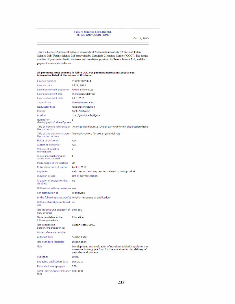
233
234
235
LIST OF REFERENCES
1. S. Patel, and A.K. Mitra, , An Overview of Recent Patents on Nanocarrier Based Ophthalmic
Drug Delivery Systems Recent patents on nanomedicine, 2 (2012) 2-16.
2. V. Tamboli, G.P. Mishra, A.K. Mitra, Polymeric vectors for ocular gene delivery,
Therapeutic delivery, 2 (2011) 523-536.
3. H. Ananthula, R.D. Vaishya, M. Barot, A.K. Mitra, Bioavailability, in: W. Tasman, E.A.
Jaeger (Eds.) Duane's Ophthalmology, Lippincott Williams & Wilkins, Philadelphia, 2009.
4. S.D. Klyce, C.E. Crosson, Transport processes across the rabbit corneal epithelium: a
review, Curr Eye Res, 4 (1985) 323-331.
5. B.J. McLaughlin, R.B. Caldwell, Y. Sasaki, T.O. Wood, Freeze-fracture quantitative
comparison of rabbit corneal epithelial and endothelial membranes, Curr Eye Res, 4 (1985)
951-961.
6. G. Sunkara, U.B. Kompella, Membrane transport processes in the eye, in: A.K. Mitra (Ed.)
Ophthalmic drug delivery systems, Marcel Dekker, Inc, New York, 2003, pp. 13-58.
7. P. Saha, K.J. Kim, V.H. Lee, A primary culture model of rabbit conjunctival epithelial cells
exhibiting tight barrier properties, Curr Eye Res, 15 (1996) 1163-1169.
8. D.H. Geroski, H.F. Edelhauser, Transscleral drug delivery for posterior segment disease,
Adv Drug Deliv Rev, 52 (2001) 37-48.
9. S.H. Kim, R.J. Lutz, N.S. Wang, M.R. Robinson, Transport barriers in transscleral drug
delivery for retinal diseases, Ophthalmic Res, 39 (2007) 244-254.
10. D. Ghate, H.F. Edelhauser, Ocular drug delivery, Expert Opin Drug Deliv, 3 (2006) 275-
287.
11. K. Hosseini, D. Matsushima, J. Johnson, G. Widera, K. Nyam, L. Kim, Y. Xu, Y. Yao, M.
Cormier, Pharmacokinetic study of dexamethasone disodium phosphate using intravitreal,
subconjunctival, and intravenous delivery routes in rabbits, J Ocul Pharmacol Ther, 24
(2008) 301-308.
12. O. Weijtens, E.J. Feron, R.C. Schoemaker, A.F. Cohen, E.G. Lentjes, F.P. Romijn, J.C.
van Meurs, High concentration of dexamethasone in aqueous and vitreous after
subconjunctival injection, Am J Ophthalmol, 128 (1999) 192-197.
13. A. Urtti, Challenges and obstacles of ocular pharmacokinetics and drug delivery, Adv Drug
Deliv Rev, 58 (2006) 1131-1135.
14. M.R. Prausnitz, J.S. Noonan, Permeability of cornea, sclera, and conjunctiva: a literature
analysis for drug delivery to the eye, J Pharm Sci, 87 (1998) 1479-1488.
15. A.K. Mitra, B.S. Anand, S. Duvvuri, Drug delivery to the eye, in: J. Fischbarg (Ed.) The
biology of the eye, Elsevier Inc, New York, 2006, pp. 307-351.
16. R. Gaudana, J. Jwala, S.H. Boddu, A.K. Mitra, Recent perspectives in ocular drug delivery,
Pharm Res, 26 (2009) 1197-1216.
17. E. Mannermaa, K.S. Vellonen, A. Urtti, Drug transport in corneal epithelium and blood-
retina barrier: emerging role of transporters in ocular pharmacokinetics, Adv Drug Deliv
Rev, 58 (2006) 1136-1163.
18. S. Dey, B.S. Anand, J. Patel, A.K. Mitra, Transporters/receptors in the anterior chamber:
pathways to explore ocular drug delivery strategies, Expert Opin Biol Ther, 3 (2003) 23-
44.
19. P.A. Constable, J.G. Lawrenson, D.E. Dolman, G.B. Arden, N.J. Abbott, P-Glycoprotein
expression in human retinal pigment epithelium cell lines, Exp Eye Res, 83 (2006) 24-30.
236
20. K.S. Vellonen, E. Mannermaa, H. Turner, M. Hakli, J.M. Wolosin, T. Tervo, P.
Honkakoski, A. Urtti, Effluxing ABC transporters in human corneal epithelium, J Pharm
Sci, 99 (2010) 1087-1098.
21. P. Saha, J.J. Yang, V.H. Lee, Existence of a p-glycoprotein drug efflux pump in cultured
rabbit conjunctival epithelial cells, Invest Ophthalmol Vis Sci, 39 (1998) 1221-1226.
22. J.J. Yang, K.J. Kim, V.H. Lee, Role of P-glycoprotein in restricting propranolol transport
in cultured rabbit conjunctival epithelial cell layers, Pharm Res, 17 (2000) 533-538.
23. B.G. Kennedy, N.J. Mangini, P-glycoprotein expression in human retinal pigment
epithelium, Mol Vis, 8 (2002) 422-430.
24. S. Duvvuri, M.D. Gandhi, A.K. Mitra, Effect of P-glycoprotein on the ocular disposition
of a model substrate, quinidine, Curr Eye Res, 27 (2003) 345-353.
25. T. Zhang, C.D. Xiang, D. Gale, S. Carreiro, E.Y. Wu, E.Y. Zhang, Drug transporter and
cytochrome P450 mRNA expression in human ocular barriers: implications for ocular drug
disposition, Drug Metab Dispos, 36 (2008) 1300-1307.
26. J.V. Aukunuru, G. Sunkara, N. Bandi, W.B. Thoreson, U.B. Kompella, Expression of
multidrug resistance-associated protein (MRP) in human retinal pigment epithelial cells
and its interaction with BAPSG, a novel aldose reductase inhibitor, Pharm Res, 18 (2001)
565-572.
27. P.K. Karla, R. Earla, S.H. Boddu, T.P. Johnston, D. Pal, A. Mitra, Molecular expression
and functional evidence of a drug efflux pump (BCRP) in human corneal epithelial cells,
Curr Eye Res, 34 (2009) 1-9.
28. B.S. Larsson, Interaction between chemicals and melanin, Pigment Cell Res, 6 (1993) 127-
133.
29. B. Leblanc, S. Jezequel, T. Davies, G. Hanton, C. Taradach, Binding of drugs to eye
melanin is not predictive of ocular toxicity, Regul Toxicol Pharmacol, 28 (1998) 124-132.
30. R.D. Schoenwald, V. Tandon, D.E. Wurster, C.F. Barfknecht, Significance of melanin
binding and metabolism in the activity of 5-acetoxyacetylimino-4-methyl-delta2-1,3,4,-
thiadiazolin e-2-sulfonamide, Eur J Pharm Biopharm, 46 (1998) 39-50.
31. L. Pitkanen, V.P. Ranta, H. Moilanen, A. Urtti, Binding of betaxolol, metoprolol and
oligonucleotides to synthetic and bovine ocular melanin, and prediction of drug binding to
melanin in human choroid-retinal pigment epithelium, Pharm Res, 24 (2007) 2063-2070.
32. L. Salminen, G. Imre, R. Huupponen, The effect of ocular pigmentation on intraocular
pressure response to timolol, Acta Ophthalmol Suppl, 173 (1985) 15-18.
33. L. Pitkanen, V.P. Ranta, H. Moilanen, A. Urtti, Permeability of retinal pigment epithelium:
effects of permeant molecular weight and lipophilicity, Invest Ophthalmol Vis Sci, 46
(2005) 641-646.
34. S. Wang, B. Lu, S. Girman, T. Holmes, N. Bischoff, R.D. Lund, Morphological and
functional rescue in RCS rats after RPE cell line transplantation at a later stage of
degeneration, Invest Ophthalmol Vis Sci, 49 (2008) 416-421.
35. N.P. Cheruvu, U.B. Kompella, Bovine and porcine transscleral solute transport: influence
of lipophilicity and the Choroid-Bruch's layer, Invest Ophthalmol Vis Sci, 47 (2006) 4513-
4522.
36. K. Cholkar, S.P. Patel, A.D. Vadlapudi, A.K. Mitra, Novel strategies for anterior segment
ocular drug delivery, J Ocul Pharmacol Ther, 29 (2013) 106-123.
237
37. G. Meseguer, P. Buri, B. Plazonnet, A. Rozier, R. Gurny, Gamma scintigraphic comparison
of eyedrops containing pilocarpine in healthy volunteers, J Ocul Pharmacol Ther, 12 (1996)
481-488.
38. L. Lapcik, Jr., L. Lapcik, S. De Smedt, J. Demeester, P. Chabrecek, Hyaluronan:
Preparation, Structure, Properties, and Applications, Chemical reviews, 98 (1998) 2663-
2684.
39. I.P. Kaur, R. Smitha, Penetration enhancers and ocular bioadhesives: two new avenues for
ophthalmic drug delivery, Drug development and industrial pharmacy, 28 (2002) 353-369.
40. B.M. Gebhardt, E.D. Varnell, H.E. Kaufman, Cyclosporine in collagen particles: corneal
penetration and suppression of allograft rejection, J Ocul Pharmacol Ther, 11 (1995) 509-
517.
41. E. Dimitrova, S. Bogdanova, M. Mitcheva, I. Tanev, E. Minkov, Development of model
aqueous ophthalmic solution of indomethacin, Drug development and industrial pharmacy,
26 (2000) 1297-1301.
42. P. Chetoni, L. Panichi, S. Burgalassi, U. Benelli, M.F. Saettone, Pharmacokinetics and
anti-inflammatory activity in rabbits of a novel indomethacin ophthalmic solution, J Ocul
Pharmacol Ther, 16 (2000) 363-372.
43. N. Vulovic, M. Primorac, M. Stupar, M.W. Brown, J.L. Ford, Some studies on the
preservation of indometacin suspensions intended for ophthalmic use, Die Pharmazie, 45
(1990) 678-679.
44. C. Bucolo, A. Spadaro, Pharmacological evaluation of anti-inflammatory pyrrole-acetic
acid derivative eye drops, J Ocul Pharmacol Ther, 13 (1997) 353-361.
45. Y. Gao, Y. Sun, F. Ren, S. Gao, PLGA-PEG-PLGA hydrogel for ocular drug delivery of
dexamethasone acetate, Drug development and industrial pharmacy, 36 (2010) 1131-1138.
46. X. Chen, X. Li, Y. Zhou, X. Wang, Y. Zhang, Y. Fan, Y. Huang, Y. Liu, Chitosan-based
thermosensitive hydrogel as a promising ocular drug delivery system: preparation,
characterization, and in vivo evaluation, Journal of biomaterials applications, 27 (2012)
391-402.
47. Y. Qian, F. Wang, R. Li, Q. Zhang, Q. Xu, Preparation and evaluation of in situ gelling
ophthalmic drug delivery system for methazolamide, Drug development and industrial
pharmacy, 36 (2010) 1340-1347.
48. S.H. Boddu, J. Jwala, R. Vaishya, R. Earla, P.K. Karla, D. Pal, A.K. Mitra, Novel
nanoparticulate gel formulations of steroids for the treatment of macular edema, J Ocul
Pharmacol Ther, 26 (2010) 37-48.
49. H. Yin, C. Gong, S. Shi, X. Liu, Y. Wei, Z. Qian, Toxicity evaluation of biodegradable and
thermosensitive PEG-PCL-PEG hydrogel as a potential in situ sustained ophthalmic drug
delivery system, Journal of biomedical materials research. Part B, Applied biomaterials,
92 (2010) 129-137.
50. Y. Cao, C. Zhang, W. Shen, Z. Cheng, L.L. Yu, Q. Ping, Poly(N-isopropylacrylamide)-
chitosan as thermosensitive in situ gel-forming system for ocular drug delivery, Journal of
controlled release, 120 (2007) 186-194.
51. T. Gratieri, G.M. Gelfuso, O. de Freitas, E.M. Rocha, R.F. Lopez, Enhancing and
sustaining the topical ocular delivery of fluconazole using chitosan solution and
poloxamer/chitosan in situ forming gel, Eur J Pharm Biopharm, 79 (2011) 320-327.
238
52. H. Gupta, S. Jain, R. Mathur, P. Mishra, A.K. Mishra, T. Velpandian, Sustained ocular
drug delivery from a temperature and pH triggered novel in situ gel system, Drug delivery,
14 (2007) 507-515.
53. F. Cao, X. Zhang, Q. Ping, New method for ophthalmic delivery of azithromycin by
poloxamer/carbopol-based in situ gelling system, Drug delivery, 17 (2010) 500-507.
54. I.P. Kaur, M. Kanwar, Ocular preparations: the formulation approach, Drug development
and industrial pharmacy, 28 (2002) 473-493.
55. R. Valls, E. Vega, M.L. Garcia, M.A. Egea, J.O. Valls, Transcorneal permeation in a
corneal device of non-steroidal anti-inflammatory drugs in drug delivery systems, The
open medicinal chemistry journal, 2 (2008) 66-71.
56. S.H. Loftsson T, Hreinsdottir D, Konradsdottir F, Stefansson E. , Dexamethasone delivery
to posterior segment of the eye, J InclPhenomMacrocycl Chem, 57 (2007) 585-589.
57. T. Loftsson, E. Stefansson, Cyclodextrin nanotechnology for ophthalmic drug delivery,
US7893040, 2011.
58. T.T. Kararli, R. Bandyopadhyay, S.K. Singh, L.C. Hawlwy, Ophthalmic formulation of a
selective cyclooxygenase-2 inhibitory drug, WO02005815, 2002.
59. A.R. Bary, I.G. Tucker, N.M. Davies, Considerations in the use of hydroxypropyl-beta-
cyclodextrin in the formulation of aqueous ophthalmic solutions of hydrocortisone, Eur J
Pharm Biopharm, 50 (2000) 237-244.
60. J.K. Kristinsson, H. Fridriksdottir, S. Thorisdottir, A.M. Sigurdardottir, E. Stefansson, T.
Loftsson, Dexamethasone-cyclodextrin-polymer co-complexes in aqueous eye drops.
Aqueous humor pharmacokinetics in humans, Invest Ophthalmol Vis Sci, 37 (1996) 1199-
1203.
61. R.M. Mainardes, M.C. Urban, P.O. Cinto, N.M. Khalil, M.V. Chaud, R.C. Evangelista,
M.P. Gremiao, Colloidal carriers for ophthalmic drug delivery, Curr Drug Targets, 6 (2005)
363-371.
62. L. Rabinovich-Guilatt, P. Couvreur, G. Lambert, C. Dubernet, Cationic vectors in ocular
drug delivery, J Drug Target, 12 (2004) 623-633.
63. A.C. Amrite, U.B. Kompella, Size-dependent disposition of nanoparticles and
microparticles following subconjunctival administration, J Pharm Pharmacol, 57 (2005)
1555-1563.
64. A.C. Amrite, H.F. Edelhauser, S.R. Singh, U.B. Kompella, Effect of circulation on the
disposition and ocular tissue distribution of 20 nm nanoparticles after periocular
administration, Mol Vis, 14 (2008) 150-160.
65. G.P. Mishra, M. Bagui, V. Tamboli, A.K. Mitra, Recent applications of liposomes in
ophthalmic drug delivery, Journal of drug delivery, 2011 (2011) 863734.
66. I.P. Kaur, A. Garg, A.K. Singla, D. Aggarwal, Vesicular systems in ocular drug delivery:
an overview, International journal of pharmaceutics, 269 (2004) 1-14.
67. M.K. Kothuri, S. Pinnamaneni, N.G. Das, S.K. Das, Microparticles and nanoparticles in
ocular drug delivery, in: A.K. Mitra (Ed.) Ophthalmic drug delivery systems, Marcel
Dekker Inc, New York, 2003, pp. 437-466.
68. A. Samad, Y. Sultana, M. Aqil, Liposomal drug delivery systems: an update review, Curr
Drug Deliv, 4 (2007) 297-305.
69. S.M. Ansell, T.O. Harasym, P.G. Tardi, S.S. Buchkowsky, M.B. Bally, P.R. Cullis,
Antibody conjugation methods for active targeting of liposomes, Methods Mol Med, 25
(2000) 51-68.
239
70. R.M. Hathout, S. Mansour, N.D. Mortada, A.S. Guinedi, Liposomes as an ocular delivery
system for acetazolamide: in vitro and in vivo studies, AAPS PharmSciTech, 8 (2007) 1.
71. M.S. Nagarsenker, V.Y. Londhe, G.D. Nadkarni, Preparation and evaluation of liposomal
formulations of tropicamide for ocular delivery, International journal of pharmaceutics,
190 (1999) 63-71.
72. H.E. Schaeffer, D.L. Krohn, Liposomes in topical drug delivery, Invest Ophthalmol Vis
Sci, 22 (1982) 220-227.
73. L. Peeters, N.N. Sanders, K. Braeckmans, K. Boussery, J. Van de Voorde, S.C. De Smedt,
J. Demeester, Vitreous: a barrier to nonviral ocular gene therapy, Invest Ophthalmol Vis
Sci, 46 (2005) 3553-3561.
74. C. Hatzifoti, A. Bacon, H. Marriott, P. Laing, A.W. Heath, Liposomal co-entrapment of
CD40mAb induces enhanced IgG responses against bacterial polysaccharide and protein,
PLoS One, 3 (2008) e2368.
75. R. Zeimer, M.F. Goldberg, Novel ophthalmic therapeutic modalities based on noninvasive
light-targeted drug delivery to the posterior pole of the eye, Adv Drug Deliv Rev, 52 (2001)
49-61.
76. S.K. Dharma, P.H. Fishman, G.A. Peyman, A preliminary study of corneal penetration of
125I-labelled idoxuridine liposome, Acta ophthalmologica, 64 (1986) 298-301.
77. O. Felt, P. Furrer, J.M. Mayer, B. Plazonnet, P. Buri, R. Gurny, Topical use of chitosan in
ophthalmology: tolerance assessment and evaluation of precorneal retention, International
journal of pharmaceutics, 180 (1999) 185-193.
78. A.S. Monem, F.M. Ali, M.W. Ismail, Prolonged effect of liposomes encapsulating
pilocarpine HCl in normal and glaucomatous rabbits, International journal of
pharmaceutics, 198 (2000) 29-38.
79. S.L. Law, K.J. Huang, C.H. Chiang, Acyclovir-containing liposomes for potential ocular
delivery. Corneal penetration and absorption, Journal of controlled release, 63 (2000) 135-
140.
80. Y. Shen, J. Tu, Preparation and ocular pharmacokinetics of ganciclovir liposomes, AAPS
J, 9 (2007) E371-377.
81. K.M. Hosny, Ciprofloxacin as ocular liposomal hydrogel, AAPS PharmSciTech, 11 241-
246.
82. V.H. Lee, P.T. Urrea, R.E. Smith, D.J. Schanzlin, Ocular drug bioavailability from
topically applied liposomes, Surv Ophthalmol, 29 (1985) 335-348.
83. K.M. Hosny, Preparation and evaluation of thermosensitive liposomal hydrogel for
enhanced transcorneal permeation of ofloxacin, AAPS PharmSciTech, 10 (2009) 1336-
1342.
84. T.M. Allen, L. McAllister, S. Mausolf, E. Gyorffy, Liposome-cell interactions. A study of
the interactions of liposomes containing entrapped anti-cancer drugs with the EMT6, S49
and AE1 (transport-deficient) cell lines, Biochim Biophys Acta, 643 (1981) 346-362.
85. P.I. Campbell, Toxicity of some charged lipids used in liposome preparations, Cytobios,
37 (1983) 21-26.
86. S.C. Chang, H. Bundgaard, A. Buur, V.H. Lee, Improved corneal penetration of timolol by
prodrugs as a means to reduce systemic drug load, Invest Ophthalmol Vis Sci, 28 (1987)
487-491.
87. E. Yoshihara, T. Nakae, Cytolytic activity of liposomes containing stearylamine, Biochim
Biophys Acta, 854 (1986) 93-101.
240
88. K.G. Janoria, S. Hariharan, C.R. Dasari, A.K. Mitra, Recent patents and advances in
ophthalmic drug delivery, Recent Pat Drug Deliv Formul, 1 (2007) 161-170.
89. R. Pignatello, C. Bucolo, P. Ferrara, A. Maltese, A. Puleo, G. Puglisi, Eudragit RS100
nanosuspensions for the ophthalmic controlled delivery of ibuprofen, European journal of
pharmaceutical sciences, 16 (2002) 53-61.
90. R. Pignatello, C. Bucolo, G. Spedalieri, A. Maltese, G. Puglisi, Flurbiprofen-loaded
acrylate polymer nanosuspensions for ophthalmic application, Biomaterials, 23 (2002)
3247-3255.
91. Y. Rojanasakul, L.Y. Wang, M. Bhat, D.D. Glover, C.J. Malanga, J.K. Ma, The transport
barrier of epithelia: a comparative study on membrane permeability and charge selectivity
in the rabbit, Pharm Res, 9 (1992) 1029-1034.
92. P. Calvo, J.L. Vila-Jato, M.J. Alonso, Comparative in vitro evaluation of several colloidal
systems, nanoparticles, nanocapsules, and nanoemulsions, as ocular drug carriers, J Pharm
Sci, 85 (1996) 530-536.
93. E. Vega, M.A. Egea, O. Valls, M. Espina, M.L. Garcia, Flurbiprofen loaded biodegradable
nanoparticles for ophtalmic administration, J Pharm Sci, 95 (2006) 2393-2405.
94. A.M. De Campos, A. Sanchez, M.J. Alonso, Chitosan nanoparticles: a new vehicle for the
improvement of the delivery of drugs to the ocular surface. Application to cyclosporin A,
International journal of pharmaceutics, 224 (2001) 159-168.
95. X.B. Yuan, Li, H., Yuan, Y.B., Preparation of cholesterol-modified chitosan self-
aggregated nanoparticles for delivery of drugs to ocular surface, Carbohydrate Polymers,
65 (2006) 337–345.
96. X.B. Yuan, Y.B. Yuan, W. Jiang, J. Liu, E.J. Tian, H.M. Shun, D.H. Huang, X.Y. Yuan,
H. Li, J. Sheng, Preparation of rapamycin-loaded chitosan/PLA nanoparticles for
immunosuppression in corneal transplantation, International journal of pharmaceutics, 349
(2008) 241-248.
97. H.K. Ibrahim, I.S. El-Leithy, A.A. Makky, Mucoadhesive nanoparticles as carrier systems
for prolonged ocular delivery of gatifloxacin/prednisolone bitherapy, Molecular
pharmaceutics, 7 (2010) 576-585.
98. F.M.J. Alonso, S.P. Calvo, L.C. Remunan, J.J.L. Vila, Stabilization of colloidal systems
by the formation of ionic lipid-polysaccharide complexs, EP0771566, 2006.
99. A.K. Mitra, G.P. Mishra, Pentablock Polymers, US0250283, 2011.
100. L. Contreras-Ruiz, M. de la Fuente, J.E. Parraga, A. Lopez-Garcia, I. Fernandez, B. Seijo,
A. Sanchez, M. Calonge, Y. Diebold, Intracellular trafficking of hyaluronic acid-chitosan
oligomer-based nanoparticles in cultured human ocular surface cells, Mol Vis, 17 (2011)
279-290.
101. M. de la Fuente, B. Seijo, M.J. Alonso, Novel hyaluronic acid-chitosan nanoparticles for
ocular gene therapy, Invest Ophthalmol Vis Sci, 49 (2008) 2016-2024.
102. A. Enriquez de Salamanca, Y. Diebold, M. Calonge, C. Garcia-Vazquez, S. Callejo, A.
Vila, M.J. Alonso, Chitosan nanoparticles as a potential drug delivery system for the
ocular surface: toxicity, uptake mechanism and in vivo tolerance, Invest Ophthalmol Vis
Sci, 47 (2006) 1416-1425.
103. U.B. Kompella, S. Sundaram, S. Raghava, E.R. Escobar, Luteinizing hormone-releasing
hormone agonist and transferrin functionalizations enhance nanoparticle delivery in a
novel bovine ex vivo eye model, Mol Vis, 12 (2006) 1185-1198.
241
104. B.E. Rabinow, Nanosuspensions in drug delivery, Nat Rev Drug Discov, 3 (2004) 785-
796.
105. D.R. Sanders, B. Goldstick, C. Kraff, R. Hutchins, M.S. Bernstein, M.A. Evans, Aqueous
penetration of oral and topical indomethacin in humans, Archives of ophthalmology, 101
(1983) 1614-1616.
106. M.A. Kassem, A.A. Abdel Rahman, M.M. Ghorab, M.B. Ahmed, R.M. Khalil,
Nanosuspension as an ophthalmic delivery system for certain glucocorticoid drugs,
International journal of pharmaceutics, 340 (2007) 126-133.
107. J.D. Quintana-Hau, E. Cruz-Olmos, M.I. Lopez-Sanchez, V. Sanchez-Castellanos, L.
Baiza-Duran, J.R. Gonzalez, M. Tornero, R. Mondragon-Flores, A. Hernandez-Santoyo,
Characterization of the novel ophthalmic drug carrier Sophisen in two of its derivatives:
3A Ofteno and Modusik-A Ofteno, Drug development and industrial pharmacy, 31
(2005) 263-269.
108. S. Panmai, L.L. Alani, Ophthalmic nanoparticulate formulation of a cyclooxygenase-2
selective inhibitor, WO2006062875, 2006.
109. V.B. Patravale, A.A. Date, R.M. Kulkarni, Nanosuspensions: a promising drug delivery
strategy, J Pharm Pharmacol, 56 (2004) 827-840.
110. A.A. Badawi, H.M. El-Laithy, R.K. El Qidra, H. El Mofty, M. El dally, Chitosan based
nanocarriers for indomethacin ocular delivery, Archives of pharmacal research, 31
(2008) 1040-1049.
111. L. Gan, Y. Gan, C. Zhu, X. Zhang, J. Zhu, Novel microemulsion in situ electrolyte-
triggered gelling system for ophthalmic delivery of lipophilic cyclosporine A: in vitro
and in vivo results, International journal of pharmaceutics, 365 (2009) 143-149.
112. L. Baydoun, C.C. Muller-Goymann, Influence of n-octenylsuccinate starch on in vitro
permeation of sodium diclofenac across excised porcine cornea in comparison to
Voltaren ophtha, Eur J Pharm Biopharm, 56 (2003) 73-79.
113. M. ShaulMuchtar, Joseph Frucht-Pery, Simon Benita, , Ex-vivo permeation study of
indomethacin from a submicron emulsion through albino rabbit cornea, , Journal of
controlled release, 44 (1997) 55-64.
114. S. Benita, G. Lambert, Method and composition for dry eye treatment, US0108626, 2003.
115. Y. Gan, L. Gan, J. Shen, C. Zhu, Ophthalmic flurbiprofen ester nano-emulsion in-situ gel
formulation and the preparation method there of, WO2010048788, 2010.
116. S.H. Hyon, K. Jamshidi, Y. Ikada, Synthesis of polylactides with different molecular
weights, Biomaterials, 18 (1997) 1503-1508.
117. J.M. Lu, X. Wang, C. Marin-Muller, H. Wang, P.H. Lin, Q. Yao, C. Chen, Current
advances in research and clinical applications of PLGA-based nanotechnology, Expert
Rev Mol Diagn, 9 (2009) 325-341.
118. V.P. Torchilin, Structure and design of polymeric surfactant-based drug delivery systems,
Journal of controlled release, 73 (2001) 137-172.
119. C. Civiale, M. Licciardi, G. Cavallaro, G. Giammona, M.G. Mazzone,
Polyhydroxyethylaspartamide-based micelles for ocular drug delivery, International
journal of pharmaceutics, 378 (2009) 177-186.
120. A.K. Mitra, P. Velagaleti, U.M. Grau, Topical drug delivery systems for ophthalmic use,
WO2010144194, 2010.
121. A.K. Mitra, P. Velagaleti, N. Subramanian, Ophthalmic compositions comprising
calcineurin inhibitors or mtor inhibitors, US0092665, 2009.
242
122. L. Gan, S. Han, J. Shen, J. Zhu, C. Zhu, X. Zhang, Y. Gan, Self-assembled liquid
crystalline nanoparticles as a novel ophthalmic delivery system for dexamethasone:
Improving preocular retention and ocular bioavailability, International journal of
pharmaceutics, 396 (2010) 179-187.
123. J.L. Bourges, C. Bloquel, A. Thomas, F. Froussart, A. Bochot, F. Azan, R. Gurny, D.
BenEzra, F. Behar-Cohen, Intraocular implants for extended drug delivery: therapeutic
applications, Adv Drug Deliv Rev, 58 (2006) 1182-1202.
124. T.J. Smith, P.A. Pearson, D.L. Blandford, J.D. Brown, K.A. Goins, J.L. Hollins, E.T.
Schmeisser, P. Glavinos, L.B. Baldwin, P. Ashton, Intravitreal sustained-release
ganciclovir, Archives of ophthalmology, 110 (1992) 255-258.
125. J.I. Lim, R.A. Wolitz, A.H. Dowling, H.R. Bloom, A.R. Irvine, D.M. Schwartz, Visual
and anatomic outcomes associated with posterior segment complications after
ganciclovir implant procedures in patients with AIDS and cytomegalovirus retinitis, Am
J Ophthalmol, 127 (1999) 288-293.
126. G.E. Sanborn, R. Anand, R.E. Torti, S.D. Nightingale, S.X. Cal, B. Yates, P. Ashton, T.
Smith, Sustained-release ganciclovir therapy for treatment of cytomegalovirus retinitis.
Use of an intravitreal device, Archives of ophthalmology, 110 (1992) 188-195.
127. T. Yasukawa, Y. Ogura, H. Kimura, E. Sakurai, Y. Tabata, Drug delivery from ocular
implants, Expert Opin Drug Deliv, 3 (2006) 261-273.
128. K. Okabe, H. Kimura, J. Okabe, A. Kato, N. Kunou, Y. Ogura, Intraocular tissue
distribution of betamethasone after intrascleral administration using a non-biodegradable
sustained drug delivery device, Invest Ophthalmol Vis Sci, 44 (2003) 2702-2707.
129. G.J. Jaffe, R.M. McCallum, B. Branchaud, C. Skalak, Z. Butuner, P. Ashton, Long-term
follow-up results of a pilot trial of a fluocinolone acetonide implant to treat posterior
uveitis, Ophthalmology, 112 (2005) 1192-1198.
130. P.A. Pearson, G.J. Jaffe, D.F. Martin, G.J. Cordahi, H. Grossniklaus, E.T. Schmeisser, P.
Ashton, Evaluation of a delivery system providing long-term release of cyclosporine,
Archives of ophthalmology, 114 (1996) 311-317.
131. L.B. Enyedi, P.A. Pearson, P. Ashton, G.J. Jaffe, An intravitreal device providing
sustained release of cyclosporine and dexamethasone, Curr Eye Res, 15 (1996) 549-557.
132. H. Kimura, Y. Ogura, Biodegradable polymers for ocular drug delivery,
Ophthalmologica. Journal international d'ophtalmologie. International journal of
ophthalmology, 215 (2001) 143-155.
133. N. Kunou, Y. Ogura, T. Yasukawa, H. Kimura, H. Miyamoto, Y. Honda, Y. Ikada, Long-
term sustained release of ganciclovir from biodegradable scleral implant for the treatment
of cytomegalovirus retinitis, Journal of controlled release, 68 (2000) 263-271.
134. P.E. Rubsamen, P.A. Davis, E. Hernandez, G.E. O'Grady, S.W. Cousins, Prevention of
experimental proliferative vitreoretinopathy with a biodegradable intravitreal implant for
the sustained release of fluorouracil, Archives of ophthalmology, 112 (1994) 407-413.
135. T. Zhou, H. Lewis, R.E. Foster, S.P. Schwendeman, Development of a multiple-drug
delivery implant for intraocular management of proliferative vitreoretinopathy, Journal
of controlled release, 55 (1998) 281-295.
136. L. Xie, J. Sun, Z. Yao, Heparin drug delivery system for prevention of posterior capsular
opacification in rabbit eyes, Graefe's archive for clinical and experimental
ophthalmology, 241 (2003) 309-313.
243
137. T. Yasukawa, H. Kimura, Y. Tabata, H. Miyamoto, Y. Honda, Y. Ogura, Sustained
release of cis-hydroxyproline in the treatment of experimental proliferative
vitreoretinopathy in rabbits, Graefe's archive for clinical and experimental
ophthalmology, 240 (2002) 672-678.
138. C. Koelwel, S. Rothschenk, B. Fuchs-Koelwel, B. Gabler, C. Lohmann, A. Gopferich,
Alginate inserts loaded with epidermal growth factor for the treatment of
keratoconjunctivitis sicca, Pharmaceutical development and technology, 13 (2008) 221-
231.
139. C.F. Yang, T. Yasukawa, H. Kimura, H. Miyamoto, Y. Honda, Y. Tabata, Y. Ikada, Y.
Ogura, Experimental corneal neovascularization by basic fibroblast growth factor
incorporated into gelatin hydrogel, Ophthalmic Res, 32 (2000) 19-24.
140. M. Hashizoe, Y. Ogura, T. Takanashi, N. Kunou, Y. Honda, Y. Ikada, Implantable
biodegradable polymeric device in the treatment of experimental proliferative
vitreoretinopathy, Curr Eye Res, 14 (1995) 473-477.
141. M. Hashizoe, Y. Ogura, H. Kimura, T. Moritera, Y. Honda, M. Kyo, S.H. Hyon, Y. Ikada,
Scleral plug of biodegradable polymers for controlled drug release in the vitreous,
Archives of ophthalmology, 112 (1994) 1380-1384.
142. H. Miyamoto, Y. Ogura, M. Hashizoe, N. Kunou, Y. Honda, Y. Ikada, Biodegradable
scleral implant for intravitreal controlled release of fluconazole, Curr Eye Res, 16 (1997)
930-935.
143. E. Sakurai, M. Nozaki, K. Okabe, N. Kunou, H. Kimura, Y. Ogura, Scleral plug of
biodegradable polymers containing tacrolimus (FK506) for experimental uveitis, Invest
Ophthalmol Vis Sci, 44 (2003) 4845-4852.
144. J. Okabe, H. Kimura, N. Kunou, K. Okabe, A. Kato, Y. Ogura, Biodegradable intrascleral
implant for sustained intraocular delivery of betamethasone phosphate, Invest
Ophthalmol Vis Sci, 44 (2003) 740-744.
145. Y. Morita, A. Ohtori, M. Kimura, K. Tojo, Intravitreous delivery of dexamethasone
sodium m-sulfobenzoate from poly(DL-lactic acid) implants, Biological &
pharmaceutical bulletin, 21 (1998) 188-190.
146. M.R. Prausnitz, Microneedles for transdermal drug delivery, Adv Drug Deliv Rev, 56
(2004) 581-587.
147. J. Jiang, H.S. Gill, D. Ghate, B.E. McCarey, S.R. Patel, H.F. Edelhauser, M.R. Prausnitz,
Coated microneedles for drug delivery to the eye, Invest Ophthalmol Vis Sci, 48 (2007)
4038-4043.
148. J. Jiang, J.S. Moore, H.F. Edelhauser, M.R. Prausnitz, Intrascleral drug delivery to the
eye using hollow microneedles, Pharm Res, 26 (2009) 395-403.
149. S. Patel, V. Tamboli, G.P. Mishra, A.K. Mitra, Biodegradable Polymers for Ophthalmic
Applications, in: A.K. Mitra (Ed.) Treatise on Ocular Drug Delivery, Bentham Science,
2013, pp. 96-113.
150. J.L. Arias, M.A. Ruiz, M. Lopez-Viota, A.V. Delgado, Poly(alkylcyanoacrylate) colloidal
particles as vehicles for antitumour drug delivery: a comparative study, Colloids Surf B
Biointerfaces, 62 (2008) 64-70.
151. R.J. Tamargo, J.I. Epstein, C.S. Reinhard, M. Chasin, H. Brem, Brain biocompatibility of
a biodegradable, controlled-release polymer in rats, J Biomed Mater Res, 23 (1989) 253-
266.
152. H. Brem, Polymers to treat brain tumours, Biomaterials, 11 (1990) 699-701.
244
153. N. Kumar, R.S. Langer, A.J. Domb, Polyanhydrides: an overview, Adv Drug Deliv Rev,
54 (2002) 889-910.
154. J.C. Middleton, A.J. Tipton, Synthetic biodegradable polymers as orthopedic devices,
Biomaterials, 21 (2000) 2335-2346.
155. R. Jain, N.H. Shah, A.W. Malick, C.T. Rhodes, Controlled drug delivery by biodegradable
poly(ester) devices: different preparative approaches, Drug development and industrial
pharmacy, 24 (1998) 703-727.
156. R. Tang, R.N. Palumbo, W. Ji, C. Wang, Poly(ortho ester amides): acid-labile
temperature-responsive copolymers for potential biomedical applications,
Biomacromolecules, 10 (2009) 722-727.
157. J. Heller, J. Barr, S.Y. Ng, K.S. Abdellauoi, R. Gurny, Poly(ortho esters): synthesis,
characterization, properties and uses, Adv Drug Deliv Rev, 54 (2002) 1015-1039.
158. S. Einmahl, S. Capancioni, K. Schwach-Abdellaoui, M. Moeller, F. Behar-Cohen, R.
Gurny, Therapeutic applications of viscous and injectable poly(ortho esters), Adv Drug
Deliv Rev, 53 (2001) 45-73.
159. J.A. Tielsch, Vision problems in the US: a report on blindness and vision impairment in
adults age 40 and older, Prevent Blindness America, (1994).
160. C.C. Hu, J.D. Ho, H.C. Lin, Neovascular age-related macular degeneration and the risk
of stroke: a 5-year population-based follow-up study, Stroke, 41 (2010) 613-617.
161. A.D. Kulkarni, B.D. Kuppermann, Wet age-related macular degeneration, Adv Drug
Deliv Rev, 57 (2005) 1994-2009.
162. T. Inai, M. Mancuso, H. Hashizume, F. Baffert, A. Haskell, P. Baluk, D.D. Hu-Lowe,
D.R. Shalinsky, G. Thurston, G.D. Yancopoulos, D.M. McDonald, Inhibition of vascular
endothelial growth factor (VEGF) signaling in cancer causes loss of endothelial
fenestrations, regression of tumor vessels, and appearance of basement membrane ghosts,
The American journal of pathology, 165 (2004) 35-52.
163. B.T. Ozturk, H. Kerimoglu, B. Bozkurt, S. Okudan, Comparison of intravitreal
bevacizumab and ranibizumab treatment for diabetic macular edema, J Ocul Pharmacol
Ther, 27 373-377.
164. M.I. Canut, A. Alvarez, J. Nadal, R. Abreu, J.A. Abreu, J.S. Pulido, Anterior segment
changes following intravitreal bevacizumab injection for treatment of neovascular
glaucoma, Clin Ophthalmol, 5 715-719.
165. F. Bock, J. Onderka, T. Dietrich, B. Bachmann, F.E. Kruse, M. Paschke, G. Zahn, C.
Cursiefen, Bevacizumab as a potent inhibitor of inflammatory corneal angiogenesis and
lymphangiogenesis, Invest Ophthalmol Vis Sci, 48 (2007) 2545-2552.
166. M.S. Alforja, N. Sabater, J. Giralt, A. Adan, L. Pelegrin, R. Casaroli-Marano, Intravitreal
bevacizumab injection for peripheral exudative hemorrhagic chorioretinopathy, Japanese
journal of ophthalmology, 55 (2011) 425-427.
167. A. Sayen, I. Hubert, J.P. Berrod, Age related macular degeneration, La Revue du
praticien, 61 (2011) 159-164.
168. S. Saati, R.N. Agrawal, S. Louie, G.J. Chader, M.S. Humayun, Effect of multiple
injections of small divided doses vs single injection of intravitreal bevacizumab on retinal
neovascular model in rabbits, Graefe's archive for clinical and experimental
ophthalmology, 248 457-466.
245
169. J. Kang, S.P. Schwendeman, Comparison of the effects of Mg(OH)2 and sucrose on the
stability of bovine serum albumin encapsulated in injectable poly(D,L-lactide-co-
glycolide) implants, Biomaterials, 23 (2002) 239-245.
170. A. Budhian, S.J. Siegel, K.I. Winey, Production of haloperidol-loaded PLGA
nanoparticles for extended controlled drug release of haloperidol, Journal of
microencapsulation, 22 (2005) 773-785.
171. J. Jwala, S.H. Boddu, S. Shah, S. Sirimulla, D. Pal, A.K. Mitra, Ocular sustained release
nanoparticles containing stereoisomeric dipeptide prodrugs of acyclovir, J Ocul
Pharmacol Ther, 27 (2011) 163-172.
172. G.A. Peyman, E.M. Lad, D.M. Moshfeghi, Intravitreal injection of therapeutic agents,
Retina, 29 (2009) 875-912.
173. R.D. Jager, L.P. Aiello, S.C. Patel, E.T. Cunningham, Jr., Risks of intravitreous injection:
a comprehensive review, Retina, 24 (2004) 676-698.
174. K.M. Sampat, S.J. Garg, Complications of intravitreal injections, Curr Opin Ophthalmol,
21 (2010) 178-183.
175. S.R. Van Tomme, G. Storm, W.E. Hennink, In situ gelling hydrogels for pharmaceutical
and biomedical applications, International journal of pharmaceutics, 355 (2008) 1-18.
176. M. Gou, C. Gong, J. Zhang, X. Wang, Y. Gu, G. Guo, L. Chen, F. Luo, X. Zhao, Y. Wei,
Z. Qian, Polymeric matrix for drug delivery: honokiol-loaded PCL-PEG-PCL
nanoparticles in PEG-PCL-PEG thermosensitive hydrogel, J Biomed Mater Res A, 93
(2010) 219-226.
177. B. Jeong, Y.H. Bae, S.W. Kim, In situ gelation of PEG-PLGA-PEG triblock copolymer
aqueous solutions and degradation thereof, J Biomed Mater Res, 50 (2000) 171-177.
178. S. Singh, D.C. Webster, J. Singh, Thermosensitive polymers: synthesis, characterization,
and delivery of proteins, International journal of pharmaceutics, 341 (2007) 68-77.
179. C.B. Liu, C.Y. Gong, M.J. Huang, J.W. Wang, Y.F. Pan, Y.D. Zhang, G.Z. Li, M.L. Gou,
K. Wang, M.J. Tu, Y.Q. Wei, Z.Y. Qian, Thermoreversible gel-sol behavior of
biodegradable PCL-PEG-PCL triblock copolymer in aqueous solutions, J Biomed Mater
Res. Part B, Applied biomaterials, 84 (2008) 165-175.
180. C. Gong, S. Shi, L. Wu, M. Gou, Q. Yin, Q. Guo, P. Dong, F. Zhang, F. Luo, X. Zhao, Y.
Wei, Z. Qian, Biodegradable in situ gel-forming controlled drug delivery system based
on thermosensitive PCL-PEG-PCL hydrogel. Part 2: sol-gel-sol transition and drug
delivery behavior, Acta Biomater, 5 (2009) 3358-3370.
181. M.-H. Huang, S. Li, M. Vert, Synthesis and degradation of PLA–PCL–PLA triblock
copolymer prepared by successive polymerization of 3-caprolactone and DL-lactide.,
Polymer, 45 (2004) 8675-8681.
182. M.L. Houchin, E.M. Topp, Chemical degradation of peptides and proteins in PLGA: a
review of reactions and mechanisms, J Pharm Sci, 97 (2008) 2395-2404.
183. Y. Zhang, S.P. Schwendeman, Minimizing acylation of peptides in PLGA microspheres,
Journal of controlled release, 162 (2012) 119-126.
184. M.J. Hwang, J.M. Suh, Y.H. Bae, S.W. Kim, B. Jeong, Caprolactonic poloxamer analog:
PEG-PCL-PEG, Biomacromolecules, 6 (2005) 885-890.
185. A. Prabhu, C.E. Shelburne, D.F. Gibbons, Cellular proliferation and cytokine responses
of murine macrophage cell line J774A.1 to polymethylmethacrylate and cobalt-chrome
alloy particles, J Biomed Mater Res, 42 (1998) 655-663.
246
186. D. Lee, M. Shim, S. Kim, H. Lee, I. Park, T. Chang, Novel thermoreversible gelation of
biodegradable PLGA-block-PEOblock-PLGA triblock copolymers in aqueous solution,
Macromolecular rapid communications, 22 (2001) 587-592.
187. B. Jeong, Y.H. Bae, S.W. Kim, Drug release from biodegradable injectable
thermosensitive hydrogel of PEG-PLGA-PEG triblock copolymers, Journal of controlled
release, 63 (2000) 155-163.
188. S.H. Lee, Z. Zhang, S.S. Feng, Nanoparticles of poly(lactide)-tocopheryl polyethylene
glycol succinate (PLA-TPGS) copolymers for protein drug delivery, Biomaterials, 28
(2007) 2041-2050.
189. W. Jia, Y. Gu, M. Gou, M. Dai, X. Li, B. Kan, J. Yang, Q. Song, Y. Wei, Z. Qian,
Preparation of biodegradable polycaprolactone/poly (ethylene glycol)/polycaprolactone
(PCEC) nanoparticles, Drug delivery, 15 (2008) 409-416.
190. U. Bilati, E. Allemann, E. Doelker, Poly(D,L-lactide-co-glycolide) protein-loaded
nanoparticles prepared by the double emulsion method--processing and formulation
issues for enhanced entrapment efficiency, Journal of microencapsulation, 22 (2005)
205-214.
191. C. Perez, I.J. Castellanos, H.R. Costantino, W. Al-Azzam, K. Griebenow, Recent trends
in stabilizing protein structure upon encapsulation and release from bioerodible
polymers, J Pharm Pharmacol, 54 (2002) 301-313.
192. S.D. Putney, Encapsulation of proteins for improved delivery, Curr Opin Chem Biol, 2
(1998) 548-552.
193. T.R. Kumar, K. Soppimath, S.K. Nachaegari, Novel delivery technologies for protein and
peptide therapeutics, Curr Pharm Biotechnol, 7 (2006) 261-276.
194. M. van de Weert, W.E. Hennink, W. Jiskoot, Protein instability in poly(lactic-co-glycolic
acid) microparticles, Pharm Res, 17 (2000) 1159-1167.
195. H. Sah, Stabilization of proteins against methylene chloride/water interface-induced
denaturation and aggregation, Journal of controlled release, 58 (1999) 143-151.
196. K. Fu, D.W. Pack, A.M. Klibanov, R. Langer, Visual evidence of acidic environment
within degrading poly(lactic-co-glycolic acid) (PLGA) microspheres, Pharm Res, 17
(2000) 100-106.
197. E. Zablotna, A. Jaskiewicz, A. Legowska, H. Miecznikowska, A. Lesner, K. Rolka,
Design of serine proteinase inhibitors by combinatorial chemistry using trypsin inhibitor
SFTI-1 as a starting structure, J Pept Sci, 13 (2007) 749-755.
198. J. Wang, K.M. Chua, C.H. Wang, Stabilization and encapsulation of human
immunoglobulin G into biodegradable microspheres, J Colloid Interface Sci, 271 (2004)
92-101.
199. J. Jwala, R.K. Vadlapatla, A.D. Vadlapudi, S.H. Boddu, D. Pal, A.K. Mitra, Differential
expression of folate receptor-alpha, sodium-dependent multivitamin transporter, and
amino acid transporter (B (0, +)) in human retinoblastoma (Y-79) and retinal pigment
epithelial (ARPE-19) cell lines, J Ocul Pharmacol Ther, 28 (2012) 237-244.
200. D. Pissuwan, C. Boyer, K. Gunasekaran, T.P. Davis, V. Bulmus, In vitro cytotoxicity of
RAFT polymers, Biomacromolecules, 11 (2010) 412-420.
201. U. Bilati, E. Allemann, E. Doelker, Sonication parameters for the preparation of
biodegradable nanocapsules of controlled size by the double emulsion method,
Pharmaceutical development and technology, 8 (2003) 1-9.
247
202. M.I. Sabir, X. Xu, L. Li, A review on biodegradable polymeric materials for bone
tissueengineering applications, Journal of material science, 44 ( 2009) 5713-5724.
203. M.H. Huang, S. Li, M. Vert, Synthesis and degradation of PLA–PCL–PLA triblock
copolymer prepared by successive polymerization of ε-caprolactone and DL-lactide,
Polymer, 45 (2004) 8675-8681.
204. A. Frank, S.K. Rath, S.S. Venkatraman, Controlled release from bioerodible polymers:
effect of drug type and polymer composition, Journal of controlled release, 102 (2005)
333-344.
205. M.H. Huang, S. Li, D.W. Hutmacher, J.T. Schantz, C.A. Vacanti, C. Braud, M. Vert,
Degradation and cell culture studies on block copolymers prepared by ring opening
polymerization of epsilon-caprolactone in the presence of poly(ethylene glycol), J
Biomed Mater Res A, 69 (2004) 417-427.
206. S. Li, I. Molina, M.B. Martinez, M. Vert, Hydrolytic and enzymatic degradations of
physically crosslinked hydrogels prepared from PLA/PEO/PLA triblock copolymers, J
Mater Sci Mater Med, 13 (2002) 81-86.
207. S. Li, P. Dobrzynski, J. Kasperczyk, M. Bero, C. Braud, M. Vert, Structure-property
relationships of copolymers obtained by ring-opening polymerization of glycolide and
epsilon-caprolactone. Part 2. Influence of composition and chain microstructure on the
hydrolytic degradation, Biomacromolecules, 6 (2005) 489-497.
208. T.G. Park, Degradation of poly (D,L-lactic acid) microspheres: Effect of molecular
weight, Journal of Controlled Release, 30 (1994) 161–173.
209. S. Du, Q. Zhang, S. Zhang, L. Wang, J. Lian, Heat shock protein 70 expression induced
by diode laser irradiation on choroid-retinal endothelial cells in vitro, Mol Vis, 18 (2012)
2380-2387.
210. V. Bhardwaj, D.D. Ankola, S.C. Gupta, M. Schneider, C.M. Lehr, M.N. Kumar, PLGA
nanoparticles stabilized with cationic surfactant: safety studies and application in oral
delivery of paclitaxel to treat chemical-induced breast cancer in rat, Pharm Res, 26 (2009)
2495-2503.
211. S. Basu, R. Harfouche, S. Soni, G. Chimote, R.A. Mashelkar, S. Sengupta, Nanoparticle-
mediated targeting of MAPK signaling predisposes tumor to chemotherapy, Proc Natl
Acad Sci U S A, 106 (2009) 7957-7961.
212. Q.T. Shubhra, T. Feczko, A.F. Kardos, J. Toth, H. Mackova, D. Horak, G. Dosa, J.
Gyenis, Co-encapsulation of human serum albumin and superparamagnetic iron oxide in
PLGA nanoparticles: Part II. Effect of process variables on protein model drug
encapsulation efficiency, Journal of microencapsulation, (2013).
213. H. Mu, R. Ohashi, S. Yan, H. Chai, H. Yang, P. Lin, Q. Yao, C. Chen, Adipokine resistin
promotes in vitro angiogenesis of human endothelial cells, Cardiovasc Res, 70 (2006)
146-157.
214. A. Kasai, N. Shintani, M. Oda, M. Kakuda, H. Hashimoto, T. Matsuda, S. Hinuma, A.
Baba, Apelin is a novel angiogenic factor in retinal endothelial cells, Biochem Biophys
Res Commun, 325 (2004) 395-400.
215. Y. Xu, H. Zhao, Y. Zheng, Q. Gu, J. Ma, X. Xu, A novel antiangiogenic peptide derived
from hepatocyte growth factor inhibits neovascularization in vitro and in vivo, Mol Vis,
16 (2010) 1982-1995.
248
216. W.Z. Yu, H. Zou, X.X. Li, Z. Yan, J.Q. Dong, Effects of the phosphatidylinositol 3-kinase
inhibitor in a mouse model of retinal neovascularization, Ophthalmic Res, 40 (2008) 19-
25.
217. M. Gou, C. Gong, J. Zhang, X. Wang, X. Wang, Y. Gu, G. Guo, L. Chen, F. Luo, X.
Zhao, Y. Wei, Z. Qian, Polymeric matrix for drug delivery: honokiol-loaded PCL-PEG-
PCL nanoparticles in PEG-PCL-PEG thermosensitive hydrogel, J Biomed Mater Res A,
93 (2010) 219-226.
218. Y. Tang, J. Singh, Biodegradable and biocompatible thermosensitive polymer based
injectable implant for controlled release of protein, International journal of
pharmaceutics, 365 (2009) 34-43.
219. M.H. Huang, S. Li, J. Coudane, M. Vert, Synthesis and Characterization of Block
Copolymers of e-Caprolactone and DL-Lactide Initiated by Ethylene Glycol or
Poly(ethylene glycol), Macromolecular chemistry and physics, 204 (2003) 1994-2001.
220. K.F. Pistel, T. Kissel, Effects of salt addition on the microencapsulation of proteins using
W/O/W double emulsion technique, Journal of microencapsulation, 17 (2000) 467-483.
221. J.X. Zhang, K.J. Zhu, D. Chen, Preparation of bovine serum albumin loaded poly (D, L-
lactic-co-glycolic acid) microspheres by a modified phase separation technique, Journal
of microencapsulation, 22 (2005) 117-126.
222. H. Tan, C.M. Ramirez, N. Miljkovic, H. Li, J.P. Rubin, K.G. Marra, Thermosensitive
injectable hyaluronic acid hydrogel for adipose tissue engineering, Biomaterials, 30
(2009) 6844-6853.
223. L. Bian, D.Y. Zhai, E. Tous, R. Rai, R.L. Mauck, J.A. Burdick, Enhanced MSC
chondrogenesis following delivery of TGF-beta3 from alginate microspheres within
hyaluronic acid hydrogels in vitro and in vivo, Biomaterials, 32 (2011) 6425-6434.
224. A.M. Kloxin, M.W. Tibbitt, K.S. Anseth, Synthesis of photodegradable hydrogels as
dynamically tunable cell culture platforms, Nature protocols, 5 (2010) 1867-1887.
225. N. Bhattarai, J. Gunn, M. Zhang, Chitosan-based hydrogels for controlled, localized drug
delivery, Adv Drug Deliv Rev, 62 (2010) 83-99.
226. C.H. Wang, Y.S. Hwang, P.R. Chiang, C.R. Shen, W.H. Hong, G.H. Hsiue, Extended
release of bevacizumab by thermosensitive biodegradable and biocompatible hydrogel,
Biomacromolecules, 13 (2012) 40-48.
227. J.J. Kang Derwent, W.F. Mieler, Thermoresponsive hydrogels as a new ocular drug
delivery platform to the posterior segment of the eye, Transactions of the American
Ophthalmological Society, 106 (2008) 206-213; discussion 213-204.
249
VITA
Sulabh P. Patel was born on 3rd June, 1983, in Ahmedabad, Gujarat, India. He
completed his Bachelor of Pharmacy from Hemchandracharya North Gujarat University in
2004 and received his Master of Science degree with the specialization in Pharmaceutical
Analysis (2006) from National Institute of Pharmaceutical Education and Research (NIPER),
S.A.S. Nagar, Mohali, Punjab, India. After completion of his M.S. degree, he joined the
Bioanalytical division of Cadila Pharmaceuticals, India and worked as Research Associate
until August, 2007.
To pursue a Ph.D. degree, Sulabh Patel initiated Doctoral study in the Interdisciplinary
Ph.D. program at UMKC in January, 2009. He is an active member of American Association
of Pharmaceutical Scientists (AAPS), Association of Research in Vision and Ophthalmology
(ARVO) and Pharmaceutical Sciences Graduate Student Association (PSGSA). He served as
the Secretary of American Association of Pharmaceutical Scientists (AAPS) student chapter,
UMKC, for two consecutive academic years (2010-11 and 2011-12). He received the first prize
at Interdisciplinary community of scholar poster presentation, April 2011. He completed his
doctoral studies in January, 2014 under the guidance of Dr. Ashim K. Mitra. He has
authored/co-authored several peer reviewed publications and book chapters, and has also
presented his work in several annual national conferences.