cardiorespiratory phase-coupling is reduced in patients with obstructive sleep apnea
TRANSCRIPT
Cardiorespiratory Phase-Coupling Is Reduced in Patientswith Obstructive Sleep ApneaMuammar M. Kabir1*, Hany Dimitri2, Prashanthan Sanders2, Ral Antic3, Eugene Nalivaiko4, Derek
Abbott1, Mathias Baumert5
1 Centre for Biomedical Engineering, School of Electrical and Electronic Engineering, University of Adelaide, Adelaide, South Australia, Australia, 2 Cardiovascular Research
Centre, School of Medicine, University of Adelaide, Adelaide, South Australia, Australia, 3 Thoracic Medicine Department, Royal Adelaide Hospital, Adelaide, South
Australia, Australia, 4 School of Biomedical Sciences, University of Newcastle, Callaghan, New South Wales, Australia, 5 School of Medicine, School of Paediatrics and
Reproductive Health, University of Adelaide, Adelaide, South Australia, Australia
Abstract
Cardiac and respiratory rhythms reveal transient phases of phase-locking which were proposed to be an important aspect ofcardiorespiratory interaction. The aim of this study was to quantify cardio-respiratory phase-locking in obstructive sleepapnea (OSA). We investigated overnight polysomnography data of 248 subjects with suspected OSA. Cardiorespiratoryphase-coupling was computed from the R-R intervals of body surface ECG and respiratory rate, calculated from abdominaland thoracic sensors, using Hilbert transform. A significant reduction in phase-coupling was observed in patients withsevere OSA compared to patients with no or mild OSA. Cardiorespiratory phase-coupling was also associated with sleepstages and was significantly reduced during rapid-eye-movement (REM) sleep compared to slow-wave (SW) sleep. Therewas, however, no effect of age and BMI on phase coupling. Our study suggests that the assessment of cardiorespiratoryphase coupling may be used as an ECG based screening tool for determining the severity of OSA.
Citation: Kabir MM, Dimitri H, Sanders P, Antic R, Nalivaiko E, et al. (2010) Cardiorespiratory Phase-Coupling Is Reduced in Patients with Obstructive SleepApnea. PLoS ONE 5(5): e10602. doi:10.1371/journal.pone.0010602
Editor: Keertan Dheda, University of Cape Town, South Africa
Received October 27, 2009; Accepted April 18, 2010; Published May 13, 2010
Copyright: � 2010 Kabir et al. This is an open-access article distributed under the terms of the Creative Commons Attribution License, which permitsunrestricted use, distribution, and reproduction in any medium, provided the original author and source are credited.
Funding: The research was supported by the Australian Research Council (grant #DP0663345). The funders had no role in study design, data collection andanalysis, decision to publish, or preparation of the manuscript.
Competing Interests: The authors have declared that no competing interests exist.
* E-mail: [email protected]
Introduction
Obstructive sleep apnea (OSA) is a disorder of breathing during
sleep that affects over 4% of men and 2% of women [1].
Obstructive sleep apnea (OSA) is characterized by repetitive
partial or complete closure of the upper airways that causes
alterations in the functioning of cardiovascular and respiratory
systems.
Cardiac autonomic nerve activity in OSA patients has been
mainly studied using heart rate variability (HRV) methodology
that is based on the assessment of ECG RR-interval changes [2].
Some indices of HRV were shown to be an independent predictor
of cardiac mortality in different patient populations, including
myocardial infarction [3], dilated cardiomyopathy [4], and
congestive heart failure [5]. Autonomic modulation of the heart
rate is altered during sleep in OSAS patients [6,7,8] and it has
been proposed to use HRV as a screening tool [2,9].
OSA patients have elevated sympathetic nerve activity [10,11]
and their HRV is altered [10], primarily by a great increase in
very low frequency oscillations (VLF) that are caused by the heart
rate bouts associated with repeated arousals [12]. The physio-
logical mechanisms underlying HRV are not completely
understood. Respiratory sinus arrhythmia is one of the main
contributors to HRV. Spectral analysis of HRV typically reveals
a high frequency (HF) component, which closely follows the
respiratory frequency [13,14] and is regarded as the most distinct
feature of HRV [15].
Cardiorespiratory coordination is a concept based on physics
that aims to quantify the interaction [16] between respiratory and
heart rhythm, assuming they are generated by two independent
systems. It was initially described as short intermittent periods
[17,18,19] during which the phases of heart rate and respiratory
rate coincide with different integer ratios known as phase locking
ratios [20,21,22]. Cardiorespiratory coordination has been
reported in healthy adults [22,23], athletes [17,20], sleeping
humans [24,25], infants [26] and anesthetized rats [27]. Although
the mechanisms and physiological significance underlying coordi-
nation between respiration and heart rate are not understood, its
quantification might have clinical merit e.g. estimating the
prognosis of cardiac diseases after myocardial infarction in patients
[28,29].
This is the first study to explore cardiorespiratory coordination
during sleep in a large cohort of patients with OSA. We
hypothesize that cardiorespiratory coordination is reduced due
to sleep apnea and its assessment provides markers of cardiore-
spiratory system disturbances.
Methods
1. Ethics StatementThe study conformed to principles outlined in the Declaration
of Helsinki and was approved by the local ethics committee,
‘‘Human Ethics Committee, Royal Adelaide Hospital’’. Since
de-identified data were collected from participants for this study,
PLoS ONE | www.plosone.org 1 May 2010 | Volume 5 | Issue 5 | e10602
the ethics committee waived the need for written informed
consents.
2. SubjectsOvernight sleep studies were performed in 248 patients (157
males/91 females) with suspected OSA. We excluded 35 patients
with diabetes mellitus from this study due to suspected diabetic
autonomic neuropathy that might potentially confound our
results [30]. The age and BMI of the patients ranged 20–77
years (mean 6 SD: 49.4612.3 yrs) and 20.1–73.3 kg/m2 (mean
6 SD: 34.168.14 kg/m2) respectively. In this cohort, 69 patients
were reported to have cardiovascular disease. Initially, we
separately analysed 144 patients without heart diseases and 69
patients with heart diseases and found no significant differences in
the results between the two groups (see Table 1). Accordingly, the
subsequent results were reported taking 213 subjects into
account. However, the effect of age, gender and BMI were
studied using linear regression model on the 144 patients without
heart diseases.
3. Data recordings and analysis3.1 Overnight polysomnography. Overnight polysomn-
ography was performed using a E seriesH system (Compumedics,
Australia). For sleep staging and arousal scoring standard surface
electrodes were applied to the face and scalp, including two-
channel electroencephalograms (EEG, C3-A2 and C4-A1), left
and right electrooculograms (EOG) and a submental
electromyogram. Leg movements were recorded from surface
electrodes to tibialis anterior muscle of both legs. Respiratory
depth and frequency was monitored using chest and abdominal
respiratory inductance plethysmography bands. Sleep stages were
assigned to consecutive 30 s epochs. Sleep scoring was carried out
according to standard rules [31].
3.2 ECG. The ECG signal (lead II) was digitized at 128 Hz
and saved for off-line analysis. ECG R-wave peaks were detected
using the programming library libRASCH (www.librasch.org).
The RR intervals time series were visually scanned for artifacts
and, if necessary, manually edited.
3.3 Respiration. Abdominal and thoracic respiratory signals,
digitized at 32 Hz, were used for the analysis of cardiorespiratory
coordination. To remove noise, the signals were low-pass filtered
at 0.5 Hz using a Zero-phase forward and reverse digital filter,
which first filtered the raw signal in the forward direction using
Butterworth filter, then reversed the filtered signal and
subsequently filtered the reversed sequence. The resultant signal
had precisely zero phase distortion which was examined by
superimposing the filtered signal on the raw signal. Custom written
computer software developed under MATLABH was used to
detect inspiratory onsets for each respiratory cycle. First, the offset
of the signal was removed by subtracting its mean value.
Subsequently, the inspiratory and expiratory onsets were
determined as the zero-crossings of the first derivative of the
respiratory signal. All zero-crossings less than 1.0 second apart
were considered as artifact and hence discarded. The inspiratory
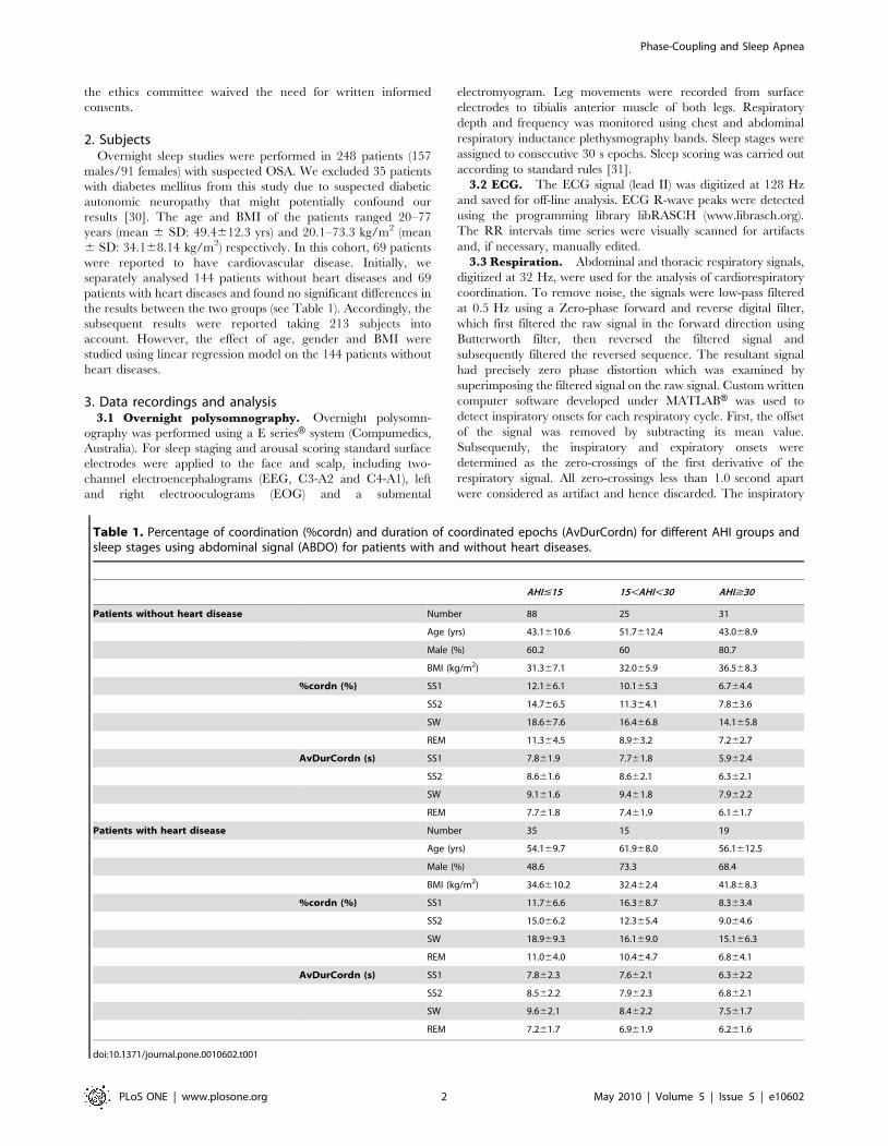
Table 1. Percentage of coordination (%cordn) and duration of coordinated epochs (AvDurCordn) for different AHI groups andsleep stages using abdominal signal (ABDO) for patients with and without heart diseases.
AHI#15 15,AHI,30 AHI$30
Patients without heart disease Number 88 25 31
Age (yrs) 43.1610.6 51.7612.4 43.068.9
Male (%) 60.2 60 80.7
BMI (kg/m2) 31.367.1 32.065.9 36.568.3
%cordn (%) SS1 12.166.1 10.165.3 6.764.4
SS2 14.766.5 11.364.1 7.863.6
SW 18.667.6 16.466.8 14.165.8
REM 11.364.5 8.963.2 7.262.7
AvDurCordn (s) SS1 7.861.9 7.761.8 5.962.4
SS2 8.661.6 8.662.1 6.362.1
SW 9.161.6 9.461.8 7.962.2
REM 7.761.8 7.461.9 6.161.7
Patients with heart disease Number 35 15 19
Age (yrs) 54.169.7 61.968.0 56.1612.5
Male (%) 48.6 73.3 68.4
BMI (kg/m2) 34.6610.2 32.462.4 41.868.3
%cordn (%) SS1 11.766.6 16.368.7 8.363.4
SS2 15.066.2 12.365.4 9.064.6
SW 18.969.3 16.169.0 15.166.3
REM 11.064.0 10.464.7 6.864.1
AvDurCordn (s) SS1 7.862.3 7.662.1 6.362.2
SS2 8.562.2 7.962.3 6.862.1
SW 9.662.1 8.462.2 7.561.7
REM 7.261.7 6.961.9 6.261.6
doi:10.1371/journal.pone.0010602.t001
Phase-Coupling and Sleep Apnea
PLoS ONE | www.plosone.org 2 May 2010 | Volume 5 | Issue 5 | e10602
onsets of respiration were later used to calculate the average
respiratory time period.
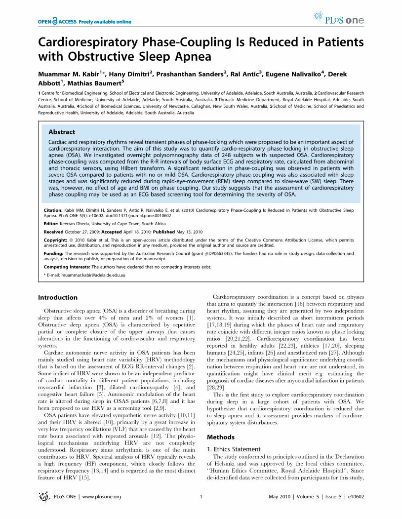
3.4 Cardiorespiratory coordination analysis. We used
Hilbert transform to calculate the phases of the respiratory signal,
and determined relationship between the respiratory phases at
different R-peak instants. If we denote the phase of heartbeat as Wc
and of respiratory signal as Wr and considering that the heart
makes m heartbeats in n respiratory cycles, then phase
coordination is defined as the locking of the corresponding
phases given by
DmWc{nWrDƒi,
where i is a constant.
In other words, if the phase difference between the two oscillators
was within a certain threshold value, i and remained stable for n
respiratory cycles, the oscillators were considered coordinated. If tk is
the time of the appearance of a kth R-peak, then by observing the
phase of the respiration at the instants tk, denoted by Wr(tk) and
wrapping the respiratory phase into a [0, 2pm] interval, we can
generate cardiorespiratory synchrogram. This provides a visual tool
to detect cardiorespiratory coordination (Figure 1, second panel), by
plotting Yn against tk which, in case of m:n coordination, results in m
horizontal lines. Here Yn is given by the equation
Yn~1
2pwr tkð Þmod 2pnð Þ:
In order to determine the values of m and n, we selected one
value of n at a time and looked for coordination at different values
of m. The study was carried for the following m:n coordination:
n = 1: m = 2,…,8; n = 2: m = 5,7,9,11,13 and n = 3: m =
7,8,10,11,13,14,16,17,19,20. We used a threshold value of
i = 0.025 for the phase difference as suggested by Cysarz et al. [24].
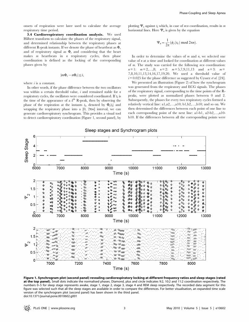
We presented an illustration (Figure 2) of how the synchrogram
was generated from the respiratory and ECG signals. The phases
of the respiratory signal, corresponding to the time points of the R-
peaks, were plotted as normalized phases between 0 and 2.
Subsequently, the phases for every two respiratory cycles formed a
relatively vertical line: a1,a2,…,a10; b1,b2,…,b10; and so on. We
then determined the differences between each point of one line to
each corresponding point of the next line: a1-b1, a2-b2,…,a10-
b10. If the differences between all the corresponding points were
Figure 1. Synchrogram plot (second panel) revealing cardiorespiratory locking at different frequency ratios and sleep stages (ratedat the top panel). Small dots indicate the normalised phases. Diamond, plus and circle indicates 9:2, 10:2 and 11:2 coordination respectively. Thenumbers 0–5 for sleep stage represents awake, stage 1, stage 2, stage 3, stage 4 and REM sleep respectively. The recorded data segment for thisfigure was selected such that all the sleep stages are available in order to compare the differences. For better visualisation, an expanded time scaleversion of the synchrogram plot (second panel) has been shown in the third panel.doi:10.1371/journal.pone.0010602.g001
Phase-Coupling and Sleep Apnea
PLoS ONE | www.plosone.org 3 May 2010 | Volume 5 | Issue 5 | e10602
below the threshold value of 0.025, the respective R-peaks were
considered as coordinated. In Figure 2 (third panel), if lines were
drawn between the points a1:b1, a2:b2,…,a10:b10, a structure of
parallel horizontal lines, as termed by Cysarz et al. [24], would be
observed. Similarly, from figure 2 (first panel), it could be observed
that every two respiratory cycles consisted of ten equidistant R-
peaks, indicating a phase locking ratio of 10:2 (or 5:1).
We employed two parameters for characterising cardiorespiratory
coordination: Firstly, we calculated the percentage of coordination,
%cordn, by adding up the time of all coordinated epochs observed in
a particular segment or sleep stage and then dividing it by the total
duration of the segments or sleep stage. Consequently, we computed
%cordnTotal and %cordnSleepStage. Secondly, we measured the average
duration of all coordinated epochs, AvDurCordn, for every sleep stage
by calculating the arithmetic mean of the durations of coordinated
epochs. Further, the phase locking ratio for each coordinated epoch
was recorded and the total number of a particular locking ratio for
every sleep stage was calculated (see Table 2 and 3) to obtain a
histogram of different locking ratios.
3.5 Surrogate data analysis. In order to determine whether
randomness of heart rate variations play any role with respect to
cardiorespiratory coordination in OSA patients, we used surrogate
data for our analysis. The surrogate data were obtained by
randomizing the order of RR intervals separately for every sleep
stage and constructing the new R time series by starting with the
first original R time and cumulating the randomized RR intervals.
Accordingly, the phases of the respiratory signal corresponding to
the new time points of the R-peaks were calculated and analysed
for cardiorespiratory coordination.
3.6 Statistical analysis. Statistics computer software SPSS,
Inc. version 15.0 and GraphPad Prism, Inc. version 5.0 were used
to analyse the data. To determine the effects of age, gender, and
BMI on cardiorespiratory coordination, linear regression models
were developed for the 144 patients without heart disease. To
determine the effect of OSA severity on cardiorespiratory
coordination, the cohort was trichotomized based on the apnea-
hypopnea index (AHI): AHI#15, 15,AHI,30, and AHI$30.
Differences in cardiorespiratory coordination between the three
OSA groups as well as between different sleep stages were assessed
with 2-way ANOVA for repeated measurements. For post-hoc
analysis, Tukey’s multiple comparison test was used. Values
p,0.05 were considered statistically significant.
Results
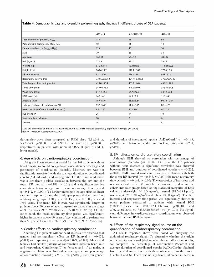
1. Polysomnographic findingsDemographic and polysomnographic data are presented in
Table 4. Of the 213 patients, 133 (70 m) presented with no or mild
Figure 2. Illustration of the generation of synchrogram from the respiratory and ECG signals. a1, a2,…,a10 and b1, b2,….,b10 in thesynchrogram plot represent the respiratory phases, based on the time points of R-peaks, for the first two and the following two respiratory cycles.doi:10.1371/journal.pone.0010602.g002
Phase-Coupling and Sleep Apnea
PLoS ONE | www.plosone.org 4 May 2010 | Volume 5 | Issue 5 | e10602
OSA (AHI#15), 40 (26 m) patients with mild to moderate OSA
(15,AHI,30) and 50 patients (38 m) with severe OSA
(AHI$30). Neither age nor BMI were significantly different
between the three subgroups.
Sleep time, wake time and percentage of REM sleep were not
significant between the three OSA subgroups (p.0.5). The
number of arousals per hour was significantly higher in patients
with severe OSA (50.7619.6 #/h) compared to patients with
moderate (25.368.6 #/h) or no/mild (16.468.4 #/h, p,0.0001)
OSA,
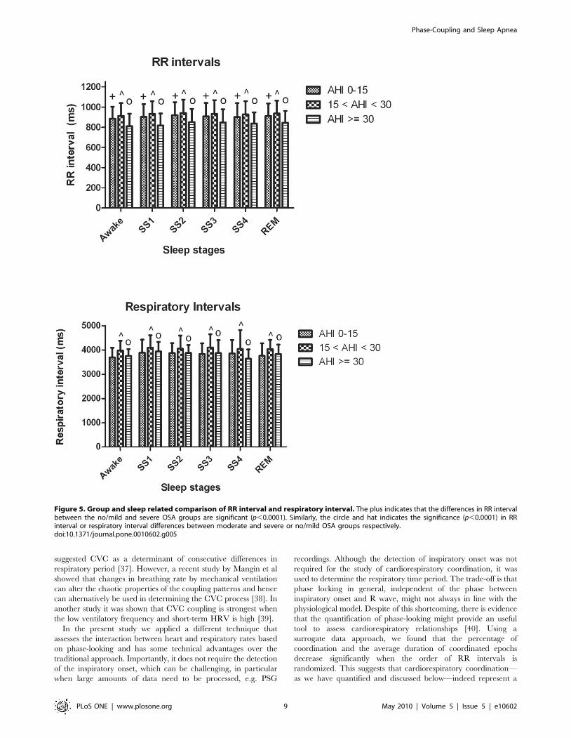
Heart rate and respiratory rate were significantly elevated in
patients with severe OSA compared to patients with no/mild OSA
(Table 4).
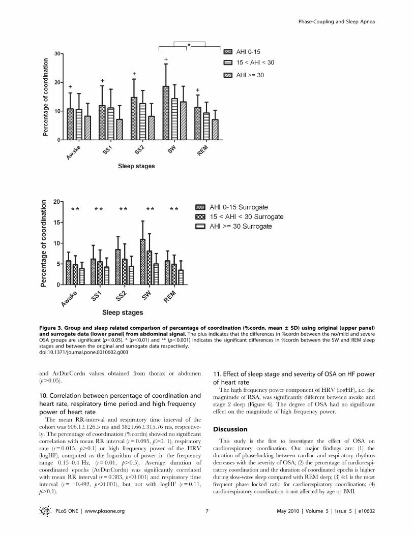
Cardiorespiratory coordination was significantly associated with
the degree of severity of OSA. Patients with severe OSA had a
significantly lower percentage of heart rate - respiratory rate
phase-locking states compared to patients with no or mild OSA
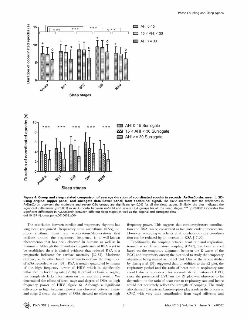
(Figure 3, upper panel). The average duration of coordinated
epochs was significantly decreased in patients with severe OSA
compared to patients with moderate or no/mild OSA (Figure 4,
upper panel).
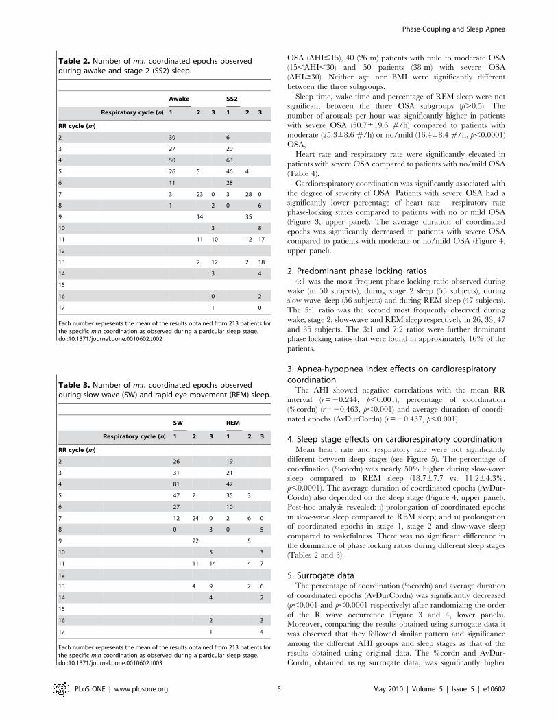
2. Predominant phase locking ratios4:1 was the most frequent phase locking ratio observed during
wake (in 50 subjects), during stage 2 sleep (55 subjects), during
slow-wave sleep (56 subjects) and during REM sleep (47 subjects).
The 5:1 ratio was the second most frequently observed during
wake, stage 2, slow-wave and REM sleep respectively in 26, 33, 47
and 35 subjects. The 3:1 and 7:2 ratios were further dominant
phase locking ratios that were found in approximately 16% of the
patients.
3. Apnea-hypopnea index effects on cardiorespiratorycoordination
The AHI showed negative correlations with the mean RR
interval (r = 20.244, p,0.001), percentage of coordination
(%cordn) (r = 20.463, p,0.001) and average duration of coordi-
nated epochs (AvDurCordn) (r = 20.437, p,0.001).
4. Sleep stage effects on cardiorespiratory coordinationMean heart rate and respiratory rate were not significantly
different between sleep stages (see Figure 5). The percentage of
coordination (%cordn) was nearly 50% higher during slow-wave
sleep compared to REM sleep (18.767.7 vs. 11.264.3%,
p,0.0001). The average duration of coordinated epochs (AvDur-
Cordn) also depended on the sleep stage (Figure 4, upper panel).
Post-hoc analysis revealed: i) prolongation of coordinated epochs
in slow-wave sleep compared to REM sleep; and ii) prolongation
of coordinated epochs in stage 1, stage 2 and slow-wave sleep
compared to wakefulness. There was no significant difference in
the dominance of phase locking ratios during different sleep stages
(Tables 2 and 3).
5. Surrogate dataThe percentage of coordination (%cordn) and average duration
of coordinated epochs (AvDurCordn) was significantly decreased
(p,0.001 and p,0.0001 respectively) after randomizing the order
of the R wave occurrence (Figure 3 and 4, lower panels).
Moreover, comparing the results obtained using surrogate data it
was observed that they followed similar pattern and significance
among the different AHI groups and sleep stages as that of the
results obtained using original data. The %cordn and AvDur-
Cordn, obtained using surrogate data, was significantly higher
Table 2. Number of m:n coordinated epochs observedduring awake and stage 2 (SS2) sleep.
Awake SS2
Respiratory cycle (n) 1 2 3 1 2 3
RR cycle (m)
2 30 6
3 27 29
4 50 63
5 26 5 46 4
6 11 28
7 3 23 0 3 28 0
8 1 2 0 6
9 14 35
10 3 8
11 11 10 12 17
12
13 2 12 2 18
14 3 4
15
16 0 2
17 1 0
Each number represents the mean of the results obtained from 213 patients forthe specific m:n coordination as observed during a particular sleep stage.doi:10.1371/journal.pone.0010602.t002
Table 3. Number of m:n coordinated epochs observedduring slow-wave (SW) and rapid-eye-movement (REM) sleep.
SW REM
Respiratory cycle (n) 1 2 3 1 2 3
RR cycle (m)
2 26 19
3 31 21
4 81 47
5 47 7 35 3
6 27 10
7 12 24 0 2 6 0
8 0 3 0 5
9 22 5
10 5 3
11 11 14 4 7
12
13 4 9 2 6
14 4 2
15
16 2 3
17 1 4
Each number represents the mean of the results obtained from 213 patients forthe specific m:n coordination as observed during a particular sleep stage.doi:10.1371/journal.pone.0010602.t003
Phase-Coupling and Sleep Apnea
PLoS ONE | www.plosone.org 5 May 2010 | Volume 5 | Issue 5 | e10602
during slow-wave sleep compared to REM sleep (9.963.9 vs.
5.762.4%, p,0.0001 and 5.961.9 vs. 4.461.8 s, p,0.0001
respectively, in patients with no/mild OSA) (Figure 3 and 4,
lower panels).
6. Age effects on cardiorespiratory coordinationUsing the linear regression model for the 144 patients without
heart disease, we found no significant association between age and
percentage of coordination (%cordn). Likewise, age was not
significantly associated with the average duration of coordinated
epochs (AvDurCordn) and locking ratio. On the other hand, there
was a significant positive correlation between the age and the
mean RR interval (r = 0.188, p,0.01) and a significant positive
correlation between age and mean respiratory time period
(r = 0.242, p,0.001). To further investigate the age effect on heart
rate and respiratory rate, the study group was divided into four
arbitrary subgroups: ,30 years, 30–45 years, 46–60 years and
.60 years. The mean RR interval was significantly longer in
patients above 60 years of age, compared to patients in the range
of 30–45 years (967.92688.55 vs. 877.016114.32 ms). On the
other hand, the mean respiratory time period was significantly
higher in patients above 60 years of age, compared to patients less
than 30 years of age (4051.326173.67 vs. 3579.956325.63 ms).
7. Gender effects on cardiorespiratory coordinationAnalysing 144 patients without heart diseases, we observed that
gender had no significant effect on RR interval (r = 20.072,
p.0.1) or respiratory time period(r = 0.029, p.0.1). Males and
females had similar patterns of coordination between heart rate
and respiration. Considering ‘0’ as females and ‘1’ as males, a
negative correlation was observed between gender and percentage
of coordination (%cordn) (r = 20.180, p,0.01), between gender
and duration of coordinated epochs (AvDurCordn) (r = 20.149,
p,0.05) and between gender and locking ratio (r = 20.204,
p,0.01).
8. BMI effects on cardiorespiratory coordinationAlthough BMI showed no correlation with percentage of
coordination (%cordn) (r = 20.007, p.0.1) in the 144 patients
without heart diseases, a significant correlation was observed
between BMI and duration of coordinated epochs (r = 20.262,
p,0.01). BMI showed significant negative correlations with both
the mean RR interval (r = 20.343, p,0.001) the mean respiratory
time period(r = 20.144, p,0.05). The association of heart rate and
respiratory rate with BMI was further assessed by dividing the
cohort into four groups based on the statistical categories of BMI
values: underweight (,18.5 kg/m2), normal (18.5–25 kg/m2),
overweight (25.1–30 kg/m2) and obese (.30 kg/m2). The RR
interval and respiratory time period was significantly shorter in
obese patients compared to patients with normal BMI
(938.396135.75 vs. 881.616111.65 ms, p,0.001 and
3997.346296.03 vs. 3888.536355.231 ms, p,0.01). No signifi-
cant difference in cardiorespiratory coordination was observed
between the four BMI categories.
9. Effects of the respiratory signal source on thequantification of cardiorespiratory coordination
All results reported above were based on analysing the
abdominal respiratory signal. To determine whether the origin
of the respiratory signal (abdomen vs. thorax) affects our results,
we compared the percentage of coordination (%cordn) and
average duration of coordinated epochs (AvDurCordn) obtained
from the abdominal trace with those obtained from the thorax
(Tables 5 and 6). There was no significant difference in %cordn
Table 4. Demographic data and overnight polysomnography findings in different groups of OSA patients.
AHI#15 15,AHI,30 AHI$30
Total number of patients, NTotal 133 51 64
Patients with diabetes mellitus, NDM 10 11 14
Patients analysed, N (NTotal2NDM) 123 40 50
Male 70 26 38
Age (yrs) 46611 56612 48612
BMI (kg/m2) 3268 3265 3969
Weight (kg) 91.2621.4 93.4619.6 111.2623.6
Height (cm) 168.669.2 170.2610.2 170.668.3
RR interval (ms) 9116120 9566131 8456123
Respiratory interval (ms) 3797.56335.5 3947.46315.8 3703.76434.2
Total length of recording (min) 428.8653.4 431.1664.6 438.2627.1
Sleep time (min) 346.0655.4 346.9660.6 352.8664.8
Wake time (min) 61.1642.4 64.7642.3 74.1656.8
REM sleep (%) 14.764.1 14.663.8 12.364.5
Arousals (#/h) 16.468.4* 25.368.6* 50.7619.6*
Total percentage of coordination (%) 13.566.2* 11.665.1* 8.864.5*
Mean duration of coordinated epochs (s) 8.361.8* 8.162.0* 6.562.1*
Hypertension 26 14 18
Structural heart disease 18 8 6
Smoker 14 2 8
Data are presented as mean 6 standard deviation. Astericks indicate statistically significant changes (p,0.001).doi:10.1371/journal.pone.0010602.t004
Phase-Coupling and Sleep Apnea
PLoS ONE | www.plosone.org 6 May 2010 | Volume 5 | Issue 5 | e10602
and AvDurCordn values obtained from thorax or abdomen
(p.0.05).
10. Correlation between percentage of coordination andheart rate, respiratory time period and high frequencypower of heart rate
The mean RR-interval and respiratory time interval of the
cohort was 906.16126.5 ms and 3821.666315.76 ms, respective-
ly. The percentage of coordination (%cordn) showed no significant
correlation with mean RR interval (r = 0.095, p.0. 1), respiratory
rate (r = 0.015, p.0.1) or high frequency power of the HRV
(logHF), computed as the logarithm of power in the frequency
range 0.15–0.4 Hz, (r = 0.01, p.0.5). Average duration of
coordinated epochs (AvDurCordn) was significantly correlated
with mean RR interval (r = 0.383, p,0.001) and respiratory time
interval (r = 20.492, p,0.001), but not with logHF (r = 0.11,
p.0.1).
11. Effect of sleep stage and severity of OSA on HF powerof heart rate
The high frequency power component of HRV (logHF), i.e. the
magnitude of RSA, was significantly different between awake and
stage 2 sleep (Figure 6). The degree of OSA had no significant
effect on the magnitude of high frequency power.
Discussion
This study is the first to investigate the effect of OSA on
cardiorespiratory coordination. Our major findings are: (1) the
duration of phase-locking between cardiac and respiratory rhythms
decreases with the severity of OSA; (2) the percentage of cardiorespi-
ratory coordination and the duration of coordinated epochs is higher
during slow-wave sleep compared with REM sleep; (3) 4:1 is the most
frequent phase locked ratio for cardiorespiratory coordination; (4)
cardiorespiratory coordination is not affected by age or BMI.
Figure 3. Group and sleep related comparison of percentage of coordination (%cordn, mean ± SD) using original (upper panel)and surrogate data (lower panel) from abdominal signal. The plus indicates that the differences in %cordn between the no/mild and severeOSA groups are significant (p,0.05). * (p,0.01) and ** (p,0.001) indicates the significant differences in %cordn between the SW and REM sleepstages and between the original and surrogate data respectively.doi:10.1371/journal.pone.0010602.g003
Phase-Coupling and Sleep Apnea
PLoS ONE | www.plosone.org 7 May 2010 | Volume 5 | Issue 5 | e10602
The association between cardiac and respiratory rhythms has
long been recognized. Respiratory sinus arrhythmia (RSA), i.e.
subtle rhythmic heart rate accelerations/decelerations that
oscillate around the respiratory frequency is a well-known
phenomenon that has been observed in humans as well as in
mammals. Although the physiological significance of RSA is yet to
be established there is clinical evidence that reduced RSA is a
prognostic indicator for cardiac mortality [32,33]. Moderate
exercise, on the other hand, has shown to increase the magnitude
of RSA recorded at rest [34]. RSA is usually quantified by means
of the high frequency power of HRV which is significantly
influenced by breathing rate [35,36]. It provides a basic surrogate,
but completely lacks information on the respiratory system. We
determined the effects of sleep stage and degree of OSA on high
frequency power of HRV (figure 6). Although a significant
difference in high frequency power was observed between awake
and stage 2 sleep, the degree of OSA showed no effect on high
frequency power. This suggests that cardiorespiratory coordina-
tion and RSA can be considered as two independent phenomena.
However, according to Schafer et al, cardiorespiratory coordina-
tion can be reduced by an increase in RSA [17,20].
Traditionally, the coupling between heart rate and respiration,
termed as cardioventilatory coupling (CVC), has been studied
based on the temporary alignment between the R waves of the
ECG and inspiratory onsets; the plot used to study the temporary
alignment being named as the RI plot. One of the recent studies
by Tzeng et al. [37] suggested that, in addition to the RI plot, the
respiratory period and the ratio of heart rate to respiratory rate
should also be considered for accurate determination of CVC,
since the presence of CVC on the RI plot was observed to be
dependent on the ratio of heart rate to respiratory rate and hence
would not accurately reflect the strength of coupling. The study
also showed that arterial baroreceptors play a role in the process of
CVC with very little contribution from vagal afferents and
Figure 4. Group and sleep related comparison of average duration of coordinated epochs in seconds (AvDurCordn, mean ± SD)using original (upper panel) and surrogate data (lower panel) from abdominal signal. The circle indicates that the differences inAvDurCordn between the moderate and severe OSA groups are significant (p,0.01) for all the sleep stages. Similarly, the plus indicates thesignificant differences (p,0.001) in AvDurCordn between no/mild and severe OSA groups for all the sleep stages. *** (p,0.0001) indicates thesignificant differences in AvDurCordn between different sleep stages as well as the original and surrogate data.doi:10.1371/journal.pone.0010602.g004
Phase-Coupling and Sleep Apnea
PLoS ONE | www.plosone.org 8 May 2010 | Volume 5 | Issue 5 | e10602
suggested CVC as a determinant of consecutive differences in
respiratory period [37]. However, a recent study by Mangin et al
showed that changes in breathing rate by mechanical ventilation
can alter the chaotic properties of the coupling patterns and hence
can alternatively be used in determining the CVC process [38]. In
another study it was shown that CVC coupling is strongest when
the low ventilatory frequency and short-term HRV is high [39].
In the present study we applied a different technique that
assesses the interaction between heart and respiratory rates based
on phase-looking and has some technical advantages over the
traditional approach. Importantly, it does not require the detection
of the inspiratory onset, which can be challenging, in particular
when large amounts of data need to be processed, e.g. PSG
recordings. Although the detection of inspiratory onset was not
required for the study of cardiorespiratory coordination, it was
used to determine the respiratory time period. The trade-off is that
phase locking in general, independent of the phase between
inspiratory onset and R wave, might not always in line with the
physiological model. Despite of this shortcoming, there is evidence
that the quantification of phase-looking might provide an useful
tool to assess cardiorespiratory relationships [40]. Using a
surrogate data approach, we found that the percentage of
coordination and the average duration of coordinated epochs
decrease significantly when the order of RR intervals is
randomized. This suggests that cardiorespiratory coordination—
as we have quantified and discussed below—indeed represent a
Figure 5. Group and sleep related comparison of RR interval and respiratory interval. The plus indicates that the differences in RR intervalbetween the no/mild and severe OSA groups are significant (p,0.0001). Similarly, the circle and hat indicates the significance (p,0.0001) in RRinterval or respiratory interval differences between moderate and severe or no/mild OSA groups respectively.doi:10.1371/journal.pone.0010602.g005
Phase-Coupling and Sleep Apnea
PLoS ONE | www.plosone.org 9 May 2010 | Volume 5 | Issue 5 | e10602
deterministic feature of heart rate and respiration and is not a
random observation. However, it also suggests that randomized
RR intervals can show some coordination; up to 10% of the whole
sleep duration, depending on the variability of the original RR
interval series and probably also on the regularity of respiration.
According to a recent study by Kenwright et al [41], the possibility
of having an interaction between cardiac and respiratory
oscillators is reduced by an increase in variation of respiratory
frequency. It was shown that cardiorespiratory coordination was
significantly decreased during exercise due to an increase in
cardiac and respiratory frequencies as a result of greater demand
for nutrients and oxygen from the blood by various body organs
[41]. From our results it appears that heart rate and/or respiration
are more regular during slow-wave sleep compared to REM sleep,
since cardiorespiratory coordination is higher in slow-wave sleep
compared to REM sleep, even when the order of RR intervals is
random.
In our study we found that phase locking between heart and
respiratory rhythms lasts for time intervals of on average
8.3 seconds in subjects with no/mild OSA. Although these
phase-locked periods are rather short they account for up to
20% of the whole sleep duration. In accordance with a previous
study [25], we found a profound sleep stage effect on
cardiorespiratory coordination. Phase-locking occurs more often
and for longer periods in slow-wave sleep compared to REM sleep.
This might be the effect of less regular breathing patterns in REM
sleep paralleled by sympathetic cardiac activation, making both
rhythms more erratic and therefore phase-locking less likely. In
line with that RSA has shown to increases during SW sleep [42].
REM sleep, characterized by irregular and high-frequency waves
in the electroencephalogram is associated with a lack of synchrony
between neuron firing rates, possibly due to desynchronized
nervous activity [43,44]. It has been suggested by Hamann et al
[45] that cardiorespiratory coordination can be observed as long
as the noise from higher brain regions affects the cardiac and
respiratory oscillators is uncorrelated. However, during REM
sleep, when the higher brain regions are more active, long-term
correlated noise might be imposed on the two oscillators and
thereby cause a reduction in the cardiorespiratory coordination
[45]. It seems probable that the decrease in the amount of phase-
locking between the cardiac and respiratory signals during REM
sleep is caused by the desynchronized activity of the nervous
system. Some of the earlier studies have also found changes in
heart rate with the change in sleep stages [46,47,48]. However, in
this study, the heart rate and respiratory rate showed no significant
change with sleep stages. As almost one-third of our study group
consisted of patients with heart disease their medication might
partly explain the different heart rate behaviour.
In our study cardiorespiratory coordination was neither affected
by age nor by BMI. These results are consistent with an earlier
study by Bartsch et al [25]. However, in a recent study by Shiogai
et al [49], it was reported that although cardiorespiratory
coordination showed no correlation with age for males, it
significantly increased with age for females. A different approach
for detection of phases, Hilbert transform, used in this study,
compared to marked events method used for cardiac phase
detection by Shiogai et al [49], might be a possible cause of the
variation in results. Similarly, the low sampling frequency and
filtering approach to the respiratory signal used in this study could
also be the contributing factors for the differences. According to
another study, autonomic modulation of heart rate diminishes
with age [50]. Interestingly, we did not observe a significant effect
of age on cardiorespiratory coordination, suggesting that there is
little association between HRV and the phase-locking between
heart and respiratory rhythms. Also, in our study, a positive
correlation between age and RR interval was unexpected as heart
rate (reciprocal of RR interval) was reported to increase with age
[47,48].
The BMI has an effect on OSA and is also associated with an
increased cardiac sympathetic tone [51,52]. Patients with higher
BMI are more likely to be affected by OSA. However, in our
study, although OSA had an effect on the phase locking between
heart rate and respiration, BMI showed no association with
cardiorespiratory coordination. On the other hand, the respiratory
time period had a negative correlation with BMI.
Autonomic modulation of heart rate is altered in OSA patients
[10]. Our results indicate a significant increase in heart rate and
respiratory rate in patients with moderate and severe OSA
compared to patients with mild or no OSA. This increase in heart
rate might be a result of a decrease in cardiac vagal outflow, of an
increase in cardiac sympathetic outflow or both [53]. On the other
hand cardiorespiratory coordination decreased significantly with
the severity of OSA. This suggests that the amount of phase
locking is influenced by autonomic control, which is affected by
the repetitive obstructive episodes in OSA patients. Measures of
cardiorespiratory coordination seem to be more sensitive to OSA
than conventional HRV metrics alone, since they incorporate
both, respiratory and cardiac domains.
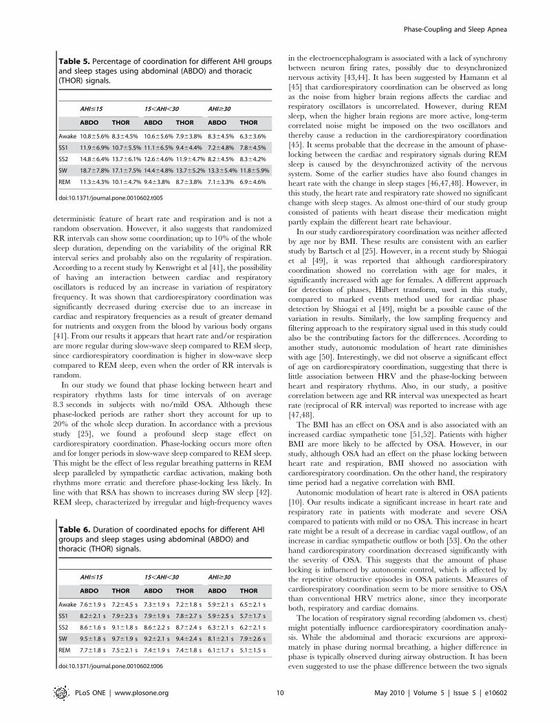
The location of respiratory signal recording (abdomen vs. chest)
might potentially influence cardiorespiratory coordination analy-
sis. While the abdominal and thoracic excursions are approxi-
mately in phase during normal breathing, a higher difference in
phase is typically observed during airway obstruction. It has been
even suggested to use the phase difference between the two signals
Table 5. Percentage of coordination for different AHI groupsand sleep stages using abdominal (ABDO) and thoracic(THOR) signals.
AHI#15 15,AHI,30 AHI$30
ABDO THOR ABDO THOR ABDO THOR
Awake 10.865.6% 8.364.5% 10.665.6% 7.963.8% 8.364.5% 6.363.6%
SS1 11.966.9% 10.765.5% 11.166.5% 9.464.4% 7.264.8% 7.864.5%
SS2 14.866.4% 13.766.1% 12.664.6% 11.964.7% 8.264.5% 8.364.2%
SW 18.767.8% 17.167.5% 14.464.8% 13.765.2% 13.365.4% 11.865.9%
REM 11.364.3% 10.164.7% 9.463.8% 8.763.8% 7.163.3% 6.964.6%
doi:10.1371/journal.pone.0010602.t005
Table 6. Duration of coordinated epochs for different AHIgroups and sleep stages using abdominal (ABDO) andthoracic (THOR) signals.
AHI#15 15,AHI,30 AHI$30
ABDO THOR ABDO THOR ABDO THOR
Awake 7.661.9 s 7.264.5 s 7.361.9 s 7.261.8 s 5.962.1 s 6.562.1 s
SS1 8.262.1 s 7.962.3 s 7.961.9 s 7.862.7 s 5.962.5 s 5.761.7 s
SS2 8.661.6 s 9.161.8 s 8.662.2 s 8.762.4 s 6.362.1 s 6.262.1 s
SW 9.561.8 s 9.761.9 s 9.262.1 s 9.462.4 s 8.162.1 s 7.962.6 s
REM 7.761.8 s 7.562.1 s 7.461.9 s 7.461.8 s 6.161.7 s 5.161.5 s
doi:10.1371/journal.pone.0010602.t006
Phase-Coupling and Sleep Apnea
PLoS ONE | www.plosone.org 10 May 2010 | Volume 5 | Issue 5 | e10602
to detect sleep apnea [54]. For the purpose of this study we
primarily used the respiratory signal obtained from the abdominal
transducer. However, our analyses indicate that respiratory signals
derived from the thorax can be equally used. There was no
significant difference in the mean percentage of coordination for
different AHI groups and sleep stages. Further, the sampling
frequency has an influence on the accuracy of measurement of R-
peaks, with a significant decrease in the amplitude of R-peak
observed at a sampling rate of 125 Hz, compared to 500 Hz, but
with no significant influence on the R-R interval [55]. The ECG
sampling frequency for this study was relatively low (128 Hz).
This, together with the technique used for quantifying synchro-
nization and the short intervals of synchronized epochs considered
as signature for cardiorespiratory coordination, could have
influenced our results.
In conclusion, OSA perturbs the phase locking between cardiac
and respiratory rhythms. The assessment of cardiorespiratory
coordination may thus provide an ECG based screening tool for
OSA.
Author Contributions
Conceived and designed the experiments: MMK. Performed the
experiments: MMK. Analyzed the data: MMK. Contributed reagents/
materials/analysis tools: MMK HD PS RA EN DA MB. Wrote the paper:
MMK. Assisted in writing the paper: EN DA MB. Assisted in study design
and data analysis: MB.
References
1. Young T, Palta M, Dempsey J, Skatrud J, Weber S, et al. (1993) The occurrence
of sleep-disordered breathing among middle-aged adults. N Engl J Med 328:
1230–1235.
2. Roche F, Gaspoz JM, Court-Fortune I, Minini P, Pichot V, et al. (1999)
Screening of obstructive sleep apnea syndrome by heart rate variability analysis.
Circulation 100: 1411–1415.
3. Tsuji H, Larson MG, Venditti FJ, Jr., Manders ES, Evans JC, et al. (1996)
Impact of reduced heart rate variability on risk for cardiac events. The
Framingham Heart Study. Circulation 94: 2850–2855.
4. Hoffmann J, Grimm W, Menz V, Knop U, Maisch B (1996) Heart rate
variability and major arrhythmic events in patients with idiopathic dilated
cardiomyopathy. Pacing Clin Electrophysiol 19: 1841–1844.
5. Sandercock GR, Brodie DA (2006) The role of heart rate variability in prognosis
for different modes of death in chronic heart failure. Pacing Clin Electrophysiol
29: 892–904.
6. Coruzzi P, Gualerzi M, Bernkopf E, Brambilla L, Brambilla V, et al. (2006)
Autonomic Cardiac Modulation in Obstructive Sleep Apnea: Effect of an Oral
Jaw-Positioning Appliance. Chest 130: 1362–1368.
7. Wiklund U, Olofsson BO, Franklin K, Blom H, Bjerle P, et al. (2000) Autonomic
cardiovascular regulation in patients with obstructive sleep apnoea: a study
based on spectral analysis of heart rate variability. Clin Physiol 20: 234–241.
8. Aydin M, Altin R, Ozeren A, Kart L, Bilge M, et al. (2004) Cardiac autonomic
activity in obstructive sleep apnea: time-dependent and spectral analysis of heart rate
variability using 24-hour Holter electrocardiograms. Tex Heart Inst J 31: 132–136.
9. Roche F, Duverney D, Court-Fortune I, Pichot V, Costes F, et al. (2002)
Cardiac interbeat interval increment for the identification of obstructive sleep
apnea. Pacing Clin Electrophysiol 25: 1192–1199.
10. Narkiewicz K, Montano N, Cogliati C, van de Borne PJH, Dyken ME, et al.
(1998) Altered Cardiovascular Variability in Obstructive Sleep Apnea.
Circulation 98: 1071–1077.
11. Somers VK, Dyken ME, Clary MP, Abboud FM (1995) Sympathetic neural
mechanisms in obstructive sleep apnea. J Clin Invest 96: 1897–1904.
12. Baumert M, Smith J, Catcheside P, McEvoy RD, Abbott D, et al. (2008)
Variability of QT interval duration in obstructive sleep apnea: an indicator of
disease severity. Sleep 31: 959–966.
13. Hayano J, Mukai S, Sakakibara M, Okada A, Takata K, et al. (1994) Effects of
respiratory interval on vagal modulation of heart rate. Am J Physiol Heart Circ
Physiol 267: H33–40.
14. Song H-S, Lehrer PM (2003) The Effects of Specific Respiratory Rates on Heart
Rate and Heart Rate Variability. Appl Psychophysiol Biofeedback 28: 13–23.
15. Berntson GG, Bigger JT, Jr., Eckberg DL, Grossman P, Kaufmann PG, et al.
(1997) Heart rate variability: origins, methods, and interpretive caveats.
Psychophysiology 34: 623–648.
16. Toledo E, Akselrod S, Pinhas I, Aravot D (2002) Does synchronization reflect a
true interaction in the cardiorespiratory system? Med Eng Phys 24: 45–52.
17. Schafer C, Rosenblum MG, Kurths J, Abel HH (1998) Heartbeat synchronized
with ventilation. Nature 392: 239–240.
18. Hoyer D, Hader O, Zwiener U (1997) Relative and intermittent cardiorespi-
ratory coordination. IEEE Eng Med Biol Mag 16: 97–104.
19. Bettermann H, Cysarz D, Van Leeuwen P (2002) Comparison of two different
approaches in the detection of intermittent cardiorespiratory coordination
during night sleep. BMC Physiol 2: 18.
20. Schafer C, Rosenblum MG, Abel HH, Kurths J (1999) Synchronization in the
human cardiorespiratory system. Phys Rev E Stat Phys Plasmas Fluids Relat
Interdiscip Topics 60: 857–870.
21. Censi F, Calcagnini G, Lino S, Seydnejad S, Kitney R, et al. (2000) Transient
phase locking patterns among respiration, heart rate and blood pressure during
cardiorespiratory synchronisation in humans. Med Biol Eng Comput 38:
416–426.
22. Lotric MB, Stefanovska A (2000) Synchronization and modulation in the human
cardiorespiratory system. Physica A 283: 451–461.
23. Kotani K, Takamasu K, Ashkenazy Y, Stanley HE, Yamamoto Y (2002) Model
for cardiorespiratory synchronization in humans. Phys Rev E Stat Nonlin Soft
Matter Phys 65: 051923.
24. Cysarz D, Bettermann H, Lange S, Geue D, van Leeuwen P (2004) A
quantitative comparison of different methods to detect cardiorespiratory
coordination during night-time sleep. Biomed Eng Online 3: 44.
25. Bartsch R, Kantelhardt JW, Penzel T, Havlin S (2007) Experimental Evidence
for Phase Synchronization Transitions in the Human Cardiorespiratory System.
Phys Rev Lett 98: 054102–054104.
Figure 6. Group and sleep related comparisons of log transformed high frequency power (logHF) (mean ± SD). (* represents p,0.05).doi:10.1371/journal.pone.0010602.g006
Phase-Coupling and Sleep Apnea
PLoS ONE | www.plosone.org 11 May 2010 | Volume 5 | Issue 5 | e10602
26. Mrowka R, Patzak A, Rosenblum M (2000) Quantitative analysis of
cardiorespiratory synchronization in infants. Int J Bifurc Chaos 10: 2479–2488.27. Stefanovska A, Haken H, McClintock PV, Hozic M, Bajrovic F, et al. (2000)
Reversible transitions between synchronization states of the cardiorespiratory
system. Phys Rev Lett 85: 4831–4834.28. Hoyer D, Leder U, Hoyer H, Pompe B, Sommer M, et al. (2002) Mutual
information and phase dependencies: measures of reduced nonlinear cardiore-spiratory interactions after myocardial infarction. Med Eng Phys 24: 33–43.
29. Leder U, Hoyer D, Sommer M, Baier V, Haueisen J, et al. (2000)
Cardiorespiratory desynchronization after acute myocardial infarct. Z Kardiol89: 630–637.
30. Javorka M, Trunkvalterova Z, Tonhajzerova I, Javorkova J, Javorka K, et al.(2008) Short-term heart rate complexity is reduced in patients with type 1
diabetes mellitus. Clin Neurophysiol 119: 1071–1081.31. Rechtschaffen A, Kales A (1968) A manual of standarized terminology,
techniques and scoring system for sleep stages in human subjects. Los Angeles:
Brain Information Service/Brain Research Institute.32. Casolo GC, Stroder P, Signorini C, Calzolari F, Zucchini M, et al. (1992) Heart
rate variability during the acute phase of myocardial infarction. Circulation 85:2073–2079.
33. Moser M, Lehofer M, Sedminek A, Lux M, Zapotoczky H, et al. (1994) Heart
rate variability as a prognostic tool in cardiology. A contribution to the problemfrom a theoretical point of view. Circulation 90: 1078–1082.
34. Meersman RE (1992) Respiratory sinus arrhythmia alteration following trainingin endurance athletes. Eur J Appl Physiol 64: 434–436.
35. Hirsch JA, Bishop B (1981) Respiratory sinus arrhythmia in humans: howbreathing pattern modulates heart rate. Am J Physiol Heart Circ Physiol 241:
H620–629.
36. Penttila J, Helminen A, Jartti T, Kuusela T, Huikuri HV, et al. (2001) Timedomain, geometrical and frequency domain analysis of cardiac vagal outflow:
effects of various respiratory patterns. Clin Physiol 21: 365–376.37. Tzeng YC, Larsen PD, Galletly DC (2007) Mechanism of cardioventilatory
coupling: insights from cardiac pacing, vagotomy, and sinoaortic denervation in
the anesthetized rat. Am J Physiol Heart Circ Physiol 292: H1967–1977.38. Mangin L, Clerici C, Similowski T, Poon C-S (2009) Chaotic dynamics of
cardioventilatory coupling in humans: effects of ventilatory modes. Am J PhysiolRegul Integr Comp Physiol 296: R1088–1097.
39. Tzeng Y, Larsen P, Galletly D (2003) Cardioventilatory coupling in restinghuman subjects. Exp Physiol 88: 775–782.
40. Pereda E, Cruz DMDl, Vera LD, Gonzalez JJ (2005) Comparing generalized
and phase synchronization in cardiovascular and cardiorespiratory signals. IEEETrans Biomed Eng 52: 578–583.
41. Kenwright D, Bahraminasab A, Stefanovska A, McClintock P (2008) The effect
of low-frequency oscillations on cardio-respiratory synchronization. Eur
Phys J B Condensed Matter and Complex Systems 65: 425–433.
42. Brandenberger G, Buchheit M, Ehrhart J, Simon C, Piquard F (2005) Is slow
wave sleep an appropriate recording condition for heart rate variability analysis?
Auton Neurosci 121: 81–86.
43. Guyton AC, Hall JE (2006) States of brain activity - sleep, brain waves, epilepsy,
psychoses. In: Schmitt W, Gruliow R, eds. Textbook of medical physiology
Elsevier Saunders. pp 739–747.
44. Gottesmann C (1999) Neurophysiological support of consciousness during
waking and sleep. Prog Neurobiol 59: 469–508.
45. Hamann C, Bartsch RP, Schumann AY, Penzel T, Havlin S, et al. (2009)
Automated synchrogram analysis applied to heartbeat and reconstructed
respiration. Chaos 19: 015106–015108.
46. Snyder F, Hobson JA, Morrison DF, Goldfrank F (1964) Changes in respiration,
heart rate, and systolic blood pressure in human sleep. J Appl Physiol 19:
417–422.
47. Penzel T, Kantelhardt JW, Lo CC, Voigt K, Vogelmeier C (2003) Dynamics of
heart rate and sleep stages in normals and patients with sleep apnea.
Neuropsychopharmacology 28 Suppl 1: S48–53.
48. Nalivaiko E, Catcheside PG, Adams A, Jordan AS, Eckert DJ, et al. (2007)
Cardiac changes during arousals from non-REM sleep in healthy volunteers.
Am J Physiol Regul Integr Comp Physiol 292: R1320–1327.
49. Shiogai Y, Stefanovska A, McClintock PVE (2010) Nonlinear dynamics of
cardiovascular ageing. Physics Reports 488: 51–110.
50. Umetani K, Singer DH, McCraty R, Atkinson M (1998) Twenty-four hour time
domain heart rate variability and heart rate: relations to age and gender over
nine decades. J Am Coll Cardiol 31: 593–601.
51. Ferguson KA, Ono T, Lowe AA, Ryan CF, Fleetham JA (1995) The
Relationship Between Obesity and Craniofacial Structure in Obstructive Sleep
Apnea. Chest 108: 375–381.
52. Busetto L, Enzi G, Inelmen EM, Costa G, Negrin V, et al. (2005) Obstructive
sleep apnea syndrome in morbid obesity: effects of intragastric balloon. Chest
128: 618–623.
53. Narkiewicz K, Somers VK (2003) Sympathetic nerve activity in obstructive sleep
apnoea. Acta Physiol Scand 177: 385–390.
54. Varady P, Bongar S, Benyo Z (2003) Detection of airway obstructions and sleep
apnea by analyzing the phase relation of respiration movement signals. IEEE
Trans Instrum Meas 52: 2–6.
55. Pizzuti GP, Cifaldi S, Nolfe G (1985) Digital sampling rate and ECG analysis.
J Biomed Eng 7: 247–250.
Phase-Coupling and Sleep Apnea
PLoS ONE | www.plosone.org 12 May 2010 | Volume 5 | Issue 5 | e10602