obstructive sleep apnea–hypopnea and neurocognitive functioning in the sleep heart health study
TRANSCRIPT
Original article
Obstructive sleep apnea–hypopnea and neurocognitive functioning
in the Sleep Heart Health Study
Stuart F. Quan a,*, Ron Wright b, Carol M. Baldwin c, Kristine L. Kaemingk d,James L. Goodwin e, Tracy F. Kuo f, Alfred Kaszniak g, Lori L. Boland h,
Elise Caccappolo i, Richard R. Bootzin j
a Department of Medicine, Arizona Respiratory, Sleep Disorders and General Clinical Research Centers,
University of Arizona College of Medicine, 1501 North Campbell, Room 2305 Tucson, AZ 85724, USAb Department of Psychiatry and General Clinical Research Center, University of Arizona College of Medicine, Tucson, AZ, USA
c College of Nursing (Southwest Borderlands), Arizona State University, Tempe, AZ, USAd Department of Pediatrics, Steele Memorial Children’s Research and General Clinical Research Centers,
University of Arizona College of Medicine, Tucson, AZ, USAe Department of Medicine and Arizona Respiratory Center, University of Arizona College of Medicine, Tucson, AZ, USA
f Department of Psychiatry and Behavioral Sciences, Stanford University, Stanford, CA, USAg Department of Psychology, University of Arizona, Tucson, AZ, USA
h Division of Epidemiology, School of Public Health, University of Minnesota, Minneapolis, MN, USAi Department of Neurology, Columbia University, New York, NY, USA
j Departments of Psychology and Psychiatry, Sleep Disorders Center, University of Arizona, Tucson, AZ, USA
Received 19 October 2005; received in revised form 8 February 2006; accepted 12 February 2006
Available online 3 July 2006
Abstract
Background and purpose: Obstructive sleep apnea–hypopnea (OSAH) is associated with sleep fragmentation and nocturnal hypoxemia. In
clinical samples, patients with OSAH frequently are found to have deficits in neuropsychological function. However, the nature and severity
of these abnormalities in non-clinical populations is less well defined.
Patients and methods: One hundred and forty-one participants from the Tucson, AZ and New York, NY field centers of the Sleep Heart
Health Study completed a battery of neuropsychological tests for 9–40 months (meanZ24 months, SDZ7 months) after an unattended home
polysomnogram. Sixty-seven participants had OSAH (AHIO10) and 74 did not have OSAH (control (CTL), apnea–hypopnea index
(AHI)!5). In addition to the individual tests, composite variables representing attention, executive function, MotorSpeed and processing
speed were constructed from the neuropsychological test battery.
Results: There were no significant differences in any individual neuropsychological test or composite variable between the OSAH and CTL
groups. However, when time spent with O2 saturations less than 85% was dichotomized into those participants in the top quartile of the
distribution and those in the lower three quartiles, motor speed was significantly impaired in those who were more hypoxemic. In addition,
poorer motor speed (model adjusted R2Z0.242, P!0.001) and processing speed performance (model adjusted R2Z0.122, P!0.001) were
associated with more severe oxygen desaturation even after controlling for degree of daytime sleepiness, age, gender and educational level.
Conclusions: Mild to moderate OSAH has little impact on the selected measures of attention, executive function, motor speed and processing
speed. However, hypoxemia adversely affects both motor and processing speed. These results suggest that in middle-aged to elderly adults
the neuropsychological effects of clinically unrecognized mild to moderate OSAH are neither global nor large.
q 2006 Elsevier B.V. All rights reserved.
Keywords: Sleep disordered breathing; Obstructive sleep apnea; Neuropsychologic function; Hypoxemia; Neurocognitive function; epidomiology
Sleep Medicine 7 (2006) 498–507
www.elsevier.com/locate/sleep
1389-9457/$ - see front matter q 2006 Elsevier B.V. All rights reserved.
doi:10.1016/j.sleep.2006.02.005
* Corresponding author. Tel.: C1 520 626 6115; fax: C1 520 626 6970.
E-mail addresses: [email protected] (S.F. Quan), [email protected] (R. Wright), [email protected] (C.M. Baldwin), kaemingk@peds.
arizona.edu (K.L. Kaemingk), [email protected] (J.L. Goodwin), [email protected] (T.F. Kuo), [email protected] (A. Kaszniak),
[email protected] (L.L. Boland), [email protected] (E. Caccappolo), [email protected] (R.R. Bootzin).

S.F. Quan et al. / Sleep Medicine 7 (2006) 498–507 499
1. Background
Obstructive sleep apnea–hypopnea (OSAH) is now
recognized as a major public health problem due to its
high prevalence, increased morbidity and mortality, high
medical costs and increased public safety risk [1–3]. The
impact of OSAH is potentially profound and wide-ranging
with a number of studies noting adverse effects on physical,
emotional, and intellectual capacities [4–7], as well as
functional quality of life [8,9]. Some of the most serious
symptoms that have been associated with OSAH include
attention deficits, impaired concentration, and memory
problems [10].
A number of studies have found patients diagnosed with
OSAH to have poorer cognitive outcomes [11–16]. For
example, Montplaisir et al. reported executive and
psychomotor deficits, as well as attention and memory
problems associated with impaired vigilance [14]. Green-
berg et al. reported deficits on measures of motor and
perceptual-organization ability [12]. Memory deficits have
been related to the number of apneas and hypopneas
per hour of sleep, while typical frontal lobe-related
abnormalities appeared consistent with the level of
nocturnal hypoxemia [15,16].
Several studies suggest that cognitive deficits persist
despite standard interventions for OSAH. Valencia-Flores
et al. examined OSAH patients on their neuropsychological
test performance prior to and immediately after successful
treatment with continuous positive airway pressure (CPAP)
[17]. Analyses showed that the effects of severe OSAH
appear to impair functions that were not easily modified by
treatment with continuous positive airway pressure (CPAP),
such as sustained attention in repetitive arithmetic tasks. In
their re-evaluation study of attention, short-term memory
span, learning abilities, planning capacities, categorizing
activities, and verbal fluency 4–6 months following CPAP
treatment, Naegele et al. noted that patients had normalized
their cognitive executive and learning problems; however,
short-term memory impairment persisted [16]. More
recently, a meta-analytic review of the neuropsychological
effects of OSAH indicated that vigilance, motor coordi-
nation and executive functions ranged from moderately to
markedly affected, while intelligence, verbal and visual-
perceptual abilities were not affected in adults with
untreated OSAH [18].
Reports of cognitive impairment have not been limited
only to patients with severe OSAH. Redline et al. found that
in comparison to controls, individuals with mild sleep apnea
performed more poorly on a visual vigilance task and the
digits backward test of working memory [19]. Notably, Kim
et al. found that mild OSAH was equivalent to a reduction in
psychomotor efficiency associated with five additional years
of age, or to half of the decrement related to use of
hypnosedatives [20].
Although, there is evidence to suggest that cognitive
deficits occur in both clinical and non-clinical samples with
varying levels of OSAH severity, neuropsychological
findings differ in type and/or degree as well as their
association with the number of OSAH events and/or
severity of hypoxemia. These inconsistencies could be due
to the sensitivity of tests used to assess neuropsychological
function, sample size, and/or criteria used to assess the
severity of OSAH [21]. Whether the results of the preceding
studies are generalizable to untreated community-based
persons with mild to severe OSAH remains unclear. The
intent of the present study is to characterize the effect of
mild to severe OSAH on neuropsychological functioning in
a subset of persons from the Sleep Heart Health Study
(SHHS), a community-dwelling cohort recruited to inves-
tigate the cardiovascular consequences of OSAH. It is
hypothesized that persons with OSAH will manifest
neuropsychological deficits compared to persons who do
not have OSAH, and the deficits will be proportional with
the severity of the OSAH.
2. Methods
2.1. Recruitment and participants
Participants were recruited from the Tucson, AZ and
New York, NY sites of the SHHS. The SHHS is a multi-
center prospective cohort study implemented by the
National Heart, Lung, and Blood Institute to investigate
the cardiovascular consequences of OSAH. The specific
aims, design and participant characteristics of the SHHS
have been previously reported [22,23]. They are also
available at www.jhucct.com/shhs. In brief, SHHS subjects
were recruited from ongoing parent cohort studies of
cardiovascular or respiratory disease. Inclusion criteria
were age 40 years or older, no history of treatment of
sleep apnea with CPAP, no tracheostomy and no current
home oxygen therapy.
The demographic characteristics of participants at the
Tucson and New York sites have been reported [24]. All
prospective participants had to be between the ages of 40
and 75 years. Because final scoring of an unattended
polysomnogram (PSG) was not available for months after
the study had been completed, potential index cases (OSAH
group) were identified based on preliminary scoring of the
PSG. Preliminary scoring of the PSG defined the apnea
hypopnea index (AHI) as the number of apneas or
hypopneas associated with a 3% oxygen desaturation per
hour of total sleep time (AHI3%). Initial assignment to the
OSAH group was based on participants having an AHI3%
between 10 and 50. However, after final PSG scoring, only
participants with a sufficient number of apneas or hypopneas
associated with a 4% oxygen desaturation per hour of total
sleep time (AHI4%) were included in the OSAH group (vide
infra). A total of 97 women and 245 men were identified as
potential index-case subjects who met OSAH selection
criterion. Potential control participants were persons who
S.F. Quan et al. / Sleep Medicine 7 (2006) 498–507500
had an AHI3%!5, with a total of 559 women and 380 men
identified as possible control cases (CTL group).
To control for important confounding conditions that
may contribute to neuropsychological impairments, poten-
tial participants were excluded if they were currently being
treated for cancer and/or in remission for !5 years, on
antipsychotic or anticonvulsant medication or being treated
for OSAH, had a history of alcohol-related problems that
interfered with work and/or personal life, myocardial
infarction within the last 3 years, stroke, head injuries, or
surgical treatment for OSAH. Potential participants were
recruited by telephone. In Tucson, recruitment also included
a mailed SHHS newsletter announcement.
A brief phone screen was conducted to determine
whether or not a prospective subject met selection criteria
after he/she expressed interest in participating. In order to
maximize the potential of finding group differences, an
effort was made to enroll cases with the highest AHIs first.
Recruitment then moved downward toward the lower end of
the AHI cutoff. Recruitment of the CTL subjects was
staggered and on average lagged behind the OSAH subjects
by 4 months. A total of 144 persons (107:46 women and 61
men from Tucson; and 37; 17 women and 20 men from New
York) were enrolled. As previously discussed, recruitment
was based on a preliminary determination of the AHI
(AHI3%). Thus, one Tucson participant and two New York
participants ultimately were excluded because their final
AHI (AHI4%) was between 5 and 10, which did not meet
selection criteria. Therefore, the final sample consisted of 67
OSAH (53% male) and 74 CTL participants (63% male).
Non-Hispanic Whites represented 86.5% of the study
population. The remaining participants were primarily of
African-American (7.8%) or Hispanic (5.0%) descent.
2.2. Polysomnography
As part of the SHHS protocol, all participants underwent
unattended PSG at home prior to the neuropsychological
testing. The average time lag between the PSG and the
neuropsychological testing was 24 months (SDZ7, rangeZ9–40 months). A questionnaire on sleep habits, requesting
information on snoring history, sleep apnea symptoms,
diagnosis and treatment, and daytime sleepiness assessed by
the Epworth sleepiness scale (ESS) was administered prior
to the PSG [25]. Detailed descriptions of the home visit and
the methods used for unattended PSG have been previously
published [22,23]. In summary, overnight PSG was
performed using the Compumedics Portable PS-2 System
(Abottsville, AU) [23]. Sensors were placed and equipment
was calibrated during an evening visit by a certified
technician. Data collection included C3/A2 and C4/A1
electroencephalograms (EEGs); right and left electrooculo-
grams (EOGs); a bipolar submental electromyogram
(EMG); thoracic and abdominal excursions (inductive
plethysmography bands); ‘airflow’ (detected by a nasal-
oral thermocouple (Protec, Woodinville, WA)); oximetry
using finger pulse oximetry (Nonin, Minneapolis, MN);
electrocardiogram (ECG) and heart rate (using a bipolar
ECG lead); body position (using a mercury gauge sensor);
and ambient light (on/off, by a light sensor secured to the
recording garment). Following equipment retrieval, the
data, stored in real time on Personal Computer Memory
Card International Association (PCMCIA) cards, were
downloaded to the computers at each respective clinical
site, locally reviewed, and forwarded to the central SHHS
Reading Center (Case Western Reserve University, Cleve-
land, OH). Sleep stages were scored according to the
guidelines developed by Rechtschaffen and Kales [26].
Stages 3 and 4 were combined and classified as ‘slow wave
sleep’. Arousals were identified according to American
Sleep Disorders Association criteria [27]. An apnea was
defined as a complete or almost complete cessation of
airflow (at least !25% of baseline), as measured by the
amplitude of the thermocouple signal, lasting O10 s.
Hypopneas were identified if the amplitude of a measure
of flow or volume (detected by the thermocouple or thorax
or abdominal inductance band signals) decreased to !70%
of the amplitude of ‘baseline’ breathing for O10 s, but did
not meet the criteria for apnea. For this study, only apneas or
hypopneas associated with R4% oxyhemoglobin desatura-
tion were considered in the calculation of the AHI (AHI4%).
In addition to the AHI4%, the number of arousals per hour
(arousal index, AI) was used as a measure of sleep
disruption. The percentage of sleep time in which oxygen
saturation was !85% (O2SAT85) was used as an index of
hypoxemia. Variables representing sleep architecture
included total sleep time (TST), sleep efficiency, and
percent of stages 1, 2, 3/4, and REM sleep.
2.3. Neuropsychological assessment
After successful recruitment, participants were sched-
uled for a neuropsychological screening conducted by
trained psychometricians that employed commonly used
measures dependent on attention, processing speed, execu-
tive function and motor speed. Measures included the
Wechsler Adult Intelligence Test-Third Edition (WAIS-III)
Picture Completion, Digit Span, Letter-Number Sequen-
cing, Digit Symbol Coding, and Symbol Search subtests
[28]; the Stroop Color and Word Test [29]; the Trail Making
Test [30]; and the Grooved Pegboard [31].
Picture Completion, Digit Span, and Letter-Number
Sequencing are commonly used measures of attention
[28,32,33]. They require the ability to identify missing
elements of pictures, repetition of numbers (forward and
reverse order), and recall letters and numbers in order
(numerical, alphabetical), respectively. Digit Symbol
Coding and Symbol Search are measures of processing
speed that assess rapid copying of symbols and scanning
speed and accuracy, respectively [28,32]. Both measures
are timed and require a motor response. These Wechsler
measures have a mean of 10 and SD of 3. Executive function

S.F. Quan et al. / Sleep Medicine 7 (2006) 498–507 501
was measured using the Interference T-score (meanZ50,
SDZ10) from the Stroop [29]. This score is an indicator of
response inhibition and a commonly used measure of
executive function [32,33]. The time to complete Trail
Making Part B (in seconds) was also used to assess
executive function [30]. This measure of executive skills
requires rapid sequencing and alternating attention [32,33].
Motor speed was assessed with Grooved Pegboard [31], a
task that requires rapid placement of pegs in holes with the
dominant hand and then the non-dominant hand. Com-
pletion time with each hand was used as the dependent
measure.
2.4. Data analysis
In order, to focus on specific components of neuropsycho-
logical function, we created composite variables for (a)
attention (Attention), (b) processing speed (Procspeed), (c)
executive function (Execfunct) and (d) motor speed (Motor-
Speed). The composite variable for the Attention domain
combined data from the Picture Completion, Digit Span, and
Letter-Number Sequencing tests. Procspeed combined Digit
Symbol Coding and Symbol Search. Execfunct was a
composite of the Stroop Interference T-score and the time to
complete the Trail Making B test. Motorspeed was assessed by
summing the scores on the Grooved Pegboard. In order to give
every component variable equal weight in the composite, each
component was first standardized and these Z-scores were
averaged to create the composite variables. Within a given
composite, component variables were positively correlated,
and composites retained the original direction for the
construct. Thus, high scores indicated better performance for
the Attention and Procspeed composites, while low scores
indicated better performance for the ExecFunct and Motor-
Speed composites.
Although, the overall AHI4% distribution here was
bimodal (hence, non-normal) by study design, in past research,
AHI4% has generally not been normally distributed, and the
usual practice is to apply a logarithmic transformation prior to
Table 1
Participant characteristics
CTL, NZ74: 39 men 35 women
Mean SD
Demographics
Age (years) 57.4 9.2
Education (years) 14.9 2.7
Ethnicity (% non-hispanic white) 85.1
Body mass index (kg/m2) 25.9 4.3
Married (%) 62.4
Right handed (%) 67.6
Habits
Usual caffeine intake 3.7 4.4
Current smoker (%) 5.4
Daytime sleepiness
Epworth sleepiness scale 7.0 4.3
analysis. We have followed that procedure as well
(LogAHI4%). For cases in which AHI4% was 0, the value
0.07 was used. This represents a trivial number that is 50% of
the lowest non-zero value of AHI4%. In addition, the
difference in the median AHI4% between the OSAH and
CTL groups was assessed utilizing the Mann–WhitneyU-test.
Student’s unpaired t-test was employed to compare mean
differences between the OSAH and CTL groups. The c2
statistic was used to compare proportions between nominal
variables.
The relationships between the four composite variables
of neuropsychological function (Attention, Procspeed, Exec-
funct and Motorspeed), and potential explanatory and
demographic variables were explored using simple and
multiple linear regression. For each composite variable,
performance was assessed in the following models: (1) AHI
group (OSAH vs. CTL) alone, and in combination with the
covariates age, educational level and gender because each
potentially could affect neuropsychological performance; (2)
LogAHI4% alone, and in combination with age, educational
level and gender; (3) O2SAT85 alone, and in combination
with age, educational level and gender; (4) ESS alone and in
combination with age, educational level and gender; (5) ESS
with either LogAHI4% or O2SAT85, and age, educational
level and gender; and (6) each of models 1 and 3–5 within
both AHI groups individually. In preliminary analyses, AI
was found to predict worse Motorspeed performance. Thus,
AI was included along with age, educational level and
gender in prediction models for only this composite variable.
No other sleep architecture or demographic variables were
related to neuropsychological performance on exploratory
analyses. Models including both LogAHI4% and O2SAT85
were not constructed because there were no instances where
both variables in a simple regression predicted performance
on any of the composite variables.
Analyses were performed using Systat (Version 10,
Systat Software, Inc., Richmond, CA) and SPSS for
Windows, Version 11.5, SPSS, Inc., Chicago, IL).
OSAH, NZ67: 42 men 22 women P value
Mean SD
59.4 9.2 n.s.
15.2 2.6 n.s.
88.1 n.s.
30.8 6.2 !001
73.8 n.s.
76.1 n.s.
2.8 2.8 n.s.
9.0 n.s.
9.1 4.9 .007

S.F. Quan et al. / Sleep Medicine 7 (2006) 498–507502
3. Results
Table 1 shows the demographic and social characteristics
of the CTL and OSAH groups. Persons with OSAH had
higher ESS scores and body mass index (BMI). Otherwise,
there were no significant differences between groups.
Data pertaining to sleep and respiration for the CTL and
OSAH groups are displayed in Table 2. Except for a slightly
greater percentage of stage 1 sleep in persons with OSAH,
no differences were observed between the two groups in
sleep architecture. However, OSAH participants had a
greater number of arousals. By definition, the AHI4% was
higher in the OSAH group and as might be expected, greater
amounts of oxygen desaturation (higher O2SAT85) were
observed in the OSAH group as well.
The mean actual and Z-scores for individual neuropsy-
chological tests are displayed in Table 3. There were no
significant differences between the OSAH and CTL groups.
Table 4 shows the mean Z-scores for the composite
variables Motorspeed, Execfunct, Procspeed and Attention
for the OSAH and CTL groups as well as for a comparison
of the upper quartile of O2SAT85 against the lower three
quartiles. Although, no differences were observed between
AHI groups in any of the composite variables, Motorspeed
performance was significantly impaired in those with
greater amounts of oxygen desaturation.
Table 5 shows selected multiple regression models
illustrating the important relationships between the four
composite neuropsychological domains and age, edu-
cational level, gender, AHI Group, LogAHI4% and
O2SAT85 for all participants. With respect to covariates,
increasing age was observed to be associated with worse
performance on tests of Motorspeed and Execfunct and
slightly better function in Procspeed. Educational level was
a significant factor only for Attention and for the effect of
LogAHI4% on Procspeed. Men scored slightly worse in
Motorspeed and better in Procspeed. More notably,
severity of nocturnal oxygen desaturation was found to
be predictive of worse performance in the domains of
motor speed and Procspeed. Furthermore, regression
Table 2
Sleep architecture and sleep disordered breathing
CTL, NZ74: 39 men 35 women
Mean SD
Sleep architecture
Total sleep time (min) 352 67
Sleep efficiency (%) 82 9
Stage 1 (%) 4.7 3.5
Stage 2 (%) 58.4 10.7
Stages 3 and 4 (%) 16.5 11.5
Stage REM (%) 20.4 5.9
Arousal index 16.2 8.6
OSAH measures
Median AHI 0.7 –
LogAHI4 % K0.6 1.1
O2SAT!85% (% of TST) 0.0002 0.002
analyses within each AHI Group (CTL and OSAH)
confirmed that severity of oxygen desaturation within
OSAH participants was responsible for the adverse impact
on Motorspeed (bZ0.081G0.034, PZ0.02) and Procspeed
(bZ0.080G0.038, PZ0.04) in the models using all
participants. In order to assess the contribution of oxygen
desaturation in explaining the variance in the models of
Motorspeed and Procspeed, the unadjusted coefficient of
variation (R2) was calculated for these models including
only the covariates age, education and gender. Comparison
was then made with the R2 of models with oxygen
desaturation included. For Motorspeed, adding O2SAT85
to the regression model increases R2 from 0.234 to 0.264
(PZ0.02). For Procspeed, adding O2SAT85 increases R2
from 0.113 to 0.148 (PZ0.02). Thus, although small in
absolute terms, oxygen desaturation accounts for 12.8 and
31.0% of the explained variance in our models. Sleepiness
was found to be predictive of poorer performance only for
Procspeed (bZK0.044G0.014; PZ0.004). In contrast,
neither the AHI Group nor LogAHI4% was found to be
predictive of performance in any of the four domains of
neuropsychological function. In addition, AI was unrelated
to Motorspeed performance in multivariate analyses (bZ0.001G0.007, PZn.s.).
4. Discussion
This study investigated the effects of OSAH on
performance of commonly used measures of attention,
executive function, motor speed and processing speed in a
sample of adults from the community-based Sleep Heart
Health Study. Excessive daytime sleepiness was more
severe in persons with OSAH and affected processing
speed function. However, the presence of mild to
moderate OSAH was not associated with impaired
performance on any of the measures of neuropsychologi-
cal function used in this study. In contrast, severity of
hypoxemia adversely impacted both processing speed and
motor speed performance.
OSAH, NZ67: 42 men 22 women P value
Mean SD
356 76 n.s.
81 12 n.s.
6.5 6.1 !05
60.5 9.4 n.s.
14.6 10.3 n.s.
18.5 6.6 n.s.
24.8 10.6 !001
22.4 – 0.001
3.1 0.4 0.001
1.5 2.8 0.001
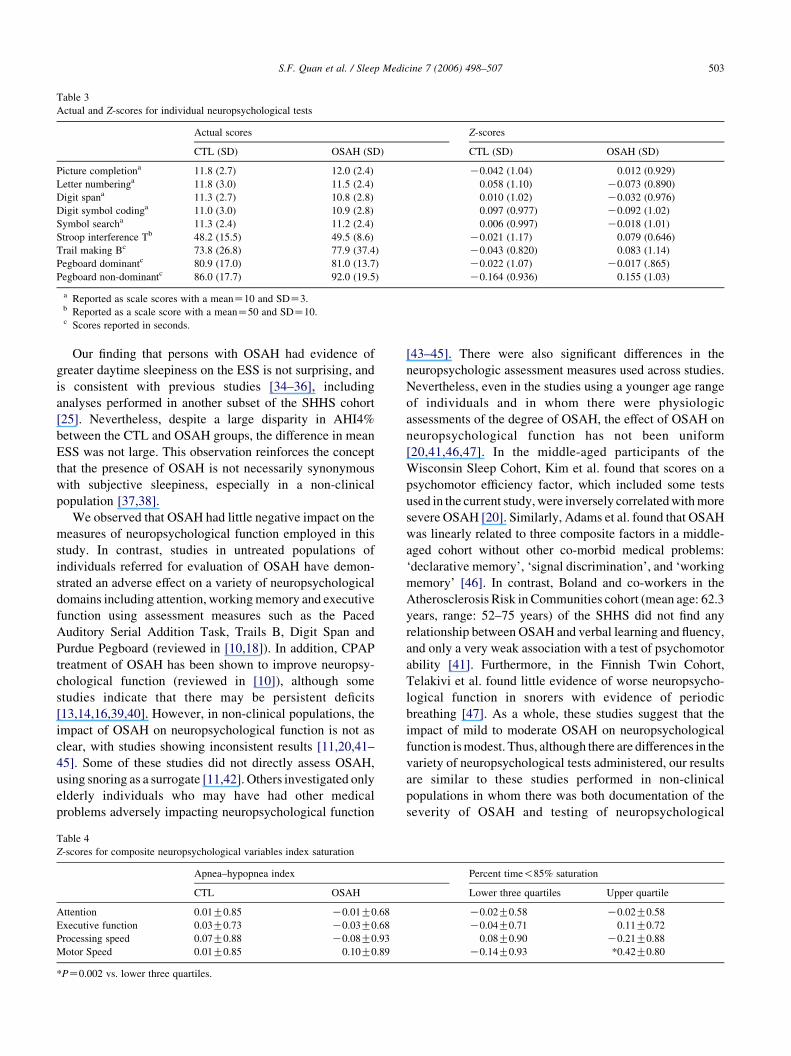
Table 3
Actual and Z-scores for individual neuropsychological tests
Actual scores Z-scores
CTL (SD) OSAH (SD) CTL (SD) OSAH (SD)
Picture completiona 11.8 (2.7) 12.0 (2.4) K0.042 (1.04) 0.012 (0.929)
Letter numberinga 11.8 (3.0) 11.5 (2.4) 0.058 (1.10) K0.073 (0.890)
Digit spana 11.3 (2.7) 10.8 (2.8) 0.010 (1.02) K0.032 (0.976)
Digit symbol codinga 11.0 (3.0) 10.9 (2.8) 0.097 (0.977) K0.092 (1.02)
Symbol searcha 11.3 (2.4) 11.2 (2.4) 0.006 (0.997) K0.018 (1.01)
Stroop interference Tb 48.2 (15.5) 49.5 (8.6) K0.021 (1.17) 0.079 (0.646)
Trail making Bc 73.8 (26.8) 77.9 (37.4) K0.043 (0.820) 0.083 (1.14)
Pegboard dominantc 80.9 (17.0) 81.0 (13.7) K0.022 (1.07) K0.017 (.865)
Pegboard non-dominantc 86.0 (17.7) 92.0 (19.5) K0.164 (0.936) 0.155 (1.03)
a Reported as scale scores with a meanZ10 and SDZ3.b Reported as a scale score with a meanZ50 and SDZ10.c Scores reported in seconds.
S.F. Quan et al. / Sleep Medicine 7 (2006) 498–507 503
Our finding that persons with OSAH had evidence of
greater daytime sleepiness on the ESS is not surprising, and
is consistent with previous studies [34–36], including
analyses performed in another subset of the SHHS cohort
[25]. Nevertheless, despite a large disparity in AHI4%
between the CTL and OSAH groups, the difference in mean
ESS was not large. This observation reinforces the concept
that the presence of OSAH is not necessarily synonymous
with subjective sleepiness, especially in a non-clinical
population [37,38].
We observed that OSAH had little negative impact on the
measures of neuropsychological function employed in this
study. In contrast, studies in untreated populations of
individuals referred for evaluation of OSAH have demon-
strated an adverse effect on a variety of neuropsychological
domains including attention, working memory and executive
function using assessment measures such as the Paced
Auditory Serial Addition Task, Trails B, Digit Span and
Purdue Pegboard (reviewed in [10,18]). In addition, CPAP
treatment of OSAH has been shown to improve neuropsy-
chological function (reviewed in [10]), although some
studies indicate that there may be persistent deficits
[13,14,16,39,40]. However, in non-clinical populations, the
impact of OSAH on neuropsychological function is not as
clear, with studies showing inconsistent results [11,20,41–
45]. Some of these studies did not directly assess OSAH,
using snoring as a surrogate [11,42]. Others investigated only
elderly individuals who may have had other medical
problems adversely impacting neuropsychological function
Table 4
Z-scores for composite neuropsychological variables index saturation
Apnea–hypopnea index
CTL OSAH
Attention 0.01G0.85 K0.01G0.68
Executive function 0.03G0.73 K0.03G0.68
Processing speed 0.07G0.88 K0.08G0.93
Motor Speed 0.01G0.85 0.10G0.89
*PZ0.002 vs. lower three quartiles.
[43–45]. There were also significant differences in the
neuropsychologic assessment measures used across studies.
Nevertheless, even in the studies using a younger age range
of individuals and in whom there were physiologic
assessments of the degree of OSAH, the effect of OSAH on
neuropsychological function has not been uniform
[20,41,46,47]. In the middle-aged participants of the
Wisconsin Sleep Cohort, Kim et al. found that scores on a
psychomotor efficiency factor, which included some tests
used in the current study, were inversely correlated with more
severe OSAH [20]. Similarly, Adams et al. found that OSAH
was linearly related to three composite factors in a middle-
aged cohort without other co-morbid medical problems:
‘declarative memory’, ‘signal discrimination’, and ‘working
memory’ [46]. In contrast, Boland and co-workers in the
Atherosclerosis Risk in Communities cohort (mean age: 62.3
years, range: 52–75 years) of the SHHS did not find any
relationship between OSAH and verbal learning and fluency,
and only a very weak association with a test of psychomotor
ability [41]. Furthermore, in the Finnish Twin Cohort,
Telakivi et al. found little evidence of worse neuropsycho-
logical function in snorers with evidence of periodic
breathing [47]. As a whole, these studies suggest that the
impact of mild to moderate OSAH on neuropsychological
function is modest. Thus, although there are differences in the
variety of neuropsychological tests administered, our results
are similar to these studies performed in non-clinical
populations in whom there was both documentation of the
severity of OSAH and testing of neuropsychological
Percent time!85% saturation
Lower three quartiles Upper quartile
K0.02G0.58 K0.02G0.58
K0.04G0.71 0.11G0.72
0.08G0.90 K0.21G0.88
K0.14G0.93 *0.42G0.80

Table 5
Selected regression models for composite
Neuropsychological outcomes b SE P
Motor speed
Effect of LogAHI4%
NZ139, adjusted R2Z0.211, P!0.001
Age 0.045 0.008 !0.001
Education K0.032 0.027 n.s.
Gender 0.298 0.245 0.043
LogAHI4% K0.002 K0.003 n.s.
Effect of O2Sat85
NZ138, adjusted R2Z0.242, P!0.001
Unadjusted R2Z0.264, full model
Unadjusted R2Z0.234; age, education, gender only
Age 0.045 0.008 !0.001
Education K0.032 0.026 n.s.
Gender 0.279 0.143 0.053
O2Sat85 0.077 0.033 0.022
Processing speed
Effect of LogAHI4%
NZ139, adjusted R2Z0.094, PZ0.002
Age 0.026 0.008 0.002
Education 0.058 0.028 0.04
Gender K0.371 0.150 0.014
LogAHI4% K0.038 0.037 n.s.
Effect of O2Sat85
NZ138, adjusted R2Z0.122, P!0.001
Unadjusted R2Z0.148, full model
Unadjusted R2Z0.113, age, education, gender only
Age 0.024 0.008 0.002
Education 0.058 0.027 n.s.
Gender K0.360 0.148 0.016
O2Sat85 K0.081 0.034 0.020
Attention
Effect of O2Sat85
NZ138, R2Z0.114, PZ0.032
Age 0.004 0.007 n.s.
Education 0.078 0.024 0.002
Gender K0.056 0.130 n.s.
O2Sat85 K0.021 0.030 n.s.
Effect of LogAHI4%
NZ139, R2Z0.046, PZ0.036
Age 0.005 0.007 n.s.
Education 0.077 0.024 0.002
Gender K0.062 0.130 n.s.
LogAHI4% K0.007 0.032 n.s.
Executive functiona
Effect of LogAHI4%
NZ130, adjusted R2Z0.047, PZ0.034
Age 0.023 0.006 !0.001
Education K0.014 0.022 n.s.
Gender 0.164 0.119 n.s.
LogAHI4% K0.051 0.029 n.s.
Effect of O2Sat85
NZ129, adjusted R2Z0.086, PZ0.004
Age 0.022 0.006 !0.001
Education K0.015 0.022 n.s.
Gender 0.154 0.120 n.s.
O2Sat85 K0.038 0.030 n.s.
a The variable Execfunct was constructed by combining scores on the Stroop Interference T (Stroop) and the Trail Making B (Trails B) tests. However,
unlike component variables for the other scales, which correlated positively with one another (C0.22 to C0.71), the components of the Execfunct scale were
uncorrelated (rZ0.001). Individually, performance on the Stroop and the Trails B was predicted by O2SAT85, but in opposite directions. LogRDI4% was
associated with better performance on the Stroop, but did not predict scores on the Trails B.
S.F. Quan et al. / Sleep Medicine 7 (2006) 498–507504

S.F. Quan et al. / Sleep Medicine 7 (2006) 498–507 505
function. Furthermore, they provide additional data extend-
ing these findings to a cohort with a broader age range.
Although, we observed little impact of OSAH by itself on
selected measures of neuropsychological function, hypo-
xemia was associated with decrements in motor and
processing speed performance. In clinical populations,
OSAH patients with hypoxemia have been found to be
more cognitively impaired than persons without hypoxemia
[12,48]. Some studies performed in non-clinical samples
have also found a relationship between severity of
hypoxemia and neuropsychological function [46,47].
There are some differences, however. Adams et al. found
that hypoxemia was associated with worse performance on a
wider spectrum of neuropsychological functioning than our
data [46]. In contrast, Telakivi et al. noted only an
impairment in verbal memory, an area we did not examine
[47]. Nevertheless, our observations are consistent with a
more limited impact of hypoxemia on neuropsychological
function. The reason for these discrepancies is not clear, but
differences in the age range of participants, the severity of
hypoxemia and the types of tests administered are possible
explanations (e.g. no visuospatial or memory tests were
used) [21].
The underlying mechanism responsible for neuropsycho-
logical deficits associated with OSAH is still undecided.
Nocturnal hypoxemia is a commonly proposed causative
factor (reviewed in [49]), and our data would support this
theory at least in those persons with mild to moderate
OSAH. Intermittent hypoxia in experimental paradigms has
been shown to result in worse neurobehavioral function in
rodents and correlates with evidence of cellular dysfunction
and apoptosis in the hippocampus [50]. Alternatively, sleep
fragmentation with resultant sleepiness and diminished
vigilance may be responsible (reviewed in [49]). Although,
we observed a slight increase in stage 1 sleep and a higher
arousal index among persons with OSAH, these factors
were not correlated with neuropsychological performance.
A number of studies have implicated sleepiness as an
important factor affecting neuropsychological function
[46,47,51]. However, our data do not completely support
this hypothesis. Although, the ESS did predict processing
speed, our results with respect to both motor and processing
speed persisted after controlling for the ESS. Perhaps, the
most parsimonious explanation is that both hypoxemia and
the consequences of sleep fragmentation are responsible for
the adverse effects of OSAH on neuropsychological
function [49], and the relative impact will depend on the
severity of OSAH, degree of hypoxemia and the affected
individual.
In our regression models, we controlled for the effects of
age, education and gender. In most models, these variables
were not significant predictors of our composite outcome
variables. However, as might be expected, increasing age
was associated with worse performance in Motorspeed and
Execfunct, and higher educational achievement predicted
better scores on Execfunct. Nevertheless, why increasing
age would result in better Procspeed performance and why
there are gender differences in Procspeed and Motorspeed is
unexplained.
We acknowledge that our study has some limitations.
First, the neuropsychological testing was done some
months after the PSG was performed. Thus, it is possible
that the severity of OSAH was greater at the time of the
neuropsychological testing, leading to some misclassifi-
cation. Although, OSAH severity can increase in some
individuals over time, this is not a universal finding
(discussed in [52]). Furthermore, in the absence of weight
change, worsening of OSAH over several years is slight
[53]. Because assignment to the control group was limited
to those with a AHI4%!5 and to the OSAH group to those
with a AHI4%O10, we believe that there was a minimal
misclassification. Moreover, we found LogAHI4%, a
continuous variable, was not predictive of neuropsycho-
logical performance. Second, although this cohort was
recruited from several large non-clinical cohorts, there is
some inherent survivor bias in the sample used in this
analysis as well as in the component cohorts of SHHS [22].
This may be particularly true in the Tucson participants
where individuals are the survivors of a 25-year longitudi-
nal cohort study. They are perhaps healthier than their
counterparts in the general population, and they have a
history of ‘volunteering’ for participation in this study. It is
also possible that individuals with better neuropsychologi-
cal functioning were more likely to agree to participate,
thus biasing the results towards the null hypothesis.
Therefore, we acknowledge that our results may not be
representative of those that might be found in a general
population sample. Third, it is difficult to compare our
results to others given that there are differences in types of
neuropsychological tests administered and because we
aggregated test results into four domains. However, except
for Execfunct, individual test components correlated with
each other suggesting that our approach was reasonable.
Fourth, in comparison to the overall SHHS cohort, the
sample size selected for testing is small and thus limits
statistical power. Irrespective of this latter limitation, we
nonetheless were able to detect a significant effect related
to hypoxemia.
Despite these limitations, however, our study has several
strengths. First, in contrast to a number of studies that have
used snoring as a surrogate for OSAH and/or have used
symptoms as an indicator of neuropsychological function, it
is one of the few studies in a non-clinical sample in which
OSAH has been directly measured and neuropsychological
testing has been utilized. Second, as a non-clinical sample,
participants were not being treated or evaluated for OSAH,
and, thus, our results may be more reflective of the
neuropsychological consequences of clinically unrecog-
nized OSAH. Third, our participants were middle-aged to
elderly persons and thus are representative of the age range
with the highest prevalence of OSAH.

S.F. Quan et al. / Sleep Medicine 7 (2006) 498–507506
5. Conclusions
We found that mild to moderate OSAH had minimal
impact on selected measures of attention, executive
function, motor speed and processing speed. However,
hypoxemia modestly affected both motor and processing
speed. These results suggest the neuropsychological effects
of clinically unrecognized mild to moderate OSAH in
middle-aged to elderly adults are neither uniform nor large,
and are primarily related to hypoxemia.
Acknowledgements
This work was supported by National Heart, Lung and
Blood Institute cooperative agreements U01HL53940
(University of Washington), U01HL53941 (Boston Univer-
sity), U01HL53938 (University of Arizona), U01HL53916
(University of California, Davis), U01HL53934 (University
of Minnesota), U01HL53931 (New York University),
U01HL53937 and U01HL64360 (Johns Hopkins Univer-
sity), U01HL63463 (Case Western Reserve University), and
U01HL63429 (Missouri Breaks Research). In addition,
statistical support for this analysis was provided by the
University of Arizona General Clinical Research Center.
Sleep Heart Health Study (SHHS) acknowledges the
Atherosclerosis Risk in Communities Study (ARIC), the
Cardiovascular Health Study (CHS), the Framingham Heart
Study (FHS), the Cornell/Mt. Sinai Worksite and Hyperten-
sion Studies, the Strong Heart Study (SHS), the Tucson
Epidemiologic Study of Airways Obstructive Diseases
(TES) and the Tucson Health and Environment Study
(H&E) for allowing their cohort members to be part of the
SHHS and for permitting data acquired by them to be used
in the study. SHHS is particularly grateful to the members of
these cohorts who agreed to participate in SHHS as well.
SHHS further recognizes all of the investigators and staff
who have contributed to its success. A list of SHHS
investigators, staff and their participating institutions is
available on the SHHS website (www.jhucct.com/shhs).
References
[1] Baumel MJ, Maislin G, Pack AI. Population and occupational
screening for obstructive sleep apnea: are we there yet? Am J Respir
Crit Care Med 1997;155:9–14.
[2] Kapur V, Blough DK, Sandblom RE. The medical cost of
undiagnosed sleep apnea. Sleep 1999;22:749–55.
[3] Kryger MH, Roos L, Delaive K, et al. Utilization of health care
services in patients with severe obstructive sleep apnea. Sleep 1996;
19:S11–S16.
[4] Aikens JE, Caruana-Montaldo B, Vanable PA, et al. MMPI correlates
of sleep and respiratory disturbance in obstructive sleep apnea. Sleep
1999;22:362–9.
[5] Bliwise DL, Yesavage JA, Sink J, et al. Depressive symptoms and
impaired respiration in sleep. J Consult Clin Psychol 1986;54:734–5.
[6] Kales A, Caldwell AB, Cadieux RJ. Severe obstructive sleep apnea-II:
associated psychopathology and psychosocial consequences. J Chron
Dis 1985;38:427–84.
[7] Pillar G, Lavie P. Psychiatric symptoms in sleep apnea syndrome:
effects of gender and respiratory disturbance index. Chest 1998;114:
697–703.
[8] Baldwin CM, Griffith KA, Nieto FJ, et al. The association of sleep-
disordered breathing and sleep symptoms with quality of life in the
Sleep Heart Health Study. Sleep 2001;24:96–105.
[9] Finn L, Young T, Palta M, Fryback DG. Sleep-disordered breathing
and self-reported general health status in the Wisconsin sleep cohort
study. Sleep 1998;21:701–6.
[10] Engleman HM, Kingshott RN, Martin SE, Douglas NJ. Cognitive
function in the sleep apnea/hypopnea syndrome (SAHS). Sleep 2000;
23:S102–S8.
[11] Dealberto MJ, Pajot N, Courbon D, Alperovitch A. Breathing
disorders during sleep and cognitive performance in an older
community sample: the EVA study. J Am Geriatr Soc 1996;44:
1287–94.
[12] Greenberg GD, Watson RK, Deptula D. Neuropsychological
dysfunction in sleep apnea. Sleep 1987;10:254–62.
[13] Kotterba S, Rasche K, Widdig W, et al. Neuropsychological
investigations and event-related potentials in obstructive sleep
apnea syndrome before and during CPAP-therapy. J Neurol Sci
1998;159:45–50.
[14] Montplaisir J, Bedard MA, Richer F, Rouleau I. Neurobehavioral
manifestations in obstructive sleep apnea syndrome before and after
treatment with continuous positive airway pressure. Sleep 1992;15:
S17–S19.
[15] Naegele B, Thouvard V, Pepin JL. Deficits of cognitive executive
functions in patients with sleep apnea syndrome. Sleep 1995;18:
43–52.
[16] Naegele B, Pepin JL, Levy P, et al. Cognitive executive dysfunction in
patients with obstructive sleep apnea syndrome (OSA) after CPAP
treatment. Sleep 1998;21:392–7.
[17] Valencia-Flores M, Bliwise DL, Guilleminault C, et al. Cognitive
function in patients with sleep apnea after acute nocturnal nasal
continuous positive airway pressure (CPAP) treatment: sleepiness and
hypoxemia effects. J Clin Exp Neuropsychol 1996;18:197–210.
[18] Beebe DW, Groesz L, Wells C, et al. The neuropsychological effects
of obstructive sleep apnea: a meta-analysis of norm-referenced and
case-controlled data. Sleep 2003;26:298–307.
[19] Redline S, Strauss ME, Adams N. Neuropsychological function in
mild sleep-disordered breathing. Sleep 1997;20:160–7.
[20] Kim HC, Young T, Matthews CG, et al. Sleep-disordered breathing
and neuropsychological deficits: a population-based study. Am
J Respir Crit Care Med 1997;156:1813–9.
[21] Weaver TE. Outcome measurement in sleep medicine practice and
research. Part 2: assessment of neurobehavioral performance and
mood. Sleep Med Rev 2001;5:223–36.
[22] Quan SF, Howard BV, Iber C, et al. The Sleep Heart Health Study:
design, rationale, and methods. Sleep 1997;20:1077–85.
[23] Redline S, Sanders MH, Lind BK, et al. Methods for obtaining and
analyzing unattended polysomnography data for a multicenter study.
Sleep 1998;21:759–67.
[24] Lind BK, Goodwin JL, Hill JG, et al. Recruitment of healthy adults
into a study of overnight sleep monitoring in the home: experience of
the sleep heart health study. Sleep Breath 2003;7:13–24.
[25] Johns MW. Reliability and factor analysis of the Epworth Sleepiness
Scale. Sleep 1992;15:376–81.
[26] Rechtschaffen A, Kales A. A manual of standardized terminology,
techniques and scoring system for sleep stages of human subjects.
Washington, DC: US Government Printing Office; 1968.
[27] American Sleep Disorders Association Taskforce. EEG arousals;
scoring rules and examples: a preliminary report from the atlas task
force of the American Sleep Disorders Association. Sleep 1992;15:
173–84.

S.F. Quan et al. / Sleep Medicine 7 (2006) 498–507 507
[28] Wechsler D. Wechsler adult intelligence scale-third edition (WAIS-
III). San Antonio: The Psychological Corporation; 1997.
[29] Golden C. Stroop color and word test: a manual for clinical and
experimental uses. Wood Dale, IL: Stoelting Company; 1978.
[30] Reitan R, Wolfson D. The Halstead-Reitan neuropsychological test
battery. Tucson, AZ: Neuropsychology Press; 1985.
[31] Lafayette I. Instruction/owner’s manual for the grooved pegboard test.
Lafayette, IN; 1989.
[32] Lezak MD. Neuropsychological assessment. 3rd ed. London: Oxford
University Press; 1995.
[33] Spreen ES. A compendium of neuropsychological tests. New York:
Oxford University Press; 1998.
[34] Gottlieb DJ, Whitney CW, Bonekat WH, et al. Relation of sleepiness
to respiratory disturbance index: the Sleep Heart Health Study. Am
J Respir Crit Care Med 1999;159:502–7.
[35] Goncalves MA, Paiva T, Ramos E, Guilleminault C. Obstructive sleep
apnea syndrome, sleepiness, and quality of life. Chest 2004;125:2091–6.
[36] Chung KF. Use of the Epworth Sleepiness Scale in Chinese patients
with obstructive sleep apnea and normal hospital employees.
J Psychosom Res 2000;49:367–72.
[37] Chervin RD, Aldrich MS. The Epworth Sleepiness Scale may not
reflect objective measures of sleepiness or sleep apnea. Neurology
1999;52:125–31.
[38] Benbadis SR, Mascha E, Perry MC, et al. Association between the
Epworth sleepiness scale and the multiple sleep latency test in a
clinical population. Ann Intern Med 1999;130:289–92.
[39] Bedard MA, Montplaisir J, Malo J, et al. Persistent neuropsycholo-
gical deficits and vigilance impairment in sleep apnea syndrome after
treatment with continuous positive airways pressure (CPAP). J Clin
Exp Neuropsychol 1993;15:330–41.
[40] Barnes M, McEvoy RD, Banks S, et al. Efficacy of positive airway
pressure and oral appliance in mild to moderate obstructive sleep
apnea. Am J Respir Crit Care Med 2004;170:656–64.
[41] Boland LL, Shahar E, Iber C, et al. Measures of cognitive function in
persons with varying degrees of sleep-disordered breathing: the Sleep
Heart Health Study. J Sleep Res 2002;11:265–72.
[42] Jennum P, Hein HO, Suadicani P, Gyntelberg F. Cognitive function
and snoring. Sleep 1993;16:S62–S4.
[43] Hayward L, Mant A, Eyland A, et al. Sleep disordered breathing and
cognitive function in a retirement village population. Age Ageing
1992;21:121–8.
[44] Berry DT, Phillips BA, Cook YR, et al. Sleep-disordered breathing in
healthy aged persons: possible daytime sequelae. J Gerontol 1987;42:
620–6.
[45] Foley DJ, Monjan AA, Masaki KH, et al. Associations of symptoms of
sleep apnea with cardiovascular disease, cognitive impairment, and
mortality among older Japanese–American men. J Am Geriatr Soc
1999;47:524–8.
[46] Adams N, Strauss M, Schluchter M, Redline S. Relation of measures
of sleep-disordered breathing to neuropsychological functioning. Am
J Respir Crit Care Med 2001;163:1626–31.
[47] Telakivi T, Kajaste S, Partinen M, et al. Cognitive function in middle-
aged snorers and controls: role of excessive daytime somnolence and
sleep-related hypoxic events. Sleep 1988;11:454–62.
[48] Findley LJ, Barth JT, Powers DC, et al. Cognitive impairment in
patients with obstructive sleep apnea and associated hypoxemia.
Chest 1986;90:686–90.
[49] Engleman HM, Douglas NJ. Sleep. 4: sleepiness, cognitive function,
and quality of life in obstructive sleep apnoea/hypopnoea syndrome.
Thorax 2004;59:618–22.
[50] Row BW, Liu R, Xu W, et al. Intermittent hypoxia is associated with
oxidative stress and spatial learning deficits in the rat. Am J Respir
Crit Care Med 2003;167:1548–53.
[51] Roehrs T, Merrion M, Pedrosi B, et al. Neuropsychological
function in obstructive sleep apnea syndrome (OSAS) compared
to chronic obstructive pulmonary disease (COPD). Sleep 1995;18:
382–8.
[52] Quan SF. Evolution of OSA. Thorax 1998;53:532.
[53] Peppard PE, Young T, Palta M, et al. Longitudinal study of moderate
weight change and sleep-disordered breathing. J Am Med Assoc
2000;284:3015–21.