brain putamen volume changes in newly-diagnosed patients with obstructive sleep apnea
TRANSCRIPT

AUTHOR QUERY FORM
Journal: YNICL Please e-mail or fax your responses and any corrections to:Young, Samuel
Article Number: 222
Dear Author,
Please check your proof carefully and mark all corrections at the appropriate place in the proof (e.g., by using on-screen annotationin the PDF file) or compile them in a separate list. Note: if you opt to annotate the file with software other than Adobe Reader thenplease also highlight the appropriate place in the PDF file. To ensure fast publication of your paper please return your correctionswithin 48 hours.
For correction or revision of any artwork, please consult http://www.elsevier.com/artworkinstructions.
Any queries or remarks that have arisen during the processing of your manuscript are listed below and highlighted by flags in theproof. Click on the ‘Q’ link to go to the location in the proof.
Location in article Query / Remark: click on the Q link to goPlease insert your reply or correction at the corresponding line in the proof
Q1 Please confirm that given names and surnames have been identified correctly.
Q2 = sign in this entry was duplicated, thus the second occurrence was deleted. Please check if thisdeletion is appropriate, and amend if necessary.
Q3 Please provide the specific name of the department for affiliation 7.
Q4 Uncited reference: This section comprises references that occur in the reference list but not in the bodyof the text. Please position each reference in the text or, alternatively, delete it. Thank you.
Please check this box if you have nocorrections to make to the PDF file. □
Thank you for your assistance.
Our reference: YNICL 222 P-authorquery-v11
Page 1 of 1

UNCO
RRECTED P
RO
OF
1 Highlights
2 NeuroImage: Clinical xxx (2014) xxx–xxx
4
5 Brain putamen volume changes in newly-diagnosed patients with obstructivesleep apnea
Rajesh Kumar a,b,e,⁎, Salar Farahvar c, Jennifer A. Ogren d, Paul M. Macey d,e, Paul M. Thompson f,g, Mary A. Woo d,Frisca L. Yan-Go f, Ronald M. Harper c,e
a Department of Anesthesiology, UCLA School of Nursing, University of California at Los Angeles, Los Angeles, CA 90095, USAb Department of Radiological Sciences, UCLA School of Nursing, University of California at Los Angeles, Los Angeles, CA 90095, USAc Department of Neurobiology, UCLA School of Nursing, University of California at Los Angeles, Los Angeles, CA 90095, USAd David Geffen School of Medicine at UCLA, UCLA School of Nursing, University of California at Los Angeles, Los Angeles, CA 90095, USAe The Brain Research Institute, University of California at Los Angeles, Los Angeles, CA 90095, USAf Department of Neurology, UCLA School of Nursing, University of California at Los Angeles, Los Angeles, CA 90095, USAg Departments of UCLA School of Nursing, University of California at Los Angeles, Los Angeles, CA 90095, USA
• Global and regional putamen volumes were examined in newly-diagnosed OSA.• Global volumes are higher, but subareas showed increases and decreases.• The volume increases suggest transient tissue swelling from hypoxic action.• Altered sites likely contribute to motor and other functional deficits in OSA.
NeuroImage: Clinical xxx (2014) xxx
YNICL-00222; No. of pages: 1; 4C:
2213-1582/$ – see front matter © 2014 The Authors. Published by Elsevier Inc. All rights reserved.http://dx.doi.org/10.1016/j.nicl.2014.01.009
Contents lists available at ScienceDirect
NeuroImage: Clinical
j ourna l homepage: www.e lsev ie r .com/ locate /yn ic l
Please cite this article as: Kumar, R., et al., Brain putamen volume changes in newly-diagnosed patients with obstructive sleep apnea,NeuroImage: Clinical (2014), http://dx.doi.org/10.1016/j.nicl.2014.01.009
1
Q1
Q3
2
34567
89101112131415
16
17
18
19
20
21
22
23
24
25
NeuroImage: Clinical xxx (2014) xxx–xxx
YNICL-00222; No. of pages: 9; 4C:
Contents lists available at ScienceDirect
NeuroImage: Clinical
j ourna l homepage: www.e lsev ie r .com/ locate /yn ic l
Brain putamen volume changes in newly-diagnosed patients withobstructive sleep apnea☆
RO
OFRajesh Kumar a,b,e,⁎, Salar Farahvar c, Jennifer A. Ogren d, PaulM.Macey d,e, PaulM. Thompson f,g, Mary A.Woo d,
Frisca L. Yan-Go f, Ronald M. Harper c,e
a Department of Anesthesiology, UCLA School of Nursing, University of California at Los Angeles, Los Angeles, CA 90095, USAb Department of Radiological Sciences, UCLA School of Nursing, University of California at Los Angeles, Los Angeles, CA 90095, USAc Department of Neurobiology, UCLA School of Nursing, University of California at Los Angeles, Los Angeles, CA 90095, USAd David Geffen School of Medicine at UCLA, UCLA School of Nursing, University of California at Los Angeles, Los Angeles, CA 90095, USAe The Brain Research Institute, University of California at Los Angeles, Los Angeles, CA 90095, USAf Department of Neurology, UCLA School of Nursing, University of California at Los Angeles, Los Angeles, CA 90095, USAg Departments of UCLA School of Nursing, University of California at Los Angeles, Los Angeles, CA 90095, USA
UNCO
Abbreviations: OSA, Obstructive sleep apnea; 3D, Thrresonance imaging; AHI, Apnea–hypopnea index; ESS, EPittsburgh Sleep Quality Index; BDI-II, Beck DepressionInventory; PD, Proton density; MNI, Montreal Neurologicfluid; TIV, Total intracranial volume; MPRAGE, Magnetizagradient-echo; TR, Repetition time; TE, Echo time; FA,GRAPPA, Generalized autocalibrating partially parallel acq☆ This is an open-access article distributed under the tAttribution-NonCommercial-No Derivative Works License,use, distribution, and reproduction in any medium, provideare credited.⁎ Corresponding author at: Departments of Anesthesio
David Geffen School of Medicine at UCLA, 56-132 CHS, 1California at Los Angeles, Los Angeles, CA 90095-1763, U310 206 6133; fax: +1 310 825 2224.
E-mail address: [email protected] (R. Kumar).
2213-1582/$ – see front matter © 2014 The Authors. Pubhttp://dx.doi.org/10.1016/j.nicl.2014.01.009
Please cite this article as: Kumar, R., et alNeuroImage: Clinical (2014), http://dx.doi.o
Pa b s t r a c t
a r t i c l e i n f o26
27
28
Article history:Received 2 August 2013Received in revised form 17 January 2014Accepted 21 January 2014Available online xxxx
Keywords:Magnetic resonance imagingCognition3D surface morphometryBasal gangliaIntermittent hypoxiaAutonomicMotor
29
30
31
32
33
RRECTEDObstructive sleep apnea (OSA) is accompanied by cognitive, motor, autonomic, learning, and affective abnormali-ties. The putamen serves several of these functions, especially motor and autonomic behaviors, but whether globaland specific sub-regions of that structure are damaged is unclear. We assessed global and regional putamen vol-umes in 43 recently-diagnosed, treatment-naive OSA (age, 46.4 ± 8.8 years; 31 male) and 61 control subjects(47.6 ± 8.8 years; 39 male) using high-resolution T1-weighted images collected with a 3.0-Tesla MRI scanner.Global putamen volumes were calculated, and group differences evaluated with independent samples t-tests, aswell as with analysis of covariance (covariates; age, gender, and total intracranial volume). Regional differencesbetween groups were visualized with 3D surface morphometry-based group ratio maps. OSA subjects showedsignificantly higher global putamen volumes, relative to controls. Regional analyses showed putamen areas withincreased and decreased tissue volumes in OSA relative to control subjects, including increases in caudal, mid-dorsal, mid-ventral portions, and ventral regions, while areas with decreased volumes appeared in rostral, mid-dorsal,medial-caudal, andmid-ventral sites. Global putamenvolumeswere significantly higher in theOSA subjects,but local sites showedboth higher and lower volumes. The appearance of localized volume alterations points to dif-ferential hypoxic or perfusion action on glia and other tissues within the structure, and may reflect a stage in pro-gression of injury in these newly-diagnosed patients toward the overall volume loss found in patients with chronicOSA. The regional changes may underlie some of the specific deficits in motor, autonomic, and neuropsychologicfunctions in OSA.
© 2014 The Authors. Published by Elsevier Inc. All rights reserved.
343537
36
43
44
45
46
47
48
49
50
51
52
53
ee dimensional; MRI, Magneticpworth Sleepiness Scale; PSQI,Inventory II; BAI, Beck Anxietyal Institute; CSF, Cerebrospinaltion prepared rapid acquisitionFlip angle; FOV, Field of view;uisition.erms of the Creative Commonswhich permits non-commerciald the original author and source
logy and Radiological Sciences,0833 Le Conte Av, University ofSA. Tel.: +1 310 206 1679, +1
lished by Elsevier Inc. All rights reser
., Brain putamen volume chrg/10.1016/j.nicl.2014.01.009
1. Introduction
Obstructive sleep apnea (OSA) patients show multiple neurobehav-ioral difficulties, including deficits in attention, psychomotor and execu-tive functioning, memory, and affect (Beebe et al., 2003). However, theprincipal deficits in the syndrome involve the loss of motor controlover the upper airway muscles, which fail to discharge in a timely fash-ion, when they should dilate the airway during diaphragmatic descent,and a loss of control over autonomic motor activity, with chronic, exag-gerated sympathetic discharge (Somers et al., 1995), and inappropriatelagged or muted dynamic sympathetic responses to blood pressure orventilatory challenges (Harper et al., 2003; Henderson et al., 2004;Macey et al., 2006). Those somatic and autonomic motor defects suggestcentral injury in motor and autonomic regulatory structures, and grossdamage has been found in multiple sites, based on manual and voxel-based morphometry of high-resolution T1-weighted images (Kumaret al., 2008; Macey et al., 2002), diffusion tensor imaging (Kumar et al.,
ved.
anges in newly-diagnosed patients with obstructive sleep apnea,

T
54
55
56
57
58
59
60
61
62
63
64
65
66
67
68
69
70
71
72
73
74
75
76
77
78
79
80
81
82
83
84
85
86
87
88
89
90
91
92
93
94
95
96
97
98
99
100
101
102
103
104
105
106
107
108
109
110
111
112
113
114
115
116
117
118
119
120
121
122
123
124
125
126
127
128
129
130
131
132
133
134
135
136
137
138
139
140
141
142
143
144
145
146
147
148
149
150
151
152
153
154
155
156
157
158
159
160
161
162
163
164
165
166
167
168
169
170
171
172
173
174
2 R. Kumar et al. / NeuroImage: Clinical xxx (2014) xxx–xxx
UNCO
RREC
2012; Macey et al., 2008), and T2-relaxometry procedures (Cross et al.,2008; Kumar et al., 2009b). Several of the affected brain sites includestructures that serve both somato motor and autonomic motor roles;one such structure is the putamen,which showsoverall injury,metabolicabnormalities, and functional deficits (Alkan et al., 2013; EminAkkoyunlu et al., 2013; Kumar et al., 2012; Macey et al., 2006).
The evidence for putamen roles in autonomic regulation is well-known, especially from pathological conditions with dysautonomia,such as Multiple Systems Atrophy (Pastakia et al., 1986). Such a role isexpected, considering themajor projections received from the autonom-ic regulatory insular cortices (Saper, 1982). The putamen somatic motorrole appears to incorporate sensory and other signals to initiate effectivemotor action, with putamen stroke inducing conditions of “motor ne-glect” (Sapir et al., 2007). OSA represents a prime example of how signalsfail to initiate and maintain upper airway muscle discharge, and theputamen may be playing a significant role in that upper airway muscle“neglect,” particularly considering its projections to brain areas regulat-ing oral motor functions (Kunzle, 1978; Miyata and Sasaki, 1984).
Theputamenalso contributes to regulation of other neuropsychologic,cognitive, and learning functions which are deficient in OSA. These func-tions includememory,mood, language, andmotivation, showdifferentialtopographic organization (Alexander and Crutcher, 1990; Badgaiyanet al., 2008; Booth et al., 2007; Bowman et al., 1996; Husain et al., 1991;Saper, 1982), and the sub-regions within that organization receive infor-mation fromprefrontal, insular, and thalamic areas, and send dopaminer-gic projections to the ventral tegmentum, substantia nigra, and globuspallidus (Avendano et al., 2006; DeVito et al., 1980; Ferry et al., 2000;Gorbachevskaya, 1997; Saper, 1982; Schultz and Romo, 1987). Putamenvolumes are reduced in depressed patients (Husain et al., 1991), andover half of OSApatients are depressed (Asghari et al., 2012); themajorityof OSA subjects also show learning deficits (Wallace and Bucks, 2013).
Regional structural changeswithin the putamen can thus alter infor-mation transfer between various brain sites. Manual volumetric proce-dures, using high-resolution T1-weighted images, can evaluate globalputamen volume (Kumar et al., 2011), and 3D surface morphometrytechniques can assess regional tissue changes, as well as extent ofchanges within the structures (Butters et al., 2009; Sabattoli et al.,2008; Shi et al., 2013; Thompson et al., 2004). Both procedures havebeen used to examine global and regional basal ganglia structures(Kumar et al., 2009a), including the putamen (Kumar et al., 2011). Theextent of tissue changes within putamen sub-regions which serve spe-cialized functions, or project to other brain areas serving unique roles,is unclear in OSA. Knowledge of localized changes is essential to disclosefunctional outcomes, since the functional topography of the putamen isnot uniform.
Our aim was to examine global and regional putamen volumes inOSA relative to control subjects using high-resolution T1-weighted im-ages withmanual volumetric and 3D surface morphometry procedures,and to determine the nature of any volume changes. We selectednewly-diagnosed, untreated OSA patients to avoid confounding mea-sures with treatment interventions, and to determine tissue changesas early as possible in the syndrome development. Since OSA subjectsshow overall putamen damage, altered metabolites, and functional def-icits (Alkan et al., 2013; Emin Akkoyunlu et al., 2013; Kumar et al., 2012;Macey et al., 2006) and severe autonomic, behavioral andmotor deficitsappear in the condition, we hypothesized that both left and right globaland regional putamen integrity would be altered in OSA, compared tocontrol subjects.
2. Materials and methods
2.1. Subjects
We studied 43 recently-diagnosed (based on overnightpolysomnography; apnea–hypopnea-index ≥ 5) OSA subjects be-fore any treatment, and 61 healthy control subjects. Both OSA and
Please cite this article as: Kumar, R., et al., Brain putamen volume chNeuroImage: Clinical (2014), http://dx.doi.org/10.1016/j.nicl.2014.01.009
ED P
RO
OF
control subjects included here were subjects in previously-publishedmanuscripts related to other issues in OSA (Cross et al., 2008; Kumaret al., 2008, 2009b, 2012; Macey et al., 2008, 2010, 2012, 2013). AllOSA subjects were recruited from the Sleep Disorders Laboratory atUCLA, and control subjects were recruited from the UCLA campus andWest Los Angeles area. OSA subjects were not taking any medications(β-blockers, α-agonists, angiotension-converting enzyme inhibitors,vasodilators, or serotonin reuptake inhibitors), and were without anydiagnosed history of stroke, heart failure, or mental illness that mightinfluence brain tissue composition. Control subjects were in goodhealth, without any brain disorder which may induce brain tissuechanges. We interviewed the control subjects, as well as their sleeppartners, when available, to determine the potential for sleep disor-dered breathing, and subjects suspected of having such disturbed pat-terns underwent an overnight sleep study. OSA and control subjectswith body weight more than 125 kg (scanner limitation), or with MRIincompatible metallic implants were excluded, as described at theMRI safety website (http://www.mrisafety.com/). All OSA and controlsubjects gave written and informed consent prior to the study, andthe study protocol was approved by the Institutional Review Board atthe University of California at Los Angeles.
2.2. Overnight polysomnography (PSG)
Overnight sleep studies were performed on all OSA subjects at theUCLA Sleep Disorders Center and Laboratory, and consisted of a7–10 hour period (9:00 PM to 6:00 AM) monitoring of electroencepha-logram (EEG— central and occipital), digastric electromyogram (EMG),electrocardiogram (EKG— lead II), right and left extra-ocular eyemove-ment (EOG), thoracic and abdominal wall movement, air flow, O2 satu-ration, end-tidal CO2 levels, snore volume, bilateral leg movement, andsleep position. Acquired data were digitized, and stored on a computerfor sleep evaluation.
2.3. Examination of daytime sleepiness and sleep quality
We used the Epworth Sleepiness Scale (ESS) and the PittsburghSleep Quality Index (PSQI) questionnaires to examine daytime sleepi-ness and sleep quality, respectively, in both OSA and control subjects(Johns, 1991; Knutson et al., 2006). These commonly-used measureswere self-administered, either before or after MRI data acquisition.
2.4. Assessment of depression and anxiety symptoms
We administered the Beck Depression Inventory (BDI)-II to examinedepressive symptoms, and the Beck Anxiety Inventory to assess anxietysymptoms inOSA and control subjects (Beck et al., 1988, 1996), either be-fore or after MRI data collection. Both inventories are self-administeredquestionnaires, with scores between 0 and 63 based on the severity ofsymptoms (Beck et al., 1988, 1996).
2.5. Magnetic resonance imaging
Brain studies were performed using a 3.0-Tesla MRI scanner(Magnetom Tim-Trio; Siemens, Erlangen, Germany), with a receive-only8-channel phased-array head-coil, and a whole-body transmitter coil.Foampadswere used on both sides of the head to reduce headmotion re-lated artifacts, and subjects lay supine during data collection.We collectedtwo high-resolution T1-weighted image scans using a magnetizationprepared rapid acquisition gradient-echo (MPRAGE) pulse sequence[repetition time (TR) = 2200 ms; echo time (TE) = 2.2 ms; inversiontime = 900 ms; flip angle (FA) = 9°; matrix size = 256 × 256; field ofview (FOV) = 230 × 230 mm; slice thickness = 1.0 mm; slices = 176].Proton density (PD) and T2-weighted images were collected usinga dual-echo turbo spin-echo pulse sequence (TR = 10,000 ms; TE1,2 = 17, 134 ms; FA = 130°; matrix size = 256 × 256; FOV =
anges in newly-diagnosed patients with obstructive sleep apnea,
T
175
176
177
178
179
180
181
182
183
184
185
186
187
188
189
190
191
192
193
194
195
196
197
198
199
200
201
202
203
204
205
206
207
208
209
210
211
212
213
214
215
216
217
218
219
220
221
222
223
224
225
226
227
228
229
230
231
232
233
234
235
236
237
238
239
240
241
242
243
244
245
246
247
248
249
250
251
252
253
254
255
256
257
258
259
260
261
262
263
264
265
266
267
268
269
270
271
272
273
274
275
276
277
278
279
280
281
282
283
284
285
286
287
288
289
290
291
292
293
294
3R. Kumar et al. / NeuroImage: Clinical xxx (2014) xxx–xxx
UNCO
RREC
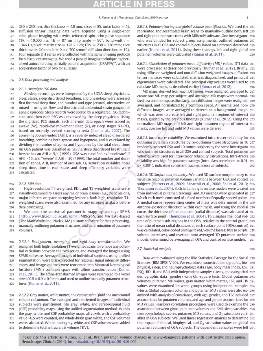
230 × 230mm; slice thickness=4.0mm; slices= 55; turbo factor= 5).Diffusion tensor imaging data were acquired using a single-shotecho-planar imaging with twice-refocused spin-echo pulse sequence(TR = 10,000 ms; TE = 87 ms; flip angle = 90°; band-width =1346 Hz/pixel; matrix size = 128 × 128; FOV = 230 × 230 mm; slicethickness = 2.0 mm, b = 0 and 700 s/mm2, diffusion directions = 12).Four separate DTI series were collected with the same imaging protocolfor subsequent averaging. We used a parallel imaging technique, “gener-alized autocalibrating partially parallel acquisition (GRAPPA)”, with anacceleration factor of two for all scans.
2.6. Data processing and analysis
2.6.1. Overnight PSG dataAll sleep recordings were interpreted by the UCLA sleep physicians.
Sleep states, sleep disordered breathing, and physiology were assessedfirst for total sleep time, and number and type (central, obstructive, ormixed — using air flow and thoracic and abdominal strain gauges) ofapneic episodes. Sleep records were scored by a registered PSG techni-cian, and then each PSG was reviewed by the sleep physician. Usingthe digitized PSG signals, each one-min data epoch were scored asawake (W), rapid-eye movement sleep (R), or sleep stages N1–N3,based on recently-revised scoring criteria (Iber et al., 2007). Theapnea–hypopnea-index (AHI), is a severity index of sleep disorderedbreathing, combining both apneas and hypopneas, and is calculated bydividing the number of apnea and hypopnea by the total sleep time.An OSA patient was classified as having sleep disordered breathing ifhe/she has an AHI ≥ 5/h (1999). OSA was classified as “moderate” ifAHI N 15, and “severe” if AHI N 30 (1999). The total number and dura-tion of apnea, AHI, number of arousals, O2 saturation variables, totalsleep time, time in each state, and sleep efficiency variables werecalculated.
2.6.2. MRI dataHigh-resolution T1-weighted, PD-, and T2-weighted scans were
visually-examined to assess any major brain lesions (e.g., cystic lesions,major infarcts, or space occupying lesions). Both high-resolution T1-weighted scans were also examined for any imaging artifacts beforedata processing.
We used the statistical parametric mapping package SPM8(http://www.fil.ion.ucl.ac.uk/spm/), MRIcroN, and MATLAB-based(TheMathWorks Inc., Natick, MA) custom software for data processing,manually-outlining putamen structures, and determination of putamenvolumes.
2.6.2.1. Realignment, averaging, and rigid-body transformation. Werealigned both high-resolution T1-weighted scans to remove any poten-tial variations between the two series, and averaged the images usingSPM8 software. Averaged images of individual subjects, using unifiedsegmentation, were bias-corrected for regional signal intensity differ-ences, and image volumes were reoriented into Montreal NeurologicalInstitute (MNI) common space with affine transformation (Kumaret al., 2011). The affine-transformed images were resampled to a voxelsize of 0.9 × 0.9 × 0.9 mm, and used to outline manually putamen struc-tures (Kumar et al., 2011).
2.6.2.2. Gray matter, white matter, and cerebrospinal fluid and intracranialvolume calculation. The averaged and reoriented images of individualsubjects were partitioned into gray, white, and cerebrospinal fluid(CSF) probability maps using the unified segmentation method. Withinthe gray, white, and CSF probability maps, all voxels with a probabilityvalueN0.5were counted, andwhole-brain gray,white, and CSF volumeswere calculated.Whole-brain gray, white, and CSF volumeswere addedto determine total intracranial volume (TIV).
Please cite this article as: Kumar, R., et al., Brain putamen volume chNeuroImage: Clinical (2014), http://dx.doi.org/10.1016/j.nicl.2014.01.009
ED P
RO
OF
2.6.2.3. Putamen tracing and global volume quantification. We used thereoriented and resampled brain scans to manually-outline both leftand right putamen structures with MRIcroN software. One investigator,who was blinded for subject group assignments, outlined putamenstructures in all OSA and control subjects, based on a protocol describedearlier (Kumar et al., 2011). Using these tracings, left and right globalputamen volumes were calculated (Kumar et al., 2011).
2.6.2.4. Calculation of putamen mean diffusivity (MD) values. DTI datawere processed as described previously (Kumar et al., 2012). Briefly,using diffusion weighted and non-diffusion weighted images, diffusiontensor matrices were calculated, matrices diagonalized, and principaleigenvalues were calculated. The principal eigenvalues were used tocalculate MD maps, as described earlier (Kumar et al., 2012).
MDmaps, derived from each DTI series, were realigned, averaged tocreate one MD map per subject, and averaged MD maps were normal-ized to a common space. Similarly, non-diffusion imageswere realigned,averaged, and normalized to a common space. All normalized non-diffusion images were averaged to determine a background image,which was used to create left and right putamen regions-of-interestmasks, guided by the previous findings (Kumar et al., 2012). Using thenormalized MD maps and left and right putamen region-of-interestmasks, average left and right MD values were derived.
2.6.2.5. Intra-tracer reliability. We examined intra-tracer reliability foroutlining putamen structures by re-outlining those structures in 10randomly-selected OSA and 10 control subjects by the same investigatorwho outlined structures in all OSA and control subjects. Total putamenvolumes were used for intra-tracer reliability calculations. Intra-tracerreliability was high for putamen tracings (intra-class correlation = 0.91,p b 0.001), indicating consistent tracings across all subjects.
2.6.2.6. 3D Surface morphometry. We used 3D surface morphometry tovisualize regional putamen volume variations betweenOSA and controlsubjects (Butters et al., 2009; Sabattoli et al., 2008; Shi et al., 2013;Thompson et al., 2004). Both left and right surface models were createdfrom individual putamen tracings, and 3D meshes were generated inwhich each mesh consisted of a fixed number of equally-spaced points.A medial curve representing center of mass was determined in theanterior–posterior direction within each mesh, and using the medialcurve, the thickness of the putamen (radial distance) was calculated ateach surface point (Thompson et al., 2004). To visualize the local vol-ume of putamen sub-regions in the OSA, relative to the control group,the ratio of mean radial distances at each surface point (OSA/control)was calculated, color-coded (orange to red, volume losses; blue to purple,volume increases), and overlaid onto averaged 3D putamen surfacemodels, determined by averaging all OSA and control surface models.
2.7. Statistical analysis
Data were evaluated using the IBM Statistical Package for the SocialSciences (IBM SPSS, V 20). We examined numerical demographic, bio-physical, sleep, and neuropsychologic data (age, body mass index, ESS,PSQI, BDI-II, and BAI) with independent samples t-tests, and categoricaldemographic data (gender) with Chi-square tests. Global putamenvolumes, putamen MD values, gray matter, white matter, CSF, and TIVvalues were examined between groups using independent samplest-tests. Global putamen volumes and putamenMD values were also ex-amined with analysis of covariance, with age, gender, and TIV includedas covariates for putamen volumes, and age and gender as covariates forMD values. Pearson's correlation procedures were used to examine theassociation between global putamen volumes and BMI, sleep variables,neuropsychologic scores, putamen MD values, and O2 saturation vari-ables in OSA subjects. We used linear regression analysis to determinethe impact of clinical, biophysical, and O2 saturation values on globalputamen volumes of OSA subjects. The dependent variables were left
anges in newly-diagnosed patients with obstructive sleep apnea,

295
296
297
298
299
300
301
302
303
304
305
306
307
308
309
310
311
312
313
314
315
316
317
318
319
320
321
322
323
324
325
326
327
t1:1
t1:1
t1:1
t1:1
t1:1
t1:1
t1:1
t1:1
t1:1
t1:1
t1:1
t1:1
t1:1
t1:1
t1:1
t1:1
t1:1
t1:1
t1:1
t1:1
t1:1
t1:1
t1:1
t1:1
t1:1
t1:1
4 R. Kumar et al. / NeuroImage: Clinical xxx (2014) xxx–xxx
and right putamen volumes, and independent variables were AHI, BMI,ESS, PSQI, BDI-II, BAI, and O2 saturation nadir values. We establishedintra-tracer reliability with an intra-class correlation procedure.
3. Results
3.1. Demographics
Demographic data are summarized in Table 1. Neither age norgender differed significantly between OSA and control subjects (age,p = 0.56; gender, p = 0.38). Body mass indices were significantlyhigher in OSA compared to control subjects (p b 0.001).
3.2. Sleep, O2 saturation, and neuropsychologic variables
Sleep, O2 saturation, and neuropsychologic variables of OSA andcontrol subjects are summarized in Table 1. Both ESS and PSQI scores sig-nificantly differed between OSA and control subjects (ESS, p b 0.001;PSQI, p b 0.001). BDI-II and BAI values were significantly higher in OSA,compared to control subjects (BDI-II, p b 0.001; BAI, p = 0.001).
UNCO
RRECT
Table 1Characteristics of OSA and control subjects.
Variable type Variables OSA(Mea[A]
Demographic Age (years) 46.4(n =
Gender (Male:Female) 31:12Biophysical BMI (kg/m2) 30.1
(n =Sleep AHI (events/h) 31.4
(n =ESS 10.3
(n =PSQI 9.3 ±
(n =O2 saturation Baseline SaO2 (%) 96.3
(n =SaO2 nadir (%) 77.9
(n =SaO2 nadir change (%) 18.3
(n =Obstructive length of nadir (s) 40.6
(n =Neuropsychological BDI-II 9.3 ±
(n =BAI 10.7
(n =Brain (volume) Gray matter (mm3) 700,4
(n =White matter (mm3) 445,3
(n =CSF (mm3) 315,2
(n =TIV (mm3) 1,461
(n =Putamen (volume) Left putamen (mm3) 4146
4117(n =
Right putamen (mm3) 41524125(n =
Putamen (MD values) Left putamen (×10−6 mm2/s) 803.1802.5n =
Right putamen (×10−6 mm2/s) 784.7784.2n =
SD, standard deviation; BMI, body mass index; AHI, apnea–hypopnea-index; ESS, EpworthDepression Inventory II; BAI, Beck Anxiety Inventory; CSF, cerebrospinal fluid; TIV, total intraTIV; ##, p value controlled for age and gender.
Please cite this article as: Kumar, R., et al., Brain putamen volume chNeuroImage: Clinical (2014), http://dx.doi.org/10.1016/j.nicl.2014.01.009
OF
3.3. Global gray, white, and CSF volumes and TIV
Global gray matter, white matter, CSF volumes, and TIV values ofOSA and control group are summarized in Table 1. Gray matter, whitematter, and CSF volumes, aswell as TIV values, did not differ significant-ly between OSA and control subjects (gray matter, p = 0.40; whitematter, p = 0.27; CSF, p = 0.62; TIV, p = 0.82).
3.4. Global putamen volumes and MD value differences
Both left and right global putamen volumes of OSA and control sub-jects are summarized in Table 1, and scatter plots are shown in Fig. 1.The left and right global putamen volumes were significantly higher inOSA, compared to control subjects (independent samples t-tests;left, p = 0.041; right, p = 0.025); values were significantly higher inOSA, even after controlling for age, gender, and TIV (ANCOVA; left,p = 0.043; right, p = 0.027).
Average putamen MD values of OSA and control subjects aresummarized in Table 1. Both left and right putamenMDvalueswere sig-nificantly lower in OSA, compared to control subjects (independentsamples t-tests; left, p = 0.017; right, p = 0.001); values were
ED P
RO
n ± SD)Controls(Mean ± SD)[B]
[A] vs [B]p values
± 8.843)
47.4 ± 8.9(n = 61)
0.56
39:22 0.38± 5.243)
24.8 ± 3.7(n = 61)
b 0.001*
± 19.343)
– –
± 4.943)
5.5 ± 3.5(n = 61)
b 0.001*
4.043)
3.8 ± 2.5(n = 61)
b 0.001*
± 240)
– –
± 10.336)
– –
± 10.636)
– –
± 21.430)
– –
8.343)
3.9 ± 4.4(n = 61)
b0.001*
± 12.443)
3.9 ± 5.0(n = 61)
0.001*
69.7 ± 73,557.743)
713,303.3 ± 77,941.7(n = 61)
0.40
47.5 ± 56,252.443)
458,578.8 ± 63,000.5(n = 61)
0.27
75.5 ± 167,583.043)
298,249.2 ± 175,544.6(n = 61)
0.62
,092.6 ± 206,276.643)
1,470,131.3 ± 199,550.1(n = 61)
0.82
.6 ± 666.0
.9 ± 461.643)
3909.1 ± 502.43929.3 ± 461.1(n = 61)
0.0410.043**
.8 ± 654.5
.2 ± 465.243)
3897.6 ± 487.83917.0 ± 472.3(n = 61)
0.0250.027**
7 ± 75.648 ± 84.5219
870.00 ± 92.30870.50 ± 84.48n = 21
0.0170.016##
5 ± 67.943 ± 91.6819
889.12 ± 112.18889.58 ± 91.65n = 21
0.0010.001##
Sleepiness Scale; PSQI, Pittsburgh Sleep Quality Index; SaO2, O2 saturation; BDI-II, Beckcranial volume; *, equal variances not assumed; **, p value adjusted for age, gender, and
anges in newly-diagnosed patients with obstructive sleep apnea,

328
329
330
331
332
333
334
335
336
337
338
339
340
341
342
343
344
345
346
347
348
349
350
351
352
353
354
355
356
357
358
359
360
361
362
363
364
365
366
367
368
369
370
371
372
373
374
375
376
377
378
379
380
381
382
Fig. 1. Global putamen volumes of OSA and control subjects. Both left and right putamenvolumes were significantly increased in recently-diagnosed, treatment naïve OSA overcontrol subjects, controlling for age, gender, and TIV (left, p = 0.043; right, p = 0.027).
5R. Kumar et al. / NeuroImage: Clinical xxx (2014) xxx–xxx
significantly reduced in OSA over controls, even accounting for age andgender (ANCOVA; left, p = 0.016; right, p = 0.001).
T383
384
385
386
387
388
389
390
391
392
393
394
395
396
397
398
RREC3.5. Relationships between global putamen volumes and BMI, sleep, O2
saturation, left and right putamenmd values, and neuropsychologic variables
Coefficients of correlation and significant levels among putamenvolumes, BMI, sleep, O2 saturation, left and right putamen MD values,and neuropsychologic variables are tabulated in Table 2. Significant neg-ative correlations appeared between global putamen volumes, BMI(left; r=−0.35 p= 0.022; right, r=−0.32, p= 0.036), and putamenMD values (left; r=−0.48, p= 0.035; right, r=−0.46, p= 0.047) inOSA subjects. However, no significant correlations emerged betweenputamen volumes and AHI, PSQI, and ESS scores. The BAI scores signifi-cantly correlated with putamen volumes (left; r = −0.39 p = 0.01;right, r = −0.40, p = 0.008); however, BDI-II scores did not showany correlations. The O2 desaturation nadir showed a significant associ-ation with left putamen volume (r =−0.34, p = 0.045); but, other O2
saturation variables did not show any relationships.
399400
401
402
403
404
405
406
407
408
409
410
411
UNCO3.6. Regional putamen volume changes
Multiple regions within the putamen showed increased and de-creased ratios of radial distance of surface points from the medialcurve, indicating increased or decreased regional volume in OSA, com-pared to control subjects. Regions with decreased volumes within theputamen in OSA subjects included bilateral rostral (Fig. 2a, b, f, n, andq), mid-dorsal (Fig. 2c, g, and h), medial-caudal (Fig. 2d, j, and p), andmid-ventral sites (Fig. 2k, p), and increased volumes included the leftcaudal-dorsal (Fig. 2e), right mid-dorsal (Fig. 2i), left mid-ventralportions (Fig. 2l, m), and right caudal-ventral area (Fig. 2o).
412
413
414
415
416
417
418
419
420
3.7. Putamen volume and linear regression analyses
Regression analyses findings are summarized in Table 3. Oxygensaturation nadir (p= 0.045, B =−20.46) was an independent predic-tor for left global putamen volume in OSA subjects. However, novariable independently predicted right global putamen volume inthese subjects.
Please cite this article as: Kumar, R., et al., Brain putamen volume chNeuroImage: Clinical (2014), http://dx.doi.org/10.1016/j.nicl.2014.01.009
ED P
RO
OF
4. Discussion
4.1. Overview
Newly-diagnosed, treatment-naïve OSA subjects show increasedglobal putamen volumes, but localized areas within the putamenshow both higher and lower volumes compared to control subjects.The appearance of both increased and decreased volumes suggeststhat we obtained a snapshot of progressive putamen tissue changesearly in the development of the chronic OSA condition, in which bothtissue swelling and loss occur, likely depending on the nature of thecomposite structure and local perfusion. The putamen sites with alteredvolume in OSA are associated with somatic and autonomic motor andneuropsychologic regulatory roles, all of which are deficient in thecondition (Bedard et al., 1991; Beebe et al., 2003; Berry et al., 1986).
4.2. OSA and brain injury
The putamen, togetherwith other basal ganglia and brain structures,show gray matter volume loss, gray matter and fiber injury, and meta-bolic abnormalities in patients in chronic stages of the OSA syndrome(Kamba et al., 1997; Macey et al., 2002; Morrell et al., 2003), ratherthan the increased volume in the newly-diagnosed cases studied here.Those prior studies in chronic OSA subjects, based on volumetric proce-dures, showed reduced regional tissue volume changes, indicatingchronic tissue injury with late OSA diagnosis (Macey et al., 2002;Morrell et al., 2003). However, in newly-diagnosed OSA subjects,acute tissue changes are predominantly accompanied by axonal, glial,and neuronal swelling, i.e., larger volumes, but show decreased meandiffusivity values in the sameOSA subject pool, indicative of acute tissueinjury (Kumar et al., 2012). The increased overall structural volumesfound here should be viewed in this context of apparent injury fromdif-fusion tensor imaging, but global increased volume from resultantswelling; the progression of the syndrome will be accompanied bylater volume declines. The localized declines in volume may followdifferent time courses for different brain structures; selected brainareas, such as the mammillary bodies, show reduced volumes early inthe disease process (Kumar et al., 2008). Moreover, subareas withinthe putamen may vary in vascularization and tissue composition withdifferent time courses of tissue loss.
4.3. Global and regional putamen volumes and OSA functional changes
Global putamen volumes were significantly increased in OSA overcontrol subjects, despite sub-regions showing both increased and de-creased volumes. The putamen contains dopaminergic neurons, sendsthose projections to the substantia nigra and ventral tegmentum(Gorbachevskaya, 1997; Schultz and Romo, 1987), and receives projec-tions from the thalamus, insular cortices, and prefrontal cortex(Avendano et al., 2006; Ferry et al., 2000), areas which are also injuredin OSA subjects (Kumar et al., 2012; Macey et al., 2002, 2008). Putamensub-regions are involved in various functions, including those heavilyinfluenced by dopamine released by midbrain projections (Inase et al.,1997; Schultz, 1986).
Various putamen sites, including bilateral rostral, mid-dorsal,medial-caudal, mid-ventral and ventral, and caudal-dorsal regionsshowed either increased or decreased volume in OSA subjects; bothtypes of tissue changesmay be associatedwith altered function. The pu-tamen primarily serves motor regulation functions, receiving projec-tions from motor and somatosensory areas for that role (Kunzle,1977). The rostral putamen receives projections from the prerubralarea of the substantia nigra through the lateral hypothalamus, internalcapsule and the head of the caudate nucleus (Usunoff et al., 1976).Somatosensory and motor cortex areas representing the lower limbs,ventromedial portions of facial regions, and upper limb regions projectto the dorsolateral putamen, and these projecting sites send fibers in a
anges in newly-diagnosed patients with obstructive sleep apnea,

UNCO
RRECT
421
422
423
424
425
426
427
428
429
430
431
432
433
434
435
436
437
438
439
440
441
442
443
444
445
446
447
448
449
450
451
452
453
454
455
456
457
458
459
460
461
462
463
464
465
466
467
468
469
470
471
472
473
474
475
476
477
478
479
480
481
482
483
484
t2:2
Table2
t2:2
Coefficien
tsof
correlationan
dsign
ificant
values
amon
gpu
tamen
volumes,B
MI,slee
p,ne
urop
sych
olog
icva
riab
les,O2saturation
,putam
enMDva
lues,and
AHIinOSA
subjects.
Leftpu
tamen
volume
n=
43Righ
tputam
envo
lume
n=
43BM
In=
43ES
Sn=
43PS
QI
n=
43BD
I-II
n=
43BA
In=
43O2saturation
nadir
n=
36Leftpu
tamen
MD
n=
19Righ
tpu
tamen
MD
n=
19AHI
n=
43t2:2
Leftpu
tamen
volume
r=
0.98
0p=
0.00
0r=
−0.34
8p=
0.02
2r=
−0.21
2p=
0.17
2r=
−0.06
0p=
0.70
0r=
−0.21
3p=
0.17
0r=
−0.38
6p=
0.01
1r=
−0.33
6p=
0.04
5r=
−0.48
5p=
0.03
5r=
−0.44
0p=
0.05
9r=
−0.08
3p=
0.59
7t2:2
Righ
tputam
envo
lume
r=
0.98
0p=
0.00
0r=
−0.32
0p=
0.03
6r=
−.019
4p=
0.21
2r=
−0.09
8p=
0.53
2r=
−0.20
9p=
0.17
9r=
−0.39
7p=
0.00
8r=
−0.30
7p=
0.06
9r=
−0.53
8p=
0.01
7r=
−0.46
1p=
0.04
7r=
−0.08
9p=
0.56
9t2:2
BMI
r=
−0.34
8p=
0.02
2r=
−0.32
0p=
0.03
6r=
0.02
6p=
0.86
7r=
0.03
9p=
0.80
2r=
−0.02
9p=
0.85
4r=
0.09
0p=
0.56
4r=
0.17
2p=
0.31
7r=
0.31
5p=
0.18
9r=
0.46
8p=
0.04
3r=
0.43
3p=
0.00
4t2:2
ESS
r=
−0.21
2p=
0.17
2r=
−0.19
4p=
0.21
2r=
0.02
6p=
0.86
7r=
0.07
9p=
0.61
5r=
0.24
1p=
0.11
9r=
0.18
6p=
0.23
2r=
−0.03
4p=
0.84
5r=
0.22
7p=
0.35
0r=
0.28
3p=
0.24
1r=
0.06
4p=
0.68
2t2:2
PSQI
r=
−0.06
0p=
0.70
0r=
−0.09
8p=
0.53
2r=
0.03
9p=
0.80
2r=
=0.07
9p=
0.61
5r=
0.61
1p=
.000
r=
0.46
4p=
0.00
2r=
−0.12
1p=
0.48
2r=
0.22
4p=
0.35
6r=
0.03
6p=
0.88
5r=
0.03
4p=
0.82
7t2:2
BDI-II
r=
−0.21
3p=
0.17
0r=
−0.20
9p=
0.17
9r=
0.02
9p=
0.85
4r=
0.24
1p=
0.11
9r=
0.61
1p=
.000
r=
0.80
6p=
0.00
0r=
0.24
2p=
0.15
6r=
0.51
7p=
0.02
3r=
0.38
8p=
0.10
1r=
−0.01
0p=
0.95
1t2:2
BAI
r=
−0.38
6p=
0.01
1r=
−0.39
7p=
0.00
8r=
0.09
0p=
0.56
4r=
0.18
6p=
0.23
2r=
0.46
4p=
0.00
2r=
0.80
6p=
0.00
0r=
0.39
0p=
0.01
9r=
0.35
4p=
0.13
8r=
0.24
3p=
0.31
6r=
−0.06
1p=
0.69
9t2:2
O2saturation
nadir
r=
−0.33
6p=
0.04
5r=
−0.30
7p=
0.06
9r=
0.17
2p=
0.31
7r=
Q2
−0.03
4p=
0.84
5r=
−0.12
1p=
0.48
2r=
0.24
2p=
0.15
6r=
0.39
0p=
0.01
9r=
0.07
2p=
0.78
5r=
0.15
2p=
0.56
0r=
−0.20
6p=
0.22
8t2:2
Leftpu
tamen
MD
r=
−0.48
5p=
0.03
5r=
−0.53
8p=
0.01
7r=
0.31
5p=
0.18
9r=
0.22
7p=
0.35
0r=
0.22
4p=
0.35
6r=
0.51
7p=
0.02
3r=
0.35
4p=
0.13
8r=
0.07
2p=
0.78
5r=
0.81
7p=
0.00
0r=
0.41
4p=
0.07
8t2:2
Righ
tputam
enMD
r=
−0.44
0p=
0.05
9r=
−0.46
1p=
0.04
7r=
0.46
8p=
0.04
3r=
0.28
3p=
0.24
1r=
0.03
6p=
0.88
5r=
0.38
8p=
0.10
1r=
0.24
3p=
0.31
6r=
0.15
2p=
0.56
0r=
0.81
7p=
0.00
0r=
0.42
5p=
0.07
0t2:2
AHI
r=
−0.08
3p=
0.59
7r=
−0.08
9p=
0.56
9r=
0.43
3p=
0.00
4r=
0.06
4p=
0.68
2r=
0.03
4p=
0.82
7r=
−0.01
0p=
0.95
1r=
−0.06
1p=
0.69
9r=
−0.20
6p=
0.22
8r=
0.41
4p=
0.07
8r=
0.42
5p=
0.07
0t2:2
t2:2
BMI,bo
dymassinde
x;ES
S,Ep
worth
Slee
pine
ssScale;
PSQI,Pittsburgh
Slee
pQua
lityInde
x;BD
I-II,
Beck
Dep
ressionInve
ntoryII;
BAI,Be
ckAnx
iety
Inve
ntory;
MD,M
eandiffu
sivity;A
HI,ap
nea–
hypo
pnea-ind
ex.
6 R. Kumar et al. / NeuroImage: Clinical xxx (2014) xxx–xxx
Please cite this article as: Kumar, R., et al., Brain putamen volume chNeuroImage: Clinical (2014), http://dx.doi.org/10.1016/j.nicl.2014.01.009
ED P
RO
OF
rostro-caudal direction (Kunzle, 1975, 1977). Both premotor and sup-plementary motor areas representing the face (Muakkassa and Strick,1979), also send projections to the ventral medial putamen (Kunzle,1978; Miyata and Sasaki, 1984). The ventrolateral globus pallidus pro-jects to the ventrolateral thalamus,which represents oral and upper air-way regions (DeVito and Anderson, 1982). Supplementary motor areasand the caudolateral substantia nigra are involved in orofacial control(Boussaoud and Joseph, 1985). Both the globus pallidus and substantianigra sites receive projections from rostroventral putamen (DeVitoet al., 1980). Since a principal concern in OSA is somatic motor regula-tion of the oral airway, localized damage to subareas of the putameninvolved in such regulation is of great interest in the syndrome.
Multiple autonomic motor and neurobehavioral issues, includingaltered memory, attention, psychomotor and executive function, andemotional regulation are affected in OSA subjects (Bedard et al., 1991;Beebe et al., 2003; Berry et al., 1986). The putamen contains dopaminer-gic neurons, the neurotransmitter principally involved in mediating re-ward. Injury to ventral putamen sub-regions served bydopamine-basedneurotransmitters would have significant effects on affective behaviorsdependent on reward (Seeman et al., 2006); those behaviors are heavilyaffected in OSA (Asghari et al., 2012). A primary characteristic of OSA isimpaired autonomic nervous system regulation, reflected especially inexcessive sympathetic activity and impaired timing to autonomic chal-lenges (Harper et al., 2003; Henderson et al., 2002; Somers et al., 1995).Injury to both the insular cortices and dorsal putamen likely contributesto that deficit, since both regions serve autonomic roles; the putamenreceives projections from the insular cortices (Saper, 1982), and bothstructures show overall injury and functional deficits during autonomicand respiratory challenges (Henderson et al., 2004; Kumar et al., 2012;Macey et al., 2006). Since a close relationship exists between transientelevation in blood pressure with both diaphragmatic (Trelease et al.,1985) and upper airway action (Marks and Harper, 1987), and upperairwaymuscle suppression ismore sensitive to blood pressure elevationthan the diaphragm, distorted regulation of dynamic sympatheticpatterns has the potential to contribute to the “motor neglect” ofupper airway muscles found in OSA.
4.4. Tissue volume changes: processes
Several mechanisms may underlie the global and regional putamentissue changes. OSA subjects are intermittently exposed to hypoxia fromthe repeated, successive cessations of airflow during sleep, which isfollowed by rapid re-oxygenation at apnea termination. Moreover, ex-aggerated blood pressure swings occur over the course of each apneicevent, with substantial changes in perfusion accompanying the vascularalterations (Yan et al., 2009). Hypoxic and ischemic conditions induceenergy pump failure, altering sodium and potassium ion distribution,and affecting intracellular and extracellular water, resulting in neuronaland axonal swelling (Hossmann, 1971; Lowry et al., 1964;Mintorovitchet al., 1994; Oehmichen et al., 2001). The axonal and neuronal swellingincreases volumes of affected brain structures. However, chronic stagesof hypoxia/ischemia would be accompanied by a reduction in axons,neurons, and glia, leaving a net loss of tissue volume over control values.Since OSA subjects showed higher global putamen volumes, relative tocontrol subjects, we believe that the tissue changes are representativeof acute changes. These changes likely contribute to manifestations ofcharacteristics in OSA.
Both increased and decreased localized volume changes appeared inOSA subjects within the putamen over controls, suggesting that thechanges result from both early and long-standing tissue alterations inthese newly-diagnosed, treatment-naïve subjects, or that portions ofthe putamen may be more vulnerable to hypoxic or perfusion changesthrough the nature of tissue composition or extent of vascularization.Overall, the predominant changes appear to be in acute stages, sinceglobal putamen volumes were found to be significantly increased inOSA over controls. Thesefindings suggest that a series of changes occurs
anges in newly-diagnosed patients with obstructive sleep apnea,

T
RO
OF
485
486
487
488
489
490
491
492
493
494
495
496
497
498
499
500
501
502
503
504
505
506
507
508
509
510
511
512
513
514
515
516
517
518
519
520
Fig. 2.Areas of regional putamen volume increase and decrease in OSA compared to control subjects, with areas shown from a dorsal perspective (Left), and a ventral perspective (Right).The color coding represents ratios of radial distance of each surface point from control reference values, with warm colors (orange to red) indicating volume losses and cool colors (blue topurple) representing volume increases; those corresponding sites are labeledwith letters (a–q). Bilateral siteswith increased putamen volume in the OSA group includedmid and caudal-ventral portions, based on OSA to control group ratios, and decreased volumes emerged in bilateral rostral and mid-dorsal regions, extending to ventral areas (a–q).
t3:3
t3:3
t3:3
t3:3
t3:3
t3:3
t3:3
t3:3
t3:3
t3:3
t3:3
t3:3
t3:3
t3:3
t3:3
t3:3
t3:3
t3:3
t3:3
t3:3
t3:3
t3:3
7R. Kumar et al. / NeuroImage: Clinical xxx (2014) xxx–xxx
RREC
within tissue before axonal and neuronal loss, with early, acute stages ofpathology associatedwith increased tissue volume,while chronic stagesof pathology are accompanied by decreased volumes. Tissue changesin both acute and chronic stages of pathology likely contribute to func-tional deficits served by subareas within the structure.
It is possible, although speculative, that theOSA conditionmay resultfromputamen tissue changes, i.e., that the failure in upper airway actionresults from initial putamen dysfunction from injury which may prog-ress, cumulating with the exaggerated autonomic and other manifesta-tions found in OSA. Initial putamen injury, perhaps by carbonmonoxidepoisoning, accidental hypoxic exposure, or reaction to toxic substancesacting on the multiple neurotransmitters in the structure, has the po-tential to interferewith normal putamen functioning, disrupting normalregulation of the upper airway. This initial dysfunctionmay escalate to acondition that self-perpetuates or worsens, with all of the autonomicand somatomotor outcomes reported for OSA. Determination of thefeasibility of such an outcome would require basic studies of insults tothe putamen in an animal model that results in interference withupper airway muscle action paralleling the coordination loss found inhuman OSA.
UNCO
Table 3Comparison ofmultivariate p values and B values of AHI, BMI, sleep, neuropsychologic, leftand right putamenMD values, and O2 saturation variables for the linear regression analy-ses in OSA subjects.
Side Covariates Multivariate p values B
Left putamen volume AHI 0.573 –
BMI 0.175 –
ESS 0.892 –
PSQI 0.950 –
BDI–II 0.897 –
BAI 0.567 –
O2 saturation nadir 0.045 –20.46Right putamen volume AHI – –
BMI – –
ESS – –
PSQI – –
BDI-II – –
BAI – –
O2 saturation nadir – –
AHI, apnea–hypopnea-index; BMI, bodymass index; ESS, Epworth Sleepiness Scale; PSQI,Pittsburgh Sleep Quality Index; BDI-II, Beck Depression Inventory II; BAI, Beck AnxietyInventory; MD, mean diffusivity.
Please cite this article as: Kumar, R., et al., Brain putamen volume chNeuroImage: Clinical (2014), http://dx.doi.org/10.1016/j.nicl.2014.01.009
ED P4.5. Limitations
One limitation of this study is the absence of data on the pre-diagnosis duration of OSA subjects. However, we are reasonably confi-dent that the predominant brain tissue changes in the OSA subjectsare acute in nature. MRI-based mean diffusivity (MD) procedures,which measure water diffusion within tissue, and are influenced bythe presence of tissue barriers and extracellular/extra-axonal fluid,show reduced MD values in acute disease stages of ischemic/hypoxicpathology, but show increased values in chronic stages of such patholo-gy (Ahlhelm et al., 2002; Chan et al., 2009; Hossmann et al., 1994;Loubinoux et al., 1997; Matsumoto et al., 1995; Shereen et al., 2011;Warach et al., 1996). Since these same OSA subjects showed reducedMD values across various brain sites (Kumar et al., 2012), we believethat the predominant tissue pathology in this sample derives fromdamage in acute stages.
521
522
523
524
525
526
527
528
529
530
531
532
533
534
535
536
5. Conclusions
Newly-diagnosedOSA subjectswithout treatment showsignificantlyincreased global putamen volumes over control subjects, with sub-regionswithin the structure showing both increased and decreased vol-umes. Regional sites with decreased volumes emerged in rostral, mid-dorsal, medial-caudal, and mid-ventral areas, and increased volumesappeared in caudal-dorsal, mid-dorsal, mid-ventral, and caudal-ventralportions. These areas contribute to regulation of neuropsychologic,autonomic motor, and somatomotor functions deficient in OSA, and es-pecially to oral motor functioning, a special concern in the syndromedue to airway collapse in obstructive apnea. The pathological processescontributing to tissue changes may include intermittent hypoxia andimpaired perfusion, both of which can affect glial tissue necessary forneuronal support; such tissue is extremely sensitive to hypoxia andischemia. The finding of regional increased and decreased volumevariation indicates that final volume loss earlier found in long-termOSA occurs with a progression of changes in the tissue.
537Q4
538
6. Uncited reference
Unknown, 1999
anges in newly-diagnosed patients with obstructive sleep apnea,

T
539
540
541
542
543
544
545
546547548549550551552553554555556557558559560561562563564565566567568569570571572573574575576577578579580581582583584585586587588589590591592593594595596597598599600601602603604605606607608609610611612613614615616617618
619620621622623624625626627628629630631632633634635636637638639640641642643644645646647648649650651652653654655656657658659660661662663664665666667668669670671672673674675676677678679680681682683684685686687688689690691692693694695696697698699700701702703704
8 R. Kumar et al. / NeuroImage: Clinical xxx (2014) xxx–xxx
UNCO
RREC
Acknowledgments
The authors thank Ms. Rebecca Harper, Mr. Edwin Valladares, andDrs. Rebecca Cross and Stacy Serber for their help with data collection,and Mr. Jose Palomares for his help in table preparation. This researchwork was supported by the National Institutes of Health R01 HL-113251.
References
Ahlhelm, F., Schneider, G., Backens, M., Reith, W., Hagen, T., 2002. Time course of theapparent diffusion coefficient after cerebral infarction. Eur. Radiol. 12, 2322–2329.
Alexander, G.E., Crutcher, M.D., 1990. Preparation for movement: neural representationsof intended direction in three motor areas of the monkey. J. Neurophysiol. 64,133–150.
Alkan, A., Sharifov, R., Akkoyunlu, M.E., Kilicarslan, R., Toprak, H., Aralasmak, A., Kart, L.,2013. MR spectroscopy features of brain in patients with mild and severe obstructivesleep apnea syndrome. Clin. Imaging 37, 989–992.
Asghari, A., Mohammadi, F., Kamrava, S.K., Tavakoli, S., Farhadi, M., 2012. Severity of depres-sion and anxiety in obstructive sleep apnea syndrome. Eur. Arch. Otorhinolaryngol. 269,2549–2553.
Avendano, C., de Las Heras, S., Gimenez-Amaya, J.M., 2006. Striatal projections from thelateral and posterior thalamic complexes. An anterograde tracer study in the cat.Histochem. Cell Biol. 125, 265–271.
Badgaiyan, R.D., Fischman, A.J., Alpert, N.M., 2008. Explicit motor memory activates thestriatal dopamine system. Neuroreport 19, 409–412.
Beck, A.T., Epstein, N., Brown, G., Steer, R.A., 1988. An inventory for measuring clinicalanxiety: psychometric properties. J. Consult. Clin. Psychol. 56, 893–897.
Beck, A.T., Steer, R.A., Brown, G.K., 1996. Manual for the Beck Depression Inventory-II. ThePsychological Corporation, San Antonio, Texas.
Bedard, M.A., Montplaisir, J., Richer, F., Rouleau, I., Malo, J., 1991. Obstructive sleep apneasyndrome: pathogenesis of neuropsychological deficits. J. Clin. Exp. Neuropsychol. 13,950–964.
Beebe, D.W., Groesz, L., Wells, C., Nichols, A., McGee, K., 2003. The neuropsychologicaleffects of obstructive sleep apnea: a meta-analysis of norm-referenced and case-controlled data. Sleep 26, 298–307.
Berry, D.T., Webb, W.B., Block, A.J., Bauer, R.M., Switzer, D.A., 1986. Nocturnal hypoxia andneuropsychological variables. J. Clin. Exp. Neuropsychol. 8, 229–238.
Booth, J.R., Wood, L., Lu, D., Houk, J.C., Bitan, T., 2007. The role of the basal ganglia andcerebellum in language processing. Brain Res. 1133, 136–144.
Boussaoud, D., Joseph, J.P., 1985. Role of the cat substantia nigra pars reticulata in eye andhead movements. II. Effects of local pharmacological injections. Exp. Brain Res. 57,297–304.
Bowman, E.M., Aigner, T.G., Richmond, B.J., 1996. Neural signals in the monkey ventralstriatum related to motivation for juice and cocaine rewards. J. Neurophysiol. 75,1061–1073.
Butters, M.A., Aizenstein, H.J., Hayashi, K.M., Meltzer, C.C., Seaman, J., Reynolds 3rd, C.F.,Toga, A.W., Thompson, P.M., Becker, J.T., 2009. Three-dimensional surface mappingof the caudate nucleus in late-life depression. Am. J. Geriatr. Psychiatry 17, 4–12.
Chan, K.C., Khong, P.L., Lau, H.F., Cheung, P.T., Wu, E.X., 2009. Latemeasures of microstruc-tural alterations in severe neonatal hypoxic-ischemic encephalopathy by MR diffu-sion tensor imaging. Int. J. Dev. Neurosci. 27, 607–615.
Cross, R.L., Kumar, R., Macey, P.M., Doering, L.V., Alger, J.R., Yan-Go, F.L., Harper, R.M., 2008.Neural alterations and depressive symptoms in obstructive sleep apnea patients. Sleep31, 1103–1109.
DeVito, J.L., Anderson, M.E., 1982. An autoradiographic study of efferent connections ofthe globus pallidus in Macaca mulatta. Exp. Brain Res. 46, 107–117.
DeVito, J.L., Anderson, M.E., Walsh, K.E., 1980. A horseradish peroxidase study of afferentconnections of the globus pallidus in Macaca mulatta. Exp. Brain Res. 38, 65–73.
Emin Akkoyunlu, M., Kart, L., Kilicarslan, R., Bayram, M., Aralasmak, A., Sharifov, R., Alkan,A., 2013. Brain diffusion changes in obstructive sleep apnoea syndrome. Respiration86, 414–420.
Ferry, A.T., Ongur, D., An, X., Price, J.L., 2000. Prefrontal cortical projections to the striatumin macaque monkeys: evidence for an organization related to prefrontal networks.J. Comp. Neurol. 425, 447–470.
Gorbachevskaya, A.I., 1997. Projections of the ventral tegmental area of the midbrain, thesubstantia nigra, and the amygdaloid body in different parts of the putamen in thedog. Neurosci. Behav. Physiol. 27, 496–502.
Harper, R.M., Macey, P.M., Henderson, L.A., Woo, M.A., Macey, K.E., Frysinger, R.C., Alger,J.R., Nguyen, K.P., Yan-Go, F.L., 2003. fMRI responses to cold pressor challenges in con-trol and obstructive sleep apnea subjects. J. Appl. Physiol. 94, 1583–1595.
Henderson, L.A., Macey, P.M., Macey, K.E., Frysinger, R.C., Woo, M.A., Harper, R.K., Alger,J.R., Yan-Go, F.L., Harper, R.M., 2002. Brain responses associated with the Valsalvamaneuver revealed by functional magnetic resonance imaging. J. Neurophysiol. 88,3477–3486.
Henderson, L.A., Richard, C.A., Macey, P.M., Runquist, M.L., Yu, P.L., Galons, J.P., Harper,R.M., 2004. Functional magnetic resonance signal changes in neural structures tobaroreceptor reflex activation. J. Appl. Physiol. 96, 693–703.
Hossmann, K.A., 1971. Cortical steady potential, impedance and excitability changesduring and after total ischemia of cat brain. Exp. Neurol. 32, 163–175.
Hossmann, K.A., Fischer, M., Bockhorst, K., Hoehn-Berlage, M., 1994. NMR imaging of theapparent diffusion coefficient (ADC) for the evaluation of metabolic suppression andrecovery after prolonged cerebral ischemia. J. Cereb. Blood Flow Metab. 14, 723–731.
Please cite this article as: Kumar, R., et al., Brain putamen volume chNeuroImage: Clinical (2014), http://dx.doi.org/10.1016/j.nicl.2014.01.009
ED P
RO
OF
Husain, M.M., McDonald,W.M., Doraiswamy, P.M., Figiel, G.S., Na, C., Escalona, P.R., Boyko,O.B., Nemeroff, C.B., Krishnan, K.R., 1991. A magnetic resonance imaging study ofputamen nuclei in major depression. Psychiatry Res. 40, 95–99.
Iber, C., Ancoli-Israel, S., Chesson, A., Quan, S.F., 2007. for the American Academy of SleepMedicine, The AASM Manual for the Scoring of Sleep and Associated Events: Rules,Terminology and Technical Specifications1st ed. American Academy of SleepMedicine, Westchester, Illinois.
Inase, M., Li, B.M., Tanji, J., 1997. Dopaminergic modulation of neuronal activity in themonkey putamen through D1 and D2 receptors during a delayed Go/Nogo task.Exp. Brain Res. 117, 207–218.
Johns, M.W., 1991. A new method for measuring daytime sleepiness: the Epworth sleep-iness scale. Sleep 14, 540–545.
Kamba, M., Suto, Y., Ohta, Y., Inoue, Y., Matsuda, E., 1997. Cerebral metabolism in sleepapnea. Evaluation by magnetic resonance spectroscopy. Am. J. Respir. Crit. CareMed. 156, 296–298.
Knutson, K.L., Rathouz, P.J., Yan, L.L., Liu, K., Lauderdale, D.S., 2006. Stability of thePittsburgh Sleep Quality Index and the Epworth Sleepiness Questionnaires over 1year in early middle-aged adults: the CARDIA study. Sleep 29, 1503–1506.
Kumar, R., Birrer, B.V., Macey, P.M., Woo, M.A., Gupta, R.K., Yan-Go, F.L., Harper, R.M.,2008. Reduced mammillary body volume in patients with obstructive sleep apnea.Neurosci. Lett. 438, 330–334.
Kumar, R., Ahdout, R., Macey, P.M., Woo, M.A., Avedissian, C., Thompson, P.M., Harper,R.M., 2009a. Reduced caudate nuclei volumes in patients with congenital centralhypoventilation syndrome. Neuroscience 163, 1373–1379.
Kumar, R., Macey, P.M., Cross, R.L., Woo, M.A., Yan-Go, F.L., Harper, R.M., 2009b. Neuralalterations associated with anxiety symptoms in obstructive sleep apnea syndrome.Depress. Anxiety 26, 480–491.
Kumar, R., Nguyen, H.D., Ogren, J.A., Macey, P.M., Thompson, P.M., Fonarow, G.C.,Hamilton, M.A., Harper, R.M., Woo, M.A., 2011. Global and regional putamen volumeloss in patients with heart failure. Eur. J. Heart Fail. 13, 651–655.
Kumar, R., Chavez, A.S., Macey, P.M., Woo, M.A., Yan-Go, F.L., Harper, R.M., 2012. Alteredglobal and regional brain mean diffusivity in patients with obstructive sleep apnea.J. Neurosci. Res. 90, 2043–2052.
Kunzle, H., 1975. Bilateral projections from precentral motor cortex to the putamen andother parts of the basal ganglia. An autoradiographic study in Macaca fascicularis.Brain Res. 88, 195–209.
Kunzle, H., 1977. Projections from the primary somatosensory cortex to basal ganglia andthalamus in the monkey. Exp. Brain Res. 30, 481–492.
Kunzle, H., 1978. An autoradiographic analysis of the efferent connections from premotorand adjacent prefrontal regions (areas 6 and 9) in macaca fascicularis. Brain Behav.Evol. 15, 185–234.
Loubinoux, I., Volk, A., Borredon, J., Guirimand, S., Tiffon, B., Seylaz, J., Meric, P., 1997.Spreading of vasogenic edema and cytotoxic edema assessed by quantitative diffu-sion and T2 magnetic resonance imaging. Stroke 28, 419–426 (discussion 426–417).
Lowry, O.H., Passonneau, J.V., Hasselberger, F.X., Schulz, D.W., 1964. Effect of ischemia onknown substrates and cofactors of the glycolytic pathway in brain. J. Biol. Chem. 239,18–30.
Macey, P.M., Henderson, L.A., Macey, K.E., Alger, J.R., Frysinger, R.C., Woo, M.A., Harper,R.K., Yan-Go, F.L., Harper, R.M., 2002. Brain morphology associated with obstructivesleep apnea. Am. J. Respir. Crit. Care Med. 166, 1382–1387.
Macey, K.E., Macey, P.M., Woo, M.A., Henderson, L.A., Frysinger, R.C., Harper, R.K., Alger,J.R., Yan-Go, F., Harper, R.M., 2006. Inspiratory loading elicits aberrant fMRI signalchanges in obstructive sleep apnea. Respir. Physiol. Neurobiol. 151, 44–60.
Macey, P.M., Kumar, R., Woo, M.A., Valladares, E.M., Yan-Go, F.L., Harper, R.M., 2008. Brainstructural changes in obstructive sleep apnea. Sleep 31, 967–977.
Macey, P.M., Woo, M.A., Kumar, R., Cross, R.L., Harper, R.M., 2010. Relationship betweenobstructive sleep apnea severity and sleep, depression and anxiety symptoms innewly-diagnosed patients. PLoS One 5, e10211.
Macey, P.M., Kumar, R., Yan-Go, F.L., Woo, M.A., Harper, R.M., 2012. Sex differences inwhite matter alterations accompanying obstructive sleep apnea. Sleep 35,1603–1613.
Macey, P.M., Kumar, R., Woo, M.A., Yan-Go, F.L., Harper, R.M., 2013. Heart rate responsesto autonomic challenges in obstructive sleep apnea. PLoS One 8, e76631.
Marks, J.D., Harper, R.M., 1987. Differential inhibition of the diaphragm and posteriorcricoarytenoid muscles induced by transient hypertension across sleep states inintact cats. Exp. Neurol. 95, 730–742.
Matsumoto, K., Lo, E.H., Pierce, A.R., Wei, H., Garrido, L., Kowall, N.W., 1995. Role ofvasogenic edema and tissue cavitation in ischemic evolution on diffusion-weightedimaging: comparison with multiparameter MR and immunohistochemistry. Am.J. Neuroradiol. 16, 1107–1115.
Mintorovitch, J., Yang, G.Y., Shimizu, H., Kucharczyk, J., Chan, P.H., Weinstein, P.R., 1994.Diffusion-weighted magnetic resonance imaging of acute focal cerebral ischemia:comparison of signal intensity with changes in brain water and Na+, K(+)-ATPaseactivity. J. Cereb. Blood Flow Metab. 14, 332–336.
Miyata, M., Sasaki, K., 1984. Horseradish peroxidase studies on thalamic and striatalconnections of the mesial part of area 6 in the monkey. Neurosci. Lett. 49, 127–133.
Morrell, M.J., McRobbie, D.W., Quest, R.A., Cummin, A.R., Ghiassi, R., Corfield, D.R., 2003.Changes in brain morphology associated with obstructive sleep apnea. Sleep Med.4, 451–454.
Muakkassa, K.F., Strick, P.L., 1979. Frontal lobe inputs to primate motor cortex: evidencefor four somatotopically organized ‘premotor’ areas. Brain Res. 177, 176–182.
Oehmichen, M., Ochs, U., Meissner, C., 2001. Regional potassium distribution in the brainin forensic relevant types of intoxication preliminary morphometric evaluation usinga histochemical method. Neurotoxicology 22, 99–107.
Pastakia, B., Polinsky, R., Di Chiro, G., Simmons, J.T., Brown, R., Wener, L., 1986. Multiplesystem atrophy (Shy-Drager syndrome): MR imaging. Radiology 159, 499–502.
anges in newly-diagnosed patients with obstructive sleep apnea,

705706707708709710711712713714715716717718719720721722723724725726
727728729730731732733734735736737738739740741742743744745746747748
9R. Kumar et al. / NeuroImage: Clinical xxx (2014) xxx–xxx
Sabattoli, F., Boccardi, M., Galluzzi, S., Treves, A., Thompson, P.M., Frisoni, G.B., 2008.Hippocampal shape differences in dementia with Lewy bodies. Neuroimaging 41,699–705.
Saper, C.B., 1982. Convergence of autonomic and limbic connections in the insular cortexof the rat. J. Comp. Neurol. 210, 163–173.
Sapir, A., Kaplan, J.B., He, B.J., Corbetta, M., 2007. Anatomical correlates of directionalhypokinesia in patients with hemispatial neglect. J. Neurosci. 27, 4045–4051.
Schultz, W., 1986. Responses of midbrain dopamine neurons to behavioral trigger stimuliin the monkey. J. Neurophysiol. 56, 1439–1461.
Schultz, W., Romo, R., 1987. Responses of nigrostriatal dopamine neurons to high-intensity somatosensory stimulation in the anesthetized monkey. J. Neurophysiol.57, 201–217.
Seeman, P., Wilson, A., Gmeiner, P., Kapur, S., 2006. Dopamine D2 and D3 receptors inhuman putamen, caudate nucleus, and globus pallidus. Synapse 60, 205–211.
Shereen, A., Nemkul, N., Yang, D., Adhami, F., Dunn, R.S., Hazen, M.L., Nakafuku, M., Ning,G., Lindquist, D.M., Kuan, C.Y., 2011. Ex vivo diffusion tensor imaging and neuropath-ological correlation in a murine model of hypoxia–ischemia-induced thromboticstroke. J. Cereb. Blood Flow Metab. 31, 1155–1169.
Shi, J., Wang, Y., Ceschin, R., An, X., Lao, Y., Vanderbilt, D., Nelson, M.D., Thompson, P.M.,Panigrahy, A., Lepore, N., 2013. A multivariate surface-based analysis of the putamenin premature newborns: regional differences within the ventral striatum. PLoS One 8,e66736.
UNCO
RRECT
Please cite this article as: Kumar, R., et al., Brain putamen volume chNeuroImage: Clinical (2014), http://dx.doi.org/10.1016/j.nicl.2014.01.009
F
Somers, V.K., Dyken, M.E., Clary, M.P., Abboud, F.M., 1995. Sympathetic neural mecha-nisms in obstructive sleep apnea. J. Clin. Invest. 96, 1897–1904.
Thompson, P.M., Hayashi, K.M., De Zubicaray, G.I., Janke, A.L., Rose, S.E., Semple, J., Hong,M.S., Herman, D.H., Gravano, D., Doddrell, D.M., Toga, A.W., 2004. Mappinghippocampal and ventricular change in Alzheimer disease. Neuroimaging 22,1754–1766.
Trelease, R.B., Sieck, G.C., Marks, J.D., Harper, R.M., 1985. Respiratory inhibition inducedby transient hypertension during sleep in unrestrained cats. Exp. Neurol. 90,173–186.
Sleep-related breathing disorders in adults: recommendations for syndrome definitionand measurement techniques in clinical research. The Report of an AmericanAcademy of Sleep Medicine Task Force. Sleep 22, 667–689.
Usunoff, K.G., Hassler, R., Romansky, K., Usunova, P., Wagner, A., 1976. The nigrostriatalprojection in the cat. Part 1. Silver impregnation study. J. Neurol. Sci. 28, 265–288.
Wallace, A., Bucks, R.S., 2013.Memory and obstructive sleep apnea: a meta-analysis. Sleep36, 203–220.
Warach, S., Mosley, M., Sorensen, A.G., Koroshetz, W., 1996. Time course of diffusionimaging abnormalities in human stroke. Stroke 27, 1254–1256.
Yan, B., Li, L., Harden, S.W., Gozal, D., Lin, Y., Wead, W.B., Wurster, R.D., Cheng, Z.J., 2009.Chronic intermittent hypoxia impairs heart rate responses to AMPA and NMDA andinduces loss of glutamate receptor neurons in nucleus ambiguous of F344 rats. Am.J. Physiol. Regul. Integr. Comp. Physiol. 296, R299–R308.
ED P
RO
O
anges in newly-diagnosed patients with obstructive sleep apnea,