seizures initially diagnosed as panic attacks: case series
TRANSCRIPT

For Review O
nly
Seizures initially diagnosed as panic attacks: A case series
Journal: Australian and New Zealand Journal of Psychiatry
Manuscript ID: ANP-2009-00170
Manuscript Type: Case Report
Date Submitted by the Author:
10-May-2009
Complete List of Authors: Plotnick, Adam; Royal Melbourne Hospital, Department of Neurosurgery Carney, Patrick; Royal Melbourne Hospital and University of Melbourne, Departments of Neurology, Medicine and Surgery Schweder, Patrick; Royal Melbourne Hospital, Department of Neurosurgery O'Brien, Terence; Royal Melbourne Hospital and University of Melbourne, Departments of Neurology, Medicine and Surgery Velakoulis, Dennis; Neuropsychiatry Unit, Royal Melbourne Hospital Drummond, Katharine; Royal Melbourne Hospital and University of
Melbourne, Departments of Neurology, Medicine and Surgery; Royal Melbourne Hospital, Department of Neurosurgery
Keywords: panic attack, epilepsy, organic disorders
Australian and New Zealand Journal of Psychiatry

For Review O
nly
1
Seizures initially diagnosed as panic attacks: A case series
Running title: Epilepsy presenting as panic attacks
1Adam N. Plotnik MBBS
2Patrick Carney
1Patrick Schweder
2Terence J. O’Brien, MB, BS, MD, FRACP
3Dennis Velakoulis
1, 2Katharine J. Drummond MD FRACS
1Department of Neurosurgery, The Royal Melbourne Hospital, Parkville,
Victoria, Australia
2Departments of Neurology, Medicine and Surgery, The Royal Melbourne
Hospital, The University of Melbourne, Parkville, Victoria, Australia
3Neuropsychiatry Unit, Melbourne Neuropsychiatry Centre, The Royal
Melbourne Hospital and The University of Melbourne, Parkville, Victoria,
Australia
Correspondence to:
Dr Katharine J. Drummond
Department of Neurosurgery
The Royal Melbourne Hospital
Parkville VIC 3050
Page 1 of 20 Australian and New Zealand Journal of Psychiatry
123456789101112131415161718192021222324252627282930313233343536373839404142434445464748495051525354555657585960

For Review O
nly
2
Australia
Ph: +61 3 9342 8408
Fax: +61 3 9342 7273
E-mail: [email protected]
Word count (including abstract and references): 2859
Page 2 of 20Australian and New Zealand Journal of Psychiatry
123456789101112131415161718192021222324252627282930313233343536373839404142434445464748495051525354555657585960

For Review O
nly
3
Abstract
Panic attacks and partial epileptic seizures are both brief paroxysmal episodes
that may present with similar clinical features. We present three patients with
varying intracranial pathology, in whom the presenting seizures were initially
diagnosed as panic attacks, delaying definitive treatment. The relationship
between seizures and anxiety or panic is complex and the two conditions can
co-exist. Thus, any patient with panic attacks should have a careful history and
examination. Atypical features, or failure to respond to treatment, warrant
further investigation with imaging or specialist epilepsy referral for video-EEG
monitoring.
Keywords; Panic attacks, complex partial seizures, epilepsy
Page 3 of 20 Australian and New Zealand Journal of Psychiatry
123456789101112131415161718192021222324252627282930313233343536373839404142434445464748495051525354555657585960

For Review O
nly
4
Introduction
Lesions of the central nervous system may present with symptomatology
suggestive of psychiatric conditions. This has been dramatically demonstrated
previously with psychiatric presentations of large anterior cranial fossa
meningiomas, leading to the recommendation that patients presenting with
psychiatric syndromes in middle to later life and those refractory to treatment
should undergoing imaging to avoid delay of appropriate treatment.1,2 With the
ready availability of high resolution neuroimaging, patients with persistent or
severe psychiatric symptoms should be considered for radiological and/or
electrophysiological investigation.
We present three patients seen at our institution, and the Epilepsy Unit at
the Austin Hospital, Melbourne, with brain lesions causing complex partial
seizures that were initially diagnosed and treated as panic attacks using DSM
IV criteria.3 Only the onset of generalised seizures or poor response to
treatment led to subsequent imaging or referral for video-EEG monitoring at
which time the correct diagnosis was made leading to the instigation of
successful treatment.
Page 4 of 20Australian and New Zealand Journal of Psychiatry
123456789101112131415161718192021222324252627282930313233343536373839404142434445464748495051525354555657585960
For Review O
nly
5
Patients
Patient 1
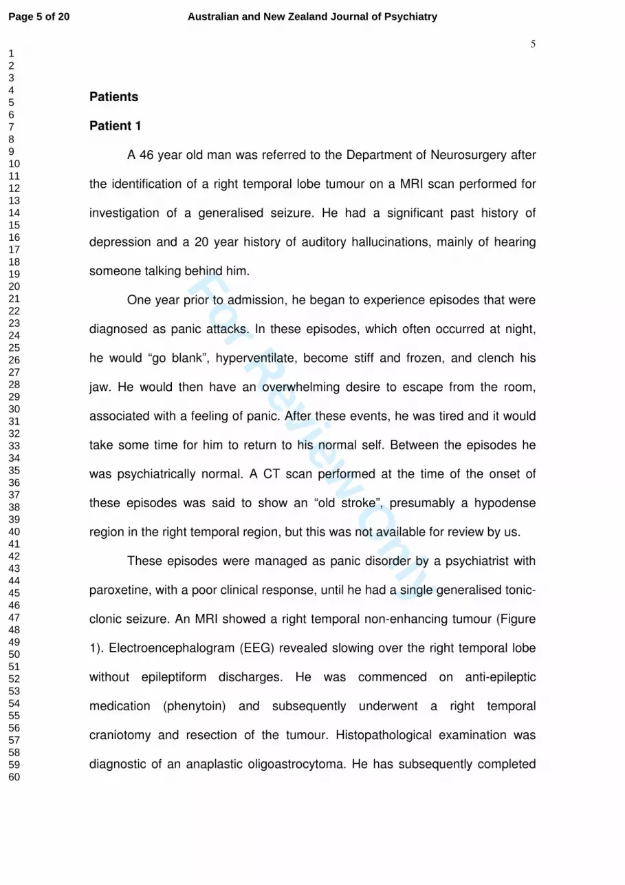
A 46 year old man was referred to the Department of Neurosurgery after
the identification of a right temporal lobe tumour on a MRI scan performed for
investigation of a generalised seizure. He had a significant past history of
depression and a 20 year history of auditory hallucinations, mainly of hearing
someone talking behind him.
One year prior to admission, he began to experience episodes that were
diagnosed as panic attacks. In these episodes, which often occurred at night,
he would “go blank”, hyperventilate, become stiff and frozen, and clench his
jaw. He would then have an overwhelming desire to escape from the room,
associated with a feeling of panic. After these events, he was tired and it would
take some time for him to return to his normal self. Between the episodes he
was psychiatrically normal. A CT scan performed at the time of the onset of
these episodes was said to show an “old stroke”, presumably a hypodense
region in the right temporal region, but this was not available for review by us.
These episodes were managed as panic disorder by a psychiatrist with
paroxetine, with a poor clinical response, until he had a single generalised tonic-
clonic seizure. An MRI showed a right temporal non-enhancing tumour (Figure
1). Electroencephalogram (EEG) revealed slowing over the right temporal lobe
without epileptiform discharges. He was commenced on anti-epileptic
medication (phenytoin) and subsequently underwent a right temporal
craniotomy and resection of the tumour. Histopathological examination was
diagnostic of an anaplastic oligoastrocytoma. He has subsequently completed
Page 5 of 20 Australian and New Zealand Journal of Psychiatry
123456789101112131415161718192021222324252627282930313233343536373839404142434445464748495051525354555657585960

For Review O
nly
6
adjuvant radiotherapy. He has had no further seizures, including those
associated with panic, for 12 months, but had an episode of depression
associated with commencing a second anti-epileptic medication (levetiracetam).
Patient 2
A 45 year old man was referred to the Department of Neuropsychiatry
with a 3 year history of episodes diagnosed and managed as panic attacks. The
attacks were frequent, occurring almost daily, and would on occasion wake him
from sleep. Treatment with the serotonin reuptake inhibitor, paroxetine, had not
altered the frequency or severity of the attacks. The only significant past history
was of a motor vehicle accident 20 years previously during which he sustained
a right frontal lobe injury requiring a craniotomy.
The episodes generally commenced with autonomic symptoms, including
palpitations, a cold sweat, dry mouth, shallow breathing and a sense of
impending doom. In association with these attacks he described a ‘funny
feeling’ in his stomach which rose into his chest. At times, he would also display
unusual motor activity including repetitive rubbing of his fingers, chewing and lip
smacking. He did not lose consciousness during these episodes but described
a feeling of ‘detachment’. The episodes were followed by fatigue.
A routine EEG followed by video-EEG monitoring was performed. During
the inter-ictal recording there were frequent epileptiform spikes over the right
posterior frontal region. Several typical events were recorded during monitoring
which were accompanied on the EEG by focal rhythmic seizure activity over this
region. MRI scan demonstrated a large area of encephalomalacia in the right
Page 6 of 20Australian and New Zealand Journal of Psychiatry
123456789101112131415161718192021222324252627282930313233343536373839404142434445464748495051525354555657585960

For Review O
nly
7
posterior frontal lobe, behind the region of the previous craniotomy (Figure 2) as
well as signal abnormality in the right mesial temporal region. He was
commenced on carbamazepine and has had no further events for over 12
months.
Patient 3
A 30 year old woman was referred to the Epilepsy Unit at the Austin
Hospital, Melbourne, following a generalised seizure. This seizure was
associated with sleep deprivation and excessive alcohol intake, which was
unusual for her. She had a history of depression and episodes that had been
diagnosed and managed as panic attacks. These occurred at least weekly and
usually in clusters of two or three per day. She had been referred to a
psychologist 2 years previously for counselling to manage these episodes,
which resulted in no significant change in their frequency of occurrence.
These episodes began 4 years prior to her first generalised seizure. The
initial symptoms consisted of an olfactory or gustatory aura of either burning
hair or coins. This was followed by a sense of fear or dread that she described
as “as if my family were being killed in front of me”. Witnesses observed that the
patient would usually stare and repeatedly swallow. She was unable to
communicate during the episodes but did not lose consciousness. They lasted
less than one minute and were followed by fatigue. They would often occur in
clusters and without obvious provocation.
An EEG showed slowing and sharp waves over the right temporal lobe.
MRI revealed a heterogenous lesion in the right parahippocampal gyrus
Page 7 of 20 Australian and New Zealand Journal of Psychiatry
123456789101112131415161718192021222324252627282930313233343536373839404142434445464748495051525354555657585960

For Review O
nly
8
consistent with a cavernous haemangioma (Figure 3). She was commenced on
carbamazepine, which only partially controlled her seizures. Other anti-epileptic
drugs, including lamotrigine and sodium valproate, also resulted in only partial
control. Video EEG monitoring and ictal single proton emission computed
tomography (SPECT) suggested she would benefit from resection of the lesion
in terms of seizure control.
She underwent resection of the cavernous malformation with access
through the middle temporal gyrus. The lesion was resected as a single piece
with aspiration of surrounding haemosiderin-stained brain. She made an
uncomplicated recovery and has been seizure free for more than 3 months on
her usual medications.
Discussion
These three patients, although harbouring differing intracerebral
pathology, presented with similar seizure related symptomatology with features
that led to misdiagnoses of panic attacks. A small number of similar reports in
the literature, have highlighted the diagnostic complexity of such cases and the
importance of further investigation of patients presenting panic attacks which
have atypical features or are resistant to treatment.4-9 In addition to symptoms
of anxiety or panic, on careful history such patients also manifest features
suggestive of focal epilepsy, including auras, automatisms and post-ictal
fatigue, which could reasonably have been considered an indication for early
radiological or EEG investigation. The cases highlight the similar clinical
Page 8 of 20Australian and New Zealand Journal of Psychiatry
123456789101112131415161718192021222324252627282930313233343536373839404142434445464748495051525354555657585960

For Review O
nly
9
symptomatology of panic attacks and seizures, particularly those arising in the
frontal and temporal lobes.
Temporal and frontal lobe epilepsies may feature simple partial seizures,
complex partial seizures, secondarily generalized seizures, or combinations of
these seizure types. Simple partial seizures arising from the temporal lobe are
typified by autonomic, psychological and sensory phenomena including
olfactory and auditory hallucinations and déjà vu. There is not uncommonly an
epigastric rising sensation or abdominal discomfort. Complex partial seizures of
the temporal lobe often begin with these same symptoms followed by motor
arrest and loss of awareness, typically accompanied by oroalimentary
automatism. Post ictal confusion is common.10 Electrical stimulation studies of
the temporal lobe have elicited panic and associated somatic signs and
symptoms including dizziness, nausea, tachycardia, chest pain, altered
temperature sensations and depersonalisation.11 Seizures with features similar
to panic attack have been particularly linked to epilepsy arising in the right
temporal lobe.12 In our series, patients 1 and 3 had lesions of the right temporal
lobe. Patient 2, whose seizures appeared to arise in the right posterior frontal
lobe, also exhibited abnormalities of the right mesial temporal lobe on MRI.
Seizures in the posterior frontal lobe commonly rapidly spread to the temporal
region.
Frontal lobe partial seizures are sometimes mistaken for psychogenic
nonepileptic seizures (PNES) or “pseudoseizures”. They can occur several
times a day and frequently arise out of sleep. Complex gestural automatisms
are common. Complex partial seizures arising from the frontal lobe often have
Page 9 of 20 Australian and New Zealand Journal of Psychiatry
123456789101112131415161718192021222324252627282930313233343536373839404142434445464748495051525354555657585960

For Review O
nly
10
minimal or no post ictal confusion. Rapid secondary generalization is a frequent
feature, with status epilepticus a not-uncommon complication.10
Based on these descriptions of temporal and frontal lobe seizures, the
“panic attacks” in each of the three patients presented had features strongly
suggestive of epilepsy. Two of the patients had a type of mass lesion, including
an intrinsic brain neoplasm and a cavernous haemangioma, which has been
associated with seizures in up to 80% of patients.13-17
Panic attacks, like seizures, are unexpected, brief, paroxysmal, and self-
limiting phenomena. DSM IV classifies a panic attack as a discrete period of
intense fear or discomfort that is associated with at least four of the following:
palpitations, pounding heart or accelerated heart rate; sweating; trembling or
shaking; sensations of shortness of breath or smothering; feeling of choking;
chest pain or discomfort; nausea or abdominal distress; feeling dizzy, unsteady,
lightheaded or faint; derealisation or depersonalization; fear of losing control or
going crazy; fear of dying; paraesthesias; chills or hot flushes.3 Each of the
patients presented here had episodes with features that also typified panic
attacks, thus contributing to the diagnostic difficulty.
In addition to anxiety or panic occurring as a feature of the seizure or its
aura, anxiety symptoms are common in patients with epilepsy (reviewed in18,19).
Anxiety may occur as a pre-ictal psychological reaction to symptoms that alert
the patient to seizure onset, as a feature of the post ictal state or as a distinct
interictal behaviour.20 Patients with epilepsy have a higher incidence of
comorbid non-seizure panic attacks or anxiety disorders than the general
population,18,21,22 and these may be misdiagnosed as seizures.23 Anxiety or
Page 10 of 20Australian and New Zealand Journal of Psychiatry
123456789101112131415161718192021222324252627282930313233343536373839404142434445464748495051525354555657585960

For Review O
nly
11
panic may be a feature of PNES in patient with or without co-existent epilepsy,
where the differential diagnosis may be particularly difficult.24 With careful
clinical evaluation, these similar symptoms can often be differentiated. For
example, the onset of ictal fear is paroxysmal and the duration is brief (30-120
sec)25 whereas panic attacks often build up over minutes with symptomatology
lasting more than five minutes.26 Furthermore, simple partial seizures
characterized by fear, anxiety or autonomic symptoms generally progress to
complex partial seizures, with change in consciousness, or generalise at some
time.4
Nonetheless, differentiating between seizures and panic attacks may be
difficult on clinical grounds alone. The patients reported here were all initially
diagnosed with, and had features of panic attacks, yet all had atypical features
to suggest seizures. Patient 1, with a right temporal anaplastic
oligoastrocytoma, had paroxysmal panic with a significant past history of
psychiatric illness, but also had oral automatisms and post-ictal fatigue. Patient
2, with post-traumatic right frontal encephalomalacia, had paroxysmal panic that
was resistant to paroxetine, but also had episodes that would wake him from
sleep, with manual and oral automatisms and post-ictal fatigue. Patient 3, with a
right parahippocampal gyrus cavernoma, had paroxysmal panic that was
resistant to psychotherapy, but the episodes were also preceded by an olfactory
or gustatory aura and had oral automatisms and post-ictal fatigue. While there
are differences between each presentation, a careful history from witnesses to
the episodes elicited a history consistent with oral automatisms and post-ictal
fatigue in all three patients.
Page 11 of 20 Australian and New Zealand Journal of Psychiatry
123456789101112131415161718192021222324252627282930313233343536373839404142434445464748495051525354555657585960

For Review O
nly
12
In conclusion, when evaluating a patient with panic attacks, it is important
to recognise that seizure symptomatology may also fulfil the DSM IV criteria for
panic disorder3,5 and that co-morbid psychiatric illness and seizures is
common.27 Failure to make the correct diagnosis may lead to a failure to control
symptoms, with prolonged suffering or delayed diagnosis of a neoplasm, or
treatable lesion, as well as exposing the patient to the physical and medical risk
of ongoing seizures. Patients with a first presentation of “panic attacks” require
a detailed history and clinical evaluation, with a high index of clinical suspicion
for epilepsy. A detailed history from witnesses to the episodes can provide
further information which leads to the diagnosis of a seizure disorder. With
readily available access to imaging, any unusual presentation of psychiatric
symptoms, or poor response to treatment, should warrant further investigation,
including referral to a specialist epilepsy unit with video-EEG monitoring.
Page 12 of 20Australian and New Zealand Journal of Psychiatry
123456789101112131415161718192021222324252627282930313233343536373839404142434445464748495051525354555657585960

For Review O
nly
13
References
1. Hutchinson G, Austin H, Neehall JE. Psychiatric symptoms and an
anterior cranial fossa meningioma. West Indian Med J 1998;47(3):111-2.
2. Wessling H, Simosono CL, Escosa-Bage M, de Las Heras-
Echeverria P. Anton's syndrome due to a giant anterior fossa
meningioma. The problem of routine use of advanced diagnostic imaging
in psychiatric care. Acta Neurochir (Wien) 2006;148(6):673-5.
3. American Psychiatric Society. Diagnostic and Statistical Manual of
Mental Disorders IV-Text Revision. 4th ed. Arlington VA: American
Psychiatric Publishing Inc.; 2000.
4. Young GB, Chandarana PC, Blume WT, McLachlan RS, Munoz
DG, Girvin JP. Mesial temporal lobe seizures presenting as anxiety
disorders. J Neuropsychiatry Clin Neurosci 1995;7(3):352-7.
5. Lee DO, Helmers SL, Steingard RJ, DeMaso DR. Case study:
seizure disorder presenting as panic disorder with agoraphobia. J Am
Acad Child Adolesc Psychiatry 1997;36(9):1295-8.
6. Laidlaw JD, Khin Maung Z. Epilepsy mistaken for panic attacks in
an adolescent girl. BMJ 1993;306(6879):709-10.
7. Picardi A, Di Gennaro G, Meldolesi GN, Grammaldo LG, Esposito
V, Quaranto PP. Partial seizures due to sclerosis of the right amygdala
presenting as panic disorder. On the importance of psychopathological
assessment in differential diagnosis. Psychopathology 2007;40(3):178-
83.
Page 13 of 20 Australian and New Zealand Journal of Psychiatry
123456789101112131415161718192021222324252627282930313233343536373839404142434445464748495051525354555657585960
For Review O
nly
14
8. Scalise A, Placidi F, Diomedi M, De Simone R, Gigli GL. Panic
disorder or epilepsy? A case report. J Neurol Sci 2006;246(1-2):173-5.
9. Saegusa S, Takahashi T, Moriya J, Yamakawa J, Itoh T, Kusaka
K et al. Panic attack symptoms in a patient with left temporal lobe
epilepsy. J Int Med Res 2004;32(1):94-6.
10. Commission on Classification and Terminology of the International
League Against Epilepsy. Proposal for revised classification of epilepsies
and epileptic syndromes. Epilepsia 1989;30(4):389-99.
11. Weilburg JB, Schachter S, Sachs GS, Worth J, Pollack MH, Ives
JR, et al. Focal paroxysmal EEG changes during atypical panic attacks. J
Neuropsychiatry Clin Neurosci 1993;5(1):50-5.
12. Sazgar M, Carlen PL, Wennberg R. Panic attack semiology in
right temporal lobe epilepsy. Epileptic Disord 2003;5(2):93-100.
13. Kilpatrick C. Epilepsy and its neurosurgical complications. In:
Kaye AH, editor. Essential Neurosurgery. Third ed; 2005. p. 269-80.
14. Hildebrand J, Lecaille C, Perennes J, Delattre JY. Epileptic
seizures during follow-up of patients treated for primary brain tumors.
Neurology 2005;65(2):212-5.
15. Stefan H, Hammen T. Cavernous haemangiomas, epilepsy and
treatment strategies. Acta Neurol Scand 2004;110(6):393-7.
16. Moran NF, Fish DR, Kitchen N, Shorvon S, Kendall BE, Stevens
JM. Supratentorial cavernous haemangiomas and epilepsy: a review of
the literature and case series. J Neurol Neurosurg Psychiatry
1999;66(5):561-8.
Page 14 of 20Australian and New Zealand Journal of Psychiatry
123456789101112131415161718192021222324252627282930313233343536373839404142434445464748495051525354555657585960

For Review O
nly
15
17. Noto S, Fujii M, Akimura T, Imoto H, Nomura S, Kajiwara K, et al.
Management of patients with cavernous angiomas presenting epileptic
seizures. Surg Neurol 2005;64(6):495-8, discussion 8-9.
18. Vazquez B, Devinsky O. Epilepsy and anxiety. Epilepsy Behav
2003;4(Suppl 4): S20-5.
19. Adams SJ, O'Brien TJ, Lloyd J, Kilpatrick CJ, Salzberg MR,
Velakoulis D. Neuropsychiatric morbidity in focal epilepsy. Br J
Psychiatry 2008;192(6):464-9.
20. Pariente PD, Lepine JP, Lellouch J. Lifetime history of panic
attacks and epilepsy: an association from a general population survey. J
Clin Psychiatry 1991;52(2):88-9.
21. Genton P, Bartolomei F, Guerrini R. Panic attacks mistaken for
relapse of epilepsy. Epilepsia 1995;36(1):48-51.
22. Mintzer S, Lopez F. Comorbidity of ictal fear and panic disorder.
Epilepsy Behav 2002;3(4):330-7.
23. Bernik MA, Corregiari FM, Braun IM. Panic attacks in the
differential diagnosis and treatment of resistant epilepsy. Depress
Anxiety 2002;15(4):190-2.
24. Witgert ME, Wheless JW, Breier JI. Frequency of panic symptoms
in psychogenic nonepileptic seizures. Epilepsy Behav 2005;6(2):174-8.
25. Devinsky O, Vazquez B. Behavioral changes associated with
epilepsy. Neurol Clin 1993;11(1):127-49.
26. Arun P, Chavan BS, Kumar N. Seizure disorder presenting as
panic attack. Indian J Med Sci 2002;56(10):486-8.
Page 15 of 20 Australian and New Zealand Journal of Psychiatry
123456789101112131415161718192021222324252627282930313233343536373839404142434445464748495051525354555657585960

For Review O
nly
16
27. Tsopelas ND, Saintfort R, Fricchione GL. The relationship of
psychiatric illnesses and seizures. Curr Psychiatry Rep 2001;3(3):235-
42.
Page 16 of 20Australian and New Zealand Journal of Psychiatry
123456789101112131415161718192021222324252627282930313233343536373839404142434445464748495051525354555657585960

For Review O
nly
17
Figure legends
Figure 1
T1-weighted axial contrast-enhanced MRI of the brain from Patient 1. There is a
hypointense region in the right temporal lobe which does not enhance with
contrast (arrow). Following resection the lesion was found to be an anaplastic
oligoastrocytoma on histological examination.
Figure 2
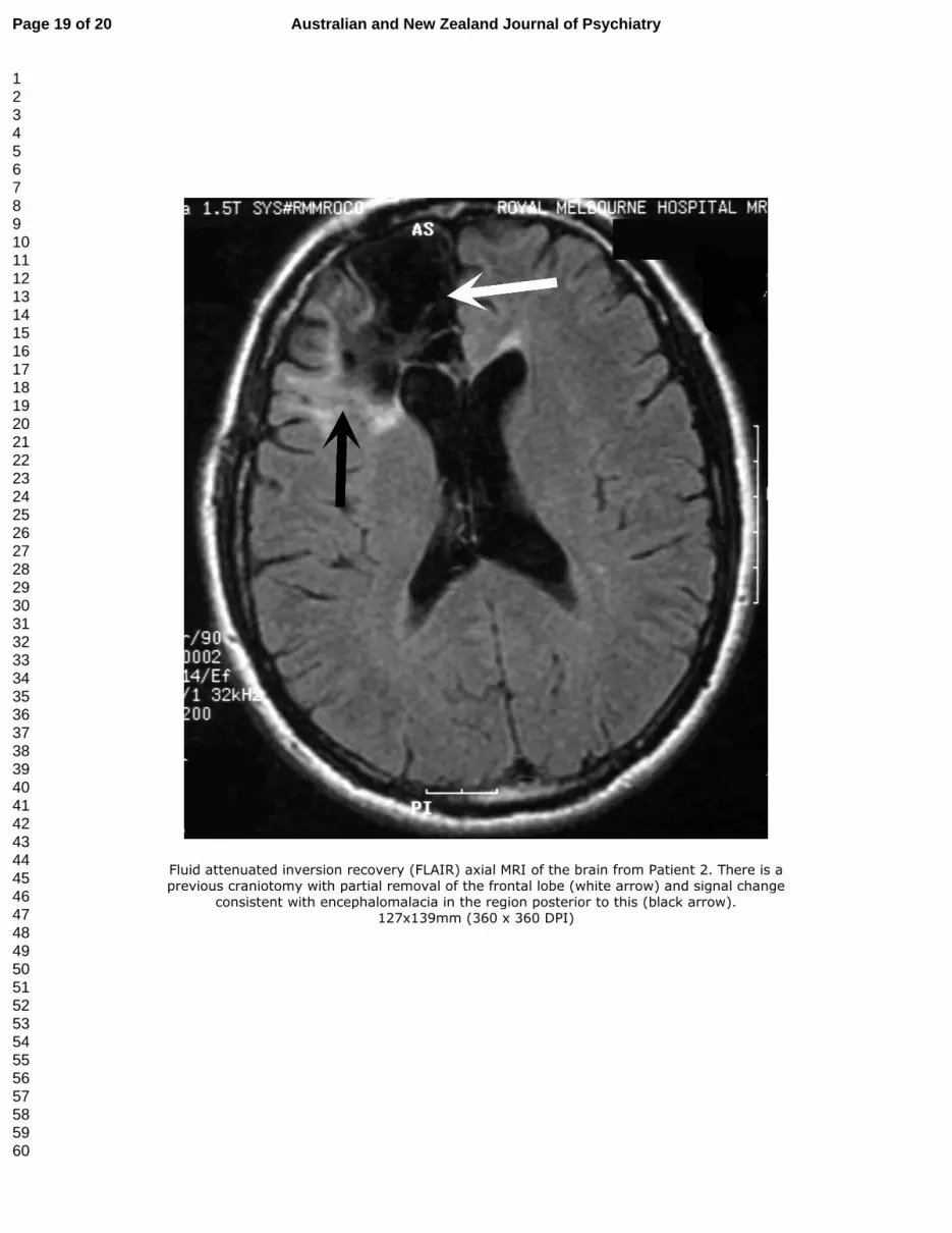
Fluid attenuated inversion recovery (FLAIR) axial MRI of the brain from Patient
2. There is a previous craniotomy with partial removal of the frontal lobe (white
arrow) and signal change consistent with encephalomalacia in the region
posterior to this (black arrow).
Figure 3
T2-weighted coronal MRI of the brain from Patient 3. There is a heterogenous
lesion in the right parahippocampal gyrus (arrow) consistent with a cavernous
haemangioma.
Page 17 of 20 Australian and New Zealand Journal of Psychiatry
123456789101112131415161718192021222324252627282930313233343536373839404142434445464748495051525354555657585960

For Review O
nly
T1-weighted axial contrast-enhanced MRI of the brain from Patient 1. There is a hypointense region in the right temporal lobe which does not enhance with contrast (arrow). Following resection the
lesion was found to be an anaplastic oligoastrocytoma on histological examination. 127x144mm (360 x 360 DPI)
Page 18 of 20Australian and New Zealand Journal of Psychiatry
123456789101112131415161718192021222324252627282930313233343536373839404142434445464748495051525354555657585960
For Review O
nly
Fluid attenuated inversion recovery (FLAIR) axial MRI of the brain from Patient 2. There is a previous craniotomy with partial removal of the frontal lobe (white arrow) and signal change
consistent with encephalomalacia in the region posterior to this (black arrow). 127x139mm (360 x 360 DPI)
Page 19 of 20 Australian and New Zealand Journal of Psychiatry
123456789101112131415161718192021222324252627282930313233343536373839404142434445464748495051525354555657585960

For Review O
nly
T2-weighted coronal MRI of the brain from Patient 3. There is a heterogenous lesion in the right parahippocampal gyrus (arrow) consistent with a cavernous haemangioma.
127x131mm (360 x 360 DPI)
Page 20 of 20Australian and New Zealand Journal of Psychiatry
123456789101112131415161718192021222324252627282930313233343536373839404142434445464748495051525354555657585960