adiaspores of emmonsia parva var. crescens in … · fischer o. a.:adiaspores of emmonsia parva...
TRANSCRIPT

ADIASPORES OF Emmonsia parva var. crescens IN LUNGSOF SMALL RODENTS IN A RURAL AREA
O. A. FISCHERBohuslava MartinÛ 44, Brno, Czech Republic
Received February 26, 2001Accepted May 28, 2001
AbstractFischer O. A. : Adiaspores of Emmonsia parva var. crescens in Lungs of Small Rodents in aRural Area. Acta Vet. Brno 2001, 70: 345–352.
The purpose of this study was to compare the occurrence of Emmonsia parva var. crescens inpopulated and unpopulated habitats of a rural area (in a village with 615 inhabitants and insurrounding forests without human population) as indicated by findings of adiaspores in lungs ofsmall rodents captured in these habitats. Adiaspores of E. parva var. crescens were found in lungsof 13 (9.6 %) out of 135 examined rodents. Four rodent species out of 7 were infected: the bankvole (Clethrionomys glareolus, 15.6 %), the house mouse (Mus musculus, 5.6 %), the wood mouse(Apodemus sylvaticus, 3.2 %) and the yellow-necked field mouse (A. flavicollis, 16.7 %). Thehighest prevalence was found in the A. flavicollis and in the C. glareolus in the village and in theforests, respectively. Intensity of the infection was low or moderate (below 100 adiaspores peranimal). The highest prevalence of the infection was found in spring. The prevalence was notinfluenced by sex of the animals. Occurrence of both saprophytic stage of E. parva var. crescensand rodents can be enabled by plant cover of uncultivated soil in the village, where the same plantspecies as in the surrounding forests occur. Therefore the risk of infection for humans and animalsis not limited only to forest (unpopulated) habitats.
Sapronoses, emmonsiosis, Arvicolidae, Muridae, Czech Republic
Emmonsiosis (adiaspiromycosis) is a world-wide distributed, largely neglected andunderdiagnosed pulmonary disease of mammals, including humans (Hubálek et al. 1998).Only one causative agent of emmonsiosis, the fungus Emmonsia parva var. crescens(Emmons et Jel l ison 1960 van Oorschot1980), occurs in the Czech Republic(Dvofiák et al. 1973). The infection is widespread especially among rodents of the familiesArvicolidae and Muridae (Hubálek et al. 1991) and more rarely in the families Sciuridae(Kfiivanec et al. 1976) and Cricetidae (Hubálek 1999). The source of infection is thesaprophytic stage of fungus producing minute conidia. After inhalation of the conidia bymammalian host, large thick-walled spherules called adiaspores develop in host tissues, mostoften in lungs. Expanding adiaspore, a parasitic stage of the fungus, causes inflammatoryreaction in lung tissue (Koìousek et al. 1971; Halouzka et al. 1989; Ooi and Lin1996), which leads to collapse of the adjacent alveoli resulting, in case of massive infection,to respiratory distress or even lung failure (Hubálek 1999). Macroscopicpathomorphological changes are densely disseminated whitish nodules about 1 to 2 mm indiameter, described by Koìousek et al. (1971) in a case human emmonsiosis. After deathof the infected host, the saprophytic stage can grow from the adiaspores released from deadhost’s body (Kfiivanec and Otãená‰ek 1977). Although the main source of conidia is thesoil (Dvofiák et al. 1973; Prokopiã and ·tûrba 1978), conditions of natural infectionwere not studied sufficiently. Because of danger for humans (Koìousek et al. 1971;England and Hochholzer 1993; Nuorva et al. 1997; de Montprévi l le et al. 1999)and close taxonomic relations of Emmonsia to other pathogenic fungi of the anamorphousgenera Paracoccidioides, Blastomyces and Histoplasma (Peterson and Sigler 1998)emmonsiosis was studied by many biologists, mycologists and microbiologists, but no
ACTA VET. BRNO 2001, 70: 345–352
Address for correspondence:Oldfiich Arno‰t FischerBohuslava MartinÛ 44CZ - 602 00 BrnoCzech Republic
Phone: + 420 5 4323 4315http://www.vfu.cz/acta-vet/actavet.htm
346
complete information about all possible routes of infection and health hazard for humans andanimals in densely populated areas, such as in villages and towns, is available. Most studieshave been hitherto performed in exoanthropic habitats. Village ecosystem includes not onlyintensively cultivated arable fields, orchards and gardens, which are not suitable for growingof the saprophytic stage (Hubálek et al. 1998), but also uncultivated areas with weeds, suchas nettle (Urtica spp.) and common elder (Sambucus nigra), and wet, shady places, enablinggrowth of the saprophytic stage of the fungus.
The aim of this study was to compare the occurrence of Emmonsia infection in village andforest habitats as indicated by findings of adiaspores in lungs of small rodents of the familiesArvicolidae and Muridae.
Materials and Methods
Area under s tudySouth-Moravian village Ketkovice near Brno (N 49o08’ E 16o06’, quadrat 6863 of the national faunal mapping
grid) with 615 inhabitants is situated 35 km west of Brno and 5 km from west margin of Rosice-Oslavany BlackCoal Basin in forest-arable hilly land at average elevation 433 m (340 – 480 m) a.s.l. (Fischer 2000). The roadfrom Rapotice to Oslavany passing through the village makes a frontier of nature reservation Oslava. The right(southwest) half of the village with adjacent pieces of land (fields and forests) belongs to the reservation, the left(northeast) one does not belong to this reservation. The climate is cold and dry, with average annual precipitationbelow 600 mm and drinking water deficits. The annual mean temperature is 7.5 oC (January – 5 oC, July 20 oC).The average year thickness of snow cover is less than 0.5 m. The air is polluted mostly by thermal powerplant andincinerator in Oslavany (7 km from Ketkovice). There are arable fields with potatoes, alfalfa, turnip, rye, wheat,barley, oat, intensively cultivated meadows, orchards and gardens around the village (Fischer 2000), but alsouncultivated soil with weeds. The village is surrounded with spruce (Picea abies, Pinus silvestris) and deciduous(Carpinus betulus, Quercus spp.) forests in a distance 250 – 1000 m from the margins of the village. Small brook(Ketkovick˘ potok) rises in the northern margin and flows through the centre of the village to the southeast. Anothersmall brook (Balinka) rises 100 m from north-eastern margin of the village and flows far away to southeast. In thevillage, the rodents were caught in a house at the eastern margin of the village. The miner family house built in 1932had three rooms, a shed, a former hen-house, two yards, a small alfalfa field and a small garden. The nearest forestmargin is about 1000 m from the house (Fischer 2000). No poultry or domestic animals were kept in this house,but rabbits, poultry, cats, dogs, pigs and goats were kept in neighbouring barns, and a cow-shed with about 200cows was at the western margin of the village, only 250 m from a forest. A stone marten (Martes foina Erxleben,1777) lived in loft of the house at this time.
Collect ion and examinat ion of rodentsSmall rodents were caught in snap-traps in the village house and in surrounding forests from July 3,1999 to
November 3, 2000. Captured rodents were determined, sexed and dissected. Macroscopic examinations of lungs wereperformed during dissections and special attention was given to any nodules in lung tissue. Whole lungs werepreserved in 10% (v/v) water solution of formaldehyde. They were warmed in the formaldehyde solution to 80 oC for30 minutes and after cooling to 40 oC prior the examination. Then they were exposed to 2% (w/v) water solution ofsodium hydroxide NaOH for 3 h. Compression preparations of small pieces of lungs were examined microscopicallyat a standard magnification 32 ×. Usually 10 slides were prepared from the whole lungs of one animal. All adiasporeswere measured. Only adiaspores with diameters above 70 µm were determined as E. parva var. crescens and counted.Intensity of infection was assessed as low (1 to 9 adiaspores), moderate (10 to 99 adiaspores), high (100 to 999adiaspores) and very high (1000 or more adiaspores per animal) according to Hubálek (1999).
Plant species composi t ionSpecial attention was given to occurrence of the most abundant plants in both habitats of the study area (Faustus
and Polívka 1976).
Stat is t icsStudent’s t-test was used to evaluate average numbers of adiaspores in animal species and mean diameters of
adiaspores (Table 2). Differences in the prevalence of infection (Tables 1 and 3) were evaluated by the Fisher’sexact test (Venãikov and Venãikov 1977).
Results
No macroscopic pathomorphological changes in lungs of trapped animals were observedand no nodules resembling Emmonsia granulomas were found. All adiaspores were found
347
in compression preparations only. A total of 13 (9.6 %) rodents out of 135 examined wereinfected (Table 1). Four out of 7 species of rodents were infected: Clethrionomys glareolus(15.6 %), Mus musculus (5.6 %), Apodemus sylvaticus (3.2 %) and A. flavicollis (16.7 %).Two rodent species, M. musculus and A. flavicollis, were infected in the village and threespecies, C. glareolus, A. sylvaticus and A. flavicollis, were infected in the forests. C.glareolus from the forests (17.5 %) was more often infected than A. flavicollis from the samehabitat (13.3 %; P > 0.05, non-significant). A. flavicollis was more often infected (P > 0.05)in the village (22.2 %) than in the forests (13.3 %). A. flavicollis in the village (22.2 %) wasmore infected (P > 0.05) than C. glareolus in the forests (17.5 %). A. flavicollis was also intotal more frequently infected than C. glareolus (16.7 % vs. 15.6 %; P > 0.05). In total,rodents captured in the forests (14.3 %) were significantly (P = 0.04) more frequentlyinfected than rodents from the village (4.6 %).
The smallest adiaspore (83 µm) and the largest one (575 µm) were found in one M. musculus captured in the village in August and in a C. glareolus captured in the forestin June, respectively. Intensity of the infections was low to moderate (Table 2). Twomoderate infections, 27 and 36 adiaspores per one host, were found in two A. flavicolliscaptured in the forests in March and April, respectively. A. flavicollis captured in the forestshad significantly greater number of adiaspores than A. flavicollis captured in the village (P < 0.01). Average diameters of adiaspores were greater (P < 0.01) in A. flavicollis fromthe village (282.1 ± 34.5 µm) than in A. flavicollis from the forests (205.9 ± 46.6 µm).
In total, adiaspores were found more often (P > 0.05) in animals captured in spring
Hosts Village Forests Both habitats
Positive/examined % Positive/examined % Positive/examined %
Bank vole 0 / 5 0 7 / 40 17.5 7 / 45 15.6
(Clethrionomys glareolus Schreber, 1780)
Common vole 0 / 14 0 0 / 0 0 0 / 14 0
(Microtus arvalisPallas, 1779)
Short-tailed vole 0 / 0 0 0 / 1 0 0 / 1 0
(M. agrestisLinnaeus, 1761)
House mouse 1 / 18 5.6 0 / 0 0 1 / 18 5.6
(Mus musculusLinnaeus, 1758)
Wood mouse 0 / 19 0 1 / 12 8.3 1 / 31 3.2
(Apodemus sylvaticusLinnaeus, 1758)
Yellow-necked field mouse 2 / 9 22.2 2 / 15 13.3 4 / 24 16.7
(A. flavicollisMelchior, 1834)
Herb field mouse 0 / 0 0 0 / 2 0 0 / 2 0
(A. micropsKratochvíl et Rosick˘, 1952)
Total 3 / 65 4.6 10 / 70 14.3 13 / 135 9.6
Table 1Findings of adiaspores of Emmonsia parva var. crescens in lungs of small rodents by their habitats

348
(19.2 %) than in winter (10.0 %) and in summer (9.8 %). The autumn prevalence (4.2 %)was not significantly lower than summer one (P > 0.05, Table 3).
Emmonsia infection was uniformly distributed between males and females. Three (13.6%) out of 22 examined males and 4 (17.4 %) out of 23 examined females of C. glareoluswere infected (P > 0.05). Two (20.0 %) out of 10 examined males and 2 (14.3 %) out of 14examined females of A. flavicollis were infected (P > 0.05). No statistically significantdiferences between prevalences in both sexes were found (P > 0.05).
Table 2Intensity of Emmonsia infection in small rodents
Table 3Emmonsia infection in seasons of the year
C. – Clethrionomys, M. – Mus, A. – Apodemus, V – village, F – forest, n – number of measured adiaspores, S.D.=
standard devation.
Total number of
adiaspores
found in one rodent
species
(average number
of adiaspores ± S.D.)
Low intensity of infection
(No. of cases)
Moderate intensity of
infection (No. of cases)
Minimal – maximal number
of adiaspores per host
Average
diameter
of adiaspores
± S.D. /µm/,
(range)
Habitat C. glareolus M. musculus A. sylvaticus A. flavicollis
V 0 1 0 7
(1.0 ± 0.0) (3.5 ± 3.5)
F 14 0 1 63
(2.0 ± 2.0) (1.0 ± 0.0) (31.5 ± 6.4)
V 0 1 0 2
F 7 0 1 0
V 0 0 0 0
F 0 0 0 2
V 0 1 0 1–6
F 1–4 0 1 27–36
V 0 83.0 ± 0.0 0 282.1 ± 34.5
n = 1 n = 7
(250 – 325)
F 377.5 ± 123.6 0 400.0 ± 0.0 205.9 ± 46.6
n = 14 n = 1 n = 63
(245 – 575) (125 – 325)
Hosts
Winter Spring Summer Autumn
(XII - II) (III - V) (VI - VIII) (IX - XI)
P. / E. % P. / E. % P. / E. % P. / E. %
Clethrionomys1 / 7 14.3 2 / 11 18.2 3 / 23 13.0 1 / 4 25.0
glareolus
Microtus arvalis 0 / 4 0 0 / 2 0 0 / 0 0 0 / 8 0
M. agrestis 0 / 0 0 0 / 0 0 0 / 0 0 0 / 1 0
Mus musculus 0 / 0 0 0 / 4 0 1 / 11 9.1 0 / 3 0
Apodemus sylvaticus 0 / 6 0 1 / 5 20.0 0 / 2 0 0 / 18 0
A. flavicollis 1 / 3 33.3 2 / 4 50.0 0 / 3 0 1 / 14 7.1
A. microps 0 / 0 0 0 / 0 0 0 / 2 0 0 / 0 0
Total 2 / 20 10.0 5 / 26 19.2 4 / 41 9.8 2 / 48 4.2
P. – number of positive hosts, E. – number of examined hosts.

349
Twenty nine abundant plant species /14 herbs, 6 shrubs and 9 trees/ occurring in largenumbers in forest habitat, village habitat or both were recorded. Sixteen plant species /8herbs: Dryopteris filix-mas (family Polypodiaceae), Anemone nemorosa (Ranunculaceae),Alliaria officinalis (Brassicaceae), Rubus idaeus (Rosaceae), Oxalis acetosella(Oxalidaceae), Impatiens parviflora (Impatientaceae), Hypericum perforatum(Hypericaceae) and Epilobium parviflorum (Oenotheraceae), one shrub: Ligustrum vulgare(Oleaceae) and 7 trees: Picea abies, Larix decidua, Pinus silvestris (Pinaceae), Carpinusbetulus, Betulla verucosa, Quercus robur (Betulaceae), Acer pseudoplatanus (Aceraceae)/were the most abundant plant species in forest habitat. Three herb species /Thlaspi arvense(Brassicaceae), Urtica urens (Urticaceae) and Pastinaca sativa (Daucaceae)/ were the mostabundant plant species in village habitat. Ten plant species /3 herbs: Chelidonium majus(Papaveraceae),Urtica dioica (Urticaceae), Daucus carota var. silvestris (Daucaceae), 5shrubs: Rubus fruticosus, Rosa canina, Prunus spinosa (Rosaceae), Corylus avellana(Betulaceae), Sambucus nigra (Loniceraceae) and 2 trees: Robinia pseudoacacia(Viciaceae) and Tilia cordata (Tiliaceae)/ occurred in both habitats of the area under study.
Discussion
Hubálek et al. (1997) examined rodents of nine species in six localities near Ketkovice(quadrat 6863) in the vicinity of Moravsk˘ Krumlov. The most infected species were C. glareolus (20.9 %), A. sylvaticus (11.4 %) and A. flavicollis (11.3 %). They recorded alsoinfection of M. arvalis (11.0 %) and one from five pine voles (M. subterraneus De Sélys-Longchamps, 1836). M. arvalis prefers fields and meadows. Most of individuals enteringthe house in Ketkovice originated from surrounding fields. Farmland habitats like arablefields, cultivated meadows, orchards and gardens are obviously less optimal thanuncultivated habitats (woods, shrubby balks or windbreaks between fields) for the growthof Emmonsia in the soil. This resulted in a lower incidence of rodent emmonsiosis in theagrocenoses (Hubálek et al. 1995a). Growth requirements of saprophytic stage of E. parvavar. crescens in nature have not been defined clearly. The most probable place of occurrenceof saprophytic stage is wet soil in shady places (Hubálek et al. 1995b, 1998) and lairs ofanimals, especially rodents (Prokopiã and ·tûrba 1978). Hubálek et al. (1998) founda higher prevalence of the infection in adult rodents from windbreaks than in those fromadjacent arable fields, 62.1 % and 8.2 %, respectively. Occurrence of Emmonsia was notinfluenced by water content and pH values of the soil, but significantly higher mean weightproportion of plant remnants was present in the soil from windbreaks than from fields.
Among 29 most abundant plant species growing in Ketkovice and surrounding forestswere ten species occuring in both habitats. C. majus, U. dioica and D. carota var. silvestrisare shadow-tolerating herbs (Faustus and Polívka 1976). R. fruticosus, R. canina, P. spinosa, C. avellana and S. nigra are bushes producing large amount of plant remnantsand they provide small rodents food (Holi‰ová 1960; Abt 1992) and shelter. R. pseudoacacia and T. cordata are deciduous trees producing plant remnants (Faustusand Polívka 1976). Because these plants grow in both habitats, small rodent populationsin village have similar living conditions as in the forests. In accordance with Hubálek etal. (1998), R. canina, S. nigra and T. cordata (growing also in the windbreaks) were foundamong plant species occurring in both habitats in Ketkovice near Brno.
Nuorva et al. (1997) described emmonsiosis in a two-year-old Finnish girl of Caucasianorigin. Her mother worked in a large garden shop and she was in contact with soil therefore.
Prokopiã and ·tûrba (1978) infected white laboratory mice (house mouse, M. musculus)by keeping them in a territory previously used by a colony of common voles (M. arvalis)spontaneously infected with E. parva var. crescens. Adiaspores 190 - 210 µm in diameter werefound in lungs of mice after four months of inhabiting of former vole lairs. Rodents from air-

350
polluted areas were more infected than rodents from non-polluted areas (Jeãn˘ andVojtûchová 1984; Hubálek et al. 1988).
High prevalence (C. glareolus 45 %, A. flavicollis 56 %, A. sylvaticus 26 %, M. agrestis13 % and M. arvalis 9 %) in rodents inhabiting the shores of fishponds in district Tfiebíã(about 40 km from Ketkovice) was found by Hubálek et al. (1995b). There are nofishponds or large water sources in Ketkovice (Fischer 2000).
The highest prevalence of infection occurs in spring and winter (Hubálek et al. 1995b).Although the age of rodents was not assessed in present study, it is well-known that adultanimals are more often infected than juveniles (Jeãn˘ and Vojtûchová 1984; Hubáleket al. 1988, 1997). Low autumn prevalence can be explained by the fact that rodentpopulations include many juvenile animals in the autumn (Rajska-Jurgiel 2000).
Emmonsia infection was not influenced by sex of animals. The same results reportedHubálek et al. (1988;1997).
Very low prevalence and intensity of infection in M. musculus can be explained by itssynanthropy. Hemisynanthropic A. flavicollis was the most infected rodent species in thevillage and the second most infected species in the forests. The most infected species in theforests was the exoanthropic bank vole (C. glareolus). Similar relations were observed inmustelid carnivores of the family Mustelidae by Kfiivanec and Otãená‰ek (1977).Whereas exoanthropic pine marten (Martes martes Linnaeus, 1758) and steppe polecat(Putorius eversmanni Lesson, 1827) captured in the Czechoslovakia had a high prevalence,72.2 % and 70.3 %, respectively, hemisynanthropic stone marten (M. foina) and dark polecat(P. putorius Linnaeus, 1758) had lower prevalence, 37.5 % and 30.6 %, respectively.Suitable living conditions for hemisynanthropic animals were indicated by presence of M. foina in house in Ketkovice during the study period.
No macroscopic pulmonary lesions were found in examined animals, because theintensity of infection was either low or moderate. Hubálek et al. (1988) found in lungs ofone short-tailed vole (M. agrestis) from air-polluted area of Kru‰né hory (Ore Mountains,Bohemia) as many as 1130 adiaspores (very high intensity of infection), which undoubtedlyinfluenced the health status of the animal.
Emmonsia infection was found not only in small rodents, but also in larger rodent speciessuch as squirrel (Sciurus vulgaris Linnaeus, 1758) and muskrat (Ondatra zibethicusLinnaeus, 1758) in the Czech Republic (Kfiivanec et al. 1976; Hubálek 1999) and beaver(Castor fiber Linnaeus, 1758) in Sweden (Mörner et al. 1999). Kfiivanec et al. (1976)found infection in lungs of 36 (20.5 %) from 176 squirrels captured in many various partsof the Czechoslovakia. The diameters of adiaspores varied from 150 to 600 µm, but mostfrequent were diameters of 400 – 500 µm. Adiaspores found in the beaver by Mörner etal. (1999) were 100 – 200 µm large.
The size of adiaspores indicates a probable time of infection of the host. Adiaspores of E. parva var. crescens reach a diameter of 130 – 230 µm within one month, that of 220 – 420µm in two months after infection (Hubálek et al. 1988). According to these data, most ofrodents were infected probably in autumn or winter.
Emmonsiosis is a neglected and underdiagnosed disease, because there are no suitablediagnostic methods. Small nodules can be overlooked or mistaken for otherpathomorphological changes, for instance miliary tuberculosis (Koìousek et al. 1971;Johnstone et al. 1993). Cultivation of Emmonsia spp. is difficult (Dvofiák et al. 1973;Kfiivanec and Otãená‰ek 1977) and serological dignostic methods are complicated bycross reactions with other soil fungi (Hubálek et al. 1998). Adiaspores can be easilydemonstrated by histological staining methods. They are large, with a typical structure(Halouzka et al. 1989), and can be well stained with gallocyanine blue (Koìousek et al.1971), PAS (Ooi and Lin 1996), Grocott (Jeãn˘ and Vojtûchová 1984; de
351
Montprévi l le et al. 1999; Mörner et al. 1999) and hematoxylin and eosin (deMontprévi l le et al. 1999; Mörner et al. 1999). However, histological examinations aretime consuming and expensive. Also reliable method of compression preparations of lungsis not performed at large scale.There is almost no information available about Emmonsiainfection in game, pets, laboratory, domestic and farm animals therefore.
Village areas provide suitable living conditions for saprophytic stage of E. parva var.crescens and for possible transmitters of adiaspores, small rodents, and risk of the infectionfor humans and animals is not limited only to the forests.
Adiaspory Emmonsia parva var. crescens v plicíchdrobn˘ch hlodavcÛ na venkovû
Úãelem studie bylo srovnání v˘skytu Emmonsia parva var. crescens v obydlené a neobydlené venkovské lokalitû (ve vesnici s 615 obyvateli a v okolních neobydlen˘chlesích) na základû nálezÛ adiaspor v plicích drobn˘ch hlodavcÛ chycen˘ch v tûchtolokalitách. Adiaspory E. parva var. crescens byly nalezeny v plicích 13 (9,6 %) ze 135vy‰etfien˘ch hlodavcÛ. Ze sedmi druhÛ hlodavcÛ byly infikovány ãtyfii: norník rud˘(Clethrionomys glareolus, 15,6 %), my‰ domácí (Mus musculus, 5,6 %), my‰ice kfiovinná(Apodemus sylvaticus, 3,2 %) a my‰ice lesní (A. flavicollis, 16,7 %). Prevalence byla vevesnici nejvy‰‰í u A. flavicollis, v lesích u C. glareolus. Intenzita infekce byla nízká nebostfiední (ménû neÏ 100 adiaspor na zvífie). Infekce byla zji‰Èována nejãastûji na jafie a pohlavízvífiat na ni nemûlo vliv. Rostlinn˘ kryt neobdûlávané pÛdy ve vesnici, v nûmÏ se vyskytujístejné rostliny jako v okolních lesích, mÛÏe podporovat jak v˘skyt hlodavcÛ tak v˘skytsaprofytního stadia E. parva var. crescens. Nebezpeãí nákazy pro lidi a zvífiata proto neníomezeno pouze na lesní (neobydlené) lokality.
References
ABT, K. F. 1992: Die Nahrung von Apodemus sylvaticus und A. flavicollis während eines Sommers im Gebiet derBornhöveder Seenkette (Schleswig-Holstein). Säugetierkd. Inf., Jena, 3: 409-419
DE MONTPRÉVILLE, V. T., HUERRE, M., DULMET, E. 1999: Adiaspiromycose, 2 cas de diagnostic fortuit.Ann. Pathol. 19: 513-515
DVO¤ÁK, J., OTâENÁ·EK, M., ROSICK¯, B. 1973: Adiaspiromycosis caused by Emmonsia crescens,Emmons et Jellison 1960. Study of the Czechoslovak Academy of Sciences No.14, Prague, Academia, 120 p.
ENGLAND, D. M., HOCHOLZER, L.1993: Adiaspiromycosis: an unusual fungal infection of the lung. Report of11 cases. Am. J. Surg. Pathol. 17: 876-886
FAUSTUS, L., POLÍVKA, F. 1976: Botanick˘ klíã. 1st ed., Prague, Státní pedagogické nakladatelství, 480 p.FISCHER, O. A. 2000: Blowflies of the genera Calliphora, Lucilia and Protophormia (Diptera, Calliphoridae)
in South-Moravian urban and rural areas with respect to Lucilia bufonivora Moniez, 1876. Acta Vet. Brno 69:225-231
HALOUZKA, R., GROCH, L., PIVNÍK, L.1989: Z alba patologické morfologie: adiaspora z plic hrabo‰e polního.Veterináfiství 39: 2
HOLI·OVÁ, V. 1960: Potrava my‰ice kfiovinné Apodemus sylvaticus L. na âeskomoravské vrchovinû. Folia Zool.9: 135-158
HUBÁLEK, Z. 1999: Emmonsiosis of wild rodents and insectivores in Czechland. J. Wildlife Dis. 35: 243-249HUBÁLEK, Z., JU¤ICOVÁ, Z., ZIMA, J. 1988: Adiaspiromycosis of mammals in an air-polluted area of
Czechoslovakia. Ekológia, Bratislava 7: 281-289HUBÁLEK, Z., KR·KA, A., GAISLER, J., ZEJDA, J., HEROLDOVÁ, M., RYCHNOVSK¯, B. 1997:
Emmonsiosis of small mammals (Rodentia, Insectivora) in southwest Moravia, Czech Republic. Folia Zool. 46:223-227
HUBÁLEK, Z., NESVADBOVÁ, J., HALOUZKA, J. 1998: Emmonsiosis of rodents in an agroecosystem. Med.Mycol. 36: 387-390
HUBÁLEK, Z., NESVADBOVÁ, J., RYCHNOVSK¯, B. 1995a: A heterogeneous distribution of Emmonsiaparva var. crescens in an agro-ecosystem. J. Med. Vet. Mycol. 33: 197-200
HUBÁLEK, Z., RYCHNOVSK¯, B., PE·KO, J. 1995b: Adiasporomycosis of rodents inhabiting the shores offishponds. Czech Mycol. 48: 139-144
JEâN¯, V., VOJTùCHOVÁ, A. 1984: Adiaspiromykóza drobn˘ch savcÛ z Mostecké kotliny Severoãeskéhnûdouhelné pánve. Sbor. Okres. Muzea, Most, 6: 11-21

JOHNSTONE, A. C., HUSSEIN, H. M., WOODGYER, A. 1993: Adiaspiromycosis in suspected cases ofpulmonary tuberculosis in the common brushtail possum (Trichosurus vulpecula). N. Z. Vet. J. 41: 175-178
KOëOUSEK, R., VORTEL, V., FINGERLAND, A., VOJTEK, V., ·ER¯, Z., HÁJEK,V., KUâERA, K. 1971:Pulmonary adiaspiromycosis in man caused by Emmonsia crescens: report of a unique case. Am. J. Clin. Pathol.56: 394-399
K¤IVANEC, K., DVO¤ÁK, J., OTâENÁ·EK, M. 1976: Emmonsia crescens Emmons et Jellison 1960 – a rarecausative agent of adiaspiromycosis in squirrels (Sciurus vulgaris L.) in the Czechoslovakia. Med. Parazitol.,Moscow, 45: 464-467 (In Russian)
K¤IVANEC, K., OTâENÁ·EK, M. 1977: Importance of free living mustelid carnivores in circulation ofadiaspiromycosis. Mycopathologia 60: 139-144
MÖRNER, T., AVENÄS, A., MATTSSON, R. 1999: Adiaspiromycosis in a European beaver from Sweden. J.Wildlife Dis. 35: 367-370
NUORVA, K., PITKÄNEN, R., ISSAKAINEN, J., HUTTUNEN, N. - P., JUHOLA, M. 1997: Pulmonaryadiaspiromycosis in a two year old girl. J. Clin. Pathol. 50: 82-85
OOI, H. - K., LIN, S. - C. 1996: Adiaspiromycosis in Niviventer coninga in Taiwan. Taiwan J. Vet. Med. Anim.Husb. 66: 253-258
PETERSON, S. W., SIGLER, L. 1998: Molecular genetic variation in Emmonsia crescens and Emmonsia parva,etiologic agents of adiaspiromycosis, and their phylogenetic relationship to Blastomyces dermatitis (Ajellomycesdermatitis) and other systemic fungal pathogens. J. Clin. Microbiol. 36: 2918-2925
PROKOPIâ, J., ·TùRBA, J. 1978: Spontaneous infection of white laboratory mice with Emmonsia crescensEmmons et Jellison, 1960 under natural conditions. Folia Parasitol. Prague 25: 371-374
RAJSKA-JURGIEL, E. 2000: Breeding dispersal in Clethrionomys glareolus females. Acta Theriol. 45: 367-376VENâIKOV, A. I., VENâIKOV, V. A. 1977: Základní metody statistického zpracování dat ve fyziologii. 1st ed.,
Prague, Avicenum, 160 p.
352

SCREENING FOR PENICILLIN PLASMA RESIDUES IN CATTLE BYENZYME-LINKED IMMUNOSORBENT ASSAY
H.J. LEE, P.D. RYU, H. LEE, M.H. CHO, M.H. LEE*
College of Veterinary Medicine and School of Agricultural Biotechnology,Seoul National University, Suwon 441-744, Korea
Received June 19, 2000Accepted August 28, 2001
Abstract
Lee, H. J . , P . D. Ryu, H. Lee, M. H. Cho, M. H. Lee:Screening for Penicillin PlasmaResidues in Cattle by Enzyme-Linked Immunosorbent Assay. Acta Vet. Brno 2001, 70: 353–358.
In this study, we established a rapid prediction test for the detection of the cattle with violativetissue residues of penicillins. The recommended therapeutic doses of two penicillins, ampicillin(withdrawal time, 6 days) and amoxicillin (withdrawal time, 14 days), were administered to twogroups of 10 cattle each. Blood was sampled and tested before drug administration and during thewithdrawal period. The concentration of penicillins in plasma, determined by a semi-quantitativeELISA, was compared to that of internal standard (4ppb as penicillin G). The absorbance ratio ofinternal standard to sample (B/Bs) was introduced as an index to determine whether drug residuesin cattle tissues are negative or positive. That means B/Bs ratio lower than 1 was considered residuepositive and that higher than 1 negative.
All 10 plasma samples from non-treated cattle showed negative results for both penicillins. Bothpenicillins were detected in plasma samples of cattle treated until the 3rd day of withdrawal period.
The present study has shown that the semi-quantitative ELISA could be easily adapted forprediction of screening plasma residues for penicillin antibiotics (ampicillin and amoxicillin) inlive cattle.
Penicillin ELISA, plasma, cattle contamination, live animal test
With the ever-growing world population, animal production practices have becomemore intensive and efficient, accompanied by increasing demands for drug treatment.Currently, approximately 80% of all food animals receive medication for part or most oftheir lives (Sternes jö et al. 1998). In the near future, nearly all animals bred in the worldfor food will receive chemotherapeutic and prophylactic agents of some type (Booth1988). A survey of all violative carcasses in the United States in 1993 revealed that the mostfrequent drug residues were penicillin (20%), streptomycin (10%), oxytetracycline (10%),and sulfamethazine (9%) (Paige 1994). According to Canadian Animal Health Institute,penicillins were the most frequently detected residues in milk in most countries(Heeschen et al. 1996). Since 1986, Department of Veterinary Service, Ministry ofAgriculture & Forestry, Korea has conducted National Residue Program (NRP) to samplemeat and poultry for residue tests at the slaughtering establishments under its inspectionauthority and from import shipments at the port of entry. In 1997, a total of 45,000 samplescomprising 10,000 beef, 23,000 pork, and 11,000 poultry meat were analyzed for five kindof antibiotics (penicillins and tetracyclines) and six sulfonamides. The results showedviolative residues of tetracyclines, sulfonamides and aminoglycosides in beef, pork, andpoultry meat.
A few cases of minor allergic reactions (e.g., skin rashes) in individuals previouslysensitized to penicillin G residues in milk and meat have been documented, as well asstrong evidence linking a widespread agricultural use of antibiotics to an increase inantibiotic resistance among the animal and human pathogens (Dewdney et al. 1984;
ACTA VET. BRNO 2001, 70: 353–358
Address for correspondence:Mun-Han Lee, DVM, Ph.D.Laboratory of biochemistryCollege of Veterinary Medicine, Seoul National UniversitySuwon 441-744, Korea
Phone: +88-31-290-2741Fax: +88-31-293-0084E-mail: [email protected]://www.vfu.cz/acta-vet/actavet.htm
354
Franco et al. 1990; Huber 1971; Kindred et al. 1993; Mitchel l et al. 1995;Ormerod et al. 1987).
The demands for reliable, simple, sensitive, rapid and low-cost methods for detectingresidues in foods continue to grow (Mitchel l et al. 1998; Lee et al. 2001). Variety ofenzyme immunoassays have been developed and adopted for detecting the generic groupsof chemical residues in milk, urine, blood, and meat samples (Gardner et al. 1996;Szekacs 1994; Lee et al. 2000; Lee et al. 2001). Enzyme-linked immunosorbent assay(ELISA) has become the most popular method for chemical residue detection in food due toits extreme sensitivity, simplicity, and ability to screen large number of samples (Cl i f ford1985; Gardner et al. 1996; Szekacs 1994; Lee et al. 2001).
In the present study, we developed a live animal test to predict the tissue residues ofpenicillins (ampicillin and amoxicillin) in cattle by examining the concentration of drug inblood during the withdrawal period obtained by an ELISA technique.
Materials and MethodsMaterials
Ten Holstein female cattle (7 - 8 month old, mean body mass 200 kg) were used in the experiments. BinotalInjection (100 mg/ml ampicillin natrium) was obtained from Bayer Korea Ltd. (Seoul, Korea). Clamoxyl L.A.Injection (150 mg /ml amoxicillin trihydrate) was obtained from Pfizer Korea Ltd. (Seoul, Korea). ELISA kits forß-lactams, manufactured by Idetek, were purchased from Korea Media Ltd.
Drug adminis t ra t ion and samples Ampicillin was administered intramuscularly to each of the 10 cattle at the rate of 11 mg per kg body weight per
day for seven consecutive days, and amoxicillin twice (24 h interval) intramuscularly to each of 10 cattle at 15 mgper kg body weight. Blood samples were collected from all cattle before administration of the drugs and on days 1,3, 5, 6, and 10 after the last ampicillin injection. From the cattle treated with amoxicillin, blood samples werecollected on days 1, 3, 7, 10, and 14. Ten ml of blood from each cattle were collected in heparinized tubes andcentrifuged at 4500 × g for 10 minutes to collect the plasma.
Preparat ion of s tandard curvesStock standard solution of 1000 µg/ml of each ampicillin and amoxicillin were prepared using USP standards in
saline. These stock solutions were further diluted with saline or blank serum to prepare 0, 1, 2, 5, 10, 20, 50, 100,500, and 1000 µg/ml working standard solutions. Standard curves of each antibiotic were constructed using thestandard solutions fortified into serum to determine the detection limit for the ELISA kit.
Analysis of penici l l ins in plasma ELISA tests for β-lactams were applied to each plasma sample in duplicate using a modified methodology
described by Boison et al. (1995), in which the manufacturer’s protocol for milk screening was adapted for plasmascreening. Briefly, 250 µl of the internal standard solution (equivalent to 4 ppb penicillin G) was pippetted into a test tube containing immobilized β-lactam antibodies. The plasma (250 µl, diluted 1 : 10 w/PBS) was pippettedinto individually labeled tubes. An equal volume of tracer solution (enzyme conjugate, lyophilized horseradishperoxidase labeled β-lactam conjugate with preservative) was added, and the test tubes were incubated at roomtemperature for 3 minutes with continuous shaking. The excess sample and conjugate reactants were then washedout with saline. A colour developer (0.5 ml, enzyme substrate) made up of 2,2’-azino-bis(3-ethylbenzothiazoline-6-sulfonic acid) and hydrogen peroxide in citrate buffer was added to the test tubes, and the mixture was incubatedat room temperature for 3 minutes with continuous shaking. Dilute sodium dodecyl sulfate solution (0.5 ml) wasadded to each test tube to stop the reaction. The absorbance was read at the wavelength of 405 nm with a photometricdetector (Idetek Reader, Awareness Technology, Inc., USA, operated in the 0.9 ratio mode) and compared withthat of the internal standard (4 ppb). Samples with absorbance higher than that of the internal standard wereconsidered to be negative (β-lactam drug free), and those with absorbance lower than that of the internal standardwere considered as positive. In this analysis, no more than 5 samples were processed simultaneously, and the assaywas completed within 10 minutes (Boison et al. 1995; Cullor et al. 1994).
Results
Standard curves and detect ion l imitsThe standard curves of ampicillin and amoxicillin were constructed to determine the
detection limits of each drug. The detection limits of ampicillin and amoxicillin were foundto be lower than 1 ppb based on the B/Bo ratio of 0.8 in the ELISA system (Figs. 1 and 2).

355
Live animal tes t for penici l l ins in plasma Ampici l l in . Results of plasma analysis are shown in Table 1. As the absorbance ratios
of normal 10 cattle of the control group were higher than 1.0, that is, the concentrations ofampicillin in the diluted plasma (× 10) of this group were higher than 4 ppb, the control groupwas negative. On day 1 of withdrawal, 8 of the 10 samples were found positive. The numberof positive samples on day 3 was 5. All samples showed negative reaction after day 5 ofwithdrawal (B/Bs ratio ≥1.0).
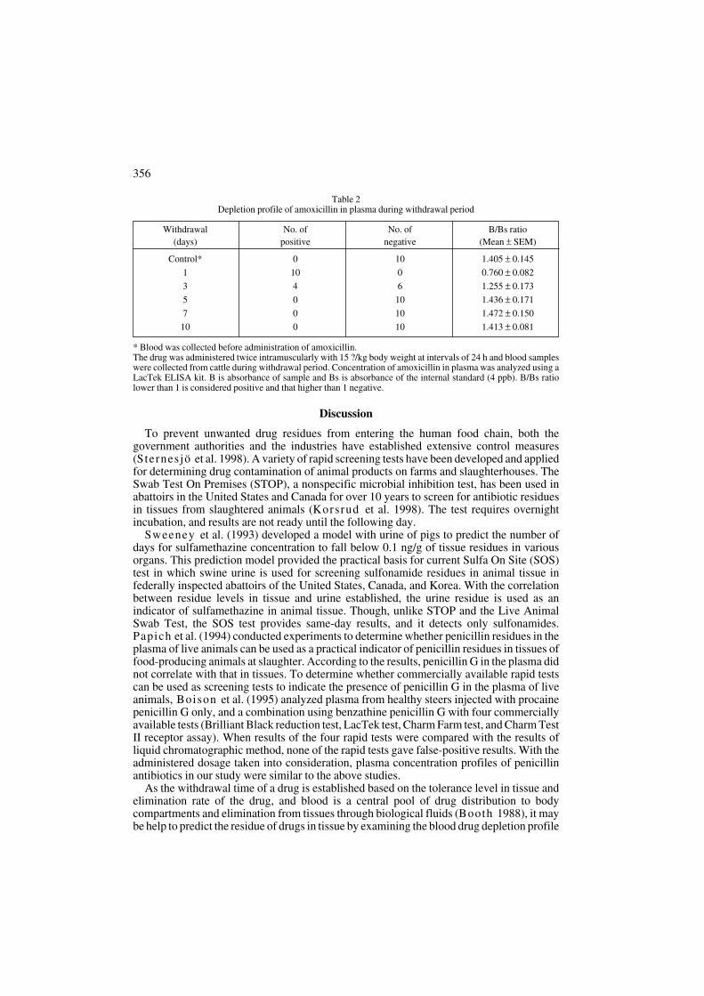
Amoxici l l in . Results of plasma analysis are shown in Table 2. As the absorbance ratiosof normal 10 cattle of the control group were higher than 1.0, that is, the concentrations ofamoxicillin in the diluted plasma (× 10) of this group were higher than 4 ppb, the group testednegative. All samples tested positive on day 1 of withdrawal. On day 3, 4 of the 10 samples werepositive. After day 5 of withdrawal, all samples showed negative reaction (B/Bs ratio ≥ 1.0).
1.0
0.8
0.6
0.4
0.2
0.0
B/B
o
0 1 10 100 1000
Ampicillin concentration (ng/ml)
PBSSerum
1.0
0.8
0.6
0.4
0.2
0.0B
/Bo
0 1 10 100 1000
Amoxicillin concentration (ng/ml)
PBSSerum
Fig. 1. Standard curves of ampicillin in phosphate buffersolution (PBS) and serum. Detection limit of ampicillinwas calculated as less as 1 ppb. The detection limit ofELISA kit was determined as the point of the B/Bs ratioof 0.8. B/Bo: Absorbance ratio of the standard (Bo) andPBS or control serum (B).
Fig. 2. Standard curves of amoxicillin in phosphatebuffer solution (PBS) and serum. Detection limit ofamoxicillin was calculated as less as 1 ppb. Thedetection limit of ELISA kit was determined as the pointof the B/Bs ratio of 0.8. B/Bo: Absorbance ratio of thestandard (Bo) and PBS or control serum (B).
Table 1Depletion profile of ampicillin in plasma during withdrawal period
* Blood was collected before administration of ampicillin.The drug was administered intramuscularly with 11 mg/kg body weight once daily for seven consecutive days, andblood samples were collected from cattle during the withdrawal period. Concentration of ampicillin in plasma wasanalyzed using a LacTek ELISA kit. B is absorbance of sample and Bs is absorbance of the internal standard (4 ppb). B/Bs ratio lower than 1.0 is considered positive and that higher than 1.0 negative.
Withdrawal No. of No. of B/Bs ratio
(days) positive negative (Mean ± SEM)
Control* 0 10 1.374 ± 0.079
1 8 2 0.818 ± 0.171
3 5 5 1.126 ± 0.457
5 0 10 1.409 ± 0.119
6 0 10 1.443 ± 0.070
356
Discussion
To prevent unwanted drug residues from entering the human food chain, both thegovernment authorities and the industries have established extensive control measures(Sternesjö et al. 1998). A variety of rapid screening tests have been developed and appliedfor determining drug contamination of animal products on farms and slaughterhouses. TheSwab Test On Premises (STOP), a nonspecific microbial inhibition test, has been used inabattoirs in the United States and Canada for over 10 years to screen for antibiotic residuesin tissues from slaughtered animals (Korsrud et al. 1998). The test requires overnightincubation, and results are not ready until the following day.
Sweeney et al. (1993) developed a model with urine of pigs to predict the number ofdays for sulfamethazine concentration to fall below 0.1 ng/g of tissue residues in variousorgans. This prediction model provided the practical basis for current Sulfa On Site (SOS)test in which swine urine is used for screening sulfonamide residues in animal tissue infederally inspected abattoirs of the United States, Canada, and Korea. With the correlationbetween residue levels in tissue and urine established, the urine residue is used as anindicator of sulfamethazine in animal tissue. Though, unlike STOP and the Live AnimalSwab Test, the SOS test provides same-day results, and it detects only sulfonamides.Papich et al. (1994) conducted experiments to determine whether penicillin residues in theplasma of live animals can be used as a practical indicator of penicillin residues in tissues offood-producing animals at slaughter. According to the results, penicillin G in the plasma didnot correlate with that in tissues. To determine whether commercially available rapid testscan be used as screening tests to indicate the presence of penicillin G in the plasma of liveanimals, Boison et al. (1995) analyzed plasma from healthy steers injected with procainepenicillin G only, and a combination using benzathine penicillin G with four commerciallyavailable tests (Brilliant Black reduction test, LacTek test, Charm Farm test, and Charm TestII receptor assay). When results of the four rapid tests were compared with the results ofliquid chromatographic method, none of the rapid tests gave false-positive results. With theadministered dosage taken into consideration, plasma concentration profiles of penicillinantibiotics in our study were similar to the above studies.
As the withdrawal time of a drug is established based on the tolerance level in tissue andelimination rate of the drug, and blood is a central pool of drug distribution to bodycompartments and elimination from tissues through biological fluids (Booth 1988), it maybe help to predict the residue of drugs in tissue by examining the blood drug depletion profile
Table 2Depletion profile of amoxicillin in plasma during withdrawal period
* Blood was collected before administration of amoxicillin.The drug was administered twice intramuscularly with 15 ?/kg body weight at intervals of 24 h and blood sampleswere collected from cattle during withdrawal period. Concentration of amoxicillin in plasma was analyzed using aLacTek ELISA kit. B is absorbance of sample and Bs is absorbance of the internal standard (4 ppb). B/Bs ratiolower than 1 is considered positive and that higher than 1 negative.
Withdrawal No. of No. of B/Bs ratio
(days) positive negative (Mean ± SEM)
Control* 0 10 1.405 ± 0.145
1 10 0 0.760 ± 0.082
3 4 6 1.255 ± 0.173
5 0 10 1.436 ± 0.171
7 0 10 1.472 ± 0.150
10 0 10 1.413 ± 0.081

during withdrawal period (Korsrud et al. 1995; Boison et al. 1995; Lee et al. 2000; Leeet al. 2001). According to our results, the developed methods can be adapted easily to pre-detect residues of penicillin antibiotics (ampicillin and amoxicillin) in live cattle usingdiluted blood plasma (× 10) with the modified ELISA test kits.
It is conceivable that the veterinary inspector in the abattoir may be able to use this methodto screen for penicillin antibiotics in plasma from live cattle in holding pens prior to slaughterand obtain same-day results. Cattle that show positive can then be held in the pens until retestresults come up negative before they are slaughtered.
Aplikace metody ELISA pro stanovení penicilínovích reziduí v krevní plasmû skotu
V této studii byl vypracován postup pro detekci prchav˘ch zbytkÛ penicilínu v tkaniváchskotu. Doporuãované dávky dvou penicilínÛ, jmenovitû ampicilínu (doba odbourání 6 dní)a amoxicilínu (doba odbourání 14 dní) byly podány dvûma skupinám skotu (n = 10). Krevbyla odebrána a testována pfied podáním lékÛ i poãas jejich odbourávání. Koncentracepenicilínu v plasmû byla stanovena pomocí semikvantitativní metody ELISA a bylaporovnána s koncentrací interního standardu (4ppb penicilínu G). Pomûr absorbanceinterního standardu k absorbanci vzorky (B/Bs) byl definován jako index k urãení ãi zbytekantibiotik ve tkáních byl pozitivní anebo negativní. Pomûr B/Bs men‰í neÏ 1 byl pokládánjako reziduálnû pozitivní a pomûr vet‰í neÏ 1 jako reziduálnû negativní.
KaÏd˘ z deseti vzorkÛ odebran˘ch skotu, jemuÏ nebylo podáno Ïádne antibiotikum bylnegativní na oba penicilíny. Oba v‰ak byly detekovány v plasmû zvífiat do tfietího dne odpodání dávky.
Táto studie potvrdila, Ïe semikvantitativní metoda ELISA mÛÏe b˘t snadno adaptovánapro stanovení reziduí antibiotik penicilínové fiady (ampicilínu a amoxicilínu) v krevníplasmû skotu.
Acknowledgements
This work was supported by the Brain Korea 21 Project and a grant from Agricultural Research and PromotionCenter.
References
BOISON, J. O., KORSRUD, G. O., PAPICH, M. G., MACNEIL, J. D. 1995: Comparison of four commerciallyavailable rapid test kits with liquid chromatography for detecting penicillin G residues in bovine plasma. J. Assoc. Off. Anal. Chem. Int. 78: 1144-1152
BOOTH, N. H.1988: Veterinary pharmacology and therapeutics. In: BOOTH, N. H., MCDONALD, L. E.:Toxicology of drug and chemical residues. Iowa State University Press, Ames, Iowa, pp. 1149-1205
CULLOR, J. S., VAN EENENNAAM, A., GARDNER, I., PERANI, L., DELLINGER, J., SMIT, W. L.,THOMPSON, T., PAYNE, M. A., JENSEN, L., GUTERBOCK, W. M.1994: Performance of various testsused to screen antibiotic residues in milk samples from individual animals. J. Assoc. Off. Anal. Chem. Int.77: 862-870
DEWDNEY, J. M., EDWARDS, R. G. 1984: Penicillin hypersensitivity is milk a significant hazard?: a review. J. Rech. Soc. Med. 77: 866-877
FRANCO, D. A., WEBB, J., TAYLOR, C. E. 1990: Antibiotic and sulfonamide residues in meat: implications forhuman health. J. Food Prot. 53: 178-185
GARDNER, I. A., CULLOR, J. S., GALEY, F. D., SISCHO, W., SALMAN, M., SLENNING, B., ERB, H. N.,TYLER, J. W.1996: Alternatives for the validation of diagnostic assays used to detect antibiotic residues in milk.J. Am. Vet. Med. Assoc. 209: 46-52
HEESCEN, W. H., SUHREN, G. 1996: Principles of and practical experiences with an integrated system for thedetection of antimicrobials in milk. Milchwissenschaft. 51: 154-164
HUBER, W. G.1971: The impact of antibiotic drugs and their residues. Adv. Vet. Sci. Comp. Med. 15: 101-132KINDRD, T. P., HUBBERT, W. T. 1993: Residue prevention strategies in the United States. J. Am. Vet. Med.
Assoc. 202: 46-49KORSRUD, G. O., SALIBURY, C.D. C., FESSER, A. C. E., MACNEIL, J. D. 1995: Laboratory evaluation of the
Charm Farm test for antimicrobial residues in meat. J. Food Prot. 58: 1129-1132KORSRUD, G. O., SALISBURY, C. D. C., RODES, C. S., PAPICH, M. G., YATES, W. D. G., BULMER, W. S.,
357

MACNEIL, J. D., LANDRY, D. A., LAMBERT, G., YONG, M. S., RITTERS, L. 1998: Depletion of penicillinG residues in tissues, plasma and injection sites of market pigs injected intramuscularly with procaine penicillinG. Food Addit. Contam. 15: 421-426
LEE, H. J., LEE, M. H., HAN, I. K. 2001: Application of ELISA for the detection of oxytetracycline residue in liveanimal. Asian-Aust. J. Anim. Sci. 14: 378-381
LEE, H. J., LEE, M. H., HAN, I. K. 2000: Application of ELISA for the detection of penicillin antibiotic residuesin live animal. Asian-Aust. J. Anim. Sci. 13: 1604-1608
LEE, H. J., LEE, M. H., RYU, P. D., LEE, H., CHO, M. H. 2001: Enzyme-linked immunosorbent assay forscreening the residues of tetracycline antibiotics in pigs. J. Vet. Med. Sci. 63: 553-556
LEE, M. H., LEE, H. J., RYU, P. D. 2001: Public health risks: chemical and antibiotic residues. Asian-Aust. J. Anim. Sci. 14: 402-413
MITCHELL, J. M., YEE, A. J. 1995: Antibiotic use and transfer of drug resistance: does it mean we should stoptreating animals with these drugs? Dairy Food Environ. Sanit. 15: 484-487
MITCHELL, J. M., GRIFFITHS, M. W., MCEWEN, S. A., MCNAB, W. B., Yee, A. J. 1998: ntimicrobial drugresidues in milk and meat: causes, concerns, prevalence, regulations, tests, and test performance. J. Food Prot.61: 742-56
ORMEROD, A. D., REID, T. M. S., MAIN, R. A. 1987: Penicillin in milk-its importance in urticaria. Clin. Allergy.17: 229-234
PAIGE, J. C. 1994: Analysis of tissue residues. FDA Vet. 9: 4-6PAPICH, M. G., KORSRUD, G. O., BOISON, J. O., YATES, W. D., MACNEIL, J. D., JANZEN, E. D.,
MCKINNON, J. J., LANDRY, D. A. 1994: Disposition of penicillin G after administration of benzathinepenicillin G, or a combination of benzathine penicillin G and procaine penicillin G in cattle. Am. J. Vet. Res. 55:825-30
STERNESJÖ, Ω., JOHNSSON, G. 1998: A Novel Rapid Enzyme Immunoassay (Fluorophos BetaScreen) forDetection of ß-Lactam Residues in Ex-Farm Raw Milk. J. Food Prot. 61: 808-811
SWEENEY, R. W., BARDALAYE, P. C., SMITH, C. M., SOMA, L. R., UBOH, C. E. 1993: Pharmacokineticmodel for predicting sulfamethazine disposition in pigs. Am. J. Vet. Res. 54: 750-754
SZEKACS, A. 1994: Development of enzyme-linked immunosorbent assay (ELISA) systems for environmentalmonitoring. Acta Biol. Hung. 45: 77-80
358
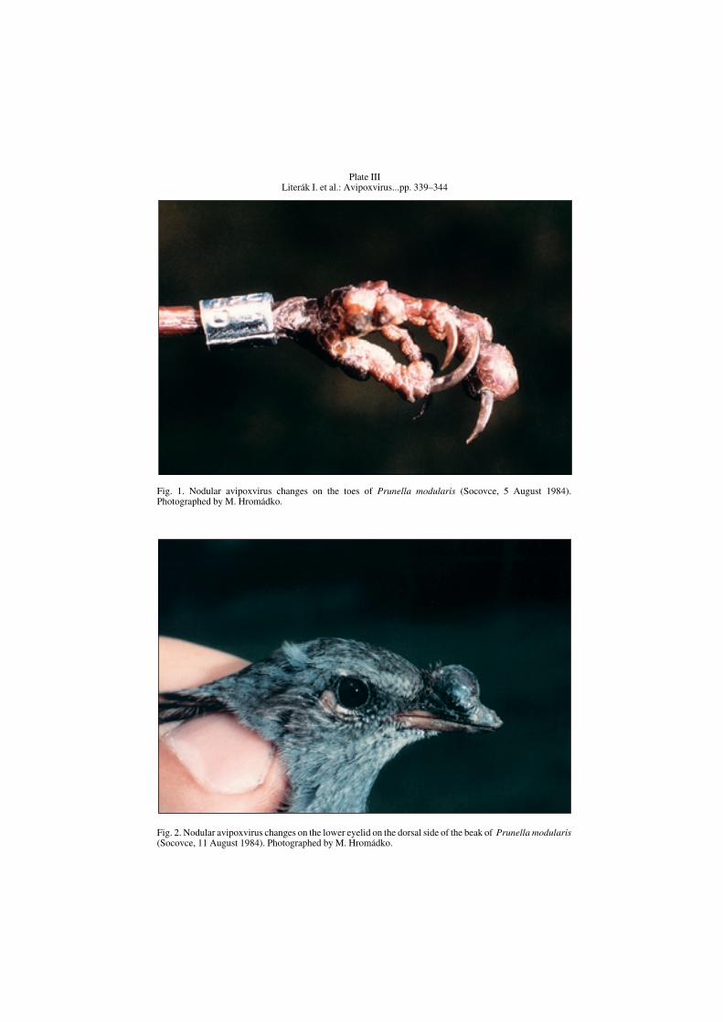
Plate IIILiterák I. et al.: Avipoxvirus...pp. 339–344
Fig. 1. Nodular avipoxvirus changes on the toes of Prunella modularis (Socovce, 5 August 1984).Photographed by M. Hromádko.
Fig. 2. Nodular avipoxvirus changes on the lower eyelid on the dorsal side of the beak of Prunella modularis(Socovce, 11 August 1984). Photographed by M. Hromádko.

Plate IV
Fig. 3. Avipoxvirus changes on the eyelid, i n the corner of the beak, on the distal end of the wing and on thetoes of Sylvia atricapilla (Socovce, 27 July 1999). Photographed by I. Literák.
Fig. 4. Detail of hypertrophic keratinocytes with Bollinger bodies. Deep zone of epidermal nodularhyperplasia. On the right side inflammatory infiltrate in the dermis. (Sylvia atricapilla, Socovce, 27 July1999, HE, × 500). Photographed by R. Halouzka.