thyroid-specific and camp-dependent hypersensitive regions in thyroglobulin gene chromatin
TRANSCRIPT

Eur. .I. Biochem. 178, 387-393 (1988) (<) FEBS 1988
Thyroid-specific and CAMP-dependent hypersensitive regions in thyroglobulin gene chromatin Carole HANSEN I , Catherine GERARD I , Gilbert VASSART’, Patrick STORDEUR’ and Daniel CHRISTOPHE’ ’ Institut de Recherche Interdisciplinaire en Biologie Humaine et Nucleaire and Unite de Genetique Moleculaire,
Service de Gknetique Medicale, Universite Libre de Bruxelles
(Received April 20/August 30, 1988) - EJB 88 0458
Two regions hypersensitive to DNase I digestion were found in a 7-kb segment of thyroglobulin gene 5‘ - flanking sequences in the chromatin from bovine thyroid.
The most upstream site (-2000 to -1600 bp relative to the transcriptional start) was found in thyroid chromatin only, but independently of actual expression of the gene. It therefore represents a tissue-specific characteristic which may be associated with the commitment of the thyroglobulin gene to transcriptional activity.
The very 5’ end of the gene and the proximal promoter sequences (- 100 to f 6 0 bp relative to transcriptional start), constitute the second site, the hypersensitive character of which could be directly correlated with trans- criptional activity. The structural changes occurring in this region of the chromatin were dependent on cAMP stimulation of the thyroid cells.
Specialized cells in eukaryotes selectively express only a small subset of genes during development and differentiation. It has been shown that expressed genes are more sensitive to DNase I digestion than inactive genes [l], suggesting a more open conformation of the active chromatin. The domain of increased sensitivity includes the structural gene and the re- gions flanking it [2]. Moreover, short specific sequences, lo- cated in the vicinity (5’ or 3’) of transcription units are approximatively ten times more sensitive to DNase I digestion than the bulk of the active gene regions. They are referred to as hypersensitive sites [3], reviewed in [4]. DNase-I-hypersensi- tive sites seem to indicate positions along the chromatin fiber which have lost specific chromosomal proteins and where the DNA sequence is more accessible to regulatory proteins.
For developmentally regulated genes [ 5 - 71 the appear- ance of DNase-I-hypersensitive sites usually precedes the ini- tiation of transcription and displays tissue- or stage-speci- ficity. For several hormonally controlled genes, the appear- ance of hypersensitivity at fixed positions could be directly correlated with transcriptional activity [8, 91. In the case of the human histone H4 gene, modifications were detected in the extent of hypersensitivity which parallel the cell-cycle- dependent changes in expression of this gene [lo].
Some of the regions which displayed the hypersensitive character upon activation of transcription were shown to correspond to critical promoter elements [I I] or, more directly, to constitute target sequences for trans-acting agents [12, 131.
Correspondence to C. Hansen, lnstitut de Recherche Inter- disciplinaire en Biologic Humaine et Nucleaire, Universite Libre de Bruxelles, Campus Erasme, Bitiment C, Route de Lennik 808, B-1070 Bruxelles, Belgium
Ahhreviations. BME, basal Eagle’s medium; DMEM, Dulbecco’s modified basal Eagle’s medium; EGF, epidermal growth factor; FCS, fetal calf serum; PhMeS02F, phenylmethylsulfonyl fluoride; NaCI/ Pi, phosphate-buffered saline; NaCl/Cit, 0.1 5 M sodium chloride, 0.015 M trisodium citrate pH 7.0.
_ _ _ ~ -
Enz.vrne. Deoxyribonuclease I, DNase I (EC 3.1.21.1).
In the present work, we examine the chromatin struc- ture of the promoter region of the thyroglobulin gene. This gene is only expressed in differentiated thyroid cells under the positive control of the pituitary hormone thyrotropin. Van Heuverswyn et al. [14] demonstrated that this control occurs, at least partly, at the transcriptional level and is mediated by cAMP [15]. The recent cloning of 5’ genomic fragments from the bovine thyroglobulin gene [16] provided the starting ma- terial to investigate what changes in chromatin structure are involved in the tight tissue-specific and CAMP-mediated con- trol of thyroglobulin gene expression.
MATERIALS AND METHODS
Cell culture
Primary cultures of bovine thyrocytes were obtained as described in [17]. Briefly, thyroid follicles were obtained by enzymatic digestion (2 h at 39°C in collagenase 1 mg/ml, dis- pase 5 mg/ml, DNase 400 pg/ml in Eagle’s basal medium (BME) from minced calf thyroid tissue collected at the slaugh- terhouse. Follicles were purified by several low-speed centrifugations in BME. They were seeded in 90-mm culture dishes and adhered to the plastic substrate within 1 or 2 days. They were maintained in a medium containing DMEM, Ham’s F12 (Flow Laboratories), MCDB104 (Gibco) ( 2 : 1 : 1) supplemented with antibiotics (penicillin 100 U/ml, strepto- mycin 100 pg/ml), 2.5 pg/ml amphotericin B, 2 mM gluta- mine, 5 pg/ml insulin, 1.25 pg/ml transferrin, 10 ng/ml glycyl- histidyllysyl acetate, 10 ng/ml somatostatin and 40 pg/ml ascorbic acid. The culture dishes were placed in a water- saturated incubator at 37°C in an atmosphere of 5% COz in air. The medium was changed every 2 days. Drugs and hormones were added and maintained in the culture medium as mentioned in Results.
388
Isolation of nuclei and digestion
From fresh tissue. Beef thyroid glands and pieces of liver were collected at the abattoir soon after slaughtering and immediately placed on ice. All the following steps were performed at 4°C. The tissue was cleaned, cut into small pieces and transferred into ice-cold NaCl/Cit (0.1 5 M NaCI, 0.01 5 M sodium citrate) + 10 mM Tris/HCl, pH 7.4 (10 ml/g tissue). The tissue was then minced, resuspended in the same buffer and allowed to settle. The supernatant was discarded and the process was repeated three times. The last time, the suspension was centrifuged for 5 rnin at 1000 x g. The pellet was resus- pended (10 ml/g tissue) in 10 mM Tris/HCl pH 7.4, I0 mM NaCl, 3 mM MgC12 containing 0.5% Nonidet P-40 and 1 mM PhMeS02F, and homogenized (teflon/glass Potter homoge- nizer). The homogenate was filtered through gauze and centri- fuged for 10 rnin at 2000 rpm in a Sorvall HS4 rotor. The crude nuclear pellet was washed twice in the same buffer (without PhMeS02F) and finally resuspended in 10 mM Tris/ HC1 pH 7.4, 10 mM NaCl, 3 mM MgC12 (0.5 ml/g tissue). 0.7 ml nuclear suspension was incubated for 10 min at 37°C in 10 mM Tris/HCl pH 7.4,lO mM NaCl, 3 mM MgC12 sup- plemented with 0.1 mM CaC1, and with variable amounts of DNase I (Sigma; 0.05 - 2 pg/ml). The reaction was stopped by adding an equal volume of 1 YO SDS, 0.6 M NaCl, 20 mM Tris/HCl pH 7.4,lO mM EDTA, 400 pg/ml proteinase K and left overnight at room temperature. Aliquots of the nuclear suspension were treated with proteinase K without being pre- viously incubated to provide controls for endogenous nuclease activity. The mixture was then extracted twice with phenol and the DNA was ethanol-precipitated. The DNA pellet (800 pg) was dissolved in 500 pl 10 mM Tris/HCl pH 7 . 5 , l mM EDTA.
From cultured cells. Cells were scraped from the dishes, washed twice in NaCI/Pi and resuspended in 20 mM Tris/HCl pH 7.4, 3 mM CaCI2, 2 mM MgCI2. Nonidet P-40 was then added to a final concentration of 0.3% and the suspension was kept at 0°C for 10 min. The cells were disrupted (teflon/ glass Potter homogenizer) and the homogenate was centri- fuged at 1000 x g for 10 rnin at 4'C. The crude nuclear pellet was resuspended in buffer containing 10 mM Tris/HCl pH 7.4, 100 mM NaCl, 6 mM MgC12, 1 mM 2-mercapto- ethanol, 0.1 mM EDTA and 2 mM PhMeS02F, and was washed twice in the same buffer. It was finally suspended at a concentration of 1 mg DNA/ml (as estimated by alkaline lysis and measurement of absorbance at 260 nm of samples [18]) in the absence of PhMeS02F. Of nuclear suspension, 200 pl was incubated either for 10 rnin at 37°C with 0.05- 8 pg/ml DNase I or for 30 rnin without addition of exogenous DNase 1. Reactions were stopped by adding EDTA and SDS to 10 mM and 0.5%, respectively. The samples were digested with 400 pg/ml proteinase K overnight at 37°C. The mixture was then extracted twice with phenol and the DNA was ethanol-precipitated. The DNA pellet (200 pg) was dissolved in 500 p1 10 mM Tris/HCl pH 7.5, 1 mM EDTA.
Southern blotting
DNA samples (15 pg) were digested with restriction enyzmes according to the recommendations of the manu- facturers (Boehringer, Amersham). The DNA fragments were resolved in 1 YO agarose gel in 40 mM Tris, 38 mM acetic acid, 1 mM EDTA and blotted onto nitrocellulose filters (Schleicher & Schull) according to the manufacturer's proto- col. The filters were baked for 2 h at 80°C. Prehybridization
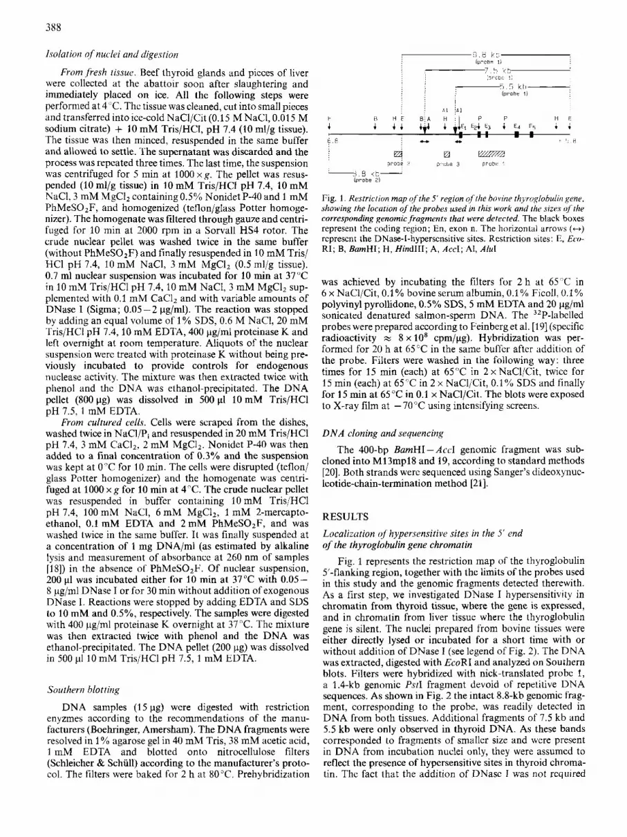
Fig. 1. Restriction mup of'the 5' region of the bovine thyroglobulin gene, showing the location of the probes used in this work and lhe sizes ojthe corresponding genomic fragments that were detected. The black boxes represent the coding region; En, exon n. The horizontal arrows (H) represent the DNase-I-hypersensitive sites. Restriction sites: E, Eco- RI; B, BumHI; H, HindIII; A. Accl; Al, Ah1
was achieved by incubating the filters for 2 h at 65 'C in 6 x NaCl/Cit, 0.1 YO bovine serum albumin, 0.1 % Ficoll, 0.1 'YO polyvinyl pyrollidone, 0.5% SDS, 5 mM EDTA and 20 pg/ml sonicated denatured salmon-sperm DNA. The 32P-labelled probes were prepared according to Feinberg et al. [19] (specific radioactivity z 8 x 10' cpm/pg). Hybridization was per- formed for 20 h at 65 "C in the same buffer after addition of the probe. Filters were washed in the following way: three times for 15 rnin (each) at 65°C in 2 x NaCl/Cit, twice for 15 rnin (each) at 65°C in 2 x NaCl/Cit, 0.1 '10 SDS and finally for 15 min at 65 "C in 0.1 x NaCl/Cit. The blots were exposed to X-ray film at - 70°C using intensifying screens.
D N A cloning and sequencing
The 400-bp BamHI-Ace1 genomic fragment was sub- cloned into M13mp18 and 19, according to standard methods [20]. Both strands were sequenced using Sanger's dideoxynuc- leotide-chain-termination method [21].
RESULTS
Localization of hypersensitive sites in the 5' end of the thyroglobulin gene chromatin
Fig. 1 represents the restriction map of the thyroglobulin 5'-flanking region, together with the limits of the probes used in this study and the genomic fragments detected therewith. As a first step, we investigated DNase I hypersensitivity in chromatin from thyroid tissue, where the gene is expressed, and in chromatin from liver tissue where the thyroglobulin gene is silent. The nuclei prepared from bovine tissues were either directly lysed or incubated for a short time with or without addition of DNase I (see legend of Fig. 2). The DNA was extracted, digested with EcoRI and analyzed on Southern blots. Filters were hybridized with nick-translated probe 1, a 1.4-kb genomic PstI fragment devoid of repetitive DNA sequences. As shown in Fig. 2 the intact 8.8-kb genomic frag- ment, corresponding to the probe, was readily detected in DNA from both tissues. Additional fragments of 7.5 kb and 5.5 kb were only observed in thyroid DNA. As these bands corresponded to fragments of smaller size and were present in DNA from incubation nuclei only, they were assumed to reflect the presence of hypersensitive sites in thyroid chroma- tin. The fact that the addition of DNase I was not rcquired

389
Fig. 2. Autoradiogruphy ofblots,from liver and thyroid DNA after hybridization with probe 1. Lanes L, T: no incubation of nuclei; lanes LA, TA: 10-min incubation without addition of DNasc I; lanes L1, L2 and T1, T2: 10-min incubation with DNase I at 0.05 pg/ml and 0.1 pg/ml respectively. Lane M, labclled (Hind111 + EcoR1)-digested ADNA. Sizcs are given in kb
to observe the thyroid-specific fragments indicated the pres- ence of endogenous nuclease activity in our nuclei prep- arations.
Nuclear endo-deoxyri bonuclease activity is known to be activated during isolation of nuclei from eukaryotic cells, depending on the ionic conditions used [22, 231. It has also been shown that the extent of nuclease activity varies from one tissue to another but that active gene sequences are digest- ed first [24]. As the addition of up to 0.1 pg/ml of exogenous DNase I did not modify the pattern of bands observed after self-digestion of the nuclei, the cutting positions detected in the cxperiment must constitute the major hypersensitive sites.
We expected that the 7.5-kb and 5.5-kb fragments resulted from cleavage occurring at hypersensitive sites located in the 5’ part of the 8.8-kb EcoRI fragment (in other words, 5’ to the gene). However, as our probe was nearly central relative to the 8.8-kb fragment, we had to verify that the 3‘ extremities (relative to gene polarity) of these shorter fragments were indeed provided by the EcoRI restriction site in intron 5 (Fig. 1). In order to gain a more precise determination of the limits of the hypersensitive regions at the same time, the same DNA preparations were used with different probe/restriction enzyme combinations in order to map the two sites. To deter- mine the position of the DNase-I-hypersensitive site which should lie near the cap site, the genomic DNA was cleaved with HindIII and the resulting blot was hybridized with probe 3 (see Fig. 1) which extends from - 760 bp to -470 bp, relative to the transcription start. As shown in Fig. 3A, a smear was revealed corresponding to fragments ranging over 660 - 820 bp in size. This result localized the hypersensitive region between positions - 100 and +60 relative to the cap site (+ 1). The second hypersensitive site lying more upstream, was located using again HindIII-cleaved genomic DNA, but probing the blot with the upstream probe 2 (see Fig. 1). By running two cloned sub-fragments in parallel on the gel, a Hindlll-BurnH1 frag’nent extending from p3.4 kb to -2 kb (lS4 kb in length) and a Hindlll-Accl fragment ex- tending from - 3.4 kb to - .8 kb in length), the se- quences which are cleaved by the nucleases were directly shown to be contained within a 400-bp-long BurnHI- AccI fragment (the difference between the two fragments used as
Fig. 3. DNAfrom selfdigested thyroidnuclei cleavad ir.itli 1 lindlll. (A) The resulting blot was probed with probe 3 (see Fig. 1). M, size markers, labelled (HindIII + EcoR1)-digested LDNA; sizes are given in kb. (B) The corresponding blot was probed with probe 2 (scc Fig. 1). R, size references: two subcloned genomic fragments, Hind111 - A N I (1.8 kb) and HindIII-BamHl (1.4 kb); see also Fig. 1. The vertical arrows represent the DNase-I-hypersensitive sites
kb

390
Fig. 4. Autoradiography uf’hlots,from liver and thyroid DNA after hybridization with probe 2. Lanes L, T: no incubation of the nuclei; lanes L i , T1; L2, T2; L3, T3; L4, T4: incubation with DNase I at 0.05 pg/ml, 0.1 pg/ml, 0.2 pg/ml and 1 pg/ml respectively. Lane M ; size markers, labelled (Hind111 + EcoR1)-cleaved IDNA; sizes are given in kb
n CAGCAGCTTCTAACCCTTCTCCCTGGAAGGCTCCCAAGATGGCCCTGGCCCTATGGGTCTTCGGTCTGCTGGACTTAATCTGCTTGGCATCCGCCAACAT
t 60 U
m m GGATCCAGGAGGGAGAGGTGAGAGGGCCAGAGGTGAGGAGCCTGCAGAGAGGATGGAGACCCAAGCCAGGAGCGGGCTCACGGAGGCCAGACCAAGGAGl
I - - I
TGGTGCTGAGTTTCCGTAGAGCATCCTCCTGGCTCACGAGGCATCAGATGATGGTAGGAGCTGAGTCATCGTAGAAAGAGTTTTCACGGTTGAGGCAGCG
CATGGGTGGGGGAAGCATGAGGAGTGGGCTTTCTGGTAGG
Fig. 5. D N A sequences. (A) The 5’ end of the bovine thyroglobulin gene. The sequences which are conserved between thyroglobulin gcne promoters (see text), the TATA box and the translation initiator codon are boxed. Underlined sequences correspond to the hypersensitive region detected in thyroid chromatin. (B) The BumHI-AccI genomic fragment located between -2000 bp and -1600 bp from the cap site. The purine-rich stretch is underlined; the repeats are boxed. The ‘GC’ box corresponding to a putative Spl-binding site is also boxed
references, as the corresponding smear was bordered by these markers; Fig. 3 B).
In order to investigate the possibility that other sequences, located more upstream from the gene, would also be involved in the formation of hypersensitive chromatin, probe 2 (see Fig. 1) was used to probe blots of EcoRI-restricted DNA from thyroid and liver nuclei. Fig. 4 shows that only the intact 3.8- kb genomic fragment corresponding to the probe was detected
in both cases, indicating the absence of nuclease hyper- sensitivity in this region of the chromatin in both cell types.
In conclusion, in a region spanning from -6.8 kb to + 5.8 kb relative to the transcription initiation site of the thyroglobulin gene, we found only two major hypersensitive sites in thyroid chromatin, both located in the 5’-flanking sequences, close to the gene. By contrast, such sites were not found in hepatic chromatin.

391
Fig. 6. Analysis of DNase-]-hypersensitive sites in chromatin from cultured cells. (A) Lanes T, T1, T2: nuclei from thyrotropin-treated cells were lysed directly (T) or after digestion with DNase I at 2 pg/ml (TI) or 4 pg/ml (T2); lanes S , S1: nuclei from FCS-treated cells were lybed directly (S) or after digestion with DNase I at 4 pg/ml (SI); lane K: nuclei from MDBK cells were digested with DNase I at 4 pg/ml prior to lysis. Lane M, size markers, labelled (Hind111 + EcoR1)-cleaved ADNA (sizes are given in kb). (B) Nuclei from cells cultured in the presencc of forskolin (F, F1, F2) or EGF (E, El) were lysed directly (F, E) or after digestion with DNase I at 2 pg/ml (Fl, E l ) or 4 pg/ml (F2). Lane M, size markers (as in A)
Characterization of the DNA sequences involved in DNase I hypersensitivity
Since the 5’ end of the bovine thyroglobulin gene had been sequenced previously [ 161, the DNA sequences corresponding to the hypersensitive chromatin structure which overlaps the cap site were readily identified (Fig. 5A). These include part of a DNA element which has been found to be conserved in human [24] and rat [25] thyroglobulin gene promoters, the only ones whose sequences are known so far. The sequence of the region corresponding to the DNase-I-hypersensitive site located more upstream was determined from a BamHI - Accl genomic fragment subcloned (see Fig. 1) into M13mp18 and mp19 bacteriophages (Fig. 5B). It displays a 34-b-long purine-rich stretch containing a duplicated motif of 9 nucleotides in length: AGAGGTGAG. This is reminiscent of the 38-b-long purine-rich segment identified previously in the proximal 5’-flanking region of the gene ( - 556 to - 518, rela- tive to cap site) which also contains an internal repetition of a 8-b-long motif: AGGAGGGA [16]. However, as exemplified by the difference in repeat units, there is no direct sequence similarity betwen these two regions. A classical ‘GC-box’ which could correspond to a binding site for the transcription factor SP 1 [26] was also identified about 250 nucleotides
downstream (closer to the gene) from the purine-rich element (Fig. 5B).
Correlation between change in chromatin structure and expression of the thyroglobulin gene
To study the relationship between the formation of DNase-I-hypersensitive structures and transcription of the thyroglobulin gene, we analyzed chromatin from primary cul- tured calf thyroid cells maintained in different culture con- ditions [17] (and C . Gerard et al., unpublished results). When cultured in the presence of thyrotropin (0.1 mU/ml) or the adenylate cyclase activator, forskolin (10 pM), these cells maintain a fully differentiated phenotype, with a readily de- tectable content of thyroglobulin mRNA. They lose most differentiated functions when treated with fetal calf serum (FCS: 1%) or epidermal growth factor (EGF, 25 ngiml), in particular, cytoplasmic thyroglobulin mRNA level becomes undetectable under these conditions. Subsequent addition of thyrotropin or forskolin reverses this phenomenon and results in the re-accumulation of thyroglobulin mRNA in the cyto- plasm of the cells.
We first compared the structure of the chromatin in thy- roid cells cultured in the presence of thyrotropin (which main-

392
in the presence of thyrotropin for 6 days, a period which had been shown to allow maximal ‘redifferentiation’ of the cells [17] (and C. Gerard et al., unpublished results). Fig. 7 shows that both nuclease-dependent bands (7.5 kb and 5.5 kb) were detected after culture in these conditions. The DNase-I-hyper- sensitive structure which involves the sequences surrounding the transcriptional start point appears therefore to be directly linked to the actual transcriptional activity of the thyroglo- bulin gene.
DISCUSSION
We have analyzed the changes in chromatin structure oc- curring at the 5’ extremity of the thyroglobulin gene when this gene is transcribed. This gene is only expressed in differen- tiated thyroid cells under the positive control of the pituitary hormone thyrotropin, which exerts its action inside the cell by activating the adenylate cyclase and increasing the cAMP content. Thyroglobulin gene expression in the thyroid is as- sociated with the presence of two ma,or D N ~ ~ ~ - I - ~ ~ ~ ~ ~ ~ ~ ~ ~ ~ - tive regions of the chromatin: at the extremity of the gene ( - 100 to +60 relative to the cap site) and in the 5‘-flanking sequences (- 2000 to - 1600).
Using a system of primary cultured thyroid cells in which
Fig. 7. Change in chromatin structure during ‘re-differentiation’ ofpri- mar\‘ cultured thyrocytes. Nuclei from FCS-treated cells (day 8) were digested with DNase I at 2 pg/ml; nuclei from cells that were sub- sequently cultured in the presence of thyrotropin were digested with DNasc 1 at 2 pg/ml. TSH, thyrotropin
tains thyroglobulin gene expression) or of FCS (which abolishes expression of the gene). The nuclei from both cell preparations were digested by various quantities of DNase I. The DNA was extracted, cleaved with EcoRI and analyzed according to the same protocol as described in Fig. 2, using probe 1 (see Fig. 1). Nuclei from MDBK cells [27] were used in parallel as controls, representing the state of chromatin found in cells which do not express the thyroglobulin gene.
Fig. 6A shows that both hypersensitive sites identified previously in the chromatin from bovine thyroid tissue were present in the chromatin from cells cultured in the continuous presence of thyrotropin (lanes T, TI, T2). When thyrotropin was omitted and 1 ‘YO FCS was included in the culture medium, the loss of thyroglobulin gene expression in the cells was reflected by the disappearance of the smaller (5.5 kb) of the two bands originating from DNase I digestion (lanes S, Sl). This indicated that the DNA sequences at the very 5’ end of the gene (- 100 to + 60) were no longer hypersensitive under these conditions. As expected, no DNase I hypersensitivity was observed in thyroglobulin gene chromatin from MDBK cells (lane K).
Identical results were obtained when the cells were kept in a differentiated state by culture in the presence of forskolin instead of thyrotropin (Fig. 6 B, lanes F, F1, F2) or ‘de-differ- entiated’ by culture in the presence of EGF instead of FCS (Fig. 6B, lanes E, El).
As the ‘de-differentiating action’ of FCS or EGF, in terms of thyroglobulin gene expression, can be reversed during a subsequent culture period in the presence of thyrotropin or forskolin [17] (and C. Gerard et al., unpublished results), we investigated the possibility that such a change would be reflected at the chromatin level by reappearance of the - 100 to +60 hypersensitive site. Cells from bovine thyroid tissue were seeded in a medium containing 1% FCS and cultured for 8 days. Part of the cells were then scraped and the structure of thyroglobulin gene chromatin was probed by digesting the nuclci with DNase I. As shown in Fig. 7, only the 7.5-kb band corresponding to the - 2000 to - 1600 hypersensitive site was detected at that time. The remaining cells were then cultured
the expression of the thyroglobulin gene can be altered through the composition of the culture medium, we have been able to show that the hypersensitive characters of these two regions are differently related to the expression of the gene. While the upstream site displays hypersensitivity to DNase I digestion in thyrocyte, irrespective of whether the gene is expressed or not, on-going transcription of the gene is needed to confer the hypersensitive character to the cap-site-proximal sequences.
Both of these hypersensitive sites are thus tissue-specific but one of them is dependent on effective expression of the thyroglobulin gene under control of thyrotropin or CAMP. Interestingly, this site involves, at least in part, DNA se- quences which are conserved in all three thyroglobulin gene promoter regions sequenced to date (see Fig. 5A). Transient expression studies also strongly suggest that these sequences are involved in the control of thyroglobulin gene transcription by cAMP (D. Christophe, unpublished results). DNase I hypersensitivity in this region of the chromatin could therefore result from the binding of trans-activating factor(s) which mediate(s) the effect of CAMP, in a way analogous to that described by others [ll, 121.
For example, in the case of the chicken adult b-globin gene it has been clearly demonstrated that DNase I hypersensitivity could result from the interaction with DNA-binding factors [28]. Other mechanisms, however, are likely to participate in the establishment of this altered property of chromatin, amongst which DNA supercoiling [29] and DNA methylation [30] are likely candidates.
Hypersensitive chromatin structures, such as the ~ 2000 to -1600 site, that are tissue-specific but do not depend on actual transcription of the gene have also been reported in several other systems [8, 9, 31, 321. Conceivably, they reflect changes associated with the establishment of a ‘trans- criptionally committed state’ of the gene chromatin. These events take place during the differentiation process and stably characterize given cell types [8, 321. They may also reflect somehow the targeting of the gene into an appropriate nuclear location [33]. The DNA sequence of the -2000 to -1600 segment has been determined. Unfortunately due to the lack

393
of knowledge of the corresponding sequences in other species, n o conclusion could be reached regarding their evolutionary conservation.
Although modifications in chromatin structure related to gene expression are not restricted to the 5’ parts of genes (see e.g. [34]), the events occurring in the promoter regions are most probably pertinent to the control of initiation of the transcription process. In this view, current studies aim at understanding the precise role of the sequences identified in this work and investigating their possible interaction with tissue-specific and CAMP-dependent trans-acting factors.
The continuous support and critical interest of Dr J.E. Dumont are deeply acknowledged. We thank Mrs D. Leemans for the prep- aration of the manuscript. This work was supported by grants from the Belgian Ministi.re ile lu Politiyue Scientfique (‘Action de Recherche Concert&’), from the National Instilutes of Health, from Solvay Company and from A.R.B.D. asbl. C.H. is a fellow of the fnstiiut pour I’Encourugemeni de la Recherche Scienrifique duns l’lndustrie et 1;4griculture.
REFERENCES 1. Weintraub, H. & Groudine, M. (1976) Science (Wash. DC) 193,
2. Stalder, J., Larsen, A., Engel, J. D., Dolan, M., Groudinc, M. &
3. Elgin, S. C. R . (1984) Nature (Lond.) 309, 213-214. 4. Reeves, R. (1984) Bioclzim. Biophys. Actu 782, 343-393. 5. Weintraub, H. (1979) Mucleic Acids Res. 7, 781 -792. 6. Weintraub, H., Bcug, H., Groudine, M. & Graf, T. (1982) Cell
7. Benvenisty, N. & Reshef, L. (1987) Proc. Nutl Acad. Sci. USA
8. Burch, J . R. E. & Weintraub, H. (1983) Cell 33, 65-76. 9. Kaye, J . S., Pratt-Kaye, S., Bcllard, M., Dretzen, G., Bellard,
F. & Chambon, P. (1986) EMBO J . 5,277-285. 10. Chrysogelos, S., Riley, D. E., Stein, G. & Stein, J. (1985) Proc.
Nut1 Acad. Sci. USA 82,7535 - 1539. 11. Siebenlist, U., Durand, D. B., Brcssler, P., Holbrook, N. J.,
Norris, C. A,, Kamoun, M.. Kanl, J . A. & Crartree, G. R. (1 986) Mol. Cell Biol. 6 , 3042 - 3049.
848 - 856.
Weintraub, H. (1980) Cell 20,451 -460.
28,931 - 940.
84, 1132-1136.
12. Zarct, K. S. & Yamanioto, K. R. (1984) CeN38, 29-38.
13. Emerson, B. M., Lewis, C. D. & Felscnfcld, G. (1985) Cell 41,
14. Van Heuverswyn, B., Slreydio, C., Brocas, H., Refetorf, S.. Dumont, J. E. & Vassart, G. (1984) Proc. Nutl Acod. Sci. USA
15. Van Heuverswyn, B., Leriche, A., Van Sande, J., Dumont, J . E. & Vassart, G. (1985) FEBS Lett. 188, 192-196.
16. de Martynoff, G., Pohl, V., Mercken, L., Van Ommen, G. J. & Vassart, G. (1987) Eur. J . Biochem. 164, 591 -599.
17. Gerard, C. M. & Roger, P. P. (1986) Frontiers in thyroidologj. vol. 1 (Medeiros-Neto, G. & Gaitan, E., cds), Plenum Publishing Corporation, New York.
18. Lawson, G. M., Tsai, M. J. & O’Malley, B. (1980) Biocliemistry
19. Feinberg, A. P. & Vogelstein, B. (1984) Anal. Biochem. 132. 6- 13.
20. Maniatis, T., Fritsch, E. F. & Sanibrook, J . (1982) Molecular. cloning, a laboratory manual, Cold Spring Harbor Laboratory, NY.
21. Sanger, F., Nicklcn, S. & Coulson, A. R. (1977) Proc. Mat1 Acud
22. Hewish, D. R., Burgoyne, L. A. (1973) Biochem. Biuphys. Rex.
23. Vanderbilt, J. N., Bloom, K. S. & Anderson, J. N. (1982) J . Biol.
24. Christophe, D., Cabrer, B., Bacolla, A,, Targovnik, H., Pohl,
25. Musti, A. M., Ursini, M. V. & Dilauro, R. (1986) Ann. Endocrirzzol.
26. Dynan, W. S. & Tjian, R. (1983) Cell 35, 79-87. 27. Madin, S. H. & Darby, N. B. (1958) Proc. Soc. Exp. Bid . Meti.
28. Emerson, B. M . & Felsenfeld, G. (1984) Proc. Nail Acad. Sci.
29. Villeponteau, B., Lundell, M. & Martinson, H. (1984) Cell 39,
30. Keshet, I . , Lieman-Hurwitz, J . &Cedar, H. (1986) Cell44, 535-
31. Tratner, I., Nahon, J. L., Sala-Trepat, J. M. & Vcnetianer. A.
32. Turcotte, B., Guertin, M., Chevrette, M., Lahue, H. & Belangcr,
33. Hutchinson, N. & Weintraub, H. (1985) Cell 43, 471 -482. 34. Bellard, M., Dretzen, G., Bellard, F., Kaye, J. S., Pratt-Kaye,
S. & Chambon, P. (1986) EMBO J . 5, 567- 574.
21 -30.
81, 5941 - 5949.
19,4403-4411.
Sci. USA 74, 5463 - 5467.
C‘ommun. 52, 504-510.
Chem. 257, 13009-13017.
V. & Vassart, G. (1983) Nucleic Acids Res. 13, 5121-5144.
47, 36.
98, 574.
USA K1,95-99.
469 -478.
543.
(1987) Mol. Cell Biol. 7, 1856-1864.
L. (1986) Nucleic Acids Res. 14, 9827-9840.