transcription elongation through a chromatin template
TRANSCRIPT
Biochimie 89 (2007) 516e527www.elsevier.com/locate/biochi
Transcription elongation through a chromatin template
Christophe Lavelle*
Laboratoire de Microscopie Moleculaire et Cellulaire, UMR 8126, Institut Gustave Roussy, 39 rue Camille Desmoulins, 94805 Villejuif, France
Received 2 August 2006; accepted 26 September 2006
Available online 17 October 2006
Abstract
DNA transaction events occurring during cell life (replication, transcription, recombination, repair, cell division) are always linked to severechanges in the topological state of the double helix. However, since naked DNA almost does not exist in eukaryote nucleus but rather interactswith various proteins, including ubiquitous histones, these topological changes happen in a chromatin context. This review focuses on the role ofchromatin fiber structure and dynamics in the regulation of transcription, with an almost exclusive emphasis on the elongation step. Beside a briefoverview of our knowledge about transcribed chromatin, we will see how recent mechanistic and biochemical studies give us new insights intothe way cell could modulate DNA supercoiling and chromatin conformational dynamics. The participation of topoisomerases in this complexballet is discussed, since recent data suggest that their role could be closely related to the precise chromatin structure. Lastly, some future pros-pects to carry on are proposed, hoping this review will help in stimulating discussions and further investigations in the field.� 2006 Elsevier Masson SAS. All rights reserved.
Keywords: DNA topology; Nucleosome; Chromatin; Topoisomerase; Transcription
1. Introduction
1.1. Chromatin: a compact ‘‘but’’ dynamic structure.
In vivo, DNA is not naked but complexed with histones,which lead naturally to the ‘‘nucleosome barrier’’ conceptwhere nucleosomes are supposed to act as both transcriptioninitiation (by simply hiding promoter) and elongation (bystopping polymerase progression) repressors. A clear evidencethat nucleosomes are indeed not ‘‘transparent’’ (as sometimeswrongly claimed) to the transcription machinery came fromgenetic studies which showed that histone depletion de-repressed certain important genes [1]. At the same time,many DNase I or micrococcal nuclease digestion assaysgave evidences that genes are still packaged within the nucleusas chromatin, even during transcription [2e4], although chro-matin structure may be altered during gene activity [2,5e8]. Infact, some studies suggest that canonical nucleosomes are lost
* Tel.: þ33 6 24 71 44 03; fax: þ33 1 42 11 54 94.
E-mail address: [email protected]
0300-9084/$ - see front matter � 2006 Elsevier Masson SAS. All rights reserv
doi:10.1016/j.biochi.2006.09.019
during transcription (as found for the RNA polymerase I tran-scribed ribosomal genes [9] and for RNA polymerase IIheavily transcribed genes [10]) while others showed they arestill present (as found on a number of inducible and house-keeping genes (see chapter 8 in [11] and references therein)).Interestingly, transcribed yeast chromatin and total yeast chro-matin are equally sensitive to DNase I digestion, which couldmean that the entire yeast genome exists in a state that repre-sents a restricted proportion of total chromatin in higher eu-karyotes [12]. In the same way, the control for ribosomegene activity has recently been shown by microscopy studiesnot to be mediated by changes in chromatin structure [13], a re-sult which confirms former biochemical studies of Sogo andcoworkers [9,14e16] and could be interpreted as follow: genestranscribed at high rates (i.e. densely loaded with RNA poly-merases) have a disrupted chromatin and lack canonical nucle-osomes. However, it remains elusive whether nucleosomesdissociate or simply unfold when polymerase get through(see further discussion below).
Hence, if chromatin is sometimes merely seen as a way topack two meters of DNA into the nucleus volume, the regula-tory role of this polymorphic and highly dynamic structure is
ed.
517C. Lavelle / Biochimie 89 (2007) 516e527
now largely acknowledged. This means that a nucleosomemust act both as a compaction and regulation tool, or, in otherwords, be reasonably stable while keeping some dynamicproperties to allow chromatin ‘‘opening’’ for promoter accessand subsequent transcription elongation.
1.2. .polymerases have to go through
Now, if transcription initiation mainly relies on promoteraccess (potentially regulated through nucleosome positioningand remodeling; see [17] for a review), do nucleosomes alsopresent a significant obstacle for RNA polymerase once ithas left the initiation complex and begun moving along theDNA? Since RNA polymerase needs to pass through nucleo-somes, transcription elongation should be repressed by chro-matin. Indeed, transcription on chromatin complex is slowerin vitro than in vivo, and also slower than on naked DNA tem-plates in vitro, probably because enhancement of pausing[18e20]. Elongation raises in fact many questions, some ofthe most puzzling being the way RNA polymerase progressesalong chromatin template (reviewed in [21]), the subsequentfate of nucleosomes [22,23] and the need for numerous elon-gation factors (including ATP-dependent remodeling factors,histone chaperones and histone modification enzymes; see[24e26] for reviews and Fig. 2D) as well as topoisomerase ac-tivity [27e34] (see Fig. 2F). Also poorly documented is thequestion whether polymerase moves/rotates along DNA orDNA moves/rotates along a fixed polymerase [35]. All ofthis will be discussed below.
2. When polymerase meets nucleosome
2.1. A thirty-year-old question
Given the fact that even a ‘‘simple’’ prokaryotic polymer-ase is a molecular motor closely associated with about 40 bpof DNA during transcription elongation, it seems inconceiv-able that it simply progresses around nucleosome, causing nei-ther displacement nor structural rearrangements. At least,histones must be displaced from the transcribed strand to allowpassage of the polymerase. Indeed, in vivo studies designed toevaluate the nucleosomal state of active genes have generallyshown that these structures are in more ‘‘open’’ or even disrup-ted state [36e38]. But at the same time, in vitro studies haveshowed limited disruption, however depending on the experi-mental conditions. So, what happens to nucleosomes duringtranscription (or, equivalently, what happens to polymeraseas it encounters nucleosomes) is a question that, although reg-ularly reviewed [22,23,39e47], has not lost much of its mys-tery. As we will see, difficulty to clearly answer thesequestions emerges mainly from the different experimental ap-proaches used (in vitro versus in vivo, linear mononucleosomeversus reconstituted circular plasmid, prokaryotic versus eu-karyotic system) and the difficulty to obtain non ambiguousresults. In particular, it is often hard to make the difference be-tween direct (mechanical, through contact) or distal
(topological, through DNA elastic constraints propagation) ef-fects of polymerase tracking.
2.2. Overcoming the nucleosome barrier
A simple energetic calculation shows that to transcribe the150 or so base pairs of DNA associated with a nucleosomewill require about 1500 kcal mol�1 (only taking into accountthe 10 kcal mol�1 required for each phosphate bond of the na-scent RNA transcript) while complete displacement of the his-tone octamer requires 10e20 kcal mol�1 [48]. Thus, one canreasonably ask whether the histone octamer is irrelevant tothe transcribing polymerase [49]. One obvious way to testthis hypothesis is to use reconstituted in vitro system. How-ever, complexity of eukaryotic polymerases (and its necessaryinitiation factors) led most researchers to utilize simpler pro-karyotic polymerases [23]. Experiments of this kind havebeen carried very soon after the nucleosome ‘‘discovery’’[50,51] but led quickly to controversial interpretation[52,53]. At the same time, De Bernardin et al. were the firstto show that nucleosomes (perhaps in an altered form) onSV40 minichromosomes do not prevent transcription elonga-tion by RNA polymerase II in vivo [54]. However, far from be-ing ‘‘transparent’’ to the transcription machinery (as alreadydiscussed above), nucleosomes interact with polymerase invarious ways which make interpretation of the numerous ex-perimental studies rather puzzling.
2.3. Many transcription assays.
Several in vitro studies showed that transcription elongationby RNA polymerase II (Pol II) is stopped by nucleosomes inphysiological salt conditions [55]. However, elongation canbe recovered in high salt (>300 mM) [50,56] or with acetyla-tion of the histone tails [57]. The common reasons could bethe neutralization of charges, suggesting either that electro-static interactions between histone octamer and DNA haveto be broken for RNA polymerase to transcribe DNA orga-nized into nucleosomes [50], or that these histone tails interactdirectly with components of the transcriptional machinery[57]. Another usually proposed explanation is H2AeH2B di-mers destabilization [56,58,59], consistent with the rapid ex-change of these dimers, compare to (H3eH4)2 tetramer,upon transcription [47,60e62]. Furthermore, some additionalfactors that facilitate these exchanges have been characterizedas essential elongation factors, including ATP-dependent re-modeling factors such as SWI/SNF [63,64] and histone chap-erones such as FACT (FAcilitates Chromatin Transcription)[58,65,66] or NAP1 (Nucleosome Assembly Protein 1 [67e69] (see also Figs. 1B and 2D).
Transcription by RNA polymerase III (Pol III) is also muchreduced on nucleosomal template with a tendency for the poly-merase to terminate at natural pause sites in the DNA se-quence that is wrapped around the nucleosome andfurthermore depends on chromatin folding mediated by inter-nucleosomal interactions [70e72]. However, as pointed out byJackson [22], these observations obtained on tandem arrays
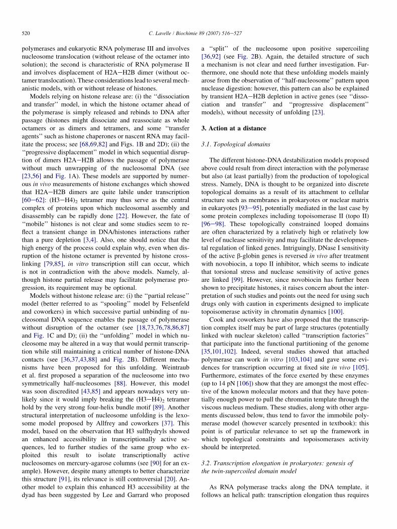
518 C. Lavelle / Biochimie 89 (2007) 516e527
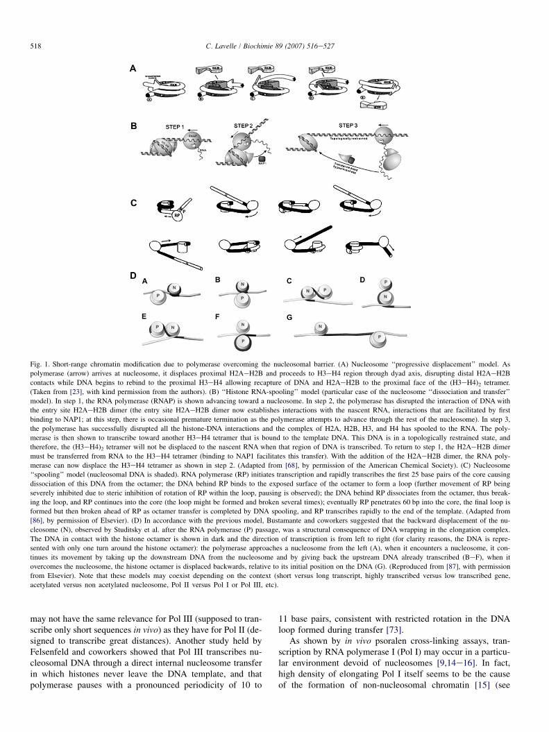
Fig. 1. Short-range chromatin modification due to polymerase overcoming the nucleosomal barrier. (A) Nucleosome ‘‘progressive displacement’’ model. As
polymerase (arrow) arrives at nucleosome, it displaces proximal H2AeH2B and proceeds to H3eH4 region through dyad axis, disrupting distal H2AeH2B
contacts while DNA begins to rebind to the proximal H3eH4 allowing recapture of DNA and H2AeH2B to the proximal face of the (H3eH4)2 tetramer.
(Taken from [23], with kind permission from the authors). (B) ‘‘Histone RNA-spooling’’ model (particular case of the nucleosome ‘‘dissociation and transfer’’
model). In step 1, the RNA polymerase (RNAP) is shown advancing toward a nucleosome. In step 2, the polymerase has disrupted the interaction of DNA with
the entry site H2AeH2B dimer (the entry site H2AeH2B dimer now establishes interactions with the nascent RNA, interactions that are facilitated by first
binding to NAP1; at this step, there is occasional premature termination as the polymerase attempts to advance through the rest of the nucleosome). In step 3,
the polymerase has successfully disrupted all the histone-DNA interactions and the complex of H2A, H2B, H3, and H4 has spooled to the RNA. The poly-
merase is then shown to transcribe toward another H3eH4 tetramer that is bound to the template DNA. This DNA is in a topologically restrained state, and
therefore, the (H3eH4)2 tetramer will not be displaced to the nascent RNA when that region of DNA is transcribed. To return to step 1, the H2AeH2B dimer
must be transferred from RNA to the H3eH4 tetramer (binding to NAP1 facilitates this transfer). With the addition of the H2AeH2B dimer, the RNA poly-
merase can now displace the H3eH4 tetramer as shown in step 2. (Adapted from [68], by permission of the American Chemical Society). (C) Nucleosome
‘‘spooling’’ model (nucleosomal DNA is shaded). RNA polymerase (RP) initiates transcription and rapidly transcribes the first 25 base pairs of the core causing
dissociation of this DNA from the octamer; the DNA behind RP binds to the exposed surface of the octamer to form a loop (further movement of RP being
severely inhibited due to steric inhibition of rotation of RP within the loop, pausing is observed); the DNA behind RP dissociates from the octamer, thus break-
ing the loop, and RP continues into the core (the loop might be formed and broken several times); eventually RP penetrates 60 bp into the core, the final loop is
formed but then broken ahead of RP as octamer transfer is completed by DNA spooling, and RP transcribes rapidly to the end of the template. (Adapted from
[86], by permission of Elsevier). (D) In accordance with the previous model, Bustamante and coworkers suggested that the backward displacement of the nu-
cleosome (N), observed by Studitsky et al. after the RNA polymerase (P) passage, was a structural consequence of DNA wrapping in the elongation complex.
The DNA in contact with the histone octamer is shown in dark and the direction of transcription is from left to right (for clarity reasons, the DNA is repre-
sented with only one turn around the histone octamer): the polymerase approaches a nucleosome from the left (A), when it encounters a nucleosome, it con-
tinues its movement by taking up the downstream DNA from the nucleosome and by giving back the upstream DNA already transcribed (BeF), when it
overcomes the nucleosome, the histone octamer is displaced backwards, relative to its initial position on the DNA (G). (Reproduced from [87], with permission
from Elsevier). Note that these models may coexist depending on the context (short versus long transcript, highly transcribed versus low transcribed gene,
acetylated versus non acetylated nucleosome, Pol II versus Pol I or Pol III, etc).
may not have the same relevance for Pol III (supposed to tran-scribe only short sequences in vivo) as they have for Pol II (de-signed to transcribe great distances). Another study held byFelsenfeld and coworkers showed that Pol III transcribes nu-cleosomal DNA through a direct internal nucleosome transferin which histones never leave the DNA template, and thatpolymerase pauses with a pronounced periodicity of 10 to
11 base pairs, consistent with restricted rotation in the DNAloop formed during transfer [73].
As shown by in vivo psoralen cross-linking assays, tran-scription by RNA polymerase I (Pol I) may occur in a particu-lar environment devoid of nucleosomes [9,14e16]. In fact,high density of elongating Pol I itself seems to be the causeof the formation of non-nucleosomal chromatin [15] (see
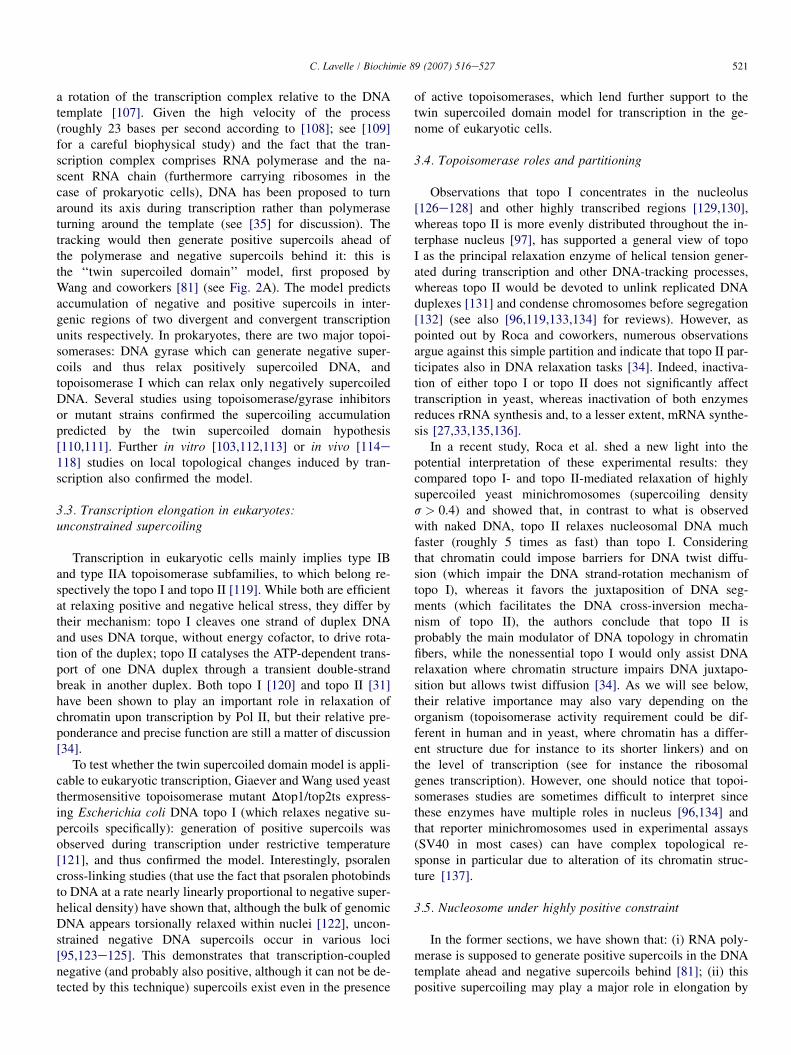
519C. Lavelle / Biochimie 89 (2007) 516e527
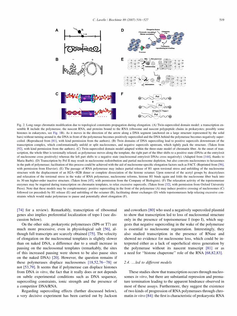
Fig. 2. Long range chromatin modification due to topological constraints propagation during elongation. (A) Twin-supercoiled domain model: a transcription en-
semble R include the polymerase, the nascent RNA, and proteins bound to the RNA (ribosome and nascent polypeptide chains in prokaryotes; possibly some
histones in eukaryotes, see Fig. 1B). As it moves in the direction of the arrow along a DNA segment (anchored on a large structure represented by the solid
bars) without turning around it, the DNA in front of the polymerase becomes positively supercoiled and the DNA behind the polymerase becomes negatively super-
coiled. (Reproduced from [81], with kind permission from the authors). (B) Twin domains of DNA supercoiling lead to positive supercoils downstream of the
transcription complex, which conformationally unfold or split nucleosomes, and negative supercoils upstream, which tightly pack the structure. (Taken from
[92], with kind permission from the authors). (C) Twin-supercoiled domain model adapted within the three-state model of chromatin fiber. At the onset of tran-
scription, the whole fiber is torsionally relaxed; as polymerase moves along the template, the right part of the fiber shifts to a positive state (DNAs at the entry/exit
of nucleosome cross positively) whereas the left part shifts to a negative state (nucleosomal entry/exit DNAs cross negatively). (Adapted from [144], thanks to
Maria Barbi). (D) Transcription by Pol II may result in nucleosome redistribution and partial nucleosome depletion, but also converts nucleosomes to hexasomes
in the path of polymerase; facilitation of this process could be achieved with the aid of nucleosome-specific elongation factors such as FACT. (Reprinted from [56],
with permission from Elsevier). (E) The passage of RNA polymerase may induce partial release of H1 upon torsional stress and unfolding of the nucleosome
structure with the displacement of an H2AeH2B dimer or complete dissociation of the histone octamer. Upon removal of the acetyl groups by deacetylases
and relaxation of the torsional stress in the wake of RNA polymerase, nucleosome reforms, histone H1 binds again and folds the nucleosome fiber back into
its 30 nm higher-order inactive structure. (Taken from [45], with permission from the Company of Biologists). (F) The relaxation activity of the topoisomerase
enzymes may be required during transcription on chromatin templates, to relax excessive supercoils. (Taken from [32], with permission from Oxford University
Press). Note that these models may be complementary: positive supercoiling in the front of the polymerase (A) may induce positive crossing of nucleosomes (C)
followed (or preceded) by H1 release (E) and unfolding of the octamer (B), facilitating dimer exchanges (D) while topoisomerases help relaxing excessive con-
straints which would make polymerase to pause and potentially abort elongation (F).
[74] for a review). Remarkably, transcription of ribosomalgenes also implies preferential localization of topo I (see dis-cussion below).
On the other side, prokaryotic polymerases (SP6 or T7) aremuch more processive, even in physiological salt [56], al-though full transcripts are scarcely obtained [75]. The velocityof elongation on the nucleosomal templates is slightly slowerthan on naked DNA, a difference due to a small increase inpausing on the nucleosomal templates (remarkably, the sitesof this increased pausing were shown to be also pause siteson the naked DNA) [20]. However, the question remains ifthese polymerases displace nucleosomes [18,52,76e78] ornot [53,79]. It seems that if polymerase can displace histonesfrom DNA in vitro, the fact that it really does or not dependson subtle experimental conditions such as DNA sequence,supercoiling constraints, ionic strength and the presence ofa competitor DNA/RNA.
Regarding supercoiling effects (further discussed below),a very decisive experiment has been carried out by Jackson
and coworkers [80] who used a negatively supercoiled plasmidto show that transcription led to loss of nucleosomal structureonly in the presence of topoisomerase I (topo I), which sug-gests that negative supercoiling in the wake of the polymeraseis essential to nucleosome regeneration. Interestingly, theyalso studied transcription in the presence of RNase andshowed no evidence for nucleosome loss, which could be in-terpreted either as a lack of superhelical stress generation bythe polymerase without its nascent transcript [81] or asa need for ‘‘histone chaperone’’ role of the RNA [68,82,83].
2.4. .led to different models
These studies show that transcription occurs through nucleo-somes in vitro, but there are substantial repression and prema-ture termination leading to the apparent hindrance observed inmost of these assays. Furthermore, they suggest the existenceof two kinds of progression of RNA polymerases through chro-matin in vitro [84]: the first is characteristic of prokaryotic RNA
520 C. Lavelle / Biochimie 89 (2007) 516e527
polymerases and eukaryotic RNA polymerase III and involvesnucleosome translocation (without release of the octamer intosolution); the second is characteristic of RNA polymerase IIand involves displacement of H2AeH2B dimer (without oc-tamer translocation). These considerations lead to several mech-anistic models, with or without release of histones.
Models relying on histone release are: (i) the ‘‘dissociationand transfer’’ model, in which the histone octamer ahead ofthe polymerase is simply released and rebinds to DNA afterpassage (histones might dissociate and reassociate as wholeoctamers or as dimers and tetramers, and some ‘‘transferagents’’ such as histone chaperones or nascent RNA may facil-itate the process; see [68,69,82] and Figs. 1B and 2D); (ii) the‘‘progressive displacement’’ model in which sequential disrup-tion of dimers H2AeH2B allows the passage of polymerasewithout much unwrapping of the nucleosomal DNA (see[23,56] and Fig. 1A). These models are supported by numer-ous in vivo measurements of histone exchanges which showedthat H2AeH2B dimers are quite labile under transcription[60e62]: (H3eH4)2 tetramer may thus serve as the centralcomplex of proteins upon which nucleosomal assembly anddisassembly can be rapidly done [22]. However, the fate of‘‘mobile’’ histones is not clear and some studies seem to re-flect a transient change in DNA/histones interactions ratherthan a pure depletion [3,4]. Also, one should notice that thehigh energy of the process could explain why, even when dis-ruption of the histone octamer is prevented by histone cross-linking [79,85], in vitro transcription still can occur, whichis not in contradiction with the above models. Namely, al-though histone partial release may facilitate polymerase pro-gression, its requirement may be optional.
Models without histone release are: (i) the ‘‘partial release’’model (better referred to as ‘‘spooling’’ model by Felsenfeldand coworkers) in which successive partial unbinding of nu-cleosomal DNA sequence enables the passage of polymerasewithout disruption of the octamer (see [18,73,76,78,86,87]and Fig. 1C and D); (ii) the ‘‘unfolding’’ model in which nu-cleosome may be altered in a way that would permit transcrip-tion while still maintaining a critical number of histone-DNAcontacts (see [36,37,43,88] and Fig. 2B). Different mecha-nisms have been proposed for this unfolding. Weintraubet al. first proposed a separation of the nucleosome into twosymmetrically half-nucleosomes [88]. However, this modelwas soon discredited [43,85] and appears nowadays very un-likely since it would imply breaking the (H3eH4)2 tetramerhold by the very strong four-helix bundle motif [89]. Anotherstructural interpretation of nucleosome unfolding is the lexo-some model proposed by Allfrey and coworkers [37]. Thismodel, based on the observation that H3 sulfhydryls showedan enhanced accessibility in transcriptionally active se-quences, led to further studies of the same group who ex-ploited this result to isolate transcriptionally activenucleosomes on mercury-agarose columns (see [90] for an ex-ample). However, despite many attempts to better characterizethis structure [91], its relevance is still controversial [20]. An-other model to explain this enhanced H3 accessibility at thedyad has been suggested by Lee and Garrard who proposed
a ‘‘split’’ of the nucleosome upon positive supercoiling[36,92] (see Fig. 2B). Again, the detailed structure of sucha mechanism is not clear and need further investigation. Fur-thermore, one should note that these unfolding models mainlyarose from the observation of ‘‘half-nucleosome’’ pattern uponnuclease digestion: however, this pattern can also be explainedby transient H2AeH2B depletion in active genes (see ‘‘disso-ciation and transfer’’ and ‘‘progressive displacement’’models), without necessity of unfolding [23].
3. Action at a distance
3.1. Topological domains
The different histone-DNA destabilization models proposedabove could result from direct interaction with the polymerasebut also (at least partially) from the production of topologicalstress. Namely, DNA is thought to be organized into discretetopological domains as a result of its attachment to cellularstructure such as membranes in prokaryotes or nuclear matrixin eukaryotes [93e95], potentially mediated in the last case bysome protein complexes including topoisomerase II (topo II)[96e98]. These topologically constrained looped domainsare often characterized by a relatively high or relatively lowlevel of nuclease sensitivity and may facilitate the developmen-tal regulation of linked genes. Intriguingly, DNase I sensitivityof the active b-globin genes is reversed in vivo after treatmentwith novobiocin, a topo II inhibitor, which seems to indicatethat torsional stress and nuclease sensitivity of active genesare linked [99]. However, since novobiocin has further beenshown to precipitate histones, it raises concern about the inter-pretation of such studies and points out the need for using suchdrugs only with caution in experiments designed to implicatetopoisomerase activity in chromatin dynamics [100].
Cook and coworkers have also proposed that the transcrip-tion complex itself may be part of large structures (potentiallylinked with nuclear skeleton) called ‘‘transcription factories’’that participate into the functional partitioning of the genome[35,101,102]. Indeed, several studies showed that attachedpolymerase can work in vitro [103,104] and gave some evi-dences for transcription occurring at fixed site in vivo [105].Furthermore, estimates of the force exerted by these enzymes(up to 14 pN [106]) show that they are amongst the most effec-tive of the known molecular motors and that they have poten-tially enough power to pull the chromatin template through theviscous nucleus medium. These studies, along with other argu-ments discussed below, thus tend to favor the immobile poly-merase model (however scarcely presented in textbook): thispoint is of particular relevance to set up the framework inwhich topological constraints and topoisomerases activityshould be interpreted.
3.2. Transcription elongation in prokaryotes: genesis ofthe twin-supercoiled domain model
As RNA polymerase tracks along the DNA template, itfollows an helical path: transcription elongation thus requires
521C. Lavelle / Biochimie 89 (2007) 516e527
a rotation of the transcription complex relative to the DNAtemplate [107]. Given the high velocity of the process(roughly 23 bases per second according to [108]; see [109]for a careful biophysical study) and the fact that the tran-scription complex comprises RNA polymerase and the na-scent RNA chain (furthermore carrying ribosomes in thecase of prokaryotic cells), DNA has been proposed to turnaround its axis during transcription rather than polymeraseturning around the template (see [35] for discussion). Thetracking would then generate positive supercoils ahead ofthe polymerase and negative supercoils behind it: this isthe ‘‘twin supercoiled domain’’ model, first proposed byWang and coworkers [81] (see Fig. 2A). The model predictsaccumulation of negative and positive supercoils in inter-genic regions of two divergent and convergent transcriptionunits respectively. In prokaryotes, there are two major topoi-somerases: DNA gyrase which can generate negative super-coils and thus relax positively supercoiled DNA, andtopoisomerase I which can relax only negatively supercoiledDNA. Several studies using topoisomerase/gyrase inhibitorsor mutant strains confirmed the supercoiling accumulationpredicted by the twin supercoiled domain hypothesis[110,111]. Further in vitro [103,112,113] or in vivo [114e118] studies on local topological changes induced by tran-scription also confirmed the model.
3.3. Transcription elongation in eukaryotes:unconstrained supercoiling
Transcription in eukaryotic cells mainly implies type IBand type IIA topoisomerase subfamilies, to which belong re-spectively the topo I and topo II [119]. While both are efficientat relaxing positive and negative helical stress, they differ bytheir mechanism: topo I cleaves one strand of duplex DNAand uses DNA torque, without energy cofactor, to drive rota-tion of the duplex; topo II catalyses the ATP-dependent trans-port of one DNA duplex through a transient double-strandbreak in another duplex. Both topo I [120] and topo II [31]have been shown to play an important role in relaxation ofchromatin upon transcription by Pol II, but their relative pre-ponderance and precise function are still a matter of discussion[34].
To test whether the twin supercoiled domain model is appli-cable to eukaryotic transcription, Giaever and Wang used yeastthermosensitive topoisomerase mutant Dtop1/top2ts express-ing Escherichia coli DNA topo I (which relaxes negative su-percoils specifically): generation of positive supercoils wasobserved during transcription under restrictive temperature[121], and thus confirmed the model. Interestingly, psoralencross-linking studies (that use the fact that psoralen photobindsto DNA at a rate nearly linearly proportional to negative super-helical density) have shown that, although the bulk of genomicDNA appears torsionally relaxed within nuclei [122], uncon-strained negative DNA supercoils occur in various loci[95,123e125]. This demonstrates that transcription-couplednegative (and probably also positive, although it can not be de-tected by this technique) supercoils exist even in the presence
of active topoisomerases, which lend further support to thetwin supercoiled domain model for transcription in the ge-nome of eukaryotic cells.
3.4. Topoisomerase roles and partitioning
Observations that topo I concentrates in the nucleolus[126e128] and other highly transcribed regions [129,130],whereas topo II is more evenly distributed throughout the in-terphase nucleus [97], has supported a general view of topoI as the principal relaxation enzyme of helical tension gener-ated during transcription and other DNA-tracking processes,whereas topo II would be devoted to unlink replicated DNAduplexes [131] and condense chromosomes before segregation[132] (see also [96,119,133,134] for reviews). However, aspointed out by Roca and coworkers, numerous observationsargue against this simple partition and indicate that topo II par-ticipates also in DNA relaxation tasks [34]. Indeed, inactiva-tion of either topo I or topo II does not significantly affecttranscription in yeast, whereas inactivation of both enzymesreduces rRNA synthesis and, to a lesser extent, mRNA synthe-sis [27,33,135,136].
In a recent study, Roca et al. shed a new light into thepotential interpretation of these experimental results: theycompared topo I- and topo II-mediated relaxation of highlysupercoiled yeast minichromosomes (supercoiling densitys > 0.4) and showed that, in contrast to what is observedwith naked DNA, topo II relaxes nucleosomal DNA muchfaster (roughly 5 times as fast) than topo I. Consideringthat chromatin could impose barriers for DNA twist diffu-sion (which impair the DNA strand-rotation mechanism oftopo I), whereas it favors the juxtaposition of DNA seg-ments (which facilitates the DNA cross-inversion mecha-nism of topo II), the authors conclude that topo II isprobably the main modulator of DNA topology in chromatinfibers, while the nonessential topo I would only assist DNArelaxation where chromatin structure impairs DNA juxtapo-sition but allows twist diffusion [34]. As we will see below,their relative importance may also vary depending on theorganism (topoisomerase activity requirement could be dif-ferent in human and in yeast, where chromatin has a differ-ent structure due for instance to its shorter linkers) and onthe level of transcription (see for instance the ribosomalgenes transcription). However, one should notice that topoi-somerases studies are sometimes difficult to interpret sincethese enzymes have multiple roles in nucleus [96,134] andthat reporter minichromosomes used in experimental assays(SV40 in most cases) can have complex topological re-sponse in particular due to alteration of its chromatin struc-ture [137].
3.5. Nucleosome under highly positive constraint
In the former sections, we have shown that: (i) RNA poly-merase is supposed to generate positive supercoils in the DNAtemplate ahead and negative supercoils behind [81]; (ii) thispositive supercoiling may play a major role in elongation by

522 C. Lavelle / Biochimie 89 (2007) 516e527
destabilizing and possibly releasing H2AeH2B dimers, whichsubsequent reincorporation on tetramers/hexamers would befacilitated by negative supercoiling in the wake of the poly-merase [60,80]; (iii) this model is consistent with the observa-tion of dimers exchange during in vivo transcription [60e62],although octamers could also alternatively be translocated asa whole [79]. Now, in the absence of conclusive direct exper-imental data on the role of a positive wave, nucleosome recon-stitution was studied on positive DNA substrates. The firstresult is that nucleosome can form on positively supercoiledplasmid without any visible alteration [138,139], although fur-ther similar study showed a possible altered form of positivelyconstrained nucleosome [140]. Another system using ethidiumbromide intercalation in the loop of mononucleosome on DNAminicircle failed in releasing dimer [141]. The reason is how-ever obvious taking into account the ability of nucleosome en-try/exit DNAs to form a positive crossing, as demonstrated byPrunell’s group [142,143].
In the study already mentioned, Roca and coworkers pro-duced highly supercoiled chromatin substrates and character-ized their conformation under topological changes in vivo[34]. Indeed, when DNA undergoes positive helical tension,the bulk of histones remain bound to DNA but a few addi-tional micrococcal nuclease cleavage sites indicate an alter-ation of chromatin structure (as already observed witha similar approach by Lee and Garrard [92]), apparently re-versible under DNA relaxation. As the authors noticed, inter-mediate states might occur, and individual nucleosomes mayequilibrate differently according to their DNA sequences andhistone modifications. This observation is much consistentwith our own study on nucleosome polymorphism, whereboth histone tails and DNA sequence effects were character-ized [143]. Furthermore, a recent work we carried out on sin-gle chromatin fiber under torsional stress led us to propose anoriginal interpretation of positively supercoiled chromatindata and to draw a new mechanism that could facilitate thetracking of RNA polymerase through chromatin [144] (seeFig. 2C). This rather sophisticated experimental set up al-lowed us for the first time to manipulate under torsion singlenucleosome arrays reconstituted on tandem repeat of 5S po-sitioning sequence. As we will see below, this system re-vealed an unsuspected resilience of the chromatin fiberwhich can accommodate torsional stress relatively more eas-ily than naked DNA.
3.6. Chromatin: a highly resilient structure
The conventional biochemical and biophysical techniquesused to study chromatin structure and dynamics have been re-cently complemented by an array of single-molecule ap-proaches, in which chromatin fibers are investigated one ata time [145]. Among them, micromanipulation (either withmagnetic or optical tweezers) is a powerful technique to testsingle chromatin fiber assembly [146] and response to variousmechanical constraints, either under tension [147e150] or tor-sion [144]. Namely, force measurement has revealed the
existence of an internucleosomal attraction that maintainsthe higher-order chromatin structure under physiological con-dition [149] and shown the multi-step (and partially reversible)pealing of nucleosomal DNA [148,150].
Only recently has the first torsional manipulation of singlechromatin fibers using magnetic tweezers been carried out[144]. This study showed that chromatin fiber can accommo-date a surprisingly large amount of torsional stress, eithernegative or positive, without much change in its length. Ex-perimental results were interpreted in the framework of thethree-state model of chromatin fiber (natural extension ofthe three-state nucleosome model arisen from minicirclestudies of Prunell and coworkers [142,143,151]) and pro-vided a prediction of the torque as a function of torsion:this torque is less than 3 pN nm rad�1 (compared to 6 pNnm rad�1 for naked DNA), that is, substantially smallerthan that exerted by advancing polymerase (>5 pNnm rad�1) [107] and than the value predicted for nucleosometorsional ejection (9 pN nm rad�1) [152]. Now, if we take forexample a 10 kbp domain clamped at both ends making a to-pologically constrained fiber containing 50 nucleosomes (seeFig. 2C), a rough estimation shows that this fiber can accom-modate the supercoiling generated by the transcription ofabout 100 bp (1% of the domain) without the help of topoi-somerase and without exceeding the torque exerted by thepolymerase [144]. Transposed to a whole chromatin domainof roughly 100 kbp, these figures mean polymerase couldtranscribe a single gene of 1000 bp (i.e. almost 100 turnsof the template around the fixed polymerase) without topoi-somerase activity. Consistent with this finding, Parvin et al.have shown that, in the absence of topoisomerase, the syn-thesis of transcripts <100 nucleotides were unaffected bychromatin reconstituted on a 3000 bp linear template (thisfigure could however be overestimated due to the lack ofsupercoiling constraints at the extremities of the fragment, al-though transient torques develop even in topologically opensystem [153]).
This result give a scheme to explain how torsional resil-ience of chromatin could lead to a small driving torqueand thus to a low activity of topo I, while at the sametime chromatin might favor DNA transport activity of topoII by increasing the juxtaposition probability of DNA seg-ments and facilitate the nucleosome entry/exit DNAs to shiftfrom a closed negative state to closed positive one. However,in regions where the transcription rate is high, nucleosomemay be depleted (see discussion above) and, as pointed outby Roca [34], DNA-pulling forces exerted by polymerasesmay prevent the local formation of supercoils: helical tensionwould then deform DNA mostly by twist, allowing topo I tobe more efficient than topo II. This scheme is consistent withthe preferential localization of topo I in highly transcribed re-gions, such as rDNA genes [27,29,33,129,130,154]. Thus,perturbation occurring throughout a transcribed gene seemsto be dependent on the relative frequency of transcription[155] which in turn favor the activity of one relaxing mech-anism (topo I twist relaxation) compare to another (topo IIwrithe relaxation).
523C. Lavelle / Biochimie 89 (2007) 516e527
4. Conclusion and further discussion
4.1. Chromatin dynamics and nucleosome polymorphismact as a topological buffer during elongation
Transcription is a multiscale process whose regulation takesplace at multiple steps, including initiation and elongation. Ini-tiation is not discussed here since it would deserve in itselfa whole review. However, one should remind that it has alsoa lot to do with DNA supercoiling, but puzzlingly in an oppo-site way as for elongation: namely, transcription initiation canbe stimulated owing to an accumulation of negative supercoilswhile synthesis of full-length RNA requires a DNA relaxingactivity [33].
As far as elongation is concerned, some relevant featureshave been discussed here. Basically, we have shown that a mo-lecular motor such as a polymerase push, pull and twist DNA,developing transient torques chromatin has to deal with. Forinstance, movement of the polymerase through a nucleosomecreates approximately 20 positive supercoils (taking into ac-count the linker DNA) while only one would be released bynucleosome disruption. If one obvious role for topoisomerasesmight be to relax the excessive supercoils in order to preventtranscription stalling, nucleosome itself plays a big part in this.Indeed, as stressed by Owen-Hughes [156], nucleosomes arefar from being ‘‘tuna cans’’ but are instead highly polymorphicand dynamic entities which: (i) continuously fluctuate betweendifferent conformation at the entry/exit DNAs [151]; (ii)change structure to create subparticles (tetrasome [157], hexa-some [56]) and other altered forms, to appropriately respondand adapt to surrounding events such as transcription or repli-cation. This highly dynamical feature of chromatin and itscomponents explains why this structure is paradoxicallymore flexible than a renormalized naked DNA of the samecontour length [144]: in fact, because rotary friction is linearlydependent on the length, but has a higher dependence on ra-dius, chromatin should be harder to spin than DNA [158].However, this would be true if chromatin was merely seenas a compact rigid cylinder, which is obviously not the case.In fact, it is a highly tunable structure of which elastic con-stants are strongly sensitive to its architectural details (suchas linker length, or acetylation status of the histone) and mightlocally reach very low values [159]. This tunable elasticitymight be a key feature for chromatin function in the regulationof transcription. For instance, as transcription elongation isconcerned, the capacity of chromatin to accommodate torsionshould favor the smooth progression of tracking enzymes,maintain a low torque to enable polymerase tracking and pro-tect nucleosomes from unsolicited destruction by positivesupercoiling [144].
4.2. Transcription and replication elongation: sameproblems, same solutions?
Remarkably, most of the concept presented could apply foranother crucial cellular process: the replication. Indeed, repli-cation elongation shares obvious similarities with transcription
elongation [35]: it also happens in fixed structures (so-calledreplication factories) [160] and implies nucleosome disruptionat the replication fork and reincorporation in the wake of thepolymerase [161,162]. Progress of the replication fork gener-ates positive supercoils ahead of the replication machineryand removal of these supercoils seems a prerequisite to allowfurther progress of the replication machinery [163]. Stilla more obvious need for topoisomerases occurs at the replica-tion termination, when two replication forks converge at theend of DNA synthesis: decatenation of the two double-strandmolecules by type II enzymes is required before cell division(see [119,133,134] for discussion and comparison of topoiso-merases roles in these two processes).
4.3. Experimental perspectives
First, biophysical studies using cryoelectron microscopy oratomic force microscopy should help us to better characterizepotential transcription intermediate such as tetrasome, hexa-some or lexosome, particularly in the way they fold DNA.Then, further single chromatin fiber micromanipulation mayenable us to test torsional response of chromatin bearing his-tone variants (H2AZ, macroH2A, etc). Ultimately, some enzy-mology on these single fibers, with purified polymerases,topoisomerases and additional cofactors such as transcriptionfactors or histone chaperone, should help us to better charac-terize the transcription mechanisms and the different rolesplayed by the many actors at work. These are some experi-ments to be done within the next decade..
Acknowledgments
I would like to thank Gilles Mirambeau, Eric Le Cam andClaire Heride for discussion, and Ariel Prunell for criticalreading of the manuscript and fruitful comments. I wouldlike also to take this ‘‘special issue’’ opportunity to conveythe great pleasure I had to have Michel Duguet as a refereeof my PhD thesis and how I miss the exciting ‘‘topological’’debates we had at that time.
References
[1] M. Han, M. Grunstein, Nucleosome loss activates yeast downstream
promoters in vivo, Cell 55 (1988) 1137e1145.
[2] D. Lohr, The chromatin structure of an actively expressed, single copy
yeast gene, Nucleic Acids Res. 11 (1983) 6755e6773.
[3] G.A. Nacheva, D.Y. Guschin, O.V. Preobrazhenskaya, V.L. Karpov,
K.K. Ebralidse, A.D. Mirzabekov, Change in the pattern of histone
binding to DNA upon transcriptional activation, Cell 58 (1989) 27e36.
[4] M.J. Solomon, P.L. Larsen, A. Varshavsky, Mapping protein-DNA inter-
actions in vivo with formaldehyde: evidence that histone H4 is retained
on a highly transcribed gene, Cell 53 (1988) 937e947.
[5] D.S. Gross, W.T. Garrard, Nuclease hypersensitive sites in chromatin,
Annu. Rev. Biochem. 57 (1988) 159e197.
[6] R. Reeves, Transcriptionally active chromatin, Biochim. Biophys. Acta.
782 (1984) 343e393.
[7] Y.L. Sun, Y.Z. Xu, M. Bellard, P. Chambon, Digestion of the chicken
beta-globin gene chromatin with micrococcal nuclease reveals the
524 C. Lavelle / Biochimie 89 (2007) 516e527
presence of an altered nucleosomal array characterized by an atypical
ladder of DNA fragments, EMBO J. 5 (1986) 293e300.
[8] C. Wu, Y.C. Wong, S.C. Elgin, The chromatin structure of specific
genes: II. Disruption of chromatin structure during gene activity, Cell
16 (1979) 807e814.
[9] A. Conconi, R.M. Widmer, T. Koller, J.M. Sogo, Two different chroma-
tin structures coexist in ribosomal RNA genes throughout the cell cycle,
Cell 57 (1989) 753e761.
[10] R.M. Widmer, R. Lucchini, M. Lezzi, B. Meyer, J.M. Sogo,
J.E. Edstrom, T. Koller, Chromatin structure of a hyperactive secretory
protein gene (in Balbiani ring 2) of Chironomus, EMBO J. 3 (1984)
1635e1641.
[11] K.E. van Holde, Chromatin, Springer, Berlin, 1989.
[12] D. Lohr, L. Hereford, Yeast chromatin is uniformly digested by DNase-
I, Proc. Natl. Acad. Sci. U.S.A. 76 (1979) 4285e4288.
[13] M. Derenzini, G. Pasquinelli, M.F. O’Donohue, D. Ploton, M. Thiry,
Structural and functional organization of ribosomal genes within the
mammalian cell nucleolus, J. Histochem. Cytochem. 54 (2006) 131e
145.
[14] R. Dammann, R. Lucchini, T. Koller, J.M. Sogo, Chromatin structures
and transcription of rDNA in yeast Saccharomyces cerevisiae, Nucleic
Acids Res. 21 (1993) 2331e2338.
[15] R. Dammann, R. Lucchini, T. Koller, J.M. Sogo, Transcription in the
yeast rRNA gene locus: distribution of the active gene copies and chro-
matin structure of their flanking regulatory sequences, Mol. Cell. Biol.
15 (1995) 5294e5303.
[16] J.M. Sogo, P.J. Ness, R.M. Widmer, R.W. Parish, T. Koller, Psoralen-
crosslinking of DNA as a probe for the structure of active nucleolar
chromatin, J. Mol. Biol. 178 (1984) 897e919.
[17] R.G. Roeder, Transcriptional regulation and the role of diverse coacti-
vators in animal cells, FEBS Lett. 579 (2005) 909e915.
[18] J. Bednar, V.M. Studitsky, S.A. Grigoryev, G. Felsenfeld,
C.L. Woodcock, The nature of the nucleosomal barrier to transcription:
direct observation of paused intermediates by electron cryomicroscopy,
Mol. Cell 4 (1999) 377e386.
[19] M.G. Izban, D.S. Luse, Transcription on nucleosomal templates by
RNA polymerase II in vitro: inhibition of elongation with enhancement
of sequence-specific pausing, Genes Dev. 5 (1991) 683e696.
[20] R.U. Protacio, J. Widom, Nucleosome transcription studied in a real-
time synchronous system: test of the lexosome model and direct mea-
surement of effects due to histone octamer, J. Mol. Biol. 256 (1996)
458e472.
[21] G. Orphanides, D. Reinberg, RNA polymerase II elongation through
chromatin, Nature 407 (2000) 471e475.
[22] V. Jackson, What happens to nucleosomes during transcription? in:
J. Zlatanova, S.H. Leuba (Eds.), Chromatin Structure and Dynamics:
State-of-the-Art Elsevier, Amsterdam, 2004.
[23] K.E. van Holde, D.E. Lohr, C. Robert, What happens to nucleosomes
during transcription? J. Biol. Chem. 267 (1992) 2837e2840.
[24] G.J. Narlikar, H.Y. Fan, R.E. Kingston, Cooperation between complexes
that regulate chromatin structure and transcription, Cell 108 (2002)
475e487.
[25] R.J. Sims 3rd, R. Belotserkovskaya, D. Reinberg, Elongation by RNA
polymerase II: the short and long of it, Genes Dev. 18 (2004) 2437e
2468.
[26] G.A. Hartzog, Transcription elongation by RNA polymerase II, Curr.
Opin. Genet. Dev. 13 (2003) 119e126.
[27] S.J. Brill, S. DiNardo, K. Voelkel-Meiman, R. Sternglanz, Need for
DNA topoisomerase activity as a swivel for DNA replication for tran-
scription of ribosomal RNA, Nature 326 (1987) 414e416.
[28] S.J. Brill, R. Sternglanz, Transcription-dependent DNA supercoiling in
yeast DNA topoisomerase mutants, Cell 54 (1988) 403e411.
[29] G. Cavalli, D. Bachmann, F. Thoma, Inactivation of topoisomerases af-
fects transcription-dependent chromatin transitions in rDNA but not in
a gene transcribed by RNA polymerase II, EMBO J. 15 (1996) 590e
597.
[30] I. Collins, A. Weber, D. Levens, Transcriptional consequences of topoi-
somerase inhibition, Mol. Cell. Biol. 21 (2001) 8437e8451.
[31] N. Mondal, J.D. Parvin, DNA topoisomerase IIalpha is required for
RNA polymerase II transcription on chromatin templates, Nature 413
(2001) 435e438.
[32] N. Mondal, Y. Zhang, Z. Jonsson, S.K. Dhar, M. Kannapiran,
J.D. Parvin, Elongation by RNA polymerase II on chromatin templates
requires topoisomerase activity, Nucleic Acids Res. 31 (2003) 5016e
5024.
[33] M.C. Schultz, S.J. Brill, Q. Ju, R. Sternglanz, R.H. Reeder, Topoiso-
merases and yeast rRNA transcription: negative supercoiling stimulates
initiation and topoisomerase activity is required for elongation, Genes
Dev. 6 (1992) 1332e1341.
[34] J. Salceda, X. Fernandez, J. Roca, Topoisomerase II, not topoisomerase
I, is the proficient relaxase of nucleosomal DNA, EMBO J. 25 (2006)
2575e2583.
[35] P.R. Cook, The organization of replication and transcription, Science
284 (1999) 1790e1795.
[36] M.S. Lee, W.T. Garrard, Transcription-induced nucleosome ‘splitting’:
an underlying structure for DNase I sensitive chromatin, EMBO J. 10
(1991) 607e615.
[37] C.P. Prior, C.R. Cantor, E.M. Johnson, V.C. Littau, V.G. Allfrey, Revers-
ible changes in nucleosome structure and histone H3 accessibility in
transcriptionally active and inactive states of rDNA chromatin, Cell
34 (1983) 1033e1042.
[38] H. Weintraub, M. Groudine, Chromosomal subunits in active genes
have an altered conformation, Science 193 (1976) 848e856.
[39] C.C. Adams, J.L. Workman, Nucleosome displacement in transcription,
Cell 72 (1993) 305e308.
[40] G. Felsenfeld, D. Clark, V. Studitsky, Transcription through nucleo-
somes, Biophys. Chem. 86 (2000) 231e237.
[41] R.D. Kornberg, Y. Lorch, Irresistible force meets immovable object:
transcription and the nucleosome, Cell 67 (1991) 833e836.
[42] R.D. Kornberg, Y. Lorch, Chromatin structure and transcription, Annu.
Rev. Cell Biol. 8 (1992) 563e587.
[43] F. Thoma, Structural changes in nucleosomes during transcription: strip,
split or flip? Trends Genet. 7 (1991) 175e177.
[44] D.J. Steger, J.L. Workman, Remodeling chromatin structures for tran-
scription: what happens to the histones? Bioessays 18 (1996) 875e884.
[45] J. Ausio, Structure and dynamics of transcriptionally active chromatin,
J. Cell Sci. 102 (Pt 1) (1992) 1e5.
[46] J.L. Workman, Nucleosome displacement in transcription, Genes Dev.
20 (2006) 2009e2017.
[47] C. Thiriet, J.J. Hayes, Histone dynamics during transcription: exchange
of H2A/H2B dimers and H3/H4 tetramers during pol II elongation, Re-
sults Probl. Cell Differ. 41 (2006) 77e90.
[48] J.M. Gottesfeld, K. Luger, Energetics and affinity of the histone octamer
for defined DNA sequences, Biochemistry 40 (2001) 10927e10933.
[49] B.M. Turner, Chromatin and Gene Regulation, Blackwell Science, Ox-
ford, 2001.
[50] B. Wasylyk, P. Chambon, Transcription by eukaryotic RNA polymer-
ases A and B of chromatin assembled in vitro, Eur. J. Biochem. 98
(1979) 317e327.
[51] P. Williamson, G. Felsenfeld, Transcription of histone-covered T7 DNA
by Escherichia coli RNA polymerase, Biochemistry 17 (1978) 5695e5705.
[52] Y. Lorch, J.W. LaPointe, R.D. Kornberg, Nucleosomes inhibit the initi-
ation of transcription but allow chain elongation with the displacement
of histones, Cell 49 (1987) 203e210.
[53] R. Losa, D.D. Brown, A bacteriophage RNA polymerase transcribes in
vitro through a nucleosome core without displacing it, Cell 50 (1987)
801e808.
[54] W. De Bernardin, T. Koller, J.M. Sogo, Structure of in-vivo transcribing
chromatin as studied in simian virus 40 minichromosomes, J. Mol. Biol.
191 (1986) 469e482.
[55] C.H. Chang, D.S. Luse, The H3/H4 tetramer blocks transcript elonga-
tion by RNA polymerase II in vitro, J. Biol. Chem. 272 (1997)
23427e23434.
[56] M.L. Kireeva, W. Walter, V. Tchernajenko, V. Bondarenko, M. Kashlev,
V.M. Studitsky, Nucleosome remodeling induced by RNA polymerase
525C. Lavelle / Biochimie 89 (2007) 516e527
II: loss of the H2A/H2B dimer during transcription, Mol. Cell 9 (2002)
541e552.
[57] M. Pineiro, P.J. Gonzalez, F. Hernandez, E. Palacian, Interaction of
RNA polymerase II with acetylated nucleosomal core particles, Bio-
chem. Biophys. Res. Commun. 177 (1991) 370e376.
[58] R. Belotserkovskaya, S. Oh, V.A. Bondarenko, G. Orphanides,
V.M. Studitsky, D. Reinberg, FACT facilitates transcription-dependent
nucleosome alteration, Science 301 (2003) 1090e1093.
[59] P.J. Gonzalez, E. Palacian, Interaction of RNA polymerase II with struc-
turally altered nucleosomal particles. Transcription is facilitated by loss
of one H2A.H2B dimer, J. Biol. Chem. 264 (1989) 18457e18462.
[60] V. Jackson, In vivo studies on the dynamics of histone-DNA interaction:
evidence for nucleosome dissolution during replication and transcrip-
tion and a low level of dissolution independent of both, Biochemistry
29 (1990) 719e731.
[61] L. Louters, R. Chalkley, Exchange of histones H1, H2A, and H2B in
vivo, Biochemistry 24 (1985) 3080e3085.
[62] S. Schwager, J.D. Retief, P. de Groot, C. von Holt, Rapid exchange of
histone H2A and H2B in sea urchin embryo chromatin, FEBS Lett. 189
(1985) 305e309.
[63] R. Bash, H. Wang, C. Anderson, J. Yodh, G. Hager, S.M. Lindsay,
D. Lohr, AFM imaging of protein movements: histone H2AeH2B re-
lease during nucleosome remodeling, FEBS Lett. 580 (2006) 4757e4761.
[64] J.K. Davie, C.M. Kane, Genetic interactions between TFIIS and the
Swi-Snf chromatin-remodeling complex, Mol. Cell. Biol. 20 (2000)
5960e5973.
[65] R. Belotserkovskaya, D. Reinberg, Facts about FACT and transcript
elongation through chromatin, Curr. Opin. Genet. Dev. 14 (2004)
139e146.
[66] G. Orphanides, G. LeRoy, C.H. Chang, D.S. Luse, D. Reinberg, FACT,
a factor that facilitates transcript elongation through nucleosomes, Cell
92 (1998) 105e116.
[67] Y. Ishimi, J. Hirosumi, W. Sato, K. Sugasawa, S. Yokota, F. Hanaoka,
M. Yamada, Purification and initial characterization of a protein which
facilitates assembly of nucleosome-like structure from mammalian
cells, Eur. J. Biochem. 142 (1984) 431e439.
[68] V. Levchenko, V. Jackson, Histone release during transcription: NAP1
forms a complex with H2A and H2B and facilitates a topologically de-
pendent release of H3 and H4 from the nucleosome, Biochemistry 43
(2004) 2359e2372.
[69] J. Mazurkiewicz, J.F. Kepert, K. Rippe, On the mechanism of nucleo-
some assembly by histone chaperone NAP1, J. Biol. Chem. 281
(2006) 16462e16472.
[70] S.J. Felts, P.A. Weil, R. Chalkley, Transcription factor requirements for
in vitro formation of transcriptionally competent 5S rRNA gene chro-
matin, Mol. Cell. Biol. 10 (1990) 2390e2401.
[71] J.C. Hansen, A.P. Wolffe, Influence of chromatin folding on transcrip-
tion initiation and elongation by RNA polymerase III, Biochemistry
31 (1992) 7977e7988.
[72] R.H. Morse, Nucleosomes inhibit both transcriptional initiation and
elongation by RNA polymerase III in vitro, EMBO J. 8 (1989)
2343e2351.
[73] V.M. Studitsky, G.A. Kassavetis, E.P. Geiduschek, G. Felsenfeld, Mech-
anism of transcription through the nucleosome by eukaryotic RNA
polymerase, Science 278 (1997) 1960e1963.
[74] M. Toussaint, G. Levasseur, M. Tremblay, M. Paquette, A. Conconi,
Psoralen photocrosslinking, a tool to study the chromatin structure of
RNA polymerase Ietranscribed ribosomal genes, Biochem. Cell Biol.
83 (2005) 449e459.
[75] T.E. O’Neill, M. Roberge, E.M. Bradbury, Nucleosome arrays inhibit
both initiation and elongation of transcripts by bacteriophage T7
RNA polymerase, J. Mol. Biol. 223 (1992) 67e78.
[76] D.J. Clark, G. Felsenfeld, A nucleosome core is transferred out of the
path of a transcribing polymerase, Cell 71 (1992) 11e22.
[77] M.F. O’Donohue, I. Duband-Goulet, A. Hamiche, A. Prunell, Octamer
displacement and redistribution in transcription of single nucleosomes,
Nucleic Acids Res. 22 (1994) 937e945.
[78] V.M. Studitsky, D.J. Clark, G. Felsenfeld, A histone octamer can step
around a transcribing polymerase without leaving the template, Cell
76 (1994) 371e382.
[79] T.E. O’Neill, J.G. Smith, E.M. Bradbury, Histone octamer dissociation
is not required for transcript elongation through arrays of nucleosome
cores by phage T7 RNA polymerase in vitro, Proc. Natl. Acad. Sci.
U.S.A. 90 (1993) 6203e6207.
[80] P. Pfaffle, V. Gerlach, L. Bunzel, V. Jackson, In vitro evidence that tran-
scription-induced stress causes nucleosome dissolution and regenera-
tion, J. Biol. Chem. 265 (1990) 16830e16840.
[81] L.F. Liu, J.C. Wang, Supercoiling of the DNA template during tran-
scription, Proc. Natl. Acad. Sci. U.S.A. 84 (1987) 7024e7027.
[82] H.F. Peng, V. Jackson, Measurement of the frequency of histone dis-
placement during the in vitro transcription of nucleosomes: RNA is
a competitor for these histones, Biochemistry 36 (1997) 12371e
12382.
[83] B. ten Heggeler-Bordier, S. Muller, M. Monestier, W. Wahli, An im-
muno-electron microscopical analysis of transcribing multinucleosomal
templates: what happens to the histones? J. Mol. Biol. 299 (2000) 853e858.
[84] V.M. Studitsky, W. Walter, M. Kireeva, M. Kashlev, G. Felsenfeld,
Chromatin remodeling by RNA polymerases, Trends Biochem. Sci.
29 (2004) 127e135.
[85] H.J. Gould, G.J. Cowling, N.R. Harborne, J. Allan, An examination of
models for chromatin transcription, Nucleic Acids Res. 8 (1980) 5255e
5266.
[86] V.M. Studitsky, D.J. Clark, G. Felsenfeld, Overcoming a nucleosomal
barrier to transcription, Cell 83 (1995) 19e27.
[87] C. Rivetti, S. Codeluppi, G. Dieci, C. Bustamante, Visualizing RNA ex-
trusion and DNA wrapping in transcription elongation complexes of
bacterial and eukaryotic RNA polymerases, J. Mol. Biol. 326 (2003)
1413e1426.
[88] H. Weintraub, A. Worcel, B. Alberts, A model for chromatin based
upon two symmetrically paired half-nucleosomes, Cell 9 (1976) 409e417.
[89] K. Luger, A.W. Mader, R.K. Richmond, D.F. Sargent, T.J. Richmond,
Crystal structure of the nucleosome core particle at 2.8 A resolution,
Nature 389 (1997) 251e260.
[90] J. Walker, T.A. Chen, R. Sterner, M. Berger, F. Winston, V.G. Allfrey,
Affinity chromatography of mammalian and yeast nucleosomes. Two
modes of binding of transcriptionally active mammalian nucleosomes
to organomercurial-agarose columns, and contrasting behavior of the
active nucleosomes of yeast, J. Biol. Chem. 265 (1990) 5736e5746.
[91] D.P. Bazett-Jones, E. Mendez, G.J. Czarnota, F.P. Ottensmeyer,
V.G. Allfrey, Visualization and analysis of unfolded nucleosomes asso-
ciated with transcribing chromatin, Nucleic Acids Res. 24 (1996) 321e
329.
[92] M.S. Lee, W.T. Garrard, Positive DNA supercoiling generates a chroma-
tin conformation characteristic of highly active genes, Proc. Natl. Acad.
Sci. U.S.A. 88 (1991) 9675e9679.
[93] D.A. Jackson, P. Dickinson, P.R. Cook, Attachment of DNA to the nu-
cleoskeleton of HeLa cells examined using physiological conditions,
Nucleic Acids Res. 18 (1990) 4385e4393.
[94] D.A. Jackson, P. Dickinson, P.R. Cook, The size of chromatin loops in
HeLa cells, EMBO J. 9 (1990) 567e571.
[95] P.R. Kramer, R.R. Sinden, Measurement of unrestrained negative super-
coiling and topological domain size in living human cells, Biochemistry
36 (1997) 3151e3158.
[96] V. Borde, M. Duguet, The mapping of DNA topoisomerase sites in vivo:
a tool to enlight the functions of topoisomerases, Biochimie 80 (1998)
223e233.
[97] M.O. Christensen, M.K. Larsen, H.U. Barthelmes, R. Hock,
C.L. Andersen, E. Kjeldsen, B.R. Knudsen, O. Westergaard,
F. Boege, C. Mielke, Dynamics of human DNA topoisomerases IIalpha
and IIbeta in living cells, J. Cell Biol. 157 (2002) 31e44.
[98] P.N. Cockerill, W.T. Garrard, Chromosomal loop anchorage of the
kappa immunoglobulin gene occurs next to the enhancer in a region
containing topoisomerase II sites, Cell 44 (1986) 273e282.
526 C. Lavelle / Biochimie 89 (2007) 516e527
[99] B. Villeponteau, M. Lundell, H. Martinson, Torsional stress promotes
the DNAase I sensitivity of active genes, Cell 39 (1984) 469e478.
[100] M. Cotten, D. Bresnahan, S. Thompson, L. Sealy, R. Chalkley, Novobi-
ocin precipitates histones at concentrations normally used to inhibit eu-
karyotic type II topoisomerase, Nucleic Acids Res. 14 (1986) 3671e3686.
[101] P.R. Cook, The nucleoskeleton and the topology of transcription, Eur. J.
Biochem. 185 (1989) 487e501.
[102] F.J. Iborra, A. Pombo, J. McManus, D.A. Jackson, P.R. Cook, The to-
pology of transcription by immobilized polymerases, Exp. Cell Res.
229 (1996) 167e173.
[103] P.R. Cook, F. Gove, Transcription by an immobilized RNA polymerase
from bacteriophage T7 and the topology of transcription, Nucleic Acids
Res. 20 (1992) 3591e3598.
[104] D.A. Schafer, J. Gelles, M.P. Sheetz, R. Landick, Transcription by sin-
gle molecules of RNA polymerase observed by light microscopy, Na-
ture 352 (1991) 444e448.
[105] P. Dickinson, P.R. Cook, D.A. Jackson, Active RNA polymerase I is
fixed within the nucleus of HeLa cells, EMBO J. 9 (1990) 2207e2214.
[106] H. Yin, M.D. Wang, K. Svoboda, R. Landick, S.M. Block, J. Gelles,
Transcription against an applied force, Science 270 (1995) 1653e
1657.
[107] Y. Harada, O. Ohara, A. Takatsuki, H. Itoh, N. Shimamoto,
K. Kinosita Jr., Direct observation of DNA rotation during transcription
by Escherichia coli RNA polymerase, Nature 409 (2001) 113e115.
[108] A.W. Shermoen, P.H. O’Farrell, Progression of the cell cycle through
mitosis leads to abortion of nascent transcripts, Cell 67 (1991) 303e310.
[109] S.F. Tolic-Norrelykke, A.M. Engh, R. Landick, J. Gelles, Diversity in
the rates of transcript elongation by single RNA polymerase molecules,
J. Biol. Chem. 279 (2004) 3292e3299.
[110] D. Lockshon, D.R. Morris, Positively supercoiled plasmid DNA is pro-
duced by treatment of Escherichia coli with DNA gyrase inhibitors, Nu-
cleic Acids Res. 11 (1983) 2999e3017.
[111] G.J. Pruss, K. Drlica, Topoisomerase I mutants: the gene on pBR322
that encodes resistance to tetracycline affects plasmid DNA supercoil-
ing, Proc. Natl. Acad. Sci. U.S.A. 83 (1986) 8952e8956.
[112] P. Droge, A. Nordheim, Transcription-induced conformational change
in a topologically closed DNA domain, Nucleic Acids Res. 19 (1991)
2941e2946.
[113] Y.P. Tsao, H.Y. Wu, L.F. Liu, Transcription-driven supercoiling of
DNA: direct biochemical evidence from in vitro studies, Cell 56
(1989) 111e118.
[114] J.K. Lodge, T. Kazic, D.E. Berg, Formation of supercoiling domains in
plasmid pBR322, J. Bacteriol. 171 (1989) 2181e2187.
[115] E.A. Ostrander, P. Benedetti, J.C. Wang, Template supercoiling by a chi-
mera of yeast GAL4 protein and phage T7 RNA polymerase, Science
249 (1990) 1261e1265.
[116] A.R. Rahmouni, R.D. Wells, Direct evidence for the effect of transcrip-
tion on local DNA supercoiling in vivo, J. Mol. Biol. 223 (1992) 131e
144.
[117] H.Y. Wu, S.H. Shyy, J.C. Wang, L.F. Liu, Transcription generates pos-
itively and negatively supercoiled domains in the template, Cell 53
(1988) 433e440.
[118] L. Moulin, A.R. Rahmouni, F. Boccard, Topological insulators inhibit
diffusion of transcription-induced positive supercoils in the chromo-
some of Escherichia coli, Mol. Microbiol. 55 (2005) 601e610.
[119] J.C. Wang, Cellular roles of DNA topoisomerases: a molecular perspec-
tive, Nat. Rev. Mol. Cell. Biol. 3 (2002) 430e440.
[120] A. Khobta, F. Ferri, L. Lotito, A. Montecucco, R. Rossi, G. Capranico,
Early effects of topoisomerase I inhibition on RNA polymerase II along
transcribed genes in human cells, J. Mol. Biol. 357 (2006) 127e138.
[121] G.N. Giaever, J.C. Wang, Supercoiling of intracellular DNA can occur
in eukaryotic cells, Cell 55 (1988) 849e856.
[122] R.R. Sinden, J.O. Carlson, D.E. Pettijohn, Torsional tension in the DNA
double helix measured with trimethylpsoralen in living E. coli cells:
analogous measurements in insect and human cells, Cell 21 (1980)
773e783.
[123] E.R. Jupe, R.R. Sinden, I.L. Cartwright, Stably maintained microdo-
main of localized unrestrained supercoiling at a Drosophila heat shock
gene locus, EMBO J. 12 (1993) 1067e1075.
[124] M. Ljungman, P.C. Hanawalt, Localized torsional tension in the DNA of
human cells, Proc. Natl. Acad. Sci. U.S.A. 89 (1992) 6055e6059.
[125] K. Matsumoto, S. Hirose, Visualization of unconstrained negative su-
percoils of DNA on polytene chromosomes of Drosophila, J. Cell Sci.
117 (2004) 3797e3805.
[126] M.O. Christensen, R.M. Krokowski, H.U. Barthelmes, R. Hock,
F. Boege, C. Mielke, Distinct effects of topoisomerase I and RNA poly-
merase I inhibitors suggest a dual mechanism of nucleolar/nucleoplas-
mic partitioning of topoisomerase I. J. Biol. Chem. 279 (2004) 21873e21882.
[127] M.T. Muller, W.P. Pfund, V.B. Mehta, D.K. Trask, Eukaryotic type I
topoisomerase is enriched in the nucleolus and catalytically active on
ribosomal DNA, EMBO J. 4 (1985) 1237e1243.
[128] H. Zhang, J.C. Wang, L.F. Liu, Involvement of DNA topoisomerase I in
transcription of human ribosomal RNA genes, Proc. Natl. Acad. Sci.
U.S.A. 85 (1988) 1060e1064.
[129] G. Fleischmann, G. Pflugfelder, E.K. Steiner, K. Javaherian,
G.C. Howard, J.C. Wang, S.C. Elgin, Drosophila DNA topoisomerase
I is associated with transcriptionally active regions of the genome,
Proc. Natl. Acad. Sci. U.S.A. 81 (1984) 6958e6962.
[130] Y. Mao, I.R. Mehl, M.T. Muller, Subnuclear distribution of topoisomer-
ase I is linked to ongoing transcription and p53 status, Proc. Natl. Acad.
Sci. U.S.A. 99 (2002) 1235e1240.
[131] I. Lucas, T. Germe, M. Chevrier-Miller, O. Hyrien, Topoisomerase II
can unlink replicating DNA by precatenane removal, EMBO J. 20
(2001) 6509e6519.
[132] Y. Adachi, M. Luke, U.K. Laemmli, Chromosome assembly in vitro:
topoisomerase II is required for condensation, Cell 64 (1991) 137e148.
[133] J.C. Wang, DNA topoisomerases, Annu. Rev. Biochem. 65 (1996) 635e
692.
[134] J.L. Nitiss, Investigating the biological functions of DNA topoiso-
merases in eukaryotic cells, Biochim. Biophys. Acta. 1400 (1998)
63e81.
[135] M.R. Gartenberg, J.C. Wang, Positive supercoiling of DNA greatly di-
minishes mRNA synthesis in yeast, Proc. Natl. Acad. Sci. U.S.A. 89
(1992) 11461e11465.
[136] M. Yamagishi, M. Nomura, Deficiency in both type I and type II DNA
topoisomerase activities differentially affect rRNA and ribosomal pro-
tein synthesis in Schizosaccharomyces pombe, Curr. Genet. 13 (1988)
305e314.
[137] R.M. Givens, R.A. Saavedra, J.A. Huberman, Topological complexity
of SV40 minichromosomes, J. Mol. Biol. 257 (1996) 53e65.
[138] D.J. Clark, G. Felsenfeld, Formation of nucleosomes on positively
supercoiled DNA, EMBO J. 10 (1991) 387e395.
[139] D.J. Clark, R. Ghirlando, G. Felsenfeld, H. Eisenberg, Effect of positive
supercoiling on DNA compaction by nucleosome cores, J. Mol. Biol.
234 (1993) 297e301.
[140] V. Jackson, Influence of positive stress on nucleosome assembly, Bio-
chemistry 32 (1993) 5901e5912.
[141] A. Sivolob, F. De Lucia, B. Revet, A. Prunell, Nucleosome dynamics.
II. High flexibility of nucleosome entering and exiting DNAs to positive
crossing. An ethidium bromide fluorescence study of mononucleosomes
on DNA minicircles, J. Mol. Biol. 285 (1999) 1081e1099.
[142] F. De Lucia, M. Alilat, A. Sivolob, A. Prunell, Nucleosome dynamics.
III. Histone tail-dependent fluctuation of nucleosomes between open
and closed DNA conformations. Implications for chromatin dynamics
and the linking number paradox. A relaxation study of mononucleo-
somes on DNA minicircles, J. Mol. Biol. 285 (1999) 1101e1119.
[143] A. Sivolob, C. Lavelle, A. Prunell, Sequence-dependent nucleosome
structural and dynamic polymorphism. Potential involvement of histone
H2B N-terminal tail proximal domain, J. Mol. Biol. 326 (2003) 49e63.
[144] A. Bancaud, N. Conde e Silva, M. Barbi, G. Wagner, J.F. Allemand,
J. Mozziconacci, C. Lavelle, V. Croquette, J.M. Victor, A. Prunell,
J.L. Viovy, Structural plasticity of single chromatin fibers revealed by
torsional manipulation, Nat. Struct. Mol. Biol. 13 (2006) 444e450.
527C. Lavelle / Biochimie 89 (2007) 516e527
[145] J. Zlatanova, S.H. Leuba, Chromatin fibers, one-at-a-time, J. Mol. Biol.
331 (2003) 1e19.
[146] S.H. Leuba, M.A. Karymov, M. Tomschik, R. Ramjit, P. Smith,
J. Zlatanova, Assembly of single chromatin fibers depends on the ten-
sion in the DNA molecule: magnetic tweezers study, Proc. Natl.
Acad. Sci. U.S.A. 100 (2003) 495e500.
[147] M.L. Bennink, S.H. Leuba, G.H. Leno, J. Zlatanova, B.G. de Grooth,
J. Greve, Unfolding individual nucleosomes by stretching single chro-
matin fibers with optical tweezers, Nat. Struct. Biol. 8 (2001) 606e
610.
[148] B.D. Brower-Toland, C.L. Smith, R.C. Yeh, J.T. Lis, C.L. Peterson,
M.D. Wang, Mechanical disruption of individual nucleosomes reveals
a reversible multistage release of DNA, Proc. Natl. Acad. Sci. U.S.A.
99 (2002) 1960e1965.
[149] Y. Cui, C. Bustamante, Pulling a single chromatin fiber reveals the
forces that maintain its higher-order structure, Proc. Natl. Acad. Sci.
U.S.A. 97 (2000) 127e132.
[150] L.H. Pope, M.L. Bennink, K.A. van Leijenhorst-Groener, D. Nikova,
J. Greve, J.F. Marko, Single chromatin fiber stretching reveals physi-
cally distinct populations of disassembly events, Biophys. J. 88
(2005) 3572e3583.
[151] A. Sivolob, A. Prunell, Nucleosome conformational flexibility and im-
plications for chromatin dynamics, Philos. Trans. A Math. Phys. Eng.
Sci. 362 (2004) 1519e1547.
[152] A. Sarkar, J.F. Marko, Removal of DNA-bound proteins by DNA twist-
ing, Phys. Rev. E Stat. Nonlin. Soft Matter. Phys. 64 (2001) 061909.
[153] P. Nelson, Transport of torsional stress in DNA, Proc. Natl. Acad. Sci.
U.S.A. 96 (1999) 14342e14347.
[154] P.J. Ness, T. Koller, F. Thoma, Topoisomerase I cleavage sites identified
and mapped in the chromatin of Dictyostelium ribosomal RNA genes, J.
Mol. Biol. 200 (1988) 127e139.
[155] I.L. Cartwright, S.C. Elgin, Nucleosomal instability and induction of
new upstream protein-DNA associations accompany activation of four
small heat shock protein genes in Drosophila melanogaster, Mol.
Cell. Biol. 6 (1986) 779e791.
[156] A. Flaus, T. Owen-Hughes, Mechanisms for ATP-dependent chromatin
remodelling: farewell to the tuna-can octamer? Curr. Opin. Genet. Dev.
14 (2004) 165e173.
[157] M. Alilat, A. Sivolob, B. Revet, A. Prunell, Nucleosome dynamics. Protein
and DNA contributions in the chiral transition of the tetrasome, the histone
(H3eH4)2 tetramer-DNA particle, J. Mol. Biol. 291 (1999) 815e841.
[158] F. Kouzine, J. Liu, S. Sanford, H.J. Chung, D. Levens, The dynamic re-
sponse of upstream DNA to transcription-generated torsional stress,
Nat. Struct. Mol. Biol. 11 (2004) 1092e1100.
[159] E. Ben-Haim, A. Lesne, J.M. Victor, Chromatin: a tunable spring at
work inside chromosomes, Phys. Rev. E Stat. Nonlin. Soft Mater.
Phys. 64 (2001) 051921.
[160] P. Hozak, A.B. Hassan, D.A. Jackson, P.R. Cook, Visualization of rep-
lication factories attached to nucleoskeleton, Cell 73 (1993) 361e373.
[161] A.T. Annunziato, Split decision: what happens to nucleosomes during
DNA replication? J. Biol. Chem. 280 (2005) 12065e12068.
[162] C. Gruss, J. Wu, T. Koller, J.M. Sogo, Disruption of the nucleosomes at
the replication fork, EMBO J. 12 (1993) 4533e4545.
[163] A.T. Annunziato, Inhibitors of topoisomerases I and II arrest DNA rep-
lication, but do not prevent nucleosome assembly in vivo, J. Cell Sci. 93
(Pt 4) (1989) 593e603.