l3mbtl1, a histone-methylation-dependent chromatin lock
TRANSCRIPT

L3MBTL1, a Histone-Methylation-Dependent Chromatin LockPatrick Trojer,1,2,3,7 Guohong Li,1,2,3,7 Robert J. Sims, III,2,3,7 Alejandro Vaquero,1,2,6 Nagesh Kalakonda,4
Piernicola Boccuni,4 Donghoon Lee,2 Hediye Erdjument-Bromage,5 Paul Tempst,5 Stephen D. Nimer,4
Yuh-Hwa Wang,2 and Danny Reinberg1,2,3,*1Howard Hughes Medical Institute2Department of Biochemistry, Division of Nucleic Acids Enzymology, Robert Wood Johnson Medical SchoolUniversity of Medicine and Dentistry of New Jersey, 683 Hoes Lane, Piscataway, NJ 08854, USA3Department of Biochemistry, New York University Medical School, 522 First Avenue, New York, NY 10016, USA4Laboratory of Molecular Aspects of Hematopoiesis5Protein Center and Molecular Biology ProgramSloan Kettering Institute for Cancer Research, New York, NY 10021, USA6Present address: ICREA and IBMB, CSIC/IRB, Parc Cientific de Barcelona, Josep Samitier 1-5, 08028 Barcelona, Spain.7These authors contributed equally to this work.*Correspondence: [email protected]
DOI 10.1016/j.cell.2007.03.048
SUMMARY
Distinct histone lysine methylation marks areinvolved in transcriptional repression linked tothe formation and maintenance of facultativeheterochromatin, although the underlyingmechanisms remain unclear. We demonstratethat the malignant-brain-tumor (MBT) proteinL3MBTL1 is in a complex with core histones,histone H1b, HP1g, and Rb. The MBT domainis structurally related to protein domains thatdirectly bind methylated histone residues. Con-sistent with this, we found that the L3MBTL1MBT domains compact nucleosomal arrays de-pendent on mono- and dimethylation of histoneH4 lysine 20 and of histone H1b lysine 26. TheMBT domains bind at least two nucleosomessimultaneously, linking repression of transcrip-tion to recognition of different histone marks byL3MBTL1. Consistently, L3MBTL1 was found tonegatively regulate the expression of a subsetof genes regulated by E2F, a factor that inter-acts with Rb.
INTRODUCTION
Chromatin, the organized assemblage of histones and
genomic DNA, is critical to the proper regulation of cellular
processes associated with DNA metabolism, including
transcription. Covalent histone modifications, chromatin
remodeling, and histone exchange contribute to dynamic
chromatin structure changes that impact transcriptional
regulation. Silencing of gene expression is partially
achieved by transient or stable condensation of the chro-
matin structure (facultative or constitutive heterochroma-
tin, respectively), which renders DNA inaccessible to the
transcription machinery. Histone lysine methyltrans-
ferases (HKMTs) that target H3K9, H3K27, and H4K20
are important for the establishment and maintenance of
heterochromatin, but the underlying mechanisms remain
elusive (Sims et al., 2003). These histone lysine methyla-
tion marks serve as recognition sites for chromatin-
binding proteins (‘‘readers’’), which may directly compact
chromatin structure or recruit other chromatin-effector
proteins.
The linker histone H1 is thought to be an important me-
diator of higher-order chromatin structure (Bednar et al.,
1995; Sato et al., 1999). Recently, lysine 26 was identified
as a target for acetylation and methylation within the
histone H1 variant H1b/H1.4/H1s-4 (H1bK26; Kuzmichev
et al., 2004; Vaquero et al., 2004); however, the biological
significance remains unknown.
Histone modifications such as H3K27 methylation,
H3K9 dimethylation (H3K9me2), and H4K20 monomethy-
lation (H4K20me1) are commonly found in facultative
heterochromatin (Sims et al., 2003). Methylated H3K27
is recognized by Polycomb, a component of Polycomb
repressive complex 1 (PRC1). However, recently PRC1
was shown to compact nucleosomal arrays in vitro inde-
pendent of H3K27 methylation or histone tails (Francis
et al., 2004). H3K9 di- and trimethylation are directly rec-
ognized by HP1 (Bannister et al., 2001; Jacobs et al.,
2001). HP1 localizes to facultative and constitutive hetero-
chromatin, associates with the H3K9-specific HKMT
SUV39H1 (Schotta et al., 2002), and can mediate the
spreading of H3K9 methylation by oligomerization (Hall
et al., 2002). HP1 binding to chromatin is highly dynamic
and in some instances is independent of H3K9 methyla-
tion (Meehan et al., 2003). Of the three mammalian HP1
isoforms, HP1a and -b are predominantly associated
with constitutive heterochromatin, and HP1g is found
in euchromatin and facultative heterochromatin (Minc
Cell 129, 915–928, June 1, 2007 ª2007 Elsevier Inc. 915

et al., 2000). HP1 also binds to recombinant nucleosomes,
DNA, and RNA (Maison et al., 2002; Zhao et al., 2000). In
contrast, the functional significance of H4K20me1 medi-
ated by PR-SET7 (Nishioka et al., 2002) remains far less
understood.
L3MBTL1 is of great interest in this context. The
malignant-brain-tumor (MBT) domain was first described
in the Drosophila tumor-suppressor protein L(3)mbt (Wis-
mar et al., 1995). Recessive or temperature sensitive mu-
tations of the D-l(3)mbt gene cause malignant transforma-
tion in the larval brain. Various mutations were found to
impair synchronous cell division and mitotic progression
in the early stages of Drosophila embryonic development
(Yohn et al., 2003). Human L3MBTL1 is a known transcrip-
tional repressor (Boccuni et al., 2003), which requires
its three MBT domains for silencing. A screen for novel
chromatin-binding domains revealed that the second
and third MBT domain of L3MBTL1 bound to monomethyl
H3K4 and dimethyl H4K20 (Kim et al., 2006). Another
MBT family member, Drosophila Sfmbt, was recently
shown to bind to mono- and dimethylated H3K9 and to
dimethylated H4K20 peptides (Klymenko et al., 2006).
Yet, the functional implications of this binding event are
not known.
Using sucrose gradient sedimentation to reconstitute
L3MBTL1-histone complexes followed by electron mi-
croscopy (EM) analyses, we demonstrate that L3MBTL1
compacts nucleosomal arrays in a manner that requires
specific posttranslational modifications within core and/
or linker histones. The second MBT domain of L3MBTL1
exclusively recognizes the mono- and dimethyl versions
of both H4K20 and H1bK26, respectively. Our studies
identify a novel mechanism for the readout of multiple
lysine methylation marks within histones H1b, H3, and
H4 via a singular complex of L3MBTL1 and HP1g and
suggest that a combinatorial pattern of histone modifica-
tions results in a direct functional outcome—specifically,
chromatin condensation at Rb-regulated genes.
RESULTS
L3MBTL1 Associates with Core Histones,
Histone H1 and HP1g
The human L3MBTL1 (isoform 1) protein comprises 772
amino acids (�100 kDa) and contains three identifiable
domains (Figure 1A). To tackle the mechanistic aspects
of L3MBTL1-mediated transcriptional repression (Boc-
cuni et al., 2003), we generated a 293 cell line that consti-
tutively expresses full-length L3MBTL1 containing a FLAG
epitope at its C terminus (L3MBTL1-F). L3MBTL1-F and
its associated proteins were identified after affinity purifi-
cation from nuclear extracts (Figure 1A and see below).
A cell line stably transfected with empty vector (mock)
served as control. Silver staining revealed a number of
polypeptides that specifically associate with L3MBTL1-
F. These polypeptides were found to be core histones,
histone H1b, HP1g, and the Retinoblastoma protein (Rb;
Figure 1A).
916 Cell 129, 915–928, June 1, 2007 ª2007 Elsevier Inc.
The MBT Domains of L3MBTL1 Compact Chromatin
in a Core-Histone-Modification-Dependent Manner
To determine the functional relevance of core histones
present in a complex with L3MBTL1, we first investigated
whether their modifications contributed to this interaction.
Nucleosomal arrays were reconstituted with bacterially
expressed histones (recombinant chromatin) or histones
purified from HeLa cells (native chromatin) and then incu-
bated with a recombinant protein encompassing the three
MBT repeats of L3MBTL1 fused to GST (GST-3MBT;
Figure 1A). Initially the association of GST-3MBT with his-
tones was analyzed by sucrose gradient sedimentation
followed by EM. Fractions derived from the sucrose gradi-
ents were analyzed by western blot using anti-3MBT anti-
bodies (Figure 1B). The GST-3MBT protein migrated near
top of the gradient but shifted toward the bottom upon the
addition of native chromatin templates (Figure 1B). Impor-
tantly, this shift was not observed in the presence of
recombinant chromatin. A similar profile was observed
upon inspection of the sedimentation profile of nucleoso-
mal DNA (Figure 1C).
The peak fractions from the sucrose gradient (see
Figure 1C) were analyzed by EM to explore any visible
changes to the chromatin template upon GST-3MBT
binding. In the absence of GST-3MBT, recombinant and
native nucleosomal arrays displayed a typical ‘‘beads on
a string’’ configuration (11 nm fiber), confirming a proper
chromatin reconstitution in both cases (Figure 1D). The re-
combinant chromatin was treated with the HAT-p300 and
acetyl coenzyme A to allow acetylation and the establish-
ment of 11 nm fibers (Loyola et al., 2001). Since the native
and recombinant chromatin both contained acetylated
residues (data not shown), we concluded that acetylation
per se does not impair binding of MBT to chromatin
(see Experimental Procedures; Figures 1D, 2B, 5B, and
5C; Table 1).
Strikingly, the native chromatin particles became highly
compacted upon GST-3MBT addition (Figure 1D; magni-
fied image of one compacted particle in upper right panel).
Quantification of the observed structural changes re-
vealed that �70% of the molecules became fully com-
pacted by GST-3MBT (data not shown). In contrast, the
recombinant nucleosome arrays were unaffected and
remained in the 11 nm fiber configuration. Addition of
GST protein alone was ineffectual (data not shown).
L3MBTL1 can homodimerize via its SPM domain (Boccuni
et al., 2003), and GST-3MBT can do so through the GST
domain. To analyze if dimerization was a factor, we pro-
duced a hexahistidine-tagged 3MBT protein (His-3MBT).
Superdex-200 size-exclusion chromatography verified
that His-3MBT was monomeric (Figure S1). The mono-
meric His-3MBT polypeptide also compacts native chro-
matin but not recombinant chromatin (Figure S2). We con-
clude that the 3MBT not only binds modified core histones
in nucleosome arrays but also compacts these arrays in
a strictly histone-modification-dependent manner.
We next tested which, if any, of the six major histone-
methylation sites on core histones is/are responsible for

Figure 1. Purification of L3MBTL1-Associated Proteins and Chromatin Compaction by L3MBTL1 in a Histone-Modification-
Dependent Manner
(A) Left panel: Schematic of the L3MBTL1 domain organization and constructs used for eukaryotic expression (L3MBTL1-F) or prokaryotic expression
(GST-3MBT). Right panel: Silver staining of proteins purified from 293 cells expressing L3MBTL1-F or empty vector (Mock) using anti-FLAG (M2)
agarose. Proteins identified by mass spectrometry or western blot are indicated.
(B) Western blot of sucrose gradient fractions containing GST-3MBT or recombinant or native oligonucleosomes incubated with GST-3MBT using
anti-3MBT antibodies.
(C) Sucrose gradient fractions as in (B) analyzed by agarose gel electrophoresis and ethidium bromide staining.
(D) Sucrose gradient peak fractions as determined in (C) subjected to electron microscopy (EM) analysis. Scale bar is 100 nm, and bar in the insert is
25 nm.
L3MBTL1 binding and compaction using, initially, pep-
tide-affinity binding assays. GST-3MBT protein bound
specifically to H4K20me1/2 but not to unmodified
H4K20 or H4K20me3 (Figure 2A). GST-3MBT did not
bind to methylated H3K4, H3K9, and H3K27 peptides,
but HP1g bound to H3K9me2 (Figure S3), and hCHD1
bound to H3K4me (data not shown). Collectively these
results establish that in peptide pull-down assays, GST-
3MBT binds specifically to mono- and dimethylated
H4K20.
To address the chromatin-compaction potential of
GST-3MBT in the context of H4K20me1 and nucleosome
arrays, we employed PR-SET7, the enzyme that catalyzes
the addition of a single methyl group to H4K20 (Nishioka
et al., 2002). PR-SET7 was used to methylate recombinant
nucleosomal arrays in vitro, and subsequent addition of
GST-3MBT protein was analyzed by sucrose gradient
sedimentation. Only chromatin that was premethylated
(+S-adenosyl-methionine [SAM]) induced a migration shift
upon GST-3MBT addition (data not shown). Using EM, we
observed a number of compacted chromatin particles as
a function of GST-3MBT binding (Figure 2B; Table 1).
We repeated these experiments using monomeric His-
3MBT protein and observed a similar outcome, chromatin
compaction (Table 1). This compaction was dependent on
the presence of SAM, confirming that L3MBTL1 could
bind and compact nucleosome arrays containing
H4K20me1.
L3MBTL1 Interacts with HP1g
We next explored the basis of HP1g association with
L3MBTL1. To score for specificity, we compared the
proteins present in the L3MBTL1-F fraction to those asso-
ciated with its two close homologs, L3MBTL2-F and
Cell 129, 915–928, June 1, 2007 ª2007 Elsevier Inc. 917

Figure 2. H4K20 Monomethylation Is Sufficient to Recruit L3MBTL1 with Resultant Chromatin Compaction
(A) Immunoblotting (anti-GST antibody) of peptide affinity chromatography fractions (flow-through [FT]); bound fraction [Elu]) using crosslinked
histone H4 peptides, either unmodified, mono-, di-, or tri-methylated at K20 (H4K20me1/2/3) and GST-3MBT.
(B) EM analysis of sucrose gradient peak fractions performed similarly to the experiment shown in Figure 1C using recombinant reconstituted nucle-
osomal arrays monomethylated at H4K20 with PR-SET7. Scale bar is 100 nm.
(C) Western blot of polypeptides associated with L3MBTL1-F, L3MBTL2-F, and L3MBTL3-F using the antibodies indicated. Nuclear extract (NE) was
used as input, and anti-SNF2H antibody was used as negative control.
(D) 293 cells were transiently transfected with expression vectors encoding HP1g-HA and L3MBTL1-F. Extracts were subjected to anti-FLAG (M2)
agarose, and bound proteins were monitored by immunoblot with anti-FLAG and anti-HA antibodies.
(E) Top panel: Representation indicating the sizes of recombinant L3MBTL1 fragments used for GST pull-downs. Bottom panel: Recombinant
GST and GST-L3MBTL1 fragments and in-vitro-translated [35S]-labeled HP1g used for GST pull-downs. Precipitated proteins were monitored by
autoradiography.
L3MBTL3-F. We analyzed the association of HP1 using
specific antibodies that discriminate between the three
mammalian HP1 isoforms in western blot. Biochemical
fractionation of cellular extracts revealed that HP1g is
more abundant in nuclear extracts compared to the
a or b isoforms (Figure S4A; Nielsen et al., 2001). HP1a
and -b were not present, but HP1g was detected in the
L3MBTL1 and L3MBTL2 affinity-purified fractions
(Figure 2C), a result consistent with previous findings
(Ogawa et al., 2002). This interaction is specific, as the
L3MBTL3-F affinity-purified fraction was devoid of
HP1g. Using two antibodies from different sources, we
also detected the human Rb protein within the affinity-
purified L3MBTL1 and L3MBTL2 samples, but not in the
case of L3MBTL3 (Figure 3A and data not shown).
Anti-FLAG-immunoprecipitation of cotransfected cells
demonstrates that HA-tagged HP1g coimmunoprecipi-
tated only in the presence of L3MBTL1-F (Figure 2D). As
918 Cell 129, 915–928, June 1, 2007 ª2007 Elsevier Inc.
well, in-vitro-translated full-length L3MBTL1 precipitated
with GST-HP1g (Figure S4B). The N terminus of
L3MBTL1 interacted with HP1g (Figure 2E), but since
these studies were performed using protein translated
in vitro, we cannot conclude that the interaction is direct;
however, we can rule out an indirect association mediated
through core histones as these interact exclusively with
the MBT domains and not the N-terminal region of
L3MBTL1 (data not shown).
L3MBTL1 Directly and Specifically Binds
to H1bK26me1/2
Given the presence of H1b in the affinity-purified prepara-
tion of L3MBTL1-F, we analyzed for their interaction
in vivo using cotransfection experiments in 293 cells with
either L3MBTL1-F or L3MBTL2-F and HA-tagged histone
H1b/H1.4 (H1b-HA). H1b coprecipitated with L3MBTL1-F
but not with L3MBTL2-F using anti-FLAG antibodies

Figure 3. L3MBTL1 Interacts with H1b In Vivo and In Vitro
(A) IP performed on extracts from 293 cells cotransfected with expression vectors encoding H1b-HA and either L3MBTL1-F or L3MBTL2-F.
(B) As in (A) but with L3MBTL1-F and either H1b-HA or H1.0-HA.
(C) As in (A) but with L3MBTL1-F and H1b-HA, either wild-type or with K26A point mutation.
(D) Peptides comprising amino acids 20–37 of histone H1b either unmodified, mono-, di-, or tri-methylated at K26 (H1K26me0/1/2/3) immobilized to
sulfolink resin and incubated with recombinant GST or GST-3MBT. Bound (Elu) and unbound (FT) proteins were monitored by silver staining.
(E) Silver stain of interactions between immobilized H1b-K26me2 peptides and recombinant GST-3MBT either wild-type or with point mutations P1a,
P2a, and P3a in the first, second, and third MBT domain, respectively.
(F) Autoradiography of interactions between immobilized H1b-K26me1 or H4-K20me1 peptides and full-length in-vitro-translated [35S]-labeled
L3MBTL1, either wild-type or with P1a or P1b, P2a or P2b, or P3a or P3b point mutants (see Supplemental Data for details) in the first, second,
and third MBT domain, respectively.
(Figure 3A). A similar result was obtained in reciprocal
immunoprecipitation assays (Figure S5). We analyzed if
L3MBTL1-F interacts with additional H1 isotypes. Co-
transfection experiments with the histone variants H1o/
H1.0 were performed. Histone H1b, but not H1o, inter-
acted with L3MBTL1-F (Figure 3B). Histone H1b can be
methylated at lysine 26 (H1bK26me; Kuzmichev et al.,
2005), and this residue is not present in H1o. Similar to
the H1o case, a mutant form of H1b containing a substitu-
tion of lysine-26 to alanine (H1b-K26A; Kuzmichev et al.,
2004) did not immunoprecipitate with L3MBTL1-F
(Figure 3C).
Since all HP1 isoforms recognize and bind dimethylated
H1b-K26 (Daujat et al., 2005), the observed interaction of
HP1g with L3MBTL1-F (Figure 1A) could be mediated
through its interaction with H1. However, the amount of en-
dogenous HP1g in the anti-FLAG immunoprecipitates was
relatively similar regardless of whether H1b was coprecipi-
tated or not (Figure 3C). Collectively, our data indicate that
L3MBTL1 is associated with HP1g, nucleosomes contain-
ing H4K20me1 and H1b and that the K26 residue of H1b is
important for its interaction with L3MBTL1. Moreover, the
presenceof Rb in theL3MBTL1complex suggests a means
of directing L3MBTL1 to target genes.
To determine if the interaction between L3MBTL1 and
H1b is direct and if methylation of K26 is required, we
initially used peptide-affinity chromatography. Affinity
columns were generated with H1b peptides comprising
residues 20–37 in which K26 was un-, mono-, di-, or trime-
thylated and incubated with GST-3MBT. GST-3MBT
binds to the mono- and dimethylated peptides but not to
the unmethylated or trimethylated versions (Figure 3D).
We next tested GST-3MBT proteins containing single
amino acid substitutions in each of the MBT domains
(P1a, P2a, and P3a; Figure S6) at residues which are pre-
dicted to be important for binding (Wang et al., 2003). The
P1a and P3a mutant proteins bound, but not the P2a mu-
tant (Figure 3E). We expanded the binding studies and
Cell 129, 915–928, June 1, 2007 ª2007 Elsevier Inc. 919

found that full-length in-vitro-translated L3MBTL1 protein
bound mono- and dimethylated H1 peptides exclusively
(Figure 3F and data not shown). Notably, the in-vitro-
translated full-length P2a mutant protein (but not the
recombinant GST-3MBT P2a mutant) migrates faster in
SDS-PAGE; the reason for this is not known. In this exper-
iment, we used a different set of mutant proteins, each
with a single amino acid substitution (P1b, P2b, and
P3b; Figure S6) and again observed that only mutation
in the second MBT domain abolished interaction
(Figure 3F). A point mutation in the third pocket led to
decreased binding, suggesting that both pockets might
cooperate in binding methylated histone lysines. Inter-
estingly, the P2a mutation also abolished binding to
H4K20me1, but the P2b mutant was unaffected
(Figure 3F). These data suggest that both H1K26 and
H4K20 are bound via the second MBT domain; however,
a different set of residues seems to be involved in
H1K26me versus H4K20me binding.
To validate these findings, interaction experiments were
performed using GST-3MBT and bacterially expressed
full-length histone H1b containing an HA tag. As expected,
bacterially produced (unmodified) H1b did not interact
with GST-3MBT (data not shown). It was shown previously
that G9a can dimethylate H3K9 and histone H1 in vitro
(Tachibana et al., 2001). When G9a was used to methylate
wild-type H1b and mutant H1bK26A proteins, GST-3MBT
bound to G9a-methylated H1b but not to the methylation-
deficient H1bK26A mutant. Kinetic and substrate-speci-
ficity studies determined that G9a is capable of mono-
and dimethylation in a time-dependent manner (Patnaik
et al., 2004). Indeed, using an antibody that specifically
recognizes H1bK26me2, we confirmed that G9a dimethy-
lates H1K26 and that H1b dimethylated at K26 binds to
GST-3MBT (Figure 4A).
Of note, the H1b sequence containing lysine-26 is
similar to the sequences surrounding H3K9 and H3K27
(ARKS). However, we did not observe binding of GST-
3MBT to the H3K9 and H3K27 peptides, regardless of
their methylation status (Figure S3). Collectively, our
results show that the MBT repeats of L3MBTL1 directly
interact with mono- or dimethylated histone H1bK26.
MBT Domains Compact Chromatin in an
H1bK26me1/2-Dependent Manner
Our results show that L3MBTL1 interacts with
H1bK26me1/2. Histone H1 is implicated in the formation
of higher-order chromatin and can function on its own
in compaction of chromatin templates in vitro (Bednar
et al., 1995). We next investigated the role of the MBT
domains in conjunction with H1 in this process. Recombi-
nant or native histone H1b was incorporated onto spaced
nucleosomes that were confirmed as such by MNase
digestion (data not shown). Chromatin compaction in the
presence or absence of GST-3MBT was analyzed by
sucrose gradient sedimentation followed by EM as
described above. Under the conditions used whereby
recombinant core histones are acetylated with p300,
920 Cell 129, 915–928, June 1, 2007 ª2007 Elsevier Inc.
a preparation of human histone H1 by itself did not effi-
ciently compact the chromatin templates (Figure 4B, left
panel). Remarkably, and in contrast to its inability to do
so alone, GST-3MBT was observed to compact recombi-
nant nucleosome arrays upon the incorporation of native,
but not recombinant, H1 (Figure 4B, right panel; Table 1;
data not shown).
The native H1 preparation that successfully gave rise
to compacted molecules in a GST-3MBT-dependent
manner is presumably comprised of different H1 variants
with multiple posttranslational modifications. Thus, the
compaction assay was repeated using recombinant H1b
that was methylated by G9a before incorporation into
nucleosomes. GST-3MBT compacted a significant num-
ber of these particles (Figure 4C; Table 1). Two types of
molecules were generally observed. In one case, the mol-
ecules contained a loop, the other type of molecules did
not loop, but the array appeared to be compacted
(Figure 4C). Regardless, the increase in looping and/or
compaction was dependent on H1b methylation (Table 1).
These results provided further evidence that the MBT
domains bind H1K26me1/2 in the context of nucleosome
arrays and, more importantly, suggest that L3MBTL1
binding to H1bK26me1/2 yields a distinct functional out-
come, the compaction of chromatin fibers.
The MBT Domains of L3MBTL1 Can Bind Two
Nucleosomes Simultaneously
The specificity of binding of the three MBT domains to two
different methylated histone lysine residues, together with
the mutational analyses presented above (Figure 3), raised
an interesting question: Can a single 3MBT domain bind
simultaneously to two different histone lysine methylation
marks? To address this question we used the monomeric
His-3MBT protein, which was incubated with various
histone peptides. The peptide-protein complexes were
then loaded, either immediately or after a preincubation
step, onto a column conjugated with H4K20me1 pep-
tides (outlined in the left panels of Figures 4D and 4E).
His-3MBT protein bound to H4K20me1 in the presence
of H1K26me3 peptides, but binding was completely in-
hibited by the presence of the H1K26me2 peptide (Figures
4D and 4E, right panels). This suggests that H1K26me2
and H4K20me1 bind to the same pocket within the three
MBT domains. Interestingly, if equimolar amounts of
H4K20me1 peptide were mixed with His-3MBT, competi-
tion was not complete, and a fraction of His-3MBT still
bound to the H4K20me1 peptide column (Figure 4D, see
lanes 2 and 3).
We next examined if the molecular basis of L3MBTL1-
mediated chromatin compaction involves simultaneous
binding to more than one nucleosome. A linearized DNA
template containing nucleosome-positioning sequences
exclusively at the 50- and 30-ends was generated. Native
octamers were added to cover both nucleosome-posi-
tioning regions at the DNA ends, leaving the intervening
DNA relatively naked (see Experimental Procedures). This
chromatin template is suitable to screen for intra- and
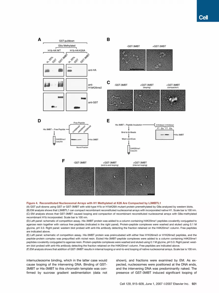
Figure 4. Reconstituted Nucleosomal Arrays with H1 Methylated at K26 Are Compacted by L3MBTL1
(A) GST pull-downs using GST or GST-3MBT with wild-type H1b or H1bK26A mutant protein premethylated by G9a analyzed by western blots.
(B) EM analysis shows that L3MBTL1 can compact recombinant reconstituted nucleosomal arrays with incorporated native H1. Scale bar is 100 nm.
(C) EM analysis shows that GST-3MBT caused looping and compaction of recombinant reconstituted nucleosomal arrays with G9a-methylated
recombinant H1b incorporated. Scale bar is 100 nm.
(D) Left panel: schematic of competition assay. His-3MBT protein was added to a column containing H4K20me1 peptides covalently conjugated to
agarose resin together with various free peptides (indicated in the right panel). Protein-peptide complexes were washed and eluted using 0.1 M
glycine, pH 3.0. Right panel: western blot probed with anti-His antibody detecting the fraction retained on the H4K20me1 column. Free peptides
are indicated above.
(E) Left panel: schematic of competition assay. His-3MBT protein was preincubated with either free H1K26me3 or H1K26me2 peptides, and the
peptide-protein complex was prepurified with nickel resin. Eluted His-3MBT-peptide complexes were added to a column containing H4K20me1
peptides covalently conjugated to agarose resin. Protein-peptide complexes were washed and eluted using 0.1 M glycine, pH 3.0. Right panel: west-
ern blot probed with anti-His antibody detecting the fraction retained on the H4K20me1 column. Free peptides are indicated above.
(F) EM analysis shows that addition of GST-3MBT results in internal looping or end-to-end looping of native nucleosomal arrays. Scale bar is 100 nm.
internucleosome binding, which in the latter case would
cause looping of the intervening DNA. Binding of GST-
3MBT or His-3MBT to this chromatin template was con-
firmed by sucrose gradient sedimentation (data not
shown), and fractions were examined by EM. As ex-
pected, nucleosomes were positioned at the DNA ends,
and the intervening DNA was predominantly naked. The
presence of GST-3MBT induced significant looping of
Cell 129, 915–928, June 1, 2007 ª2007 Elsevier Inc. 921

Table 1. Quantification of Chromatin Compaction/Looping Experiments
Type of Assay Proteins Incubated with Nucleosomal Arrays
Total Molecules
Analyzed
Looping and
Compaction (%)
Compaction nH1 � GST-3MBT 800 21.0
+ GST-3MBT 900 45.0
rH1b/G9a � SAM � GST-3MBT 400 15.5
+ GST-3MBT 400 15.0
+ SAM � GST-3MBT 400 14.75
+ GST-3MBT 400 32.75
PR-SET7 � SAM � GST-3MBT 500 12.8
+ GST-3MBT 500 13.4
+ SAM � GST-3MBT 600 14.2
+ GST-3MBT 400 39.75
� His-3MBT 600 15.8
+ His-3MBT (WT) 600 50.8
+ His-3MBT (P2a) 600 26.1
+ His-3MBT (P2b) 600 33.3
Looping � GST-3MBT 1000 4.6
+ GST-3MBT 1000 35.6
+ GST-3MBT (P2) 400 15.75
+ GST-3MBT (P1, P2, P3) 400 6.5
+ His-3MBT-C2HC 500 35.2
+ His-3MBT 500 17.8
Compaction assays have been performed using recombinant reconstituted nucleosomes, and looping assays have been
performed with native nucleosomal arrays (subsaturating amounts of octamers). Percentage of compacted chromatin particlesor particles showing looping upon addition of recombinant MBT domains in comparison to total number of analyzed particles is
shown. The chromatin-compaction experiments carried out with monomeric His-3MBT protein are indicated in bold. All compac-
tion assays have been carried out three to four times, and the looping assays (� GST-3MBT, + GST-3MBT) have been carried outfour times or two times (all other looping experiments).
the DNA template with both nucleosomal regions being
brought in close vicinity to each other (Figure 4F; see Table
1 for quantification). His-3MBT did induce looping but did
so to a lesser extent than GST-3MBT (Figure S7; Table 1).
To analyze the putative contribution of the Zn-finger
domain of L3MBTL1 in looping, we generated a protein
that is comprised of the three MBT domains and the
Zn-finger domain (His-3MBT-C2HC; see Figure 1A). His-
3MBT-C2HC protein migrated as a monomer in size-
exclusion chromatography (Figure S1) but behaved in
the looping assay similarly to the GST-3MBT protein
(Figure S1; Table 1). Looping was reduced in the presence
of the GST-3MBT-P2 mutant protein and completely abol-
ished in the case of the triply mutated GST-3MBT protein
(single point mutation in each of the three MBT domains;
Table 1). Collectively, the results indicate that a monomeric
form of L3MBTL1 allows internucleosome binding but that
the C2HC domain or dimerization (via the SPM domain of
the native protein or GST of the bacterially produced pro-
tein) contributes to this process (Table 1).
922 Cell 129, 915–928, June 1, 2007 ª2007 Elsevier Inc.
If monomeric His-3MBT is able to bind two nucleo-
somes simultaneously (Figure S7), then a single molecule
might accommodate two methylated histone lysine resi-
dues. We tested this using our chromatin-compaction
assay. Recombinant chromatin was monomethylated
at H4K20 by PR-SET7, incubated with His-3MBT, and
fractionated by sucrose gradient sedimentation (data not
shown). The fractions analyzed by EM confirmed that His-
3MBT indeed compacted chromatin solely dependent on
a single histone lysine methylation mark, H4K20me1
(Figure S8; Table 1). However, if we used mutant His-
3MBT containing a single amino acid substitution in the
second MBT domain that is completely defective in
binding to H4K20me1 (P2a), the number of compacted
molecules decreased substantially (Figure S8; Table 1).
A mutation in the second pocket that still exhibited bind-
ing to H4K20me1 (P2b) showed a reduced number
of compacted particles compared to wild-type His-
3MBT, but significantly more compared to the P2a mutant
(Table 1).

Figure 5. ChIP Experiments Identify
L3MBTL1 Target Genes
(A) 293 cells stably expressing L3MBTL1-F in
ChIP experiments using anti-FLAG mono-
(M2) and polyclonal antibodies. Screening of
E2F target genes revealed that L3MBTL1-F oc-
cupies c-myc and ccne1 promoter regions. For
cdc25, c-myc, c-fos, and actin genes a region
around the transcriptional start site (+1) was
analyzed, and for cyclin E1 (ccne1) the regions
indicated were analyzed.
(B) HeLa cells were used for ChIP experiments
to screen for the presence of endogenous
L3MBTL1 on the c-myc promoter region.
(C) The �900 promoter region upstream of the
c-myc transcriptional start site is occupied by
L3MBTL1, H1, HP1g, H4K20me1, H3K9me2,
and H1K26me2. The �3000 region is not
occupied by L3MBTL1, but H3K9me2 and
H4K20me1 are present.
(D) ShRNA mediated decrease in L3MBTL1
protein levels. Lentiviral based stable knock-
down of L3MBTL1 using two different l3mbtl1
shRNA sequences (1 and 2) leads to increased
MYC protein levels as analyzed by immuno-
blotting of nuclear extracts. A 293 cell line sta-
bly expressing ectopic L3MBTL1 (L3MBTL1-F)
and a 293 cell line transduced with shRNA
against an unrelated protein (Control shRNA)
are also analyzed.
L3MBTL1 Occupies E2F Target Genes Together
with HP1g
Rb controls cell-cycle progression by binding to members
of the E2F family of transcription factors to prevent gene
activation. Since Rb copurified with L3MBTL1 (see Fig-
ure 1A) and the Drosophila L3MBTL1-homolog copurifies
with a E2F/RBF complex (Lewis et al., 2004), we tested if
L3MBTL1 also localizes to E2F-regulated genes such as
c-myc and cyclin E1 (ccne1). Chromatin immunoprecipita-
tion (ChIP) experiments using 293 cells stably expressing
L3MBTL1-F demonstrated binding of L3MBTL1-F to the
proximal promoter regions of c-myc and ccne1 genes
(Figure 5A). L3MBTL1-F was not detectable downstream
of the transcription start site of the cyclin E gene, nor at
the cdc25, c-fos, and actin promoters (Figure 5A), demon-
strating specificity of the assay.
To explore chromatin binding of the endogenous
L3MBTL1 protein, we generated a specific anti-L3MBTL1
antibody (Figure S9). ChIP experiments confirmed binding
of endogenous L3MBTL1 to the c-myc gene (Figure 5B).
The biochemical experiments (see above) strongly sug-
gested that H4K20me1 and H1K26me1/2 should be pres-
ent at these genes, and we found this to be the case for
c-myc using ChIP assays (Figure 5C). Importantly, within
the c-myc promoter region (�900 bp from the transcrip-
tional start site, TSS) the presence of H1bK26me1/2,
HP1g, and H4K20me1 correlates well with the presence
of L3MBTL1. Interestingly, H3K9me2 but not H3K9me3
was also present at these regions of the c-myc gene.
H3K9me2 likely serves as a site for HP1g binding. Se-
quences upstream (�3000) or downstream of the TSS
were devoid of L3MBTL1, H1b, or HP1g (Figures 5B and
5C; data not shown).
Our results suggest that L3MBTL1 functions as a tran-
scriptional repressor, at least in part, by compacting
chromatin. To analyze further whether L3MBTL1 impacts
c-myc gene expression, we used RNA interference. Short
hairpin RNAs (shRNA) against l3mbtl1 led to a decrease in
L3MBTL1 protein levels concomitant with a significant
increase in MYC protein levels (Figure 5D). These results
are consistent with our model that L3MBTL1 negatively
regulates c-myc gene expression. This is also consistent
with the finding that Drosophila L(3)MBT functions in tran-
scriptional repression (Lewis et al., 2004).
DISCUSSION
A Novel Facet of Facultative Chromatin Compaction
The transcriptional repressor L3MBTL1 compacts chroma-
tin in a manner that is strictly dependent on histone methyl-
ation marks—specifically H4K20me1/2 and H1K26me1/2
as shown in this report. The apparent exclusivity of
L3MBTL1 for mono- and dimethylated states supports
a model in which different degrees of methylation at a par-
ticular site can give rise to different readouts. The chromo-
domains (Fischle et al., 2003; Flanagan et al., 2005; Sims
Cell 129, 915–928, June 1, 2007 ª2007 Elsevier Inc. 923

Figure 6. Alternate Models for L3MBTL1
Compaction of Nucleosomal Arrays
See text for details.
Bridging model. One L3MBTL1 monomer or di-
mer binds to two nucleosomes moving them
into close vicinity. In the monomeric case,
one molecule containing three MBT repeats
can accommodate either: (A) two H4K20me1
or (B) two H1K26me1/2 marks on the histone
tails of adjacent nucleosomes or chromato-
somes, respectively. For simplicity, in the
L3MBTL1-homodimer case only one of two
marks bound by each monomer is illustrated.
The homodimer could accommodate four of
one type of mark, (A) H4K20me1 or (B)
H1K26me1/2, or it could accommodate (C)
two of each type on adjacent nucleosomes.
Association model: Each homodimeric or
monomeric L3MBTL1 binds one nucleosome
and facilitates linker DNA bending or inter-
nucleosomal interactions. As shown in the
monomeric case, L3MBTL1 molecules would
be positioned on (D) the surface of the nucleo-
some or (E) the chromatosome by specific
binding to either H4K20me1 or H1K26me1/2,
respectively, leading to a compacted chroma-
tin state. The same is true for the L3MBTL1
homodimer except that (F) both marks could
be accommodated.
et al., 2005) and PHD-domains (Shi et al., 2006; Wysocka
et al., 2006; Li et al., 2006; Pena et al., 2006) found in sev-
eral proteins provide a paradigm for this model, given their
preference for di- and trimethylated lysines as compared
to the monomethyl state.
The binding specificities of L3MBTL1 raise an important
question. How do the MBT repeats bind specifically to two
different methylated lysine residues (H4K20me1/2 and
H1K26me1/2)? Even more intriguing is that the MBT
domains bind H1K26me1/2 but not H3K9me2/3 or
H3K27me1/2/3, yet these lysine residues are located
within a conserved consensus sequence (ARKS). The sim-
plest explanation would be that each one of the three MBT
domains binds a different ligand. However, our data and
that of others (Kim et al., 2006) suggest otherwise. The
second MBT domain is important for H1K26me1/2 as
well as for H4K20me1/2 binding (Figure 3F), as pre binding
with H1K26me2 peptides abolished 3MBT binding to
H4K20me1 (Figures 4D and 4E). Moreover, since we iden-
tified a mutant in the second MBT domain that abolishes
binding to both ligands and a second mutant that selec-
tively abolishes H1K26me, but not H4K20me, binding,
we suggest that both methylated residues are bound via
the second MBT domain but that different aromatic resi-
dues are involved in caging the methylated lysine residue.
Caging through aromatic residues is a property of all
proteins that specifically recognize methylated lysine
residues (Flanagan et al., 2005; Huang et al., 2006; Jacobs
and Khorasanizadeh, 2002; Li et al., 2006; Nielsen et al.,
2002; Pena et al., 2006).
With respect to chromatin compaction, we envision two
different scenarios, taking into account that the P2 domain
924 Cell 129, 915–928, June 1, 2007 ª2007 Elsevier Inc.
of L3MBTL1 can bind H4K20me1 or H1K26me1/2 but
apparently not both modifications simultaneously. Impor-
tantly, monomeric 3MBT can still compact chromatin in
our assay conditions, suggesting that the P2 domain can
accommodate two modified histone marks on two nucle-
osomes. However, given that pre binding with H1K26me2
peptides abolished 3MBT binding to H4K20me1, the two
marks accommodated by the monomer must be identical.
Thus, in the case of the dimeric L3MBTL1, each of the
monomers would bind two identical marks such that
four H4K20me1, four H1K26me1/2, or two of each mark
are bound.
In the ‘‘bridging model’’ L3MBTL1 functions either as a
monomer or a dimer, and adjacent nucleosomes or chro-
matosomes are bound simultaneously, thereby bridging
the linker DNA and moving the nucleosomes closer to-
gether (Figure 6A). L3MBTL1 does exist as a homodimer
in vivo (Boccuni et al., 2003), and dimerization is one
mechanism by which two L3MBTL1 molecules bind to
H4K20me1 and H1K26me1/2 on adjacent nucleosomes/
chromatosomes. This is also supported by our looping
experiments (Figures 4F and S7). Yet, repression does
not depend on the SPM domain responsible for
L3MBTL1 homodimerization, and a monomeric 3MBT
molecule lacking the SPM domain still shows compaction
in our assays (Figures S2 and S7) and can repress tran-
scription when directed to a reporter (Boccuni et al.,
2003). Nonetheless, chromatin compaction by a single
3MBT molecule is still consistent with the bridging model.
A similar mechanism for binding and compacting of multi-
ple nucleosomes by a single molecule was reported re-
cently in the case of the PRC1 component PSC (Francis

et al., 2004), but the histone tails or histone lysine methyl-
ation marks were not required. In this case, the com-
pacted particles resembled the chromatin structures
that we observed in the presence of L3MBTL1.
In the ‘‘association model’’ a single L3MBTL1 molecule
(monomer or dimer) could compact chromatin by posi-
tioning itself on the surface of the nucleosome/chromato-
some in a fashion that promotes bending of the linker DNA
or facilitates histone-histone interactions (Figure 6B). In
this model, correct positioning of L3MBTL1 is accom-
plished by the specific recognition of H4K20me1 or
H1K26me1/2 on the nucleosome or chromatosome sur-
face, respectively. The linker histone itself functions in a
similar manner (Wollfe, 1998). Moreover, proteins contain-
ing HMG boxes, the transcription factor HNF3, the myeloid
and erythroid nuclear termination stage-specific protein
(MENT; Springhetti et al., 2003), or PARP-1 (Kim et al.,
2004) can also bind to nucleosomes and alter chromatin
conformation. In the case of L3MBTL1, compaction is
dependent on specific methylated lysine residues adding
a distinct regulatory parameter.
Concerted Actions of Histone H1b and L3MBTL1
during Chromatin Compaction
Linker histone H1 functions as a transcriptional repressor
in vitro (Cheung et al., 2002; Croston et al., 1991; Laybourn
and Kadonaga, 1991) and is important in chromatin fold-
ing in vitro (van Holde, 1989; Wolffe, 1997). The C-terminal
region of H1 is required for its binding to DNA between nu-
cleosomes (Bednar et al., 1998), and H1 phosphorylation
changes its ability to bind to chromatin (Dou et al.,
1999). Here we show that L3MBTL1 interacts with H1 in
a methylation-dependent manner and that H1bK26me1/2
is important for chromatin compaction by the MBT
domains (Figure 4C). H1 has been detected on both tran-
scriptionally active and inactive genes (reviewed by Parse-
ghian and Hamkalo, 2001), and its binding to chromatin is
dynamic in vivo (Lever et al., 2000; Misteli et al., 2000), yet
the number of factors affecting H1 mobility is unknown
(discussed in van Holde and Zlatanova, 2006). It is possible
that methylation of histone H1 at lysine-26 in the presence
of L3MBTL1 increases H1 residence time on chromatin,
thereby facilitating a compacted chromatin state.
MBT Domain Proteins and Gene Expression
The specificity of the proteins involved in establishing
a type of facultative heterochromatin is likely dictated by
the interaction of regulators (E2F) with other regulators
(Rb) and factors that function in compacting chromatin.
L3MBTL1 is a member of a large family of mammalian
MBT proteins that contain variable numbers of MBT do-
mains. These domains are not identical in sequence, as
is also the case with the chromo- and bromo-domains.
Given this, the different members of the MBT family might
recognize different patterns of histone methyl marks to
establish repression of specific genes through the formation
of facultative heterochromatin.
One of the L3MBTL1 targets shown here is c-myc, the
expression of which is tightly regulated, with increased
myc expression often correlated with cancer (for review
see Nilsson and Cleveland, 2003). We found that reduc-
tion of L3MBTL1 levels significantly increases MYC pro-
tein levels. Ectopic expression of L3MBTL1, however,
does not affect MYC protein levels (Figure 5D). This is
perhaps not surprising given that our in vitro data show
a specific requirement for histone methylation marks in
binding; thus, increased expression of L3MBTL1 would
not necessarily lead to its increased chromatin binding.
It remains to be investigated if overexpression of myc in
cancer correlates with aberrant L3mbtl1 gene expression
and/or localization. The human L3MBTL1 gene is located
on chromosome 20q within the region commonly deleted
in patients with myeloproliferative disorders (MacGrogan
et al., 2001). It is also possible that the chromatin signature
(e.g., histone methylation marks) of the c-myc promoter
region is abnormal in malignant cells, thereby altering
L3MBTL1 binding and the regulated expression of c-myc.
EXPERIMENTAL PROCEDURES
Biochemical Purification of L3MBTL1-F and Associated
Polypeptides
Full-length l3mbtl1 cDNA was inserted into pCMV-Tag4A and trans-
fected in 293 cells using FuGENE (Roche), and clones were selected
that stably expressed the L3MBTL1-FLAG fusion protein. Nuclear ex-
tracts (�300 mg) were prepared from 45 liters of culture following the
Dignam protocol (Dignam et al., 1983) and subjected to anti-FLAG (M2)
agarose (Sigma). Bound proteins were eluted with 200 mg/ml FLAG
peptide (Sigma). Affinity-purified L3MBTL1-F fractions were resolved
by SDS-PAGE and analyzed by silver staining, western blotting, and
mass spectrometry. Gel-resolved proteins were digested with trypsin,
the mixtures fractionated on a Poros 50 R2 RP microtip, and the
resulting peptide pools analyzed by matrix-assisted laser-desorption/
ionization reflectron time-of-flight (MALDI-reTOF) MS using a BRUKER
UltraFlex TOF/TOF instrument (Bruker; Bremen, Germany) as de-
scribed (Devroe et al., 2004).
Peptide Affinity Chromatography
Peptide-affinity columns were generated using SulfoLink coupling gel
(Pierce). Histone H1b peptides comprised residues 20–37 with K26
either unmodified, mono-, di-, or trimethylated. Additional peptides
include H3K4 (residues 1–8), H3K9 (residues 5–13), H3K27 (20–33),
and H4K20 (residues 16–25). Peptide-bound proteins (either in-vitro-
translated or 10 ug of recombinant protein) were washed extensively
(60 column volumes of 25 mM Tris, pH 8, 150 mM NaCl, 2 mM
EDTA, and 0.5% NP40) and eluted with either 0.5 mg/ml peptide or
low pH buffer (100 mM glycine, pH 3.0; Sims et al., 2006).
Chromatin-Compaction Assay
Two micrograms of nucleosome arrays were reconstituted as previ-
ously described (Nishioka and Reinberg, 2003) and incubated with re-
combinant 3MBT proteins in HE buffer containing 25 mM KCl at a molar
ratio of 1:4 at RT for 60 min. The protein-nucleosome complexes were
loaded onto a 5%–30% sucrose gradient in HE buffer containing
25 mM KCl and centrifugated for 6.5–15 hr at 25,000 RPM, and the
fractions were analyzed by 1.0% agarose gel electrophoresis. Peak
fractions of the protein-nucleosome complexes were analyzed by EM.
Cell 129, 915–928, June 1, 2007 ª2007 Elsevier Inc. 925

EM
Protein-nucleosome complexes were fixed with 0.6% glutaraldehyde,
and DNA-protein complexes were purified by gravity-flow gel filtration
(2 ml of BIO-GEL A-5M resin or Sepharose CL-4B, BioRad) using TE
buffer. Purified protein-nucleosome complexes were mixed with
a buffer containing spermidine to a final concentration of 2 mM, ad-
sorbed to glow-charged carbon-coated grids, washed with a water/
graded ethanol series, and rotary shadow cast with tungsten (Griffith
and Christiansen, 1978). Samples were examined using a JEOL 1200
EX transmission electron microscope. Micrographs are shown in
reverse contrast. A Cohu CCD camera attached to a Macintosh
computer programmed with National Institute of Health (NIH) IMAGE
software was used to prepare the images.
ChIP and RNAi
ChIP assays were performed as described (Lewis et al., 2005; Vaquero
et al., 2004). ChIP samples were prepared from 293 cells stably
expressing L3MBTL1-F and from HeLa cells. Primer sets were chosen
to amplify approximately 200 bp around the indicated region. Primer
sequences are available upon request. Five different l3mbtl1 short hair-
pin (sh) RNA constructs were obtained from the MISSION TRC-Hs 1.0
(Human) shRNA library (SIGMA) and used as described previously
(Zufferey et al., 1998).
Supplemental Data
Supplemental Data include nine figures and can be found with this
article online at http://www.cell.com/cgi/content/full/129/5/915/DC1/.
ACKNOWLEDGMENTS
We are grateful to Dr. Lynne Vales for critical reading of our manuscript
and for helpful comments. We thank Drs. Pierre Chambon and Regine
Losson for the generous gift of antibodies. This work is supported by
an Erwin Schrodinger Fellowship from the Austrian Science Founda-
tion (FWF) to P.T. (J2354-B12), a NIH postdoctoral fellowship to
R.J.S. (GM-71166), and by grants from the National Institutes of Health
(CA085826 and CA113863 to Y.-H.W., CA 102202 to S.D.N., and GM-
64844 to D.R.) and the Howard Hughes Medical Institute to D.R.
Received: July 24, 2006
Revised: December 27, 2006
Accepted: March 12, 2007
Published: May 31, 2007
REFERENCES
Bannister, A.J., Zegerman, P., Partridge, J.F., Miska, E.A., Thomas,
J.O., Allshire, R.C., and Kouzarides, T. (2001). Selective recognition
of methylated lysine 9 on histone H3 by the HP1 chromo domain.
Nature 410, 120–124.
Bednar, J., Horowitz, R.A., Dubochet, J., and Woodcock, C.L. (1995).
Chromatin conformation and salt-induced compaction: three-dimen-
sional structural information from cryoelectron microscopy. J. Cell
Biol. 131, 1365–1376.
Bednar, J., Horowitz, R.A., Grigoryev, S.A., Carruthers, L.M., Hansen,
J.C., Koster, A.J., and Woodcock, C.L. (1998). Nucleosomes, linker
DNA, and linker histone form a unique structural motif that directs
the higher-order folding and compaction of chromatin. Proc. Natl.
Acad. Sci. USA 95, 14173–14178.
Boccuni, P., MacGrogan, D., Scandura, J.M., and Nimer, S.D. (2003).
The human L(3)MBT polycomb group protein is a transcriptional re-
pressor and interacts physically and functionally with TEL (ETV6).
J. Biol. Chem. 278, 15412–15420.
Cheung, E., Zarifyan, A.S., and Kraus, W.L. (2002). Histone H1 re-
presses estrogen receptor alpha transcriptional activity by selectively
926 Cell 129, 915–928, June 1, 2007 ª2007 Elsevier Inc.
inhibiting receptor-mediated transcription initiation. Mol. Cell. Biol. 22,
2463–2471.
Croston, G.E., Kerrigan, L.A., Lira, L.M., Marshak, D.R., and
Kadonaga, J.T. (1991). Sequence-specific antirepression of histone
H1-mediated inhibition of basal RNA polymerase II transcription. Sci-
ence 251, 643–649.
Daujat, S., Zeissler, U., Waldmann, T., Happel, N., and Schneider, R.
(2005). HP1 binds specifically to Lys26-methylated histone H1.4,
whereas simultaneous Ser27 phosphorylation blocks HP1 binding.
J. Biol. Chem. 280, 38090–38095.
Devroe, E., Erdjument-Bromage, H., Tempst, P., and Silver, P.A.
(2004). Human Mob proteins regulate the NDR1 and NDR2 serine-
threonine kinases. J. Biol. Chem. 279, 24444–24451.
Dignam, J.D., Lebovitz, R.M., and Roeder, R.G. (1983). Accurate
transcription initiation by RNA polymerase II in a soluble extract from
isolated mammalian nuclei. Nucleic Acids Res. 11, 1475–1489.
Dou, Y., Mizzen, C.A., Abrams, M., Allis, C.D., and Gorovsky, M.A.
(1999). Phosphorylation of linker histone H1 regulates gene expression
in vivo by mimicking H1 removal. Mol. Cell 4, 641–647.
Fischle, W., Wang, Y., Jacobs, S.A., Kim, Y., Allis, C.D., and
Khorasanizadeh, S. (2003). Molecular basis for the discrimination of
repressive methyl-lysine marks in histone H3 by Polycomb and HP1
chromodomains. Genes Dev. 17, 1870–1881.
Flanagan, J.F., Mi, L.Z., Chruszcz, M., Cymborowski, M., Clines, K.L.,
Kim, Y., Minor, W., Rastinejad, F., and Khorasanizadeh, S. (2005). Dou-
ble chromodomains cooperate to recognize the methylated histone H3
tail. Nature 438, 1181–1185.
Francis, N.J., Kingston, R.E., and Woodcock, C.L. (2004). Chromatin
compaction by a polycomb group protein complex. Science 306,
1574–1577.
Griffith, J.D., and Christiansen, G. (1978). Electron microscope visual-
ization of chromatin and other DNA-protein complexes. Annu. Rev.
Biophys. Bioeng. 7, 19–35.
Hall, I.M., Shankaranarayana, G.D., Noma, K., Ayoub, N., Cohen, A.,
and Grewal, S.I. (2002). Establishment and maintenance of a hetero-
chromatin domain. Science 297, 2232–2237.
Huang, Y., Fang, J., Bedford, M.T., Zhang, Y., and Xu, R.M. (2006).
Recognition of histone H3 lysine-4 methylation by the double tudor do-
main of JMJD2A. Science 312, 748–751.
Jacobs, S.A., and Khorasanizadeh, S. (2002). Structure of HP1 chro-
modomain bound to a lysine 9-methylated histone H3 tail. Science
295, 2080–2083.
Jacobs, S.A., Taverna, S.D., Zhang, Y., Briggs, S.D., Li, J., Eissenberg,
J.C., Allis, C.D., and Khorasanizadeh, S. (2001). Specificity of the HP1
chromo domain for the methylated N-terminus of histone H3. EMBO J.
20, 5232–5241.
Kim, J., Daniel, J., Espejo, A., Lake, A., Krishna, M., Xia, L., Zhang, Y.,
and Bedford, M.T. (2006). Tudor, MBT and chromo domains gauge the
degree of lysine methylation. EMBO Rep. 4, 397–403.
Kim, M.Y., Mauro, S., Gevry, N., Lis, J.T., and Kraus, W.L. (2004).
NAD+-dependent modulation of chromatin structure and transcription
by nucleosome binding properties of PARP-1. Cell 119, 803–814.
Klymenko, T., Papp, B., Fischle, W., Kocher, T., Schelder, M., Fritsch,
C., Wild, B., Wilm, M., and Muller, J. (2006). A Polycomb group protein
complex with sequence-specific DNA-binding and selective methyl-
lysine-binding activities. Genes Dev. 20, 1110–1122.
Kuzmichev, A., Jenuwein, T., Tempst, P., and Reinberg, D. (2004). Dif-
ferent EZH2-containing complexes target methylation of histone H1 or
nucleosomal histone H3. Mol. Cell 14, 183–193.
Kuzmichev, A., Margueron, R., Vaquero, A., Preissner, T.S., Scher, M.,
Kirmizis, A., Ouyang, X., Brockdorff, N., Abate-Shen, C., Farnham, P.,
and Reinberg, D. (2005). Composition and histone substrates of

polycomb repressive group complexes change during cellular differ-
entiation. Proc. Natl. Acad. Sci. USA 102, 1859–1864.
Laybourn, P.J., and Kadonaga, J.T. (1991). Role of nucleosomal cores
and histone H1 in regulation of transcription by RNA polymerase II.
Science 254, 238–245.
Lever, M.A., Th’ng, J.P., Sun, X., and Hendzel, M.J. (2000). Rapid
exchange of histone H1.1 on chromatin in living human cells. Nature
408, 873–876.
Lewis, B.A., Sims, R.J., 3rd, Lane, W.S., and Reinberg, D. (2005).
Functional characterization of core promoter elements: DPE-specific
transcription requires the protein kinase CK2 and the PC4 coactivator.
Mol. Cell 18, 471–481.
Lewis, P.W., Beall, E.L., Fleischer, T.C., Georlette, D., Link, A.J., and
Botchan, M.R. (2004). Identification of a Drosophila Myb-E2F2/RBF
transcriptional repressor complex. Genes Dev. 18, 2929–2940.
Li, H., Ilin, S., Wang, W., Duncan, E.M., Wysocka, J., Allis, C.D., and
Patel, D.J. (2006). Molecular basis for site-specific read-out of histone
H3K4me3 by the BPTF PHD finger of NURF. Nature 442, 91–95.
Loyola, A., LeRoy, G., Wang, Y.H., and Reinberg, D. (2001). Reconsti-
tution of recombinant chromatin establishes a requirement for histone-
tail modifications during chromatin assembly and transcription. Genes
Dev. 15, 2837–2851.
MacGrogan, D., Alvarez, S., DeBlasio, T., Jhanwar, S.C., and Nimer,
S.D. (2001). Identification of candidate genes on chromosome band
20q12 by physical mapping of translocation breakpoints found in
myeloid leukemia cell lines. Oncogene 20, 4150–4160.
Maison, C., Bailly, D., Peters, A.H., Quivy, J.P., Roche, D., Taddei, A.,
Lachner, M., Jenuwein, T., and Almouzni, G. (2002). Higher-order
structure in pericentric heterochromatin involves a distinct pattern of
histone modification and an RNA component. Nat. Genet. 30, 329–
334.
Meehan, R.R., Kao, C.F., and Pennings, S. (2003). HP1 binding to na-
tive chromatin in vitro is determined by the hinge region and not by the
chromodomain. EMBO J. 22, 3164–3174.
Minc, E., Courvalin, J.C., and Buendia, B. (2000). HP1gamma associ-
ates with euchromatin and heterochromatin in mammalian nuclei and
chromosomes. Cytogenet. Cell Genet. 90, 279–284.
Misteli, T., Gunjan, A., Hock, R., Bustin, M., and Brown, D.T. (2000).
Dynamic binding of histone H1 to chromatin in living cells. Nature
408, 877–881.
Nielsen, P.R., Nietlispach, D., Mott, H.R., Callaghan, J., Bannister, A.,
Kouzarides, T., Murzin, A.G., Murzina, N.V., and Laue, E.D. (2002).
Structure of the HP1 chromodomain bound to histone H3 methylated
at lysine 9. Nature 416, 103–107.
Nielsen, S.J., Schneider, R., Bauer, U.M., Bannister, A.J., Morrison, A.,
O’Carroll, D., Firestein, R., Cleary, M., Jenuwein, T., Herrera, R.E., and
Kouzarides, T. (2001). Rb targets histone H3 methylation and HP1 to
promoters. Nature 412, 561–565.
Nilsson, J.A., and Cleveland, J.L. (2003). Myc pathways provoking cell
suicide and cancer. Oncogene 22, 9007–9021.
Nishioka, K., and Reinberg, D. (2003). Methods and tips for the
purification of human histone methyltransferases. Methods 31, 49–58.
Nishioka, K., Rice, J.C., Sarma, K., Erdjument-Bromage, H., Werner,
J., Wang, Y., Chuikov, S., Valenzuela, P., Tempst, P., Steward, R.,
et al. (2002). PR-Set7 is a nucleosome-specific methyltransferase
that modifies lysine 20 of histone H4 and is associated with silent chro-
matin. Mol. Cell 9, 1201–1213.
Ogawa, H., Ishiguro, K., Gaubatz, S., Livingston, D.M., and
Nakatani, Y. (2002). A complex with chromatin modifiers that
occupies E2F- and Myc-responsive genes in G0 cells. Science 296,
1132–1136.
Parseghian, M.H., and Hamkalo, B.A. (2001). A compendium of the his-
tone H1 family of somatic subtypes: an elusive cast of characters and
their characteristics. Biochem. Cell Biol. 79, 289–304.
Patnaik, D., Chin, H.G., Esteve, P.O., Benner, J., Jacobsen, S.E., and
Pradhan, S. (2004). Substrate specificity and kinetic mechanism of
mammalian G9a histone H3 methyltransferase. J. Biol. Chem. 279,
53248–53258.
Pena, P.V., Davrazou, F., Shi, X., Walter, K.L., Verkhusha, V.V., Gozani,
O., Zhao, R., and Kutateladze, T.G. (2006). Molecular mechanism of
histone H3K4me3 recognition by plant homeodomain of ING2. Nature
442, 100–103.
Sato, M.H., Ura, K., Hohmura, K.I., Tokumasu, F., Yoshimura, S.H.,
Hanaoka, F., and Takeyasu, K. (1999). Atomic force microscopy
sees nucleosome positioning and histone H1-induced compaction in
reconstituted chromatin. FEBS Lett. 452, 267–271.
Schotta, G., Ebert, A., Krauss, V., Fischer, A., Hoffmann, J., Rea, S.,
Jenuwein, T., Dorn, R., and Reuter, G. (2002). Central role of Drosoph-
ila SU(VAR)3–9 in histone H3–K9 methylation and heterochromatic
gene silencing. EMBO J. 21, 1121–1131.
Shi, X., Hong, T., Walter, K.L., Ewalt, M., Michishita, E., Hung, T.,
Carney, D., Pena, P., Lan, F., Kaadige, M.R., et al. (2006). ING2 PHD
domain links histone H3 lysine 4 methylation to active gene repression.
Nature 442, 96–99.
Sims, R.J., 3rd, Nishioka, K., and Reinberg, D. (2003). Histone lysine
methylation: a signature for chromatin function. Trends Genet. 19,
629–639.
Sims, R.J., 3rd, Chen, C.F., Santos-Rosa, H., Kouzarides, T., Patel,
S.S., and Reinberg, D. (2005). Human but not yeast CHD1 binds
directly and selectively to histone H3 methylated at lysine 4 via its
tandem chromodomains. J. Biol. Chem. 280, 41789–41792.
Sims, R.J., 3rd, Trojer, P., Li, G., and Reinberg, D. (2006). Methods to
identify and functionally analyze factors that specifically recognize his-
tone lysine methylation. Methods 40, 331–338.
Springhetti, E.M., Istomina, N.E., Whisstock, J.C., Nikitina, T.,
Woodcock, C.L., and Grigoryev, S.A. (2003). Role of the M-loop and
reactive center loop domains in the folding and bridging of nucleosome
arrays by MENT. J. Biol. Chem. 278, 43384–43393.
Tachibana, M., Sugimoto, K., Fukushima, T., and Shinkai, Y. (2001).
Set domain-containing protein, G9a, is a novel lysine-preferring
mammalian histone methyltransferase with hyperactivity and specific
selectivity to lysines 9 and 27 of histone H3. J. Biol. Chem. 276,
25309–25317.
van Holde, K., and Zlatanova, J. (2006). Scanning chromatin: a new
paradigm? J. Biol. Chem. 281, 12197–12200.
van Holde, K.E. (1989). Chromatin, Volume 1, First Edition, (New York:
Springer-Verlag).
Vaquero, A., Scher, M., Lee, D., Erdjument-Bromage, H., Tempst, P.,
and Reinberg, D. (2004). Human SirT1 interacts with histone H1 and
promotes formation of facultative heterochromatin. Mol. Cell 16, 93–
105.
Wang, W.K., Tereshko, V., Boccuni, P., MacGrogan, D., Nimer, S.D.,
and Patel, D.J. (2003). Malignant brain tumor repeats: a three-leaved
propeller architecture with ligand/peptide binding pockets. Structure
11, 775–789.
Wismar, J., Loffler, T., Habtemichael, N., Vef, O., Geissen, M., Zirwes,
R., Altmeyer, W., Sass, H., and Gateff, E. (1995). The Drosophila
melanogaster tumor suppressor gene lethal(3)malignant brain tumor
encodes a proline-rich protein with a novel zinc finger. Mech. Dev.
53, 141–154.
Wolffe, A.P. (1997). Histone H1. Int. J. Biochem. Cell Biol. 29, 1463–
1466.
Wollfe, A. (1998). Chromatin: Structure and Function (Wiltshire, Great
Britain: Academic Press).
Cell 129, 915–928, June 1, 2007 ª2007 Elsevier Inc. 927

Wysocka, J., Swigut, T., Xiao, H., Milne, T.A., Kwon, S.Y., Landry, J.,
Kauer, M., Tackett, A.J., Chait, B.T., Badenhorst, P., et al. (2006). A
PHD finger of NURF couples histone H3 lysine 4 trimethylation with
chromatin remodelling. Nature 442, 86–90.
Yohn, C.B., Pusateri, L., Barbosa, V., and Lehmann, R. (2003). l(3)ma-
lignant brain tumor and three novel genes are required for Drosophila
germ-cell formation. Genetics 165, 1889–1900.
928 Cell 129, 915–928, June 1, 2007 ª2007 Elsevier Inc.
Zhao, T., Heyduk, T., Allis, C.D., and Eissenberg, J.C. (2000). Hetero-
chromatin protein 1 binds to nucleosomes and DNA in vitro. J. Biol.
Chem. 275, 28332–28338.
Zufferey, R., Dull, T., Mandel, R.J., Bukovsky, A., Quiroz, D., Naldini, L.,
and Trono, D. (1998). Self-inactivating lentivirus vector for safe and
efficient in vivo gene delivery. J. Virol. 72, 9873–9880.