topoisomerase ii, scaffold component, promotes chromatin compaction in vitro in a linker-histone...
TRANSCRIPT
Nucleic Acids Research, 2007, 1–13doi:10.1093/nar/gkm116
Topoisomerase II, scaffold component, promoteschromatin compaction in vitro in a linker-histoneH1-dependent mannerKohji Hizume1, Sumiko Araki2, Kenichi Yoshikawa2 and Kunio Takeyasu1, *
1Laboratory of Plasma Membrane and Nuclear Signaling, Graduate School of Biostudies, Kyoto University,Kitashirakawa-oiwake-cho, Sakyo-ku, Kyoto, 606-8502, Japan and 2Department of Physics, Graduate School ofScience, Kyoto University, Kitashirakawa-oiwake-cho, Sakyo-ku, Kyoto, 606-8502, Japan
Received September 15, 2006; Revised February 7, 2007; Accepted February 7, 2007
ABSTRACT
TopoisomeraseII (Topo II) is a major component ofchromosomal scaffolds and essential for mitoticchromosome condensation, but the mechanism ofthis action remains unknown. Here, we used an invitro chromatin reconstitution system in combina-tion with atomic force and fluorescence micro-scopic analyses to determine how Topo II affectschromosomal structure. Topo II bound to bare DNAand clamped the two DNA strands together, even inthe absence of ATP. In addition, Topo II promotedchromatin compaction in a manner dependent onhistone H1 but independent of ATP. Histone H1-induced 30-nm chromatin fibers were convertedinto a large complex by Topo II. Fluorescencemicroscopic analysis of the Brownian motion ofchromatin stained with 40,6-diamidino-2-phenylin-dole showed that the reconstituted chromatinbecame larger following the addition of Topo IIin the presence but not the absence of histone H1.Based on these findings, we propose that chromatinpacking is triggered by histone H1-dependent, TopoII-mediated clamping of DNA strands.
INTRODUCTION
High-order packing of genomic DNA into eukaryoticinterphase nuclei is necessary for the maintenance ofgenomic information and the complex regulation of geneactivities. Chromosomal packing is also critical during celldivision because an aberration in the packing of thegenome results in defects in the formation and correctseparation of sister chromatids.
Many structural studies have been carried out toinvestigate the molecular mechanism of the higher ordergenome packing. Nanoscale observations using electron
microscopy and atomic force microscopy (AFM) haverevealed that the nucleosome, which forms a ‘beads-on-a-string’ structure with linker DNA, is the fundamentalstructural unit of the chromosome (1–3). These nucleoso-mal arrays are packed into 30-nm fibers with the help oflinker-histone H1 (4–7). Although the mechanisms of howstructures larger than 30-nm fibers are generated remaincontroversial, it has been proposed that chromatin fibersform loop structures that are attached to an axis called ascaffold or matrix. Indeed, DNA loops extending from anaxis have been observed in histone-depleted metaphasechromosomes (8).Biochemical studies on interphase nuclei have suggested
that genomic DNA contains regions associated with thescaffold/matrix approximately every 100 kb, a regioncalled the scaffold/matrix attachment region (SAR/MAR) (9). Some of the components associated with thisscaffold/matrix region have been identified (10). Forexample, the metaphase scaffold/matrix contains thecondensin complex and topoisomerase II (Topo II) (11–14). Both of these components are essential for mitoticchromosome condensation (15–17) and are located on theaxis of condensed chromosomes (18). The interphasescaffold/matrix, on the other hand, contains RNA andribonucleoprotein as major components (19,20). Also,NuMA, actin, DNA and RNA polymerases, histoneacetyl transferase and Topo II are now considered to becomponents of the interphase scaffold/matrix.Interestingly, Topo II is a component of the scaffold at
both mitosis and interphase. Topo II catalyzes thedecatenation of two DNA strands using energy fromthe hydrolysis of ATP (21). Topo II is a homodimerwith a ring-like structure (22,23). In a mutant strain offission yeast lacking functional Topo II, chromosomescannot condense (15). In addition, Topo II-immunodepletedmitotic frog extracts cannot induce chromatin condensationin nuclei from HeLa cells or chicken erythrocytes (24).
� 2007 The Author(s).
This is an Open Access article distributed under the terms of the Creative Commons Attribution Non-Commercial License (http://creativecommons.org/licenses/
by-nc/2.0/uk/) which permits unrestricted non-commercial use, distribution, and reproduction in any medium, provided the original work is properly cited.
Nucleic Acids Research Advance Access published April 11, 2007
In previous studies, we established an in vitro chromatinreconstitution system, wherein beads-on-a-string nucleo-some arrays are produced upon removal of salt bydialysis (3) and 30-nm chromatin fibers can be generatedby the addition of histone H1 (7). Here, we conductedmolecular imaging analyses on the interaction betweenTopo II and in vitro-reconstituted chromatin fibers.We found that, in the absence of ATP, Topo IIpromotes further folding of 30-nm fibers but not ofbeads-on-a-string arrays. This means that linker-histoneH1 is crucial for the formation of 30-nm chromatin fibersand that the subsequent function of Topo II is essentialfor achieving higher order structures.
MATERIALS AND METHODS
Materials
The plasmids used for chromatin reconstitution were kindgifts from Dr W. De Laat (Erasmus University MedicalCenter, Department of Cell Biology, Netherlands). Theentire �-globin gene (�170 kb) was cloned into thepCYPAC2 vector, producing a 185-kb plasmid (25).The locus control region (LCR) of the human �-globingene (21.5 kb) was isolated by SalI/ClaI digestion andsubcloned into pBR322, yielding a 26-kb plasmid.The plasmids to be tested for the Topo II binding were
constructed as follows: The 6-kb LCR region of thehuman �-globin gene including nuclease hypersensitivesites 4 and 5 was amplified by PCR using human genomicDNA as a template and then cloned into pT7Blue-2(Merck, Germany), yielding pBGa. The region betweennuclease hypersensitive sites 4 and 5 contains the SAR/MAR sequence (26).Core histones were purified from HeLa cells according
to the method developed by O’Neill et al. (27), with slightmodifications. The cells were harvested, washed with PBSand lysed with L-buffer (140mM NaCl, 10mM Tris-HCl,pH 7.5 and 0.5% Triton X-100). Nuclei were isolatedby low-speed centrifugation and washed three times withW-buffer (350mM NaCl and 10mM Tris-HCl, pH 7.5).The nuclei were then treated with micrococcal nuclease(40 units/mg of DNA) at 378C for 15min in D-buffer(10mM Tris-HCl pH 7.5, 1.5mM MgCl2, 1mM CaCl2,0.25M sucrose and 0.1mM phenylmethylsulfonylfluoride. The reaction was stopped by the addition ofEGTA to a final concentration of 2mM, and the nucleiwere sedimented by centrifugation at 10 000� g for 5min.The pellet was resuspended in N-buffer (10mM Tris-HCl,pH 6.8, 5mM EDTA, and 0.1mM phenylmethylsulfonylfluoride), and dialyzed against N-buffer overnight at48C. The sample was centrifuged at 10 000� g for10min, and the soluble chromatin supernatant wasredialyzed against HA-buffer (0.1M NaPO4, pH 6.7and 0.63M NaCl) and mixed with hydroxyapatite resin(Bio-Rad). After batch binding at 48C for 1 h, the resinwas packed into a column and washed with five volumesof HA-buffer. The core histones were eluted with E-buffer(0.1M NaPO4, pH 6.7 and 2M NaCl). The eluate wasapplied to a gel filtration column (HiPrep 16/60 S-200;Amersham Biosciences) to separate the octamer from
the H3-H4 tetramer, H2A-H2B dimer and othercontaminants.
Histone H1 was purified from HeLa cells accordingto the method developed by Mirzabekov et al. (28) withslight modifications. The cells were harvested, washedwith PBS, lysed in 140mM NaCl, 10mM Tris-HCl(pH 7.5) and 0.5% Triton X-100, and washed threetimes with the same buffer without the detergent.The nuclei were collected, washed with a buffer of0.35M NaCl and 10mM Tris-HCl (pH 7.5), resuspendedin 5% CCl3COOH and rotated at 48C for 80min.After centrifugation at 4000� g for 15min, the solublehistone H1-containing supernatant was dialyzed against10mM HCl containing 2mM �-mercaptoethanol. Thedialyzed sample was lyophilized and stored at �808C. Forchromatin reconstitution, the lyophilized protein wasresuspended in 10mM Tris-HCl (pH 7.5), 1mM EDTA,500mM NaCl, 0.05% NP-40 and 20% glycerol.
Chromatin reconstitution
Equal amounts (0.5 mg) of the purified DNA andthe histone octamer were mixed in Hi-buffer (10mMTris-HCl, pH 7.5, 2M NaCl, 1mM EDTA, 0.05% NP-40and 5mM �-mercaptoethanol), and placed in a dialysistube (total volume, 50 ml). The dialysis was startedwith 150ml of Hi-buffer by stirring at 48C. Lo-buffer(10mM Tris-HCl, pH 7.5, 1mM EDTA, 0.05% NP-40and 5mM 2-mercaptoethanol) was added to the dialysisbuffer at a rate of 0.46ml/min, and simultaneously, thedialysis buffer was pumped out at the same speed with aperistaltic pump so that the final dialysis buffer contained50mMNaCl after 20 h. The sample was collected from thedialysis tube and stored at 48C until use.
For experiments in which reconstitution with histoneH1 was performed, histone H1 was added after the saltdialysis was completed (at a NaCl concentration of50mM). The molar ratio of histone H1 to histone octamerwas 1:1. After standing on ice for 30min, the sample wasused for the Topo II treatment.
Topo II binding
Topo II used in this study was topoisomerase II�(TopoGEN, Port Orange, FL). Various amounts ofTopo II were mixed with 80 ng of supercoiled pBGain 50mM NaCl buffer (10mM Tris-HCl, pH 7.5, 1mMEDTA, 50mM NaCl, 0.05% NP-40 and 5mM�-mercaptoethanol) at 378C for 30min. In some experi-ments, a 1/10 volume of ATP-Mg buffer (0.3mM ATPand 12.5mM MgCl2) was added to the reaction mixture.The reaction samples were diluted 10-fold by addingHMgG buffer (10mM HEPES, pH 7.5, 2mM MgCl2 and0.3% glutaraldehyde) and kept for 30min at roomtemperature.
The reconstituted chromatin sample with or without H1was treated with Topo II at 378C for 30min. Afterincubation, the sample was analyzed by AFM.
AFM observation
The reconstituted chromatin solution was fixed with0.3% glutaraldehyde for 30min at room temperature
2 Nucleic Acids Research, 2007
and then dropped onto a freshly cleaved mica surfacethat had been pretreated with 10mM spermidine.After 10min at room temperature, the mica was washedwith water and dried under nitrogen gas. AFM observa-tion was performed with a Nanoscope IIIa or IV (DigitalInstruments) in air in the tapping mode. The cantilever(OMCL-AC160TS-W2; Olympus) was 129mm in lengthwith a spring constant of 33–62N/m. The scanningfrequency was 2–3Hz, and images were captured withthe height mode in a 512� 512-pixel format. The obtainedimages were plane-fitted and flattened using the softwaresupplied in the imaging module. Except where noted,AFM images in this study are presented as surface plots,which are efficient for displaying the entire structure.In the Supplementary Data, all AFM images are shownusing the top view with a height scale.
Pull-down assay
The cDNA encoding histone H1 was a kind gift fromMichael J. Hendzel (University of Alberta, Canada). Theplasmid for overexpressing of histone H1 with anN-terminal GST tag was constructed as follows: theORF of the histone H1 gene was isolated by EcoRIdigestion and inserted into pGEX5X-2, yielding pGSTH1.
Plasmids pGSTH1 and pGEX5X-2 were introducedinto Escherichia coli and grown in 30ml of LB. During thelog phase of growth, the medium was adjusted by theaddition of isopropyl-�-D-thiogalactopyranoside (IPTG)into the culture medium (30ml of LB) to the finalconcentration of 1mM. After 4 h, the cells were harvestedand resuspended in 1.5ml of 50mM NaCl buffer (asdescribed in the ‘Topo II binding’ section). The suspendedcells were treated with 0.2mg/ml lysozyme for 30min at48C. The cells were then sonicated, and the cell lysate wascentrifuged at 11 000� g for 30min. The supernatant wascollected and used for purification of the fusion proteins.
The supernatant (300ml) was mixed with 30 ml ofglutathione-Sepharose 4B beads (GE HealthcareBioscience) equilibrated with 50mM NaCl buffer at 48Cfor 1.5 h. The beads were collected by gentle centrifugationand washed five times with 300 ml of 50mM NaCl buffer.
After incubating the GST- or GST-H1-bound beads at48C for 1.5 h with 200 ng of Topo II in 80ml 50mM NaClbuffer, the supernatant was collected as the unboundfraction. The beads were then washed five times with100 ml of 50mM NaCl buffer, and proteins remainingbound were used for Western blot analyses.
Fluorescence observation in solution and statistical analysis
The Brownian motion of chromatin was examinedby fluorescence microscopy as described previously (29).The reconstituted chromatin samples were treated withHMgG buffer (see the ‘Topo II binding’ section) andstained with 0.1mM 40,6-diamidino-2-phenylindole(DAPI). DNA staining was observed under UV irradia-tion at 365 nm using a Carl Zeiss Axiovert 135 TVmicroscope equipped with a �100 oil immersion objectivelens. Images were recorded on video tape at 30 frames persecond through a high-sensitivity EB-CCD camera withan image-processing system (Hamamatsu Photonics).
When we observed the chromatin complex existingin the bulk buffer solution (not attached to a coverglass),the complex moved by Brownian motion. To evaluatethe hydrodynamic radius of the chromatin in bulksolution, we measured the Brownian motion of individualchromatin complexes as described previously (30).Due to a blurring of the fluorescent images of individualchromatin complexes, the observed DAPI signals wereslightly (�0.3 mm) larger than the actual sizes (31).We plotted the location of the center of mass of theDAPI signal every 0.03 s and determined the timedependence of the mean-square displacement for eachcomplex. From these values, we evaluated the diffusionconstant, D, as the projection onto the 2D plane:
½rð0Þ � rðtÞ�2� �
¼ 4Dt,
where r is the 2D spatial position of the center of themass of a chromatin complex, and the symbol hi denotes atime average of squared displacements. We also calculatedthe hydrodynamic radius, RH, which is a measure of thechromatin conformation, according to the followingequation:
RH ¼ kBT=6��D,
where kB is the Boltzmann constant and � is the viscosityof the solvent.
RESULTS
Sequence-independent action of Topo II on bare DNA
To understand how Topo II alters the moleculararchitecture of chromosomes, we first used agarose gelelectrophoresis to examine the structure of bare DNA inthe presence of Topo II. Mixing Topo II with a 9-kbsupercoiled plasmid harboring the SAR/MAR region ofthe �-globin gene in the presence of ATP and MgCl2altered the topology of the supercoiled plasmid (lanes 3–5in Figure 1A). Similar results were observed whentopoisomerase I was added instead of Topo II in theabsence of ATP (lane 7). For 80 ng of plasmid DNA, 80 ng(lane 3), but not 2 (lane 5) or 20 ng (lane 4), of Topo IIwas sufficient to completely remove the supercoiling.Next, we examined samples of the reactions containing
different amounts of Topo II by AFM (Figure 1B–E).As seen by agarose gel electrophoresis (Figure 1A),supercoiling of the plasmids was observed in the absenceof Topo II (Figure 1B). Supercoiling was progressivelylost as the amount of Topo II was increased, andthe plasmids became completely relaxed in the presence80 ng of Topo II (Figure 1C–E).Topo II, however, did not mediate relaxation of
the supercoiled plasmids in the absence of ATP(Figure 2A–D), although formed regions where twoDNA strands ran in parallel or crossed over. As seen inthe enlarged image (Figure 2C), two DNA strands wereattached to each other and clamped by a particle witha size (36.6� 5.1 nm) similar to a Topo II dimer (23).These results demonstrated that Topo II can clamptwo DNA strands in the absence of ATP. The number
Nucleic Acids Research, 2007 3

of Topo II particles (Topo II dimers) on the DNAmolecules is shown in Figure 1E. As the amount of TopoII was increased, the number of Topo II–DNA complexesincreased to �70%, with �30% of the DNA moleculesremaining free of Topo II. Although we mixed 18 Topo IIdimers per plasmid molecule in the experiments shown inFigures 1E and 2D, less than four Topo II dimers wereassociated with each plasmid (Figure 2E). These resultssuggest that Topo II binds weakly to bare DNA.We next examined the sequence dependency of
Topo II–DNA binding. We mixed Topo II with the 9-kblinear plasmid DNA containing the SAR/MAR sequenceand analyzed the interaction between Topo II and DNAby AFM (Figure 2F). A histogram of the frequency ofTopo II binding to the DNA (Figure 2G) suggests that it isindependent of the SAR/MAR sequence. We previouslyreported that AFM can detect the sequence-specificbinding of transcription factors, including AP2 (32) andBach1 (33). Therefore, the present results suggest thatTopo II has little if any sequence specificity.
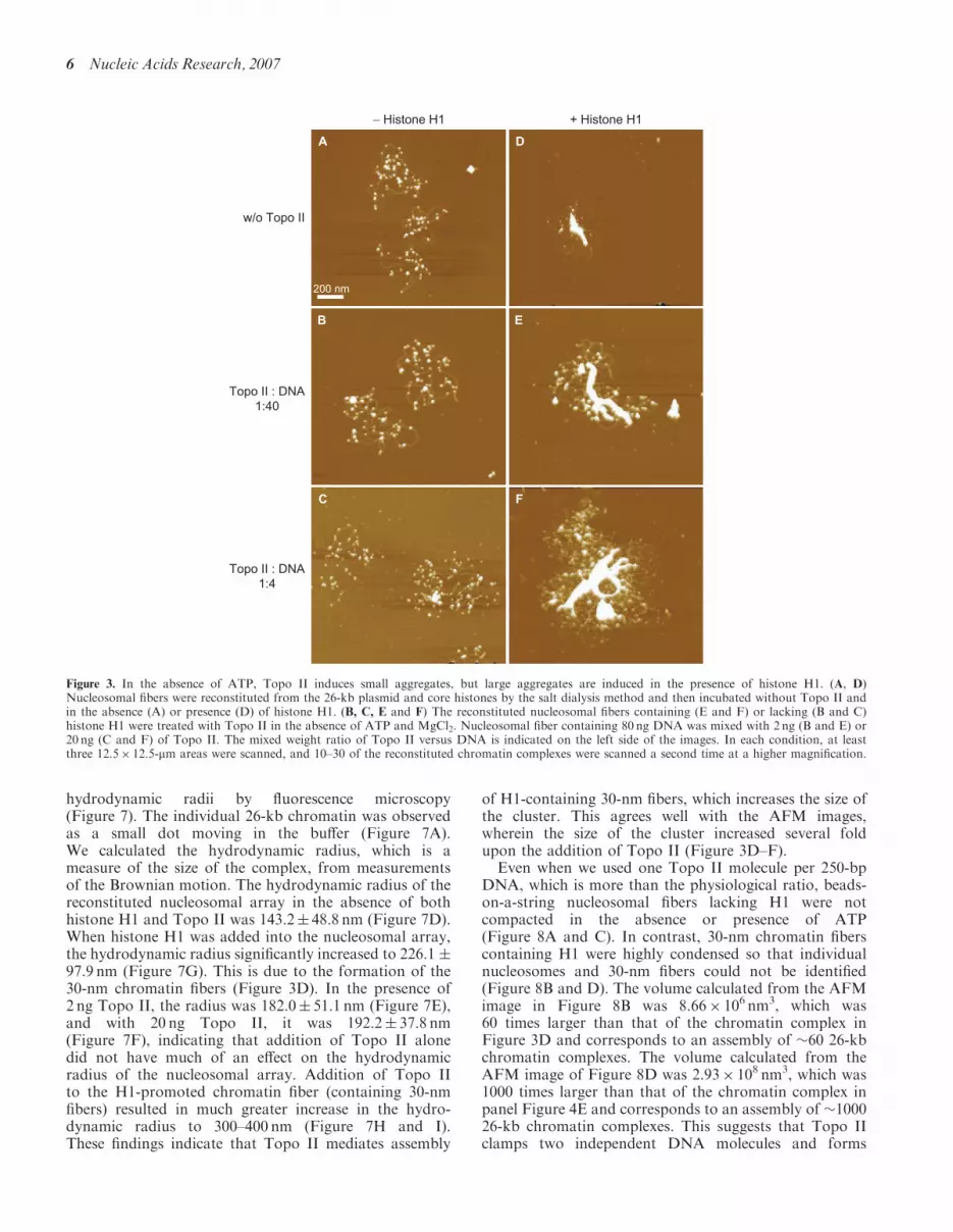
Topo II promotes chromatin compaction in a histoneH1-dependent manner
When we used a 26-kb supercoiled plasmid (21.5-kbLCR fragment of the �-globin locus in pBR322) forreconstitution of nucleosomal fibers, �50 nucleosomeswere formed on each plasmid DNA (Figure 3A).
The number of nucleosomes is below saturation because,in vivo, a 26-kb DNA should contain 104–130 nucleo-somes. As previously described (3), however, this efficiency(50 nucleosome per 26-kb DNA) is the upper limit forreconstitution by salt dialysis, which does not requireother proteins such as histone chaperons. We addedvarious amounts of Topo II to these reconstituted‘beads-on-a-string’ nucleosomes. We did not detect anystructural changes (Figure 3B and C), although Topo IIcould bind weakly to the DNA (Figures 1 and 2). Incontrast, a large complex was formed in the absence ofATP when Topo II was added to 30-nm chromatin fibers(7) that had been reconstituted from the 26-kb nucleo-somes by the addition of histone H1 (Figures 2D–F).
Nucleosome–nucleosome interactions are stronglyaffected by the salt environment. This accounts for thefact that the addition of ATP and MgCl2 to thereconstituted system caused the formation of smallaggregates of nucleosomes (Figure 4A) that were notsignificantly affected by the addition of Topo II (Figure 4Band C). Again, in this system, large complexes wereformed only from 30-nm chromatin fibers (Figure 4D–F).
The finding that histone H1 is essential for Topo II-induced chromatin compaction (Figures 3 and 4) raisedthe possibility that the compaction is caused by a directinteraction between Topo II and histone H1. To test thispossibility, we performed a pull-down assay usingGST-H1. GST-H1 was expressed in E. coli and trapped
A
c0802022zoom
1 : 4 1 : 1200 nm
c0801057zoom
c0801053zoom−+
++++ +
−−
−−−−−−
B
C D E
w/o Topo II
(Topo II : DNA ) 1 : 40
1 2 3 4 5 6 7 8
Topo II:
Topo I:
Incubate:
Figure 1. Topo II decatenates DNA in the presence of ATP. (A) DNA samples (80 ng) were treated with or without topoisomerases, separated byagarose gel electrophoresis and stained with ethidium bromide. Lanes 1 and 8, �DNA digested by HindIII as a size marker. Lane 2, untreated 9-kbplasmid. The lower band indicates the supercoiled form, and the upper band indicates the nicked relaxed form. Lanes 3–5, plasmids treated withTopo II in the presence of ATP and 80 ng (lane 3), 20 ng (lane 4) or 2 ng (lane 5) of Topo II at 378C for 30min. Lane 6, plasmid incubated withTopo II (80 ng) at 378C for 30min in the absence of ATP. Lane 7, plasmid treated with topoisomerase I. (B–E) AFM analysis of plasmid (80 ng)treated in the presence of ATP and MgCl2 and without (B) or with 80 ng (E), 20 ng (D), or 2 ng (C) of Topo II. The mixed weight ratio of Topo IIversus DNA is indicated in the lower right corner of the images. In each condition, 18 individual 2� 2-mm areas were scanned, and at least 59molecules of plasmid were analyzed.
4 Nucleic Acids Research, 2007

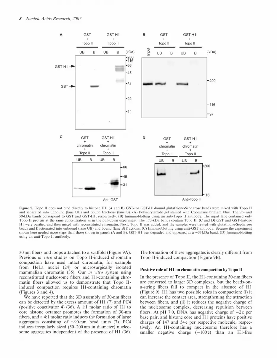
on glutathione-Sepharose beads. After the beads weremixed with Topo II, the unbound and the bound fractionswere separated by centrifugation. As shown in Figure 5Aand B, Topo II was present in the unbound fraction;however, when Topo II was mixed with GST-H1 andreconstituted chromatin, Topo II was present in the boundfraction (Figure 5C and D). These results suggest thatthe chromatin compaction by Topo II is not due to adirect interaction between Topo II and histone H1 butrather an interaction between Topo II and H1-containingchromatin.
In the nucleus, chromatin fibers are held by scaffold/matrix structures that occur every several tens or hundredsof kilobases (9). We therefore monitored the behavior
of chromatin fibers longer than 100 kb. When we mixedTopo II with 30-nm fibers reconstituted on a 186-kbplasmid DNA containing the entire �-globin gene, thefibers became tangled and formed a partial aggregate(Figure 6B). Addition of excess Topo II causedthe formation of large aggregates (Figure 6C). Theseaggregates consisted of a bead unit with a diameter of30.5� 7.3 nm (mean�SD; n¼ 50; Figure 6C inset and D).
Fluorescence microscopy reveals Topo II-dependentcompaction of 30-nm chromatin fibers
To determine the level of compaction, we stained thechromatin complexes with DAPI and determined their
c0826033zoom
0.0 0.2 0.4 0.6 0.8 1.0 1.2 1.40
4
8
MAR? nm
Fre
quen
cy
E
G
F
200 nm
200 nm
c0424002c0424025zzoomc0424019zzoom
A B
0 10 20 30 40 50 60 70 80 90 100
: 0 : 1 : 2 : 3 : 4Topo II
w/o
1:40
1:4
1:1
w/o
1:40
1:4
1:1
D
ATP
w/o
+
w/o ATP
1:4 1:1
C
w/o Topo II (Topo II : DNA ) 1:40
50 nm
PercentageT
Figure 2. Topo II clamps two strands together in the absence of ATP. (A–D) AFM analysis of plasmid (80 ng) treated in the absence of ATP andMgCl2 and without (A) or with 80 ng (D), 20 ng (C) or 2 ng (B) of Topo II. The mixed weight ratio of Topo II versus DNA is indicated in the lowerright corner of the images. In each condition, 18 individual 2� 2-mm areas were scanned, and at least 65 molecules of plasmid were analyzed. Theinset in (C) shows an enlarged image in which Topo II binding to the DNA can be observed. The apparent diameter of Topo II was 36.6� 5.1 nm(mean� SD; n¼ 63), which is �5 nm larger than the apparent size of the nucleosome. This apparent enlargement is due to a tip effect of the AFMimaging. According to electron microscopic observation (46), the diameter of Topo II dimer is �15 nm (9-nm large and 6-nm small domains), whichis 4 nm larger than the real diameter (11 nm) of the nucleosome (47). (E) Concentration dependence of Topo II binding. In the AFM images, thenumber of bound Topo II molecules was counted per plasmid and plotted on a histogram. The plasmid used in this experiment was purified using aQIAGEN-tip 100 column, but it was impossible to remove all the contamination. The results show that a few plasmids possessed small particles evenin the absence of Topo II. (F) AFM image of Topo II binding to linearized DNA containing the MAR sequence. (G) Frequency of Topo II bindingto DNA containing a MAR sequence. The MAR sequence is 0.1–0.3 mm from the end of the DNA as shown in the illustration.
Nucleic Acids Research, 2007 5
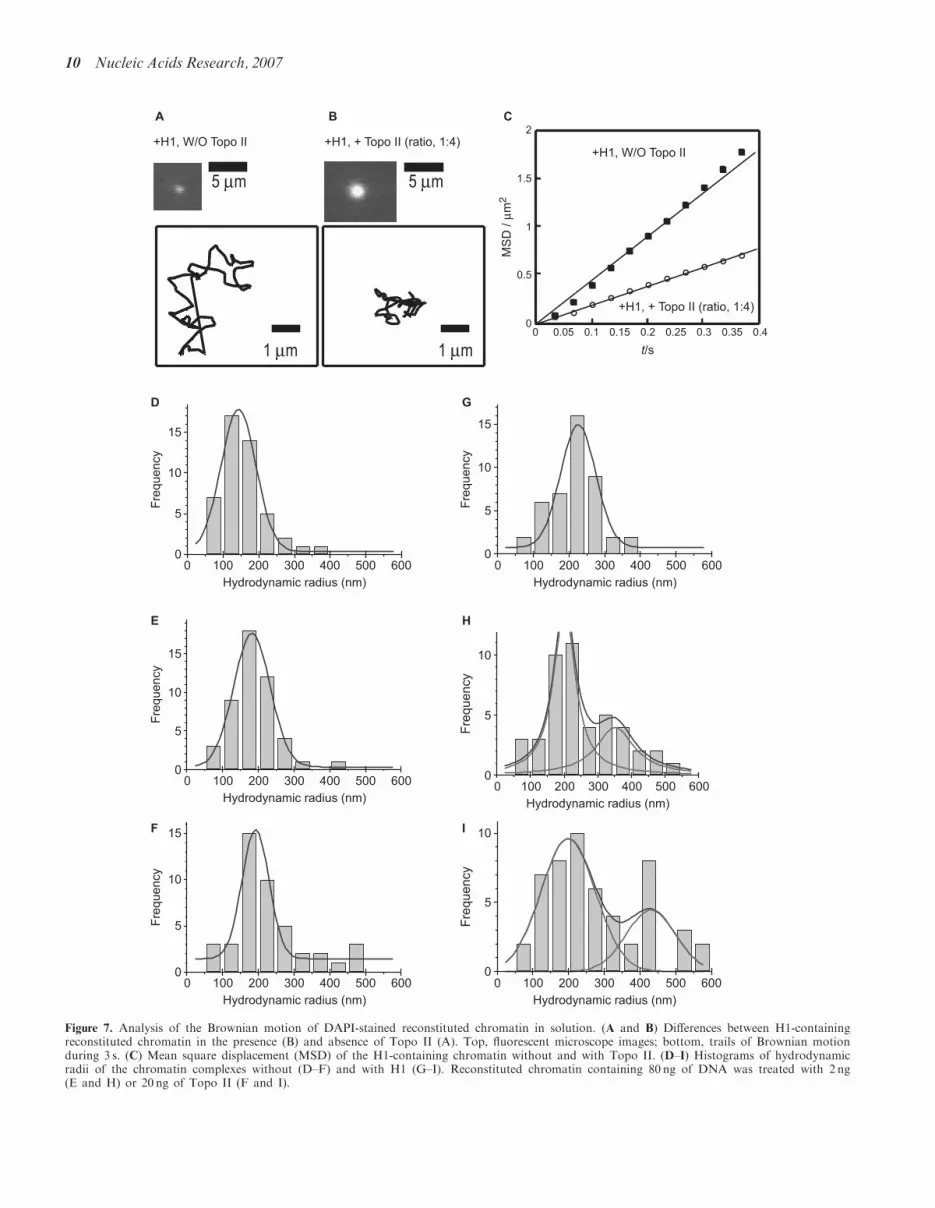
hydrodynamic radii by fluorescence microscopy(Figure 7). The individual 26-kb chromatin was observedas a small dot moving in the buffer (Figure 7A).We calculated the hydrodynamic radius, which is ameasure of the size of the complex, from measurementsof the Brownian motion. The hydrodynamic radius of thereconstituted nucleosomal array in the absence of bothhistone H1 and Topo II was 143.2� 48.8 nm (Figure 7D).When histone H1 was added into the nucleosomal array,the hydrodynamic radius significantly increased to 226.1 �97.9 nm (Figure 7G). This is due to the formation of the30-nm chromatin fibers (Figure 3D). In the presence of2 ng Topo II, the radius was 182.0� 51.1 nm (Figure 7E),and with 20 ng Topo II, it was 192.2� 37.8 nm(Figure 7F), indicating that addition of Topo II alonedid not have much of an effect on the hydrodynamicradius of the nucleosomal array. Addition of Topo IIto the H1-promoted chromatin fiber (containing 30-nmfibers) resulted in much greater increase in the hydro-dynamic radius to 300–400 nm (Figure 7H and I).These findings indicate that Topo II mediates assembly
of H1-containing 30-nm fibers, which increases the size ofthe cluster. This agrees well with the AFM images,wherein the size of the cluster increased several foldupon the addition of Topo II (Figure 3D–F).
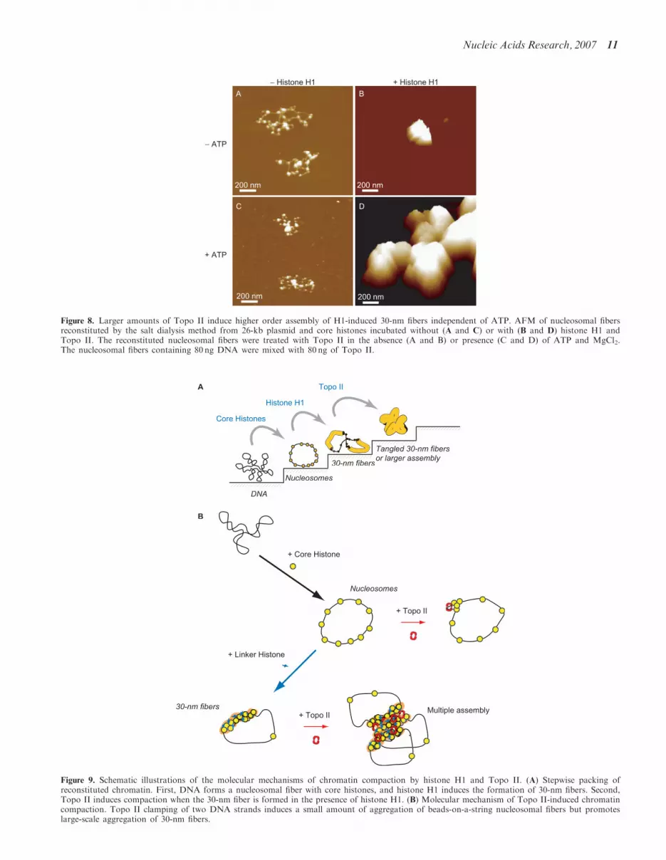
Even when we used one Topo II molecule per 250-bpDNA, which is more than the physiological ratio, beads-on-a-string nucleosomal fibers lacking H1 were notcompacted in the absence or presence of ATP(Figure 8A and C). In contrast, 30-nm chromatin fiberscontaining H1 were highly condensed so that individualnucleosomes and 30-nm fibers could not be identified(Figure 8B and D). The volume calculated from the AFMimage in Figure 8B was 8.66� 106 nm3, which was60 times larger than that of the chromatin complex inFigure 3D and corresponds to an assembly of �60 26-kbchromatin complexes. The volume calculated from theAFM image of Figure 8D was 2.93� 108 nm3, which was1000 times larger than that of the chromatin complex inpanel Figure 4E and corresponds to an assembly of �100026-kb chromatin complexes. This suggests that Topo IIclamps two independent DNA molecules and forms
− Histone H1 + Histone H1
D
E
F
A
200 nm
B
C
Topo II : DNA1:40
Topo II : DNA1:4
w/o Topo II
Figure 3. In the absence of ATP, Topo II induces small aggregates, but large aggregates are induced in the presence of histone H1. (A, D)Nucleosomal fibers were reconstituted from the 26-kb plasmid and core histones by the salt dialysis method and then incubated without Topo II andin the absence (A) or presence (D) of histone H1. (B, C, E and F) The reconstituted nucleosomal fibers containing (E and F) or lacking (B and C)histone H1 were treated with Topo II in the absence of ATP and MgCl2. Nucleosomal fiber containing 80 ng DNA was mixed with 2 ng (B and E) or20 ng (C and F) of Topo II. The mixed weight ratio of Topo II versus DNA is indicated on the left side of the images. In each condition, at leastthree 12.5� 12.5-mm areas were scanned, and 10–30 of the reconstituted chromatin complexes were scanned a second time at a higher magnification.
6 Nucleic Acids Research, 2007
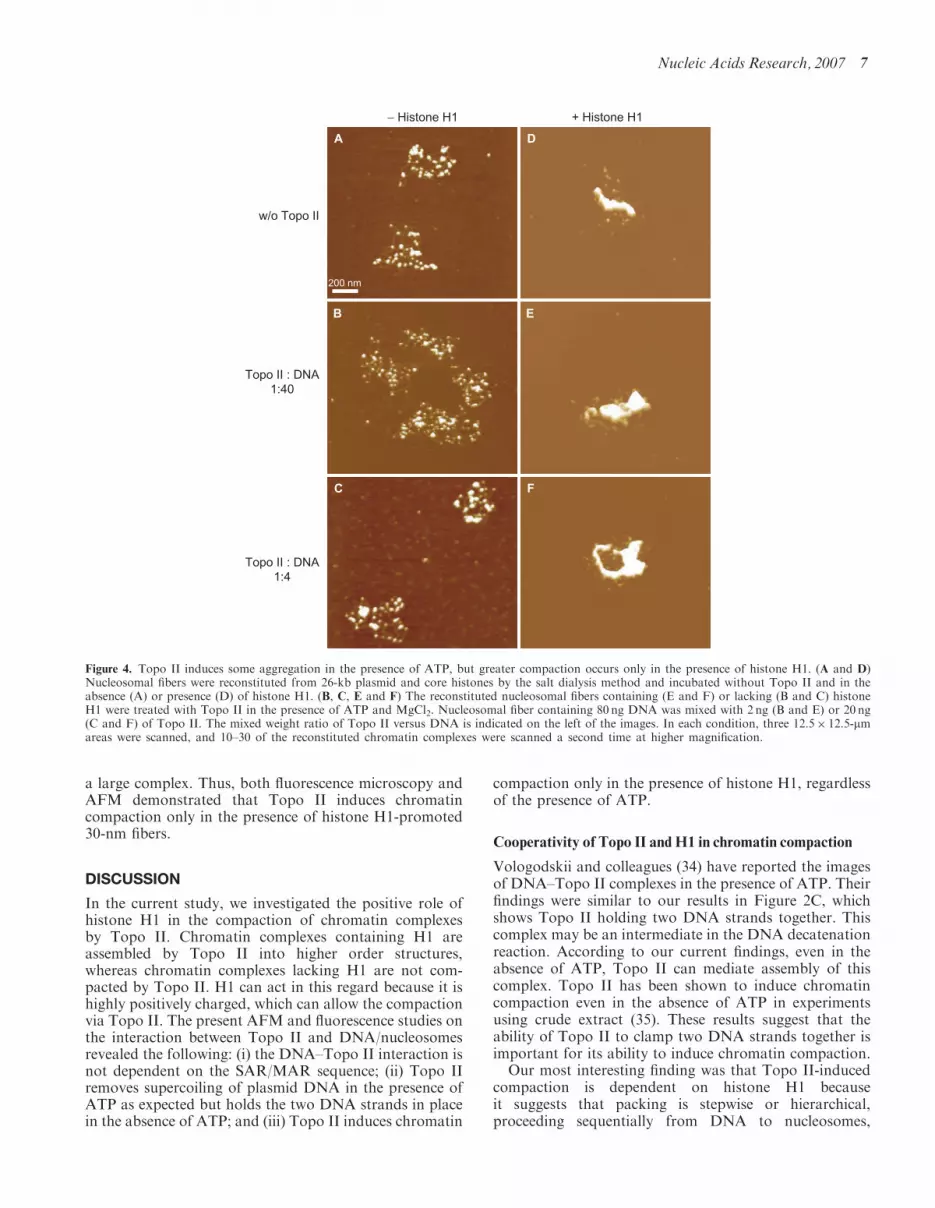
a large complex. Thus, both fluorescence microscopy andAFM demonstrated that Topo II induces chromatincompaction only in the presence of histone H1-promoted30-nm fibers.
DISCUSSION
In the current study, we investigated the positive role ofhistone H1 in the compaction of chromatin complexesby Topo II. Chromatin complexes containing H1 areassembled by Topo II into higher order structures,whereas chromatin complexes lacking H1 are not com-pacted by Topo II. H1 can act in this regard because it ishighly positively charged, which can allow the compactionvia Topo II. The present AFM and fluorescence studies onthe interaction between Topo II and DNA/nucleosomesrevealed the following: (i) the DNA–Topo II interaction isnot dependent on the SAR/MAR sequence; (ii) Topo IIremoves supercoiling of plasmid DNA in the presence ofATP as expected but holds the two DNA strands in placein the absence of ATP; and (iii) Topo II induces chromatin
compaction only in the presence of histone H1, regardlessof the presence of ATP.
Cooperativity of Topo II and H1 in chromatin compaction
Vologodskii and colleagues (34) have reported the imagesof DNA–Topo II complexes in the presence of ATP. Theirfindings were similar to our results in Figure 2C, whichshows Topo II holding two DNA strands together. Thiscomplex may be an intermediate in the DNA decatenationreaction. According to our current findings, even in theabsence of ATP, Topo II can mediate assembly of thiscomplex. Topo II has been shown to induce chromatincompaction even in the absence of ATP in experimentsusing crude extract (35). These results suggest that theability of Topo II to clamp two DNA strands together isimportant for its ability to induce chromatin compaction.Our most interesting finding was that Topo II-induced
compaction is dependent on histone H1 becauseit suggests that packing is stepwise or hierarchical,proceeding sequentially from DNA to nucleosomes,
− Histone H1 + Histone H1
D
E
F
A
200 nm
B
C
w/o Topo II
Topo II : DNA1:40
Topo II : DNA1:4
Figure 4. Topo II induces some aggregation in the presence of ATP, but greater compaction occurs only in the presence of histone H1. (A and D)Nucleosomal fibers were reconstituted from 26-kb plasmid and core histones by the salt dialysis method and incubated without Topo II and in theabsence (A) or presence (D) of histone H1. (B, C, E and F) The reconstituted nucleosomal fibers containing (E and F) or lacking (B and C) histoneH1 were treated with Topo II in the presence of ATP and MgCl2. Nucleosomal fiber containing 80 ng DNA was mixed with 2 ng (B and E) or 20 ng(C and F) of Topo II. The mixed weight ratio of Topo II versus DNA is indicated on the left of the images. In each condition, three 12.5� 12.5-mmareas were scanned, and 10–30 of the reconstituted chromatin complexes were scanned a second time at higher magnification.
Nucleic Acids Research, 2007 7
30 nm fibers and loops attached to a scaffold (Figure 9A).Previous in vitro studies on Topo II-induced chromatincompaction have used intact chromatin, for examplefrom HeLa nuclei (24) or microsurgically isolatedmammalian chromatin (35). Our in vitro system usingreconstituted nucleosome fibers and H1-containing chro-matin fibers allowed us to demonstrate that Topo II-induced compaction requires H1-containing chromatin(Figures 3 and 4).We have reported that the 3D assembly of 30-nm fibers
can be detected by the excess amount of H1 (7) and PC4(positive coactivator 4) (36). A 1:1 molar ratio of H1 tocore histone octamer promotes the formation of 30-nmfibers, and a 4:1 molar ratio induces the formation of largeaggregates consisting of �90-nm bead units (7). PC4induces irregularly sized (50–200 nm in diameter) nucleo-some aggregates independent of the presence of H1 (36).
The formation of these aggregates is clearly different fromTopo II-induced compaction (Figure 9B).
Positive role of H1 on chromatin compaction by Topo II
In the presence of Topo II, the H1-containing 30-nm fibersare converted to larger 3D complexes, but the beads-on-a-string fibers fail to compact in the absence of H1(Figure 9). H1 has two possible roles in compaction: (i) itcan increase the contact area, strengthening the attractionbetween fibers, and (ii) it reduces the negative charge ofthe nucleosome complex, decreasing repulsion betweenfibers. At pH 7.0, DNA has negative charge of �2 e perbase pair, and histone core and H1 proteins have positivecharges of 147 and 54 e per respective molecule, respec-tively. An H1-containing nucleosome therefore has asmaller negative charge (�100 e) than an H1-free
A B
200
116
97
Input
UB UB BBUB UB BB
GST
+
Topo II
GST-H1
+
Topo II
GST
+
Topo II
GST-H1
+
Topo II
200116
66
45
31
22
14
(kDa) (kDa)
GST
GST-H1
C
UB UB BB
GST+
chromatin+
Topo II
GST-H1+
chromatin+
Topo II
Anti-GST
66
45
31
GST+
chromatin+
Topo II
GST-H1+
chromatin+
Topo II
200
116
UB UB BB
D
Anti-Topo II
Figure 5. Topo II does not bind directly to histone H1. (A and B) GST- or GST-H1-bound glutathione-Sepharose beads were mixed with Topo IIand separated into unbound (lane UB) and bound fractions (lane B). (A) Polyacrylamide gel stained with Coomassie brilliant blue. The 26- and59-kDa bands correspond to GST and GST-H1, respectively. (B) Immunoblotting using an anti-Topo II antibody. The input lane contained onlyTopo II protein at the same concentration as in the pull-down experiment. The 170-kDa bands contain Topo II. (C and D) GST and GST-histoneH1 were purified and then mixed with reconstituted chromatin. Next, Topo II was added, and the samples were treated with glutathione-Sepharosebeads and fractionated into unbound (lane UB) and bound (lane B) fractions. (C) Immunoblotting using anti-GST antibody. Because the experimentshown here needed more steps than those shown in panels (A and B), GST-H1 was degraded and appeared as a �33 kDa band. (D) Immunoblottingusing an anti-Topo II antibody.
8 Nucleic Acids Research, 2007

nucleosome (�150 e), suggesting that the strength ofCoulombic repulsion is smaller when H1 is present.Chromatin formation and the interaction between nucleo-somes have been discussed in terms of the charges
[(37) and references therein]. Our studies in bulk solutionby fluorescence microscopy (Figure 7) indicated that the30-nm fibers, which are generated in the presence of H1,do not aggregate, suggesting the fiber has a chargedcolloidal nature.Topo II appears to induce the parallel alignment of
double-stranded DNA chains and to promote formationof thick filaments in the absence of ATP (Figure 2),indicating that Topo II mediates assembly of DNAstrands independently of its enzymatic activity. It isreasonable that this interaction also occurs withinDNA–histone complexes (i.e. nucleosomes). The markeddifference in the effect of Topo II on reconstitutedchromatin in the absence and presence of histone H1can be attributed to the difference in the fundamentalgeometric structures containing the 30-nm fibers. In theabsence of H1, each Topo II molecule mediates a weakattraction between the nucleosome cores. In the presenceof H1, as the contact areas between the fibers areincreased, the attraction between two clusters alsoincreases due to the added attraction via Topo II.Therefore, the attraction between 30-nm fibers would bemuch stronger than the attraction between the pair ofnucleosomes, and it could be enhanced by Topo II.The free energy of the cluster of 30-nm fibers, which
determines the stability, can be described with thefollowing equation:
F ¼ �aNþ bN� þ cN�,
where N is the number of 30-nm fibers in the assembly,and a, b and c are positive constants and � and � areexponents. For simplicity, the fibers are assumed to havethe same lengths. The first term in the equationcorresponds to the stabilization due to the attractiveinteraction mediated by Topo II; the second is the surfaceenergy and the exponent, where � is generally51; and thethird is derived from the destabilization due to theremaining negative charge in the assembly, where theexponent � is larger than 1. In an idealized system with aspherical symmetry, � is 5/3. For the assembly ofnonspherical objects, � is somewhat smaller than 5/3 butis always41. This equation predicts that the assembly of30-nm fibers will stop at a finite size, which depends on theremaining negative charges and attractions, and it furtherimplies the formation of a larger assembly when theremaining negative charges are further reduced.In summary, in addition to favorable geometry of
the attractive interaction between the 30-nm fibers, thereduction of the negative charge in the polynucleosomestructure by H1 may decrease the instability due toelectrostatic repulsion. Such a charge effect of H1 can alsostabilize the assembly of 30-nm fibers.
Physiological significance of Topo II-inducedchromatin compaction
The matrix attachment region (MAR) has been experi-mentally identified as a segment of genomic DNA that isresistant to digestion by nucleases (38) or that remainsattached to the insoluble fraction of the nucleusafter digestion with restriction endonucleases (39).
D
500 nm
A
500 nm
B
1:40
500 nm
C
1:4
0 10 20 30 40 500
5
10
Fre
quency
Diameter of beads (nm)
(peak-to-peak distance)
100 nm
w/o Topo II
Figure 6. Compaction of 186-kb chromatin by Topo II. (A) AFM ofnucleosomal fibers (beads-on-a-string) reconstituted from the 186-kbplasmid and treated with histone H1 to induce the assembly of 30-nmfibers. (B and C) AFM of chromatin fibers containing 80 ng DNAtreated with 2 ng (B) or 20 ng (C) of Topo II. The mixed weight ratio ofTopo II versus DNA is indicated in the right lower corner of theimages. The inset shows an enlarged view in which the globular surfaceof the complex can be observed. Except for the inset of panel (C),which is a top view, all other images are surface plots. (D) Thediameters of the globules on the surface of the complex were measuredand are plotted as a histogram.
Nucleic Acids Research, 2007 9
G
H
I
Hydrodynamic radius (nm)
Fre
quency
E
F
D
B CA
+H1, + Topo II (ratio, 1:4)+H1, W/O Topo II
0
0.5
1
1.5
2
0 0.05 0.1 0.15 0.2 0.25 0.3 0.35 0.4
t/s
+H1, W/O Topo II
0 100 200 300 400 500 600
Hydrodynamic radius (nm)
0 100 200 300 400 500 600
Hydrodynamic radius (nm)
0 100 200 300 400 500 600
Hydrodynamic radius (nm)
0 100 200 300 400 500 600
Hydrodynamic radius (nm)
0 100 200 300 400 500 600
Hydrodynamic radius (nm)
0 100 200 300 400 500 6000
5
10
15
Fre
quency
0
5
10
15
Fre
quency
0
5
10
15
Fre
quency
0
5
10
15
Fre
quency
0
5
10
Fre
quency
0
5
10
+H1, + Topo II (ratio, 1:4)
MS
D / µ
m2
Figure 7. Analysis of the Brownian motion of DAPI-stained reconstituted chromatin in solution. (A and B) Differences between H1-containingreconstituted chromatin in the presence (B) and absence of Topo II (A). Top, fluorescent microscope images; bottom, trails of Brownian motionduring 3 s. (C) Mean square displacement (MSD) of the H1-containing chromatin without and with Topo II. (D–I) Histograms of hydrodynamicradii of the chromatin complexes without (D–F) and with H1 (G–I). Reconstituted chromatin containing 80 ng of DNA was treated with 2 ng(E and H) or 20 ng of Topo II (F and I).
10 Nucleic Acids Research, 2007
− Histone H1 + Histone H1
BA
200 nm
DC
− ATP
+ ATP
200 nm
200 nm200 nm
Figure 8. Larger amounts of Topo II induce higher order assembly of H1-induced 30-nm fibers independent of ATP. AFM of nucleosomal fibersreconstituted by the salt dialysis method from 26-kb plasmid and core histones incubated without (A and C) or with (B and D) histone H1 andTopo II. The reconstituted nucleosomal fibers were treated with Topo II in the absence (A and B) or presence (C and D) of ATP and MgCl2.The nucleosomal fibers containing 80 ng DNA were mixed with 80 ng of Topo II.
DNA
Core Histones
+ Core Histone
Nucleosomes
Histone H1
Topo II
30-nm fibers
Tangled 30-nm fibersor larger assembly
A
B
+ Linker Histone
+ Topo II30-nm fibers
Nucleosomes
+ Topo II
Multiple assembly
Figure 9. Schematic illustrations of the molecular mechanisms of chromatin compaction by histone H1 and Topo II. (A) Stepwise packing ofreconstituted chromatin. First, DNA forms a nucleosomal fiber with core histones, and histone H1 induces the formation of 30-nm fibers. Second,Topo II induces compaction when the 30-nm fiber is formed in the presence of histone H1. (B) Molecular mechanism of Topo II-induced chromatincompaction. Topo II clamping of two DNA strands induces a small amount of aggregation of beads-on-a-string nucleosomal fibers but promoteslarge-scale aggregation of 30-nm fibers.
Nucleic Acids Research, 2007 11
A MAR has been located in the histone gene cluster (38)and the �-globin locus (39), which contain the consensussequence for interaction with Topo II (38,40). In ourexperiments, Topo II did not show detectable affinity forthe MAR region of the �-globin gene (Figure 2G). Basedon these results, we concluded that the MAR region doesnot influence the effect of Topo II in the in vitro system.Mechanisms for loading Topo II on the MAR region mustexist in vivo, and Topo II is expected to condense thechromatin with a help of other scaffold proteins such asthe condensin complex. A comparison of in vitro and invivo chromatin structures is needed to determine thesignificance of the MAR region in chromatin compactionby Topo II.Proteomic analysis of the metaphase chromosome has
shown that the molar ratio of Topo II to histone H4 is0.72:100 (41). This corresponds to our experimentalcondition of a weight ratio of 1:40 Topo II to DNA inFigures 3E, 4E and 6B. Because this weight ratiocorresponds to one Topo II molecule per 10 kb of DNAand given an in vivo efficiency of one nucleosome per200 bp of DNA, it is expected that there is one Topo IImolecule per 50 nucleosomes (100 molecules of histoneH4). Therefore, our experimental system (1:40 weightratio) may reflect the in vivo situation, although we cannotprecisely determine the molar ratio in the reconstitutedchromatin at a single-molecule level. In this regard, it isinteresting that 30-nm fibers loop out from the condensedpart (Figure 6B), which might reflect the highly condensedscaffold region and chromatin looping that has beenpreviously observed by electron microscopy (42). One ofthe key results of our study is that Topo II does notconvert 30-nm fibers into larger complexes. Even in theaggregated complex (Figure 6D), the structural units were30 nm, indicating that the Topo II may mediate chromatincompaction simply by bringing the 30-nm fibers intocontact. In other words, Topo II cannot induce largerchromosomal units such as 80-nm (43), 100-nm (44) and300-nm (45) beads or fibers, which have been observed byelectron microscopy.
ACKNOWLEDGEMENTS
This work was supported by the Japanese Ministry ofEducation, Culture, Sports, Science and Technology(Grant-in-Aid for Scientific Research on Priority Areasto K.T. and K.Y.) and the Japan Society for thePromotion of Science (Grant-in-Aid for YoungScientists (B) to K.H.). Funding to pay the Open Accesspublication charge was provided by the Grant-in-Aid forYoung Scientists (B) from the Japan Society for thePromotion of Science.
Conflict of interest statement. None declared.
REFERENCES
1. Germond,J.E., Hirt,B., Oudet,P., Gross-Bellark,M. and Chambon,P.(1975) Folding of the DNA double helix in chromatin-like structuresfrom simian virus 40. Proc. Natl. Acad. Sci. USA, 72, 1843–1847.
2. Leuba,S.H., Yang,G., Robert,C., Samori,B., van Holde,K.,Zlatanova,J. and Bustamante,C. (1994) Three-dimensional structureof extended chromatin fibers as revealed by tapping-mode scanningforce microscopy. Proc. Natl. Acad. Sci. USA, 91, 11621–11625.
3. Hizume,K., Yoshimura,S.H. and Takeyasu,K. (2004) Atomic forcemicroscopy demonstrates a critical role of DNA superhelicity innucleosome dynamics. Cell Biochem. Biophys., 40, 249–262.
4. Thoma,F., Koller,T. and Klug,A. (1979) Involvement of histone H1in the organization of the nucleosome and of the salt-dependentsuperstructures of chromatin. J. Cell Biol., 83, 403–427.
5. Leuba,S.H., Bustamante,C., van Holde,K. and Zlatanova,J. (1998)Linker histone tails and N-tails of histone H3 are redundant:scanning force microscopy studies of reconstituted fibers.Biophys. J., 74, 2830–2839.
6. Leuba,S.H., Bustamante,C., Zlatanova,J. and van Holde,K. (1998)Contributions of linker histones and histone H3 to chromatinstructure: scanning force microscopy studies on trypsinized fibers.Biophys. J., 74, 2823–2829.
7. Hizume,K., Yoshimura,S.H. and Takeyasu,K. (2005) Linker histoneH1 per se can induce three-dimensional folding of chromatin fiber.Biochemistry, 44, 12978–12989.
8. Paulson,J.R. and Laemmli,U.K. (1977) The structure ofhistone-depleted metaphase chromosomes. Cell, 12, 817–828.
9. Jackson,D.A., Dickinson,P. and Cook,P.R. (1990) The size ofchromatin loops in HeLa cells. EMBO J., 9, 567–571.
10. Hancock,R. (2000) A new look at the nuclear matrix. Chromosoma,109, 219–225.
11. Berrios,M., Osheroff,N. and Fisher,P.A. (1985) In situ localizationof DNA topoisomerase II, a major polypeptide component of theDrosophila nuclear matrix fraction. Proc. Natl. Acad. Sci. USA, 82,4142–4146.
12. Gasser,S.M., Laroche,T., Falquet,J., Boy de la Tour,E. andLaemmli,U.K. (1986) Metaphase chromosome structure.Involvement of topoisomerase II. J. Mol. Biol., 188, 613–629.
13. Saitoh,N., Goldberg,I.G., Wood,E.R. and Earnshaw,W.C. (1994)ScII: an abundant chromosome scaffold protein is a member of afamily of putative ATPases with an unusual predicted tertiarystructure. J. Cell Biol., 127, 303–318.
14. Kimura,K. and Hirano,T. (1997) ATP-dependent positivesupercoiling of DNA by 13S condensin: a biochemicalimplication for chromosome condensation. Cell, 90, 625–634.
15. Uemura,T., Ohkura,H., Adachi,Y., Morino,K., Shiozaki,K. andYanagida,M. (1987) DNA topoisomerase II is required forcondensation and separation of mitotic chromosomes in S. pombe.Cell, 50, 917–925.
16. Saka,Y., Sutani,T., Yamashita,Y., Saitoh,S., Takeuchi,M.,Nakaseko,Y. and Yanagida,M. (1994) Fission yeast cut3 and cut14,members of a ubiquitous protein family, are required forchromosome condensation and segregation in mitosis. EMBO J.,13, 4938–4952.
17. Hirano,T. and Mitchison,T.J. (1994) A heterodimeric coiled-coilprotein required for mitotic chromosome condensation in vitro.Cell, 79, 449–458.
18. Maeshima,K. and Laemmli,U.K. (2003) A two-step scaffoldingmodel for mitotic chromosome assembly. Dev. Cell, 4, 467–480.
19. Fey,E.G., Krochmalnic,G. and Penman,S. (1986) Thenonchromatin substructures of the nucleus: the ribonucleoprotein(RNP)-containing and RNP-depleted matrices analyzed bysequential fractionation and resinless section electron microscopy.J. Cell Biol., 102, 1654–1665.
20. Nickerson,J. (2001) Experimental observations of a nuclear matrix.J. Cell Sci., 114, 463–474.
21. Wang,J.C. (2002) Cellular roles of DNA topoisomerases: amolecular perspective. Nat. Rev. Mol. Cell Biol., 3, 430–440.
22. Berger,J.M., Gamblin,S.J., Harrison,S.C. and Wang,J.C. (1996)Structure and mechanism of DNA topoisomerase II. Nature, 379,225–232.
23. Nettikadan,S.R., Furbee,C.S., Muller,M.T. and Takeyasu,K. (1998)Molecular structure of human topoisomerase II alpha revealed byatomic force microscopy. J. Electron Microsc. (Tokyo), 47,671–674.
24. Adachi,Y., Luke,M. and Laemmli,U.K. (1991) Chromosomeassembly in vitro: topoisomerase II is required for condensation.Cell, 64, 137–148.
12 Nucleic Acids Research, 2007
25. Patrinos,G.P., de Krom,M., de Boer,E., Langeveld,A., Imam,A.M.,Strouboulis,J., de Laat,W. and Grosveld,F.G. (2004) Multipleinteractions between regulatory regions are required to stabilize anactive chromatin hub. Genes Dev., 18, 1495–1509.
26. van Drunen,C.M., Sewalt,R.G., Oosterling,R.W., Weisbeek,P.J.,Smeekens,S.C. and van Driel,R. (1999) A bipartite sequence elementassociated with matrix/scaffold attachment regions. Nucleic AcidsRes., 27, 2924–2930.
27. O’Neill,T.E., Roberge,M. and Bradbury,E.M. (1992) Nucleosomearrays inhibit both initiation and elongation of transcripts bybacteriophage T7 RNA polymerase. J. Mol. Biol., 223, 67–78.
28. Mirzabekov,A.D., Pruss,D.V. and Ebralidse,K.K. (1990) Chromatinsuperstructure-dependent crosslinking with DNA of the histone H5residues Thr1, His25 and His62. J. Mol. Biol., 211, 479–491.
29. Nakai,T., Hizume,K., Yoshimura,S.H., Takeyasu,K. andYoshikawa,K. (2005) Phase transition in reconstituted chromatin.Europhys. Lett., 69, 1024–1030.
30. Araki,S., Nakai,T., Hizume,K., Takeyasu,K. and Yoshikawa,K.(2006) Hydrodynamic radius of circular DNA is larger than that oflinear DNA. Chem. Phys. Lett., 418, 255–259.
31. Yoshikawa,K., Takahashi,M., Vasilevskaya,V.V. andKhokhlov,A.R. (1996) Large discrete transition in a single DNAmolecule appears continuous in the ensemble. Phys. Rev. Lett., 76,3029–3031.
32. Nettikadan,S., Tokumasu,F. and Takeyasu,K. (1996) Quantitativeanalysis of the transcription factor AP2 binding to DNA by atomicforce microscopy. Biochem. Biophys. Res. Commun., 226, 645–649.
33. Yoshimura,S.H., Yoshida,C., Igarashi,K. and Takeyasu,K. (2000)Atomic force microscopy proposes a ‘kiss and pull’ mechanism forenhancer function. J. Electron Microsc. (Tokyo), 49, 407–413.
34. Vologodskii,A.V., Zhang,W., Rybenkov,V.V.,Podtelezhnikov,A.A., Subramanian,D., Griffith,J.D. andCozzarelli,N.R. (2001) Mechanism of topology simplification bytype II DNA topoisomerases. Proc. Natl. Acad. Sci. USA, 98,3045–3049.
35. Bojanowski,K., Maniotis,A.J., Plisov,S., Larsen,A.K. andIngber,D.E. (1998) DNA topoisomerase II can drive changes inhigher order chromosome architecture without enzymaticallymodifying DNA. J. Cell Biochem., 69, 127–142.
36. Das,C., Hizume,K., Batta,K., Kumar,B.R., Gadad,S.S., Ganguly,S.,Lorain,S., Verreault,A., Sadhale,P.P. et al. (2006) Transcriptionalcoactivator PC4, a chromatin-associated protein, induces chromatincondensation. Mol. Cell. Biol., 26, 8303–8315.
37. Muhlbacher,F., Holm,C. and Schiessel,H. (2006) Controlled DNAcompaction within chromatin: the tail-bridging effect. Europhys.Lett., 73, 135–141.
38. Gasser,S.M. and Laemmli,U.K. (1986) The organisation ofchromatin loops: characterization of a scaffold attachment site.EMBO J., 5, 511–518.
39. Jarman,A.P. and Higgs,D.R. (1988) Nuclear scaffold attachmentsites in the human globin gene complexes. EMBO J., 7, 3337–3344.
40. Adachi,Y., Kas,E. and Laemmli,U.K. (1989) Preferential,cooperative binding of DNA topoisomerase II to scaffold-associatedregions. EMBO J., 8, 3997–4006.
41. Uchiyama,S., Kobayashi,S., Takata,H., Ishihara,T., Hori,N.,Higashi,T., Hayashihara,K., Sone,T., Higo,D. et al. (2005)Proteome analysis of human metaphase chromosomes. J. Biol.Chem., 280, 16994–17004.
42. Marsden,M.P. and Laemmli,U.K. (1979) Metaphase chromosomestructure: evidence for a radial loop model. Cell, 17, 849–858.
43. Kobori,T., Kodama,M., Hizume,K., Yoshimura,S.H., Ohtani,T.and Takeyasu,K. (2006) Comparative structural biology of thegenome: nano-scale imaging of single nucleus from differentkingdoms reveals the common physicochemical property ofchromatin with a 40 nm structural unit. J. Electron Microsc.(Tokyo), 55, 31–40.
44. Belmont,A.S. and Bruce,K. (1994) Visualization of G1chromosomes: a folded, twisted, supercoiled chromonema modelof interphase chromatid structure. J. Cell Biol., 127, 287–302.
45. Rattner,J.B. and Lin,C.C. (1985) Radial loops and helical coilscoexist in metaphase chromosomes. Cell, 42, 291–296.
46. Schultz,P., Olland,S., Oudet,P. and Hancock,R. (1996) Structureand conformational changes of DNA topoisomerase IIvisualized by electron microscopy. Proc. Natl. Acad. Sci. USA,93, 5936–5940.
47. Luger,K., Mader,A.W., Richmond,R.K., Sargent,D.F. andRichmond,T.J. (1997) Crystal structure of the nucleosome coreparticle at 2.8A resolution. Nature, 389, 251–260.
Nucleic Acids Research, 2007 13