t-cell responses before and after the fifth consecutive acellular pertussis vaccination in...
TRANSCRIPT

T-Cell Responses before and after the Fifth Consecutive AcellularPertussis Vaccination in 4-Year-Old Dutch Children
Rose-Minke Schure,a,b Lotte H. Hendrikx,c Lia G. H. de Rond,a Kemal Oztürk,a Elisabeth A. M. Sanders,b Guy A. M. Berbers,a
and Anne-Marie Buismana
Laboratory for Infectious Disease and Perinatal Screening, Center for Infectious Diseases Control, National Institute for Public Health, Bilthoven, The Netherlandsa;Department of Pediatric Immunology and Infectious Diseases, UMC, Utrecht, The Netherlandsb; and Department of Pediatrics, Free University of Amsterdam (VUMC),Amsterdam, The Netherlandsc
Immunization with acellular pertussis vaccine (aP) induces higher specific antibody levels and fewer adverse reactions than doesimmunization with the whole-cell vaccine (wP). However, antibody levels in infants induced by both types of pertussis vaccineswane already after 1 year. Therefore, long-term T-cell responses upon vaccination might play a role in protection against pertus-sis. In a cross-sectional study (ISRCTN65428640), we investigated T-helper (Th) cell immune responses in wP- or aP-vaccinatedchildren before and after an aP low-dose or high-dose preschool booster at 4 years of age in The Netherlands. T cells were stimu-lated with pertussis vaccine antigens. The numbers of gamma interferon-producing cells and Th1, Th2, Th17, and interleukin-10(IL-10) cytokine concentrations were determined. In addition, pertussis-specific IgE levels were measured in plasma. Childrenbeing vaccinated with aP vaccinations at 2, 3, 4, and 11 months of age still showed higher pertussis-specific T-cell responses at 4years of age than did wP-vaccinated children. These T-cell responses failed to show a typical increase in cytokine production af-ter a fifth aP vaccination but remained high after a low-dose booster and seemed to decline even after a high-dose booster. Im-portantly, elevated IgE levels were induced after this booster vaccination. In contrast, wP-vaccinated children had only low pre-booster T-cell responses, and these children showed a clear postbooster T-cell memory response even after a low-dose boostervaccine. Four high-dose aP vaccinations in infancy induce high T-cell responses still present even 3 years after vaccination andenhanced IgE responses after preschool booster vaccination. Therefore, studies of changes in vaccine dosage, timing of pertussis(booster) vaccinations, and the possible association with local side effects are necessary.
Recently, during a large pertussis outbreak in California 10 in-fants have died (19), and 9,154 cases of whooping cough have
been reported by the California Department of Public Health (4).Already in the 1990s, many developed countries replaced thewhole-cell pertussis component (wP) with the acellular pertussiscomponent (aP) in the DTP-IPV-Hib combination vaccine in or-der to achieve higher antigen-specific antibody levels and fewerside effects. This, however, did not stop the reemergence of per-tussis in these countries (8, 38, 39). The high incidence of pertussisworldwide can be (partly) explained by adaptation of the circulat-ing bacterial strains to vaccine pressure as well as waning immu-nity after vaccination and natural infection (2, 27).
In The Netherlands, wP vaccines have been used since the early1950s, resulting in a decline of pertussis disease. Despite a highvaccination coverage, the incidence of pertussis increasedagain after 1996, and for this reason, an aP preschool boostervaccination (aP) was introduced at 4 years of age in 2001. From2005 onwards, the infant wP vaccine component administeredat 2, 3, 4, and 11 months of age has also been replaced by the aPvaccine.
Pertussis-specific antibody levels are induced by vaccinationand natural infection and protect against disease; however, theselevels decline very rapidly after vaccination (9, 13). Several studieshave shown that protection against disease also relies on T-helper(Th) cells, as well as antibodies (7, 26). Multiple Th cell lineagesmay be involved, like the Th1, Th2, and Th17 cells, and each lin-eage is characterized by specific cytokine repertoires (6). However,the induction of long-term T-cell memory responses and the rel-ative contribution of each Th cell lineage upon vaccination arelargely unknown. Previous studies have shown that aP may lead to
Th2 cytokine repertoires in infants and children, whereas wPrather primes for Th1 immune responses (1, 23). The aP vaccinesconsist of some purified pertussis proteins that may differ fromwP vaccines in the induction of Th cell responses, which includemany other biological components. Information about T-cell im-munity after pertussis vaccination and comparison between wP-and aP-primed infants is scarce. Moreover, Th2 responses mightbe associated with atopic reactions (31), and pertussis-specific IgEhas been found after aP vaccinations in infants (28). The aim ofthis study is to assess the Th1, Th2, and Th17 as well as inter-leukin-10 (IL-10) cytokine responses to pertussis vaccine anti-gens in children 4 years of age who received either a low-dose ora high-dose antigen aP preschool booster vaccination. Wecompared groups of children who have been primed either bywP or by aP in infancy. Apart from T-cell kinetics, also pertus-sis antigen-specific IgE responses in these groups of childrenare studied.
MATERIALS AND METHODSSubjects and study design. In this study, a cohort of children 4 years of ageforms a subset of a cross-sectional observational study in The Netherlands
Received 11 May 2012 Returned for modification 30 July 2012Accepted 24 September 2012
Published ahead of print 26 September 2012
Address correspondence to Anne-Marie Buisman, [email protected].
Copyright © 2012, American Society for Microbiology. All Rights Reserved.
doi:10.1128/CVI.00277-12
November 2012 Volume 19 Number 11 Clinical and Vaccine Immunology p. 1879–1886 cvi.asm.org 1879
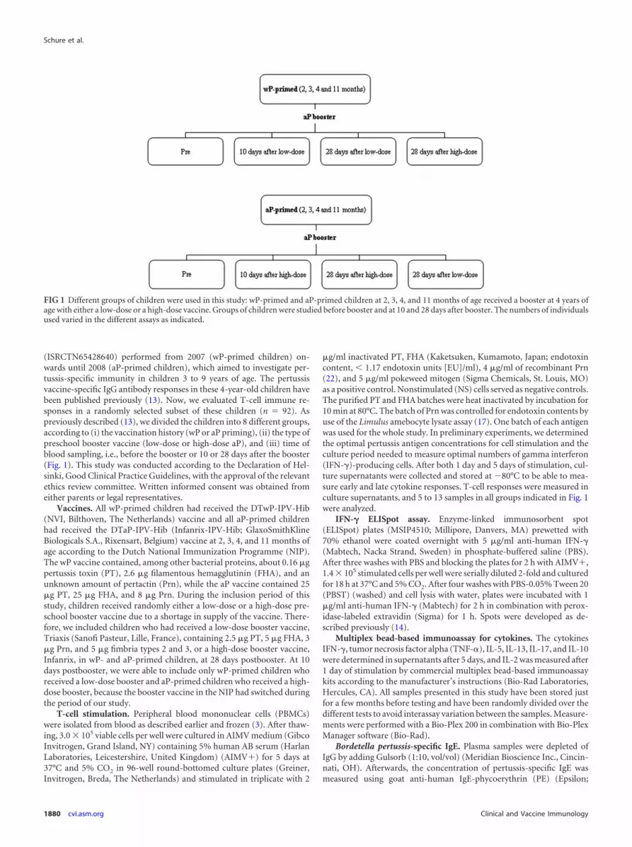
(ISRCTN65428640) performed from 2007 (wP-primed children) on-wards until 2008 (aP-primed children), which aimed to investigate per-tussis-specific immunity in children 3 to 9 years of age. The pertussisvaccine-specific IgG antibody responses in these 4-year-old children havebeen published previously (13). Now, we evaluated T-cell immune re-sponses in a randomly selected subset of these children (n � 92). Aspreviously described (13), we divided the children into 8 different groups,according to (i) the vaccination history (wP or aP priming), (ii) the type ofpreschool booster vaccine (low-dose or high-dose aP), and (iii) time ofblood sampling, i.e., before the booster or 10 or 28 days after the booster(Fig. 1). This study was conducted according to the Declaration of Hel-sinki, Good Clinical Practice Guidelines, with the approval of the relevantethics review committee. Written informed consent was obtained fromeither parents or legal representatives.
Vaccines. All wP-primed children had received the DTwP-IPV-Hib(NVI, Bilthoven, The Netherlands) vaccine and all aP-primed childrenhad received the DTaP-IPV-Hib (Infanrix-IPV-Hib; GlaxoSmithKlineBiologicals S.A., Rixensart, Belgium) vaccine at 2, 3, 4, and 11 months ofage according to the Dutch National Immunization Programme (NIP).The wP vaccine contained, among other bacterial proteins, about 0.16 �gpertussis toxin (PT), 2.6 �g filamentous hemagglutinin (FHA), and anunknown amount of pertactin (Prn), while the aP vaccine contained 25�g PT, 25 �g FHA, and 8 �g Prn. During the inclusion period of thisstudy, children received randomly either a low-dose or a high-dose pre-school booster vaccine due to a shortage in supply of the vaccine. There-fore, we included children who had received a low-dose booster vaccine,Triaxis (Sanofi Pasteur, Lille, France), containing 2.5 �g PT, 5 �g FHA, 3�g Prn, and 5 �g fimbria types 2 and 3, or a high-dose booster vaccine,Infanrix, in wP- and aP-primed children, at 28 days postbooster. At 10days postbooster, we were able to include only wP-primed children whoreceived a low-dose booster and aP-primed children who received a high-dose booster, because the booster vaccine in the NIP had switched duringthe period of our study.
T-cell stimulation. Peripheral blood mononuclear cells (PBMCs)were isolated from blood as described earlier and frozen (3). After thaw-ing, 3.0 � 105 viable cells per well were cultured in AIMV medium (GibcoInvitrogen, Grand Island, NY) containing 5% human AB serum (HarlanLaboratories, Leicestershire, United Kingdom) (AIMV�) for 5 days at37°C and 5% CO2 in 96-well round-bottomed culture plates (Greiner,Invitrogen, Breda, The Netherlands) and stimulated in triplicate with 2
�g/ml inactivated PT, FHA (Kaketsuken, Kumamoto, Japan; endotoxincontent, � 1.17 endotoxin units [EU]/ml), 4 �g/ml of recombinant Prn(22), and 5 �g/ml pokeweed mitogen (Sigma Chemicals, St. Louis, MO)as a positive control. Nonstimulated (NS) cells served as negative controls.The purified PT and FHA batches were heat inactivated by incubation for10 min at 80°C. The batch of Prn was controlled for endotoxin contents byuse of the Limulus amebocyte lysate assay (17). One batch of each antigenwas used for the whole study. In preliminary experiments, we determinedthe optimal pertussis antigen concentrations for cell stimulation and theculture period needed to measure optimal numbers of gamma interferon(IFN-�)-producing cells. After both 1 day and 5 days of stimulation, cul-ture supernatants were collected and stored at �80°C to be able to mea-sure early and late cytokine responses. T-cell responses were measured inculture supernatants, and 5 to 13 samples in all groups indicated in Fig. 1were analyzed.
IFN-� ELISpot assay. Enzyme-linked immunosorbent spot(ELISpot) plates (MSIP4510; Millipore, Danvers, MA) prewetted with70% ethanol were coated overnight with 5 �g/ml anti-human IFN-�(Mabtech, Nacka Strand, Sweden) in phosphate-buffered saline (PBS).After three washes with PBS and blocking the plates for 2 h with AIMV�,1.4 � 105 stimulated cells per well were serially diluted 2-fold and culturedfor 18 h at 37°C and 5% CO2. After four washes with PBS-0.05% Tween 20(PBST) (washed) and cell lysis with water, plates were incubated with 1�g/ml anti-human IFN-� (Mabtech) for 2 h in combination with perox-idase-labeled extravidin (Sigma) for 1 h. Spots were developed as de-scribed previously (14).
Multiplex bead-based immunoassay for cytokines. The cytokinesIFN-�, tumor necrosis factor alpha (TNF-�), IL-5, IL-13, IL-17, and IL-10were determined in supernatants after 5 days, and IL-2 was measured after1 day of stimulation by commercial multiplex bead-based immunoassaykits according to the manufacturer’s instructions (Bio-Rad Laboratories,Hercules, CA). All samples presented in this study have been stored justfor a few months before testing and have been randomly divided over thedifferent tests to avoid interassay variation between the samples. Measure-ments were performed with a Bio-Plex 200 in combination with Bio-PlexManager software (Bio-Rad).
Bordetella pertussis-specific IgE. Plasma samples were depleted ofIgG by adding Gulsorb (1:10, vol/vol) (Meridian Bioscience Inc., Cincin-nati, OH). Afterwards, the concentration of pertussis-specific IgE wasmeasured using goat anti-human IgE-phycoerythrin (PE) (Epsilon;
FIG 1 Different groups of children were used in this study: wP-primed and aP-primed children at 2, 3, 4, and 11 months of age received a booster at 4 years ofage with either a low-dose or a high-dose vaccine. Groups of children were studied before booster and at 10 and 28 days after booster. The numbers of individualsused varied in the different assays as indicated.
Schure et al.
1880 cvi.asm.org Clinical and Vaccine Immunology

Fisher Scientific) in a multiplex bead-based assay as described previously(15, 37). IgE responses were measured in 16 to 52 plasma samples of allgroups presented in Fig. 1.
Definitions, data presentation, and statistical analysis. All individualdata or geometric mean (GM) values and 95% confidence intervals pergroup were presented. Background values of negative-control cells werededucted from each sample. Significant differences between groups (P �0.05) were determined as indicated.
RESULTS
Induction of T-cell cytokines in wP- and aP-primed children ininfancy. The concentrations of the T-cell cytokines TNF-�,IFN-�, IL-5, IL-17, and IL-10 specific for the pertussis proteinsPT, FHA, and Prn in PBMCs of aP- and wP-primed childrenbefore and 28 days after a high-dose aP preschool booster areillustrated in Fig. 2. At 4 years of age, 3 years after four aP
FIG 2 Concentrations of Th1 (TNF-� and IFN-�), Th2 (IL-5), Th17 (IL-17), and IL-10 cytokines in supernatants of PBMCs of wP- (gray bars) and aP-primed(white bars) children specific for the pertussis proteins PT, FHA, and Prn. Bars represent GMs and 95% confidence intervals of data prebooster and at 28 daysafter booster vaccination with a high-dose aP vaccine. Groups consisted of 13 individuals before booster vaccination and 5 to 8 persons at 28 days after boostervaccination. *, significantly increased values in aP-primed children compared to wP-primed children; #, significant increase at 28 days after boostervaccination compared to prebooster values in wP-primed children; ‡, significant decrease at 28 days after booster compared to prebooster values inaP-primed children.
T-Cell Responses to Preschool aP Pertussis Vaccine
November 2012 Volume 19 Number 11 cvi.asm.org 1881

vaccinations in the first year of life, most prebooster T-cellcytokine levels specific for all three pertussis proteins (11/15)were significantly higher than those of wP-primed children.The PT-specific IFN-�, IL-17, and IL-10 as well as the Prn-specific IL-10 were also higher in aP-primed children but didnot reach significance (Fig. 2).
In wP-primed children, the aP booster did enhance the pertus-sis-specific TNF-�, IFN-�, and IL-5 production, an effect whichwas significant for FHA- and Prn-specific IL-5 as well as the Prn-specific IFN-�. In contrast, the aP booster in aP-primed childrendid not enhance the T-cell cytokine concentrations. Moreover,after administration of a fifth high-dose aP vaccine, the FHA-specific IFN-� and IL-17 response even showed a decline. In gen-eral, the pertussis-specific IL-17 and IL-10 values were low com-pared with the other cytokine values, especially for PT (Fig. 2).
To compare the production levels of the Th2 cytokine andthe Th1 cytokines, the geometric mean values (GMs) of theIL-5 levels per group before and after the booster were com-pared to those of IFN-�. The GM of the Th2 cytokine IL-5specific for PT was about 6-fold higher in aP-primed childrenthan was the Th1 IFN-� GM before booster vaccination. At 28days after a fifth, high-dose aP vaccine, the IL-5 GM was about10- to 20-fold higher than that of IFN-� for all three pertussisantigens. In contrast, similar values of Th1 and Th2 cytokineswere found in wP-primed children (Fig. 2).
Kinetics of the T-cell cytokine production in wP- and aP-primed children after a preschool aP booster vaccination. Toillustrate the kinetics of the T-cell cytokine production after an aPbooster vaccination at 4 years of age, the concentrations of twoother cytokines are presented in Table 1, showing data for day 10after an aP booster vaccination, too. Also, for the Th1 cytokineIL-2 and the Th2 cytokine IL-13, prebooster T-cell cytokine levelsspecific for all pertussis antigens were significantly higher in aP-primed children than in wP-primed children, as shown for theother cytokines (Table 1).
In general, the T-cell cytokine concentrations of wP-primedchildren specific for all pertussis antigens showed increased valuesalready at day 10 after a low-dose booster vaccination. These val-ues were significantly higher for FHA- and Prn-specific IL-2 aswell as PT- and Prn-specific IL-13. During the next 18 days, no
significant differences in cytokine production were observed. Incontrast, no increased values or a tendency toward lower valueswas observed at day 10 after even a high-dose aP booster in aP-primed children (Table 1).
Due to the high prebooster values, the IL-13 cytokine levels forall three proteins in aP-primed children at day 28 after a low-dosebooster were still higher than those found in wP-primed childrenafter a low-dose booster vaccination. This was also found for PT-specific IL-2 and IL-13 at 28 days after a high-dose booster, al-though the differences were not significant due to the high varia-tion in the relatively small amount of samples (Table 1).
Induction of IFN-�-producing T cells in wP- and aP-primedchildren. The numbers of IFN-�-producing T cells in wP- andaP-primed children before and at 10 and 28 days after the boostershowed the same pattern as that found for the cytokine values inthe T-cell supernatants. At 3 years after four aP vaccinations in thefirst year of life, the numbers of IFN-�-producing cells specific forall three pertussis antigens were about 10-fold higher than those inchildren vaccinated with wP in infancy (Fig. 3).
In wP-primed children, the numbers of IFN-�-producing cellsspecific for PT increased significantly at both day 10 and day 28after a low-dose booster (P � 0.02 and P � 0.04, respectively) aswell as after a high-dose booster (P � 0.02). Those numbers spe-cific for FHA and Prn were significantly increased at day 10 (P �0.04 and P � 0.03, respectively) and were just slightly increased atday 28 after either a low-dose or a high-dose booster. At day 28, ahigh-dose booster in wP-primed children induced numbers ofIFN-�-producing cells comparable with those induced by a low-dose booster for all three antigens (Fig. 3).
The already high numbers of IFN-�-producing cells at 4 yearsof age prebooster in aP-primed children did not further increaseafter children received either a low-dose or a high-dose booster.Surprisingly, at 28 days after a high-dose aP booster vaccine, thesenumbers even tended to decrease for all three antigens, whichresulted in significantly lower numbers (P � 0.04) for Prn thanthose after a low-dose booster (Fig. 3). Furthermore, at 28 daysafter a low-dose preschool booster, the numbers of pertussis-spe-cific IFN-�-producing cells in aP-primed children were higherthan those in wP-primed children, which reached significance for
TABLE 1 IL-2 and IL-13 produced by PBMCs of wP- and aP-primed children pre- and postbooster upon stimulation with PT, FHA, and Prna
Cytokine withstimulant
Cytokine concn (pg/ml)
wP primed aP primed
Prebooster(n � 10)
10 days,low dose(n � 8 or 9)
28 days,low dose(n � 9)
28 days,high dose(n � 8)
Prebooster(n � 9)
10 days,high dose(n � 9)
28 days,high dose(n � 6)
28 days,low dose
IL-2PT 0.01 (�) 0.04 (7.2) 1.5c (501) 0.05 (1.1) 7.0b (55.2) 3.0 (15.4) 19.6 (36.6) NDFHA 0.01 (�) 0.6c (74.2) 6.0c (401) 1.6c (44.1) 36.1b (250) 22.5 (116) 37.9 (340) NDPrn 0.01 (�) 0.1c (2.4) 0.5c (12.3) 0.3c (6.4) 10.6b (42.0) 5.1 (66.8) 7.1 (20.6) ND
(n � 13) (n � 10 or 11) (n � 9 or 10) (n � 8 or 9) (n � 13) (n � 11 or 12) (n � 5 or 6) (n � 5 to 7)IL-13
PT 0.5 (28.4) 15.1c (143) 8.4 (111) 13.8c (443) 132b (3,877) 99.7 (948) 65.5 (255) 82.3 (693)FHA 1.9 (79.0) 16.9 (267) 7.0 (152) 47.7c (230) 502b (7,322) 326 (2,754) 45.8 (799) 644 (1,543)Prn 1.6 (45.1) 39.3c (174) 43.4c (157) 39.8c (212) 158b (450) 43.3 (999) 38.7 (270) 135 (148)
a Children 4 years of age were vaccinated with either a low-dose (Triaxis) or a high-dose (Infanrix) aP booster vaccine. Data are presented as geometric means (standarddeviations). ND, no data.b Significantly higher prebooster values in aP-primed children than in wP-primed children.c Significantly increased postbooster values compared to prebooster values in wP-primed children.
Schure et al.
1882 cvi.asm.org Clinical and Vaccine Immunology

the antigens PT and FHA (P � 0.04 and P � 0.03, respectively)and not just for Prn (P � 0.06) (Fig. 3).
Correlations of cytokine levels. Significant correlations werefound for the concentrations of the Th2 cytokines IL-5 and IL-13(R � 0.81), for the Th1 cytokines IFN-� and TNF-� (R � 0.45),and even for the Th17 (IL-17) response with that of IFN-� (R �0.64) using the values (10 pg/ml) of all wP- and aP-primedchildren in this study (Fig. 4). Additionally, the correlations of theTh1 cytokines IL-2, measured at 24 h, and IFN-�, at day 5, weresimilar (R � 0.48). The correlation between Th1 and Th2 cyto-kines mutually was lower (data not shown).
Differences in plasma pertussis-specific IgE levels of wP- andaP-primed children. In addition to pertussis-specific IgG levelsthat were previously reported (6), we also evaluated pertussis-specific IgE values upon the preschool booster vaccinations of 4
years old children. Pertussis-specific IgE values were similar inwP- and aP-primed children before booster vaccination (Fig. 5).Both PT- and Prn-specific IgE values increased after either a low-dose or a high-dose booster vaccination in aP-primed and wP-primed children (Fig. 5). However, in aP-primed children IgEvalues specific for Prn were significantly higher after both boosterdoses (P � 0.01 and �0.0001, respectively) compared with wP-primed children, and this was also the case for the IgE valuesspecific for PT after a high-dose booster (P � 0.001). Total IgEvalues were also higher in aP-primed children than in wP-primedchildren after a low-dose or a high-dose booster vaccination (datanot shown).
DISCUSSION
We demonstrated that infant vaccinations with high-dose acellu-lar pertussis vaccines resulted in high pertussis antigen-specificTh1 and Th2 T-cell responses that persisted in children at leastuntil 4 years of age in aP-primed children, despite waning IgGlevels. Importantly, a fifth aP preschool booster vaccine at 4 yearsof age did not show a typical memory response by increasing theseT-cell responses. In contrast, an aP booster in wP-primed childrendid induce memory T-cell responses. Apart from inducing high
FIG 3 Numbers of IFN-�-producing cells prebooster and at 10 and 28 daysafter booster vaccination in wP- (circles) and aP-primed (triangles) childrenspecific for the pertussis proteins PT, FHA, and Prn. At 4 years of age, both wP-and aP-primed children have been administered an aP booster with either alow-dose (open symbols) or a high-dose (closed symbols) vaccine. Horizontallines indicate the geometric mean per group. #, significantly increased num-bers postbooster compared to prebooster values in wP-primed children;*, significantly increased numbers in aP-primed children compared to wP-primed children; ‡, significantly higher numbers after a low-dose booster com-pared to a high-dose booster in aP-primed children at 28 days postbooster.
FIG 4 Correlation of concentrations of the Th2 cytokines IL-5 and IL-13 (A),the Th1 cytokines IFN-� and TNF-� (B), and the Th1 cytokine IFN-� and theTh17 cytokine IL-17 (C) present in T-cell culture supernatants of all wP- andaP-primed children used in this study.
T-Cell Responses to Preschool aP Pertussis Vaccine
November 2012 Volume 19 Number 11 cvi.asm.org 1883

IgG levels (13), the fifth aP vaccine elevated IgE antibody values,which are associated with Th2 and atopic responses. Moreover,pertussis-specific IgE values were increased in aP-primed childrenafter both low- and high-dose preschool booster vaccination.
Still, limited information on T-cell cytokine responses inyoung children is available since most studies that evaluated pedi-atric DTaP-IPV-Hib (HebB) and adult TdaP vaccines have fo-cused on antibody responses against the different vaccine compo-nents (13, 21, 24, 25). Pertussis-specific antibody levels found inthe 4-year-old children in this study (13) vaccinated at 2, 3, 4, and11 months of age were comparable with data from French chil-dren of the same age (12) vaccinated at 2, 4, and 18 months of age.In both countries, the prebooster anti-PT antibody levels weresimilarly low in 4-year-old children and those specific for Prn andFHA were higher after aP vaccination than those found after wPvaccination (12, 13). The comparison of the few studies on cell-mediated immune responses against pertussis and the replace-ment of wP with aP vaccines is complicated since different vac-cines, various vaccination schedules, and different age groupshave been used (1, 5, 12, 23, 32, 34). Similar Th1 responses havebeen found before and 5 weeks after the booster in 4- to 6-year-oldaP-primed children, whereas increased Th2 responses were foundafter the fourth aP vaccination (34). However, we did not findsuch an increase after a fifth aP booster vaccine. This discrepancyprobably results from the additional booster given after the pri-mary series in the first year of life in our children, which mightexplain the already higher levels of prebooster IL-5 at 4 years ofage. These IL-5 levels resemble the data of 4- to 6-year-old FrenchaP-vaccinated children, although their IFN-� data were higher.
This can be explained by the comparison of mean values withgeometric mean values in combination with a high variation be-tween samples. The higher cytokine levels in French wP-vacci-nated children could have been induced by the use of a differentwP vaccine in France than the one used in The Netherlands (12).
The immunological basis of long-term vaccine-induced pro-tection against pertussis is not clear yet. In the present study, wedetermined pertussis-specific Th1, Th2, Th17, and T-regulatorycytokine responses 3 years after infant aP or wP vaccinations butalso shortly after a preschool booster with two aP vaccines con-taining different doses of proteins. After a fifth aP vaccination inour study, the already high levels of all T-cell cytokines remainedelevated, whereas the IFN-� levels and the numbers of IFN-�-producing cells even slightly declined. The results for the IFN-�-producing cells confirm the kinetics of the Th1 responses found inT-cell supernatants, corroborating the differences in pre- andpostbooster T-cell responses between wP- and aP-primed chil-dren. Surprisingly, we did not find a typical T-cell memory re-sponse shown by an increase in cytokine production shortly aftera fifth high-dose aP vaccine. This might suggest that the T-cellresponses were still high enough to assist the T-cell-dependentrecall antibody production upon an aP booster in aP-primed chil-dren, which has resulted in significantly increased postbooster an-tibody levels (13). Although we were not able to study the effects ofa high-dose aP booster in wP-primed children, we clearly showedthat even a low-dose aP booster vaccine did increase the post-booster T-cell cytokine responses in these children at 10 days post-booster. It can be expected that a high-dose aP booster vaccine willinduce the same kinetics as those induced by a low-dose vaccine.
In aP-primed children, we did observe slightly higher Th2 re-sponses than Th1 responses and low levels of IL-10 and IL-17compared with the Th1 and Th2 cytokines. Although IL-17 playsa role in protection against intracellular pathogens (10) and inprotection against Bordetella infection in mice (16), we do notknow much about IL-17 induction in vaccinated children. In Eng-lish infants vaccinated with Mycobacterium bovis BCG, also lowIL-17 and Il-10 levels were found, whereas in infants from Malawi,higher levels of these cytokines have been shown, suggesting thatthis is influenced by the genetic background (20). In Bordetellapertussis-infected adults, however, we did measure higher levels ofIL-17 (about 100 to 300 pg/ml) by using the same method as thatin the present study (36) (R.-M. Schure, K. Oztürk, L. de Rond, E.A .M. Sanders, G. A. M. Berbers, and A. M. Buisman, unpublisheddata). Anyway, the exact mechanism of T-cell responsiveness orregulation in children is rather complex and needs to be the sub-ject of further investigation.
After the fifth aP vaccination, at 4 years of age, the number ofchildren reporting various side effects has doubled in comparisonwith that after an aP booster in wP-primed children. The localadverse effects range from nontender redness (�5 cm) to severeswelling (5 cm) at the injection site (18). Moreover, the aP-primed children in this study showed increased pertussis-specificIgE levels after both a low-dose and a high-dose booster vaccine.This finding is in agreement with other studies (11, 34); however,the clinical relevance of these increased IgE responses is not clear.Since antihistamine was not effective in treating the adverse ef-fects, the time course of these symptoms (mostly occurring within1 or 2 days after vaccination [30]) reflects the possibility of a de-layed-type hypersensitivity reaction caused by T cells and macro-phages (29). We speculate that the four infant high-dose aP vac-
FIG 5 PT- (A) and Prn-specific (B) plasma IgE values in wP- (circles) andaP-primed (triangles) children prebooster (n � 46 and n � 32, respectively)and at 28 days after a low-dose booster (gray symbols) (n � 46 and n � 16,respectively) or a high-dose booster (black symbols) (n � 52 and n � 35,respectively). A horizontal line indicates the geometric mean per group.*, significantly increased values postbooster compared to prebooster; #, signif-icantly increased values in aP-primed children compared to wP-primed chil-dren. MFI, mean fluorescence intensity.
Schure et al.
1884 cvi.asm.org Clinical and Vaccine Immunology
cinations might be responsible for the high prebooster Th1 andTh2 responses. The preschool booster vaccination additive to on-going T-cell immunity may explain the adverse local reactions,which have been associated with Th2 responses by Rowe et al. (33)and T-cell responses by Scheifele et al. (35). Further research isnecessary to assess the direct association between these high T-cellresponses and local side effects, and at the moment, a study thatrecruits samples of children who actually report the local side ef-fects is ongoing.
Our findings are in agreement with other studies (1, 23), show-ing that memory T cells persist for years while antibody responseswane rapidly after vaccination with aP or wP. This strongly sug-gests that memory T cells might play an important role in pertus-sis-specific long-term immunity. The T-cell responses in aP-primed children can be induced by vaccination and naturalboosting due to the high circulation of pertussis in the population.However, since wP-primed children showed much lower T-cellresponses than did aP-primed children of 4 years of age and theincidence of pertussis during the inclusion period of both groupswas constant, the high T-cell responses were most likely inducedby the aP vaccines administered early in life. The immune systemof young infants is still under development and therefore verysensitive to stimulation. We propose that the high doses of pro-teins in the aP vaccines administered repeatedly within a shortperiod of time, at 2, 3, and 4 months of age followed by a booster7 months later, initiate high T-cell responses, which are main-tained till even 3 years later.
The aP priming in infancy in combination with a preschool aPbooster also induced high serum antibody levels (13) and memoryB-cell responses (14). These responses together with epidemiolog-ical data indicate that protection against pertussis has improved inchildren after replacing the Dutch wP vaccine with aP vaccines.Since the effectiveness of vaccination is normally evaluated byantibody levels alone, we would like to stress that monitoring T-cell immunity in follow-up studies is necessary to better elucidatethe efficacy and safety of infant vaccines. Moreover, the aP vac-cines contain not only purified proteins but also an adjuvant stim-ulating the immune system to produce a Th2 response. In order toimprove the immune response induced by aP vaccines in the fu-ture, the nature of the vaccine, the adjuvants, and the vaccinationintervals need to be reconsidered. A vaccination schedule of 2, 4,and 6 months is employed by a third of the European countries(euvac.net) but also by the United States, Canada, and the SouthAmerican countries. Therefore, we also recommend a more dis-persed aP vaccination schedule starting at 2 months of age in TheNetherlands. A better understanding of T-cell responses and theireffect upon vaccination could result in a further improvement ofthe pertussis vaccination schedules for infants. The improvedschedules should stimulate the immune system of infants in abetter way to protect against pertussis.
ACKNOWLEDGMENTS
We thank all children who participated in this study and all research stafffrom the Linnaeus Institute, Spaarne Hospital, Hoofddorp, The Nether-lands, who were involved in this study.
This work was supported by the Dutch Ministry of Health, Welfareand Sport.
REFERENCES1. Ausiello CM, Urbani F, la Sala A, Lande R, Cassone A. 1997. Vaccine-
and antigen-dependent type 1 and type 2 cytokine induction after primary
vaccination of infants with whole-cell or acellular pertussis vaccines. In-fect. Immun. 65:2168 –2174.
2. Berbers GA, de Greeff SC, Mooi FR. 2009. Improving pertussis vaccina-tion. Hum. Vaccin. 5:497–503.
3. Buisman AM, de Rond CG, Ozturk K, Ten Hulscher HI, van Bin-nendijk RS. 2009. Long-term presence of memory B-cells specific fordifferent vaccine components. Vaccine 28:179 –186.
4. California Department of Public Health. 6 January 2012. Pertussis report. Cal-ifornia Department of Public Health, Sacramento, CA. http://www.cdph.ca.gov/programs/immunize/Documents/PertussisReport1-6-2012.pdf.
5. Cassone A, et al. 1997. Cell-mediated and antibody responses to Borde-tella pertussis antigens in children vaccinated with acellular or whole-cellpertussis vaccines. The Progetto Pertosse-CMI Working Group. Arch.Pediatr. Adolesc. Med. 151:283–289.
6. Constant SL, Bottomly K. 1997. Induction of Th1 and Th2 CD4� T cellresponses: the alternative approaches. Annu. Rev. Immunol. 15:297–322.
7. de Gouw D, Diavatopoulos DA, Bootsma HJ, Hermans PW, Mooi FR.2011. Pertussis: a matter of immune modulation. FEMS Microbiol. Rev.35:441– 474.
8. de Greeff SC, et al. 2010. Seroprevalence of pertussis in The Netherlands:evidence for increased circulation of Bordetella pertussis. PLoS One5:e14183. doi:10.1371/journal.pone.0014183.
9. de Melker HE, et al. 2000. Specificity and sensitivity of high levels ofimmunoglobulin G antibodies against pertussis toxin in a single serumsample for diagnosis of infection with Bordetella pertussis. J. Clin. Micro-biol. 38:800 – 806.
10. Dubin PJ, Kolls JK. 2008. Th17 cytokines and mucosal immunity. Im-munol. Rev. 226:160 –171.
11. Edelman K, et al. 1999. Local reactions and IgE antibodies to pertussistoxin after acellular diphtheria-tetanus-pertussis immunization. Eur. J.Pediatr. 158:989 –994.
12. Guiso N, et al. 2007. Long-term humoral and cell-mediated immunityafter acellular pertussis vaccination compares favourably with whole-cellvaccines 6 years after booster vaccination in the second year of life. Vaccine25:1390 –1397.
13. Hendrikx LH, Berbers GA, Veenhoven RH, Sanders EA, Buisman AM.2009. IgG responses after booster vaccination with different pertussis vac-cines in Dutch children 4 years of age: effect of vaccine antigen content.Vaccine 27:6530 – 6536.
14. Hendrikx LH, et al. 2011. Impact of infant and preschool pertussis vac-cinations on memory B-cell responses in children at 4 years of age. Vaccine29:5725–5730.
15. Hendrikx LH, et al. 2011. Different IgG-subclass distributions afterwhole-cell and acellular pertussis infant primary vaccinations in healthyand pertussis infected children. Vaccine 29:6874 – 6880.
16. Higgins SC, Jarnicki AG, Lavelle EC, Mills KH. 2006. TLR4 mediatesvaccine-induced protective cellular immunity to Bordetella pertussis: roleof IL-17-producing T cells. J. Immunol. 177:7980 –7989.
17. Keller GN, et al. 1999. The determination of endotoxin in the finishedcellular product. Cytotherapy 1:423– 428.
18. Kemmeren JM, Timmer SS, van der Maas NA, de Melker HE. 2011.Comparison of the tolerability of an acellular pertussis-containing vaccinegiven as the fifth booster dose in differently primed children. Vaccine29:4373– 4377.
19. Kuehn BM. 2010. Panel backs wider pertussis vaccination to curb out-breaks, prevent deaths. JAMA 304:2684 –2686.
20. Lalor MK, et al. 2011. BCG vaccination induces different cytokine pro-files following infant BCG vaccination in the UK and Malawi. J. Infect. Dis.204:1075–1085.
21. Le T, et al. 2004. Immune responses and antibody decay after immuni-zation of adolescents and adults with an acellular pertussis vaccine: theAPERT Study. J. Infect. Dis. 190:535–544.
22. Loosmore SM, et al. 1995. Hybrid genes over-express pertactin fromBordetella pertussis. Vaccine 13:571–580.
23. Mascart F, et al. 2007. Modulation of the infant immune responses by thefirst pertussis vaccine administrations. Vaccine 25:391–398.
24. Mertsola J, et al. 2010. Decennial administration of a reduced antigencontent diphtheria and tetanus toxoids and acellular pertussis vaccine inyoung adults. Clin. Infect. Dis. 51:656 – 662.
25. Miller E, et al. 1997. Effect of schedule on reactogenicity and antibodypersistence of acellular and whole-cell pertussis vaccines: value of labora-tory tests as predictors of clinical performance. Vaccine 15:51– 60.
T-Cell Responses to Preschool aP Pertussis Vaccine
November 2012 Volume 19 Number 11 cvi.asm.org 1885

26. Mills KH. 2001. Immunity to Bordetella pertussis. Microbes Infect.3:655– 677.
27. Mooi FR, et al. 1998. Polymorphism in the Bordetella pertussis virulencefactors P.69/pertactin and pertussis toxin in The Netherlands: temporaltrends and evidence for vaccine-driven evolution. Infect. Immun. 66:670 –675.
28. Nilsson L, Gruber C, Granstrom M, Bjorksten B, Kjellman NI. 1998.Pertussis IgE and atopic disease. Allergy 53:1195–1201.
29. Poulter LW, Seymour GJ, Duke O, Janossy G, Panayi G. 1982. Immu-nohistological analysis of delayed-type hypersensitivity in man. Cell. Im-munol. 74:358 –369.
30. Quinn P, et al. 2011. Recurrence of extensive injection site reactionsfollowing DTPa or dTpa vaccine in children 4 – 6 years old. Vaccine 29:4230 – 4237.
31. Renz H, et al. 2002. T(H)1/T(H)2 immune response profiles differ be-tween atopic children in eastern and western Germany. J. Allergy Clin.Immunol. 109:338 –342.
32. Rieber N, et al. 2008. Differences of humoral and cellular immune re-sponse to an acellular pertussis booster in adolescents with a whole cell oracellular primary vaccination. Vaccine 26:6929 – 6935.
33. Rowe J, et al. 2005. Th2-associated local reactions to the acellular diph-
theria-tetanus-pertussis vaccine in 4- to 6-year-old children. Infect. Im-mun. 73:8130 – 8135.
34. Ryan EJ, Nilsson L, Kjellman N, Gothefors L, Mills KH. 2000. Boosterimmunization of children with an acellular pertussis vaccine enhancesTh2 cytokine production and serum IgE responses against pertussis toxinbut not against common allergens. Clin. Exp. Immunol. 121:193–200.
35. Scheifele DW, Ochnio JJ, Halperin SA. 2009. Cellular Immunity as apotential cause of local reactions to booster vaccination with diphtheriaand tetanus toxoids and acellular pertussis antigens. Pediatr. Infect. Dis. J.28:985–989.
36. Schure RM, et al. 2012. Pertussis circulation has increased T-cell immu-nity during childhood more than a second acellular booster vaccination inDutch children 9 years of age. PLoS One 7:e41928. doi:10.1371/journal.pone.0041928.
37. van Gageldonk PG, van Schaijk FG, van der Klis FR, Berbers GA. 2008.Development and validation of a multiplex immunoassay for the simul-taneous determination of serum antibodies to Bordetella pertussis, diph-theria and tetanus. J. Immunol. Methods 335:79 – 89.
38. Ward JI, et al. 2005. Efficacy of an acellular pertussis vaccine amongadolescents and adults. N. Engl. J. Med. 353:1555–1563.
39. Zepp F, et al. 2011. Rationale for pertussis booster vaccination through-out life in Europe. Lancet Infect. Dis. 11:557–570.
Schure et al.
1886 cvi.asm.org Clinical and Vaccine Immunology