percutaneous pulmonary valve implantation in humans: results in 59 consecutive patients
TRANSCRIPT
ISSN: 1524-4539 Copyright © 2005 American Heart Association. All rights reserved. Print ISSN: 0009-7322. Online
72514Circulation is published by the American Heart Association. 7272 Greenville Avenue, Dallas, TX
DOI: 10.1161/CIRCULATIONAHA.104.523266 2005;112;1189-1197; originally published online Aug 15, 2005; Circulation
Razavi, Denis Pellerin, John Deanfield and Philipp Bonhoeffer Derrick, Victor Tsang, Jeffrey Cooper, Vivek Muthurangu, Sanjeet R. Hegde, Reza S. Sachin Khambadkone, Louise Coats, Andrew Taylor, Younes Boudjemline, Graham
Consecutive PatientsPercutaneous Pulmonary Valve Implantation in Humans: Results in 59
http://circ.ahajournals.org/cgi/content/full/112/8/1189located on the World Wide Web at:
The online version of this article, along with updated information and services, is
http://www.lww.com/reprintsReprints: Information about reprints can be found online at
[email protected]. E-mail:
Fax:Kluwer Health, 351 West Camden Street, Baltimore, MD 21202-2436. Phone: 410-528-4050. Permissions: Permissions & Rights Desk, Lippincott Williams & Wilkins, a division of Wolters
http://circ.ahajournals.org/subscriptions/Subscriptions: Information about subscribing to Circulation is online at
at UNIV COLLEGE LONDON on November 25, 2008 circ.ahajournals.orgDownloaded from
Percutaneous Pulmonary Valve Implantation in HumansResults in 59 Consecutive Patients
Sachin Khambadkone, MD, MRCP*; Louise Coats, MRCP*; Andrew Taylor, MD, MRCP, FRCR*;Younes Boudjemline, MD; Graham Derrick, MRCP; Victor Tsang, MD, FRCS; Jeffrey Cooper, SCST;
Vivek Muthurangu, MRCPCH; Sanjeet R. Hegde, MRCPCH; Reza S. Razavi, MD, MRCPCH;Denis Pellerin, MD, PhD; John Deanfield, FRCP; Philipp Bonhoeffer, MD
Background—Right ventricular outflow tract (RVOT) reconstruction with valved conduits in infancy and childhood leadsto reintervention for pulmonary regurgitation and stenosis in later life.
Methods and Results—Patients with pulmonary regurgitation with or without stenosis after repair of congenital heartdisease had percutaneous pulmonary valve implantation (PPVI). Mortality, hemodynamic improvement, freedom fromexplantation, and subjective and objective changes in exercise tolerance were end points. PPVI was performedsuccessfully in 58 patients, 32 male, with a median age of 16 years and median weight of 56 kg. The majority had avariant of tetralogy of Fallot (n�36), or transposition of the great arteries, ventricular septal defect with pulmonarystenosis (n�8). The right ventricular (RV) pressure (64.4�17.2 to 50.4�14 mm Hg, P�0.001), RVOT gradient(33�24.6 to 19.5�15.3, P�0.001), and pulmonary regurgitation (PR) (grade 2 of greater before, none greater thangrade 2 after, P�0.001) decreased significantly after PPVI. MRI showed significant reduction in PR fraction (21�13%versus 3�4%, P�0.001) and in RV end-diastolic volume (EDV) (94�28 versus 82�24 mL · beat�1 · m�2, P�0.001)and a significant increase in left ventricular EDV (64�12 versus 71�13 mL · beat�1 · m�2, P�0.005) and effective RVstroke volume (37�7 versus 42�9 mL · beat�1 · m�2, P�0.006) in 28 patients (age 19�8 years). A further 16 subjects,on metabolic exercise testing, showed significant improvement in V̇O2max (26�7 versus 29�6 mL · kg�1 · min�1,P�0.001). There was no mortality.
Conclusions—PPVI is feasible at low risk, with quantifiable improvement in MRI-defined ventricular parameters andpulmonary regurgitation, and results in subjective and objective improvement in exercise capacity. (Circulation. 2005;112:1189-1197.)
Key Words: regurgitation � pulmonary valve insufficiency � magnetic resonance imaging � exercise testing� catheterization
Right ventricular outflow tract (RVOT) reconstructionforms an integral part of surgical correction in a wide
spectrum of congenital heart disease.1–3 A homograft conduithas been most widely used both at primary repair and atreoperation for RVOT dysfunction.4–6 The longevity ofbiological valves, however, is limited because of degenera-tion and calcification. As a result, the need for multiplereoperations has been anticipated and observed in the adultcongenital surgical series.7
Six years ago, we initiated a program to develop a valvestent that could be deployed in the RVOT by a transcatheterapproach.8–10 We now report the clinical results in a series of59 consecutive patients with immediate, early, and medium-term results.
MethodsThe clinical program of percutaneous pulmonary valve implantation(PPVI) started at Hôpital Necker Enfants Malades (Paris, France)and continued at Great Ormond Street Hospital for Children and TheHeart Hospital (London, UK). The ethics committees at theseinstitutions approved the study protocol. Written informed consentwas obtained from patients and parents as appropriate.
Patients were considered for PPVI if they had previously under-gone surgery on RVOT during repair of congenital heart disease andhad symptoms or RVOT dysfunction of a sufficient degree towarrant surgical intervention on the basis of conventional indica-tions.11–14 This included right ventricular (RV) hypertension (twothirds of systemic blood pressure or greater) with outflow tractobstruction, significant pulmonary insufficiency, RV dilatation, orRV failure. Echocardiography was performed on VIVID 7 (GE,Medical Systems, Milwaukee, Wis.) in all patients. RV pressure wasestimated from the tricuspid regurgitation jet and compared with cuff
Received November 23, 2004; revision received May 8, 2005; accepted May 10, 2005.From Great Ormond Street Hospital (S.K., L.C., A.T., Y.B., G.D., V.T., J.C., J.D., P.B.); London Cardiac MR Research Group, Division of Imaging
Sciences, Kings College and Guy’s Hospital (V.M., S.R.H., R.S.R.); and Heart Hospital (D.P.), London, UK.*The first 3 authors contributed equally to this work.Correspondence to Dr Sachin Khambadkone, Consultant Cardiologist, Great Ormond Street Hospital, London WC1N 3JH, UK. E-mail
[email protected]© 2005 American Heart Association, Inc.
Circulation is available at http://www.circulationaha.org DOI: 10.1161/CIRCULATIONAHA.104.523266
1189
Valvular Heart Disease
at UNIV COLLEGE LONDON on November 25, 2008 circ.ahajournals.orgDownloaded from
blood pressure. The RVOT gradient was calculated from the velocityacross the RVOT. Color flow mapping of the RVOT and branchpulmonary arteries was used to grade the pulmonary regurgitation.This was graded into 5 categories: 0, absent; 1, trivial; 2, mild; 3,moderate; and 4, severe or free pulmonary regurgitation. Anyregurgitation more than grade 2 was defined as significant for thepurpose of this study.15
On the basis of RVOT gradient during cardiac catheterization, wecharacterized patients into 2 subgroups: gradient of �30 mm Hg(Stenosis group) and gradient �30 mm Hg (Regurgitation group).
We excluded patients �5 years of age or weighing �20 kg. Otherexclusion criteria were pregnancy, occluded central veins, activeinfection, and outflow tracts with “unfavorable” morphology (nar-rowest RVOT diameter �22 mm on angiography and conduits�16 mm in diameter at surgical insertion, as described in theoperation notes).
After a detailed history and physical examination, patients wereassigned to a New York Heart Association (NYHA) functional class.
The design of the valved stent and technique of delivery have beenreported previously.8 Under general anesthesia, vascular access wasachieved through the femoral (n�57) or right internal jugular (n�2)route. Standard right heart catheterization with invasive systemicarterial pressure monitoring was undertaken for hemodynamic as-sessment. Angiography was used to determine anatomy of the RVOTand branch pulmonary arteries so as to determine feasibility, selectappropriate site of deployment, and aid the choice of deliverysystem. Orthogonal projections were used, wherever appropriate, toobtain the dimensions of the RVOT, and projections were selecteddepending on RVOT morphology. Multitrack catheters with plati-num image bands (placed 10 mm apart) were used for angiographyand calibration of measurements. Hemodynamic measurements andangiography were repeated after valve implantation. After theprocedure, echocardiography was performed within 24 hours toassess hemodynamics and evaluate pulmonary regurgitation. Chestradiography was performed in the frontal and lateral projections tolook for stent fractures.
Magnetic Resonance ImagingMRI was performed at 1.5 T with 2 MR scanners (Symphony;Siemens Medical Systems, Erlangen, Germany; and Intera; PhilipsMedical Systems, Best, the Netherlands).
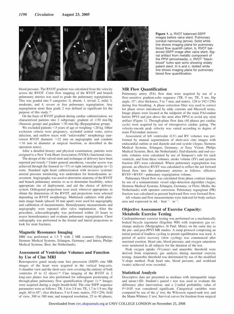
Assessment of Ventricular Volumes and Functionby Use of Cine MRIRetrospective gated steady-state free precession (SSFP) cine MRimages of the heart were acquired in the vertical long-axis,4-chamber view and the short-axis view covering the entirety of bothventricles (9 to 12 slices).16 Cine imaging of the RVOT in 2long-axis planes was also performed for subsequent positioning ofthrough-plane pulmonary flow quantification (Figure 1).16 Imageswere acquired during a single breath-hold. The cine SSFP sequenceparameters were as follows: TR, 3.4 to 3.8 ms; TE, 1.7 to 1.9 ms; flipangle, 60 to 65°; slice thickness, 5 to 8 mm; matrix, 192�256; fieldof view, 300 to 380 mm; and temporal resolution, 25 to 40 phases.
MR Flow QuantificationPulmonary artery (PA) flow data were acquired by use of aflow-sensitive gradient-echo sequence (TR, 9 ms; TE, 5 ms; flipangle, 15°; slice thickness, 5 to 7 mm; and matrix, 128 to 192�256)during free breathing. A phase correction filter was used to correctfor phase errors introduced by eddy currents and Maxwell terms.Image planes were located at the midpoint of the main PA/conduitbefore PPVI and just above the stent after PPVI to avoid any stentartifact (Figure 1). Through-plane flow data (40 phases per cardiaccycle) were acquired by use of retrospective cardiac gating. Thevelocity-encode peak velocity was varied according to degree ofmain PA/conduit stenosis.
Assessment of left ventricular (LV) and RV volumes was per-formed by manual segmentation of short-axis cine images withendocardial outline at end diastole and end systole (Argus; SiemensMedical Systems, Erlangen, Germany; or Easy Vision; PhilipsMedical Systems, Best, the Netherlands). End-diastolic and end-sys-tolic volumes were calculated by use of Simpson’s rule for eachventricle, and from these volumes, stroke volume (SV) and ejectionfraction (EF) were calculated. Where pulmonary regurgitation waspresent, an effective RVSV was calculated to reflect the net forwardblood flow into the pulmonary arteries as follows: effectiveRVSV�RVSV�pulmonary regurgitation volume.
Pulmonary blood flow was calculated from phase contrast imagesby use of a semiautomatic vessel edge-detection algorithm (Argus;Siemens Medical Systems, Erlangen, Germany; or Flow; Medis, theNetherlands) with operator correction. Pulmonary regurgitant (PR)fraction was calculated as percent backward flow over forward flow.
All volume and flow measurements were indexed for body surfacearea and expressed in mL · beat�1 · m�2.
Objective Assessment of Exercise Capacity:Metabolic Exercise TestingCardiopulmonary exercise testing was performed on a mechanicallybraked bicycle ergometer (Ergoline 900) with respiratory gas ex-change analysis (Medgraphics, St Paul, Minn), on the same day asthe pre- and post-PPVI MR studies. A ramp protocol comprising aninitial period of loadless cycling to permit equilibration was used. Aperiod of active recovery (slow cycling) was commenced aftermaximal exertion. Heart rate, blood pressure, and oxygen saturationwere monitored in all subjects for the duration of the test.
Peak oxygen uptake (V̇O2max) and anaerobic threshold werederived from respiratory gas analysis during maximal exercisetesting. Anaerobic threshold was determined by use of the modifiedV-slope method. Peak heart rate, blood pressure, and workload(watts) achieved were recorded.
Statistical AnalysisDescriptive data are presented as medians with interquartile rangeand mean�SD. Student’s paired t test was used to evaluate thedifference after intervention, and a 2-tailed probability value ofP�0.05 was considered significant. Categorical variables werecompared by use of the �2 test, the Wilcoxon signed-rank test, andthe Mann-Whitney U test. Survival curves for freedom from surgical
Figure 1. a, RVOT balanced SSFPimages before valve stent. Pulmonaryconduit narrowing (arrow). Solid whiteline shows imaging plane for pulmonaryblood flow quantifi cation. b, RVOT bal-anced SSFP image after valve stent. Sig-nal artifact from metallic component ofthe PPVI (arrowheads). c, RVOT “black-blood” turbo spin echo showing widelypatent stent. In b and c, dotted whiteline shows imaging plane for pulmonaryblood flow quantification.
1190 Circulation August 23, 2005
at UNIV COLLEGE LONDON on November 25, 2008 circ.ahajournals.orgDownloaded from
explantation for valve failure were obtained by use of Kaplan-Meierplots. Statistical analysis was performed on SPSS 11.0. and 12.0(SPSS Inc., Chicago, Ill).
ResultsBetween January 2000 and September 2004, we attemptedPPVI in 59 patients (32 male), with successful implantation in58. One patient, in whom we failed to maneuver the valve-stent assembly into the outflow tract, is awaiting a furtherattempt.
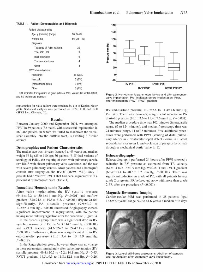
Demographics and Patient CharacteristicsThe median age was 16 years (range, 9 to 43 years) and medianweight 56 kg (25 to 110 kg); 36 patients (61%) had variants oftetralogy of Fallot, the majority of them with pulmonary atresia(n�18), 3 with absent pulmonary valve syndrome, and the restwith severe pulmonary stenosis. Most patients had a homograftconduit after surgery on the RVOT (46/59, 78%). Only 3patients had a “native” RVOT that had been augmented with apericardial or homograft patch (Table 1).
Immediate Hemodynamic ResultsAfter valve implantation, the RV systolic pressure(64.4�17.2 to 50.4�14 mm Hg, P�0.001) and outflowgradient (33�24.6 to 19.5�15.3, P�0.001) (Figure 2) fellsignificantly. PA diastolic pressure (9.9�3.7 to13.5�5.3 mm Hg, P�0.001) increased. Angiography showedsignificant improvement in regurgitation, with no patienthaving more mild regurgitation after the procedure (Figure 3).
In the Stenosis group, there was a significant drop in RVsystolic pressure (71�15.3 to 52.3�14.5 mm Hg, P�0.001)and RVOT gradient (44.6�24.3 to 24.4�15.2 mm Hg,P�0.001). Furthermore, there was a significant drop in RVend-diastolic pressure (11.7�3.4 to 10�3.9 mm Hg,P�0.018).
In the Regurgitation group, however, there was no changein these parameters immediately after valve implantation (RVsystolic pressure, 49.4�10.5 to 47.2�14.1 mm Hg, P�0.45;RVOT gradient, 14.5�9.3 to 11.8�12.1 mm Hg, P�0.26;
RV end-diastolic pressure, 10.7�2.8 to 11.4�4.6 mm Hg,P�0.43). There was, however, a significant increase in PAdiastolic pressure (10.1�3.6 to 15.4�7.6 mm Hg, P�0.001).
The median procedure time was 102 minutes (interquartilerange, 67 to 124 minutes), and median fluoroscopy time was21 minutes (range, 11 to 36 minutes). Five additional proce-dures were performed with PPVI (stenting of distal pulmo-nary arteries in 2, ventricular septal defect closure in 1, atrialseptal defect closure in 1, and occlusion of paraprosthetic leakthrough a mechanical aortic valve in 1).
EchocardiographyEchocardiography performed 24 hours after PPVI showed areduction in RV pressure as estimated from TR velocity(64�1.4 to 51.8�1.9 mm Hg, P�0.001) and RVOT gradient(63.4�23.4 to 40.5�18.2 mm Hg, P�0.001). There wassignificant reduction in grade of PR, with all patients havinggrade 2 or greater PR before, and none with more than grade2 PR after the procedure (P�0.001).
Magnetic Resonance ImagingCardiovascular MRI was performed in 28 patients (age,18.8�7.9 years; range, 9.2 to 41.6 years) a median of 6 days
TABLE 1. Patient Demographics and Diagnosis
Patient characteristics
Age, y (median) (range) 16 (9–43)
Weight, kg 56 (25–110)
Diagnosis
Tetralogy of Fallot variants 36
TGA, VSD, PS 9
Ross operation 6
Truncus arteriosus 4
Other 4
RVOT characteristics
Homograft 46 (78%)
Hancock 5 (8%)
Transannular patch 3 (5%)
Other 5 (8%)
TGA indicates transposition of great arteries; VSD, ventricular septal defect;and PS, pulmonary stenosis.
Figure 2. Hemodynamic parameters before and after pulmonaryvalve implantation. Pre- indicates before implantation; Post,after implantation; RVOT, RVOT gradient.
Figure 3. Lateral still-frame angiograms. Abolition of stenosisand regurgitation after pulmonary valve implantation.
Khambadkone et al Pulmonary Valve Implantation 1191
at UNIV COLLEGE LONDON on November 25, 2008 circ.ahajournals.orgDownloaded from
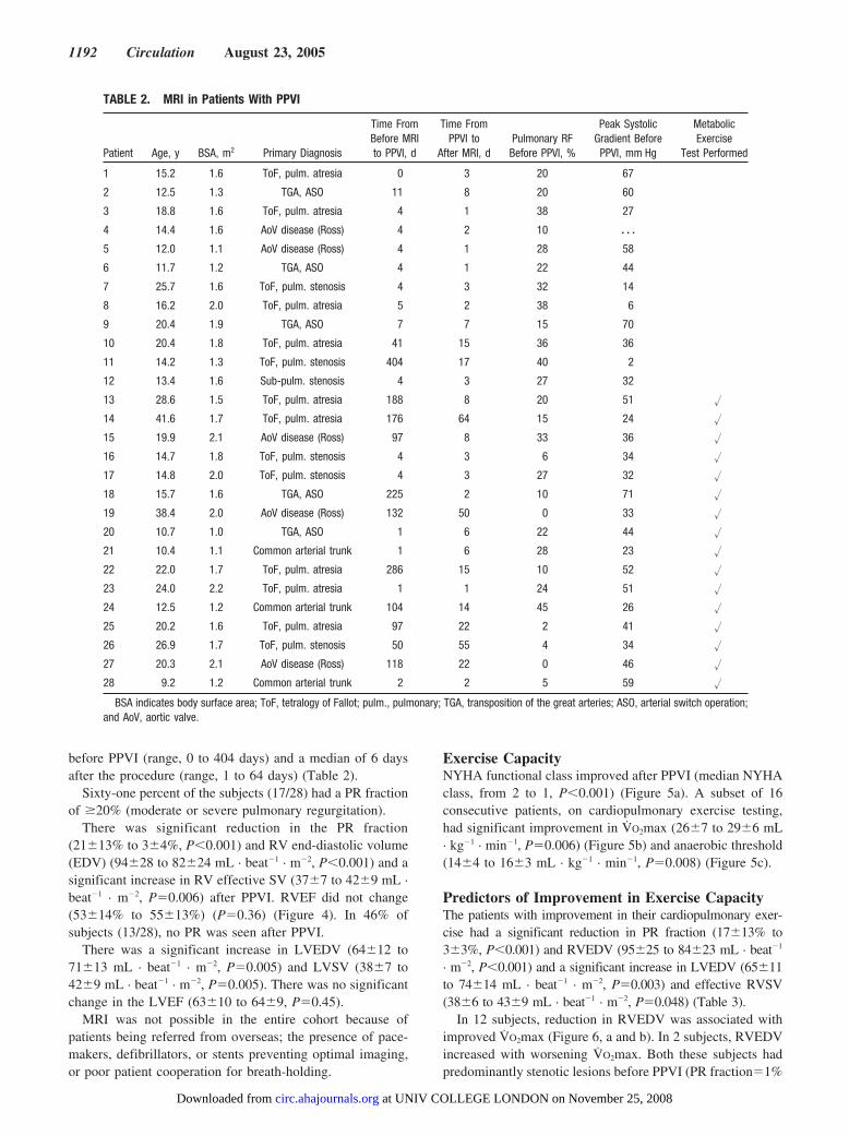
before PPVI (range, 0 to 404 days) and a median of 6 daysafter the procedure (range, 1 to 64 days) (Table 2).
Sixty-one percent of the subjects (17/28) had a PR fractionof �20% (moderate or severe pulmonary regurgitation).
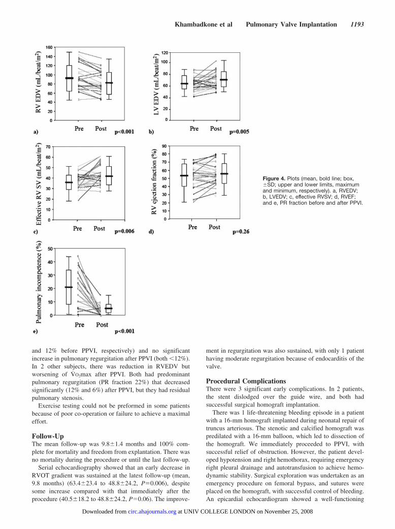
There was significant reduction in the PR fraction(21�13% to 3�4%, P�0.001) and RV end-diastolic volume(EDV) (94�28 to 82�24 mL · beat�1 · m�2, P�0.001) and asignificant increase in RV effective SV (37�7 to 42�9 mL ·beat�1 · m�2, P�0.006) after PPVI. RVEF did not change(53�14% to 55�13%) (P�0.36) (Figure 4). In 46% ofsubjects (13/28), no PR was seen after PPVI.
There was a significant increase in LVEDV (64�12 to71�13 mL · beat�1 · m�2, P�0.005) and LVSV (38�7 to42�9 mL · beat�1 · m�2, P�0.005). There was no significantchange in the LVEF (63�10 to 64�9, P�0.45).
MRI was not possible in the entire cohort because ofpatients being referred from overseas; the presence of pace-makers, defibrillators, or stents preventing optimal imaging,or poor patient cooperation for breath-holding.
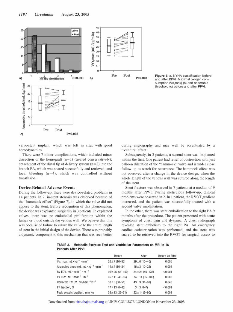
Exercise CapacityNYHA functional class improved after PPVI (median NYHAclass, from 2 to 1, P�0.001) (Figure 5a). A subset of 16consecutive patients, on cardiopulmonary exercise testing,had significant improvement in V̇O2max (26�7 to 29�6 mL· kg�1 · min�1, P�0.006) (Figure 5b) and anaerobic threshold(14�4 to 16�3 mL · kg�1 · min�1, P�0.008) (Figure 5c).
Predictors of Improvement in Exercise CapacityThe patients with improvement in their cardiopulmonary exer-cise had a significant reduction in PR fraction (17�13% to3�3%, P�0.001) and RVEDV (95�25 to 84�23 mL · beat�1
· m�2, P�0.001) and a significant increase in LVEDV (65�11to 74�14 mL · beat�1 · m�2, P�0.003) and effective RVSV(38�6 to 43�9 mL · beat�1 · m�2, P�0.048) (Table 3).
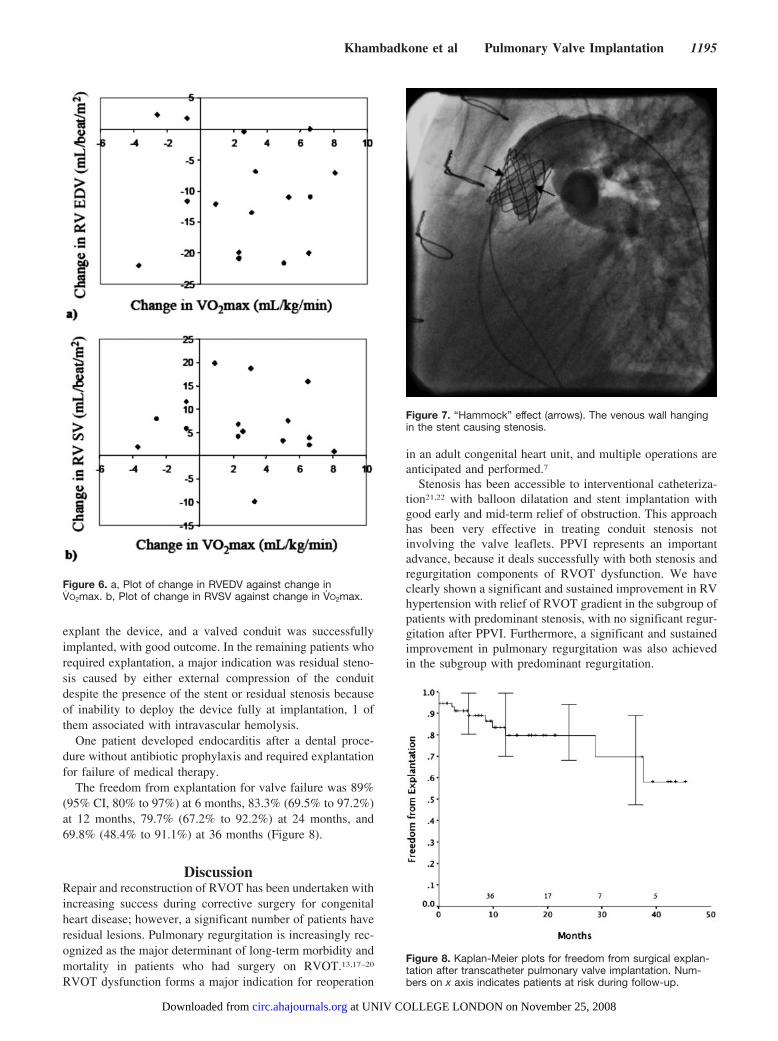
In 12 subjects, reduction in RVEDV was associated withimproved V̇O2max (Figure 6, a and b). In 2 subjects, RVEDVincreased with worsening V̇O2max. Both these subjects hadpredominantly stenotic lesions before PPVI (PR fraction�1%
TABLE 2. MRI in Patients With PPVI
Patient Age, y BSA, m2 Primary Diagnosis
Time FromBefore MRIto PPVI, d
Time FromPPVI to
After MRI, dPulmonary RF
Before PPVI, %
Peak SystolicGradient Before
PPVI, mm Hg
MetabolicExercise
Test Performed
1 15.2 1.6 ToF, pulm. atresia 0 3 20 67
2 12.5 1.3 TGA, ASO 11 8 20 60
3 18.8 1.6 ToF, pulm. atresia 4 1 38 27
4 14.4 1.6 AoV disease (Ross) 4 2 10 � � �
5 12.0 1.1 AoV disease (Ross) 4 1 28 58
6 11.7 1.2 TGA, ASO 4 1 22 44
7 25.7 1.6 ToF, pulm. stenosis 4 3 32 14
8 16.2 2.0 ToF, pulm. atresia 5 2 38 6
9 20.4 1.9 TGA, ASO 7 7 15 70
10 20.4 1.8 ToF, pulm. atresia 41 15 36 36
11 14.2 1.3 ToF, pulm. stenosis 404 17 40 2
12 13.4 1.6 Sub-pulm. stenosis 4 3 27 32
13 28.6 1.5 ToF, pulm. atresia 188 8 20 51 �
14 41.6 1.7 ToF, pulm. atresia 176 64 15 24 �
15 19.9 2.1 AoV disease (Ross) 97 8 33 36 �
16 14.7 1.8 ToF, pulm. stenosis 4 3 6 34 �
17 14.8 2.0 ToF, pulm. stenosis 4 3 27 32 �
18 15.7 1.6 TGA, ASO 225 2 10 71 �
19 38.4 2.0 AoV disease (Ross) 132 50 0 33 �
20 10.7 1.0 TGA, ASO 1 6 22 44 �
21 10.4 1.1 Common arterial trunk 1 6 28 23 �
22 22.0 1.7 ToF, pulm. atresia 286 15 10 52 �
23 24.0 2.2 ToF, pulm. atresia 1 1 24 51 �
24 12.5 1.2 Common arterial trunk 104 14 45 26 �
25 20.2 1.6 ToF, pulm. atresia 97 22 2 41 �
26 26.9 1.7 ToF, pulm. stenosis 50 55 4 34 �
27 20.3 2.1 AoV disease (Ross) 118 22 0 46 �
28 9.2 1.2 Common arterial trunk 2 2 5 59 �
BSA indicates body surface area; ToF, tetralogy of Fallot; pulm., pulmonary; TGA, transposition of the great arteries; ASO, arterial switch operation;and AoV, aortic valve.
1192 Circulation August 23, 2005
at UNIV COLLEGE LONDON on November 25, 2008 circ.ahajournals.orgDownloaded from
and 12% before PPVI, respectively) and no significantincrease in pulmonary regurgitation after PPVI (both �12%).In 2 other subjects, there was reduction in RVEDV butworsening of V̇O2max after PPVI. Both had predominantpulmonary regurgitation (PR fraction 22%) that decreasedsignificantly (12% and 6%) after PPVI, but they had residualpulmonary stenosis.
Exercise testing could not be preformed in some patientsbecause of poor co-operation or failure to achieve a maximaleffort.
Follow-UpThe mean follow-up was 9.8�1.4 months and 100% com-plete for mortality and freedom from explantation. There wasno mortality during the procedure or until the last follow-up.
Serial echocardiography showed that an early decrease inRVOT gradient was sustained at the latest follow-up (mean,9.8 months) (63.4�23.4 to 48.8�24.2, P�0.006), despitesome increase compared with that immediately after theprocedure (40.5�18.2 to 48.8�24.2, P�0.06). The improve-
ment in regurgitation was also sustained, with only 1 patienthaving moderate regurgitation because of endocarditis of thevalve.
Procedural ComplicationsThere were 3 significant early complications. In 2 patients,the stent dislodged over the guide wire, and both hadsuccessful surgical homograft implantation.
There was 1 life-threatening bleeding episode in a patientwith a 16-mm homograft implanted during neonatal repair oftruncus arteriosus. The stenotic and calcified homograft waspredilated with a 16-mm balloon, which led to dissection ofthe homograft. We immediately proceeded to PPVI, withsuccessful relief of obstruction. However, the patient devel-oped hypotension and right hemothorax, requiring emergencyright pleural drainage and autotransfusion to achieve hemo-dynamic stability. Surgical exploration was undertaken as anemergency procedure on femoral bypass, and sutures wereplaced on the homograft, with successful control of bleeding.An epicardial echocardiogram showed a well-functioning
Figure 4. Plots (mean, bold line; box,�SD; upper and lower limits, maximumand minimum, respectively). a, RVEDV;b, LVEDV; c, effective RVSV; d, RVEF;and e, PR fraction before and after PPVI.
Khambadkone et al Pulmonary Valve Implantation 1193
at UNIV COLLEGE LONDON on November 25, 2008 circ.ahajournals.orgDownloaded from
valve-stent implant, which was left in situ, with goodhemodynamics.
There were 7 minor complications, which included minordissection of the homograft (n�1) (treated conservatively);detachment of the distal tip of delivery system (n�2) into thebranch PA, which was snared successfully and retrieved; andlocal bleeding (n�4), which was controlled withouttransfusion.
Device-Related Adverse EventsDuring the follow-up, there were device-related problems in14 patients. In 7, in-stent stenosis was observed because ofthe “hammock effect” (Figure 7), in which the valve did notappose to the stent. Before recognition of this phenomenon,the device was explanted surgically in 3 patients. In explantedvalves, there was no endothelial proliferation within thelumen or blood outside the venous wall. We believe that thiswas because of failure to suture the valve to the entire lengthof stent in the initial design of the device. There was probablya dynamic component to this mechanism that was seen better
during angiography and may well be accentuated by a“Venturi” effect.
Subsequently, in 3 patients, a second stent was implantedwithin the first. One patient had relief of obstruction with justballoon dilatation of the “hammock” valve and is under closefollow-up to watch for recurrence. The hammock effect wasnot observed after a change in the device design, when thewhole length of the venous wall was sutured along the lengthof the stent.
Stent fracture was observed in 7 patients at a median of 9months after PPVI. During meticulous follow-up, clinicalproblems were observed in 2. In 1 patient, the RVOT gradientincreased, and the patient was successfully treated with asecond valve implantation.
In the other, there was stent embolization to the right PA 9months after the procedure. The patient presented with acutesymptoms of chest pain and dyspnea. A chest radiographrevealed stent embolism to the right PA. An emergencycardiac catheterization was performed, and the stent wassnared to be retrieved into the RVOT for surgical access to
Figure 5. a, NYHA classification beforeand after PPVI. Maximal oxygen con-sumption (V̇O2max) (b) and anaerobicthreshold (c) before and after PPVI.
TABLE 3. Metabolic Exercise Test and Ventricular Parameters on MRI in 16Patients After PPVI
Before After Before vs After
V̇O2 max, mL � kg�1 � min�1 26�7 (16–35) 29�6 (15–40) 0.006
Anaerobic threshold, mL � kg�1 � min�1 14�4 (10–24) 16�3 (10–22) 0.008
RV EDV, mL � beat�1 � m�2 95�25 (68–150) 84�23 (46–136) �0.001
LV EDV, mL � beat�1 � m�2 65�11 (46–85) 74�14 (55–105) 0.003
Corrected RV SV, mL/beat�1m�2 38�6 (30–51) 43�9 (31–61) 0.048
PR fraction, % 17�13 (0–45) 3�3 (0–7) �0.001
Peak systolic gradient, mm Hg 39�13 (23–71) 22�14 (9–60) 0.001
1194 Circulation August 23, 2005
at UNIV COLLEGE LONDON on November 25, 2008 circ.ahajournals.orgDownloaded from
explant the device, and a valved conduit was successfullyimplanted, with good outcome. In the remaining patients whorequired explantation, a major indication was residual steno-sis caused by either external compression of the conduitdespite the presence of the stent or residual stenosis becauseof inability to deploy the device fully at implantation, 1 ofthem associated with intravascular hemolysis.
One patient developed endocarditis after a dental proce-dure without antibiotic prophylaxis and required explantationfor failure of medical therapy.
The freedom from explantation for valve failure was 89%(95% CI, 80% to 97%) at 6 months, 83.3% (69.5% to 97.2%)at 12 months, 79.7% (67.2% to 92.2%) at 24 months, and69.8% (48.4% to 91.1%) at 36 months (Figure 8).
DiscussionRepair and reconstruction of RVOT has been undertaken withincreasing success during corrective surgery for congenitalheart disease; however, a significant number of patients haveresidual lesions. Pulmonary regurgitation is increasingly rec-ognized as the major determinant of long-term morbidity andmortality in patients who had surgery on RVOT.13,17–20
RVOT dysfunction forms a major indication for reoperation
in an adult congenital heart unit, and multiple operations areanticipated and performed.7
Stenosis has been accessible to interventional catheteriza-tion21,22 with balloon dilatation and stent implantation withgood early and mid-term relief of obstruction. This approachhas been very effective in treating conduit stenosis notinvolving the valve leaflets. PPVI represents an importantadvance, because it deals successfully with both stenosis andregurgitation components of RVOT dysfunction. We haveclearly shown a significant and sustained improvement in RVhypertension with relief of RVOT gradient in the subgroup ofpatients with predominant stenosis, with no significant regur-gitation after PPVI. Furthermore, a significant and sustainedimprovement in pulmonary regurgitation was also achievedin the subgroup with predominant regurgitation.
Figure 6. a, Plot of change in RVEDV against change inV̇O2max. b, Plot of change in RVSV against change in V̇O2max.
Figure 7. “Hammock” effect (arrows). The venous wall hangingin the stent causing stenosis.
Figure 8. Kaplan-Meier plots for freedom from surgical explan-tation after transcatheter pulmonary valve implantation. Num-bers on x axis indicates patients at risk during follow-up.
Khambadkone et al Pulmonary Valve Implantation 1195
at UNIV COLLEGE LONDON on November 25, 2008 circ.ahajournals.orgDownloaded from
We observed 3 separate categories of complications. Thefirst were complications related to balloon dilatation orstenting for conduit stenosis, including dissection, hemor-rhage from conduit rupture, and residual stenosis because ofexternal compression or undilatable conduits.21–23
The second category was related to issues of patientselection and occurred in the new substrate of patients withpredominant regurgitation who have previously been acces-sible to interventional catheterization. Dislodgment or embo-lization of the valved stent during PPVI or follow-up oc-curred because of unfavorable shape, size, and elasticproperties of the RVOT. Understanding wall characteristicsand 3D dynamic imaging of RVOT would refine patientselection and is a key issue of current research by our group.
The third category, related to device design, included the“hammock” effect and stent fracture. The hammock effectwas resolved during the series with an improved design,whereby the entire length of the stent was sutured to thevenous wall segment. In one series, 16% of patients had stentfractures in RV to PA conduits.22 In our series, 12% ofpatients had stent fracture; however, only 2 had clinicalconsequences. One was treated with a second PPVI with asuccessful hemodynamic result. The other patient with stentembolism has been described in the earlier section.
Transcatheter pulmonary valve replacement provides aunique opportunity to study the response of the RV to acutepressure and volume unloading without the confoundingeffect of cardiopulmonary bypass. Previous studies aftersurgical pulmonary valve replacement have shown reductionin RVEDVs (MRI)12 and RV dimensions (echocardiogra-phy),11 and both improved subjectively11,12 and objectively(metabolic exercise testing),24 although no association be-tween changes in ventricular parameters and exercise capac-ity has been shown.
Cardiovascular MRI assessment in patients with severepulmonary regurgitation has demonstrated markedly elevatedRVEDV and RVESV and reduced RVEF.25–28 This chronicRV volume overload has long been regarded as benign, butthere is increasing evidence that RV function may be irre-versibly compromised by such long-term changes.29 This isexemplified by 3 findings that have been demonstrated bycardiovascular MRI. First, RVEF has been shown to besignificantly lower in patients with both RV pressure andvolume overload compared with RV pressure overloadalone.28 Second, an abnormal RV response to stress (eitherpharmacological28 or physiological30) has been demonstratedin patients with repaired tetralogy of Fallot and pulmonaryregurgitation. And finally, there appears to be no significantimprovement in RVEF at rest after pulmonary valvereplacement.12,29
In our study, there was a reduction in RVEDV andimproved performance at metabolic exercise testing (12 of 16subjects), and in 2 subjects in whom RVEDV increased, therewas reduced performance at metabolic exercise test (bothpredominant stenosis patients). The reduction in RVEDV wasassociated with an increase in LVEDV, which suggests thatthe mechanism of clinical improvement may be related todiastolic ventricular interaction.31 We propose that reductionin RVEDV after reduction in PR volume by valve implanta-
tion permits an increase in LV filling and thus LVEDV. Thisoccurs within the constraints of the pericardial space and, bythe Frank-Starling mechanism, results in an increase in strokevolume, which may be responsible for the subjective andobjective improvements in functional status. This mechanismis further supported by evidence in patients with heart failure,which suggests that abnormalities of LV diastolic functionare a key determinant of exercise intolerance.32 A secondexplanation for the improved LV filling may simply berelated to increased effective RVSV as a result of a reductionin RVOT obstruction, thus increasing pulmonary venousreturn, left atrial filling, and LV preload. Further studies arein progress to define the mechanism of ventricularinteraction.
LimitationsAlthough we have demonstrated an immediate beneficialeffect of PPVI, long-term follow-up will be essential toevaluate the potential for ventricular remodeling and sus-tained symptomatic relief. The subjects in this study wereheterogeneous in terms of age, diagnosis, RVOT pathology,RV dilatation, and RV function.
As with all new procedures, whether surgical or interven-tional, we recognize the impact of learning curve on thisnovel technique. The reintervention and explantation ratesmay appear to be higher in direct comparison to surgicalreports; however, evolution of device design and moreexperience have led to further a decrease, and indeed elimi-nation, of some of the early problems. Also, our cohort ofpatients is complex, with a median of 3 operations on theirRVOT before the PPVI. Despite this, and including the wholeseries right from the first case, there has been no mortality,either during the procedure or during late follow-up.
The patient population in our series had distinct RVOTdimensions and morphology favorable for implantation of thevalved stent. We are working to expand this application topatients with dilated RVOTs with development of RVOTreducers. MRI and cardiopulmonary exercise testing couldnot be obtained in the entire cohort of patients.
SummaryOur PPVI program is the first clinical step in treatment ofvalvular regurgitation in the modern era of transcathetertherapeutics. This approach will alter strategies for transcath-eter management of other heart valves and lead to a majorchange in the lifetime management of patients with congen-ital heart disease.
AcknowledgmentsDr Taylor was funded by the Higher Education Funding Council forEngland (HEFCE). Drs Coats, Boudjemline, Deanfield, and Bon-hoeffer were funded by the British Heart Foundation. Drs Razavi,Muthurangu, and Hegde were funded by the HEFCE Joint ResearchEquipment Initiative; Philips Medical Systems, Best, the Nether-lands; the Evelina Trust; and the Charitable Foundation of Guy’s andSt Thomas NHS Trust.
DisclosureDr Bonhoeffer is a consultant to Medtronic.
1196 Circulation August 23, 2005
at UNIV COLLEGE LONDON on November 25, 2008 circ.ahajournals.orgDownloaded from
References1. Ross DN, Somerville J. Correction of pulmonary atresia with a homograft
aortic valve. Lancet. 1966;2:1446–1447.2. Rastelli GC, Wallace RB, Ongley PA. Complete repair of transposition of
the great arteries with pulmonary stenosis: a review and report of a casecorrected by using a new surgical technique. Circulation. 1969;39:83–95.
3. Bove EL, Lupinetti FM, Pridjian AK, Beekman RH III, Callow LB,Snider AR, Rosenthal A. Results of a policy of primary repair of truncusarteriosus in the neonate. J Thorac Cardiovasc Surg. 1993;105:1057–1065.
4. Conte S, Jashari R, Eyskens B, Gewillig M, Dumoulin M, Daenen W.Homograft valve insertion for pulmonary regurgitation late after valvelessrepair of right ventricular outflow tract obstruction. Eur J CardiothoracSurg. 1999;15:143–149.
5. Perron J, Moran AM, Gauvreau K, del Nido PJ, Mayer JE Jr, Jonas RA.Valved homograft conduit repair of the right heart in early infancy. AnnThorac Surg. 1999;68:542–548.
6. Bando K, Danielson GK, Schaff HV, Mair DD, Julsrud PR, Puga FJ.Outcome of pulmonary and aortic homografts for right ventricularoutflow tract reconstruction. J Thorac Cardiovasc Surg. 1995;109:509–517.
7. Dore A, Glancy DL, Stone S, Menashe VD, Somerville J. Cardiac surgeryfor grown-up congenital heart patients: survey of 307 consecutive oper-ations from 1991 to 1994. Am J Cardiol. 1997;80:906–913.
8. Bonhoeffer P, Boudjemline Y, Saliba Z, Merckx J, Aggoun Y, Bonnet D,Acar P, Le Bidois J, Sidi D, Kachaner J. Percutaneous replacement ofpulmonary valve in a right-ventricle to pulmonary-artery prostheticconduit with valve dysfunction. Lancet. 2000;356:1403–1405.
9. Bonhoeffer P, Boudjemline Y, Saliba Z, Hausse AO, Aggoun Y, BonnetD, Sidi D, Kachaner J. Transcatheter implantation of a bovine valve inpulmonary position: a lamb study. Circulation. 2000;102:813–816.
10. Bonhoeffer P, Boudjemline Y, Qureshi SA, Le Bidois J, Iserin L, Acar P,Merckx J, Kachaner J, Sidi D. Percutaneous insertion of the pulmonaryvalve. J Am Coll Cardiol. 2002;39:1664–1669.
11. Warner KG, O’Brien PK, Rhodes J, Kaur A, Robinson DA, Payne DD.Expanding the indications for pulmonary valve replacement after repairof tetralogy of Fallot. Ann Thorac Surg. 2003;76:1066–1071.
12. Vliegen HW, van Straten A, de Roos A, Roest AA, Schoof PH,Zwinderman AH, Ottenkamp J, van der Wall EE, Hazekamp MG.Magnetic resonance imaging to assess the hemodynamic effects of pul-monary valve replacement in adults late after repair of tetralogy of Fallot.Circulation. 2002;106:1703–1707.
13. Bove EL, Kavey RE, Byrum CJ, Sondheimer HM, Blackman MS,Thomas FD. Improved right ventricular function following late pulmo-nary valve replacement for residual pulmonary insufficiency or stenosis.J Thorac Cardiovasc Surg. 1985;90:50–55.
14. Discigil B, Dearani JA, Puga FJ, Schaff HV, Hagler DJ, Warnes CA,Danielson GK. Late pulmonary valve replacement after repair of tetralogyof Fallot. J Thorac Cardiovasc Surg. 2001;121:344–351.
15. Bigras JL, Boutin C, McCrindle BW, Rebeyka IM. Short-term effect ofmonocuspid valves on pulmonary insufficiency and clinical outcome aftersurgical repair of tetralogy of Fallot. J Thorac Cardiovasc Surg. 1996;112:33–37.
16. Taylor AM, Bogaert J. Imaging planes. In: Clinical Cardiac MRI.Bogaert J, Dymarkowski S, Taylor AM, eds. Heidelberg, Germany:Springer-Verlag; 2005;85–98.
17. Bove EL, Byrum CJ, Thomas FD, Kavey RE, Sondheimer HM,Blackman MS, Parker FB Jr. The influence of pulmonary insufficiency onventricular function following repair of tetralogy of Fallot: evaluationusing radionuclide ventriculography. J Thorac Cardiovasc Surg. 1983;85:691–696.
18. Therrien J, Siu SC, Harris L, Dore A, Niwa K, Janousek J, Williams WG,Webb G, Gatzoulis MA. Impact of pulmonary valve replacement onarrhythmia propensity late after repair of tetralogy of Fallot. Circulation.2001;103:2489–2494.
19. Gatzoulis MA, Till JA, Redington AN. Depolarization-repolarizationinhomogeneity after repair of tetralogy of Fallot: the substrate formalignant ventricular tachycardia? Circulation. 1997;95:401–404.
20. Gatzoulis MA, Balaji S, Webber SA, Webber SA, Siu SC, Hokanson JS,Poile C, Rosenthal M, Nakazawa M, Moller JH, Gillette PC, Webb GD,Redington AN. Risk factors for arrhythmia and sudden cardiac death lateafter repair of tetralogy of Fallot: a multicentre study. Lancet. 2000;356:975–981.
21. Pedra CA, Justino H, Nykanen DG, VanArsdell G, Coles JG, WilliamsWG, Freedom RM, Benson LN. Percutaneous stent implantation to ste-notic bioprosthetic valves in the pulmonary position. J Thorac Car-diovasc Surg. 2002;124:82–87.
22. Powell AJ, Lock JE, Keane JF, Perry SB. Prolongation of RV-PA conduitlife span by percutaneous stent implantation: intermediate-term results.Circulation. 1995;92:3282–3288.
23. Ovaert C, Caldarone CA, McCrindle BW, Nykanen D, Freedom RM,Coles JG, Williams WG, Benson LN. Endovascular stent implantation forthe management of postoperative right ventricular outflow tractobstruction: clinical efficacy. J Thorac Cardiovasc Surg. 1999;118:886–893.
24. Eyskens B, Reybrouck T, Bogaert J, Dymarkowsky S, Daenen W,Dumoulin M, Gewillig M. Homograft insertion for pulmonary regurgi-tation after repair of tetralogy of Fallot improves cardiorespiratoryexercise performance. Am J Cardiol. 2000;85:221–225.
25. Helbing WA, Niezen RA, Le Cessie S, van der Geest RJ, Ottenkamp J,de Roos A. Right ventricular diastolic function in children with pulmo-nary regurgitation after repair of tetralogy of Fallot: volumetric evaluationby magnetic resonance velocity mapping. J Am Coll Cardiol. 1996;28:1827–1835.
26. Singh GK, Greenberg SB, Yap YS, Delany DP, Keeton BR, Monro JL.Right ventricular function and exercise performance late after primaryrepair of tetralogy of Fallot with the transannular patch in infancy.Am J Cardiol. 1998;81:1378–1382.
27. Davlouros PA, Kilner PJ, Hornung TS, Li W, Francis JM, Moon JC,Smith GC, Tat T, Pennell DJ, Gatzoulis MA. Right ventricular functionin adults with repaired tetralogy of Fallot assessed with cardiovascularmagnetic resonance imaging: detrimental role of right ventricular outflowaneurysms or akinesia and adverse right-to-left ventricular interaction.J Am Coll Cardiol. 2002;40:2044–2052.
28. Tulevski II, Hirsch A, Dodge-Khatami A, Stoker J, van der Wall EE,Mulder BJ. Effect of pulmonary valve regurgitation on right ventricularfunction in patients with chronic right ventricular pressure overload.Am J Cardiol. 2003;92:113–116.
29. Therrien J, Siu SC, McLaughlin PR, Liu PP, Williams WG, Webb GD.Pulmonary valve replacement in adults late after repair of tetralogy ofFallot: are we operating too late? J Am Coll Cardiol. 2000;36:1670–1675.
30. Roest AA, Helbing WA, Kunz P, van den Aardweg JG, Lamb HJ,Vliegen HW, van der Wall EE, de Roos A. Exercise MR imaging in theassessment of pulmonary regurgitation and biventricular function inpatients after tetralogy of Fallot repair. Radiology. 2002;223:204–211.
31. Atherton JJ, Moore TD, Lele SS, Thomson HL, Galbraith AJ, Belenkie I,Tyberg JV, Frenneaux M.P. Diastolic ventricular interaction in chronicheart failure. Lancet. 1997;349:720–724.
32. Parthenakis FI, Kanoupakis EM, Kochiadakis GE, Skalidis EI, MezilisNE, Simantirakis EN, Kanakaraki MK, Vardas PE. Left ventricular dia-stolic filling pattern predicts cardiopulmonary determinants of functionalcapacity in patients with congestive heart failure. Am Heart J. 2000;140:338–344.
Khambadkone et al Pulmonary Valve Implantation 1197
at UNIV COLLEGE LONDON on November 25, 2008 circ.ahajournals.orgDownloaded from