long term outcomes following percutaneous dilatational
TRANSCRIPT
1
Long term outcomes following
percutaneous dilatational tracheostomy in
the critically ill
Thesis submitted in accordance with the requirements of the
University of Liverpool for the degree of Doctor of Medicine by
Gerard Dempsey
November 2015
2
Contents Page No
Abstract 6
Dedication 8
Acknowledgements 9
Declaration 10 List of Figures 11
List of Tables 12
List of abbreviations 13
Chapter 1 15 A history of tracheostomy and mechanical ventilation within the critical
care setting 1.1 Terminology 18 1.2 Development of tracheal cannulae 19 1.3 Changing indications 20
1.4 Positive pressure ventilation 22 1.5 Tracheostomy and positive pressure ventilation 25 1.6 Percutaneous tracheostomy 28 1.7 Modifications to the percutaneous tracheostomy technique 34
1.8 Tracheostomy outcomes 35 1.8.1 Percutaneous tracheostomy versus surgical tracheostomy 35 1.8.2 Percutaneous versus percutaneous procedures 36 1.9 Aims 38
Chapter 2 39 Major late complications following tracheostomy
2.1 The normal trachea 40
2.1.1 Tracheal anatomy 40 2.1.1.1 Anatomical relations 43
2.1.2 Tracheal physiology 45 2.1.2.1 Movement of air 45
3
2.1.2.2 Heat and moisture exchange 47 2.1.2.3 Removal of particulate debris 47
2.2 Tracheal pathology 48
2.2.1 Tracheal stenosis 48 2.2.1.1 Histopathology 48 2.2.1.2 Types of tracheal stenosis 51 2.2.1.3 Stenosis related to trans-laryngeal intubation versus
tracheostomy 56 2.2.1.4 Grading of tracheal stenosis 57 2.2.1.5 Diagnosis of tracheal stenosis 61 2.2.1.6 Management of tracheal stenosis 66
i. Interventional bronchoscopy and laser therapy 66
ii. Tracheal stenting 66 iii. Surgical management 68 iv. Adjuvant treatment 70
2.2.2 Tracheo-innominate artery fistula 73 2.2.2.1 Pathophysiology 74 2.2.2.2 Diagnosis 77 2.2.2.3 Clinical management 78
i. Haemorrhage control measures 79 ii. Surgical management 80
2.2.3 Tracheo-oesophageal fistula 83 2.2.3.1 Pathophysiology of late tracheo-oesophageal fistula 83 2.2.3.2 Diagnosis 86 2.2.3.3 Clinical management 86
i Conservative management 87 ii Surgical management 87
2.3 Summary 89 2.4 Aims 90
Chapter 3 91 Rationale for thesis
4
3.1 Systematic review 92 3.2 Single tapered dilator percutaneous tracheostomy: An 11-year
review 95
3.3 Tracheal stenosis following percutaneous tracheostomy: A MRI
study 96 3.4 Statement of aims 98
Chapter 4 99
Long-term outcome following tracheostomy in critical care: A systematic review 4.1 Search strategy 100
4.1.1 Study selection 100 4.1.2 Data extraction and outcomes 100
4.1.3 Internal validity and risk of bias assessments 101 4.1.4 Data analysis 101
4.2 Results 102 4.2.1 Search results 102
4.2.2 Study characteristics 103 4.3 Data analysis 107
4.3.1 Comparative analyses 107 4.3.2 Single-arm studies 108
4.4 Discussion 117
Chapter 5 122 Single tapered dilator percutaneous tracheostomy in the critical care unit: An
eleven-year single centre review 5.1 Methods 123 5.2 Results 125 5.3 Discussion 134
Chapter 6 139 Tracheal stenosis following percutaneous dilatational tracheostomy using the single tapered dilator: a MRI study
6.1 Methods 140 6.2 Statistical analysis 142 6.3 Results 142
5
6.4 Discussion 146
Chapter 7 152
Discussion 7.1 Gender bias 154 7.2 Timing of tracheostomy insertion 159 7.3 Reasons for tracheostomy insertion 161
7.3.1 Complex intervention 164
7.3.1.1 Defining the intervention 166 7.3.1.2 Implementation 166 7.3.1.3 Impact and outcomes 167
7.4 Aetiological factors associated with tracheal stenosis 167
7.5 Critical care survival 169 7.5.1 Survival to hospital discharge 169 7.5.2 Survival to one year from critical care admission 173
7.6 Critical care length of stay 178
7.6.1 Future work 180 7.7 Percutaneous tracheostomy outside the critical care unit 181
7.7.1 Emergent airway access 181 7.7.1.1 Future work 182
7.7.2 Percutaneous tracheostomy in the elective surgical setting 182 7.7.2.1 Future work 183
7.8 Perceptions of tracheostomy in critical illness survivors 184 7.8.1 Future work 185
7.9 Conclusion 186
Bibliography 188 Appendices 219
1. Additional data extracted from systematic review papers 220
2. Assessment of tracheal stenosis following percutaneous tracheostomy 221 3. Publications resulting from this work 222
6
Abstract Background: Percutaneous procedures are now the predominant tracheostomy
technique within the critical care setting. Complication rates for various techniques
appear to be equivalent to those achieved with surgical tracheostomy. There is a
paucity of data when comparing percutaneous procedures, particularly when
considering late complications (tracheo-innominate artery fistulae (TIF), tracheo-
oesophageal fistulae (TOF) and tracheal stenosis (TS). Given the severity of illness
and associated mortality in many of these patients the incidence of these
complications remains difficult to define. Confounding factors present in survivors of
critical illness may present difficulties in diagnosis such that underlying tracheal
pathology may go undiagnosed.
Aims: To determine:
The incidence of common early and late complications of percutaneous
dilatational tracheostomy (PDT) in relation to surgical tracheostomy (ST).
The role of peri-operative events that may contribute to the aetiology of late
complications of TS, TIF and TOF.
The incidence of early and late complications in relation to percutaneous
tracheostomy to define the safest percutaneous technique.
The utility of adjunctive techniques (bronchoscopy & ultrasound scanning) in
reducing complications of PDT.
The prevalence of sub-clinical TS following PDT using the single tapered dilator
technique (STD).
Aetiological factors for sub-clinical TS.
Whether sub-clinical TS may present atypically in critical illness survivors.
Methods: We have conducted a systematic review of all prospective studies reporting
late complications after tracheostomy performed in the critically ill. We have also
extracted data to assess the role of peri-operative events and monitoring in causing or
preventing late complications. We have undertaken an eleven-year review of all PDTs
performed within our unit to define the incidence of complications arising within our
own population. Finally, a prospective study to identify the prevalence of sub-clinical
7
TS and identify atypical presenting features in survivors of critical illness has been
performed.
Results: All surgical and percutaneous techniques are broadly similar in terms of
early and late complications. There is a higher incidence of wound infection when
comparing ST to the multiple dilator PDT. There are few studies assessing late
complications between percutaneous techniques. The TS rate varies from 2.8 to 0.6%
for ST and the STD technique respectively. Due to limited data we were unable to
identify peri-operative events that may lead to late complications. There is a very low
rate of complications attributed to the STD technique with only 9 significant late
adverse events. The rate of sub-clinical TS is low with doubtful clinical significance.
Conclusions: We have not found a significant difference in the incidence of TS
between PDT and ST. Our pooled proportions meta-analysis may indicate a tendency
toward a higher rate of stenosis for ST. The reported complication rates presented
within our cohort study may indicate that the STD PDT is one of the safer techniques
available. The rate of sub-clinical stenoses following STD PDT is low and of doubtful
clinical significance. Further work is required to define the role for percutaneous
tracheostomy outside the critical care setting and to gather qualitative data to assess
the patient’s perception of tracheostomy in the critical care setting.

8
Dedication
To my mother Maureen who, sadly, did not survive long enough to see this project
finished.
9
Acknowledgements
As with any significant body of work I would have been unable to complete this text
without the collaboration and support from a myriad of individuals.
Amongst these I would like to acknowledge the assistance of Drs Becky Hanlon, Paul
Jeanrenaud, Richard Pugh, Eoghan O’Callaghan, Carl Wright and Eoin Young
without whom the MRI study would not have reached conclusion.
Similarly, the systematic review would have not reached fruition without the hard
graft of Drs Ben Morton and Clare Hammell and the statistical expertise of Catrin
Tudor-Smith and Lisa Williams.
The prolonged and ongoing review of percutaneous tracheostomy outcomes at
Aintree University Hospital would not have been possible without the diligent
reporting of all of my critical care consultant colleagues.
I would also like to thank Professor Simon Rogers for his review of the manuscript
and constructive comments prior to submission.
Professor Terry Jones has been a longstanding collaborator in relation to this project,
predating my MD registration and his subsequent supervisory role. His sage advice
and unstinting support have been invaluable along the way. It is fair to say the project
would have remained a pipe dream were it not for his input.
Ultimately, without the initial support of both of my parents I would never have had
the opportunity to undertake this study. Their foresight and ability to see the value of
an education, regardless of how long it takes, will always leave me in their debt.
Finally, I would like to thank my wife and children. Cheryl has probably been the
individual who has suffered the most throughout this period. Her support and
encouragement through my unpredictable moods and general negativity have been
remarkable.
10
Declaration
I hereby declare that the content of this thesis “Long term outcomes following
percutaneous dilatational tracheostomy in the critically ill” from inception to
execution has been my own work.
This has been supported by Drs O’Callaghan, Wright and Young who assisted with
patient recruitment and questionnaires for the MRI study. Dr Becky Hanlon provided
radiological review of the MRI scans performed.
Drs Ben Morton and Clare Hammell assisted with data extraction for the systematic
review, whilst Catrin Tudor-Smith and Lisa Williams provided statistical advice and
support.
11
List of Figures Figure 1.1. The Drinker–Collins respirator 26 Figure 1.2. Bennett positive pressure respirator attachment 27
Figure 1.3. Shelden’s percutaneous tracheostomy 30
Figure 1.4. Toy and Weinstein’s percutaneous tracheostomy device 32
Figure 2.1. Cross sectional tracheal morphology 42
Figure 2.2. Anatomical relations of the trachea 44
Figure 2.3 Sites of tracheal stenosis 53
Figure 2.4. Classification of tracheal stenosis 60 Figure 2.5 Normal flow volume loops 64
Figure 2.6. Spirometry in upper airway obstruction 65
Figure 2.7 Relationship of the tracheostomy tube to the innominate artery 75
Figure 2.8. Mechanisms of tracheo-innominate fistula formation 76
Figure 2.9. Algorithm for management of delayed tracheal haemorrhage 82 Figure 2.10 Location and size of tracheo-oesophageal fistulae 85
Figure 4.1. Study selection flow chart 104 Figure 4.2. Forest plot comparing risk of tracheal stenosis 109
Figure 4.3. Forest plot comparing risk of all bleeding episodes 110
Figure 4.4. Forest plot comparing risk of major bleeding 111
Figure 4.5. Forest plot comparing risk of wound infection 112
Figure 5.1. Tracheostomy insertion by day of the week 126
Figure 5.2. Number of PDTs performed by year 128
Figure 5.3. Kaplan Meier plot of overall survival 131
Figure 5.4. Kaplan Meier plot of survival by age 132
Figure 5.5. Kaplan Meier plot of survival by APACHE II Score 133
Figure 6.1: Recruitment to study 143 Figure 7.1 Number of Admissions to Aintree Critical Care Unit by Gender 156
Figure 7.2 Kaplan Meier curve illustrating survival according to gender 159 Figure 7.3 Tracheostomy insertion algorithm 162 Figure 7.4 Key functions of process evaluation 165 Figure 7.5 Survival amongst elderly emergency admissions to critical care 172
Figure 7.6 Elderly survival according to functional co-morbidity score 176 Figure 7.7 Survival of patients over 75 years with an APACHE II Score ≥ 20 178 Figure 7.8 Post critical care survival according to the Sabadell Score 181
12
List of Tables Table 2.1. Degrees of tracheal stenosis (Cotton classification) 58
Table 4.1. Characteristics of studies included in analysis 105
Table 4.2. Tracheal stenosis according to tracheostomy technique 113
Table 4.3. Total bleeding episodes according to tracheostomy technique 114
Table 4.4 Major bleeding episodes according to tracheostomy technique 115
Table 4.5. Wound infection according to tracheostomy technique 116
Table 5.1. Patient characteristics for those undergoing tracheostomies 127
Table 5.2. Adverse events during technically difficult procedures 130
Table 5.3. Complications and technically difficult tracheostomies by grade 130
Table 6.1. Characteristics of patients with sub-clinical tracheal stenosis 144
Table 6.2. Tracheal stenosis, spirometric and questionnaire data 145
Table 7.1 Gender bias in landmark critical care papers 155
Table 7.2 Purported benefits of tracheostomy insertion 163
Table 7.3 Factors predicting elderly survival to hospital discharge 170
Table 7.4 Factors predicting elderly survival to hospital discharge 171
Table 7.5 Factors predicting elderly survival to 12 months 174
Table 7.6 Factors predicting elderly survival to 12 months 175
13
List of Abbreviations
APACHE Acute physiology and chronic health evaluation
AUH Aintree University Hospital NHS Foundation Trust
BD Balloon dilator
CCU Critical Care unit
CI Confidence intervals
CPDT Ciaglia percutaneous dilatational tracheostomy
CT Computed tomography
CXR Chest x-ray
DNA De-oxyribonucleic acid
EI Empey Index
ETT Endotracheal tube
FEV1 Forced expiratory volume in one second
FVC Forced vital capacity
GWDF Guide wire dilating forceps
IL-1 Interleukin-1
IL-6 Interleukin-6
IQR Inter-quartile range
MRI Magnetic resonance imaging
NICE The National Institute for Health and Care Excellence
nTS Non-tracheal stenosis (group)
PACS Picture archiving and communications system
PDT Percutaneous dilatational tracheostomy
PEFR Peak expiratory flow rate
PNR Prospective non-randomised (study)
14
PO Prospective observational (study)
RCT Randomised controlled trial
RD Risk difference
RNA Ribonucleic acid
SD Standard deviation
SE Standard error
SSRD Single step rotational dilator
ST Surgical tracheostomy
STD Single tapered dilator
TCSA Tracheal cross sectional area
TGF-β Transforming growth factor β
TIF Tracheo-innominate artery fistula
TLT Trans-laryngeal tracheostomy
TNF- α Tumour necrosis factor α
TOF Tracheo-oesophageal fistula
TS Tracheal stenosis
15
Chapter 1
A history of tracheostomy and mechanical ventilation
within the critical care setting
16
A history of tracheostomy
The earliest possible use of a tracheostomy may date as far back as the Bronze Age
description of a healing throat incision described in the book of Hindu medicine, the
Rig Veda1. Later examples of its use have been attributed to the Imhotep (c 2650–
2600 BC)2, an Egyptian polymath who lived in the 27th century BC, Alexander The
Great (356–323 BC) and the Greek physician Aretaeus of Cappadocia who practiced
in the first century AD1-4. The procedure may have been relatively common place
around 100BC with reports continuing until the second century when Galen of
Pergamon (AD 129 – c.200) credited Asclepiades of Bithynia (129 – 40BC) as the
originator of the operation5.
From the second century onwards little further is reported on the procedure. At this
point it appeared to fall in to disrepute partly from a belief that incised cartilaginous
tissue does not heal. It was referred to variously as a scandal of surgery, semi-
slaughter4 and a futile and irresponsible idea5.
At the height of the Renaissance, interest was renewed in airway surgery. In 1543
Andreas Vesalius (1514 – 1564), professor of surgery and anatomy at the University
of Padua and imperial physician to Emperor Charles V, passed a reed in to the trachea
of a dying animal and maintained respiration by blowing in to it4. The significance of
this intervention was not, however, appreciated for many years. In 1546 the first
description of a successful tracheostomy performed in a human and described by the
operating surgeon was recorded by Antonio Brasavola (1500-1555) in a patient with a
tracheal abscess5. In 1620 Nicholas Habicot (1550-1624), surgeon to the Duke of
Nemours, who had previously documented the first tracheostomy for the removal of a
foreign body and the first procedure in a paediatric patient, published the first book
solely for the description of the procedure6. From the sixteenth to nineteenth centuries
17
frequent descriptions of the operation continue to be made but it was still regarded as
a useless and dangerous procedure. During this period Goodall found evidence of
only twenty eight successful operations in the literature all but three of which were
performed in adult patients6.
At the onset of the nineteenth century tracheostomy was used cautiously for upper
airway obstruction resulting from diphtheria. In 1851 Armand Trousseau (1801-
1867), a French physician from Tours, reported the first large series of tracheostomies
inserted for diphtheria related upper airway obstruction7. From 215 procedures he
reported the survival of forty seven patients3. Consequently the process of acceptance
of tracheostomy as a legitimate surgical procedure had begun. Descriptions of the use
for upper airway obstruction due to diphtheria, croup and foreign bodies increased.
Indications also widened to include cases of laryngeal syphilis and tuberculosis3. The
mortality remained high, however, and the procedure remained feared and its use was
largely attempted only for hopeless cases.
At the beginning of the twentieth century the issues of timing and technique of
tracheostomy were largely resolved by Chevalier Jackson (1865-1958). Jackson was
an American laryngologist who studied at the Jefferson Medical College in
Philadelphia and was later at professor of laryngology at the same institution. He is
frequently referred to as the “father of endoscopy”. In 1909 he reported a series of one
hundred tracheostomies of which eighty six patients survived, ten died from non-
tracheostomy related causes with four dying as a direct result of the procedure8.
Jackson detailed the important factors in this significant improvement on previous
outcomes. He advocated the avoidance of general anaesthesia or any sedative agent to
allow preservation of spontaneous respiration and the cough reflex. Similar to
Trousseau7, he advocated an earlier operation than had previously been commonplace
18
thus avoiding the extreme dangers of hypoxia and hypercarbia when the procedure is
performed in patients in extremis. He also outlined the importance of meticulous
attention to detail during the operation itself (a luxury not afforded if the patient is in
extremis) with particular reference to a midline dissection and careful haemostasis.
He also suggested standards for post-operative management and cannula care.
In a later paper Jackson outlined the aetiological factors related to the development of
tracheal stenosis namely, high tracheostomy (cricothyroidostomy), damage to the
cricoid cartilage and ill-fitting cannulae9. Lessons that had to be re-learnt by
intensivists some sixty to eighty years later10.
1.1. Terminology
According to Goodall’s detailed history the earliest descriptions of tracheostomies
referred to by Galen and Aretaeus in the second and third centuries AD were
described using the phrases “cut to the larynx” and “make an incision in the artery”6.
At this time the “artery” and “bronchus” both referred to the trachea including the
larynx. Around this time Antyllus (second century AD) refers to the procedure as a
pharyngotomy. Later descriptions of the operation by Caelius Aurelianus in the fifth
century and Paul of Aegina in the seventh century refer to laryngotomy. In 1620
Habicot described the operation as a bronchotomy. Goodall considered the first use of
the term “tracheotomy” was by Thomas Feyens (or Fienus)(1567-1631), professor of
medicine at the University of Leuven, in the “Libri Chirurgiae XII” published in
16496. However the term was not adopted and laryngotomy and bronchotomy
remained in common use. In 1718 Lorenz Heister (1683-1758) published his best-
known work, “Institutiones Chirurgie”. In this text he re-introduced the word
“tracheotomy” to describe the procedure and also gave his opinion that the other
19
terms should be discarded. Following Heister’s work the usage of tracheotomy to
describe the procedure became increasingly common place6. After Armand
Trousseau’s paper, detailing the use of tracheostomy in a series of diphtheria patients,
the term tracheotomy largely replaced the other terms7.
1.2 Development of tracheal cannulae
Early non-human descriptions of the use of tracheostomies highlight that rudimentary
cannulae in the form of reeds were most likely utilised (see below)11. None of the
documented descriptions of tracheostomy prior to that of Fabricius of Aquapendente
(1537-1619), also a professor of anatomy and surgery at the University of Padua,
detailed the use of a tracheal cannula, leading Goodall to conclude that none of the
earliest operators used them12. At the beginning of the seventeenth century Fabricius
described the use of a small straight cannula with two wings to prevent it entering the
trachea12. Fabricius stated that the tube should be short to prevent trauma to the
posterior tracheal wall and not too wide to prevent too much air entering the lungs.
Following this description, cannula use appears to have become accepted and more
widespread. Fabricius’ pupil and successor as professor of anatomy at Padua, Julius
Casserius (1561-1616) later introduced a curved tube with tapes to secure it in-situ
although these were not adopted at the time and the straight tube remained in use (fig
1). Casserius’ tube was made of silver, although other operators also recommended
gold and lead as alternatives. Later, in 1620, Habicot developed a slightly curved
flattened tube designed to fit between the tracheal cartilages. Following this the use of
curved rather than straight tracheal cannulae came in to favour12.
The first suggestion of the utility of a dual lumen cannula, in an attempt to keep the
tube clear of bronchial secretions without the need to remove it from the trachea, was
20
described by George Martin (1702-1743), a Scottish ship’s doctor, in 1730 – a
modification that was suggested to him by a lay person. Goodall, however, was
unable to find any evidence that Martin had actually used such a tube12. By the
beginning of the nineteenth century around the time of Trousseau’s work the cannula
had the curve of a quarter circle. However, Robert W Parker, surgeon to the East
London Hospital from 1876 – 1902, noted in 1880 that such a tube did not conform to
the anatomical relation of the trachea and the skin of the anterior neck12. As a
consequence of this the tip of the tube tended to impinge on the anterior tracheal wall
causing ulceration. Parker therefore modified the design of the tube to allow the
tracheal portion of the cannula to pass posteriorly and inferiorly within the tracheal
lumen without impinging upon the tracheal wall. Interestingly, the tube designed by
Parker had the same configuration as that proposed by Julius Casserius much earlier12.
1.3 Changing indications
From the earliest descriptions of the use of tracheostomies to the middle of the
nineteenth century the overwhelming indication for the procedure was the presence of
upper airway obstruction. This was largely due to the acute infectious causes
prevalent at the time and foreign body impaction. In the late nineteenth century
Friedrich Trendelenberg (1844-1924), surgeon in chief at the University of Leipzig,
reported twenty five tracheostomies using a cuffed tube inserted to facilitate
operations on the jaw, mouth and larynx4. In Jackson’s series of one hundred patients
described above there were eleven laryngectomies and other major operations8.
Isolated reports advocating the use of tracheostomy for the resuscitation of drowning
victims, along with the use of intermittent positive pressure ventilation (see below),
were published as early as 1769 by Scottish physician William Buchan (1729-
21
1805)4,13. In the first edition of his book “Domestic Medicine” Buchan advocated
mouth-to-mouth ventilation with expired air or tobacco smoke along with insufflation
of tobacco smoke in to the intestines. In 1880 Karl Heuter (1838-1892), professor of
surgery at the University of Greifswald, suggested the use of a tracheostomy for
tracheal toilet and artificial respiration following its use in two patients with bronchial
catarrh14. However, despite such reports, the use of tracheostomy for lower airway
pathology at this time remained uncommon with the majority of procedures still being
performed for upper airway obstruction.
In one of the earliest descriptions of tracheostomy use for lower airway disease
Wilson described its use for patients with bulbar poliomyelitis15. In his report of
seventy patients with pharyngeal paralysis he states that a tracheostomy was
performed in `a few cases`. As a result of this he claimed there was an improved
ability to remove tracheal secretions, a reduction in the aspiration of said secretions
and improvements in ability to provide nutrition. Although Wilson did not use
intermittent positive pressure ventilation in any of his patients he clearly documents
the importance of positioning, pressure area and bowel care, fluid administration and
glucose control some sixty to seventy years before the introduction of care bundles in
to critical care practice. Following on from Wilson’s report, Figi reported the use of
tracheostomy for myasthenia gravis (one case in a series of 206 tracheostomies)16.
Although the initial insertion was because of upper airway obstruction its utility for
secretion clearance and muscular weakness became evident later.
Through the 1940s and 50s bulbar poliomyelitis became an increasingly accepted
indication for the use of tracheostomies. Its use was said to improve respiration, allow
secretion clearance and facilitate positive pressure respiration (see below). Following
Henry Lassen’s report on the 1952 polio epidemic in Copehagen17 the indications for
22
tracheostomy expanded significantly to incorporate trauma patients, poisonings,
thoracic and neurosurgical patients.
1.4 Positive pressure ventilation
Similar to tracheostomy the oldest references to the use of artificial respiration date
back to Egyptian times when Isis is said to have resurrected Osiris with the breath of
life18. The relevance of this and other biblical references though remain unclear.
Some of the earliest work using artificial respiration was conducted by Galen who
used a bellows to inflate the lungs of a dead animal via the trachea but failed to realize
the significance of his findings. In 1472 Paolo Bagellardo (c.1410-1492), also a
professor at the University of Padua, appeared to have appreciated the importance of
mouth to mouth artificial respiration when he advised midwives to blow in to the
mouths of newborns they found to be warm but with no respiration. The insightful
advice, however, has to be viewed in context when considering his succeeding
comment “or into its anus”18.
In 1543 Vesalius published an account detailing a mechanism to keep an animal alive
whilst its thoracic contents were examined11. Up to this point, progress in relation to
cardio-respiratory anatomy and physiology had been hampered by the fact that as
soon as the thoracic cavity was opened the animal’s lungs collapsed and death
inevitably followed. By performing a basic tracheostomy (using a reed inserted in to
the trachea) on an animal whose thorax had been opened Vesalius found that he was
able to keep the animal alive by blowing intermittently through the reed. He thus
described lung inflation and the associated improvement in cardiac output that this
action caused in the near dead animal whilst simultaneously noting the relationship
between lung collapse and diminution of cardiac activity.
23
In a similar experiment the English polymath Robert Hooke (1635 – 1703)
demonstrated the beneficial effects of artificial respiration at the Royal Society in
166718. He performed a tracheostomy in a dog and kept the animal alive, after its
thorax and abdomen had been opened, by ventilating the lungs with a bellows.
Despite these advances showing that the heart's movements and those of respiration
were independent, the use of artificial respiration was not accepted for human use.
The most likely reasons for the failure to pursue or accept these ideas were probably
in part due to fear of infectious diseases being transmitted by mouth to mouth
respiration and that of public or religious reprisals that would follow human
experimentation.
Perhaps the most startling failure to realise the utility of artificial respiration came
with the experiments of the Scottish Surgeon and Fellow of the Royal Society John
Hunter (1728-1793) in 175519. He exposed the thorax of dogs by removing the
sternum. He then performed artificial respiration with a dual chambered bellows – one
for inspired air the other for expired. He noted that when he stopped moving the
bellows the heart became gradually weaker until movement ceased. On resumption of
movement of the bellows the heart began to move again. He repeated this experiment
ten times on the same dog stopping ventilation for varying time periods up to ten
minutes at a time. Each time the heart beat returned with the resumption of movement
of the bellows. The lack of application of Hunter’s findings to clinical practice at this
time is evidenced by the failure to publish his findings for twenty-one years. When
presenting his findings to the Royal Humane Society, Hunter suggested that a similar
situation of reduced cardiac activity as a result of hypoventilation may exist in victims
of drowning and that all that may be required to restore cardiac activity and life was
the restoration of breathing. Hunter also suggested that the use of dephlogisticated air
24
(oxygen) might be more efficacious at resuscitating these drowning victims, that a
trial of electricity to stimulate the heart when other methods have failed may be
worthwhile and the injection of stimulating substances in to the veins.
During the eighteenth century, drowning became a major public health issue with
several societies founded to promote the recovery of such individuals with subsequent
reports of successful resuscitation using mouth to mouth respiration (of an apparently
dead Scottish miner James Blair)20. The use of a bellows or tracheostomy was
advocated by William Cullen (1710-1790), professor of Medicine and President of the
Edinburgh College of Physicians (1773-1775), in a letter to the then Lord Cathcart
President of the Board of Police21. Cullen describes the experience and preferences of
Alexander Munro (1733-1817), Professor of Anatomy and Surgery at Edinburgh, in
resuscitating victims of drowning. Within the letter are described techniques to
achieve mouth to mouth ventilation, alleviate upper airway obstruction, perform
tracheal intubation using a male catheter and perhaps the first description of cricoid
pressure to prevent gastro-oesophageal reflux.
By the end of the eighteenth century, books by Edward Coleman (1765-1839)22 and
the Danes John Herholdt (1764-1836) and Carl Rafn (1769-1808)23 demonstrated the
advances in the practice of resuscitation at this time. Coleman had attended the
lectures by John Hunter at the Royal Society and was interested in models of asphyxia
following his work with dogs and cats. Coleman’s suggestions for resuscitation
included the use of a gullet occluder and an endotracheal or tracheostomy tube for
lung inflation. It was suggested that the latter might be best used with a bellows and
oxygen if available. Following institution of artificial respiration an electric current
could be passed through the heart by placing electrodes over the apex and base. This
was over a century before Prevost and Battelli published their account of reversal of
25
ventricular fibrillation using electric shocks24. Herholdt and Rafn also advocated the
use of chest compressions and recognized the difficulties posed by upper airway
obstruction from either the tongue or inhaled foreign bodies. In 1793 the Dutch
Humane Society published their results of attempted resuscitations describing 990
successful cases over twenty five years25.
However, in 1827 the French physician, Jean Jacques Leroy d’Etiolles (1798-1860),
in reports that predated the ARDSnet* investigation26 by 173 years, demonstrated the
ill effects of over vigorous bellows ventilation of drowned dogs in inducing
emphysema and pneumothorax27,28. Following this report, positive pressure
ventilation was largely abandoned and would not re-emerge as a therapeutic modality
for many decades.
*The ARDSnet investigation was a landmark critical care randomised controlled trial published in
2000. Patients requiring mechanical ventilation for acute lung injury / acute respiratory distress were
randomised to receive tidal ventilation at either 12ml or 6ml/kg predicted bodyweight. The trial was
stopped early, after recruiting 861 patients, due to a lower mortality in the lower tidal volume group
(31 versus 39.8 percent).
1.5 Tracheostomy and positive pressure ventilation
Although the usage of tracheostomy and positive pressure ventilation had many
associations through their development from antiquity onwards, their combined utility
in the clinical setting outside the operating theatre was not realised until the
poliomyelitis epidemics of the 1940s and 50s.
The first use of tracheostomy and positive pressure ventilation for poliomyelitis on a
large scale was reported by Albert Bower (1890-1960), Professor of Medicine at the
University of Southern California, and Ray Bennett a biomedical engineer29. During
the Los Angeles polio epidemic of 1948-49 Bower noted that a respiratory acidosis
was a frequent finding despite the use of Drinker-Collins negative pressure
26
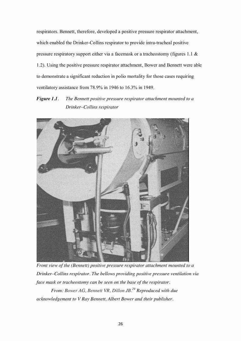
respirators. Bennett, therefore, developed a positive pressure respirator attachment,
which enabled the Drinker-Collins respirator to provide intra-tracheal positive
pressure respiratory support either via a facemask or a tracheostomy (figures 1.1 &
1.2). Using the positive pressure respirator attachment, Bower and Bennett were able
to demonstrate a significant reduction in polio mortality for those cases requiring
ventilatory assistance from 78.9% in 1946 to 16.3% in 1949.
Figure 1.1. The Bennett positive pressure respirator attachment mounted to a
Drinker–Collins respirator
Front view of the (Bennett) positive pressure respirator attachment mounted to a
Drinker–Collins respirator. The bellows providing positive pressure ventilation via
face mask or tracheostomy can be seen on the base of the respirator.
From: Bower AG, Bennett VR, Dillon JB.29 Reproduced with due
acknowledgement to V Ray Bennett, Albert Bower and their publisher.
27
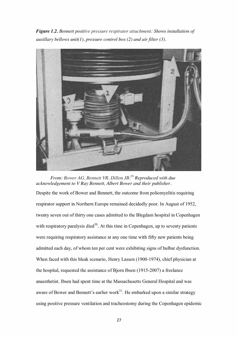
Figure 1.2. Bennett positive pressure respirator attachment: Shows installation of
auxillary bellows unit(1), pressure control box (2) and air filter (3).
From: Bower AG, Bennett VR, Dillon JB.29 Reproduced with due acknowledgement to V Ray Bennett, Albert Bower and their publisher.
Despite the work of Bower and Bennett, the outcome from poliomyelitis requiring
respirator support in Northern Europe remained decidedly poor. In August of 1952,
twenty seven out of thirty one cases admitted to the Blegdam hospital in Copenhagen
with respiratory paralysis died30. At this time in Copenhagen, up to seventy patients
were requiring respiratory assistance at any one time with fifty new patients being
admitted each day, of whom ten per cent were exhibiting signs of bulbar dysfunction.
When faced with this bleak scenario, Henry Lassen (1900-1974), chief physician at
the hospital, requested the assistance of Bjorn Ibsen (1915-2007) a freelance
anaesthetist. Ibsen had spent time at the Massachusetts General Hospital and was
aware of Bower and Bennett’s earlier work31. He embarked upon a similar strategy
using positive pressure ventilation and tracheostomy during the Copenhagen epidemic
28
of 1952. From 26th August, ultimately using a team of thirty five to forty medical
staff, 600 trained nurses and 250 medical students working in relays, 321 patients
with respiratory insufficiency were treated. Of these 265 patients had a tracheostomy
and 232 had positive pressure ventilation. Over this period the mortality reduced from
eighty seven per cent in July / August to twenty two per cent in November /
December30. This episode is now felt to be the beginning of modern intensive care
practice. Ibsen was later offered a position at Copenhagen’s Kommunehospital where
he set up what is widely regarded as the world’s first dedicated intensive care unit in
195331. Lassen and Ibsen also provided an insight in to the future problems of critical
care with their description of an increased mortality in those patients presenting with
shock, renal failure and pulmonary oedema.
1.6 Percutaneous tracheostomy
The Italian surgeon Sanctorio Sanctorious (1561-1636), using a technique described
and condemned by Trousseau7 as the expeditious method, has been suggested as the
first surgeon to have described a percutaneous technique in 1626. Although the
procedure described used a small dagger-like ripping needle and a silver perforated
cannula it is doubtful that it was ever performed by Sanctorious32. These same
instruments were also used at this time for the tapping of hydrocoeles and ascites.
Using the needle, the cannula was inserted in to the tracheal lumen and the needle was
withdrawn in a manner that appears remarkably similar to Shelden’s description in
1957 (see below)33. Although similar methods were later described by Dekker and
Heister it is unclear how often a percutaneous technique was used by such luminaries
if at all7. The French physician, Bauchot, has been credited as the first to perform a
percutaneous procedure in the mid eighteenth century using a bronchotome (with a
cutting edge) fitting in to the lumen of a flattened silver cannula32. Bauchot reportedly
29
used this technique successfully on two patients12. Gerard van Swieten (1700-1772)
of Leyden Austria, who had used a percutaneous technique on a large number of live
animals and human cadavers, considered the procedure to be extremely difficult and
not without risk7. Subsequent to this, largely because of Trousseau’s recommendation
for a large incision and Jackson8 standardising the open surgical tracheostomy, little
more was heard of the percutaneous procedure until the 1950s.
Arguably the first step towards the safe performance of a percutaneous tracheostomy
was not related to airway surgery at all. In 1953 Sven Seldinger (1921-1998), a
Swedish radiologist, published his technique of catheter insertion over a guidewire34.
In the 1940s interest in arteriography was increasing although catheter insertion to
facilitate this was problematic – usually requiring surgical exposure of the relevant
artery. With the advent of flexible guide-wires and polyethylene catheters Seldinger
was able to demonstrate the use of percutaneous puncture of a vessel followed by
guide-wire and then catheter insertion over the wire. He described this technique in
forty procedures all performed under local anaesthetic. In thirty-seven out of the forty
procedures arterial puncture and catheter insertion were achieved at the first attempt
with no significant haemorrhagic complications.
The first modern description of a percutaneous tracheostomy was described by
Shelden in 195535. Despite this being after Seldinger’s description of his guide-wire
technique Shelden did not employ it. Shelden described the use of a slotted needle and
a cutting blade with a ball like tip (figure 1.3). The needle was inserted into the
trachea below the cricoid cartilage. The ball like tip of the cutting blade was inserted
into the spherical slot on the needle and the ball passed down the needle lumen in to
the trachea. With the cutting blade in-situ the needle is removed, the cutting blades
and attached tracheostomy tube are then advanced in to the trachea. Once in place the
30
cutting blades were removed and the tracheostomy tube left in situ. Shelden had used
this device for four years at the time of a later publication and claimed use by many
other neurosurgeons throughout the world33. Despite this it is unclear how many
patients underwent this procedure. Even though the device was commercially
available and used extensively by Shelden it appears not be have been widely adopted
perhaps in part due to reported posterior tracheal wall and oesophageal
perforation36,37.
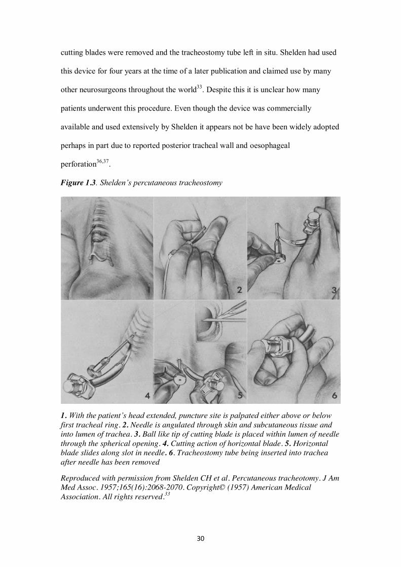
Figure 1.3. Shelden’s percutaneous tracheostomy
1. With the patient’s head extended, puncture site is palpated either above or below first tracheal ring. 2. Needle is angulated through skin and subcutaneous tissue and into lumen of trachea. 3. Ball like tip of cutting blade is placed within lumen of needle through the spherical opening. 4. Cutting action of horizontal blade. 5. Horizontal blade slides along slot in needle. 6. Tracheostomy tube being inserted into trachea after needle has been removed
Reproduced with permission from Shelden CH et al. Percutaneous tracheotomy. J Am Med Assoc. 1957;165(16):2068-2070. Copyright© (1957) American Medical Association. All rights reserved.33

31
The first description of a bedside surgical tracheostomy was provided by Roe in 1962
and suggested to be of use predominantly in the emergency setting37. In a series of
forty patients with post-operative respiratory compromise he described what might
now be referred to as a minimally invasive tracheostomy. Using a small incision, that
admitted the tip of the finger, followed by blunt dissection of the subcutaneous tissues
the cricoid and tracheal cartilages were initially palpated. Complete exposure of the
tracheal cartilage was felt to be unnecessary. The procedure had to be abandoned in
only one patient.
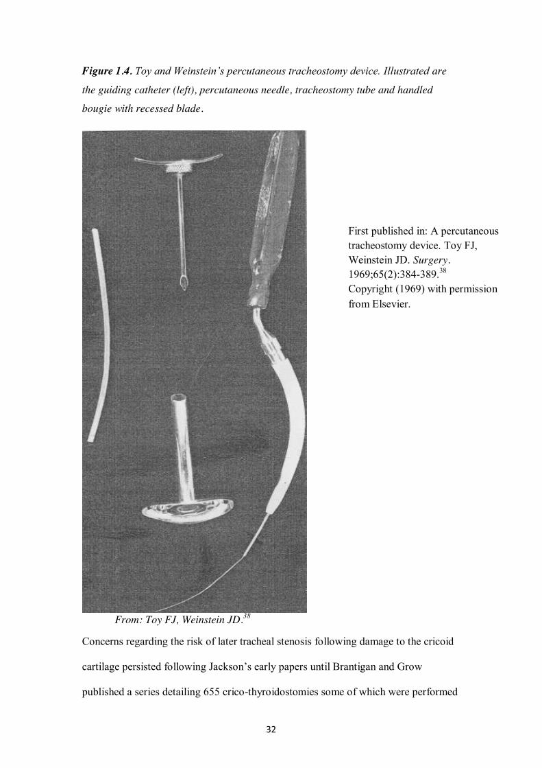
In 1969 Toy and Weinstein described the first guide assisted percutaneous
tracheostomy using a modified Seldinger technique38. They described the insertion of
a five-millimetre tracheostomy tube over a guiding bougie that had been placed via a
needle inserted in to the trachea. Using a handled bougie (figure 1.4) with recessed
blade the entire device was thrust in to the trachea. Of the six patients they described,
five had successful tracheostomy tube insertion and one had a para-tracheal
placement. Despite the potential for use within a critical care setting the authors did
not envisage this and suggested its principal uses would be in the emergency setting
and elective cases where a bleeding diathesis may be present or a concern for the
cosmetic results was present.
32
Figure 1.4. Toy and Weinstein’s percutaneous tracheostomy device. Illustrated are
the guiding catheter (left), percutaneous needle, tracheostomy tube and handled
bougie with recessed blade.
From: Toy FJ, Weinstein JD.38
Concerns regarding the risk of later tracheal stenosis following damage to the cricoid
cartilage persisted following Jackson’s early papers until Brantigan and Grow
published a series detailing 655 crico-thyroidostomies some of which were performed
First published in: A percutaneous tracheostomy device. Toy FJ, Weinstein JD. Surgery. 1969;65(2):384-389.38 Copyright (1969) with permission from Elsevier.
33
at the bedside39. Of these patients eight developed sub-glottic stenosis and seven had
sub-glottic granulomas. Pre-existing laryngeal injury was felt to be present in thirteen
of these fifteen patients due to prolonged trans-laryngeal intubation. The authors
therefore concluded that there was little risk of tracheal stenosis following
cricothyroidostomy if the larynx was normal but that it should not be performed after
prolonged trans-laryngeal intubation.
The first description of a dilatational percutaneous tracheostomy was provided by the
thoracic surgeon Pascquale Ciaglia (1912-2000) working in St. Elizabeth Hospital and
St. Luke’s Memorial Hospital Center, Utica, New York40. Following on from
Brantigan’s work39 and the minimally invasive bedside surgical tracheostomy
described by Roe37 Ciaglia set about reforming the critical care tracheostomy
procedure. Initially he undertook twenty-six fingertip sub-cricoid tracheostomies
during which a small incision was made to allow the index finger to palpate the
cricoid cartilage. A small incision was made through the crico-tracheal membrane and
the withdrawal of the tracheal tube was also palpated. As the trans-laryngeal tube was
withdrawn the tracheostomy tube was inserted. After twenty-six procedures Ciaglia
concluded the whole process could be accomplished percutaneously. Using a
modified percutaneous nephrostomy set he then performed a further twenty-six
tracheostomies (one cricothyroidostomy, one through the second to third tracheal
rings and twenty four sub-cricoid) with no significant intra-operative complications
other than one difficult dilatation in a patient who had previously had a surgical
tracheostomy. There were no instances of para-tracheal placement, pneumothorax,
subcutaneous emphysema or oesophageal injury. There was one case of sub-glottic
stenosis that was felt to be related to prolonged trans-laryngeal intubation. Ciaglia
subsequently reported the outcome of fifty two decannulated patients who had
34
undergone his dilatational tracheostomy in 199241. There were mild voice changes in
one patient, one patient had a stomal infection and there no recorded cases of tracheal
stenosis or cosmetic problems. Despite this apparent lack of tracheal stenosis reported
at this point Ciaglia suggested in this paper that a lower insertion point might be
preferable if feasible. In 1994, McFarlane reported on a series of 121 Ciaglia
tracheostomies in which there were four cases of sub-glottic stenosis10. This was
postulated to be due to the high placement of the tracheostomy tube and subsequent
damage to the cricoid cartilage with its resultant peri-chondritis and necrosis, a
finding in keeping with Jackson’s earlier assertions9. From this time onwards, most
operators have attempted to avoid crico-tracheal placement aiming for a lower
insertion point.
Subsequent to Ciaglia’s report, further modifications of the percutaneous dilatational
technique were proposed and a number of varying approaches have been developed
over the ensuing years. In 1990 William “Bill” Griggs (Adelaide, Australia) described
the use of guide-wire dilating forceps42, Antonio Fantoni (Milan, Italy) described the
trans-laryngeal tracheostomy in adults in 199643 and the single tapered dilator
modification of Ciaglia’s original procedure was reported in 200044. At the present
time, the single tapered dilator appears to be the most frequently used percutaneous
technique45,46. Subsequently lesser-used techniques have also been described47,48.
1.7 Modifications to the percutaneous tracheostomy technique
In 1989 Andreas Paul (Montreal, Canada) described the first percutaneous endoscopic
dilatational tracheostomy49. After initial testing in five anaesthetised dogs (including
post mortem examination of tracheal damage) a technique using the fibre-optic
bronchoscope to visualise the tracheal lumen during the procedure was described in
four human subjects. In one patient they identified a guide-wire misplacement thus
35
preventing para-tracheal placement. They also postulated that bronchoscopic
deployment would reduce posterior tracheal wall and oesophageal injuries whilst
allowing precise tracheostomy tube placement. He also hinted at a possible future use
of ultra-sound scanning for percutaneous tracheostomy to reduce bleeding
complications.
This latter idea was later taken up by Hatfield and Bodenham (Leeds, UK) who
undertook a study of thirty patients undergoing percutaneous tracheostomy50. In all
patients, the thyroid cartilage, carotid arteries and internal jugular veins were easily
identified, as was the mid-line and approximate level of puncture. They identified
eight patients in whom an anterior jugular vein was considered vulnerable (at or near
the mid-line). Two patients had vessels ligated and two had minor bleeding episodes.
Additionally, four patients had vulnerable arterial structures (two carotid and two
brachio-cephalic arteries). The authors concluded use of ultra-sound scanning may
reduce bleeding complications especially in patients with difficult to identify surface
anatomy.
1.8 Tracheostomy outcomes
Despite widespread adoption of percutaneous techniques from the late 1980s onwards
the outcomes, particularly long term ones, when compared to surgical tracheostomy
and amongst the percutaneous techniques themselves were the subject of some
discussion and debate.
1.8.1 Percutaneous tracheostomy versus surgical tracheostomy
The outcomes of percutaneous techniques compared to surgical tracheostomy have
been the subject of three systematic reviews dating from 2006 – 2007. In the first such
36
review, Delaney evaluated randomised controlled trials comparing surgical
tracheostomies with any percutaneous technique in the critical care setting51. He
identified seventeen trials, including 1212 patients, the commonest percutaneous
procedure evaluated being the original Ciaglia technique40. The principal findings
were equivalence for bleeding, major short and long-term complications with a
significant reduction in stomal infections for the percutaneous procedures. Oliver later
identified fourteen prospective trials (of which eight were randomised controlled
trials) comparing surgical tracheostomy with a percutaneous technique performed in
the critical care unit or the operating theatre52. They found no difference in major
complications but a greater incidence of minor complications with percutaneous
techniques along with a greater incidence of early complications for percutaneous
techniques when compared to surgical tracheostomy performed at the bedside.
Higgins53 assessed fifteen randomised controlled trials (incorporating 973 patients) all
but two of which were incorporated in Delaney’s review51. They found percutaneous
techniques resulted in fewer wound infections and cosmetic problems with no
difference in major complications. When pooled complications were analysed they
found in favour of the percutaneous procedures.
1.8.2 Percutaneous versus percutaneous procedures
For many years there appears to be an assumption of equivalence across the
percutaneous techniques described. In an attempt to address this Cabrini undertook a
systematic review of randomised controlled trials comparing two or more
percutaneous techniques54. They identified thirteen trials, incorporating 1030 patients,
the most studied techniques being the original Ciaglia multiple dilator method, guide-
wire dilating forceps and the single tapered dilator. They found that the Ciaglia and
single tapered dilator techniques appeared to have the fewest complications. There
37
appeared to be an increase in minor complications with the guide-wire dilating
forceps along with higher failure rates for both trans-laryngeal and rotational dilator
techniques (PercuTwist®). They expressed some surprise at the paucity of randomised
controlled trials when considering how widespread the use of percutaneous
tracheostomy has become. Overall it was felt that the most reliable technique for
safety and success was the single tapered dilator. In a later review Cabrini also
assessed the complication rates of the two most commonly used percutaneous
techniques, the single tapered dilator and guide wire dilating forceps55. Having
identified five eligible randomised controlled trials comprising 363 patients they
concluded that the guide wire dilating forceps technique is associated with a higher
incidence of intra-procedural bleeding and technical difficulties when compared with
the single tapered dilator. There were no differences in mid and long-term outcomes.
After many centuries of evolving surgical approaches along with advances in our
knowledge of physiology and anatomy, percutaneous procedures have become
established as the predominant tracheostomy techniques within the critical care
setting. Complication rates across the various techniques appear to be at least as low
as those achieved with surgical tracheostomies. It is possible that the single tapered
dilator method is now the most frequently used procedure with the lowest associated
complication rate. However, the paucity of data when comparing percutaneous
procedures, particularly when considering long-term outcomes, is somewhat
surprising. It is clear, at present, that equivalence between procedures in this respect
has not been fully established.
The meta-analyses described above have included only randomised controlled trials
(RCTs)51,53. The only exception to this was the analysis by Oliver which also included
non-randomised prospective studies52. The largest single study incorporated into the
38
previous analyses comprised 346 patients56. It is perhaps unsurprising, therefore, that
none of the previous meta-analyses have reported differences in late complication
rates. The exact incidence of long term complications following tracheostomy
procedures in the critically ill is difficult to quantify due to the associated mortality of
critical illness, the sub-clinical nature of many tracheal stenoses and the difficulty
maintaining follow up of these cohorts. Given this associated morbidity and the cost
associated with the management of TS a clearer picture of the risk associated with
each tracheostomy technique performed within the critical care setting is required.
1.9 Aims
To determine the utility of adjunctive techniques (bronchoscopy & ultrasound
scanning) in reducing complications of percutaneous tracheostomies
percutaneous tracheostomy technique and hence determine the safest
percutaneous technique
To determine the incidence of common early and late complications and
outcomes of PDT techniques in relation to surgical tracheostomy.
To determine the relative indices of early and late complications in relation to
percutaneous tracheostomy technique and hence determine the safest
percutaneous technique
To determine the role of early complications that may be postulated to play a
part in the genesis of the late complications of tracheal stenosis, tracheo-
innominate artery fistula and tracheo-oesophageal fistula
To determine long term survival following percutaneous tracheostomy
39
Chapter 2
Major late complications following tracheostomy
40
Both surgical and percutaneous tracheostomies are associated with a number of well-
defined early complications that may occur intra-operatively or over a variable length
of time in to the post-operative period57-59. The most common of these early
complications are haemorrhage, malpositioning of the tracheal cannula, displacement
of the tracheal tube, pneumothoraces and subcutaneous emphysema. Assessing the
relative frequency for each of these outcomes following surgical and percutaneous
techniques has been relatively well defined and the subject of a number of meta-
analyses51-55.
Late complications leading to significant morbidity and potentially mortality (tracheo-
innominate artery fistulae, tracheomalacia and tracheal stenosis) may occur after the
tracheal cannula has been removed. Given the severity of illness occurring in many of
these patients with its associated mortality, the relative incidences of each of these
complications remains difficult to define. Additionally, confounding factors present in
survivors of critical illness may present additional difficulties in diagnosis. Many
survivors will have significant residual muscle weakness, associated with underlying
parenchymal lung diseases either predating their critical care stay or as a result of it.
Consequently, a reduction in exercise capacity and shortness of breath are commonly
found in this population. Such complaints can often be attributed to the after effects of
critical illness and underlying tracheal pathology may go undiagnosed.
2.1 The normal trachea
2.1.1 Tracheal anatomy
The trachea lies in the midline of the neck and upper mediastinum. It begins at the
lower border of the cricoid cartilage at the level of the sixth cervical vertebra and
extends to its bifurcation in to right and left main bronchi at the carina. It is composed
41
of C shaped rings of cartilage that form the anterior and lateral walls and a musculo-
membranous posterior wall. The smooth muscle in the musculo-membranous portion
of the tracheal wall contains both transverse and longitudinal fibres. The transverse
fibres make up the trachealis muscle connecting the ends of the tracheal cartilages.
Tracheal size is related to the size of the individual but in the adult male there are
approximately 18-22 tracheal rings extending inferiorly from the cricoid to carina for
11-12 cm. The lateral diameter is approximately 2.3 cm coronally and 1.8 cm
sagitally60,61.
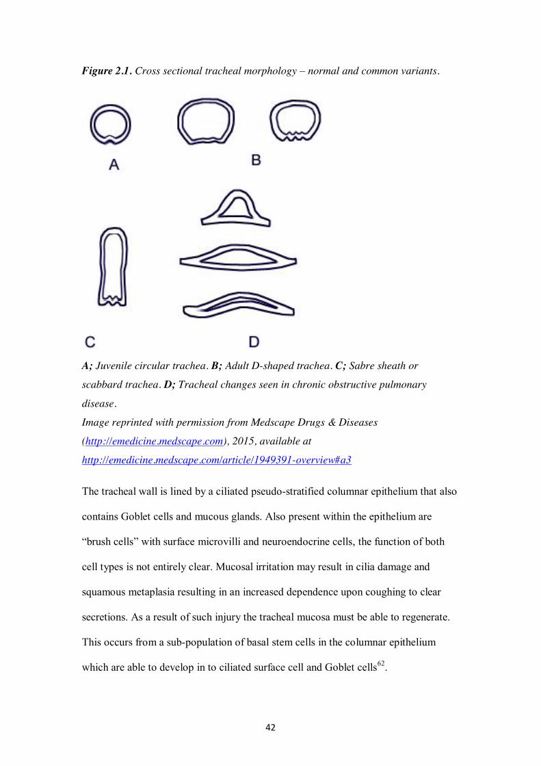
The shape of the tracheal lumen varies with age and in the presence or absence of
disease states. The lumen in the child is almost circular reaching its usual adult D
shape at adolescence (figure 2.1). Luminal shape also alters dynamically in response
to changes in intra-luminal pressure during respiration, coughing and mechanical
ventilation. During forced expiration the trachealis muscle approximates the ends of
the tracheal cartilages creating an elongated antero-posterior tracheal diameter. In the
presence of ageing or significant chronic obstructive pulmonary disease, this
reduction in lateral diameter may result in a sabre sheath or scabbard trachea. Chronic
obstructive airways disease may also result in a softening of the tracheal cartilage and
a widening of the lateral diameter and an antero-posterior narrowing (figure 2.1). This
conformational change may ultimately lead to luminal obstruction during coughing
and active expiration.
42
Figure 2.1. Cross sectional tracheal morphology – normal and common variants.
A; Juvenile circular trachea. B; Adult D-shaped trachea. C; Sabre sheath or
scabbard trachea. D; Tracheal changes seen in chronic obstructive pulmonary
disease.
Image reprinted with permission from Medscape Drugs & Diseases
(http://emedicine.medscape.com), 2015, available at
http://emedicine.medscape.com/article/1949391-overview#a3
The tracheal wall is lined by a ciliated pseudo-stratified columnar epithelium that also
contains Goblet cells and mucous glands. Also present within the epithelium are
“brush cells” with surface microvilli and neuroendocrine cells, the function of both
cell types is not entirely clear. Mucosal irritation may result in cilia damage and
squamous metaplasia resulting in an increased dependence upon coughing to clear
secretions. As a result of such injury the tracheal mucosa must be able to regenerate.
This occurs from a sub-population of basal stem cells in the columnar epithelium
which are able to develop in to ciliated surface cell and Goblet cells62.
43
The trachea receives its blood supply segmentally via the lateral walls. The cervical
trachea is supplied predominantly from the inferior thyroid artery most frequently via
three tracheo-oesophageal vessels. The mid to lower trachea receives its arterial
supply from the superior, middle and inferior bronchial arteries. At the segmental
level each artery to the trachea will branch superiorly and inferiorly over several
tracheal rings forming a series of longitudinal anastomoses. Additionally anterior and
posterior braches will run in the inter-cartilaginous space to eventually anastomose
with contralateral vessels. The posterior vessels also anastomose with oesophageal
arteries60,61.
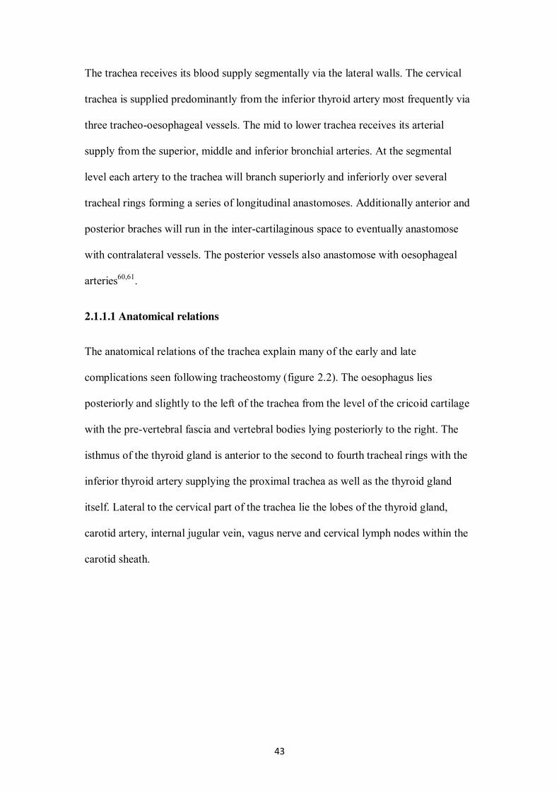
2.1.1.1 Anatomical relations
The anatomical relations of the trachea explain many of the early and late
complications seen following tracheostomy (figure 2.2). The oesophagus lies
posteriorly and slightly to the left of the trachea from the level of the cricoid cartilage
with the pre-vertebral fascia and vertebral bodies lying posteriorly to the right. The
isthmus of the thyroid gland is anterior to the second to fourth tracheal rings with the
inferior thyroid artery supplying the proximal trachea as well as the thyroid gland
itself. Lateral to the cervical part of the trachea lie the lobes of the thyroid gland,
carotid artery, internal jugular vein, vagus nerve and cervical lymph nodes within the
carotid sheath.
44
Figure 2.2. Anatomical relations of the trachea including major vessels, thyroid and nerves. Modified from: Deslauriers.61
Reprinted from Thoracic Surgery Clinics 2007;17(4):529-547. Deslauriers J. Anatomy of the neck and cervicothoracic junction. Copyright (2007), with permission from Elsevier.
45
As the trachea descends in to the superior mediastinum anteriorly lies the thymus, the
brachio-cephalic (innominate) artery and vein and the aortic arch. The tracheo-
innominate artery arises as the first branch of the aortic arch. It ascends obliquely and
posteriorly to cross the trachea at the level of the ninth tracheal cartilage (range from
sixth to thirteenth). There is however, a degree of variability in the course of the
tracheo-innominate artery with passage across the trachea being possible as high as
the second – third tracheal ring63. Posteriorly lie the oesophagus, pre-vertebral fascia
and recurrent laryngeal nerves in the tracheo-oesophageal grooves. Laterally lie the
vagus and phrenic nerves, the superior vena cava on the right, left common carotid
and left subclavian vessels with the thoracic duct on the left and azygous vein on the
right.
2.1.2 Tracheal physiology
The trachea is designed to:
Conduct air from the larynx to the main bronchi
Exchange heat and moisture with cold, dry inhaled air
Remove particulate debris and secretions from the tracheo-bronchial tract.
2.1.2.1 Movement of air
Under normal circumstances for gas to flow through the trachea a pressure gradient
has to exist to overcome the resistance of the respiratory system. Conventionally,
laminar flow through tubes is represented by the Hagen-Poiseuille Law where flow is
inversely proportional to the fourth power of the radius of the tube:
ΔP = Q8Lμ/πr4
Where:

46
ΔP = pressure change across tube
Q = flow rate
L = length of the tube
μ = gas viscosity
r = radius of the tube.
However, when considering the Reynolds number (ratio of inertial forces to viscous
forces) for flow through a given tube:
Re = QL/ μ
Where:
Q = flow rate
L= length of tube
= density
μ = viscosity
The transition to turbulent flow occurs at a Reynolds number of around 2300.
Thereafter, flow becomes completely turbulent when the Reynolds number exceeds
4000 – conditions found within the trachea. In this situation resistance across the
trachea becomes directly proportional to the gas flow rate squared and inversely
proportional to the fifth power of the radius64.
ΔP = Q2fL/rπ2r5
Where:
= density of gas
47
f = frictional factor
Consequently, a reduction in airway calibre produced by mucosal swelling,
endoluminal tumours, tracheal stenosis and tracheal tubes will all considerably
increase resistance to airflow.
2.1.2.2 Heat and moisture exchange
During inspiration the upper airways warm and humidify the inspired gas. During
quiet breathing, air is completely warmed to 370 centigrade and fully humidified at
around the level of the tracheal bifurcation – the isothermal saturation point62. The
drier and colder the inspired air the more distal this point becomes thus increasing
heat and moisture losses. This situation is further exacerbated when the upper airway
is bypassed with a tracheal or tracheostomy tube where, in addition to heat and
moisture loss, mucosal injury may result unless inspired gases are heated and
humidified.
2.1.2.3 Removal of particulate debris
Tracheo-bronchial Goblet cells produce a mucin rich secretion that protects the
underlying epithelium. The rate and volume of secretions produced is controlled by
the autonomic nervous system and inflammatory mediators. The resultant mucous
collects debris and micro-organisms and is transported in a cephalad direction by
cilial action and exhaled air with expiratory airflow becoming more prominent in the
larger central airways.
48
2.2 Tracheal pathology
2.2.1 Tracheal stenosis
Benign tracheal stenosis (TS) is a progressive narrowing of the tracheal lumen most
commonly seen after tracheal intubation and tracheostomy insertion. Pooled data from
a number of studies has shown reported incidences of TS of 0.6 – 2.8% (See Chapter
4). Such data, should however, be interpreted with caution. The reported rates are
invariably expressed as percentages of tracheostomies performed and not of survivors
who have presented for follow up58,59.
2.2.1.1 Histopathology
Despite a plethora of studies investigating the management of TS the actual
mechanisms involved in its initiation remain unclear. It is postulated that hyper-
granulation, cuff induced ischaemic mucosal injury, direct tracheal wall injury,
infection, gastro-oesophageal reflux and a genetic predisposition may all play a role65-
69. Why this reaction presents in only a small subset of patients undergoing intubation
and tracheostomy remains unknown.
Of the above factors, tracheal mucosal pressure induced necrosis seems to be of prime
importance. If the pressure within the cuff of the tracheal tube is above that of the
mucosal capillary perfusion pressure the potential result is ischaemic ulceration. As
the blood supply of the trachea is segmental with vessels perforating the tracheal wall
through the inter-ring spaces pressure on the mucosa can lead to cartilaginous
ischaemia and necrosis. Additional hypoperfusion associated with systemic
hypotensive states is not uncommon in the critically ill.
Normal wound healing progresses through three phases70:
49
c) Inflammatory phase
Tissue trauma leads to bleeding and clot formation with the release of
inflammatory mediators (prostaglandins, interleukin-1 (IL-1), interleukin-6
(IL-6), tumour necrosis factor α and transforming growth factor β (TGF-β))
that increase vessel permeability enabling the migration and accumulation of
inflammatory macrophages.
d) Proliferative phase
This phase is characterised by re-epithelialisation and new vessel formation
along with fibroblast migration. These processes are largely under the control
of platelet-derived growth factor, endothelial derived growth factor, IL-1, IL-
6, TGF-β and fibroblast growth factor. Along with fibroblasts there is also
proliferation of macrophages, keratinocytes and endothelial cells. New
connective tissue is laid down (granulation tissue)
e) Maturation phase
This phase is predominantly controlled by epidermal growth factor and TGF-
β. As the new connective tissue matures, collagen becomes the main
component.
A number of authors66,69,71 have postulated the role of infection in generating the
abnormal tissue reaction to trauma. Bacterial contamination leads to an infected peri-
chondritis and ultimately enhanced fibroblast activity. Whilst the incidence of stomal
infection following surgical tracheostomy has been shown to be higher than that
associated with percutaneous tracheostomy51 there is no definitive proof that the
incidence of TS differs between the two techniques. Furthermore, there are no studies
that demonstrate a protective effect of antimicrobials in the prevention of TS.
50
The host inflammatory reaction to this mucosal ulceration leads to healing by
secondary intention. An inflammatory cascade is initiated that results in the up-
regulation of TGF-β which promotes the growth of fibroblasts70. It is also possible
that fibroblasts may be activated as a result of tissue hypoxia leading to expression of
hypoxia inducible factor with a resultant proliferation of fibroblasts and
myofibroblasts within the tracheal mucosa72. The end result is a proliferation of
granulation tissue and subsequent fibrous scar tissue. In some patients the
proliferation of granulation tissue may lead to difficulties with decannulation although
progression to tracheal stenosis is not always seen73. The abnormal healing process
responsible for tracheal stenosis results in an imbalance of cell types leading to an
excess of scarring and granulation tissue. The fibroblasts present release collagen and
an extra-cellular matrix forms, containing types 1 and 3 collagen fibres, fibronectin
and a significantly reduced number of elastic fibres, obstructing the normal patent
airway70,72. The increase of collagen over elastic tissue makes the scarred segment
rigid and relatively avascular. The mature tracheal stenosis eventually forms within
three to six weeks of decannulation. The later accumulation of submucosal fibrous
tissue can lead to contraction, reduction and distortion of the tracheal lumen. The
abnormal inflammatory process is not usually confined to the tracheal mucosa but
also involves the deeper tracheal structures such as the peri-chondrium and
cartilage69. Tracheal patency in the presence of weakened or fractured tracheal
cartilages becomes compromised, particularly during forceful respiration and
coughing, leading to a dynamic central airway collapse.
There may be a number of additional co-factors associated with this abnormal
inflammatory cascade. A number of authors have noted a preponderance of female
patients with benign tracheal stenosis74,75 but this is by no means uniform, with other
51
studies reporting a higher incidence in males69,71. Idiopathic tracheal stenosis is also
more frequently seen in female patients76. It has been postulated that oestrogen has an
effect whereby it increases levels of TGF-β increasing collagen production and extra-
cellular matrix formation. Additionally, diabetes mellitus77, obesity and cardio-
vascular disease have been noted in up to 30% of TS patients75. It is possible that
small vessel disease present in such patients may worsen the effects of regional
ischaemia induced by the cuff of the tracheal tube. There may also be an effect of
tracheo-oesophageal reflux in promoting abnormal healing both in idiopathic and
post-intubation stenoses78.
2.2.1.2 Types of tracheal stenosis
A number of variants of normal tracheal morphology occur that must be excluded
when the diagnosis of TS is being considered. In addition to the usual adult D shaped
lumen a juvenile circular appearance may persist into adult life. A sabre sheath or
scabbard trachea may be seen in the elderly with lateral narrowing and anterior-
posterior widening and can be defined as a transverse / sagittal diameter <0.6. In
patients with chronic obstructive pulmonary disease a number of states of dynamic
tracheal collapse may occur without overt stenosis (figure 2.1).
TS can be simply defined as an abnormal narrowing of the tracheal cross sectional
lumen. A degree of stenosis is common after all tracheostomies but intervention for
symptomatic disease is only required in a minority. A number of studies investigating
the incidence of sub-clinical tracheal stenosis after percutaneous tracheostomy have
used a reduction in tracheal cross sectional area (TCSA) of 10 per cent narrowing as
a definition and found incidences in the region of 10-30%79,80. Symptomatic TS is
unlikely with reductions in TCSA of 50% with severe symptoms being present with
52
reductions in excess of 75%81.
Tracheal stenosis may be grouped according to a number of defining characteristics
related to the underlying disease process.
a) Benign / Malignant
Benign TS may present following a number of tracheal injuries and diseases.
The most frequent cause being post-intubation or post-tracheostomy discussed
herein. It may also follow from direct tracheal trauma, chemical irritation,
granulomatosis with polyangiitis (Wegener’s) and rarely be idiopathic.
Malignant stenoses result from direct infiltration of the trachea from tumours
of the oesophagus, thyroid and lung or by malignant lymph node compression.
Primary tracheal malignancies are unusual.
b) Functional behaviour
A stenosis can be further defined by its functional behaviour82. Most
commonly seen is a fixed stenotic, scarring stricture in relation to the original
trauma. Less commonly a malacic segment may follow on from the initial
injury and result in a functional stenosis with dynamic airway collapse during
the respiratory cycle. Such “dynamic stenoses” are perhaps most commonly
seen in the patient with chronic obstructive pulmonary disease.
c) Anatomical features of stenosis
Further definition of a stenotic segment is given via an anatomical description
of its nature. Lesions may be long or short, stomal or cuff related or subglottic
(see below)82.
53
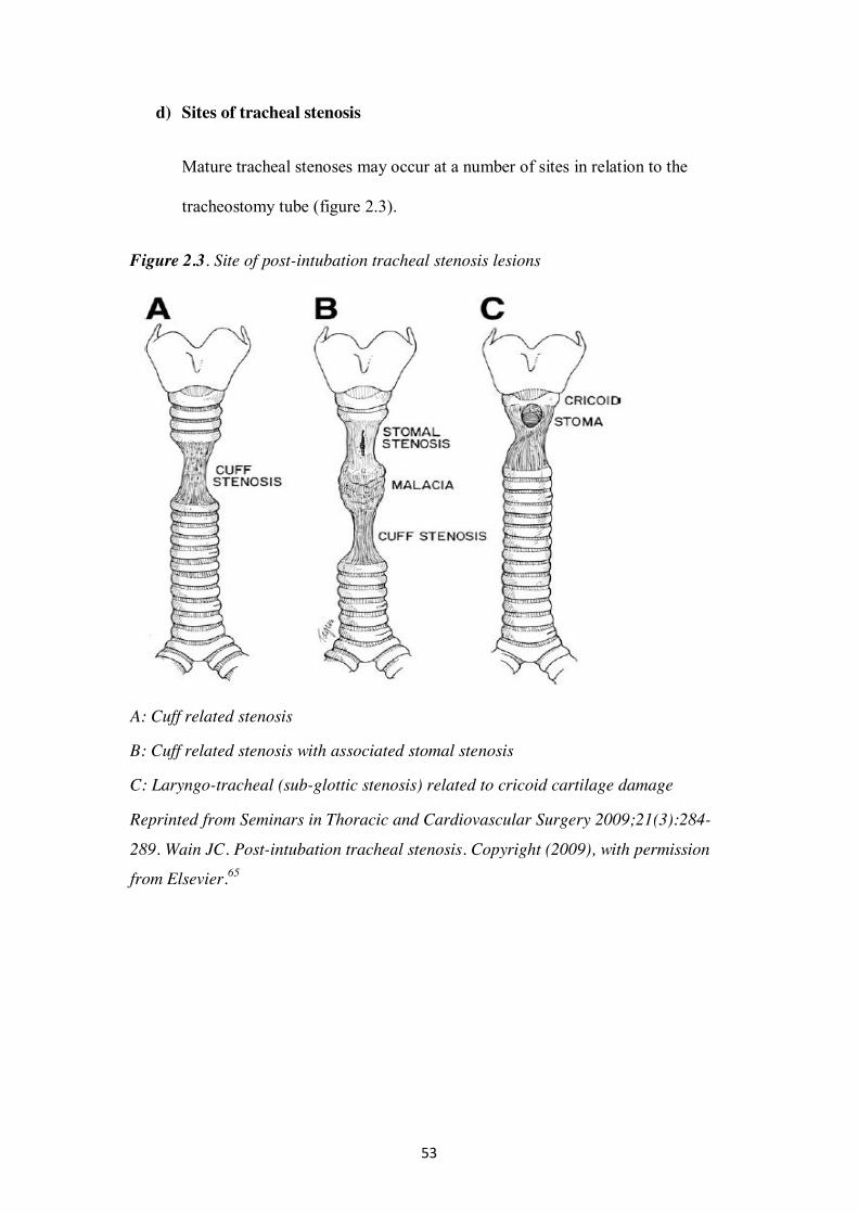
d) Sites of tracheal stenosis
Mature tracheal stenoses may occur at a number of sites in relation to the
tracheostomy tube (figure 2.3).
Figure 2.3. Site of post-intubation tracheal stenosis lesions
A: Cuff related stenosis
B: Cuff related stenosis with associated stomal stenosis
C: Laryngo-tracheal (sub-glottic stenosis) related to cricoid cartilage damage
Reprinted from Seminars in Thoracic and Cardiovascular Surgery 2009;21(3):284-
289. Wain JC. Post-intubation tracheal stenosis. Copyright (2009), with permission
from Elsevier.65
54
a) Stomal stenosis
Most commonly TS develops at the site of the tracheal stoma. Epstein73
considered that secondary bacterial infection develops in the traumatised
trachea to weaken the anterior and lateral walls.
b) Cuff related stenosis
Stenosis can develop in the region of the tracheostomy tube cuff in response to
ischaemic necrosis of the tracheal wall when the pressure within the cuff
exceeds the mucosal capillary perfusion pressure. A number of animal67,68 and
human studies83 have detailed the effects of cuff pressure on capillary mucosal
perfusion. At around 22mmHg, there is impairment of mucosal blood flow.
Above 37mmHg, there is total obstruction of blood flow to the mucosa
overlying the tracheal rings and the stretched posterior muscular wall.
Pressures over 50 mmHg for 15 minutes can cause epithelial destruction
which can lead to tracheitis, ulceration, persistent inflammation, chondritis,
fibrosis and stenosis83.
Cuff related problems associated with TS were first identified following the
poliomyelitis epidemics in the late 1940s and 50s. Between 1947 and 1959
there was a fifteen fold increase in tracheostomies performed at the
Massachusetts General Hospital84. In 462 reported cases there were two
tracheal stenoses. Reports of TS rates in the late 1960s were in the region of
20% in some studies 85,86 although up to 50% of these patients were
asymptomatic. At this time the importance of the tracheostomy tube cuff and
associated circuitry were being appreciated in the aetiology of stenosis.
Andrews demonstrated a reduction in stenosis from 17.5% to 6.9% with the
introduction of a lightweight ventilator circuit85. They attributed the reduction
55
to diminished pressure on the lateral walls of the trachea thus preventing
stomal pressure necrosis.
The first report of a high volume low pressure cuff of the tracheostomy tube
was presented by Grillo87. In a randomised trial they detailed the management
of 45 patients who had undergone a tracheostomy in the critical care setting
comparing those with the new cuff versus with a standard rubber one. Follow
up of the patients was by fibre-optic bronchoscopy of survivors or post
mortem examination of the trachea in non-survivors. Rating tracheal damage
on an objective scale from 0-4 (increasing score indicating increasing mucosal
damage) they found significantly less tracheal wall damage with the new cuff.
Average intra-cuff pressures reported were 33mmHg for the low pressure and
270 mmHg for the standard rubber cuffs. Following Grillo’s work and the use
of high volume, low-pressure cuffs along with improvements in tracheostomy
care, ventilator circuitry and tube manufacture the incidence of TS following
tracheostomy has continued to decrease.
c) Subglottic stenosis
The first recognition that stenoses after percutaneous procedures may be
different to those following surgical tracheostomy was highlighted by
McFarlane10. They described four cases of supra-stomal subglottic stenosis
following the original Ciaglia multiple dilator technique. They postulated that
damage to the cricoid cartilage was the important aetiological factor. Despite
work by Jackson9 illustrating the dangers of damage to the cricoid cartilage
early attempts to develop minimally invasive approaches to tracheostomy
predominantly used the space between the cricoid cartilage and first tracheal
ring39,40. When initial reports of TS were made in relation to these procedures
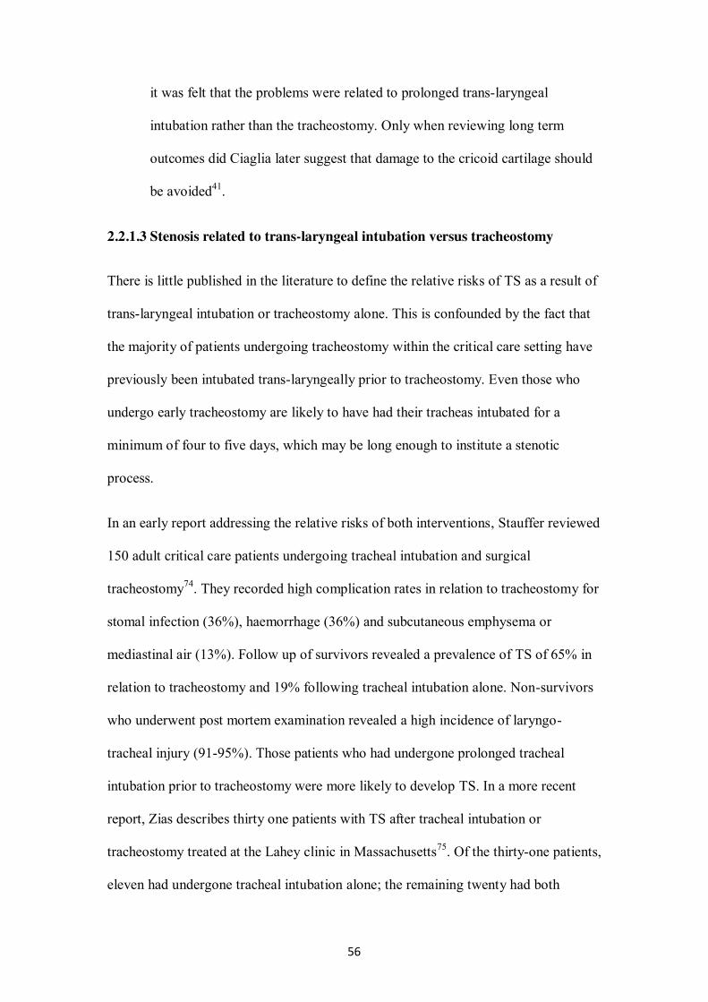
56
it was felt that the problems were related to prolonged trans-laryngeal
intubation rather than the tracheostomy. Only when reviewing long term
outcomes did Ciaglia later suggest that damage to the cricoid cartilage should
be avoided41.
2.2.1.3 Stenosis related to trans-laryngeal intubation versus tracheostomy
There is little published in the literature to define the relative risks of TS as a result of
trans-laryngeal intubation or tracheostomy alone. This is confounded by the fact that
the majority of patients undergoing tracheostomy within the critical care setting have
previously been intubated trans-laryngeally prior to tracheostomy. Even those who
undergo early tracheostomy are likely to have had their tracheas intubated for a
minimum of four to five days, which may be long enough to institute a stenotic
process.
In an early report addressing the relative risks of both interventions, Stauffer reviewed
150 adult critical care patients undergoing tracheal intubation and surgical
tracheostomy74. They recorded high complication rates in relation to tracheostomy for
stomal infection (36%), haemorrhage (36%) and subcutaneous emphysema or
mediastinal air (13%). Follow up of survivors revealed a prevalence of TS of 65% in
relation to tracheostomy and 19% following tracheal intubation alone. Non-survivors
who underwent post mortem examination revealed a high incidence of laryngo-
tracheal injury (91-95%). Those patients who had undergone prolonged tracheal
intubation prior to tracheostomy were more likely to develop TS. In a more recent
report, Zias describes thirty one patients with TS after tracheal intubation or
tracheostomy treated at the Lahey clinic in Massachusetts75. Of the thirty-one patients,
eleven had undergone tracheal intubation alone; the remaining twenty had both
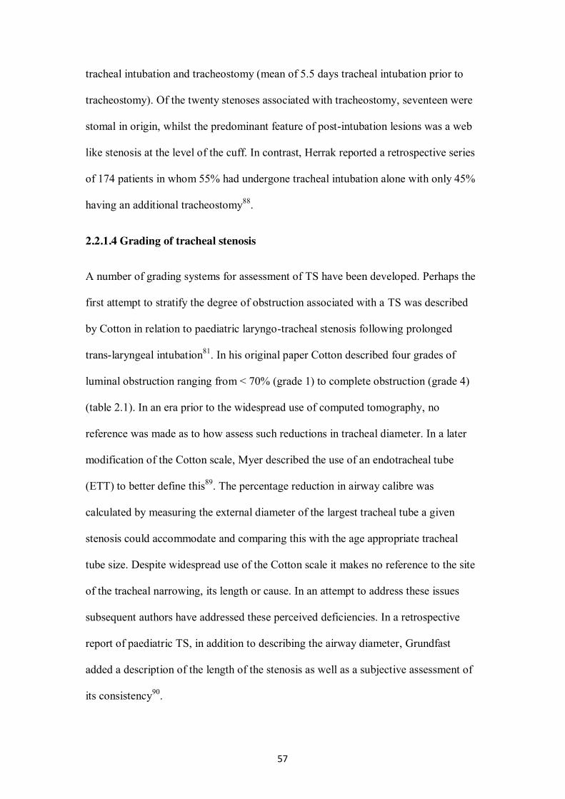
57
tracheal intubation and tracheostomy (mean of 5.5 days tracheal intubation prior to
tracheostomy). Of the twenty stenoses associated with tracheostomy, seventeen were
stomal in origin, whilst the predominant feature of post-intubation lesions was a web
like stenosis at the level of the cuff. In contrast, Herrak reported a retrospective series
of 174 patients in whom 55% had undergone tracheal intubation alone with only 45%
having an additional tracheostomy88.
2.2.1.4 Grading of tracheal stenosis
A number of grading systems for assessment of TS have been developed. Perhaps the
first attempt to stratify the degree of obstruction associated with a TS was described
by Cotton in relation to paediatric laryngo-tracheal stenosis following prolonged
trans-laryngeal intubation81. In his original paper Cotton described four grades of
luminal obstruction ranging from < 70% (grade 1) to complete obstruction (grade 4)
(table 2.1). In an era prior to the widespread use of computed tomography, no
reference was made as to how assess such reductions in tracheal diameter. In a later
modification of the Cotton scale, Myer described the use of an endotracheal tube
(ETT) to better define this89. The percentage reduction in airway calibre was
calculated by measuring the external diameter of the largest tracheal tube a given
stenosis could accommodate and comparing this with the age appropriate tracheal
tube size. Despite widespread use of the Cotton scale it makes no reference to the site
of the tracheal narrowing, its length or cause. In an attempt to address these issues
subsequent authors have addressed these perceived deficiencies. In a retrospective
report of paediatric TS, in addition to describing the airway diameter, Grundfast
added a description of the length of the stenosis as well as a subjective assessment of
its consistency90.
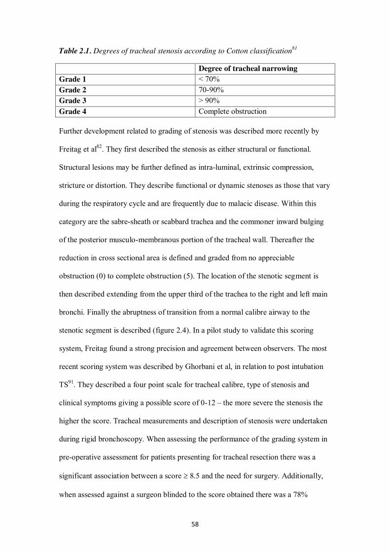
58
Table 2.1. Degrees of tracheal stenosis according to Cotton classification81
Degree of tracheal narrowing Grade 1 < 70% Grade 2 70-90% Grade 3 > 90% Grade 4 Complete obstruction Further development related to grading of stenosis was described more recently by
Freitag et al82. They first described the stenosis as either structural or functional.
Structural lesions may be further defined as intra-luminal, extrinsic compression,
stricture or distortion. They describe functional or dynamic stenoses as those that vary
during the respiratory cycle and are frequently due to malacic disease. Within this
category are the sabre-sheath or scabbard trachea and the commoner inward bulging
of the posterior musculo-membranous portion of the tracheal wall. Thereafter the
reduction in cross sectional area is defined and graded from no appreciable
obstruction (0) to complete obstruction (5). The location of the stenotic segment is
then described extending from the upper third of the trachea to the right and left main
bronchi. Finally the abruptness of transition from a normal calibre airway to the
stenotic segment is described (figure 2.4). In a pilot study to validate this scoring
system, Freitag found a strong precision and agreement between observers. The most
recent scoring system was described by Ghorbani et al, in relation to post intubation
TS91. They described a four point scale for tracheal calibre, type of stenosis and
clinical symptoms giving a possible score of 0-12 – the more severe the stenosis the
higher the score. Tracheal measurements and description of stenosis were undertaken
during rigid bronchoscopy. When assessing the performance of the grading system in
pre-operative assessment for patients presenting for tracheal resection there was a
significant association between a score 8.5 and the need for surgery. Additionally,
when assessed against a surgeon blinded to the score obtained there was a 78%

59
agreement between the score and the surgeon’s opinion regarding the need for
surgery.
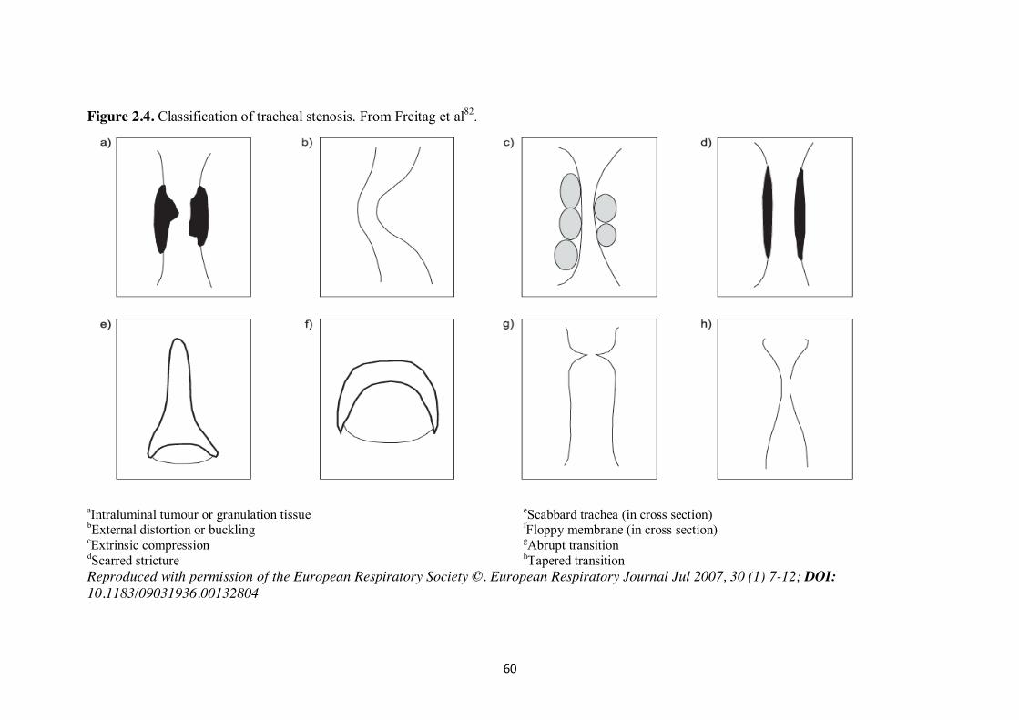
60
Figure 2.4. Classification of tracheal stenosis. From Freitag et al82.
aIntraluminal tumour or granulation tissue eScabbard trachea (in cross section) bExternal distortion or buckling fFloppy membrane (in cross section) cExtrinsic compression gAbrupt transition dScarred stricture hTapered transition Reproduced with permission of the European Respiratory Society ©. European Respiratory Journal Jul 2007, 30 (1) 7-12; DOI: 10.1183/09031936.00132804
61
2.2.1.5 Diagnosis of tracheal stenosis
2.2.1.5.i Clinical assessment
As long as a high index of suspicion is maintained, the diagnosis of TS can
largely be made from a history suggestive of airflow obstruction following on
from a period of prolonged intubation or tracheostomy. In the earlier phases of
presentation the patient may report an increase in cough associated with
difficulty expectorating secretions. With more severe stenosis, the most
frequent symptoms reported will be increasing shortness of breath associated
with inspiratory stridor. With severe stenosis, dyspnoea at rest is present. At
this point the airway diameter is likely to be <5mm. It is not uncommon that,
the presence of stridor may be mistaken for lower airway wheeze and thereby
treated as asthma92,93. In survivors of critical illness, in whom function may be
limited due to critical illness associated neuromuscular disease or ongoing
respiratory pathology, a typical presentation with stridor and shortness of
breath may not be seen. Such patients may merely report a reduction in
exercise tolerance the causes for which may be multiple and complex.
Clinical examination may note findings of tachypnoea, use of accessory
respiratory muscles, inspiratory stridor a persistently patent stoma and an
abnormal voice.
The time course for presentation after intubation may be variable. Most
patients will present within two months although a small cohort may present
very late. It is in this group of patients where misdiagnosis as lower
respiratory tract pathology may be problematic.
62
2.2.1.5.ii Investigation
a) Radiological imaging
Most patients will undergo radiological imaging either via computed
tomography (CT) or magnetic resonance imaging (MRI) prior to formal
evaluation of the stenotic segment under general anaesthesia.
Computed tomography with reformatted sagittal and coronal images can
produce an accurate representation of the underlying stenosis allowing
definition of site, length, reduction in cross sectional area and character of the
lesion. Recent advances utilising multi-detector CT imaging to produce three
dimensional images of both internal and external segments of the distorted
airway allowing a virtual endoscopy have been described94. A number of
authors have demonstrated a high degree of correlation between such images
and the findings at rigid bronchoscopy94-96. Such findings suggest that these
techniques may have an important role in defining the lesion and planning
treatment options.
b) Spirometry
Although spirometry is one of the simpler techniques used to identify upper
airway obstruction the typical changes seen occur relatively late in the disease
progression. Typically a reduction of TCSA of more than 50% must be present
for changes to be evident. With fixed TS there is a characteristic limitation of
inspiratory and expiratory flow producing the typical rhomboid curve (figure
2.5). For those lesions that are malacic and show dynamic flow limitation the
curve may be more variable with either inspiratory or expiratory abnormalities
depending upon the site of the lesion. They are of limited use in assessment
63
and planning of therapeutic intervention. A lack of correlation between
spirometry findings and both clinical symptoms and radiological findings has
been noted. In an attempt to address some of the limitations of spirometry,
Verbanck and colleagues described a forced oscillation test that was able to
define a level of critical TS that was not confounded by the presence of co-
existing peripheral airway obstruction97.
c). Diagnostic bronchoscopy
The principal role for bronchoscopy in the setting of TS is for assessment for
further surgical intervention, as it is able to define the site, length and
character of the lesion and allow planning for operative repair. It may also be
utilised with balloon dilatation to temporarily relieve symptoms prior to more
definitive treatment. However, recent National Institute for Health and
Clinical Excellence (NICE) guidance documented instances of tracheo-
bronchial rupture, tracheal laceration, laryngeal injury and bleeding as
possible cause for concern. Consequently, balloon dilatation for TS is not
recommended by NICE98.
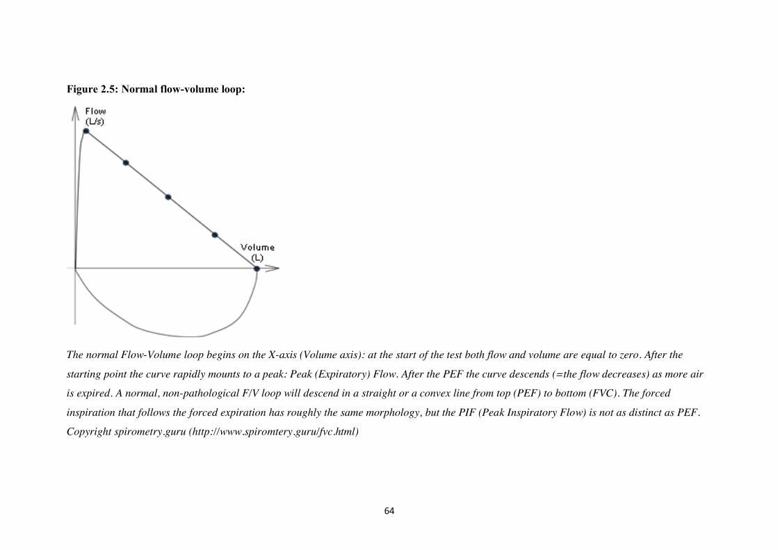
64
Figure 2.5: Normal flow-volume loop:
The normal Flow-Volume loop begins on the X-axis (Volume axis): at the start of the test both flow and volume are equal to zero. After the
starting point the curve rapidly mounts to a peak: Peak (Expiratory) Flow. After the PEF the curve descends (=the flow decreases) as more air
is expired. A normal, non-pathological F/V loop will descend in a straight or a convex line from top (PEF) to bottom (FVC). The forced
inspiration that follows the forced expiration has roughly the same morphology, but the PIF (Peak Inspiratory Flow) is not as distinct as PEF.
Copyright spirometry.guru (http://www.spiromtery.guru/fvc.html)
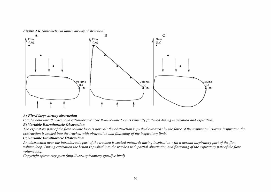
65
Figure 2.6. Spirometry in upper airway obstruction A B C
A; Fixed large airway obstruction Can be both intrathoracic and extrathoracic. The flow-volume loop is typically flattened during inspiration and expiration. B; Variable Extrathoracic Obstruction The expiratory part of the flow volume loop is normal: the obstruction is pushed outwards by the force of the expiration. During inspiration the obstruction is sucked into the trachea with obstruction and flattening of the inspiratory limb. C; Variable Intrathoracic Obstruction An obstruction near the intrathoracic part of the trachea is sucked outwards during inspiration with a normal inspiratory part of the flow volume loop. During expiration the lesion is pushed into the trachea with partial obstruction and flattening of the expiratory part of the flow volume loop. Copyright spirometry.guru (http://www.spiromtery.guru/fvc.html)
66
2.2.1.6 Management of tracheal stenosis
i. Interventional bronchoscopy and laser therapy
A number of authors have reported successful outcomes following
combination therapy of balloon dilatation in association with laser
resection75,99,100. Such interventions frequently require multiple procedures for
a successful outcome. Early work suggested that this therapy was less
effective for complex and longer stenoses and those affecting the cricoid
cartilage. Gallucio, in a study of 209 patients (mean number of interventions
per patient of 1.56-3.27) with two year follow up, reported successful
outcomes, with a combination of balloon dilatation, laser resection and
stenting, in 96% of simple stenoses and 69% of complex lesions99. Similarly
Zias, in a report of 31 cases of TS undergoing an average of 2.4 procedures
per patient, described the need for operative intervention in only one patient75.
It is possible that dynamic stenoses, particularly those involving a degree of
tracheomalacia, may be more problematic. In a series of sixty patients
undergoing combination therapy of laser resection and stenting, Plojoux
described a fifty one per cent success rate with a stent migration rate of
31%101.
It would appear, from the literature, that the lesions most suitable for endo-
luminal therapy are simple web like stenoses. There may also be a role in
more complex lesions for symptom relief prior to definitive surgery.
ii. Stenting
Traditional use of tracheal stenting has been for patients with malignant
strictures where there is a limited life expectancy102. When used for benign
67
disease with associated longer survival stent related complications become
more problematic. Whilst early results achieve relief of obstruction in up to
95% of patients long-term complications can be debilitating103,104. Long term
problems associated with tracheal stents include formation of granulation
tissue, re-stenosis, bleeding, migration, fracture and erosion. The formation of
granulation tissue and re-stenosis are two of the more problematic issues
related to stent use for benign airway strictures particularly when used in the
proximal (sub-glottic) airway. Metallic stents appear to cause more significant
problems in this region105. Additionally, the sub-cricoid area is not distensible
like the proximal trachea. Consequently, a rigid metallic stent placed here is
prone to significant shear forces generating formation of significant amounts
of granulation tissue, a propensity toward stent fracture and re-stenosis. The
resultant granulation tissue makes stent removal more difficult which may be
particularly problematic for airway management if these patients present with
upper airway obstruction with a stent in-situ.
Whilst a number of small case series published in the literature frequently
report satisfactory early symptom relief, the long term complications are
frequently excessive, with some authors suggesting that the incidence of late
complications is more related to the length of time the stent is deployed rather
than the material it is made from103. Consequently, the Food and Drug
Administration in the United States issued a series of recommendations related
to metallic stenting in benign airways disease in 2005 suggesting their use
should be avoided106.
The principal role for tracheal stenting in benign TS appears to be as a bridge
to more definitive therapies107. It should, therefore, be largely confined to
68
patients with significant co-morbidities in whom the risk of operative repair is
deemed unacceptable, those with a limited life expectancy, those who refuse
surgery, those with long complex stenoses that are deemed inoperable and to
provide symptom relief prior to definitive surgery.
Potential for an enhanced role for stenting in the future has been suggested
with the use of drug eluting stents aimed at reducing the tissue reaction and
production of granulation tissue108. Stents eluting drugs targeting the
mammalian target of rapamycin (mTOR) pathway (sirolimus, everolimus,
zotarolimus and paclitaxel) may have local immuno-modulatory effects
reducing new vessel formation and production of granulation tissue.
iii. Surgical management
Despite reports detailing the use of tracheostomy in pre-biblical times and
intermittent use through the ages tracheal surgery per se was hampered by the
belief that cartilaginous tissue heals poorly5. This opinion was stated by,
amongst others, Hippocrates and Aretaeus and persisted well in to the
twentieth century. The first description of a healing tracheal anastomosis in
dogs was reported by Gluck and Zeller who felt the technique may have a role
in humans109. The first reported human tracheal resection for post-traumatic
TS was described by Kuster in 1886110. Despite these early advances the full
utility of tracheal resection for stenotic lesions was not realised until the late
1960s and 70s with the pioneering work of Grillo111 and Pearson112. Currently
the treatment of choice for most tracheal stenoses is segmental tracheal
resection with primary anastomosis. Pre-operative assessment includes multi-
planar CT evaluation with three-dimensional reconstruction, virtual and rigid
69
bronchoscopy as detailed above. Surgical dissection is limited to preserve the
lateral segmental blood supply but segments in excess of six centimetres may
be resected in the case of complex lesions113.
Surgical outcomes have been superior to alternative non-operative procedures
with good to satisfactory results reported in excess of 90% of patients114-116.
Mortality is in the region of 0-2% with the most frequent late complications being
anastomotic failure, formation of granulation tissue and laryngeal dysfunction114.
For long complex and sub-glottic lesions the success rate appears to be around 80-
90%65. The highest failure rate is seen in patients requiring more than one
procedure to correct the defect117. Success rates may also be compromised in
patients who have previously undergone laser resection and stenting which may
damage the cricoid or lead to extension of the length of the stenotic segment118.
Where the stenosed segment extends beyond a length that is primarily resectable
surgical management becomes decidedly more complex requiring reconstruction
of the trachea itself. A reconstructed trachea requires lateral rigidity with
longitudinal flexibility. Anastomoses need to be airtight and any prosthetic
material should ideally not provoke an antigenic tissue reaction. Simple tracheal
transplantation is not possible due to the segmental nature of its blood supply.
Additionally, prolonged use of immuno-suppressants poses risks of graft infection
and breakdown. Currently approaches used have involved tracheal allografts,
aortic allografts and tracheal allografts with tissue engineered host stem cells119.
Cryopreserved and decellularised grafts appear to reduce their antigenicity and
potentially improve long-term outcome. Despite this, reports of such cases are few
in number predominantly being small cases series and isolated case reports119,120.
70
iv. Adjuvant therapy
Adjuvant treatments have been targeted at the three phases of wound healing
detailed above in an attempt to diminish the abnormal healing process responsible
for TS. Examples of strategies used include:
Inflammatory phase
Corticosteroids, antibiotics, hyperbaric oxygen.
Proliferative phase
Antibiotics, corticosteroids, mitomycin-C, 5 fluorouracil / triamcinolone,
hyperbaric oxygen.
Maturation phase
Colchicine, penicillamine, N-acetyl cysteine.
All phases
Anti-reflux therapy
Many of the potential agents have limited evidence particularly in humans with the
most studied being corticosteroids (topical, inhaled and systemic), mitomycin-C
(topical) and anti-reflux medication (proton pump inhibitors)
Corticosteroids
Theoretically corticosteroids could be expected to affect both inflammatory and
proliferative phases of wound healing.
In relation to airway injury, a number of studies have assessed the role of
dexamethasone in reducing laryngeal oedema and reducing the need for re-intubation.
A Cochrane review, of six studies in adults totalling 1953 patients, found that
71
prophylactic use of corticosteroids reduced the risk of post-extubation stridor in those
patients at high risk but did not reduce the re-intubation rate121. Although a number of
studies have shown some beneficial effects in paediatric patients there is limited
evidence for the role corticosteroids in the setting of adult post-intubation TS –
studies being confined case reports or small case series.
Mitomycin-C
Mitomycin-C is an antimicrobial agent as well as having anti-metabolite and anti-
proliferative properties. It inhibits de-oxyribonucleic acid (DNA) synthesis and in
higher concentrations suppresses ribonucleic acid (RNA) and protein synthesis.
Consequently it has been used as a chemotherapeutic agent for solid tumours. When
administered topically it has been shown to inhibit fibroblast proliferation possibly by
inducing apoptosis. Whilst mitomycin is amongst the most frequently studied
adjuvant agents for the management of TS data relating to human use is largely
confined to retrospective studies and case reports122-124.
Two randomised studies have, however, attempted to evaluate the role of mitomycin-
C in the treatment and prevention of TS. Hartnick reported a paediatric study of 24
patients undergoing a single mitomycin application after laryngotracheal
reconstruction125. Thereafter development of granulation tissue was graded on a scale
of 0-4. After recruitment of 13 patients to the intervention group and 11 to the control
group the study was stopped early, as the outcome in both groups was almost identical
(Granulation grade 3-4: placebo treated 1 patient, mitomycin treated 3 patients.
Granulation grade 0-2: placebo treated 10 patients, mitomycin treated 11 patients).
More recently Smith and Elstad reported the findings of a randomised controlled trial
comparing a one or two applications of mitomycin-C in association with endoscopic
72
laser treatment126. Twenty-six patients were recruited with idiopathic, post-intubation
and polyangiitis (Wegener’s) stenosis. All patients received mitomycin-C at the first
procedure. One month later a second endoscopic intervention was undertaken at
which patients were randomised to receive either a second application of mitomycin-
C or a saline placebo. Follow up for five years revealed an increase in the median
interval to relapse from 2.4 years in the single application group to 3.8 years in the
two application group. Relapse rates at five years, however, were identical in both
groups. Leading the authors to conclude mitomycin-C delays rather than prevents re-
stenosis.
In a review of adjuvant therapies for upper airway stenosis Hirshoren identified 11
human studies (181 patients) assessing the effects of mitomycin-C70. Only one was
randomised125, eight had no control group and six were retrospective. Of these trials,
nine claimed a positive outcome.
Mitomycin-C clearly has an effect on wound healing however optimal timing for
application remains unclear. Its efficacy for treating established stenoses is
questionable.
Treatment for gastro-oesophageal reflux disease
Initial data suggesting gastro-oesophageal reflux may play a role in the development
of TS came from case reports by Little and Bain127,128. Further work suggested that
those with reflux disease were over-represented in sub-glottic stenosis populations
particularly those with idiopathic disease129,130. A number of studies have described
less successful outcomes for surgical reconstruction in patients with uncontrolled
reflux disease131-133. However, this was not supported by the findings of a large single
blind observational study134. In an prospective, single blind observational study of 74
73
paediatric patients Zalzal failed to find any effect of gastro-oesophageal reflux disease
on the outcome of undergoing laryngotracheal reconstruction. Thus concluding that
evaluation for and treatment of reflux disease had no role in this patient population.
2.2.2 Tracheo-innominate artery fistula
The first report of a tracheo-innominate artery fistula (TIF) was in 1879 when Korte
described a fatal fistula in a five year old child with diptheria135. In a case report and
clinical review from 1976 Jones et al documented a total of 127 cases from the world
literature to that point136. The first survivor of a TIF was not reported until 1965137. In
a series of six patients, Silen reported on the management of a young female patient
who had undergone a tracheostomy for respiratory failure due to Guillain-Barre
Syndrome. Five days after tracheostomy insertion she developed a TIF. This was
managed with surgical excision of the necrotic portion of the innominate artery
followed by over-sewing of the arterial stumps. She survived for 15 days before dying
from an aspiration pneumonitis. The first long term survivor was reported by Reich in
1968 in a young female patient who again underwent resection of the necrotic arterial
segment138. Despite multiple reports in the intervening years the mortality still
remains high. A recent review139 of the surgical management of TIF in Beijing,
covering 32 years and 14 cases, reported one survivor without neurological
impairment at 11 months.
Tracheo-innominate artery fistula is an uncommon and life threatening complication
occurring after tracheostomy that is universally fatal without surgical intervention.
The true incidence of this rare complication is difficult to assess as it is most
frequently reported in small case series or isolated case reports. The incidence seems
to have reduced following the widespread introduction of high volume low pressure
74
cuffs for tracheal and tracheostomy tubes. The overall incidence of late haemorrhage
after tracheostomy has been reported at around 3%. Approximately 10% of late
bleeding is thought to be related to a TIF, however, in our own series all significant
late bleeding was due to TIF (0.35%59). Massive haemorrhage may also originate
from the aortic arch, common carotid and thyroid arteries and the innominate vein140.
Pooled data of 5530 tracheostomies recorded the incidence of delayed massive
haemorrhage as 0.3%141. In our own series of 1000 percutaneous procedures
performed using the single tapered dilator, the rate is around 0.4% (see Chapter 5).
Elsewhere, reported rates are 0.1–1% after surgical and 0.3% after percutaneous
tracheostomy, with peak incidence 7–14 days post procedure142,143.
2.2.2.1 Pathophysiology
The tracheo-innominate artery is the first branch of the aortic arch (figure 2.2). It
divides into the right common carotid and right subclavian artery 3–4 cm lateral to the
trachea, behind the right sternoclavicular joint. In its inferior proximal portion, its
relations include:
a) Anterior: left tracheo-innominate vein and thymus;
b) Posterior: trachea (6–10th ring);
c) Posterior and left: left common carotid artery;
d) Right: right tracheo-innominate vein, superior vena cava and pleura.
The tracheo-innominate artery supplies blood to the right arm and the right side of the
head and neck. Its absence on the left is explained by the direct branching of the left
common carotid and subclavian arteries from the aortic arch. A high lying innominate
artery, particularly in the thin and young, may act as a risk factor in TIF formation
following tracheostomy63.
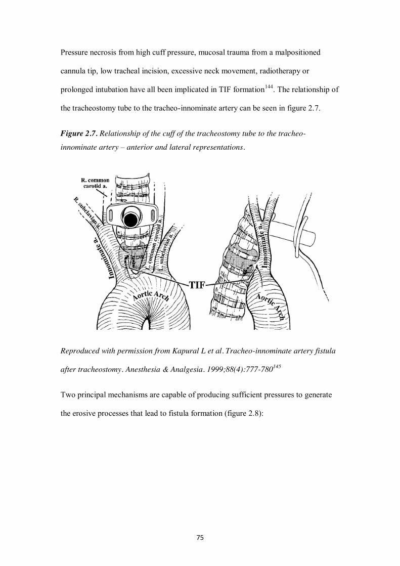
75
Pressure necrosis from high cuff pressure, mucosal trauma from a malpositioned
cannula tip, low tracheal incision, excessive neck movement, radiotherapy or
prolonged intubation have all been implicated in TIF formation144. The relationship of
the tracheostomy tube to the tracheo-innominate artery can be seen in figure 2.7.
Figure 2.7. Relationship of the cuff of the tracheostomy tube to the tracheo-
innominate artery – anterior and lateral representations.
Reproduced with permission from Kapural L et al. Tracheo-innominate artery fistula
after tracheostomy. Anesthesia & Analgesia. 1999;88(4):777-780145
Two principal mechanisms are capable of producing sufficient pressures to generate
the erosive processes that lead to fistula formation (figure 2.8):
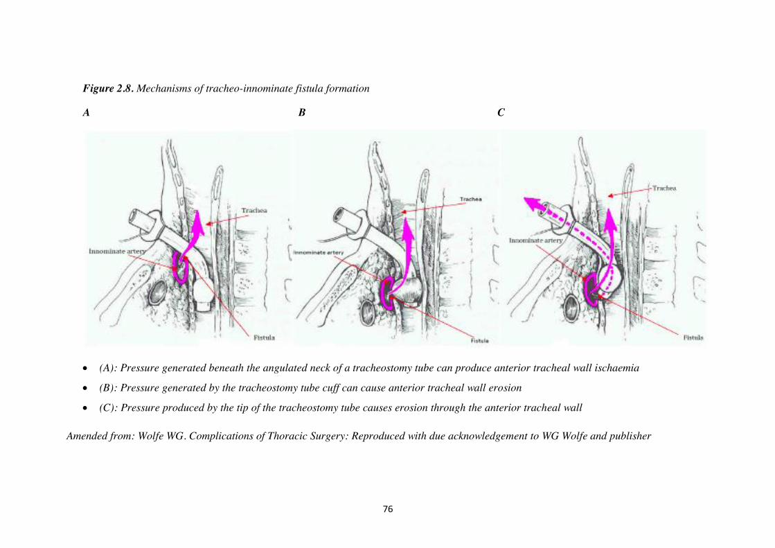
76
Figure 2.8. Mechanisms of tracheo-innominate fistula formation
A B C
(A): Pressure generated beneath the angulated neck of a tracheostomy tube can produce anterior tracheal wall ischaemia
(B): Pressure generated by the tracheostomy tube cuff can cause anterior tracheal wall erosion
(C): Pressure produced by the tip of the tracheostomy tube causes erosion through the anterior tracheal wall
Amended from: Wolfe WG. Complications of Thoracic Surgery: Reproduced with due acknowledgement to WG Wolfe and publisher
77
The first mechanism involves pressure generated beneath the angulated neck
of a tracheostomy tube (figure 2.8A). This could produce ischaemia anteriorly
on the tracheal mucosa and into the innominate artery.
A fistula may also occur between the anterior tracheal wall and artery. This is
secondary to the mechanical force generated by either the tracheostomy tube
cuff or tube tip depending on the relative positioning of the tube within the
trachea (figure 2.8B & 2.8C).
Some authors suggest that a low lying tracheostomy tube is an obvious cause of
fistula formation73. However, even when the tracheotomy incision is placed between
the second and third tracheal rings as recommended, these complications can still
occur. In a post-mortem study, Oshinsky found that ten standard vertical incisions
placed in the second and third rings resulted in all subsequently placed tracheostomy
tubes having either cuff or tip anatomically adjacent to the innominate artery,
suggesting the potential presence of this complication in all patients with
tracheostomies146.
2.2.2.2 Diagnosis
Any peri-stomal bleed or haemoptysis should lead to a full clinical investigation to
ascertain the underlying cause. A differential diagnosis is based on the lag time
between the tracheostomy and subsequent haemorrhage. Haemorrhage within 48 h is
typically associated with local factors such as traumatic puncture of anterior jugular
or inferior thyroid veins, systemic coagulopathy, erosions secondary to tracheal
suction or bronchopneumonia.
Vascular erosion from a tracheostomy tube, resulting in a TIF, requires at least 48
hours to develop even in the most friable mucosa. Haemorrhage occurring 3 days to 6
78
weeks after tracheostomy should be thought of as a result of TIF until proven
otherwise147. Other causes of catastrophic pulmonary haemorrhage include pulmonary
artery flotation catheter induced arterial rupture, thoracic aneurysm rupture and less
common vascular fistula (carotid artery, inferior thyroid). It is likely that the majority
of TIF will occur in the Critical Care Unit, as 70% of all delayed haemorrhage occurs
during the first 3 weeks148. A sentinel bleed is reported in more than 50% of patients
who then develop massive delayed haemorrhage136,149. Haemorrhage occurring after
more than 6 weeks is rarely related to TIF and more likely to be secondary to
granulation tissue, tracheo-bronchitis or malignancy.
2.2.2.3 Clinical management
Adequate oxygenation is the mainstay of immediate management with simultaneous
identification and termination of bleeding. In most TIF related deaths the cause of
death is hypoxia due to bleeding into the airway rather than exsanguination from
massive haemorrhage144.
Management of a suspected TIF will depend upon whether there is active bleeding
into the airway hindering adequate ventilation (Figure 2.9). The use of immediate
bronchoscopy to confirm the extent and source of bleeding has been advocated144.
Although bronchoscopy is unlikely to identify the fistula opening per se, it may
exclude other pathology and allows direct monitoring of attempts to obtain a blood-
free airway. Rigid bronchoscopy to clear the tracheobronchial tree of aspirated blood
and to terminate blood flow is ideal, but this may not be possible.
If bronchoscopy confirms that the main bronchi are blood-free then immediate
intervention is not required but further investigation to confirm the cause of bleeding
is vital. Other causes of haemorrhage must be excluded before a sentinel bleed
79
associated with TIF can be confirmed. If no other cause is evident then a provisional
diagnosis of TIF should be made; urgent surgical advice and consideration of stoma
exploration should follow. If, in the presence of active bleeding, bronchoscopy
indicates that the airway is clear of blood but there is ongoing external haemorrhage,
the potential for catastrophic airway contamination is real. Overinflating the cuff
provides additional airway protection and may control the bleeding temporarily. If,
however, bleeding continues then pressure dressings should be applied to the stoma
site. These manoeuvres temporarily control bleeding by a direct tamponade effect in
more than 80% of patients150. As long as the airway remains free of blood, no attempt
should be made to manipulate the tracheostomy tube. Immediate surgical exploration
should follow.
Where bronchoscopy confirms that there is active bleeding into the airway, the major
threat is respiratory compromise rather than hypovolaemia151. Airway protection is
the primary management aim. Movement of the tracheostomy tube may precipitate
disastrous airway occlusion143. The aims must be to gain temporary control of the
bleeding, get adequate oxygenation and proceed to immediate stomal exploration and
definitive treatment.
i Haemorrhage control measures
Over-inflation of the tracheostomy cuff is first line management.
If this measure fails to reduce bleeding in to the trachea then proceed
immediately to trans-laryngeal intubation with digital compression.
Trans-laryngeal intubation and digital compression of the innominate artery
against the posterior surface of the manubrium. A cuffed oral tracheal tube is
advanced so that the balloon lies distal to the tracheostomy stoma. The
80
tracheostomy tube should only be withdrawn to facilitate simultaneous trans-
laryngeal tracheal intubation. This procedure should terminate bleeding in >
90% patients and if maintained, allows transfer to the operating theatres.
If digital compression fails to stem bleed, slow withdrawal of the tracheal tube
and cuff over-inflation should follow. Manipulating the ETT tube, and its cuff
to produce tamponade is the only other manoeuvre available in this situation
and attempts should persist.
If the bleeding stops, and ventilation is acceptable, then immediate surgical
intervention should follow. There is no evidence to support any imaging prior to
immediate surgical exploration.
ii Surgical management
It is clear from the literature that the mortality for TIF without surgery is close to
100%. There remains a degree of controversy around the optimal surgical
management.
Most surgical approaches utilise a median sternotomy or upper sternotomy extended
to the second right intercostal space to access the fistula.
Thereafter the controversy revolves around the two basic strategies for fistula
management and concerns relating to post-operative neurological deficit.
Maintenance of flow within the innominate artery
Direct repair of the fistula
Graft interposition
Interruption of flow within the artery
Simple ligation of the innominate artery
81
Bypass grafting – aorto-axillary
The principal concern relating to maintenance of flow within the vessel is related to
the risk of re-bleeding. The area surrounding the fistula is highly like to have active
underlying infection and ongoing inflammation. Repair of the fistula or interposition
of a foreign body to bypass the defect are therefore highly likely to fail. In a review of
37 cases published between 1975 and 1988 Yang described the outcomes of patients
surviving to undergo operative intervention152. Fourteen patients underwent resection
or ligation of the innominate artery with a re-bleeding rate of 7% and 64% long-term
survival. Ten patients underwent procedures maintaining flow within the innominate
artery and had a re-bleeding rate of 60% and a long-term survival of 10%. Similarly
Gelman reported on the outcomes of 71 TIF survivors in 1994151. With maintenance
of flow within the vessel re-bleeding rates were 60% versus 7% when there was
disruption of flow. With maintenance of flow mortality was 86% versus 29% with
disruption. Even with ligation of the innominate artery, reported neurological
sequelae were minimal. Despite these data, continued reports of stent
interposition153,154, vascular grafting155 and endovascular embolisation156,157 continue
to appear. Reported neurological sequelae are unusual with arm weakness occurring
in around 4% of patients and more significant neurological deficit in less than 5% of
patients158 .
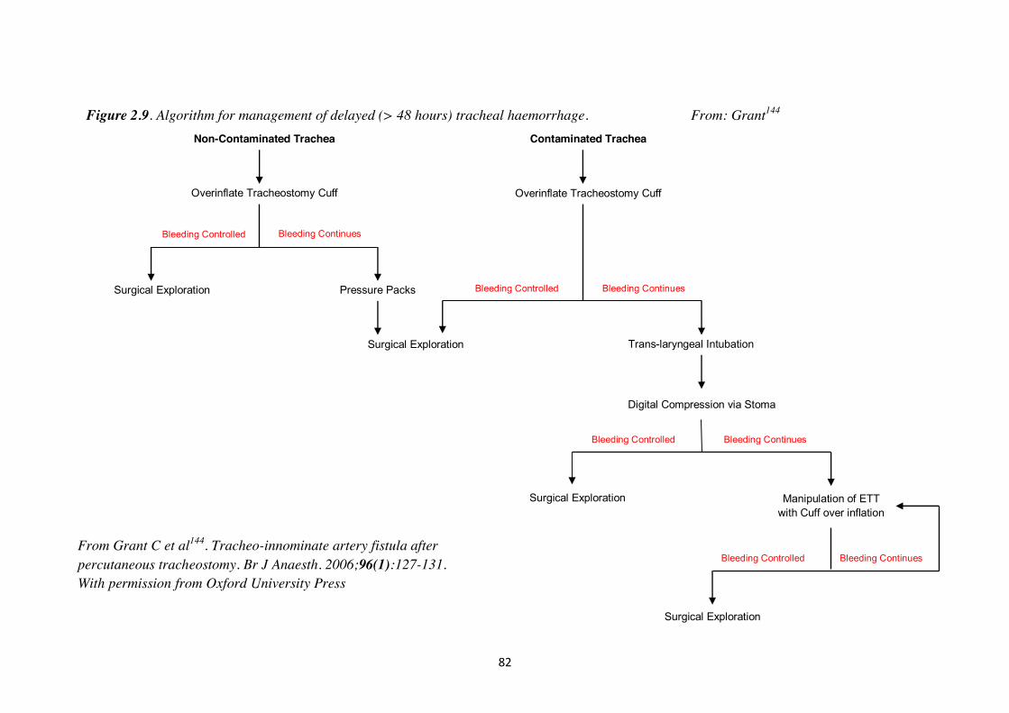
82
Figure 2.9. Algorithm for management of delayed (> 48 hours) tracheal haemorrhage. From: Grant144
Manipulation of ETT with Cuff over inflation
Surgical Exploration
Surgical Exploration
Trans-laryngeal Intubation
Overinflate Tracheostomy Cuff
Bleeding Controlled
Non-Contaminated Trachea
Overinflate Tracheostomy Cuff
Surgical Exploration Pressure Packs
Surgical Exploration
Bleeding Continues
Bleeding Continues Bleeding Controlled
Contaminated Trachea
Digital Compression via Stoma
Bleeding Continues Bleeding Controlled
Bleeding Continues Bleeding Controlled
From Grant C et al144. Tracheo-innominate artery fistula after percutaneous tracheostomy. Br J Anaesth. 2006;96(1):127-131. With permission from Oxford University Press
83
2.2.3 Tracheo-oesophageal fistula
Tracheo-oesophageal fistulae related to tracheostomy insertion may occur as both an
early and late complication. Trauma to the posterior tracheal wall during
tracheostomy insertion may lead to direct fistula formation although a simple
posterior tracheal wall tear with entry only into the mediastinum is perhaps more
likely. Such fistulae will usually be evident immediately post-operatively although
diagnosis may be delayed159,160. Features suggestive of an immediate problem include
a persistent cuff leak with the formation of subcutaneous and mediastinal emphysema
as well as pneumothoraces.
Although tracheo-oesophageal fistulae (TOF) may result from several different
aetiologies, including trauma and post-surgically, those originating after prolonged
tracheal intubation and tracheostomy remain the commonest. TOF due to
tracheostomy cuff erosion was first reported in the 1960s161-163, with Flege being the
first to postulate ischaemic necrosis, due to the cuff of the tube impairing tracheal
wall capillary perfusion, as the underlying cause162. The reported incidence is similar
to that of TIF164 formation although the presentation maybe somewhat later with
median times to diagnosis around 30 – 35 days from onset of mechanical
ventilation165,166. Similarly to TIF and TS, with the introduction of high volume low
pressure tracheostomy tube cuffs, the incidence of TOF appears to have
diminished167. Muniappan recently reviewed cases of TOF presenting over a 35 year
period extending from 1975 to 2010 and reported a falling incidence of post
intubation lesions from 71 to 47%168.
2.2.3.1 Pathophysiology of late TOF
The aetiology of late TOF is essentially the same as that described above for both TIF
84
and TS. Over inflation of the tracheostomy tube cuff will lead to pressure necrosis,
additionally malpositioning of the tube may also lead to erosions of the posterior
tracheal wall by the tip of the tracheostomy tube. After short periods of intubation,
epithelial disruption, basement membrane loss, and / or an appearance of ischemic
non-inflammatory necrosis can occur. More prolonged intubation results in broader,
deeper ulcers, disruption of basement membranes and ischemic necrosis. Similar to
the aetiology of tracheal stenosis, cuff pressures of greater than 20-30 cm of water
contribute to mucosal ischemia67,68,83. The effects of cuff related mucosal injury may
be exacerbated by repeated tracheal tube manipulations, intubations, or excessive
movements of the tracheostomy tube and ventilator circuitry. Additional factors
important in the aetiology may be the presence of a naso-gastric tube, advanced age,
diabetes mellitus, steroid therapy, prolonged hypotension, oesophageal infections and
prolonged cannulation169. However, pressure related necrosis may be more likely to
be associated with the rigid cartilaginous anterior tracheal wall than the soft tissue of
the musculo-membranous posterior wall. This may account for the more frequent
descriptions of TS and TIFs170.
As most TOF related to prolonged intubation and tracheostomy insertion are related
to mucosal ischaemia produced by the cuff of the tube their site is usually in the
proximal trachea. Muniappan in a report of 74 cases of TOF, of which 59% were post
intubation / tracheostomy, described the site of the fistula in the upper trachea in 36%,
the mid-trachea in 61% and the lower trachea in 3%168. Additionally, 35% of lesions
were < 1cm, 58% were 1 – 3cm and 8% were in excess of 3cm (figure 2.10).
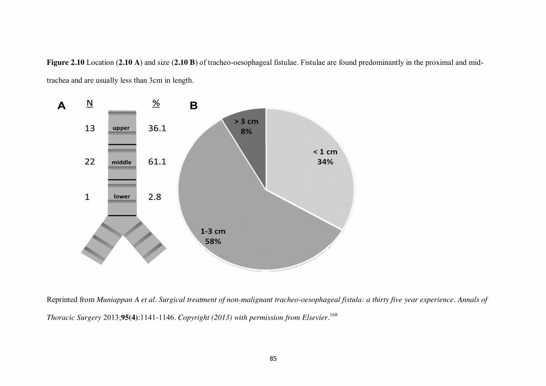
85
Figure 2.10 Location (2.10 A) and size (2.10 B) of tracheo-oesophageal fistulae. Fistulae are found predominantly in the proximal and mid-
trachea and are usually less than 3cm in length.
Reprinted from Muniappan A et al. Surgical treatment of non-malignant tracheo-oesophageal fistula: a thirty five year experience. Annals of
Thoracic Surgery 2013;95(4):1141-1146. Copyright (2013) with permission from Elsevier.168
86
2.2.3.2 Diagnosis
Hallmarks of TOF include persistent choking or coughing in relation to swallowing
leading to aspiration pneumonitis. An ongoing air leak around the cuff of the
tracheostomy tube, gastric distention and abdominal air movement synchronous with
respiration may be seen. Further signs may include subcutaneous and mediastinal
emphysema and pneumothorax171. As the underlying pathology is similar to that
responsible for both TS and TIF the timing of presentation of late TOF is likely to be
several days to weeks following the tracheostomy.
Whilst the diagnosis is largely made on clinical grounds, with associated plain
radiology showing evidence of mediastinal or pleural air, CT scanning will confirm
the diagnosis. The fistula may be visualised directly by fibre-optic bronchoscopy
although if due to erosion from the cuff it may be obscured by the tracheostomy tube
itself. Definitive visualisation, prior to operative intervention, will be achieved by
rigid bronchoscopy and oesophagoscopy.
2.2.3.3 Clinical management
When the diagnosis of TOF is confirmed the initial management involves avoiding
further aspiration of gastric secretions into the tracheo-bronchial tree. The cuff of the
tracheostomy or trans-laryngeal tube should be advanced beyond the fistula and the
nasogastric tube should be removed. Gastric reflux should be reduced by the insertion
of a gastrostomy tube and enteral feeding continued via jejunostomy. Antibiotic
treatment of associated aspiration pneumonitis may also be required. These measures
should prevent further aspiration pneumonitis and allow weaning from mechanical
ventilation before more invasive therapies are contemplated.
87
i Conservative management
Whilst spontaneous healing of a TOF, over many months, with conservative
management of gastrostomy, jejunal feeding and re-positioning of the tracheostomy
tube has been reported this is far from usual172 . Surgical treatment remains the
mainstay of treatment but a number of less invasive therapies have been described in
recent years. In a series of 25 patients with oesophageal leaks or fistulae Blackmon
reported the use of metallic stents in their management173. Only four patients had TOF
and in only two of whom was this due to prolonged intubation. The authors reported
that in two of these four patients their fistulae sealed, whether further surgery was
required or not was not reported. In another report of stenting for TOF Eleftheriadis
described the management of twelve mechanically ventilated patients166. Whilst the
authors stated that stent insertion was successful in all cases with sealing of the fistula
and no migration only three of the twelve patients survived to surgery. Whether the
stent placement resulted in a delay in referral for surgical intervention or, due to
reduced gastric aspiration, allowed an improvement in the patients’ general condition
to enable consideration of surgery is unclear168. The use of fibrin glue to close a TOF
in a mechanically ventilated patient has also been reported174.
ii Surgical management
Repair of TOF is a major undertaking in the critically ill patient. Consequently most
authors advocate waiting until the patient has been weaned from mechanical
ventilation and largely recovered from the after effects of their critical illness
including any critical illness related neuro-muscular weakness164. This will allow
improvement in nutritional status and reduce the risk of further post-operative
prolonged mechanical ventilation. In a recent report of 13 patients undergoing TOF
88
repair Foroulis described the use of a pre-sternocleidomastoid incision with
interposition of a vascularised sternocleidomastoid pedicle165. Five of the thirteen
patients were still mechanically ventilated at the time of operation, of whom three
died in the post-operative period (60% mortality). In the eight patients in whom
surgery was performed after weaning from mechanical ventilation there were no
deaths.
The surgical approach to repair is largely dependent upon the site of the fistula and
whether there is associated tracheal involvement. For those fistulae that are small (<
3cm) without tracheal involvement then a direct repair via a pre-sternocleidomastoid
approach, with or without extension in to the superior mediastinum may be the most
appropriate option. When there is associated tracheal disease (stenosis or
tracheomalacia) a combined tracheal resection and oesophageal repair, as popularised
by Grillo, is the most frequently used technique175. For more distal and complex
lesions the use of a median sternotomy or thoracotomy may be required along with
oesophageal reconstruction using gastric or colonic interposition. In addition to
repairing the defect most authors recommend the interposition of vascularised
pedicled muscle flaps (sternocleidomastoid, strap or pectoralis major muscles) to
separate the tracheal and oesophageal anastomoses168,176 although the need for these
has recently been questioned177. In their series of 25 patients Marulli reported the use
of a cervical approach in 56%, a combined cervical & sternotomy incision in 24% and
a thoracotomy in 16% of cases176. In the same patients they undertook a direct fistula
repair in 60% and a combined tracheo-oesophageal resection in 32%.
Although early reports for mortality after TOF repair were as high as 28%178,179 most
case series published after 2000 have reported mortalities of less than
89
10%168,176,177,180. Complication rates, however, remain high (32-56%)168,176 with
anastomotic breakdown and pulmonary infection being the most common.
2.3 Summary
The first reports of long-term complications of tracheostomy in significant numbers
followed the poliomyelitis epidemics in the 1940s and 50s with the first widespread
use of tracheal intubation and cuffed tracheostomy tubes for the purposes of
mechanical ventilation. Whilst the absolute incidence of each complication has fallen
with the introduction of high volume low-pressure cuffs, improved monitoring of cuff
pressures and ventilator circuitry the underlying prevalence continues to increase as
increasing numbers of patients receive these treatments.
Treatment of each complication is dependent upon the complexity of the lesion
present, the symptoms produced and co-existing co-morbidities for any given patient.
In a population of critically ill patients all long-term complications of tracheostomy
will pose significant problems with considerable morbidity and mortality.
For TS tracheal resection provides the best chance of a lasting cure with success rates
in the region of 90% for simple lesions and 80-85% for more complex ones.
Endoscopic treatments are largely reserved for symptom relief pending definitive
surgery or for those deemed unfit for tracheal resection.
For TIF outcomes are directly related to rapid diagnosis and surgical intervention.
With techniques involving ligation or resection of the innominate artery long-term
survival of 70% of patients surviving to the operating theatre is possible.
Tracheo-oesophageal fistulae require an approach to minimise ongoing gastric
aspiration to facilitate weaning from mechanical ventilation and allowing
90
improvement in general health and nutritional status prior to attempting surgical
repair. With this cautious approach mortality rates of less than 10% can be
achieved168,176,177,180, however, operating on patients who remain ventilator dependent
still results in much higher death rates165.
Whilst the incidence of TS following percutaneous tracheostomy is evident from a
number of long term cohort studies57-59 the issue of missed or sub-clinical stenoses has
not been fully addressed. Non-specific symptoms are common following critical care
admission and PDT, with frequent reports of voice changes, swallowing difficulties,
cough and shortness of breath181,182. We hypothesised that, in this cohort of patients,
there are a number of patients with undetected TS.
2.4 Aims
To identify aetiological factors in initiation of tracheal stenosis with particular
reference to peri-operative events
To determine the role of early complications that may be postulated to play a
part in the genesis of the late complications of tracheal stenosis, tracheo-
innominate artery fistula and tracheo-oesophageal fistula
To determine the prevalence of sub-clinical tracheal stenosis following
percutaneous tracheostomy using the single tapered dilator technique.
To determine possible aetiological factors for such sub-clinical stenoses
namely tracheal ring fracture and duration of cannulation
To determine whether sub-clinical tracheal stenosis may be presenting
atypically in the post-critical care population
To determine the utility of simple spirometry and a symptom based
questionnaire in identifying sub-clinical tracheal stenosis

91
Chapter 3
Rationale for thesis
92
3.1 Systematic Review
Percutaneous dilatational tracheostomy (PDT) has become widely adopted in critical
care units following Ciaglia’s first description of the dilatational technique40. Since
then multiple percutaneous techniques have been described, introduced and widely
evaluated in comparison to both each other and surgical tracheostomy (ST). Whilst
the short-term complications of these techniques are well described, the prevalence
and impact of longer-term outcomes, particularly TS, is unclear. Previous meta-
analyses have attempted to address this problem but have been confounded by the low
incidence of TS and, in particular, the limited number of studies reporting long-term
outcomes 51-55.
On examination of current evidence, the meta-analysis published by Delaney et al,
found a significantly decreased rate of wound infection after all commonly performed
percutaneous techniques compared to surgical tracheostomy51. Additionally, there
were reductions in mortality and bleeding in patients undergoing percutaneous
tracheostomy in the critical care unit as opposed to ST in the operating theatre.
Higgins et al, also reported a tendency towards a reduction in overall complications
for percutaneous procedures53. In contrast, Oliver et al, found an increased incidence
of minor early complications associated with percutaneous techniques but insufficient
evidence to suggest a difference in late complications of poor cosmetic results and
tracheo-cutaneous fistulae52. Cabrini et al, found that the six percutaneous techniques
analysed (Ciaglia multiple dilator method (CPDT), guide wire dilating forceps
(GWDF), single tapered dilator (STD), trans-laryngeal tracheostomy (TLT), balloon
dilator (BD), single step rotational dilator (SSRD)) were largely comparable with the
exception of the TLT, which was associated with an increased conversion rate to ST
or other percutaneous technique and more severe early complications54. They also
93
found the STD was associated with fewer complications and failures. In a later
analysis the same authors reported less operative bleeding and fewer technical
difficulties associated with the STD technique compared to the GWDF technique
using a composite endpoint55.
The meta-analyses described above have included only randomised controlled trials
(RCTs). The only exception to this was the analysis by Oliver which also included
non-randomised prospective studies52. The largest single study incorporated into the
previous analyses comprised 346 patients56. It is perhaps unsurprising, therefore, that
none of the previous meta-analyses have reported differences in late complication
rates. The exact incidence of long term complications following tracheostomy
procedures in the critically ill is difficult to quantify due to the associated mortality of
critical illness, the sub-clinical nature of many tracheal stenoses and the difficulty
maintaining follow up of these cohorts. From a number of prospective cohort series it
would appear that sub-clinical TS is found in around 10% of survivors79 with
clinically evident lesions presenting in 0 - 0.35%57-59.
Despite its low incidence, TS causes significant ongoing morbidity and healthcare
costs associated with its management. The management of choice for TS has been
segmental tracheal resection since Grillo183 and Pearson112 demonstrated good
outcomes. However, in some patients stenoses may not be amenable to surgery.
Management of such patients presents a challenge; alternative treatments include
endoscopic dilatation, laser ablation, tracheal stenting and cryosurgery107. These
interventions may temporarily alleviate the symptoms of TS but as re-stenosis is a
frequent occurrence repeated procedures are often necessary. Given this associated
morbidity and the cost associated with the management of TS a clearer picture of the
risk associated with each tracheostomy technique performed within the critical care
94
setting is required.
There is little direct evidence for the association of any peri-operative complications
with the aetiology of TS. In a study, pre-dating the widespread introduction of PDT,
assessing the impact of prolonged tracheal intubation and tracheostomy Stauffer
identified a high incidence of complications associated with ST (36% for stomal
bleeding, 36% for stomal infection) and a 65 % tracheal stenosis incidence74. Despite
a number of moderate sized case series from tertiary centres reporting of the
management of tracheal stenosis none have specifically looked at the role of peri-
operative events in the initiation of TS71,75,88,184. The only peri-operative event that has
been postulated to have a role in the genesis of TS is tracheal ring fracture. Whilst a
number of authors184-186 have suggested that tracheal ring fracture may be of
significance this is not necessarily borne out within large cohort studies59. Further
studies have considered a role for infection in the initiation of TS. In an animal model
of tracheal stenosis Squire inoculated the tracheas of rabbits with Staphylococcus
aureus66. When compared to those without bacterial inoculation the incidence of
tracheal stenosis was higher and the resultant lesions narrower. Additionally,
Welkoborsky examined operative specimens removed from 18 patients undergoing
surgical resection for TS and felt infection at the stenotic segment played a part in its
initiation in 4 patients69. Despite this work and evidence to suggest a greater incidence
of stomal infections for ST compared to PDT51 there is no evidence to that the
incidence of TS differs between the two techniques or that antimicrobial treatment
reduces the incidence of TS.
Despite this lack of direct evidence for the association of peri-operative events with
the development of TS we postulated that some complications maybe surrogate
markers for a more severe tracheal injury at the time of tracheostomy and thus affect
95
the healing process. When considering such problems it is possible that many of the
events listed in Appendix 1 may lead the operator to settle for sub-optimal
tracheostomy tube positioning which may have an impact on later healing or
predispose to infection. We, therefore, felt that the role such factors may play in the
initiation of tracheal stenosis was worthy of further study.
Therefore, we performed a systematic review of all prospective studies that reported
long-term outcomes and assessed potential predisposing factors. Our aim was to
determine if longer-term complication rates, with particular reference to TS, differed
between percutaneous and surgical tracheostomy techniques in the critical care
setting.
3.2 Single tapered dilator percutaneous tracheostomy: An 11-year review
Despite the popularisation of the percutaneous dilatational approach by Ciaglia40,41
and the proliferation of a number of similar techniques42-44,47,187 the long term
outcomes and survival following percutaneous tracheostomy remain unclear. A
number of large cohort studies have described late complications and short term
survival following PDT57-59,188 but survival beyond 12 months has not been previously
been reported.
Percutaneous dilatational tracheostomy has now largely replaced conventional
surgical tracheostomy in critical care patients, with benefits in terms of cost, ease of
performance, and reduced complications51. In 2000, the first description of a
modification of the original CPDT technique described by Ciaglia40 was reported
using a single tapered dilator44. The STD method has now largely supplanted the
sequential dilatational procedure and appears to be the single most common technique
for performing a PDT in the critical care setting45,46,189.
96
Several authors have published large PDT case series. Kearney, in perhaps the largest
single centre evaluation to date, reported the outcome of an eight year review of the
CPDT technique188. Diaz-Reganon reported an evaluation of both CPDT (51 patients)
and GWDF (749 patients) in a single centre study58 whilst Kost reported outcomes of
both CPDT and STD techniques across multiple units in Montreal57. Here, we update
our earlier single centre evaluation of the STD technique59 reporting long term
outcomes over an 11 year study period.
3.3 Tracheal stenosis following percutaneous tracheostomy: A MRI study
Whilst the incidence of TS following percutaneous tracheostomy is evident from a
number of long term cohort studies57-59 the issue of missed or sub-clinical stenoses has
not been fully addressed. Non-specific symptoms are common following critical care
admission and PDT, with frequent reports of voice changes, swallowing difficulties,
cough and shortness of breath181,182. We hypothesised that, in this cohort of patients,
there are a number of patients with undetected TS.
Earlier studies have attempted to identify the prevalence of TS following PDT using
CT181,190, MRI80, plain linear tomography191, spirometry with fibre-optic
laryngoscopy192, or fibre-optic laryngoscopy alone193. Norwood’s study181 using CT
evaluation of tracheal anatomy after sequential dilatation PDT and correlation with
symptoms found an incidence of TS of 31%, 20% of whom were symptomatic, but
could not determine whether voice changes or respiratory problems were a result of
critical illness or PDT itself. Spirometry has been variably described as
straightforward but with unproven utility in detecting stenosis194 and as unhelpful in
detection of TS in patients after critical illness, with poor correlation between
spirometry findings and endoscopic features192. The forced expiratory volume in 1
second (FEV1) to peak expiratory flow rate (PEFR) ratio (FEV1: PEFR) ratio was
97
demonstrated by Empey to be useful in distinguishing upper airway obstruction from
lower airways disease but has not been widely adopted195.
Despite the above attempts to delineate tracheal post-PDT changes there are limited
data pertaining to STD PDT. Although Fikkers used MRI to evaluate long-term
outcome only 14 STD patients were evaluated80. We therefore undertook a
prospective study to radiographically evaluate tracheal changes following STD PDT
in an attempt to better define the prevalence of sub-clinical TS.
98
3.4 Statement of Aims
Systematic review
To identify aetiological factors in initiation of tracheal stenosis with particular
reference to peri-operative events
To determine the utility of adjunctive techniques (bronchoscopy & ultrasound
scanning) in reducing complications of percutaneous tracheostomies
percutaneous tracheostomy technique and hence determine the safest
percutaneous technique
To determine the incidence of common early and late complications and
outcomes of PDT techniques in relation to surgical tracheostomy.
To determine the role of early complications that may be postulated to play a
part in the genesis of the late complications of tracheal stenosis, tracheo-
innominate artery fistula and tracheo-oesophageal fistula
To determine the relative indices of early and late complications in relation to
percutaneous tracheostomy technique and hence determine the safest
percutaneous technique
Cohort study
To determine the role of early complications that may be postulated to play a
part in the genesis of the late complications of tracheal stenosis, tracheo-
innominate artery fistula and tracheo-oesophageal fistula
To determine long term survival following percutaneous tracheostomy
MRI study
To determine the prevalence of sub-clinical tracheal stenosis following
percutaneous tracheostomy using the single tapered dilator technique.
To determine possible aetiological factors for such sub-clinical stenoses
namely tracheal ring fracture and duration of cannulation
To determine whether sub-clinical tracheal stenosis may be presenting
atypically in the post-critical care population
To determine the utility of simple spirometry and a symptom based
questionnaire in identifying sub-clinical tracheal stenosis
99
Chapter 4
Long-term outcome following tracheostomy in critical
care: A systematic review
100
4.1 Search strategy
We searched Embase, PubMed-Medline and the Cochrane Central Register of
Clinical Trials for prospective studies involving humans in the critical care setting that
reported tracheostomy outcome data and included patients that had received at least
one PDT either alone or in comparison with ST or another percutaneous technique. To
reflect contemporary practice we limited the search to studies published from the year
2000 onwards. The search was restricted to full text reports of papers published in
English in peer-reviewed journals. In addition, reference lists of those studies returned
in the above search were scrutinised for additional relevant papers.
4.1.1 Study selection
Titles and abstracts of the references obtained were reviewed by three independent
reviewers (G Dempsey, B Morton & C Hammell). Studies were categorised as - for
inclusion, possible inclusion and exclusion. In the absence of a decision by two
investigators to exclude a paper, data extraction of full text articles was undertaken by
two of the three reviewers independently. Discrepancies between reviewers prompted
a re-evaluation of the paper. Ongoing disagreement was resolved by the third
reviewer. If the outcome data in the original article was unclear the corresponding
author was contacted for clarification. Our protocol relating to study selection and
data extraction is published on:
http://www.crd.york.ac.uk/PROSPERO/display_record.asp?ID=CRD42014008830
4.1.2 Data extraction and outcomes
A database was constructed to incorporate outcomes common to previous systematic
reviews of percutaneous tracheostomy (additional parameters not presented in the
main text of this chapter can be found in appendix 1). Outcomes were defined a
101
priori. The primary outcome was TS. Secondary outcomes were wound infection
requiring treatment and bleeding (major and minor).
4.1.3 Internal validity and risk of bias assessment
The internal validity and risk of bias for RCTs was assessed using the Oxford Quality
Scoring System (Jadad Score)196. The score is based on randomization (0-2 points),
blinding (0-2 points), and withdrawals / dropouts (0-1 point). Studies scoring < 3 are
considered to be of low quality197. Non-randomised cohort studies were assessed
using the Newcastle-Ottawa score for cohort studies198. The scale assesses patient
selection (0-4 stars), comparability of patient cohorts (0-2 stars) and outcomes (0-3
stars). Responses indicating high quality earn a star resulting in a maximum possible
quality summary score of nine. Assessments were undertaken and confirmed by two
independent reviewers. There is a lack of formal assessment tools for observational
studies therefore no assessments were performed on the single arm observational
studies.
4.1.4 Data analysis
For each complication, we calculated the risk difference (RD) and 95% confidence
interval (CI) in each study that had compared two or more techniques. Studies that
specifically reported that no events of a particular type had occurred were included in
analyses as zero events. The RD was used as the measure of effect as the data were
sparse in many studies and we wanted to ensure that studies with zero events in both
arms contributed to meta-analyses. We used a random effects meta-analysis to pool
the RD across multiple studies199. Analyses were performed in STATA version 9.2
102
(StataCorp, 2005 Stata Statistical Software: Release 9. College Station, TX:
StataCorp LP).
As we were interested in estimating the complication risks associated with each
technique we calculated the proportion of total patients with a complication and 95%
CI in each study that provided data for a particular technique. Proportions were first
transformed via the Freeman-Tukey double arcsine method200 which were then pooled
using random effects meta-analysis199. Analyses were performed in StatsDirect
statistical software version 2.7.9 (StatsDirect Ltd, 2013 England).
We assessed statistical heterogeneity through visual inspection of the forest plot and
interpretation of the I2 statistic (percentage of variation across studies that is due to
heterogeneity rather than chance)201 and the Cochrane Q test. Where a sufficient
number of studies were included in analyses, funnel plots were examined and Egger’s
test applied to assess potential bias in meta-analyses202.
A sensitivity analysis was performed to compare results when restricted to RCTs
only.
4.2 Results
4.2.1 Search results
Database search results yielded 463 studies that were subsequently assessed for
eligibility (figure 4.1). One hundred and twenty four studies were subjected to full
text review with 95 failing to meet inclusion criteria47,48,56,181,184,185,190,203-290. One
study was excluded due to possible duplication of results and a lack of response to a
clarifying e-mail query (figure 4.1)216. A total of 29 papers reporting on 5473 patients
were included in the analysis (table 4.1).57-59,80,187,188,291-313
103
4.2.2 Study characteristics
Of the 29 studies included, nine (n=1023 patients) were RCTs that evaluated two or
more percutaneous techniques.80,187,292,295,297,298,303,305,310 Of the remaining 20 studies,
one was a prospective cohort study comparing three techniques (ST, TLT and CPDT
n=100 patients)300 , two compared prospective PDT techniques with historical
surgical control groups (n= 421; the retrospective ST patients were not included in our
analysis)294,309, three were prospective observational studies reviewing more than one
technique (n=1513)57,58,311 and 14 were single arm prospective studies
(n=2416)59,188,291,293,296,299,301,302,304,306-308,312,313. Further details of study characteristics
including risk of bias assessments can be seen in table 4.1. The mean Jadad score for
RCTs was 2.4 and mean Newcastle Ottawa score for the cohort studies was 6.25.
In all studies evaluating the CPDT, the Ciaglia Percutaneous Tracheostomy Multiple
Dilator set (Cook Critical Care, Bloomington, IN) was used. All studies assessing the
GWDF technique used the Portex kit (SIMS Portex Ltd, Hythe, Kent, UK). For
studies evaluating STD the Ciaglia Blue Rhino Percutaneous Tracheostomy
Introducer set (Cook Critical Care, Bloomington, IN, USA) was used in six
57,59,187,293,295,309 and the Portex Ultraperc (Smith Medical, Hythe, Kent, UK) was used
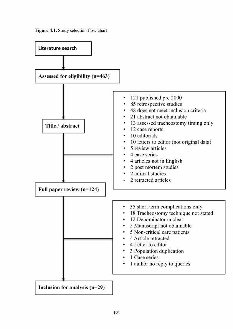
104
Figure 4.1. Study selection flow chart
Literature search
Assessed for eligibility (n=463)
Title / abstract
Full paper review (n=124)
Inclusion for analysis (n=29)
• 121 published pre 2000 • 85 retrospective studies • 48 does not meet inclusion criteria • 21 abstract not obtainable • 13 assessed tracheostomy timing only • 12 case reports • 10 editorials • 10 letters to editor (not original data) • 5 review articles • 4 case series • 4 articles not in English • 2 post mortem studies • 2 animal studies • 2 retracted articles
• 35 short term complications only • 18 Tracheostomy technique not stated • 12 Denominator unclear • 5 Manuscript not obtainable • 5 Non-critical care patients • 4 Article retracted • 4 Letter to editor • 3 Population duplication • 1 Case series • 1 author no reply to queries
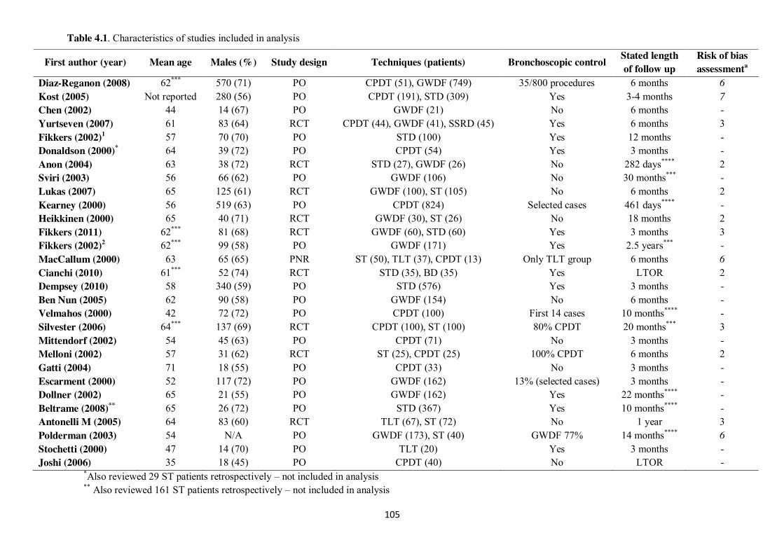
105
Table 4.1. Characteristics of studies included in analysis
*Also reviewed 29 ST patients retrospectively – not included in analysis ** Also reviewed 161 ST patients retrospectively – not included in analysis
First author (year) Mean age Males (%) Study design Techniques (patients) Bronchoscopic control Stated length of follow up
Risk of bias assessmenta
Diaz-Reganon (2008) 62*** 570 (71) PO CPDT (51), GWDF (749) 35/800 procedures 6 months 6 Kost (2005) Not reported 280 (56) PO CPDT (191), STD (309) Yes 3-4 months 7 Chen (2002) 44 14 (67) PO GWDF (21) No 6 months - Yurtseven (2007) 61 83 (64) RCT CPDT (44), GWDF (41), SSRD (45) Yes 6 months 3 Fikkers (2002)1 57 70 (70) PO STD (100) Yes 12 months - Donaldson (2000)* 64 39 (72) PO CPDT (54) Yes 3 months - Anon (2004) 63 38 (72) RCT STD (27), GWDF (26) No 282 days**** 2 Sviri (2003) 56 66 (62) PO GWDF (106) No 30 months*** - Lukas (2007) 65 125 (61) RCT GWDF (100), ST (105) No 6 months 2 Kearney (2000) 56 519 (63) PO CPDT (824) Selected cases 461 days**** - Heikkinen (2000) 65 40 (71) RCT GWDF (30), ST (26) No 18 months 2 Fikkers (2011) 62*** 81 (68) RCT GWDF (60), STD (60) Yes 3 months 3 Fikkers (2002)2 62*** 99 (58) PO GWDF (171) Yes 2.5 years*** - MacCallum (2000) 63 65 (65) PNR ST (50), TLT (37), CPDT (13) Only TLT group 6 months 6 Cianchi (2010) 61*** 52 (74) RCT STD (35), BD (35) Yes LTOR 2 Dempsey (2010) 58 340 (59) PO STD (576) Yes 3 months - Ben Nun (2005) 62 90 (58) PO GWDF (154) No 6 months - Velmahos (2000) 42 72 (72) PO CPDT (100) First 14 cases 10 months**** - Silvester (2006) 64*** 137 (69) RCT CPDT (100), ST (100) 80% CPDT 20 months*** 3 Mittendorf (2002) 54 45 (63) PO CPDT (71) No 3 months - Melloni (2002) 57 31 (62) RCT ST (25), CPDT (25) 100% CPDT 6 months 2 Gatti (2004) 71 18 (55) PO CPDT (33) No 3 months - Escarment (2000) 52 117 (72) PO GWDF (162) 13% (selected cases) 3 months - Dollner (2002) 65 21 (55) PO GWDF (162) Yes 22 months**** - Beltrame (2008)** 65 26 (72) PO STD (367) Yes 10 months**** - Antonelli M (2005) 64 83 (60) RCT TLT (67), ST (72) No 1 year 3 Polderman (2003) 54 N/A PO GWDF (173), ST (40) GWDF 77% 14 months**** 6 Stochetti (2000) 47 14 (70) PO TLT (20) Yes 3 months - Joshi (2006) 35 18 (45) PO CPDT (40) No LTOR -
106
*** Reported as median ****Reported as mean 1Fikkers BG et al Anaesthesia 2002;57(11):1094-7 2Fikkers BG et al Head Neck 2002;24(7):625-31 a Risk of bias assessments – RCT - Jadad score (0-5)
The score is based on randomisation, blinding, and withdrawals / dropouts. Studies scoring < 3 are considered to be
of low quality.197. See text for details.
Observational studies - Newcastle Ottawa Scale (0-9)
The scale assesses patient selection, comparability of patient cohorts and outcomes. Responses indicating high
quality earn a star with a maximum possible score of 9. See text for details.
Single arm studies – no assessment
Note: Scores for RCTs & observational studies are not directly comparable
PO Prospective Observational
RCT Randomised Controlled Trial
PNR Prospective Non-Randomised
CPDT Ciaglia multiple dilator method
STD Single tapered dilator
GWDF Guide wire dilating forceps
TLT Trans-laryngeal tracheostomy
BD Balloon Dilator
SSRD Single step rotational dilator
ST Surgical tracheostomy
LTOR Length of follow up not stated but long term outcomes reported
107
in one80. For the TLT technique two studies did not specify the equipment used300,310
and in the third the Mallinckrodt kit (Mallinckrodt Medical, Mirandola, Italy) was
used312. The study evaluating the BD technique187 used the Ciaglia Blue Dolphin
Percutaneous Tracheotomy Introducer Kit (Cook Critical Care Inc., Bloomington, IN,
USA) whilst the PercuTwist Set (Rüsch GmbH, Kennen, Germany) was used for the
SSRD study292. For surgical tracheostomy two studies used a modified Bjork flap
technique297,298, one used a midline vertical incision303, one used an H shaped
incision305 whilst in three others the technique used was not described300,310,311.
Three studies were conducted in multiple centres57,295,300, four were undertaken within
multiple units within the same hospital294,302,305,309 and 22 were conducted within a
single critical care unit (CCU)58,59,80,187,188,291-293,296-299,301,303,304,306-308,310-313. Of the 26
studies conducted within a single institution 23 were in a university / teaching
centre58,59,80,187,188,291-294,296,297,299,301-305,307,308,310-313. There were two studies
respectively reporting findings from neuro291,312 and cardiac surgical306,308
populations, the remaining studies were of mixed populations. In 12 studies the
primary operators were CCU physicians /
anaesthetists59,80,187,292,295,296,303,305,307,309,310,313, in six studies they were surgically
trained57,291,294,298,302,304, in a further nine studies there was a mixture of both surgical
and CCU personnel58,188,297,299-301,306,308,311 and two studies failed to specify the
background of the operator293,312.
4.3 Data analysis 4.3.1 Comparative analyses Figures 4.2 - 4.5 summarise meta-analyses of studies that provide comparative data
(RCT and prospective cohort data) between individual techniques for the
108
complications of TS, total bleeding episodes, major bleeding and wound infections
respectively. Overall there are few studies, all with small sample sizes, that provide
data comparing the different techniques, particularly in relation to those described
more recently (STD, SSRD, BD). We did not identify direct comparisons between
STD, BD or SSRD versus ST. A statistically significant risk difference was not
identified for any of the meta-analyses, apart from the comparison between ST and
CPDT in relation to wound infection (figure 4.5), with a pooled RD (95% CI) of 0.12
(0.02, 0.23) in favour of CPDT. There was evidence of heterogeneity for several
comparisons that could not be explored in detail due to the limited number of studies.
However, heterogeneity was accounted for by using a random-effects meta-analysis.
A sensitivity analysis restricting meta-analyses to only RCTs suggested that results
and conclusions were similar although for the bleeding complication results were
statistically significant for two comparisons when restricted to RCTs only [ST vs
GWDF: -0.06 (-0.25 to 0.12) RCTs and prospective observational studies, (-0.15 (-
0.23 to -0.08) RCTs only); and ST vs TLT 0.05 (-0.02 to 0.11) RCTs and prospective
observational studies, (0.08 (0 to 0.16) RCTs only].
4.3.2 Single-arm studies
To supplement the comparative analyses, a crude overall summary of incidence of TS,
bleeding episodes (total and major) and wound infection, pooled across all studies
(single-arm studies and the relevant arm data from comparative studies) according to
the random effects proportion meta-analysis, is given in Tables 4.2 - 4.5. There is a
high degree of heterogeneity (measured by the I2 statistic) between studies suggesting
that the complication risk could vary according to setting or patient. Direct
comparisons between the complication incidences for different techniques should not
be made based on these pooled estimates due to their observational nature.
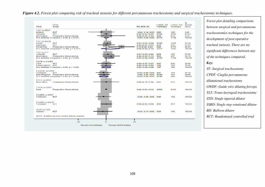
109
Figure 4.2. Forest plot comparing risk of tracheal stenosis for different percutaneous tracheostomy and surgical tracheostomy techniques.
Forest plot detailing comparisons
between surgical and percutaneous
tracheostomies techniques for the
development of post-operative
tracheal stenosis. There are no
significant differences between any
of the techniques compared.
Key: ST: Surgical tracheostomy
CPDT: Ciaglia percutaneous
dilatational tracheostomy
GWDF: Guide wire dilating forceps
TLT: Trans-laryngeal tracheostomy
STD: Single tapered dilator
SSRD: Single step rotational dilator
BD: Balloon dilator
RCT: Randomised controlled trial
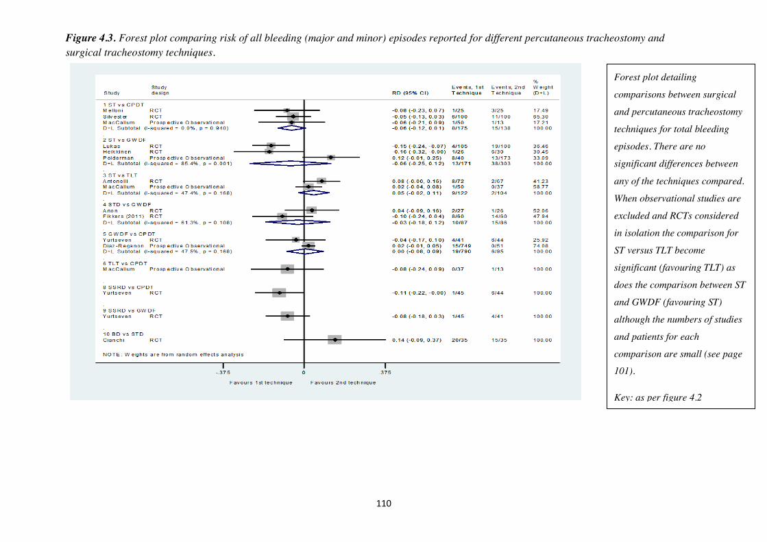
110
Figure 4.3. Forest plot comparing risk of all bleeding (major and minor) episodes reported for different percutaneous tracheostomy and surgical tracheostomy techniques.
Forest plot detailing
comparisons between surgical
and percutaneous tracheostomy
techniques for total bleeding
episodes. There are no
significant differences between
any of the techniques compared.
When observational studies are
excluded and RCTs considered
in isolation the comparison for
ST versus TLT become
significant (favouring TLT) as
does the comparison between ST
and GWDF (favouring ST)
although the numbers of studies
and patients for each
comparison are small (see page
101).
Key: as per figure 4.2
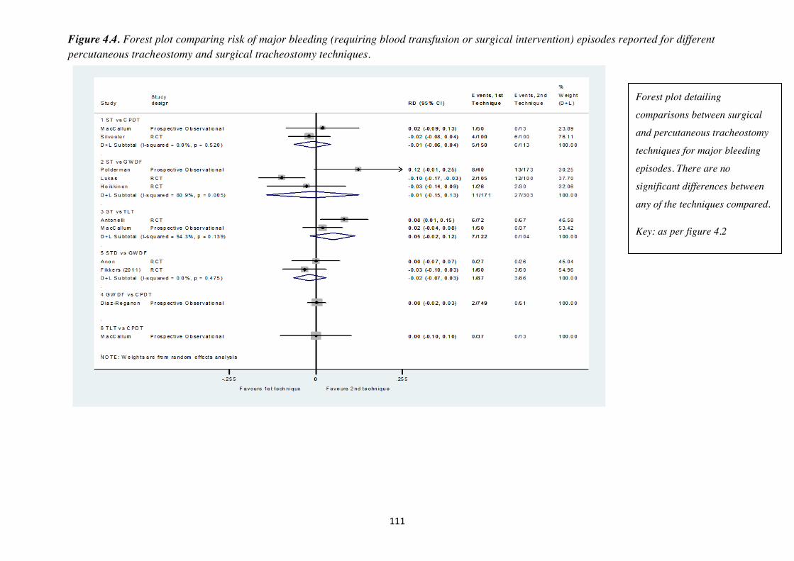
111
Figure 4.4. Forest plot comparing risk of major bleeding (requiring blood transfusion or surgical intervention) episodes reported for different percutaneous tracheostomy and surgical tracheostomy techniques.
Forest plot detailing
comparisons between surgical
and percutaneous tracheostomy
techniques for major bleeding
episodes. There are no
significant differences between
any of the techniques compared.
Key: as per figure 4.2
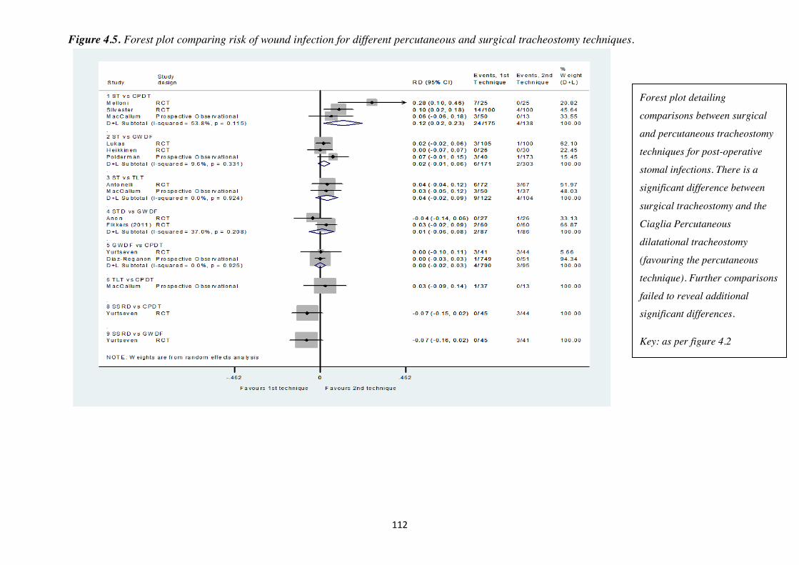
112
Figure 4.5. Forest plot comparing risk of wound infection for different percutaneous and surgical tracheostomy techniques.
Forest plot detailing
comparisons between surgical
and percutaneous tracheostomy
techniques for post-operative
stomal infections. There is a
significant difference between
surgical tracheostomy and the
Ciaglia Percutaneous
dilatational tracheostomy
(favouring the percutaneous
technique). Further comparisons
failed to reveal additional
significant differences.
Key: as per figure 4.2
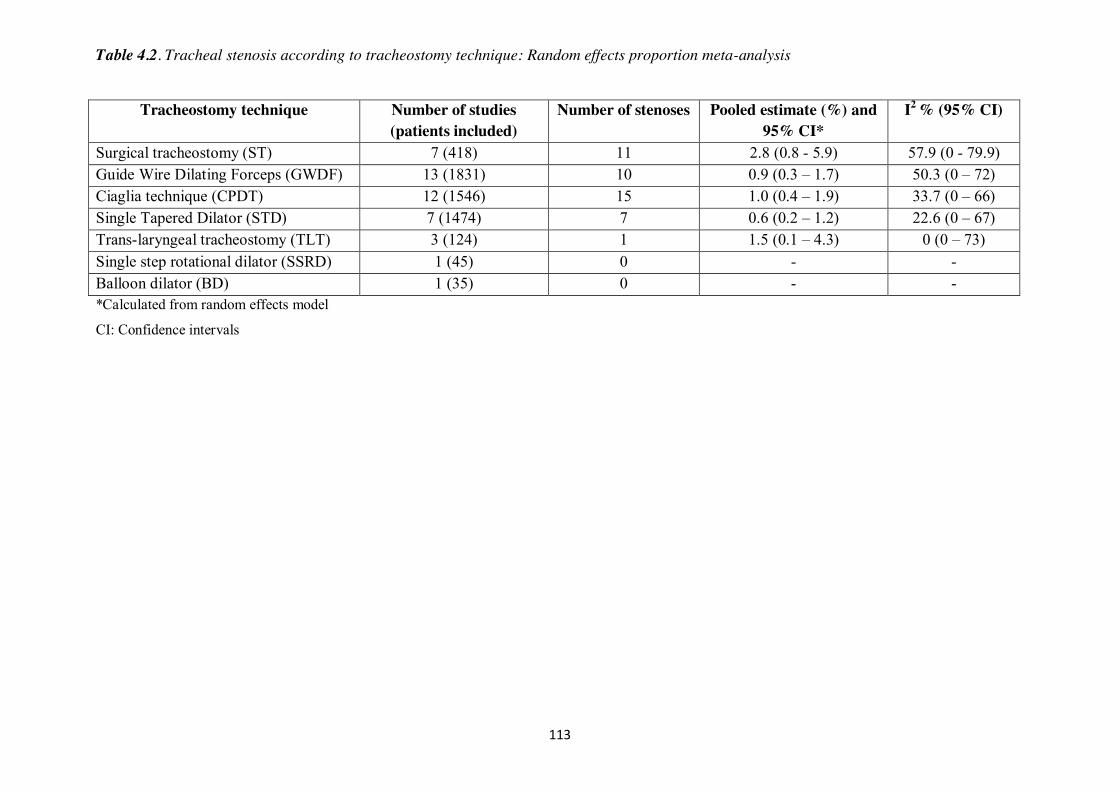
113
Table 4.2. Tracheal stenosis according to tracheostomy technique: Random effects proportion meta-analysis
Tracheostomy technique Number of studies
(patients included) Number of stenoses Pooled estimate (%) and
95% CI* I2 % (95% CI)
Surgical tracheostomy (ST) 7 (418) 11 2.8 (0.8 - 5.9) 57.9 (0 - 79.9) Guide Wire Dilating Forceps (GWDF) 13 (1831) 10 0.9 (0.3 – 1.7) 50.3 (0 – 72) Ciaglia technique (CPDT) 12 (1546) 15 1.0 (0.4 – 1.9) 33.7 (0 – 66) Single Tapered Dilator (STD) 7 (1474) 7 0.6 (0.2 – 1.2) 22.6 (0 – 67) Trans-laryngeal tracheostomy (TLT) 3 (124) 1 1.5 (0.1 – 4.3) 0 (0 – 73) Single step rotational dilator (SSRD) 1 (45) 0 - - Balloon dilator (BD) 1 (35) 0 - - *Calculated from random effects model
CI: Confidence intervals
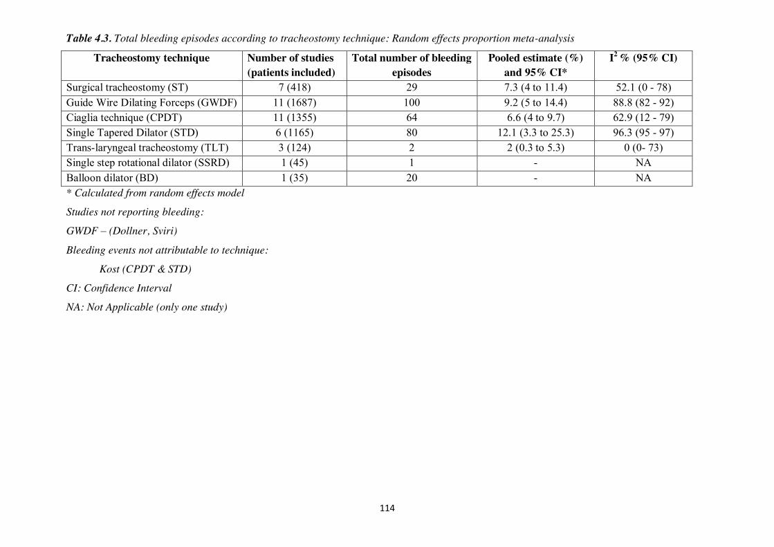
114
Table 4.3. Total bleeding episodes according to tracheostomy technique: Random effects proportion meta-analysis
Tracheostomy technique Number of studies (patients included)
Total number of bleeding episodes
Pooled estimate (%) and 95% CI*
I2 % (95% CI)
Surgical tracheostomy (ST) 7 (418) 29 7.3 (4 to 11.4) 52.1 (0 - 78) Guide Wire Dilating Forceps (GWDF) 11 (1687) 100 9.2 (5 to 14.4) 88.8 (82 - 92) Ciaglia technique (CPDT) 11 (1355) 64 6.6 (4 to 9.7) 62.9 (12 - 79) Single Tapered Dilator (STD) 6 (1165) 80 12.1 (3.3 to 25.3) 96.3 (95 - 97) Trans-laryngeal tracheostomy (TLT) 3 (124) 2 2 (0.3 to 5.3) 0 (0- 73) Single step rotational dilator (SSRD) 1 (45) 1 - NA Balloon dilator (BD) 1 (35) 20 - NA * Calculated from random effects model
Studies not reporting bleeding:
GWDF – (Dollner, Sviri)
Bleeding events not attributable to technique:
Kost (CPDT & STD)
CI: Confidence Interval
NA: Not Applicable (only one study)
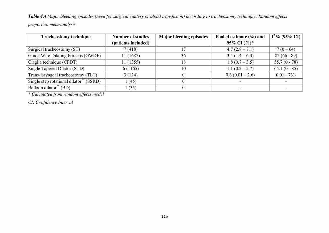
115
Table 4.4 Major bleeding episodes (need for surgical cautery or blood transfusion) according to tracheostomy technique: Random effects
proportion meta-analysis
Tracheostomy technique Number of studies (patients included)
Major bleeding episodes
Pooled estimate (%) and 95% CI (%)*
I2 % (95% CI)
Surgical tracheostomy (ST) 7 (418) 17 4.7 (2.8 – 7.1) 7 (0 – 64) Guide Wire Dilating Forceps (GWDF) 11 (1687) 36 3.4 (1.4 – 6.3) 82 (66 - 89) Ciaglia technique (CPDT) 11 (1355) 18 1.8 (0.7 – 3.5) 55.7 (0 - 78) Single Tapered Dilator (STD) 6 (1165) 10 1.1 (0.2 – 2.7) 65.1 (0 - 85) Trans-laryngeal tracheostomy (TLT) 3 (124) 0 0.6 (0.01 – 2.6) 0 (0 – 73)- Single step rotational dilator** (SSRD) 1 (45) 0 - - Balloon dilator** (BD) 1 (35) 0 - - * Calculated from random effects model
CI: Confidence Interval
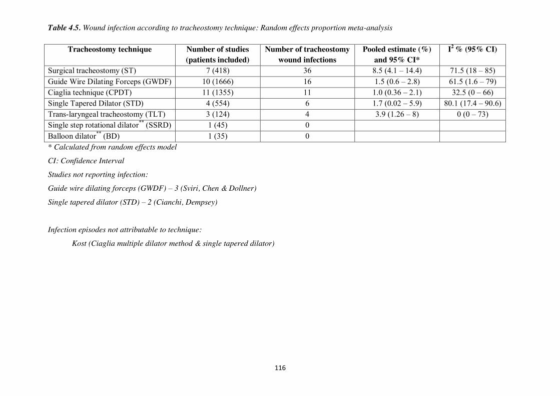
116
Table 4.5. Wound infection according to tracheostomy technique: Random effects proportion meta-analysis
Tracheostomy technique Number of studies (patients included)
Number of tracheostomy wound infections
Pooled estimate (%) and 95% CI*
I2 % (95% CI)
Surgical tracheostomy (ST) 7 (418) 36 8.5 (4.1 – 14.4) 71.5 (18 – 85) Guide Wire Dilating Forceps (GWDF) 10 (1666) 16 1.5 (0.6 – 2.8) 61.5 (1.6 – 79) Ciaglia technique (CPDT) 11 (1355) 11 1.0 (0.36 – 2.1) 32.5 (0 – 66) Single Tapered Dilator (STD) 4 (554) 6 1.7 (0.02 – 5.9) 80.1 (17.4 – 90.6) Trans-laryngeal tracheostomy (TLT) 3 (124) 4 3.9 (1.26 – 8) 0 (0 – 73) Single step rotational dilator** (SSRD) 1 (45) 0 Balloon dilator** (BD) 1 (35) 0 * Calculated from random effects model
CI: Confidence Interval
Studies not reporting infection:
Guide wire dilating forceps (GWDF) – 3 (Sviri, Chen & Dollner)
Single tapered dilator (STD) – 2 (Cianchi, Dempsey)
Infection episodes not attributable to technique:
Kost (Ciaglia multiple dilator method & single tapered dilator)
117
4.4 Discussion
We have investigated the incidence of long-term TS in patients undergoing
tracheostomy in a critical care setting. Additionally, we have estimated the incidence
of peri-procedural complications (bleeding and wound infection) that we have
postulated may have a role in the development of TS. When considering published
RCTs and comparative observational studies reporting long-term outcomes we have
found ST and all percutaneous techniques (CPDT, GWDF, STD, TLT, SSRD, BD)
broadly similar in terms of TS and bleeding. In keeping with earlier meta-analyses we
have also found a higher incidence of wound infection when comparing ST to
CPDT51. Despite the frequency with which percutaneous tracheostomies are
performed within the critical care setting there appear to be relatively few high quality
studies assessing long term outcomes between techniques.
When considering our pooled proportions meta-analysis across all studies, the TS rate
reported varies from 2.8 to 0.6% for ST and STD respectively with all percutaneous
techniques being broadly comparable (table 4.2). The point estimate of rate for total
bleeding episodes varies from 12.1% for STD to 2% for TLT (table 4.3) but the
confidence intervals around some of these estimates are wide with a substantial
degree of between study heterogeneity. Major bleeding ranges from 4.7% for ST to
0.6% for TLT, whilst wound infection for ST is 9.9% and 1.1% for CPDT, again with
wide 95% CIs and substantial between study heterogeneity.
By incorporating cohort studies as well as RCTs into our analysis we have adopted a
different methodology to earlier meta-analyses. The majority of studies incorporated
into previous analyses were RCTs of relatively small samples sizes and as such, given
the low incidence, were limited in their ability to detect a difference in risk of TS.
Whilst the incorporation of non-randomised studies may introduce an element of bias,
118
work by Golder et al has suggested this effect may be minimal314. In a meta-analysis
of meta-analyses they found a high degree of concordance between meta-analyses of
adverse events comparing data solely from RCTs with those from both RCTs and
observational studies (with less discrepancy for larger studies) concluding that meta-
analyses of adverse events should not, necessarily, be confined to specific study types.
In a sensitivity analysis, we excluded the non-randomised studies and compared meta-
analysis results to those that include both RCT and prospective observational studies.
Overall, results were similar but changed from being non- significant to statistically
significant for bleeding complication for two comparisons, suggesting that there may
be important differences between RCTs and observational studies (see below).
Another potential limitation to accurate determination of the incidence of TS,
following percutaneous tracheostomy, in the current study is the possibility that
patients who are perceived to be predisposed to a difficult tracheostomy may be
assigned to the ST group in non-randomised studies. In support of this concern, of the
seven studies assessing ST, five were RCTs 297,298,303,305,310 reporting a TS incidence
of 1.4% (4/328) and two were prospective non-randomised studies with a TS
incidence of 8.4% (7/90)300,311. In addition, the risk difference estimates for TS from
the prospective non-randomised studies were each more extreme than corresponding
estimates from RCTs in those comparisons between ST and percutaneous
tracheostomy. Of the non-randomised studies, the study by MacCallum (reporting 2
stenoses) provides no information on group allocation other than stating that
procedures were performed consecutively300. In contrast, Polderman (5 stenoses)
reports that 7 patients were allocated to the ST group because of perceived difficult
anatomy whilst the remainder of patients were allocated randomly.311 No information
as to whether those patients with difficult anatomy were also the patients who
119
developed TS was provided. The choice between surgical and percutaneous
tracheostomy in most situations is not a random event with most operators opting for
the surgical approach when potential difficulties or safety concerns are anticipated. It
is probable, therefore, that many of the surgical tracheostomies described within the
studies herein would be more prone to the occurrence of complications. It is possible,
therefore, that the observed differences in complication rates we have demonstrated in
our analyses could be due to patient factors rather than the operative technique per se.
However, as discussed above, other than a potential role for tracheal ring fracture
there is little evidence for peri-operative complications causing TS.
Accepting these potential limitations, in the critical care setting, there is a trend
toward increased risk of TS for surgical tracheostomy patients.
Our data confirm our view that in the critical care setting, in common with previous
authors, a percutaneous procedure should be the technique of choice as it is both safe
and cost effective51.
It is somewhat surprising (and a common theme across all previous meta-analyses),
given the frequency with which PDTs are now undertaken, that we have only
identified 29 studies reporting upon the long term outcomes of 5473 patients over the
preceding 14 years. We find it concerning that some techniques in relatively
widespread use (SSRD, BD, TLT) have such sparse long-term outcome data. When
specifically considering the issue of TS and the possibility of unrecognised sub-
clinical TS in this patient population, very few studies have undertaken
comprehensive radiological imaging to determine the underlying prevalence of
stenosis. Considering that the STD technique appears to be currently the most widely
used approach,45,46,189 until recently there were only 14 patients described in the
120
literature who had undergone radiological imaging after STD tracheostomy to
determine the underlying prevalence of both clinical and sub-clinical TS80.
Reporting of complications across studies remains far from standardised and posed
significant problems during data extraction. We had initially set out to collect data for
a significant number of secondary outcomes (appendix 1). However, lack of uniform
reporting made this aim much more difficult and ultimately of little value as many
complications are reported infrequently with marked differences in incidences across
studies. Even reporting of our primary and secondary outcomes was inconsistent, with
some studies failing to quote an incidence for TS, bleeding and wound infection. One
study appeared to describe an apparent incidence of TS (presence of stridor with
spirometry findings in keeping with upper airway obstruction) but failed to ascribe
these findings to a diagnosis of TS.296 A lack of a consistent definition for significant
versus minor bleeding lead us to analyse total bleeding episodes as well as those
categorised as major. Additionally, one study reported stomal decay as an outcome,
interpreted as infection for our analysis.297 Additionally, we were unable to explore
the possible relationships between bleeding and stomal infection and TS. Whilst most
of the studies included in the analysis reported TS, bleeding and stomal infection rates
there was little, if any, supplemental data reported for us to ascertain whether these
events were associated and occurring within the same patients or not. A number of
other studies that detailed complications of multiple techniques reported composite
outcomes where the complication rate for a given technique was impossible to
ascertain. In this circumstance we wrote to the author for clarification. In some
instances this resolved the problem either completely or partially. In others, where
there was no reply from the corresponding author the study was excluded from the
121
analysis. These limitations highlight the vital need to develop and report standardised
core outcome measures to improve the synthesis of trial data.315
Whilst RCTs to compare long term outcomes, particularly TS, following individual
PDT techniques would be desirable it is highly unlikely that they will be performed.
TS has a low incidence, critical illness mortality is high and long term follow up
difficult. An adequately powered study would require at least 2000 patients to detect a
reduction in TS from 2.8% to 1% (α = 0.05 β = 0.8) and this figure excludes drop out.
Thus we believe our study, despite the limitations described, is currently best placed
to inform clinicians in this area.
122
Chapter 5
Single tapered dilator percutaneous tracheostomy in
the critical care unit: An eleven-year single centre
review
123
5.1 Methods
This prospective service evaluation was carried out at Aintree University Hospital
NHS Foundation Trust (AUH). The critical care unit (CCU) at AUH is a 23-bedded
mixed medical and surgical unit undertaking between 80 - 100 PDTs per year. The
regional referral centre for head and neck cancer, the largest such unit in the UK, is
also situated at AUH. The need for PDT in any individual case was determined by the
duty consultant intensivist. After the decision to proceed with a tracheostomy, all
patients were fully evaluated and examined before operation. In all cases, when a
tracheostomy is deemed appropriate, patients are assessed as to their suitability for a
PDT. Patients undergoing ST as part of their surgical management for head and neck
cancer were not included in the study.
From November 2003 to February 2015, data were prospectively collected on all
PDTs performed at AUH. All PDTs were performed by critical care physicians at the
bedside using the Blue Rhino® Percutaneous Tracheotomy Introducer Kit (Cook
Critical Care, Bloomington, IN). All patients were over the age of 16 and each
received intravenous general anaesthesia with neuromuscular block. Each procedure
was undertaken with bronchoscopic guidance to ensure correct placement of the
guide-wire.
Before commencement of the procedure, the patient was positioned with the head
extended and the lungs were ventilated with 100% oxygen. Throughout the procedure,
heart rate, arterial pressure, and oxygen saturation were monitored continuously. After
digital palpation of the neck, direct laryngoscopy was performed to position the
tracheal tube (TT) above the site of the proposed PDT insertion point. Local
anaesthesia and vasoconstriction were achieved using 2% lidocaine with 1:100,000
124
epinephrine administered subcutaneously to the pre-tracheal tissues. A 1 cm
horizontal skin incision was made midway between the cricoid cartilage and the
sternal notch. Pre-tracheal tissues were separated by blunt dissection. Tracheal
puncture was performed with the standard 15-gauge needle. After bronchoscopic
confirmation of the position, the guide-wire was passed into the tracheal lumen. A 4.5
cm 14 Fr gauge dilator was passed over the guide-wire after which the STD was used
to expand the tract between the skin and the tracheal lumen. The appropriate-sized
tracheostomy tube was introduced into the tracheal lumen utilising a loading dilator.
Perioperative data on each patient were recorded prospectively and included age, sex,
reason for admission, admission APACHE II score, and intraoperative and
postoperative complications as detailed below.
A PDT was assigned as ‘technically difficult’ or complicated as described
previously59,316. Routine post-procedural chest X-rays (CXRs) were performed after
the first 384 PDTs. Thereafter, CXRs were limited to those PDTs considered
technically difficult.
After critical care discharge, further surveillance was provided by the AUH Critical
Care Outreach Service and after hospital discharge by the critical care follow-up
clinic allowing identification of late complications. Long-term survival data was
cross-referenced with the AUH clinical records system (System C Medway Sigma,
Maidstone, UK).
125
5.2 Results
During the study period, 1056 CCU patients required a tracheostomy. In 1025
patients, a PDT was attempted, and in 1019 (99%), the procedure was completed
successfully at the first attempt (Table 5.1). Two further patients underwent
successful PDT at the second attempt. In four patients (0.4%), PDT was abandoned
due to excess bleeding; three of these patients subsequently went on to have an ST.
Consequently, a total of 31 (3% (n=1056) patients underwent ST. In the remaining
patient, the procedure was abandoned and ST considered inappropriate due to
worsening multiple organ failure. All PDTs were undertaken between the hours of
09:00 and 17:00 and 929 (91%) were performed on a weekday (figure 5.1). Number
of PDTs performed by year is presented in figure 5.2. Nine hundred and fifty one
(93%) procedures followed an emergency critical care admission. In 813 (79%) cases,
the most senior clinician present was a consultant. For the remainder, in 190 (19%)
and 22 (2%) cases, the most senior clinician present was a senior critical care trainee
(clinical fellow) and a specialist anaesthetic registrar, respectively.
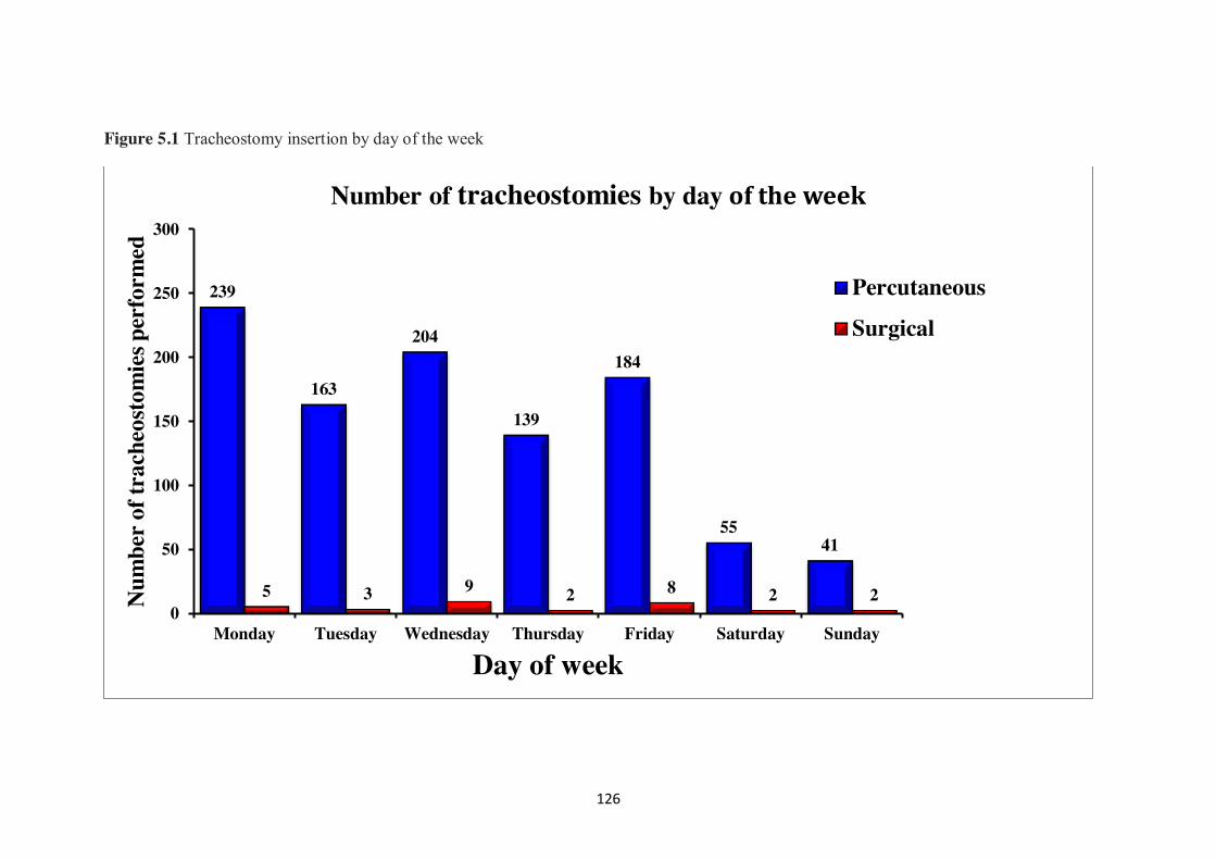
126
Figure 5.1 Tracheostomy insertion by day of the week
239
163
204
139
184
55 41
5 3 9 2 8 2 2 0
50
100
150
200
250
300
Monday Tuesday Wednesday Thursday Friday Saturday Sunday
Num
ber o
f tra
cheo
stom
ies p
erfo
rmed
Day of week
Number of tracheostomies by day of the week
Percutaneous
Surgical
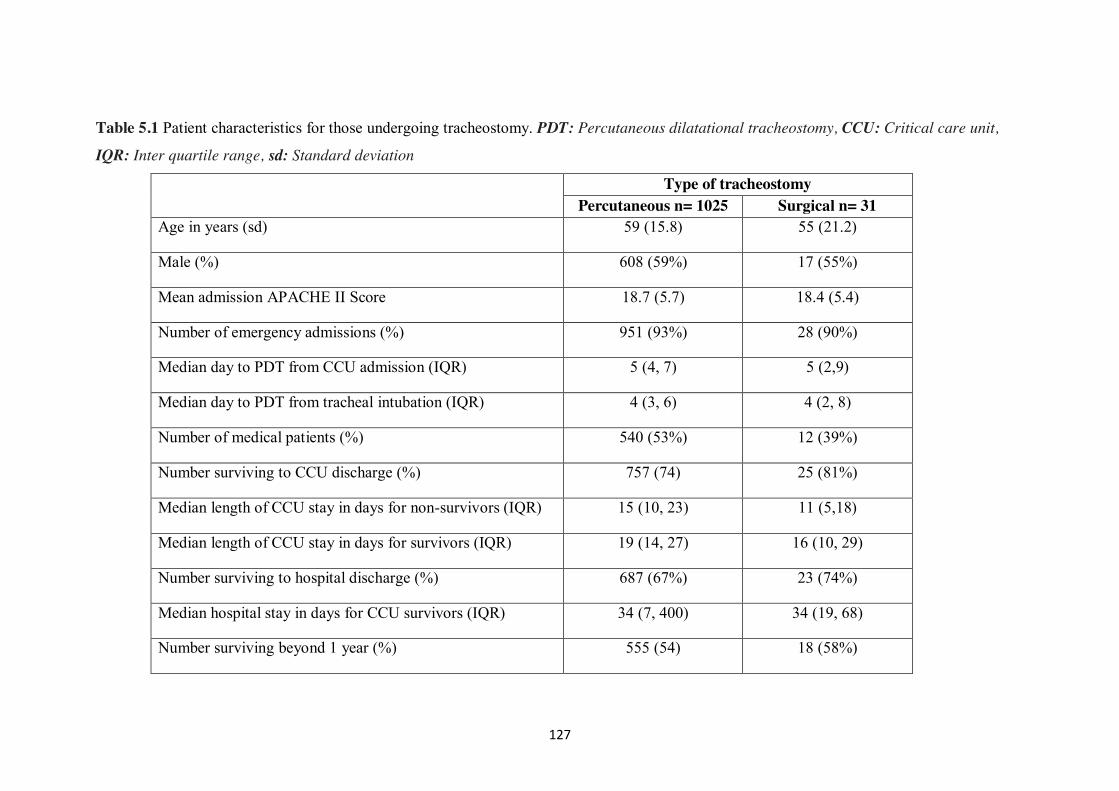
127
Table 5.1 Patient characteristics for those undergoing tracheostomy. PDT: Percutaneous dilatational tracheostomy, CCU: Critical care unit,
IQR: Inter quartile range, sd: Standard deviation
Type of tracheostomy Percutaneous n= 1025 Surgical n= 31
Age in years (sd) 59 (15.8) 55 (21.2)
Male (%) 608 (59%) 17 (55%)
Mean admission APACHE II Score 18.7 (5.7) 18.4 (5.4)
Number of emergency admissions (%) 951 (93%) 28 (90%)
Median day to PDT from CCU admission (IQR) 5 (4, 7) 5 (2,9)
Median day to PDT from tracheal intubation (IQR) 4 (3, 6) 4 (2, 8)
Number of medical patients (%) 540 (53%) 12 (39%)
Number surviving to CCU discharge (%) 757 (74) 25 (81%)
Median length of CCU stay in days for non-survivors (IQR) 15 (10, 23) 11 (5,18)
Median length of CCU stay in days for survivors (IQR) 19 (14, 27) 16 (10, 29)
Number surviving to hospital discharge (%) 687 (67%) 23 (74%)
Median hospital stay in days for CCU survivors (IQR) 34 (7, 400) 34 (19, 68)
Number surviving beyond 1 year (%) 555 (54) 18 (58%)
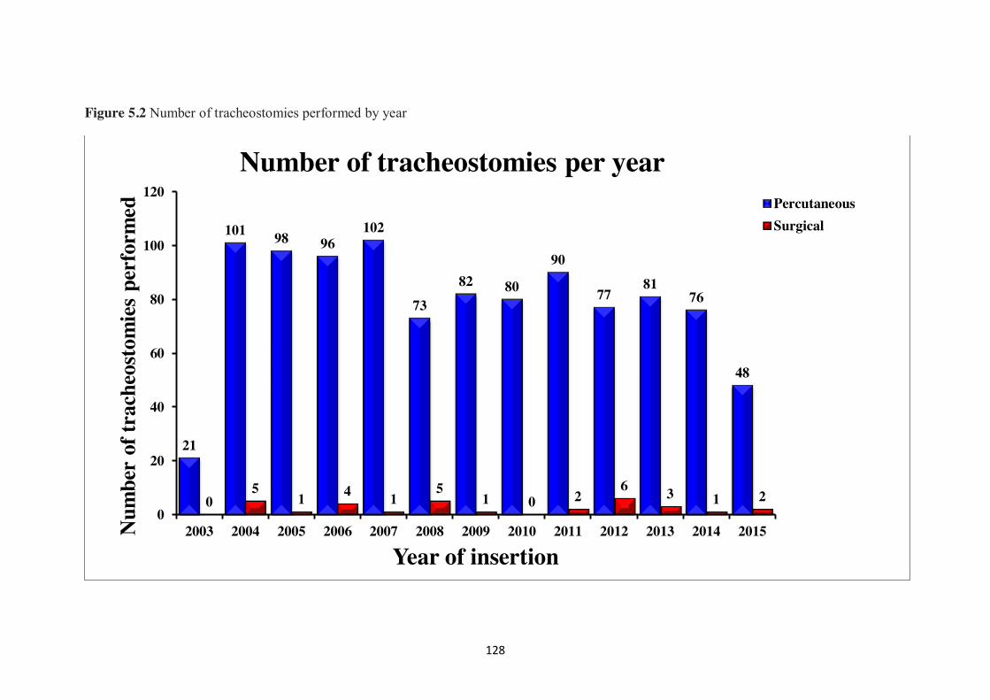
128
Figure 5.2 Number of tracheostomies performed by year
21
101 98 96 102
73 82 80
90
77 81
76
48
0 5
1 4 1 5
1 0 2 6 3 1 2
0
20
40
60
80
100
120
2003 2004 2005 2006 2007 2008 2009 2010 2011 2012 2013 2014 2015Num
ber
of tr
ache
osto
mie
s pe
rfor
med
Year of insertion
Number of tracheostomies per year PercutaneousSurgical
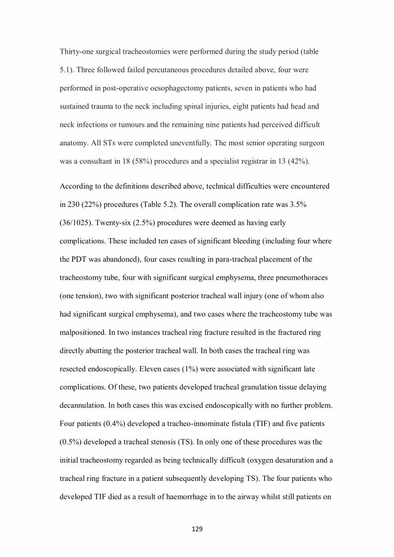
129
Thirty-one surgical tracheostomies were performed during the study period (table
5.1). Three followed failed percutaneous procedures detailed above, four were
performed in post-operative oesophagectomy patients, seven in patients who had
sustained trauma to the neck including spinal injuries, eight patients had head and
neck infections or tumours and the remaining nine patients had perceived difficult
anatomy. All STs were completed uneventfully. The most senior operating surgeon
was a consultant in 18 (58%) procedures and a specialist registrar in 13 (42%).
According to the definitions described above, technical difficulties were encountered
in 230 (22%) procedures (Table 5.2). The overall complication rate was 3.5%
(36/1025). Twenty-six (2.5%) procedures were deemed as having early
complications. These included ten cases of significant bleeding (including four where
the PDT was abandoned), four cases resulting in para-tracheal placement of the
tracheostomy tube, four with significant surgical emphysema, three pneumothoraces
(one tension), two with significant posterior tracheal wall injury (one of whom also
had significant surgical emphysema), and two cases where the tracheostomy tube was
malpositioned. In two instances tracheal ring fracture resulted in the fractured ring
directly abutting the posterior tracheal wall. In both cases the tracheal ring was
resected endoscopically. Eleven cases (1%) were associated with significant late
complications. Of these, two patients developed tracheal granulation tissue delaying
decannulation. In both cases this was excised endoscopically with no further problem.
Four patients (0.4%) developed a tracheo-innominate fistula (TIF) and five patients
(0.5%) developed a tracheal stenosis (TS). In only one of these procedures was the
initial tracheostomy regarded as being technically difficult (oxygen desaturation and a
tracheal ring fracture in a patient subsequently developing TS). The four patients who
developed TIF died as a result of haemorrhage in to the airway whilst still patients on
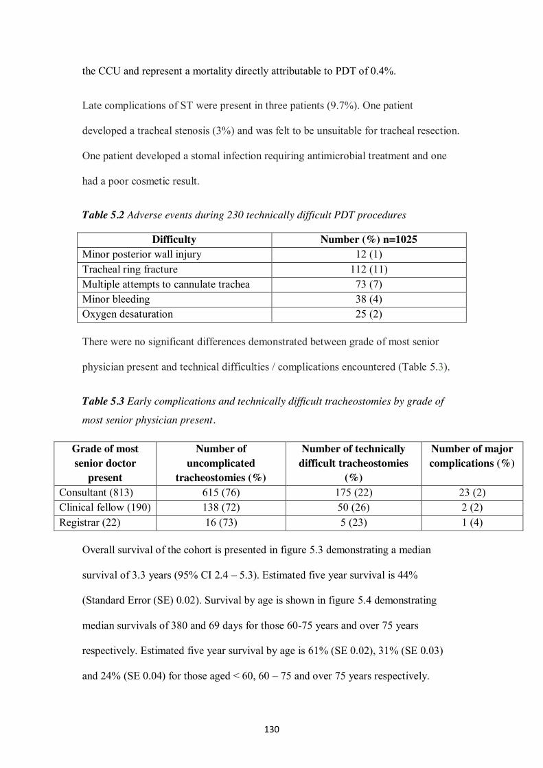
130
the CCU and represent a mortality directly attributable to PDT of 0.4%.
Late complications of ST were present in three patients (9.7%). One patient
developed a tracheal stenosis (3%) and was felt to be unsuitable for tracheal resection.
One patient developed a stomal infection requiring antimicrobial treatment and one
had a poor cosmetic result.
Table 5.2 Adverse events during 230 technically difficult PDT procedures
Difficulty Number (%) n=1025 Minor posterior wall injury 12 (1) Tracheal ring fracture 112 (11) Multiple attempts to cannulate trachea 73 (7) Minor bleeding 38 (4) Oxygen desaturation 25 (2) There were no significant differences demonstrated between grade of most senior
physician present and technical difficulties / complications encountered (Table 5.3).
Table 5.3 Early complications and technically difficult tracheostomies by grade of
most senior physician present.
Grade of most senior doctor
present
Number of uncomplicated
tracheostomies (%)
Number of technically difficult tracheostomies
(%)
Number of major complications (%)
Consultant (813) 615 (76) 175 (22) 23 (2) Clinical fellow (190) 138 (72) 50 (26) 2 (2) Registrar (22) 16 (73) 5 (23) 1 (4)
Overall survival of the cohort is presented in figure 5.3 demonstrating a median
survival of 3.3 years (95% CI 2.4 – 5.3). Estimated five year survival is 44%
(Standard Error (SE) 0.02). Survival by age is shown in figure 5.4 demonstrating
median survivals of 380 and 69 days for those 60-75 years and over 75 years
respectively. Estimated five year survival by age is 61% (SE 0.02), 31% (SE 0.03)
and 24% (SE 0.04) for those aged < 60, 60 – 75 and over 75 years respectively.
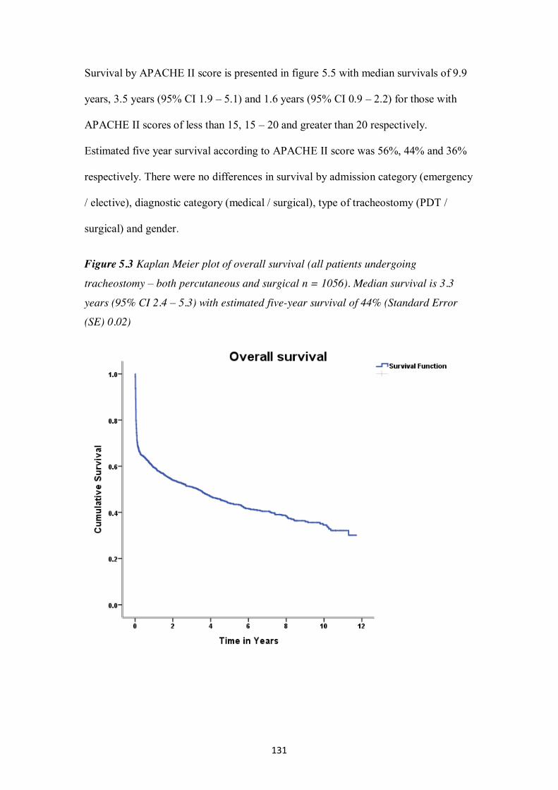
131
Survival by APACHE II score is presented in figure 5.5 with median survivals of 9.9
years, 3.5 years (95% CI 1.9 – 5.1) and 1.6 years (95% CI 0.9 – 2.2) for those with
APACHE II scores of less than 15, 15 – 20 and greater than 20 respectively.
Estimated five year survival according to APACHE II score was 56%, 44% and 36%
respectively. There were no differences in survival by admission category (emergency
/ elective), diagnostic category (medical / surgical), type of tracheostomy (PDT /
surgical) and gender.
Figure 5.3 Kaplan Meier plot of overall survival (all patients undergoing
tracheostomy – both percutaneous and surgical n = 1056). Median survival is 3.3
years (95% CI 2.4 – 5.3) with estimated five-year survival of 44% (Standard Error
(SE) 0.02)
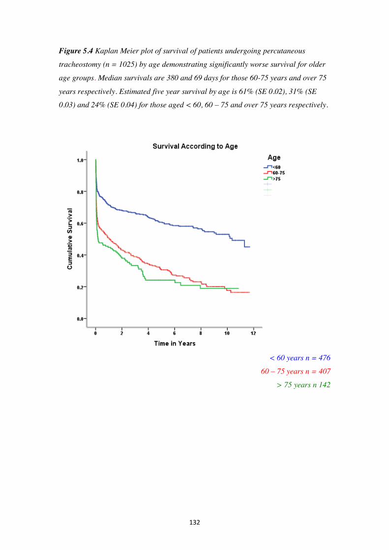
132
Figure 5.4 Kaplan Meier plot of survival of patients undergoing percutaneous
tracheostomy (n = 1025) by age demonstrating significantly worse survival for older
age groups. Median survivals are 380 and 69 days for those 60-75 years and over 75
years respectively. Estimated five year survival by age is 61% (SE 0.02), 31% (SE
0.03) and 24% (SE 0.04) for those aged < 60, 60 – 75 and over 75 years respectively.
< 60 years n = 476
60 – 75 years n = 407
> 75 years n 142
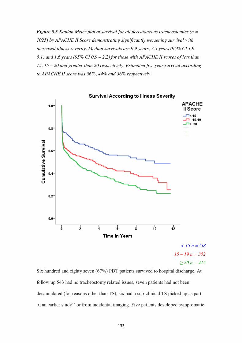
133
Figure 5.5 Kaplan Meier plot of survival for all percutaneous tracheostomies (n =
1025) by APACHE II Score demonstrating significantly worsening survival with
increased illness severity. Median survivals are 9.9 years, 3.5 years (95% CI 1.9 –
5.1) and 1.6 years (95% CI 0.9 – 2.2) for those with APACHE II scores of less than
15, 15 – 20 and greater than 20 respectively. Estimated five year survival according
to APACHE II score was 56%, 44% and 36% respectively.
< 15 n =258
15 – 19 n = 352
≥ 20 n = 415
Six hundred and eighty seven (67%) PDT patients survived to hospital discharge. At
follow up 543 had no tracheostomy related issues, seven patients had not been
decannulated (for reasons other than TS), six had a sub-clinical TS picked up as part
of an earlier study79 or from incidental imaging. Five patients developed symptomatic
134
TS, one underwent corrective surgery, one died from rapidly progressive malignant
disease and three had no surgical management. Ninety-seven patients had no
documented follow up appointment. Of these 63 had been referred from outside our
catchment area and 34 were AUH patients. Of the remaining 30 patients, 16 died
before a follow up appointment and 13 had not reached 3 months from hospital
discharge at the time of writing.
5.3 Discussion
When considering these updated results in light of our previous report the most
reassuring finding is the consistency of the outcomes. The rate of technical
difficulties, early and late complications have changed little. In our earlier paper we
described a 26% rate of technically difficult PDTs. This has reduced slightly over
time to 22% in the current paper. This would indicate a technical difficulty rate of
18% for the more recent patients (149 / 576 versus 81 / 449). Similarly, the early
complication rate remains low at 2.5 percent (26 / 1025) in comparison to the
previously reported three percent (16 / 576). Again, when considering the outcomes
for more recent patients the rates remain almost identical at 2.2% (10 / 449). The rates
for later complications have remained constant across both study periods with four
tracheo-innominate fistulae (2/576, 2/449) and five tracheal stenoses (2/576, 3/449).
Given the high consultant presence during the PDTs undertaken a reduction in peri-
operative technical difficulties with time may point to a prolonged learning curve
where continued exposure and practice may subtly diminish complications. Given the
constancy of our late complications it may also indicate that peri-operative events
have a relatively small role to play in long term outcomes with patient factors being
the most important determinant here.
When comparing our data with previous reports the most direct comparisons come
135
from large, long term observational studies similar to our own. Kearney et al, in
perhaps the first large observational cohort study of PDT, described their eight year
experience of the original CPDT reporting an overall complication rate of 15%188.
The intra-operative complication rate was six percent with the most frequent events
being premature tracheal extubation, bleeding and para-tracheal placement of the
tracheostomy tube. They also reported major peri-operative complications of tracheal
laceration requiring thoracotomy (1 patient) and tracheo-oesophageal fistulae (2
patients). Their late complications consisted of a 1.6% rate of TS with no reported
TIF. They reported a procedure related death rate of 0.6% largely related to early
post-procedural complications. Diaz-Reganon et al reported on the outcomes of 800
predominantly guide wire dilating forceps (GWDF) PDTs (749 GWDF, 51 CPDT)58.
They reported an overall complication rate of four percent with 2.1% intra-operative,
0.75% early post-operative and 1.1% late. The most common procedural complication
was minor bleeding with one reported TS. Kost reported the outcomes of 500
procedures (191 CPDT, 309 STD) performed across a number of critical care units in
Montreal57. The overall complication rate was 9.2% although considering the STD
group alone this was 6.5% with the most common events being oxygen desaturation
and bleeding. Kost also reported upon the effects of a learning curve with the CPDT
technique in that complications were more common for the first 30 patients
undergoing CPDT. This effect was not apparent for the STD group. No cases of TS
were reported.
For the above studies the overall complication rates appear to be around 15% for
CPDT, 4% for GWDF, 6.5% for STD and 3.5% for the present study. Some of this
discrepancy can be accounted for on the basis of differing definitions for
complications used by each paper and some from a degree of reporting bias. There
136
may, however, also be an inherent difference in complication rates between the
techniques that has not previously been picked up in RCTs comparing the techniques.
In two recent meta-analyses Cabrini and co-workers have tried to better quantify this
situation54,55. In their earlier paper, using data from 13 RCTs, they noted that the
different techniques were largely comparable with the exception of the retrograde
(trans-laryngeal) tracheostomy which was associated with more severe
complications54. The STD technique was associated with fewer failures and less
minor complications leading the authors to conclude that it seemed to be the most
reliable in terms of safety and success rate. A later paper by the same authors assessed
five RCTs comparing GWDF to STD PDT using a composite outcome of difficult
cannula insertion / difficult dilatation / failure55. The incidence of this composite
outcome was 15.5% for GWDF versus 4.9% for STD. They also noted more frequent
bleeding with GWDF (19.3 v 7.6%) with no difference in long-term outcomes. A
recently completed systematic review incorporating data from RCTs and
observational studies has failed to show a difference in long term outcomes of
tracheostomy techniques commonly used within the critical care setting (ST, CPDT,
GWDF, STD, TLT, BD, SSRD) from RCT data. However, when adding data from
observational studies the pooled proportions meta-analysis seems to indicate a
difference in reported TS rates from 0.6% for STD to 2.8% for ST, very similar to the
rates described herein.317
To our knowledge, long-term outcomes and survival much beyond 12 months have
not been previously reported for PDT in the CCU setting. Our data represent survival
data for up to 11 years after PDT. Six hundred and eighty seven PDT patients (67%)
survived to hospital discharge. The overall median survival for the cohort was 3.3
years (95% CI 2.4 – 4.3). Unsurprisingly, this is significantly impacted upon by age
137
with median survival reducing to 69 days (95% CI 0, 292) for those patients aged
over 75 years. Similarly, illness severity significantly reduced survival with those
with an APACHE II score in excess of 20 having a median survival of 1.6 years (95%
CI 0.9 – 2.2). Perhaps somewhat unexpectedly, there were no outcome differences
related to category of admission (elective versus emergency and medical versus
surgical). To some extent this may be explained by those patients admitted to the
CCU after an elective surgical admission who subsequently require a PDT have
invariably had a significant complication during their stay which will adversely affect
their outcome. The reason for a lack of a difference between medical and surgical
patients is less clear but may be due to a degree of selection bias on the part of the
attending intensivist with PDT not being undertaken on those patients felt to be more
likely to have poorer outcomes.
The rate of PDT has changed over the course of our study whilst the number of
admissions per year has remained relatively constant. For the earlier years of the
study we were undertaking approximately 100 PDTs per year with this number falling
to around 80 for more recent years (figure 5.2)55. The reasons for this may be several.
Firstly, whilst the number of CCU admissions has remained relatively constant, the
case mix appears to have changed with an increase in level II patients. This may be
accounted for by the introduction of a hospital wide medical emergency team (MET)
in 2009 resulting in patients being referred to the unit at an earlier stage. Secondly,
during the course of the study, a number of papers have evaluated the role of
tracheostomy, particularly in relation to timing of the procedure, in the CCU22,23,24.
The most recent meta-analysis concluding that early tracheostomy produced no
mortality benefit, no reduction in ventilator associated pneumonia, length of stay or
sedative usage25. Given that each of the above studies have demonstrated a delay in
138
performing a tracheostomy invariably results in some patients not needing the
procedure at all this work may partly explain the reduction seen here. However, the
timeline does not entirely fit with the data as presented. Our tracheostomy rate fell
from 2008 onwards, with most of the above publications being dated 2010 or later.
Additionally, whilst our rate of PDT has fallen there has been little change in the
timing of the tracheostomy from our earlier study. This is largely due to the fact that
within our unit the tracheostomy is not utilised in isolation but as part of a package of
care to facilitate early withdrawal of sedation, mobilisation, enhanced nutrition and
weaning from mechanical ventilation.
Whilst the reported complication rates presented herein are in keeping with, if not an
improvement upon, those published previously this remains a single centre evaluation
with its attendant limitations. It may, however, add weight to the gathering evidence
that the STD PDT is one of the safer techniques available. Our rate of early technical
difficulties appears to be reducing whilst long-term outcomes remain unchanged.
139
Chapter 6
Tracheal stenosis following percutaneous dilatational
tracheostomy using the single tapered dilator: a MRI
study
140
6.1 Methods
This prospective cohort study was carried out at Aintree University Hospital NHS
Foundation Trust.
A database of all patients receiving PDT, including associated outcome data,
undertaken within the CCU at AUH has been kept since 2003. All PDTs are
performed by critical care physicians at the bedside using the STD technique with
bronchoscopic guidance. All patients were over the age of 18 and each received
intravenous general anaesthesia with neuromuscular blockade during the procedure.
Following ethical approval (Liverpool (UK) Research Ethics Committee: Reference
No 08/H1005/121) patients surviving for a minimum of three months from the
insertion of PDT were identified from the database. Cross-referencing with AUH
clinical records system (System C Medway Sigma, Maidstone, UK) and following
contact with the patient’s general practitioner, when required, confirmed whether
patients were still alive. They were then contacted by telephone, informed of the
nature of the study and invited to AUH to complete a simple questionnaire, undergo
spirometry and MRI scanning (figure 6.1).
Following written informed consent, background data including age, sex, admission
illness severity (APACHE II), duration of oro-tracheal intubation prior to PDT,
complications and technical difficulties during PDT59, duration of cannulation, length
of critical care and hospital stay were collected. A PDT was considered complicated if
there were one or more technical difficulties (multiple tracheal punctures (≥ 3),
tracheal ring fracture, minor bleeding - > 3 soaked gauze swabs) or significant
complications (pneumothorax, subcutaneous emphysema, major bleeding – need for
surgical intervention or transfusion). A Health Status Screening Questionnaire
141
(Appendix 2) was devised and used to ascertain evidence of pre-existing respiratory
disease, respiratory symptoms after critical care and current exercise tolerance.
Simple spirometry was then undertaken using a micro-loop spirometer (CareFusion
Health UK 232 Ltd, Basingstoke, UK) to obtain values for FEV1, PEFR and forced
vital capacity (FVC). The best of three readings obtained were used to calculate the
FEV1/PEFR ratio. This constitutes the Empey Index (EI) (EI= FEV1/PEFR) and
previous data have reported a ratio > 10 to be clinically significant195.
MRI scanning was undertaken using a Siemens 1.5 T Avanto scanner (Siemens,
Munich, Germany). A T2-weighted coronal scan using 5 mm slices was carried out,
along with a T1 flash sagittal 3D volume scan with 1 mm slices. Multi-planar
reformats were constructed from the volume scan in the axial and coronal planes. All
sequences included the trachea from the inferior border of the cricoid cartilage to the
carina. All images were reviewed by a single consultant radiologist, unaware of the
clinical details of the patient pertaining to the PDT. Coronal and sagittal
measurements were reported at the narrowest section of the trachea at or above the
level of the tracheostomy and compared to normal tracheal dimensions above or
below the abnormality. TCSA at each level was calculated using the Picture
Archiving and Communications System (PACS – Carestream Vue PACS, Carestream
Health, Hertfordshire, UK). A significant stenosis amounting to a reduction in the
TCSA of 10% or more when compared to normal trachea above or below the
abnormal segment was defined as TS for the purpose of this study in common with
previous similar studies181,191,192. All patients with identified TS on MRI were then
referred to a consultant head and neck surgeon for further evaluation. In these cases,
each patient was initially investigated with fibre-optic laryngo-tracheoscopy,
continuing to rigid tracheo-bronchoscopy under general anaesthesia if this was
142
considered necessary.
6.2 Statistical analysis
Data were analysed initially using Microsoft® (Redmond, WA, USA) Excel™
software. GraphPad (GraphPad Software Inc, La Jolla, CA, USA) was used to carry
out Fisher’s exact test for categorical data and Mann-Whitney U tests for continuous
data. A p value of less than 0.05 was considered statistically significant.
6.3 Results
At the time of the study the database contained 745 patients who had undergone STD
PDT more than three months previously. Four hundred and eighty-three of these
patients survived to hospital discharge. A further 178 died within six months of
hospital discharge, leaving 305 eligible for inclusion. One hundred and twenty
patients were contacted by telephone. Seventy patients declined the invitation or were
unable to attend for MRI scanning, including 11 who failed to present for pre-
arranged appointments. The remaining 50 patients underwent MRI (Figure 6.1). Of
these, 49 also attended for interview, during which a standardised questionnaire was
completed (Appendix 2).
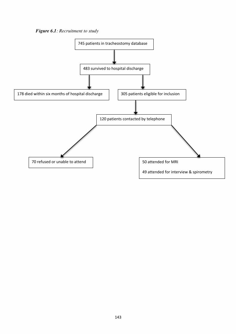
143
Figure 6.1: Recruitment to study
50 attended for MRI
49 attended for interview & spirometry
745 patients in tracheostomy database
178 died within six months of hospital discharge
120 patients contacted by telephone
70 refused or unable to attend
483 survived to hospital discharge
305 patients eligible for inclusion
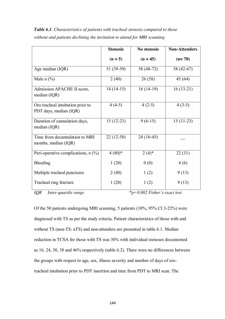
144
Table 6.1. Characteristics of patients with tracheal stenosis compared to those
without and patients declining the invitation to attend for MRI scanning
IQR Inter-quartile range *p=0.002 Fisher’s exact test
Of the 50 patients undergoing MRI scanning, 5 patients (10%, 95% CI 3-22%) were
diagnosed with TS as per the study criteria. Patient characteristics of those with and
without TS (non-TS: nTS) and non-attenders are presented in table 6.1. Median
reduction in TCSA for those with TS was 30% with individual stenoses documented
as 16, 24, 30, 38 and 46% respectively (table 6.2). There were no differences between
the groups with respect to age, sex, illness severity and number of days of oro-
tracheal intubation prior to PDT insertion and time from PDT to MRI scan. The
Stenosis
(n = 5)
No stenosis
(n = 45)
Non-Attenders
(n= 70)
Age median (IQR) 51 (39-59) 58 (48-72) 58 (42-67)
Male n (%) 2 (40) 26 (58) 45 (64)
Admission APACHE II score, median (IQR)
14 (14-15) 16 (14-19) 16 (13-21)
Oro tracheal intubation prior to PDT days, median (IQR)
4 (4-5) 4 (2-5) 4 (3-5)
Duration of cannulation days, median (IQR)
15 (12-21) 9 (6-15) 15 (11-23)
Time from decannulation to MRI months, median (IQR)
22 (12-58) 24 (10-45) __
Peri-operative complications, n (%)
Bleeding
Multiple tracheal punctures
Tracheal ring fracture
4 (80)*
1 (20)
2 (40)
1 (20)
2 (4)*
0 (0)
1 (2)
1 (2)
22 (31)
4 (6)
9 (13)
9 (13)
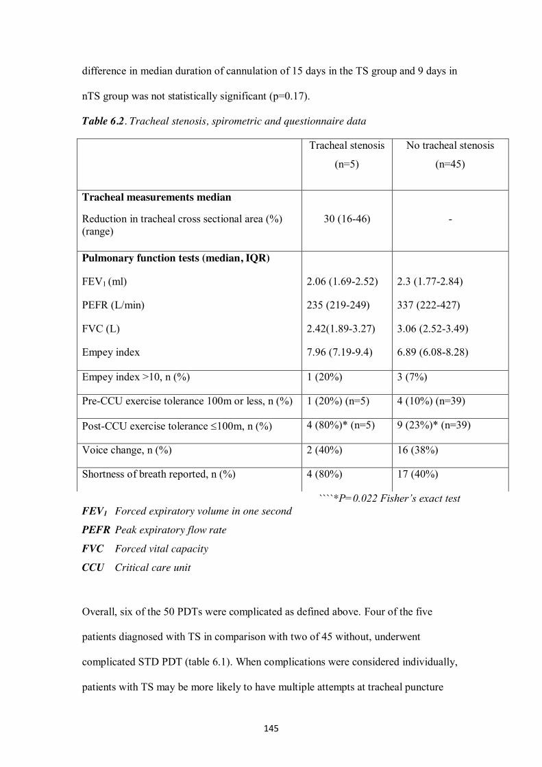
145
difference in median duration of cannulation of 15 days in the TS group and 9 days in
nTS group was not statistically significant (p=0.17).
Table 6.2. Tracheal stenosis, spirometric and questionnaire data
````*P=0.022 Fisher’s exact test FEV1 Forced expiratory volume in one second
PEFR Peak expiratory flow rate
FVC Forced vital capacity
CCU Critical care unit
Overall, six of the 50 PDTs were complicated as defined above. Four of the five
patients diagnosed with TS in comparison with two of 45 without, underwent
complicated STD PDT (table 6.1). When complications were considered individually,
patients with TS may be more likely to have multiple attempts at tracheal puncture
Tracheal stenosis
(n=5)
No tracheal stenosis
(n=45)
Tracheal measurements median
Reduction in tracheal cross sectional area (%) (range)
30 (16-46)
-
Pulmonary function tests (median, IQR)
FEV1 (ml)
PEFR (L/min)
FVC (L)
Empey index
2.06 (1.69-2.52)
235 (219-249)
2.42(1.89-3.27)
7.96 (7.19-9.4)
2.3 (1.77-2.84)
337 (222-427)
3.06 (2.52-3.49)
6.89 (6.08-8.28)
Empey index >10, n (%) 1 (20%) 3 (7%)
Pre-CCU exercise tolerance 100m or less, n (%) 1 (20%) (n=5) 4 (10%) (n=39)
Post-CCU exercise tolerance 100m, n (%) 4 (80%)* (n=5) 9 (23%)* (n=39)
Voice change, n (%) 2 (40%) 16 (38%)
Shortness of breath reported, n (%) 4 (80%) 17 (40%)
146
(2/5 versus 1/45) but there was no apparent difference between the groups in relation
to tracheal ring fracture (1/5 versus 1/45) or minor bleeding (1/5 versus 0/45) rates.
Results from the questionnaire revealed no differences in the presence of voice
changes (2/5 versus 16/45) and shortness of breath (4/5 versus 17/45) between those
with TS and those without. Two patients reported the presence of inspiratory noise –
neither were found to be stridulous or have TS on MRI scan. Data pertaining to
exercise tolerance was provided by 44 patients (5 TS and 39 nTS). Four of the five
patients with TS reported an exercise tolerance of 100 metres (m) or less, two of
whom reported unlimited exercise tolerance prior to critical care admission. In
contrast, 9 of 39 nTS patients (23%) reported an exercise tolerance of 100m or less.
Mean values for FEV1, PEFR and EI are shown in table 6.2. Neither FEV1, PEFR nor
EI were predictive of TS.
The five patients found to have TS on MRI scanning were referred to a consultant
head and neck surgeon for further evaluation and to assess the need for additional
intervention. At initial fibreoptic laryngo-tracheoscopy three of the five were deemed
to have an adequate airway and further intervention was deemed unnecessary. The
remaining two underwent rigid tracheo-bronchoscopic examination under general
anaesthesia and were again found to have an adequate airway. It was felt that further
surgical intervention would not significantly improve the airway, in a clinically
meaningful way, in any of the 5 patients with TS.
6.4 Discussion
We have found a sub-clinical TS rate of 10% in fifty patients investigated ranging
from a 16 – 46% reduction in TCSA. All patients with TS had fibre-optic laryngo-
tracheoscopic evaluation and two of the five had rigid tracheo-bronchoscopy. No
147
patient required any therapeutic intervention and all were felt to have an adequate
airway and were discharged from further follow-up. The low incidence and the sub-
clinical nature of the stenoses is reassuring, indicating the underlying prevalence of
undiagnosed TS in patients following STD PDT is of doubtful clinical significance.
There are limited studies in the literature reporting long term follow up of PDT
patients using radiological imaging (CT/MRI) with very few studying tracheal calibre
in patients who have had STD PDTs even though this is now probably the most
frequently used technique45,46,189. The 10% rate of TS described herein was less than
that found in another MRI study80. Fikkers compared long term outcome of patients
randomly allocated to either GWDF or STD PDTs. Sixty patients were assigned to
each group, 31 patients underwent MRI scanning (14 patients in the STD group), with
12 (39%) showing tracheal narrowing. Whilst the absolute number of patients with
tracheal narrowing demonstrated by Fikkers is larger than our own, the degree of
stenosis is similar in that all cases were felt to be of limited clinical consequence.
Two further studies utilised CT assessment. Norwood4 studied 100 patients who had
undergone PDT using the sequential dilators of the original Ciaglia technique
(CPDT)40. Forty-eight of these patients underwent CT scanning. Fifteen (31%)
patients were found to have more than 10% TS on CT with 10 (21%) of these being
mild (tracheal narrowing of 11-25%), 4 (8%) moderate (26-50% narrowing) and 1
(2%) severe (>50% narrowing). In Karvandian’s study, 20 patients were assessed
using CT and fibre-optic laryngotracheosopy after CPDT190. Only three of the 20
patients had cannulation times of less than three weeks (none of whom had TS) and
85% had significant sub-glottic stenosis (53% with < 50% narrowing). The study
group appeared different from our own with a mean duration of cannulation of 9
148
weeks. Additionally, 54 of 86 patients surviving four months after discharge from
hospital required permanent or long-term mechanical ventilation.
van Heurn described a TS prevalence of 26% in a study of 54 patients undergoing
plain linear tomography following CPDT191. Law used spirometry and fibre-optic
laryngo-tracheoscopy without any radiological assessment in 41 patients after
CPDT192. Three patients were found to have tracheal narrowing of between 10-30%
and one with a 40% stenosis. After a median cannulation time of 20 days, a 10% rate
of TS was reported.
The 10% rate of TS we describe is at the lower end of incidences reported above (10 –
85%). Part of this difference may be accounted for by differing definitions of TS.
However, the definition of TS as tracheal narrowing of >10% is common to most of
the studies cited above with only Fikkers80 and Karvandian190 not specifically stating
their definition of TS. The difference might also partly be attributed to shorter
tracheal cannulation times. In each of the studies described above the duration of
tracheal cannulation was longer than that described in our study (15 days in the TS
group and 9 days in the nTS group). Cannulation times ranged from 18 days in
Fikkers GWDF group80 to 9 weeks in Karvandian’s paper190. Those studies reporting
a lower prevalence of TS or only minor tracheal changes appear to have shorter
cannulation times80,192. A further consideration to take into account is the duration of
tracheal intubation prior to the insertion of PDT. PDT was carried out early in our
patients (median 4 days), whereas previous studies have reported durations from 7 –
18 days80,181,191,192. Another factor to be considered is PDT insertion technique used.
Most of the studies described above have used CPDT with only Fikkers80 evaluating
GWDF and STD techniques radiologically and then in only 15 and 14 patients
respectively. It is possible, given the paucity of data available, that the incidence of
149
post PDT TS may differ significantly between commonly used techniques.
Considering the widespread use of these techniques the lack of long-term radiological
outcome data would appear somewhat surprising.
The TS patients appear more likely to have undergone a complicated or technically
difficult PDT compared to the nTS group (table 6.1). The most frequent difficulty
encountered was the need for multiple tracheal punctures to site the cannula required
for guide-wire insertion. Whilst the significance of this finding is unclear, the
association between multiple tracheal punctures and later tracheal narrowing has not
been made previously. It is possible that several attempts to position the cannula may
ultimately lead the operator to settle for a suboptimal position for the tracheostomy
tube potentially causing tracheal injury and later narrowing.
As all cases of TS detected were clinically asymptomatic, with none requiring
surgical correction, it is difficult to explain the significance and relevance of the
reduced exercise tolerance found in these patients (four out of five reporting
significantly limited exercise tolerance, two of whom had previously been unlimited).
Conventionally TS is expected to become evident clinically when the tracheal
narrowing reaches 50 – 75%. However, many critical care survivors are likely to have
significant impairment of cardio-respiratory and neuro-muscular function. It is
possible, in the setting of such limited physiological reserve, that post critical care
patients may be symptomatic at lesser degrees of tracheal narrowing than would
usually be the case. Under normal circumstances for gas to flow through the trachea a
pressure gradient exists to overcome the resistance of the respiratory system. Flow
within the trachea is usually turbulent and in such conditions the resistance is
inversely related to r5 64. Therefore, the TS patient has to generate a greater pressure
gradient to overcome this added resistance. Consequently, for the post critical care
150
patient, the symptoms of the TS may be more in keeping with significant fatigue than
those usually associated with upper airway obstruction.
The principal limitations of this study are sample size and patient selection. The study
population may have self-selected to some extent, being limited to those who were fit
enough to travel to hospital to take part in the study. Rates or severity of TS might be
different in those patients unable to come to hospital. Conversely, those who were
well enough to have returned to full-time employment may not have been contactable
by telephone at home during office hours so may have been missed. We were,
however, unable to demonstrate any differences in patient characteristics between
those attending for scans and those unable to do so (Table 6.1). Additionally, we are
unaware of the cause of death in those patients surviving their critical care stay but
dying prior to an invitation to attend for scan. It is possible some of these patients may
have had some degree of upper airway obstruction. Given that our institution houses
the regional head & neck surgery service, and such patients would have been referred
back should they develop airway obstruction, this seems less likely.
Our initial intention was to assess 60 patients following STD PDT, specifically
recruiting 20 after uncomplicated procedures, 20 after PDT complicated by tracheal
ring fracture and a further 20 after more than 14 days cannulation. The incidences of
ring fracture and of cannulation for more than 14 days were too low for the original
aim to be achievable. Consequently, we recruited fifty eligible patients from the
database. Whilst we were unable to study these factors as initially intended, analysis
of the limited data obtained would appear to suggest that tracheal ring fracture is not
obviously implicated in TS. Data pertaining to the length of tracheal cannulation may
indicate a possible relationship between prolonged cannulation and TS (nTS 9 days
versus 15 days TS) but this was not statistically significant. Whilst we could not

151
demonstrate a statistically significant association between duration of cannulation and
TS further studies with larger numbers may be able to do so. Such studies are likely to
be confounded by differences in timing of PDT between institutions. The long follow-
up required to detect post tracheostomy TS would be another limiting factor, as would
cost – particularly if only 10% cases prove positive for TS which then may be
regarded to have limited clinical significance.

152
Chapter 7
Discussion
153
In the preceding chapters we have demonstrated the safety of percutaneous
tracheostomy and more specifically the STD technique in relation to ST. We have
shown, from the prospective studies published reporting long term outcomes, that
percutaneous techniques in common use have complication rates similar to, if not
better than ST. We have also illustrated, within our own unit59, that the complication
rate of the STD technique is amongst the lowest published when compared to similar
studies57,58,188 . Additionally, concerns relating to undiagnosed sub-clinical tracheal
stenosis seem to be unfounded79.
From the limited published data we have not found a significant difference in the
incidence of TS between a range of percutaneous techniques and surgical
tracheostomies. When considering all published data reporting long-term outcomes,
our pooled proportions meta-analysis may indicate a tendency toward a higher rate of
TS for ST but this finding is likely to be prone to selection bias. Similarly, in relation
to major bleeding, we have not found a difference between the techniques commonly
used. Again the pooled proportions meta-analysis may indicate a tendency toward
more major bleeding for ST. In relation to wound infection we have found a
significant reduction associated with CPDT compared to ST in keeping with earlier
work51. Across all percutaneous techniques for the primary and secondary outcomes
studied complication rates appear to be broadly similar but confidence intervals for
pooled risk differences are wide and include clinically important differences in both
directions. Due to limited data reporting, we were unable to identify peri-operative
events that may be of significance in the generation of TS. Neither were we able to
quantify the utility of adjuvant techniques (bronchoscopy and ultrasound scanning) in
reducing peri-operative complications.
Whilst the reported complication rates presented within our cohort study are in
154
keeping with, if not an improvement upon, those published previously57,58,188, this
remains a single centre evaluation with its attendant limitations. It may, however, add
weight to the gathering evidence55 that the STD PDT is one of the safer techniques
available. Our rate of early technical difficulties appears to be reducing whilst long-
term outcomes remain unchanged.
We have demonstrated a lower rate of sub-clinical stenosis in patients following STD
PDT than has previously been reported79. Whilst we considered that some degree of
functional limitation, in such patients, following critical care discharge might be in
part due to an underlying prevalence of TS this would appear not to be the case.
Those stenoses found were felt to be of doubtful significance with no patients
requiring corrective surgery and all, ultimately, discharged from further follow up.
Routine radiological imaging and spirometry following STD PDT would appear to be
unwarranted in asymptomatic patients.
When considering the data generated by each of the studies in the previous chapters a
number of issues arise that have not been discussed previously.
7.1 Gender Bias
In both our own prospective series (608/1025) and in all but one of the papers
reviewed for the systematic review313 there is a significant gender bias toward males.
The 29 papers reviewed incorporated a total of 5473 patients, only one paper failed to
report a male: female ratio311 (Polderman: 213 patients). Therefore, of 5260 patients
within these trials 3153 (60%) were males. A similar position is described within
Delaney’s meta-analysis51. Of the 17 papers reviewed all but two235,281 consisting of
123 out of 1212 patients describe a preponderance of males. This is not a
phenomenon that is exclusive to trials of tracheostomy insertion or indeed to critical
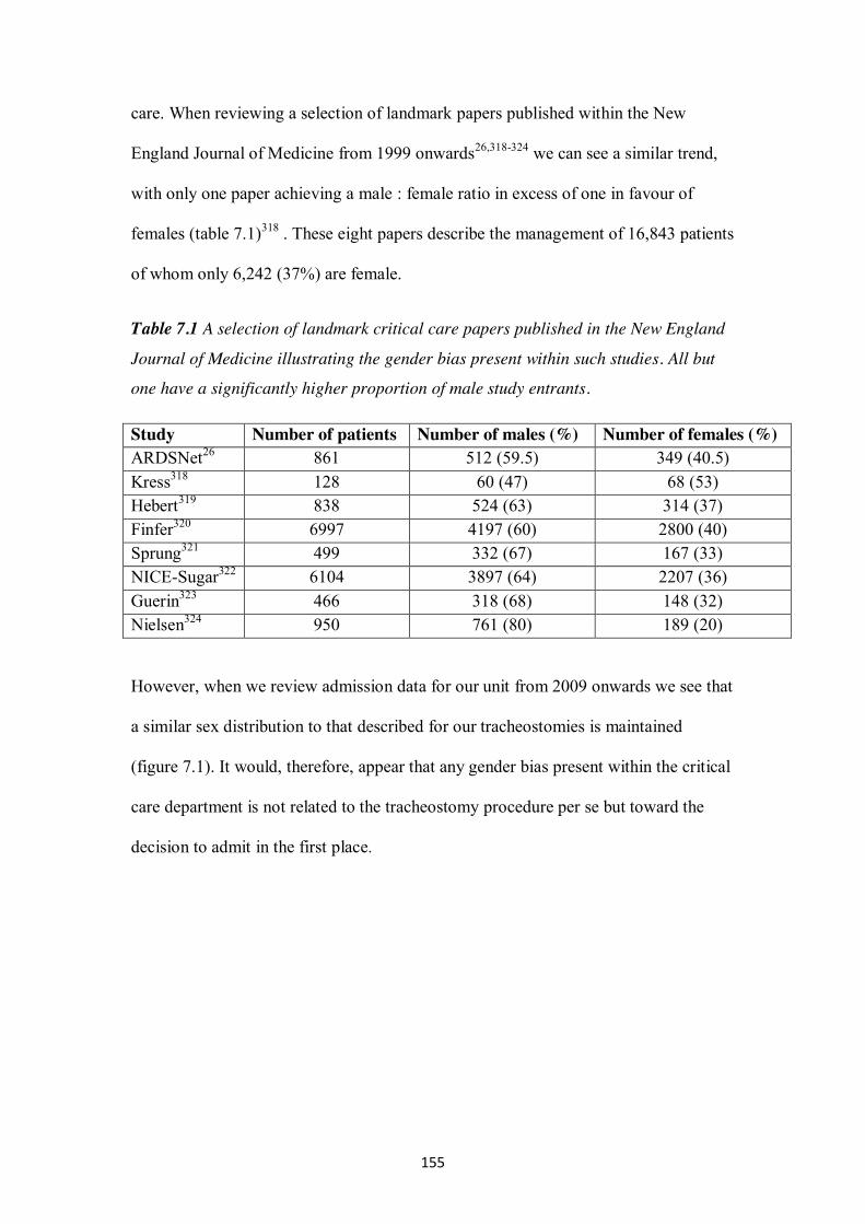
155
care. When reviewing a selection of landmark papers published within the New
England Journal of Medicine from 1999 onwards26,318-324 we can see a similar trend,
with only one paper achieving a male : female ratio in excess of one in favour of
females (table 7.1)318 . These eight papers describe the management of 16,843 patients
of whom only 6,242 (37%) are female.
Table 7.1 A selection of landmark critical care papers published in the New England
Journal of Medicine illustrating the gender bias present within such studies. All but
one have a significantly higher proportion of male study entrants.
Study Number of patients Number of males (%) Number of females (%) ARDSNet26 861 512 (59.5) 349 (40.5) Kress318 128 60 (47) 68 (53) Hebert319 838 524 (63) 314 (37) Finfer320 6997 4197 (60) 2800 (40) Sprung321 499 332 (67) 167 (33) NICE-Sugar322 6104 3897 (64) 2207 (36) Guerin323 466 318 (68) 148 (32) Nielsen324 950 761 (80) 189 (20)
However, when we review admission data for our unit from 2009 onwards we see that
a similar sex distribution to that described for our tracheostomies is maintained
(figure 7.1). It would, therefore, appear that any gender bias present within the critical
care department is not related to the tracheostomy procedure per se but toward the
decision to admit in the first place.
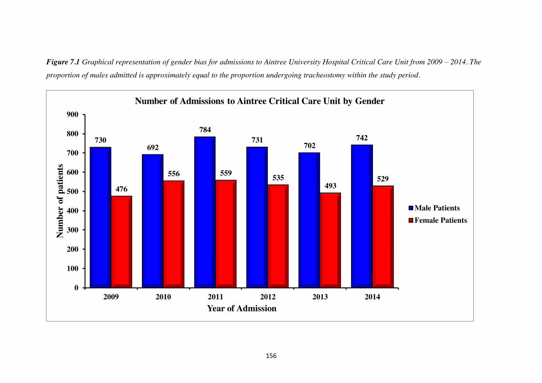
156
Figure 7.1 Graphical representation of gender bias for admissions to Aintree University Hospital Critical Care Unit from 2009 – 2014. The
proportion of males admitted is approximately equal to the proportion undergoing tracheostomy within the study period.
730 692
784 731
702 742
476
556 559 535 493
529
0
100
200
300
400
500
600
700
800
900
2009 2010 2011 2012 2013 2014
Num
ber
of p
atie
nts
Year of Admission
Number of Admissions to Aintree Critical Care Unit by Gender
Male PatientsFemale Patients
157
It is generally believed that access to critical care is determined by need and severity
of illness with differences in admission rates between genders being accounted for by
the different disease spectra between the sexes. For example, it is generally accepted
that ischaemic heart disease and trauma are commoner in males. However, a number
of studies have demonstrated some concerns in relation to gender bias within critical
care. A Canadian study analysing over 466,000 hospital admissions found that women
over 50 years of age were less likely to be admitted to a critical care unit and more
likely to die than men after a critical illness325. In contrast, in a cohort of 26,000
patients in Austria Valentin found that whilst there was a higher illness severity in
women men received an increased level of care and more invasive procedures but this
did not translate in to improved outcomes326.
A study emanating from the UK Intensive Care National Audit and Research Centre
(ICNARC) examined the influence of gender on admission to critical care327. They
examined admissions for a number of diagnostic categories for evidence of vertical
and horizontal inequity. Reviewing over 46,000 admissions from 91 critical care units
they were unable to find evidence of gender bias in admission or mortality in relation
to cardiac arrhythmias, chronic obstructive pulmonary disease, asthma, self-poisoning
and seizures. There was some evidence of vertical and horizontal inequity for females
with myocardial infarction and central nervous system bleeding. There was also
evidence suggestive of vertical inequity for male pneumonia and ventricular failure
patients.
When considering our own data we can see that the mean admission APACHE II
score for males admitted to the unit who undergo a tracheostomy is 18.3 whilst that
for females is 19.3 (p=0.005 unpaired t test). This would indicate females admitted to
our critical care unit who undergo a tracheostomy are sicker than their male
158
counterparts at critical care admission, possibly indicating an admission gender bias
within our unit. When considering survival of these cohorts the median survival for
males is 2.9 years (95% CI 1.8 – 3.9) with an estimated five-year survival of 43% (SE
0.02). For females the equivalent figures are a median survival of 3.7 years (95% CI
2.3 – 5.1) with an estimated five-year survival of 45% (SE 0.03) (figure 7.2).
Therefore, whilst the illness severity for female patients is increased this is not
seemingly translated in to worse survival figures as would be expected. The crude
APACHE II mortality prediction for the scores described above would be 29% for
males and 32% for females. Samples sizes to reliably detect such a difference should
be 511 in each group (α 0.05, power 0.8).
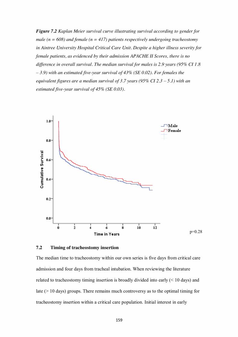
159
Figure 7.2 Kaplan Meier survival curve illustrating survival according to gender for
male (n = 608) and female (n = 417) patients respectively undergoing tracheostomy
in Aintree University Hospital Critical Care Unit. Despite a higher illness severity for
female patients, as evidenced by their admission APACHE II Scores, there is no
difference in overall survival. The median survival for males is 2.9 years (95% CI 1.8
– 3.9) with an estimated five-year survival of 43% (SE 0.02). For females the
equivalent figures are a median survival of 3.7 years (95% CI 2.3 – 5.1) with an
estimated five-year survival of 45% (SE 0.03).
p=0.28
7.2 Timing of tracheostomy insertion
The median time to tracheostomy within our own series is five days from critical care
admission and four days from tracheal intubation. When reviewing the literature
related to tracheostomy timing insertion is broadly divided into early (< 10 days) and
late (> 10 days) groups. There remains much controversy as to the optimal timing for
tracheostomy insertion within a critical care population. Initial interest in early
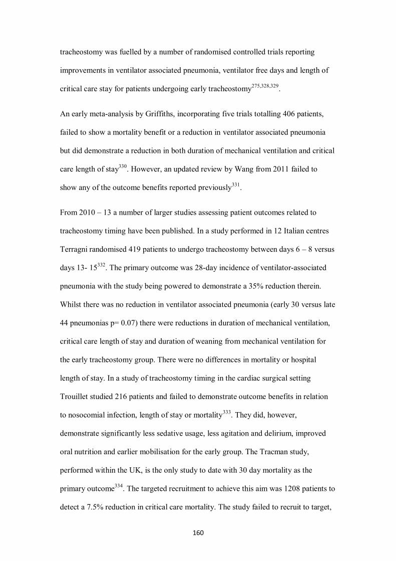
160
tracheostomy was fuelled by a number of randomised controlled trials reporting
improvements in ventilator associated pneumonia, ventilator free days and length of
critical care stay for patients undergoing early tracheostomy275,328,329.
An early meta-analysis by Griffiths, incorporating five trials totalling 406 patients,
failed to show a mortality benefit or a reduction in ventilator associated pneumonia
but did demonstrate a reduction in both duration of mechanical ventilation and critical
care length of stay330. However, an updated review by Wang from 2011 failed to
show any of the outcome benefits reported previously331.
From 2010 – 13 a number of larger studies assessing patient outcomes related to
tracheostomy timing have been published. In a study performed in 12 Italian centres
Terragni randomised 419 patients to undergo tracheostomy between days 6 – 8 versus
days 13- 15332. The primary outcome was 28-day incidence of ventilator-associated
pneumonia with the study being powered to demonstrate a 35% reduction therein.
Whilst there was no reduction in ventilator associated pneumonia (early 30 versus late
44 pneumonias p= 0.07) there were reductions in duration of mechanical ventilation,
critical care length of stay and duration of weaning from mechanical ventilation for
the early tracheostomy group. There were no differences in mortality or hospital
length of stay. In a study of tracheostomy timing in the cardiac surgical setting
Trouillet studied 216 patients and failed to demonstrate outcome benefits in relation
to nosocomial infection, length of stay or mortality333. They did, however,
demonstrate significantly less sedative usage, less agitation and delirium, improved
oral nutrition and earlier mobilisation for the early group. The Tracman study,
performed within the UK, is the only study to date with 30 day mortality as the
primary outcome334. The targeted recruitment to achieve this aim was 1208 patients to
detect a 7.5% reduction in critical care mortality. The study failed to recruit to target,
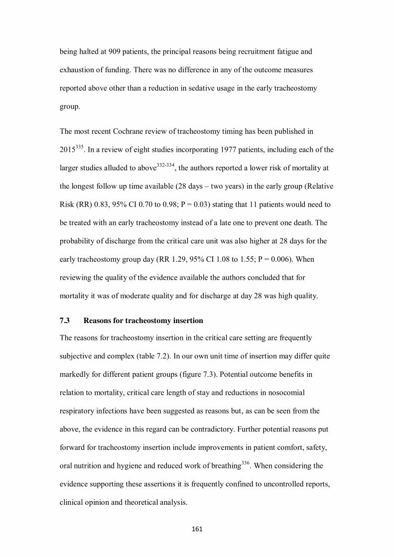
161
being halted at 909 patients, the principal reasons being recruitment fatigue and
exhaustion of funding. There was no difference in any of the outcome measures
reported above other than a reduction in sedative usage in the early tracheostomy
group.
The most recent Cochrane review of tracheostomy timing has been published in
2015335. In a review of eight studies incorporating 1977 patients, including each of the
larger studies alluded to above332-334, the authors reported a lower risk of mortality at
the longest follow up time available (28 days – two years) in the early group (Relative
Risk (RR) 0.83, 95% CI 0.70 to 0.98; P = 0.03) stating that 11 patients would need to
be treated with an early tracheostomy instead of a late one to prevent one death. The
probability of discharge from the critical care unit was also higher at 28 days for the
early tracheostomy group day (RR 1.29, 95% CI 1.08 to 1.55; P = 0.006). When
reviewing the quality of the evidence available the authors concluded that for
mortality it was of moderate quality and for discharge at day 28 was high quality.
7.3 Reasons for tracheostomy insertion
The reasons for tracheostomy insertion in the critical care setting are frequently
subjective and complex (table 7.2). In our own unit time of insertion may differ quite
markedly for different patient groups (figure 7.3). Potential outcome benefits in
relation to mortality, critical care length of stay and reductions in nosocomial
respiratory infections have been suggested as reasons but, as can be seen from the
above, the evidence in this regard can be contradictory. Further potential reasons put
forward for tracheostomy insertion include improvements in patient comfort, safety,
oral nutrition and hygiene and reduced work of breathing336. When considering the
evidence supporting these assertions it is frequently confined to uncontrolled reports,
clinical opinion and theoretical analysis.
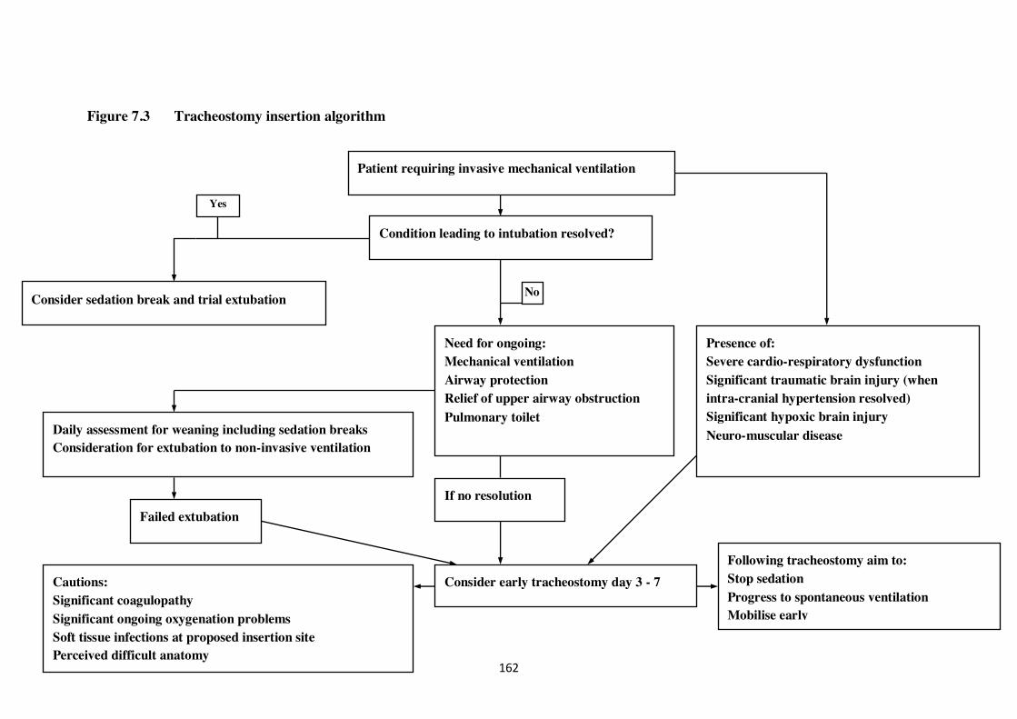
162
Figure 7.3 Tracheostomy insertion algorithm
Patient requiring invasive mechanical ventilation
Condition leading to intubation resolved?
Consider sedation break and trial extubation
Need for ongoing: Mechanical ventilation Airway protection Relief of upper airway obstruction Pulmonary toilet
Daily assessment for weaning including sedation breaks Consideration for extubation to non-invasive ventilation
Consider early tracheostomy day 3 - 7
Presence of: Severe cardio-respiratory dysfunction Significant traumatic brain injury (when intra-cranial hypertension resolved) Significant hypoxic brain injury Neuro-muscular disease
Cautions: Significant coagulopathy Significant ongoing oxygenation problems Soft tissue infections at proposed insertion site Perceived difficult anatomy
Following tracheostomy aim to: Stop sedation Progress to spontaneous ventilation Mobilise early
If no resolution
No
Yes
Failed extubation
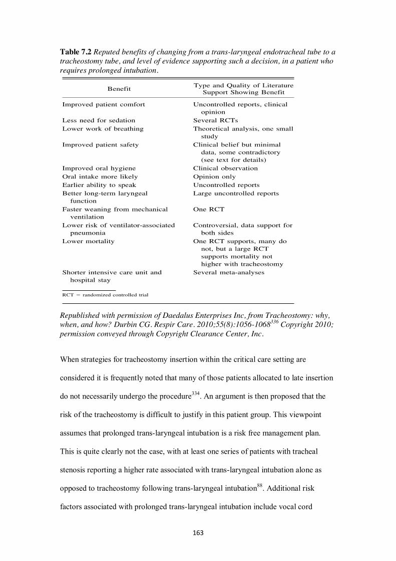
163
Table 7.2 Reputed benefits of changing from a trans-laryngeal endotracheal tube to a tracheostomy tube, and level of evidence supporting such a decision, in a patient who requires prolonged intubation.
Republished with permission of Daedalus Enterprises Inc, from Tracheostomy: why, when, and how? Durbin CG. Respir Care. 2010;55(8):1056-1068336 Copyright 2010; permission conveyed through Copyright Clearance Center, Inc.
When strategies for tracheostomy insertion within the critical care setting are
considered it is frequently noted that many of those patients allocated to late insertion
do not necessarily undergo the procedure334. An argument is then proposed that the
risk of the tracheostomy is difficult to justify in this patient group. This viewpoint
assumes that prolonged trans-laryngeal intubation is a risk free management plan.
This is quite clearly not the case, with at least one series of patients with tracheal
stenosis reporting a higher rate associated with trans-laryngeal intubation alone as
opposed to tracheostomy following trans-laryngeal intubation88. Additional risk
factors associated with prolonged trans-laryngeal intubation include vocal cord
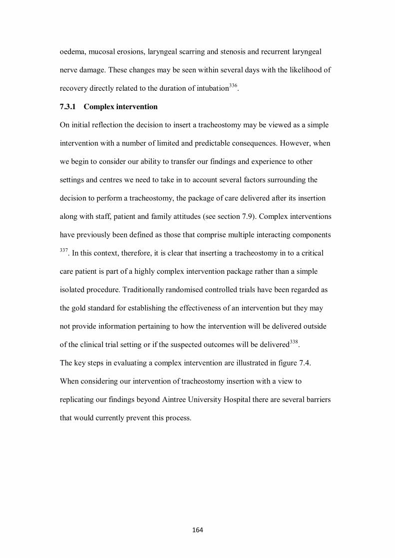
164
oedema, mucosal erosions, laryngeal scarring and stenosis and recurrent laryngeal
nerve damage. These changes may be seen within several days with the likelihood of
recovery directly related to the duration of intubation336.
7.3.1 Complex intervention
On initial reflection the decision to insert a tracheostomy may be viewed as a simple
intervention with a number of limited and predictable consequences. However, when
we begin to consider our ability to transfer our findings and experience to other
settings and centres we need to take in to account several factors surrounding the
decision to perform a tracheostomy, the package of care delivered after its insertion
along with staff, patient and family attitudes (see section 7.9). Complex interventions
have previously been defined as those that comprise multiple interacting components
337. In this context, therefore, it is clear that inserting a tracheostomy in to a critical
care patient is part of a highly complex intervention package rather than a simple
isolated procedure. Traditionally randomised controlled trials have been regarded as
the gold standard for establishing the effectiveness of an intervention but they may
not provide information pertaining to how the intervention will be delivered outside
of the clinical trial setting or if the suspected outcomes will be delivered338.
The key steps in evaluating a complex intervention are illustrated in figure 7.4.
When considering our intervention of tracheostomy insertion with a view to
replicating our findings beyond Aintree University Hospital there are several barriers
that would currently prevent this process.
165
Figure 7.4 Key functions of process evaluation and relations among them for complex interventions. Implementation, mechanisms of impact and
context are felt to be the key components of a process evaluation
Reproduced from Process evaluation of complex interventions: Medical Research Council guidance. Moore GF et al BMJ. 2015;350:h1258338. Copyright 2015 with permission from BMJ Publishing Group Ltd
166
7.3.1.1 Defining the intervention
Whilst it is a simple task to identify all critical care patients in whom a tracheostomy
has been inserted it is more difficult to quantify the change in care that results from it.
Within our unit the intervention is not simply tracheostomy insertion but the package
of care delivered thereafter. Although it is easy to observe this change, as yet, we have
not documented the process. This would require, as a minimum, assessments
addressing sedative usage, time spent out of bed and mobilising, changes in ventilator
management and additional time spent by allied health professionals with the patient.
Additionally, patient factors further complicate our ability to define the intervention
with some patients receiving a tracheostomy earlier (severe end organ dysfunction)
whilst in others this may be delayed (figure 7.3).
7.3.1.2 Implementation
The first barrier to acceptance of a process of early tracheostomy and its associated
package of care within other centres may lie in its acceptance. There is limited data
showing a mortality benefit in relation to early tracheostomy insertion with most
recent large randomised controlled trials failing to demonstrate a difference in
outcome332-334. The most recent systematic review does, however, seem to indicate
both a mortality benefit and a reduction in critical care length of stay335.
Further process evaluation to assess how the intervention would be delivered would
be required. This would need to include elements of resource allocation, assessment
of training needs (both for those undertaking the tracheostomy and for those
delivering care after insertion), staff and organisational communication issues,
measurement of delivery as well as adapting the intervention to different settings338.
167
7.3.1.3 Impact and outcomes
Any potential positive benefits related to the package of care delivered around early
tracheostomy insertion may be difficult to identify given the negative outcomes
associated with previous studies332-334. If this were to be the case this may be related
to poor initial study design with a failure to fully define the intervention or poor
implementation339. Surrogate endpoints designed to assess patient non-mortality
related outcomes are likely to be required and may include duration of mechanical
ventilation, length of critical care and hospital stay, incidence of delirium,
assessments of neuromuscular function and psychological well-being. Further
assessments of patient and family acceptance would also be required (see section 7.9).
It is also possible that the positive effects of tracheostomy insertion may be seen even
when the intervention package is not fully delivered. Whilst there may be no mortality
benefit for early tracheostomy insertion the cessation of sedation may provide
unexpected benefits that are more difficult to measure. From our own data 26% of
patients undergoing tracheostomy do not survive to critical care discharge (table 5.1).
It is possible, that in such patients, after sedation has been stopped an enhanced ability
to communicate with family and friends may contribute to a more favourable end of
life experience.
7.4 Aetiological factors associated with tracheal stenosis
In both our prospective cohort and within the systematic review we have attempted to
identify factors present at tracheostomy insertion that may be associated with the
aetiology of tracheal stenosis. To this end we have largely been unsuccessful. Within
our own series, the reason for this was largely due to the low incidence of tracheal
stenosis preventing meaningful interpretation of our data. Of the five stenoses
observed one patient had a peri-operative tracheal ring fracture and oxygen
168
desaturation, another had marked laryngeal mucosal injury from trans-laryngeal
intubation at tracheostomy insertion. The remaining three patients had uneventful
tracheostomy insertions.
The only peri-operative event described within the literature and postulated to be
associated with the development of tracheal stenosis is tracheal ring fracture at
tracheostomy insertion184-186. Within our own series we reported a rate of tracheal ring
fracture of 11% and a TS rate of 0.5%. Only one of those patients was noted to have a
tracheal ring fracture at tracheostomy insertion. Additionally, a number of authors
have noted differing rates of tracheal ring fracture with the various percutaneous
techniques with reported rates being higher with the STD technique54. However, the
pooled proportions meta-analysis from our systematic review suggested, that of the
percutaneous techniques, the rate of TS for the STD was lowest – seemingly
indicating a lack of correlation between ring fracture and TS. A further consideration
to factor in is that tracheal ring fracture is almost certainly underreported following
PDT and is largely dependent upon the bronchoscopist actively seeking this
complication. This assertion is supported by work from cadaveric studies340. Walz
undertook a clinico-pathological study of tracheal specimens following PDT from
critical care patients who had died from causes unrelated to their tracheostomy. In
twelve out of forty two specimens there was an inter-cartilaginous rupture of the
tracheal wall associated with a tracheal ring fracture and in 10 of 42 there was a
cartilage defect at the stomal site. Each of these specimens revealed denuded cartilage
with necrotic lesions that were related to duration of cannulation.
From the data presented it would seem the most important aetiological factors
associated with the genesis of TS are stomal and cuff related ischaemic damage,
duration of cannulation and inherent patient factors.
169
7.5 Critical care survival
The survival data presented within Chapter 5 demonstrates significantly worsening
outcomes with both increased illness severity and advancing age. This is in keeping
with previously published data341 and our own, as yet, unpublished observations (see
below).
7.5.1 Survival to hospital discharge
We have studied a cohort of 505 elderly patients over the age of 80 years undergoing
non-elective admissions to our critical care unit from January 2010 to December
2014. We have found the predominant predictors of survival to hospital discharge on
univariate analysis are related to category of admission (medical versus surgical) and
acute physiological derangement (APACHE II score, SOFA score & modified SOFA
score) (table 7.3). Survival to hospital discharge appears to be unrelated to indices of
chronic health and co-morbidities (Functional co-morbidity score, chronic health
evaluation of APACHE II score).
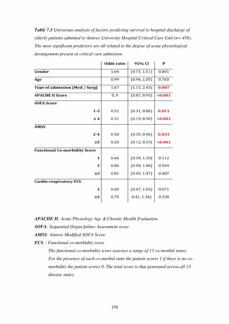
170
Table 7.3 Univariate analysis of factors predicting survival to hospital discharge of
elderly patients admitted to Aintree University Hospital Critical Care Unit (n= 458).
The most significant predictors are all related to the degree of acute physiological
derangement present at critical care admission.
APACHE II: Acute Physiology Age & Chronic Health Evaluation
SOFA: Sequential Organ failure Assessment score
AMSS: Aintree Modified SOFA Score
FCS – Functional co-morbidity score
The functional co-morbidity score assesses a range of 13 co-morbid states.
For the presence of each co-morbid state the patient scores 1 if there is no co-
morbidity the patient scores 0. The total score is that generated across all 13
disease states.
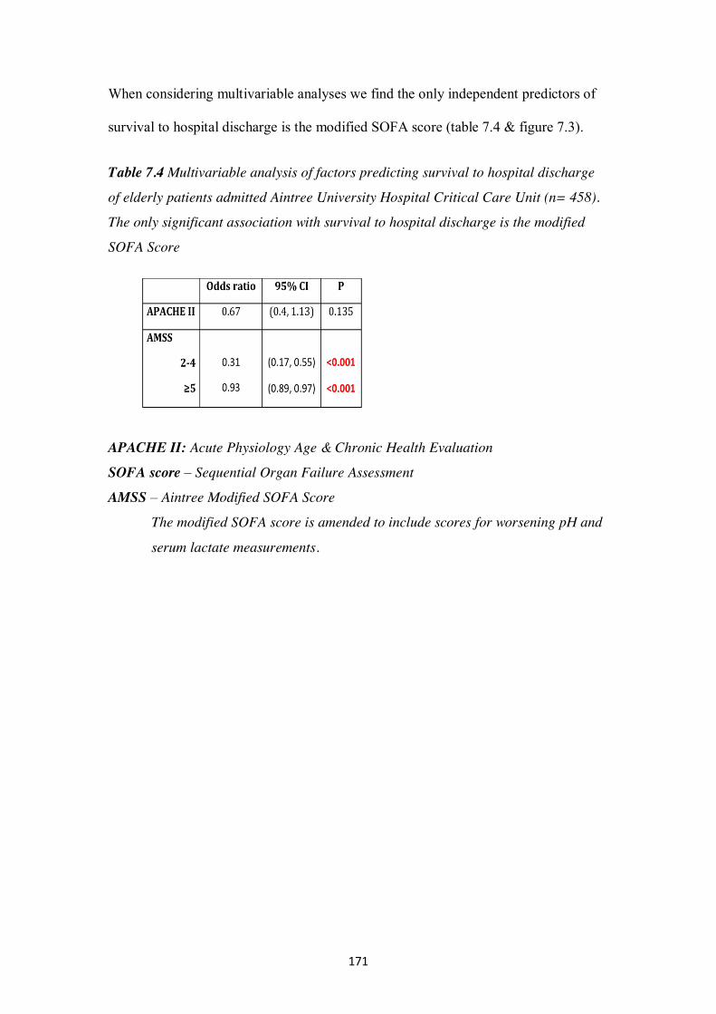
171
When considering multivariable analyses we find the only independent predictors of
survival to hospital discharge is the modified SOFA score (table 7.4 & figure 7.3).
Table 7.4 Multivariable analysis of factors predicting survival to hospital discharge
of elderly patients admitted Aintree University Hospital Critical Care Unit (n= 458).
The only significant association with survival to hospital discharge is the modified
SOFA Score
APACHE II: Acute Physiology Age & Chronic Health Evaluation
SOFA score – Sequential Organ Failure Assessment
AMSS – Aintree Modified SOFA Score
The modified SOFA score is amended to include scores for worsening pH and
serum lactate measurements.
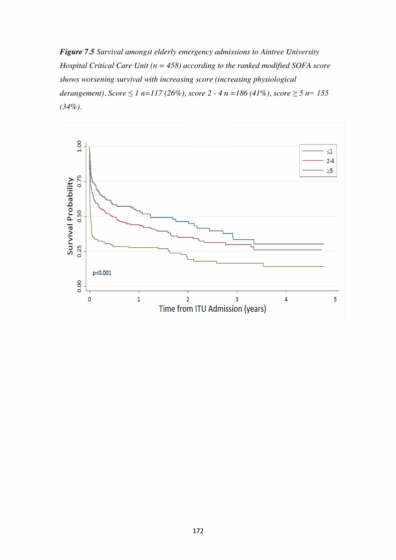
172
Figure 7.5 Survival amongst elderly emergency admissions to Aintree University
Hospital Critical Care Unit (n = 458) according to the ranked modified SOFA score
shows worsening survival with increasing score (increasing physiological
derangement). Score ≤ 1 n=117 (26%), score 2 - 4 n =186 (41%), score ≥ 5 n= 155
(34%).
173
7.5.2 Survival to one year from critical care admission
In the same cohort of elderly non-elective critical care patients when considering
univariate analyses the predominant factors predicting survival to one year remain the
degree of acute physiological derangement present at admission (higher APACHE,
SOFA and modified SOFA scores predicting worse outcomes) (table 7.5). However,
when subjected to multi-variable analyses the independent predictors of 12-month
survival are age, APACHE II score and a cardio-respiratory functional co-morbidity
score of 2 (table 7.6 and figure 7.4). With each year increase in age one-year
survival decreases by 8% and each unit increase in the APACHE II score reduces one-
year survival by 6%. Median survival for those patients with Modified SOFA scores
5 are 7 days (95% CI 3 – 11) and for a cardio-respiratory functional co-morbidity
score 2 are 43 days (95% CI 0 – 110)
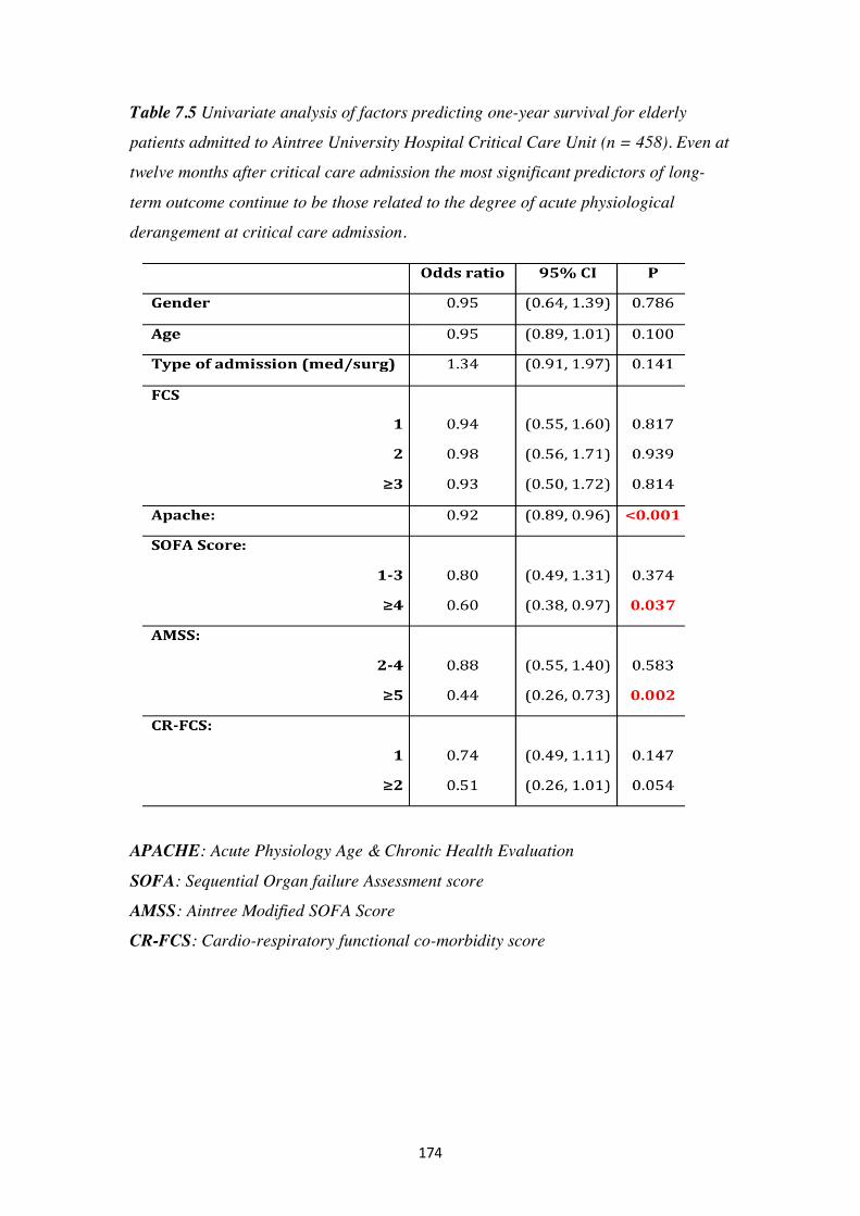
174
Table 7.5 Univariate analysis of factors predicting one-year survival for elderly
patients admitted to Aintree University Hospital Critical Care Unit (n = 458). Even at
twelve months after critical care admission the most significant predictors of long-
term outcome continue to be those related to the degree of acute physiological
derangement at critical care admission.
APACHE: Acute Physiology Age & Chronic Health Evaluation
SOFA: Sequential Organ failure Assessment score
AMSS: Aintree Modified SOFA Score
CR-FCS: Cardio-respiratory functional co-morbidity score
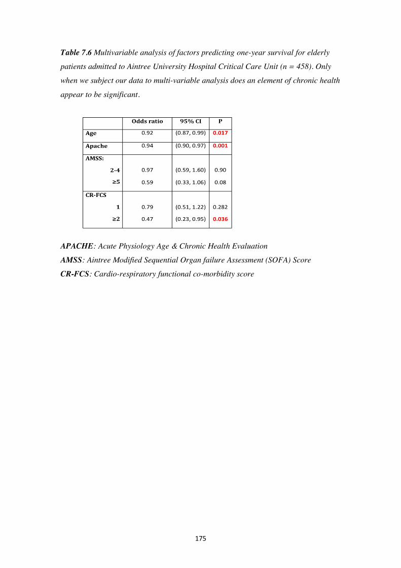
175
Table 7.6 Multivariable analysis of factors predicting one-year survival for elderly
patients admitted to Aintree University Hospital Critical Care Unit (n = 458). Only
when we subject our data to multi-variable analysis does an element of chronic health
appear to be significant.
APACHE: Acute Physiology Age & Chronic Health Evaluation
AMSS: Aintree Modified Sequential Organ failure Assessment (SOFA) Score
CR-FCS: Cardio-respiratory functional co-morbidity score
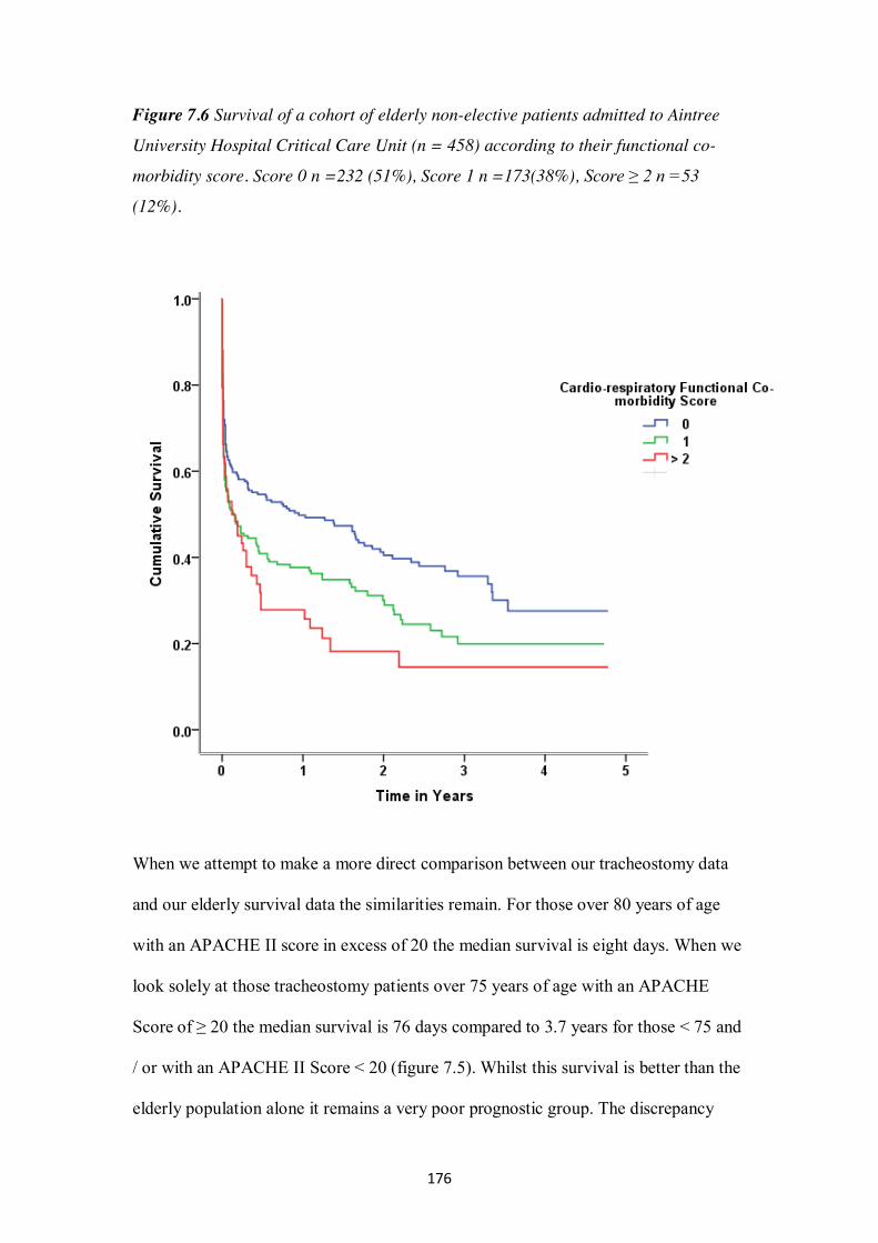
176
Figure 7.6 Survival of a cohort of elderly non-elective patients admitted to Aintree
University Hospital Critical Care Unit (n = 458) according to their functional co-
morbidity score. Score 0 n =232 (51%), Score 1 n =173(38%), Score ≥ 2 n =53
(12%).
When we attempt to make a more direct comparison between our tracheostomy data
and our elderly survival data the similarities remain. For those over 80 years of age
with an APACHE II score in excess of 20 the median survival is eight days. When we
look solely at those tracheostomy patients over 75 years of age with an APACHE
Score of ≥ 20 the median survival is 76 days compared to 3.7 years for those < 75 and
/ or with an APACHE II Score < 20 (figure 7.5). Whilst this survival is better than the
elderly population alone it remains a very poor prognostic group. The discrepancy

177
between the two groups is probably accounted for by the fact that the elderly cohort is
slightly older and an unselected group of patients reviewed from the date of
admission. For the tracheostomy group, patients must survive a median of five days
prior to tracheostomy insertion. Therefore, those patients with the worst prognoses
will not undergo the procedure.
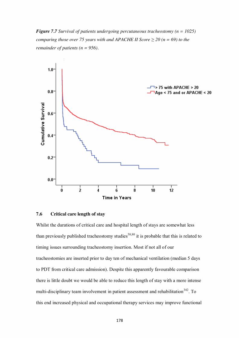
178
Figure 7.7 Survival of patients undergoing percutaneous tracheostomy (n = 1025)
comparing those over 75 years with and APACHE II Score ≥ 20 (n = 69) to the
remainder of patients (n = 956).
7.6 Critical care length of stay
Whilst the durations of critical care and hospital length of stays are somewhat less
than previously published tracheostomy studies58,80 it is probable that this is related to
timing issues surrounding tracheostomy insertion. Most if not all of our
tracheostomies are inserted prior to day ten of mechanical ventilation (median 5 days
to PDT from critical care admission). Despite this apparently favourable comparison
there is little doubt we would be able to reduce this length of stay with a more intense
multi-disciplinary team involvement in patient assessment and rehabilitation342. To
this end increased physical and occupational therapy services may improve functional
179
well-being, speech and language therapy input may improve swallowing (and thus
reduce episodes of aspiration pneumonitis), nutrition and communication,
psychological therapy may improve mental well-being and reduce the incidence of
post-traumatic stress disorder. After critical care discharge the maintenance of the
above services with enhanced input from the critical care outreach team may further
reduce length of stay and allow earlier identification of patient deterioration.
Despite this well recognised need for enhanced observation and rehabilitation
amongst the critical care patient population easily achieved interventions are not
frequently undertaken. In a recent study performed across 12 critical care units in
Australia and New Zealand incorporating 192 patients 52% of patients were found to
have critical care associated weakness which was associated with an increased risk of
death following critical care discharge343. Despite this 84% of patients did not receive
any attempts at early mobilisation (defined as any active exercise where the patient
could assist the activity using their own muscle strength).
There remains, however, a degree of uncertainty relating to the implementation and
timing of critical care rehabilitation programmes. In part this is due to a lack of
funding for resource intense interventions where the outcome is not immediately
demonstrable and in part from a lack of experience in delivering such interventions.
The evidence is further clouded by the differing levels of physiotherapy support
currently provided as the norm across units in North America, Europe and Australasia
with much higher levels provided in Europe and Australasia 342. A number of studies
have demonstrated the benefits of early initiation of a rehabilitation
programme182,342,344,345 but this has not been universal346. The benefit of rehabilitation
programmes instituted following critical care discharge have been more disappointing
180
with several trials failing to achieve a positive outcome347-349 perhaps indicating that
intervention at this point is too late.
7.6.1 Future work
Our own intention in relation to improving outcomes for patients discharged from our
unit was to attempt a targeted intervention based on our use of the Sabadell Score. We
have shown that the in hospital mortality of patients discharged from our unit who are
assigned a Sabadell 2 score is 16% (figure 7.6). We postulated that by targeting an
intense rehabilitation programme, involving increased senior medical, physiotherapy
and critical care outreach nursing input, toward a group of patients at high risk of in-
hospital mortality we may be able to improve on earlier studies where the intervention
has been delivered to all patients discharged. We have, so far, been unable to secure
funding for this project.
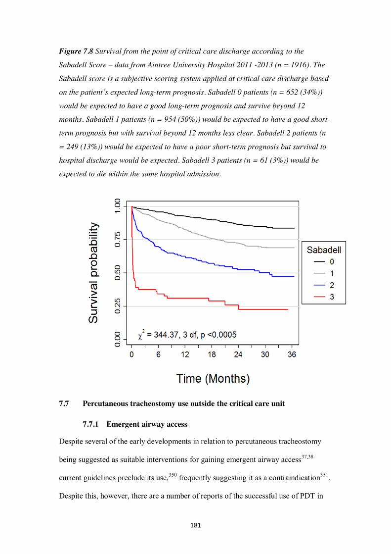
181
Figure 7.8 Survival from the point of critical care discharge according to the
Sabadell Score – data from Aintree University Hospital 2011 -2013 (n = 1916). The
Sabadell score is a subjective scoring system applied at critical care discharge based
on the patient’s expected long-term prognosis. Sabadell 0 patients (n = 652 (34%))
would be expected to have a good long-term prognosis and survive beyond 12
months. Sabadell 1 patients (n = 954 (50%)) would be expected to have a good short-
term prognosis but with survival beyond 12 months less clear. Sabadell 2 patients (n
= 249 (13%)) would be expected to have a poor short-term prognosis but survival to
hospital discharge would be expected. Sabadell 3 patients (n = 61 (3%)) would be
expected to die within the same hospital admission.
7.7 Percutaneous tracheostomy use outside the critical care unit
7.7.1 Emergent airway access
Despite several of the early developments in relation to percutaneous tracheostomy
being suggested as suitable interventions for gaining emergent airway access37,38
current guidelines preclude its use,350 frequently suggesting it as a contraindication351.
Despite this, however, there are a number of reports of the successful use of PDT in
182
the emergency setting.352-356 It seems likely, that those expert in the insertion of a PDT
would be able to achieve emergent airway access within the time frame consistent
with a satisfactory neurological outcome. It also seems likely that such individuals
would be much more comfortable gaining airway access in this setting via a PDT
rather than using those techniques set out within difficult airway guidelines.
7.7.2 Future work
We would like to further investigate the hypothesis that PDT insertion in the
emergency airway setting is contra-indicated. We would postulate that for those
expert in its insertion it may be a credible alternative to crico-thyroidotomy. We
would assess this using a bench test of timed airway access using either an airway
mannequin or pig larynx. We would assess two groups of individuals trained in
airway management but with one group also being expert in PDT insertion (intensivist
group). We will assess time to securing the airway via surgical means according to the
Difficult Airway Society surgical crico-thyroidotomy approach and also via a
standard STD PDT approach. We could potentially expect a rapid securing of the
airway with the STD PDT in the intensivist group but assessment of the speed of
airway access in the non-expert group would also be an interesting addition to the
literature.
7.8 Percutaneous versus surgical tracheostomy in the elective setting
Despite our unit housing the largest Head and Neck unit within the UK we have
limited experience in the use of PDT in the setting of elective head and neck cancer
surgery. We have used PDT in a small number of such patients but this was
abandoned for reasons other than those related to PDT insertion. Use of PDT within
an elective head and neck cancer population have formed part of some studies
183
assessing PDT outcomes357 but the majority of patients within this study were
critically ill. There are no reports detailing the use of PDT solely within a head and
neck cancer population. In addition to the low morbidity we have described herein
there are other benefits that may be realised in the head and neck oncology patient. It
has been clearly shown, both within our own meta-analysis and others, that the
incidence of stomal infection is reduced with PDT. Siting of the PDT in a surgical
patient allows the tracheostomy wound to be clearly separated from any neck wound.
This, in association with the reduced rate of stomal sepsis, may lead to a reduction in
post-operative neck wound infection in elective cancer patients if routine PDT use
were considered. Significant safety related concerns would, however, remain should
the tracheostomy become displaced in the early post-operative period. The use of
PDT within our unit stopped following a case of accidental decannulation (non-PDT
patient) on the first post-operative night in a patient undergoing tumour resection and
free tissue transfer. In this patient it proved impossible to rapidly re-secure the airway
leading to a hypoxic brain injury. Our current practice, therefore, is to use Bjork flap
surgical tracheostomies in these patients. Despite these concerns, within the critical
care unit (where these patients are routinely nursed), we have not had a significant
problem with accidental decannulation for either surgical or percutaneous
tracheostomies.
7.8.1 Future work
Whilst we have no current plans to explore the role and efficacy of percutaneous
tracheostomy in the elective surgical setting this would be a relatively simple
undertaking. A small randomised pilot study may help to evaluate a potential place for
PDT in this setting. We would assess not only the safety concerns relating to
inadvertent post-operative decannulation that have previously been raised within our
184
unit but also its effect on post-operative infection and the impact on the patient
experience. Given the general dislike of tracheostomies amongst the surgical
population further work may explore the role of smaller tracheostomy tubes, uncuffed
tubes and guidelines toward earlier decannulation for these patients.
Widespread adoption of percutaneous tracheostomy within the elective surgical
setting would, however generate its own concerns. Currently head and neck surgeons
in training gain experience required for securing the emergency surgical airway in the
elective setting. If percutaneous tracheostomy were to be widely adopted in favour of
surgical this could potentially raise significant training issues and a deskilling of the
surgical team.
7.9 Perceptions of tracheostomy in critical illness survivors
As can be seen above (table 7.2) the reasons for tracheostomy insertion within critical
care are frequently subjective with sometimes questionable evidence to support the
decision making process. It is often assumed by critical care staff that tracheostomy
insertion leads to a more settled and comfortable patient. This is in stark contrast to
the experience of our head and neck unit where the presence of the tracheostomy is
often the most complained about aspect of surgical head and neck cancer care
(personal communication). The assumption that patients are more comfortable has,
however, been relatively untested. In a review of qualitative studies of patient
experiences related to weaning from mechanical ventilation Cook described
uncertainty, stress, discomfort and a lack of control of surroundings and events as
common themes358. Whilst not all of the patients assessed within this analysis will
have had tracheostomies inserted it is easily appreciated that many of these symptoms
could be ascribed to such patients. In a small qualitative study of eight patients (not
exclusively critical care patients) Sherlock undertook a semi-structured interview to
185
identify common factors relating to their tracheostomy experience359. All but two of
the patients included had been decannulated at the time of interview. They divided
their interviews and responses into four themes namely; physical sensations,
understanding, information and feelings after tracheostomy removal. They found that
whilst most patients understood the reason for the tracheostomy and understood the
reasons for its presence there were less positive experiences reported in relation to the
physical presence of the tube, eating and drinking, tracheal suctioning and
communication. Additionally, three of the six patients who had been decannulated
reported panic attacks following removal of the tube despite happily anticipating this
event. Whilst Sherlock’s study offers some insights in to the experiences of
tracheostomised patients it does not exclusively study critical care patients.
Additionally, only patients capable of participating in 45-minute interview with a
recordable voice were included and all had previously undergone at least one
tracheostomy change. The evolving contemporaneous experiences of these patients as
they progress through their critical care stay are yet to be documented although some
insight in to this has been provided by Flinterud360. In a study of eleven patients from
a university hospital in Norway they documented similar emotions to those described
by Sherlock namely frustration, panic and anger, powerlessness, despair and loss of
control. However, they also noted a time related improvement associated with
development of coping strategies to enable easier communication.
7.9.1 Future work
It is our intention to explore the patient experience in relation to tracheostomy
insertion in more detail over the coming months and years. We initially envisage a
study conducted solely within the critical care unit undertaken with patients who have
their tracheostomy in situ at the time of interview. We would also like to capture the
186
experience of the small group of patients who tolerate oro-tracheal intubation without
sedation prior to either extubation or tracheostomy and compare their experiences
with those of patients with tracheostomies. We would then intend to follow up all
surviving patients to compare their memories and experiences following
decannulation or extubation. Additionally, we would like to compare the experiences
of these patients with those of their carers and families. To this end, we would hope to
either confirm or refute much of the subjective reasoning behind tracheostomy
insertion in the critically ill.
7.10 Conclusion
From the limited published data we have not found a significant difference in the
incidence of TS between a range of percutaneous techniques and surgical
tracheostomies. When considering all published data reporting long-term outcomes,
our pooled proportions meta-analysis may indicate a tendency toward a higher rate of
TS for ST but this finding is likely to be prone to selection bias. Similarly, in relation
to major bleeding, we have not found a difference between the techniques commonly
used. Again the pooled proportions meta-analysis may indicate a tendency toward
more major bleeding for ST. In relation to wound infection we have found a reduction
associated with CPDT compared to ST in keeping with earlier work51. Across all
percutaneous techniques for the primary and secondary outcomes studied
complication rates appear to be broadly similar but confidence intervals for pooled
risk differences are wide and include clinically important differences in both
directions.
Whilst the reported complication rates presented within our cohort study are in
keeping with, if not an improvement upon, those published previously, this remains a
187
single centre evaluation with its attendant limitations. It may, however, add weight to
the gathering evidence that the STD PDT is one of the safer techniques available. Our
rate of early technical difficulties appears to be reducing whilst long-term outcomes
remain unchanged.
We have demonstrated a lower rate of sub-clinical stenosis in patients following STD
PDT than has previously been reported. Whilst we considered that some degree of
functional limitation, in such patients, following critical care discharge might be in
part due to an underlying prevalence of TS this would appear not to be the case.
Those stenoses found were felt to be of doubtful clinical significance with no patients
requiring corrective surgery and all, ultimately, discharged from further follow up.
Routine radiological imaging and spirometry following STD PDT would appear to be
unwarranted in asymptomatic patients. The principal limitation of this study is related
to the small sample size being significantly underpowered for the stated intent. This
was in a large part due to the funding limitations placed upon us. We were able to
undertake the study due to grant monies received from the Intensive Care Society UK
but this only allowed us to perform 50 MRI scans. It is doubtful, however, given the
unremarkable nature of the findings of this study whether further scanning would
have yielded significantly different results. Additionally, given the limited numbers of
patients who experienced post-operative complications (particularly tracheal ring
fracture) and prolonged cannulation we would have needed to both capture almost all
patients in both categories and significantly extend the study period to provide a more
meaningful analysis.
188
Bibliography
1. McClelland RM. Tracheostomy: its management and alternatives. Proc R Soc
Med. 1972;65(4):401-404.
2. Musso C. Imhotep: the dean among the ancient Egyptian physicians. An
example of a complete physician. Vol 5. Humane Medicine Health
Care2005:169.
3. Frost EA. Tracing the tracheostomy. Ann Otol Rhinol Laryngol. 1976;85(5
Pt.1):618-624.
4. Szmuk P, Ezri T, Evron S, Roth Y, Katz J. A brief history of tracheostomy
and tracheal intubation, from the Bronze Age to the Space Age. Intensive Care
Med. 2008;34(2):222-228.
5. Borman J, Davidson JT. A history of tracheostomy: si spiritum ducit vivit
(Cicero). Br J Anaesth. 1963;35:388-390.
6. Goodall E. The story of tracheotomy part I. Vol 31. The British Journal of
Children’s Diseases1934:167-176.
7. Trousseau A. Lectures on clinical medicine: Tracheotomy in diptheria (trans
Cormack JR). . Vol 2. The New Sydenham Society1869:594-617.
8. Jackson C. Tracheotomy. Vol 4. The Laryngoscope1909:285-290.
9. Jackson C. High tracheotomy and other errors the chief causes of chronic
laryngeal stenosis. Vol 32. Surgery, Gynecology and Obstetrics 1921:392-398.
10. McFarlane C, Denholm SW, Sudlow CL, Moralee SJ, Grant IS, Lee A.
Laryngotracheal stenosis: a serious complication of percutaneous
tracheostomy. Anaesthesia. 1994;49(1):38-40.
11. Lee RV. Cardiopulmonary resuscitation in the eighteenth century. A historical
perspective on present practice. J Hist Med Allied Sci. 1972;27(4):418-433.
12. Goodall E. The story of tracheotomy part II. Vol 31. The British Journal of
Children’s Diseases1934:253-272.
13. Buchan WMD. Domestic Medicine; or, the Family physician ... Chiefly
calculated to recommend a proper attention to regimen and simple medicines.
Edinburgh: Balfour, Auld & Smellie; 1769.
14. Graamans K, Pirsig W, Biefel K. The shift in the indications for the
tracheotomy between 1940 and 1955: an historical review. J Laryngol Otol.
1999;113(7):624-627.
189
15. Wilson J. Acute anterior poliomyelitis: Treatment of bulbar and high spinal
types. Vol 206. New England Journal of Medicine1932:887-893.
16. Figi F. Tracheotomy: A study of 200 consecutive cases. Vol 43. Annals of
Otology, Rhinology and Laryngology1934:178-192.
17. Lassen HC. A preliminary report on the 1952 epidemic of poliomyelitis in
Copenhagen with special reference to the treatment of acute respiratory
insufficiency. Lancet. 1953;1(6749):37-41.
18. Baker AB. Artificial respiration, the history of an idea. Med Hist.
1971;15(4):336-351.
19. Hunter J. Proposals for the recovery of people apparently drowned. Vol 66.
Philosophical Transactions of the Royal Society of London1776:412-425.
20. Tossach W. A man dead in appearance recovered by distending the lungs with
air. Vol 5. Medical Essays and Observations 1744:605-608.
21. Cullen W. A letter to Lord Cathcart, President of the Board of Police in
Scotland concerning the Recovery of Persons drowned and seemingly dead.
http://woodlibrarymuseum.org/library/pdf/S_ACIF.pdf1774.
22. Coleman E. A dissertation on suspended respiration from drowning, hanging
and suffocation: In which is recommended a different mode of treatment to
any hitherto pointed out.
https://archive.org/stream/dissertationonsu00cole#page/n19/mode/2up: Cox,
London; 1791.
23. Herholdt JDaR, Hannah DW, Poulsen H, Rafn CG, Rousing AS. An Attempt
at an Historical Survey of Life-saving Measures for Drowning Persons, and
information of the best means by which they can again be brought back to life
... With a copper engraving, etc. [Edited, with a foreword, by Henning
Poulsen. Translated by D. W. Hannah and A. S. Rousing.]. [A\030Arhus];
1960.
24. Prevost J. On some effects of electrical discharges on the hearts of mammals.
In: F B, ed. Vol 129. Comptes rendus de l'Académie des Sciences1899:1267-
1268.
25. Struve C. A practical essay on the art of recovering suspended animation.
Translated from the original text in German of 1797 London: Murray and
Highley; 1802; 1797
190
26. Ventilation with lower tidal volumes as compared with traditional tidal
volumes for acute lung injury and the acute respiratory distress syndrome. The
Acute Respiratory Distress Syndrome Network. N Engl J Med.
2000;342(18):1301-1308.
27. Leroy J. Recherches sur l’asphyxie. Vol 7. Journal de physiologie
expérimentale et pathologique1827:45-65.
28. Leroy J. Seconde memoire sur l'asphyxie. Vol 8. Journal de physiologie
expérimentale et pathologique1828:97–135.
29. Bower AG, Bennett VR, Dillon JB, Axelrod B. Investigation on the care and
treatment of poliomyelitis patients. Ann West Med Surg. 1950;4(10):561-582;
contd.
30. Lassen HC. The epidemic of poliomyelitis in Copenhagen, 1952. Proc R Soc
Med. 1954;47(1):67-71.
31. Reisner-Sénélar L. The birth of intensive care medicine: Björn Ibsen's records.
Intensive Care Med. 2011;37(7):1084-1086.
32. van Heurn LW, Brink PR. The history of percutaneous tracheotomy. J
Laryngol Otol. 1996;110(8):723-726.
33. Shelden CH, Pudenz RH, Tichy FY. Percutaneous tracheotomy. J Am Med
Assoc. 1957;165(16):2068-2070.
34. Seldinger SI. Catheter replacement of the needle in percutaneous
arteriography; a new technique. Acta radiol. 1953;39(5):368-376.
35. Shelden CH, Pudenz RH, Freshwater DB, Crue BL. A new method for
tracheotomy. J Neurosurg. 1955;12(4):428-431.
36. Smith VM. Perforation of trachea during tracheotomy performed with Sheldon
tracheotome. J Am Med Assoc. 1957;165(16):2074-2076.
37. Roe BB. Bedside tracheostomy. Surg Gynecol Obstet. 1962;115:239-241.
38. Toy FJ, Weinstein JD. A percutaneous tracheostomy device. Surgery.
1969;65(2):384-389.
39. Brantigan CO, Grow JB. Cricothyroidotomy: elective use in respiratory
problems requiring tracheotomy. J Thorac Cardiovasc Surg. 1976;71(1):72-
81.
40. Ciaglia P, Firsching R, Syniec C. Elective percutaneous dilatational
tracheostomy. A new simple bedside procedure; preliminary report. Chest.
1985;87(6):715-719.
191
41. Ciaglia P, Graniero KD. Percutaneous dilatational tracheostomy. Results and
long-term follow-up. Chest. 1992;101(2):464-467.
42. Griggs WM, Worthley LI, Gilligan JE, Thomas PD, Myburg JA. A simple
percutaneous tracheostomy technique. Surg Gynecol Obstet. 1990;170(6):543-
545.
43. Fantoni A, Ripamonti D, Lesmo A, Zanoni CI. [Translaryngeal tracheostomy.
A new era?]. Minerva Anestesiol. 1996;62(10):313-325.
44. Byhahn C, Lischke V, Halbig S, Scheifler G, Westphal K. [Ciaglia blue rhino:
a modified technique for percutaneous dilatation tracheostomy. Technique and
early clinical results]. Anaesthesist. 2000;49(3):202-206.
45. Krishnan K, Elliot SC, Mallick A. The current practice of tracheostomy in the
United Kingdom: a postal survey. Anaesthesia. 2005;60(4):360-364.
46. Kluge S, Baumann HJ, Maier C, et al. Tracheostomy in the intensive care unit:
a nationwide survey. Anesth Analg. 2008;107(5):1639-1643.
47. Frova G, Quintel M. A new simple method for percutaneous tracheostomy:
controlled rotating dilation. A preliminary report. Intensive Care Med.
2002;28(3):299-303.
48. Gromann TW, Birkelbach O, Hetzer R. Balloon dilatational tracheostomy:
initial experience with the Ciaglia Blue Dolphin method. Anesth Analg.
2009;108(6):1862-1866.
49. Paul A, Marelli D, Chiu RC, Vestweber KH, Mulder DS. Percutaneous
endoscopic tracheostomy. Ann Thorac Surg. 1989;47(2):314-315.
50. Hatfield A, Bodenham A. Portable ultrasonic scanning of the anterior neck
before percutaneous dilatational tracheostomy. Anaesthesia. 1999;54(7):660-
663.
51. Delaney A, Bagshaw SM, Nalos M. Percutaneous dilatational tracheostomy
versus surgical tracheostomy in critically ill patients: a systematic review and
meta-analysis. Crit Care. 2006;10(2):R55.
52. Oliver ER, Gist A, Gillespie MB. Percutaneous versus surgical tracheotomy:
an updated meta-analysis. Laryngoscope. 2007;117(9):1570-1575.
53. Higgins KM, Punthakee X. Meta-analysis comparison of open versus
percutaneous tracheostomy. Laryngoscope. 2007;117(3):447-454.
54. Cabrini L, Monti G, Landoni G, et al. Percutaneous tracheostomy, a
systematic review. Acta Anaesthesiol Scand. 2012;56(3):270-281.
192
55. Cabrini L, Landoni G, Greco M, et al. Single dilator vs. guide wire dilating
forceps tracheostomy: a meta-analysis of randomised trials. Acta Anaesthesiol
Scand. 2014;58(2):135-142.
56. Khalili TM, Koss W, Margulies DR, Morrison E, Shabot MM. Percutaneous
dilatational tracheostomy is as safe as open tracheostomy. Am Surg.
2002;68(1):92-94.
57. Kost KM. Endoscopic percutaneous dilatational tracheotomy: a prospective
evaluation of 500 consecutive cases. Laryngoscope. 2005;115(10 Pt 2):1-30.
58. Díaz-Regañón G, Miñambres E, Ruiz A, González-Herrera S, Holanda-Peña
M, López-Espadas F. Safety and complications of percutaneous tracheostomy
in a cohort of 800 mixed ICU patients. Anaesthesia. 2008;63(11):1198-1203.
59. Dempsey GA, Grant CA, Jones TM. Percutaneous tracheostomy: a 6 yr
prospective evaluation of the single tapered dilator technique. Br J Anaesth.
2010;105(6):782-788.
60. Minnich DJ, Mathisen DJ. Anatomy of the trachea, carina, and bronchi.
Thorac Surg Clin. 2007;17(4):571-585.
61. Deslauriers J. Anatomy of the neck and cervicothoracic junction. Thorac Surg
Clin. 2007;17(4):529-547.
62. Brand-Saberi BE, Schäfer T. Trachea: anatomy and physiology. Thorac Surg
Clin. 2014;24(1):1-5.
63. Fawcett SL, Gomez AC, Hughes JA, Set P. Anatomical variation in the
position of the brachiocephalic trunk (innominate artery) with respect to the
trachea: a computed tomography-based study and literature review of
Innominate Artery Compression Syndrome. Clin Anat. 2010;23(1):61-69.
64. Bock KR, Silver P, Rom M, Sagy M. Reduction in tracheal lumen due to
endotracheal intubation and its calculated clinical significance. Chest.
2000;118(2):468-472.
65. Wain JC. Postintubation tracheal stenosis. Semin Thorac Cardiovasc Surg.
2009;21(3):284-289.
66. Squire R, Brodsky L, Rossman J. The role of infection in the pathogenesis of
acquired tracheal stenosis. Laryngoscope. 1990;100(7):765-770.
67. Stenqvist O, Bagge U. Cuff pressure and microvascular occlusion in the
tracheal mucosa. An intravital microscopic study in the rabbit. Acta
Otolaryngol. 1979;88(5-6):451-454.
193
68. Nordin U. The trachea and cuff-induced tracheal injury. An experimental
study on causative factors and prevention. Acta Otolaryngol Suppl.
1977;345:1-71.
69. Welkoborsky HJ, Hinni ML, Moebius H, Bauer L, Ostertag H. Microscopic
examination of iatrogenic subglottic tracheal stenosis: observations that may
elucidate its histopathologic origin. Ann Otol Rhinol Laryngol.
2014;123(1):25-31.
70. Hirshoren N, Eliashar R. Wound-healing modulation in upper airway stenosis-
Myths and facts. Head Neck. 2009;31(1):111-126.
71. Nair S, Mohan S, Mandal G, Nilakantan A. Tracheal stenosis: our experience
at a tertiary care centre in India with special regard to cause and management.
Indian J Otolaryngol Head Neck Surg. 2014;66(1):51-56.
72. Cai Z, Li H, Zhang H, Han S, An R, Yan X. Novel insights into the role of
hypoxia‑ inducible factor‑1 in the pathogenesis of human post‑ intubation
tracheal stenosis. Mol Med Rep. 2013;8(3):903-908.
73. Epstein SK. Late complications of tracheostomy. Respir Care.
2005;50(4):542-549.
74. Stauffer JL, Olson DE, Petty TL. Complications and consequences of
endotracheal intubation and tracheotomy. A prospective study of 150 critically
ill adult patients. Am J Med. 1981;70(1):65-76.
75. Zias N, Chroneou A, Tabba MK, et al. Post tracheostomy and post intubation
tracheal stenosis: report of 31 cases and review of the literature. BMC Pulm
Med. 2008;8:18.
76. Mark EJ, Meng F, Kradin RL, Mathisen DJ, Matsubara O. Idiopathic tracheal
stenosis: a clinicopathologic study of 63 cases and comparison of the
pathology with chondromalacia. Am J Surg Pathol. 2008;32(8):1138-1143.
77. Wright CD, Grillo HC, Wain JC, et al. Anastomotic complications after
tracheal resection: prognostic factors and management. J Thorac Cardiovasc
Surg. 2004;128(5):731-739.
78. Maronian NC, Azadeh H, Waugh P, Hillel A. Association of
laryngopharyngeal reflux disease and subglottic stenosis. Ann Otol Rhinol
Laryngol. 2001;110(7 Pt 1):606-612.
194
79. Young E, Pugh R, Hanlon R, et al. Tracheal stenosis following percutaneous
dilatational tracheostomy using the single tapered dilator: an MRI study.
Anaesth Intensive Care. 2014;42(6):745-751.
80. Fikkers BG, Staatsen M, van den Hoogen FJ, van der Hoeven JG. Early and
late outcome after single step dilatational tracheostomy versus the guide wire
dilating forceps technique: a prospective randomized clinical trial. Intensive
Care Med. 2011;37(7):1103-1109.
81. Cotton RT. Pediatric laryngotracheal stenosis. J Pediatr Surg. 1984;19(6):699-
704.
82. Freitag L, Ernst A, Unger M, Kovitz K, Marquette CH. A proposed
classification system of central airway stenosis. Eur Respir J. 2007;30(1):7-12.
83. Seegobin RD, van Hasselt GL. Endotracheal cuff pressure and tracheal
mucosal blood flow: endoscopic study of effects of four large volume cuffs.
Br Med J (Clin Res Ed). 1984;288(6422):965-968.
84. Head JM. Tracheostomy in the management of respiratory problems. N Engl J
Med. 1961;264:587-591.
85. Andrews MJ, Pearson FG. Incidence and pathogenesis of tracheal injury
following cuffed tube tracheostomy with assisted ventilation: analysis of a
two-year prospective study. Ann Surg. 1971;173(2):249-263.
86. Pearson FG, Goldberg M, da Silva AJ. A prospective study of tracheal injury
complicating tracheostomy with a cuffed tube. Ann Otol Rhinol Laryngol.
1968;77(5):867-882.
87. Grillo HC, Cooper JD, Geffin B, Pontoppidan H. A low-pressure cuff for
tracheostomy tubes to minimize tracheal injury. A comparative clinical trial. J
Thorac Cardiovasc Surg. 1971;62(6):898-907.
88. Herrak L AS, Abouqal R, Lescot B, Gharbi N. Tracheal stenosis after
intubation and/or tracheostomy. Egyptian Journal of Chest Diseases and
Tuberculosis. 2014;63:233-237.
89. Myer CM, O'Connor DM, Cotton RT. Proposed grading system for subglottic
stenosis based on endotracheal tube sizes. Ann Otol Rhinol Laryngol.
1994;103(4 Pt 1):319-323.
90. Grundfast KM, Morris MS, Bernsley C. Subglottic stenosis: retrospective
analysis and proposal for standard reporting system. Ann Otol Rhinol
Laryngol. 1987;96(1 Pt 1):101-105.
195
91. Ghorbani A, Dezfouli AA, Shadmehr MB, et al. A proposed grading system
for post-intubation tracheal stenosis. Tanaffos. 2012;11(3):10-14.
92. Barreiro TJ, Ghattas C, Valino CA. Iatrogenic tracheal stenosis presenting as
persistent asthma. Respir Care. 2013;58(9):e107-110.
93. Zubairi AB, Dildar B, Husain SJ, Khan MF. Tracheal stenosis mimicking
severe acute asthma. BMJ Case Rep. 2010;2010.
94. Mizutani L, Yazawa K, Komatsu Y. Multidetector CT evaluation for the
diagnosis of tracheal stenosis occurring shortly after intubation. BMJ Case
Rep. 2012;2012.
95. Morshed K, Trojanowska A, Szymański M, et al. Evaluation of tracheal
stenosis: comparison between computed tomography virtual
tracheobronchoscopy with multiplanar reformatting, flexible
tracheofiberoscopy and intra-operative findings. Eur Arch Otorhinolaryngol.
2011;268(4):591-597.
96. Kligerman S, Sharma A. Radiologic evaluation of the trachea. Semin Thorac
Cardiovasc Surg. 2009;21(3):246-254.
97. Verbanck S, de Keukeleire T, Schuermans D, Meysman M, Vincken W,
Thompson B. Detecting upper airway obstruction in patients with tracheal
stenosis. J Appl Physiol (1985). 2010;109(1):47-52.
98. guideline Nip. Endoscopic balloon dilatation for subglottic or tracheal
stenosis. 2012; https://www.nice.org.uk/guidance/ipg425/chapter/about-this-
guidance.
99. Galluccio G, Lucantoni G, Battistoni P, et al. Interventional endoscopy in the
management of benign tracheal stenoses: definitive treatment at long-term
follow-up. Eur J Cardiothorac Surg. 2009;35(3):429-433; discussion 933-424.
100. Mehta AC, Lee FY, Cordasco EM, Kirby T, Eliachar I, De Boer G. Concentric
tracheal and subglottic stenosis. Management using the Nd-YAG laser for
mucosal sparing followed by gentle dilatation. Chest. 1993;104(3):673-677.
101. Plojoux J, Laroumagne S, Vandemoortele T, Astoul PJ, Thomas PA, Dutau H.
Management of benign dynamic "A-shape" tracheal stenosis: a retrospective
study of 60 patients. Ann Thorac Surg. 2015;99(2):447-453.
102. Dutau H. Airway stenting for benign tracheal stenosis: what is really behind
the choice of the stent? Eur J Cardiothorac Surg. 2011;40(4):924-925.
196
103. Wu CY, Liu YH, Hsieh MJ, et al. Airway stents in management of tracheal
stenosis: have we improved? ANZ J Surg. 2007;77(1-2):27-32.
104. Merrot O, Buiret G, Gleizal A, Poupart M, Pignat JC. Management of
tracheobronchial stenoses with endoprostheses: experience with 103 patients
and 11 models. Laryngoscope. 2008;118(3):403-407.
105. Burningham AR, Wax MK, Andersen PE, Everts EC, Cohen JI. Metallic
tracheal stents: complications associated with long-term use in the upper
airway. Ann Otol Rhinol Laryngol. 2002;111(4):285-290.
106. Administration USFaD. Metallic tracheal stents in patients with benign airway
disorders. 2005;
http://www.fda.gov/medicaldevices/safety/alertsandnotices/publichealthnotific
ations/ucm062115.htm.
107. Bacon JL, Patterson CM, Madden BP. Indications and interventional options
for non-resectable tracheal stenosis. J Thorac Dis. 2014;6(3):258-270.
108. Zarogoulidis P, Darwiche K, Tsakiridis K, et al. Learning from the
Cardiologists and Developing Eluting Stents Targeting the Mtor Pathway for
Pulmonary Application; A Future Concept for Tracheal Stenosis. J Mol Genet
Med. 2013;7:65.
109. Gluck T ZA. Die prophylactische Resektion der trachea. Arch Klin Chir.
1881;26:427–436.
110. Kuster E. Uber narbige stenosen der trachea. Vol 13. Zentralbl Chir1886:759-
760.
111. Grillo HC. The management of tracheal stenosis following assisted
respiration. J Thorac Cardiovasc Surg. 1969;57(1):52-71.
112. Pearson FG, Cooper JD, Nelems JM, Van Nostrand AW. Primary tracheal
anastomosis after resection of the cricoid cartilage with preservation of
recurrent laryngeal nerves. J Thorac Cardiovasc Surg. 1975;70(5):806-816.
113. Grillo HC, Mathisen DJ, Ashiku SK, Wright CD, Wain JC. Successful
treatment of idiopathic laryngotracheal stenosis by resection and primary
anastomosis. Ann Otol Rhinol Laryngol. 2003;112(9 Pt 1):798-800.
114. Grillo HC, Donahue DM, Mathisen DJ, Wain JC, Wright CD. Postintubation
tracheal stenosis. Treatment and results. J Thorac Cardiovasc Surg.
1995;109(3):486-492; discussion 492-483.
197
115. Bisson A, Bonnette P, el Kadi NB, et al. Tracheal sleeve resection for
iatrogenic stenoses (subglottic laryngeal and tracheal). J Thorac Cardiovasc
Surg. 1992;104(4):882-887.
116. El-Fattah AM, Kamal E, Amer HE, Fouda M, Elwahab AE, Tawfik A.
Cervical tracheal resection with cricotracheal anastomosis: experience in
adults with grade III-IV tracheal stenosis. J Laryngol Otol. 2011;125(6):614-
619.
117. Donahue DM, Grillo HC, Wain JC, Wright CD, Mathisen DJ. Reoperative
tracheal resection and reconstruction for unsuccessful repair of postintubation
stenosis. J Thorac Cardiovasc Surg. 1997;114(6):934-938; discussion 938-
939.
118. Rea F, Callegaro D, Loy M, et al. Benign tracheal and laryngotracheal
stenosis: surgical treatment and results. Eur J Cardiothorac Surg.
2002;22(3):352-356.
119. Rich JT, Gullane PJ. Current concepts in tracheal reconstruction. Curr Opin
Otolaryngol Head Neck Surg. 2012;20(4):246-253.
120. Delaere PR. Tracheal transplantation. Curr Opin Pulm Med. 2012;18(4):313-
320.
121. Khemani RG, Randolph A, Markovitz B. Corticosteroids for the prevention
and treatment of post-extubation stridor in neonates, children and adults.
Cochrane Database Syst Rev. 2009(3):CD001000.
122. Wong JL, Tie ST, Samril B, Lum CL, Rahman MR, Rahman JA. Successful
treatment of tracheal stenosis by rigid bronchoscopy and topical mitomycin C:
a case report. Cases J. 2010;3:2.
123. Viveiros F, Gomes J, Oliveira A, Neves S, Almeida J, Moura e Sá J. Topical
application of mitomycin-C as an adjuvant treatment to bronchoscopic
procedures in post-intubation tracheal stenosis. Rev Port Pneumol.
2013;19(6):276-280.
124. Ortiz R, Dominguez E, De La Torre C, et al. Early endoscopic dilation and
mitomycin application in the treatment of acquired tracheal stenosis. Eur J
Pediatr Surg. 2014;24(1):39-45.
125. Hartnick CJ, Hartley BE, Lacy PD, et al. Topical mitomycin application after
laryngotracheal reconstruction: a randomized, double-blind, placebo-
controlled trial. Arch Otolaryngol Head Neck Surg. 2001;127(10):1260-1264.
198
126. Smith ME, Elstad M. Mitomycin C and the endoscopic treatment of
laryngotracheal stenosis: are two applications better than one? Laryngoscope.
2009;119(2):272-283.
127. Little FB, Koufman JA, Kohut RI, Marshall RB. Effect of gastric acid on the
pathogenesis of subglottic stenosis. Ann Otol Rhinol Laryngol. 1985;94(5 Pt
1):516-519.
128. Bain WM, Harrington JW, Thomas LE, Schaefer SD. Head and neck
manifestations of gastroesophageal reflux. Laryngoscope. 1983;93(2):175-
179.
129. Jindal JR, Milbrath MM, Shaker R, Hogan WJ, Toohill RJ. Gastroesophageal
reflux disease as a likely cause of "idiopathic" subglottic stenosis. Ann Otol
Rhinol Laryngol. 1994;103(3):186-191.
130. Walner DL, Stern Y, Gerber ME, Rudolph C, Baldwin CY, Cotton RT.
Gastroesophageal reflux in patients with subglottic stenosis. Arch Otolaryngol
Head Neck Surg. 1998;124(5):551-555.
131. Koufman JA. The otolaryngologic manifestations of gastroesophageal reflux
disease (GERD): a clinical investigation of 225 patients using ambulatory 24-
hour pH monitoring and an experimental investigation of the role of acid and
pepsin in the development of laryngeal injury. Laryngoscope. 1991;101(4 Pt 2
Suppl 53):1-78.
132. Ford CN. Evaluation and management of laryngopharyngeal reflux. JAMA.
2005;294(12):1534-1540.
133. Karkos PD, Wilson JA. Empiric treatment of laryngopharyngeal reflux with
proton pump inhibitors: a systematic review. Laryngoscope. 2006;116(1):144-
148.
134. Zalzal GH, Choi SS, Patel KM. The effect of gastroesophageal reflux on
laryngotracheal reconstruction. Arch Otolaryngol Head Neck Surg.
1996;122(3):297-300.
135. Korte W. Ueber einige seltenere Nachkrankheiten nach der Tracheotomie
wegen Diphtheritis. Vol 24. Arch Klin Chir1879:238.
136. Jones JW, Reynolds M, Hewitt RL, Drapanas T. Tracheo-innominate artery
erosion: Successful surgical management of a devastating complication. Ann
Surg. 1976;184(2):194-204.
199
137. Silen W, Spieker D. Fatal hemorrhage from the innominate artery after
tracheostomy. Ann Surg. 1965;162(6):1005-1012.
138. Reich MP, Rosenkrantz JG. Fistula between innominate artery and trachea.
Arch Surg. 1968;96(3):401-402.
139. Wang XL, Xu ZG, Tang PZ, Yu Y. Tracheo-innominate artery fistula:
Diagnosis and surgical management. Head Neck. 2013;35(12):1713-1718.
140. Brantigan CO. Delayed major vessel hemorrhage following tracheostomy. J
Trauma. 1973;13(3):235-237.
141. Dyer RK FS. Tracheal-innominate and tracheal-esophageal fistula.
Complications in Thoracic Surgery: Recognition & Management: St Louis:
Mosby Year Book; 1992.
142. Schaefer OP, Irwin RS. Tracheoarterial fistula: an unusual complication of
tracheostomy. J Intensive Care Med. 1995;10(2):64-75.
143. Allan JS, Wright CD. Tracheoinnominate fistula: diagnosis and management.
Chest Surg Clin N Am. 2003;13(2):331-341.
144. Grant CA, Dempsey G, Harrison J, Jones T. Tracheo-innominate artery fistula
after percutaneous tracheostomy: three case reports and a clinical review. Br J
Anaesth. 2006;96(1):127-131.
145. Kapural L, Sprung J, Gluncic I, et al. Tracheo-innominate artery fistula after
tracheostomy. Anesth Analg. 1999;88(4):777-780.
146. Oshinsky AE, Rubin JS, Gwozdz CS. The anatomical basis for post-
tracheotomy innominate artery rupture. Laryngoscope. 1988;98(10):1061-
1064.
147. Nelems JM. Tracheo-innominate artery fistula. Am J Surg. 1981;141(5):526-
527.
148. Bertelsen S, Jensen NM. Innominate artery rupture. A fatal complication of
tracheostomy. Ann Chir Gynaecol. 1987;76(4):230-233.
149. Courcy PA, Rodriguez A, Garrett HE. Operative technique for repair of
tracheoinnominate artery fistula. J Vasc Surg. 1985;2(2):332-334.
150. Bloss RS, Ward RE. Survival after tracheoinnominate artery fistula. Am J
Surg. 1980;139(2):251-253.
151. Gelman JJ, Aro M, Weiss SM. Tracheo-innominate artery fistula. J Am Coll
Surg. 1994;179(5):626-634.
200
152. Yang FY, Criado E, Schwartz JA, Keagy BA, Wilcox BR. Trachea-
innominate artery fistula: retrospective comparison of treatment methods.
South Med J. 1988;81(6):701-706.
153. Nakai M, Sato H, Sato M, et al. Tracheo-innominate artery fistula successfully
treated by endovascular stent-graft repair. Jpn J Radiol. 2013;31(1):65-70.
154. Deguchi J, Furuya T, Tanaka N, et al. Successful management of tracheo-
innominate artery fistula with endovascular stent graft repair. J Vasc Surg.
2001;33(6):1280-1282.
155. Jamal-Eddine H, Ayed AK, Al-Moosa A, Al-Sarraf N. Graft repair of tracheo-
innominate artery fistula following percutaneous tracheostomy. Interact
Cardiovasc Thorac Surg. 2008;7(4):654-655.
156. Hamaguchi S, Nakajima Y. Two cases of tracheoinnominate artery fistula
following tracheostomy treated successfully by endovascular embolization of
the innominate artery. J Vasc Surg. 2012;55(2):545-547.
157. Takasaki K, Enatsu K, Nakayama M, Uchida T, Takahashi H. A case with
tracheo-innominate artery fistula. Successful management of endovascular
embolization of innominate artery. Auris Nasus Larynx. 2005;32(2):195-198.
158. Bradley PJ. Bleeding around a tracheostomy wound: what to consider and
what to do? J Laryngol Otol. 2009;123(9):952-956.
159. Parker RJ, Rechner IJ, Parke TJ. Tracheo-oesophageal fistula and upper
airway leak in the intensive care unit. Br J Anaesth. 2008;100(1):139-140.
160. Altinsoy B. Tracheo-esophageal fistula secondary to tracheostomy, delayed
diagnosis. J Pak Med Assoc. 2012;62(8):851-853.
161. Hedden M, Ersoz CJ, Safar P. Tracheoesophageal fistulas following prolonged
artificial ventilation via cuffed tracheostomy tubes. Anesthesiology.
1969;31(3):281-289.
162. Flege JB. Tracheoesophageal fistula caused by cuffed tracheostomy tube. Ann
Surg. 1967;166(1):153-156.
163. Glas WW, King OJ, Lui A. Complications of tracheostomy. Arch Surg.
1962;85:56-63.
164. Macchiarini P, Verhoye JP, Chapelier A, Fadel E, Dartevelle P. Evaluation
and outcome of different surgical techniques for postintubation
tracheoesophageal fistulas. J Thorac Cardiovasc Surg. 2000;119(2):268-276.
201
165. Foroulis CN, Nana C, Kleontas A, et al. Repair of post-intubation
tracheoesophageal fistulae through the left pre-sternocleidomastoid approach:
a recent case series of 13 patients. J Thorac Dis. 2015;7(Suppl 1):S20-26.
166. Eleftheriadis E, Kotzampassi K. Temporary stenting of acquired benign
tracheoesophageal fistulas in critically ill ventilated patients. Surg Endosc.
2005;19(6):811-815.
167. Chua AP, Dalal B, Mehta AC. Tracheostomy Tube-induced
Tracheoesophageal Fistula. J Bronchology Interv Pulmonol. 2009;16(3):191-
192.
168. Muniappan A, Wain JC, Wright CD, et al. Surgical treatment of nonmalignant
tracheoesophageal fistula: a thirty-five year experience. Ann Thorac Surg.
2013;95(4):1141-1146.
169. Dartevelle P, Macchiarini P. Management of acquired tracheoesophageal
fistula. Chest Surg Clin N Am. 1996;6(4):819-836.
170. Mooty RC, Rath P, Self M, Dunn E, Mangram A. Review of tracheo-
esophageal fistula associated with endotracheal intubation. J Surg Educ.
2007;64(4):237-240.
171. Singh J OV, D’Amico TA, Wahidi MM. Adult Tracheoesophageal Fistula: A
Multidisciplinary Approach. 2008;15:145–152.
172. Lin WY, Chiu YC. Complete healing of tracheoesophageal fistula in a
ventilator-dependent patient by conservative treatment. Respirol Case Rep.
2014;2(1):27-29.
173. Blackmon SH, Santora R, Schwarz P, Barroso A, Dunkin BJ. Utility of
removable esophageal covered self-expanding metal stents for leak and fistula
management. Ann Thorac Surg. 2010;89(3):931-936; discussion 936-937.
174. Scappaticci E, Ardissone F, Baldi S, et al. Closure of an iatrogenic tracheo-
esophageal fistula with bronchoscopic gluing in a mechanically ventilated
adult patient. Ann Thorac Surg. 2004;77(1):328-329.
175. Grillo HC, Moncure AC, McEnany MT. Repair of inflammatory
tracheoesophageal fistula. Ann Thorac Surg. 1976;22(2):112-119.
176. Marulli G, Loizzi M, Cardillo G, et al. Early and late outcome after surgical
treatment of acquired non-malignant tracheo-oesophageal fistulae. Eur J
Cardiothorac Surg. 2013;43(6):e155-161.
202
177. Camargo JJ, Machuca TN, Camargo SM, Lobato VF, Medina CR. Surgical
treatment of benign tracheo-oesophageal fistulas with tracheal resection and
oesophageal primary closure: is the muscle flap really necessary? Eur J
Cardiothorac Surg. 2010;37(3):576-580.
178. Couraud L, Bercovici D, Zanotti L, Clerc P, Velly JF, Dubrez J. [Treatment of
esophagotracheal fistula following intensive care. An experience of 17 cases].
Ann Chir. 1989;43(8):677-681.
179. Thomas AN. The diagnosis and treatment of tracheoesophageal fistula caused
by cuffed tracheal tubes. J Thorac Cardiovasc Surg. 1973;65(4):612-619.
180. Shen KR, Allen MS, Cassivi SD, et al. Surgical management of acquired
nonmalignant tracheoesophageal and bronchoesophageal fistulae. Ann Thorac
Surg. 2010;90(3):914-918; discussion 919.
181. Norwood S, Vallina VL, Short K, Saigusa M, Fernandez LG, McLarty JW.
Incidence of tracheal stenosis and other late complications after percutaneous
tracheostomy. Ann Surg. 2000;232(2):233-241.
182. Needham DM, Davidson J, Cohen H, et al. Improving long-term outcomes
after discharge from intensive care unit: report from a stakeholders'
conference. Crit Care Med. 2012;40(2):502-509.
183. Grillo HC. Primary reconstruction of airway after resection of subglottic
laryngeal and upper tracheal stenosis. Ann Thorac Surg. 1982;33(1):3-18.
184. Raghuraman G, Rajan S, Marzouk JK, Mullhi D, Smith FG. Is tracheal
stenosis caused by percutaneous tracheostomy different from that by surgical
tracheostomy? Chest. 2005;127(3):879-885.
185. Dollner R, Verch M, Schweiger P, Deluigi C, Graf B, Wallner F.
Laryngotracheoscopic findings in long-term follow-up after Griggs
tracheostomy. Chest. 2002;122(1):206-212.
186. van Heurn LW, Theunissen PH, Ramsay G, Brink PR. Pathologic changes of
the trachea after percutaneous dilatational tracheotomy. Chest.
1996;109(6):1466-1469.
187. Cianchi G, Zagli G, Bonizzoli M, et al. Comparison between single-step and
balloon dilatational tracheostomy in intensive care unit: a single-centre,
randomized controlled study. Br J Anaesth. 2010;104(6):728-732.
203
188. Kearney PA, Griffen MM, Ochoa JB, Boulanger BR, Tseui BJ, Mentzer RM.
A single-center 8-year experience with percutaneous dilational tracheostomy.
Ann Surg. 2000;231(5):701-709.
189. Veenith T, Ganeshamoorthy S, Standley T, Carter J, Young P. Intensive care
unit tracheostomy: a snapshot of UK practice. Int Arch Med. 2008;1(1):21.
190. Karvandian K, Jafarzadeh A, Hajipour A, Zolfaghari N. Subglottic stenosis
following percutaneous tracheostomy: a single centre report as a descriptive
study. Acta Otorhinolaryngol Ital. 2011;31(4):239-242.
191. van Heurn LW, Goei R, de Ploeg I, Ramsay G, Brink PR. Late complications
of percutaneous dilatational tracheotomy. Chest. 1996;110(6):1572-1576.
192. Law RC, Carney AS, Manara AR. Long-term outcome after percutaneous
dilational tracheostomy. Endoscopic and spirometry findings. Anaesthesia.
1997;52(1):51-56.
193. Rosenbower TJ, Morris JA, Eddy VA, Ries WR. The long-term complications
of percutaneous dilatational tracheostomy. Am Surg. 1998;64(1):82-86;
discussion 86-87.
194. Leonard RC, Lewis RH, Singh B, van Heerden PV. Late outcome from
percutaneous tracheostomy using the Portex kit. Chest. 1999;115(4):1070-
1075.
195. Empey DW. Assessment of upper airways obstruction. Br Med J.
1972;3(5825):503-505.
196. Jadad AR, Moore RA, Carroll D, et al. Assessing the quality of reports of
randomized clinical trials: is blinding necessary? Control Clin Trials.
1996;17(1):1-12.
197. Kjaergard LL, Villumsen J, Gluud C. Reported methodologic quality and
discrepancies between large and small randomized trials in meta-analyses. Ann
Intern Med. 2001;135(11):982-989.
198. Wells G. The Newcastle-Ottawa Scale (NOS) for assessing the quality of non-
randomised studies in meta-analyses. In: B S, D OC, Peterson J, Welch V,
Losos M, Tugwell P, eds. Ottawa, ON: Ottawa Hospital Research Institute;
2011.
199. DerSimonian R, Laird N. Meta-analysis in clinical trials. Control Clin Trials.
1986;7(3):177-188.
204
200. Miller J. Inverse of the Freeman-Tukey Double Arcsine Transformation. Vol
32. The American Statistician1978:138.
201. Higgins JP, Thompson SG. Quantifying heterogeneity in a meta-analysis. Stat
Med. 2002;21(11):1539-1558.
202. Egger M, Davey Smith G, Schneider M, Minder C. Bias in meta-analysis
detected by a simple, graphical test. BMJ. 1997;315(7109):629-634.
203. Abdulla W, Netter U, Abdulla S, Isaak I. Tracheostomy under jet-ventilation--
an alternative approach to ventilating patients undergoing surgically created or
percutaneous dilational tracheostomy. Middle East J Anaesthesiol.
2008;19(4):803-818.
204. Ahmed R, Rady SR, Mohammad Siddique JI, Iqbal M. Percutaneous
tracheostomy in critically ill patients: 24 months experience at a tertiary care
hospital in United Arab Emirates. Ann Thorac Med. 2010;5(1):26-29.
205. Al Dawood A, Haddad S, Arabi Y, Dabbagh O, Cook DJ. The safety of
percutaneous tracheostomy in patients with coagulopathy or
thrombocytopenia. Middle East J Anaesthesiol. 2007;19(1):37-49.
206. Aldawood AS, Arabi YM, Haddad S. Safety of percutaneous tracheostomy in
obese critically ill patients: a prospective cohort study. Anaesth Intensive
Care. 2008;36(1):69-73.
207. Ambesh SP, Sinha PK, Tripathi M, Matreja P. Laryngeal mask airway vs
endotracheal tube to facilitate bedside percutaneous tracheostomy in critically
ill patients: a prospective comparative study. J Postgrad Med. 2002;48(1):11-
15.
208. Ambesh SP, Pandey CK, Srivastava S, Agarwal A, Singh DK. Percutaneous
tracheostomy with single dilatation technique: a prospective, randomized
comparison of Ciaglia blue rhino versus Griggs' guidewire dilating forceps.
Anesth Analg. 2002;95(6):1739-1745, table of contents.
209. Ambesh SP, Tripathi M, Pandey CK, Pant KC, Singh PK. Clinical evaluation
of the "T-Dagger": a new bedside percutaneous dilational tracheostomy
device. Anaesthesia. 2005;60(7):708-711.
210. Añón JM, Gómez V, Escuela MP, et al. Percutaneous tracheostomy:
comparison of Ciaglia and Griggs techniques. Crit Care. 2000;4(2):124-128.
205
211. Auzinger G, O'Callaghan GP, Bernal W, Sizer E, Wendon JA. Percutaneous
tracheostomy in patients with severe liver disease and a high incidence of
refractory coagulopathy: a prospective trial. Crit Care. 2007;11(5):R110.
212. Barbetti JK, Nichol AD, Choate KR, Bailey MJ, Lee GA, Cooper DJ.
Prospective observational study of postoperative complications after
percutaneous dilatational or surgical tracheostomy in critically ill patients. Crit
Care Resusc. 2009;11(4):244-249.
213. Barquist ES, Amortegui J, Hallal A, et al. Tracheostomy in ventilator
dependent trauma patients: a prospective, randomized intention-to-treat study.
J Trauma. 2006;60(1):91-97.
214. Beiderlinden M, Karl Walz M, Sander A, Groeben H, Peters J. Complications
of bronchoscopically guided percutaneous dilational tracheostomy: beyond the
learning curve. Intensive Care Med. 2002;28(1):59-62.
215. Beiderlinden M, Groeben H, Peters J. Safety of percutaneous dilational
tracheostomy in patients ventilated with high positive end-expiratory pressure
(PEEP). Intensive Care Med. 2003;29(6):944-948.
216. Ben Nun A, Orlovsky M, Best LA. Percutaneous tracheostomy in patients
with cervical spine fractures--feasible and safe. Interact Cardiovasc Thorac
Surg. 2006;5(4):427-429.
217. Bewsher MS, Adams AM, Clarke CW, McConachie I, Kelly DR. Evaluation
of a new percutaneous dilatational tracheostomy set apparatus. Anaesthesia.
2001;56(9):859-864.
218. Birbicer H, Doruk N, Yapici D, et al. Percutaneous tracheostomy: a
comparison of PercuTwist and multi-dilatators techniques. Ann Card Anaesth.
2008;11(2):131.
219. Blot F, Similowski T, Trouillet JL, et al. Early tracheotomy versus prolonged
endotracheal intubation in unselected severely ill ICU patients. Intensive Care
Med. 2008;34(10):1779-1787.
220. Börm W, Gleixner M. Experience with two different techniques of
percutaneous dilational tracheostomy in 54 neurosurgical patients. Neurosurg
Rev. 2003;26(3):188-191.
221. Byhahn C, Wilke HJ, Halbig S, Lischke V, Westphal K. Percutaneous
tracheostomy: ciaglia blue rhino versus the basic ciaglia technique of
percutaneous dilational tracheostomy. Anesth Analg. 2000;91(4):882-886.
206
222. Byhahn C, Wilke HJ, Lischke V, Westphal K. Translaryngeal tracheostomy:
two modified techniques versus the basic technique--early experience in 75
critically ill adults. Intensive Care Med. 2000;26(4):457-461.
223. Byhahn C, Wilke HJ, Lischke V, Rinne T, Westphal K. Bedside percutaneous
tracheostomy: clinical comparison of Griggs and Fantoni techniques. World J
Surg. 2001;25(3):296-301.
224. Byhahn C, Westphal K, Meininger D, Gürke B, Kessler P, Lischke V. Single-
dilator percutaneous tracheostomy: a comparison of PercuTwist and Ciaglia
Blue Rhino techniques. Intensive Care Med. 2002;28(9):1262-1266.
225. Byhahn C, Lischke V, Meininger D, Halbig S, Westphal K. Peri-operative
complications during percutaneous tracheostomy in obese patients.
Anaesthesia. 2005;60(1):12-15.
226. Cantais E, Kaiser E, Le-Goff Y, Palmier B. Percutaneous tracheostomy:
prospective comparison of the translaryngeal technique versus the forceps-
dilational technique in 100 critically ill adults. Crit Care Med.
2002;30(4):815-819.
227. Carrer S, Basilico S, Rossi S, Bosu A, Bernorio S, Vaghi GM. Outcomes of
percutaneous tracheostomy. Minerva Anestesiol. 2009;75(11):607-615.
228. Chiu CT, Chung YH, Lu HI, Lin MC. Weaning of long-term mechanically-
ventilated patients following video bronchoscopy-guided percutaneous
dilatational tracheostomy. Chang Gung Med J. 2005;28(12):829-836.
229. Domènech I, Mateu T, Cisa E, et al. [Percutanous dilation tracheotomy: our
experience]. Acta Otorrinolaringol Esp. 2004;55(7):334-337.
230. Donaldson DR, Emami AJ, Wax MK. Chest radiographs after dilatational
percutaneous tracheotomy: are they necessary? Otolaryngol Head Neck Surg.
2000;123(3):236-239.
231. Dongelmans DA, van der Lely AJ, Tepaske R, Schultz MJ. Complications of
percutaneous dilating tracheostomy. Crit Care. 2004;8(5):397-398; author
reply 397-398.
232. Ferraro F, Capasso A, Troise E, et al. Assessment of ventilation during the
performance of elective endoscopic-guided percutaneous tracheostomy:
clinical evaluation of a new method. Chest. 2004;126(1):159-164.
233. Fikkers BG, Venwiel JM, Tillmans RJ. Percutaneous tracheostomy with the
PercuTwist technique not so easy. Anaesthesia. 2002;57(9):935-936.
207
234. Fisher L, Duane D, Lafreniere L, Read D. Percutaneous dilational
tracheostomy: a safer technique of airway management using a microlaryngeal
tube. Anaesthesia. 2002;57(3):253-255.
235. Freeman BD, Isabella K, Cobb JP, et al. A prospective, randomized study
comparing percutaneous with surgical tracheostomy in critically ill patients.
Crit Care Med. 2001;29(5):926-930.
236. Gambale G, Cancellieri F, Baldini U, et al. Ciaglia percutaneous dilational
tracheostomy. Early and late complications and follow-up. Minerva
Anestesiol. 2003;69(11):825-830; 830-823.
237. Goldenberg D, Golz A, Netzer A, Joachims HZ. Tracheotomy: changing
indications and a review of 1,130 cases. J Otolaryngol. 2002;31(4):211-215.
238. Goldenberg D, Golz A, Huri A, Netzer A, Joachims HZ, Bar-Lavie Y.
Percutaneous dilation tracheotomy versus surgical tracheotomy: our
experience. Otolaryngol Head Neck Surg. 2003;128(3):358-363.
239. Guinot PG, Zogheib E, Petiot S, et al. Ultrasound-guided percutaneous
tracheostomy in critically ill obese patients. Crit Care. 2012;16(2):R40.
240. Haddad SH, Aldawood AS, Arabi YM. The diagnostic yield and clinical
impact of a chest X-ray after percutaneous dilatational tracheostomy: a
prospective cohort study. Anaesth Intensive Care. 2007;35(3):393-397.
241. Hameed AA, Mohamed H, Al-Ansari M. Experience with 224 percutaneous
dilatational tracheostomies at an adult intensive care unit in Bahrain: a
descriptive study. Ann Thorac Med. 2008;3(1):18-22.
242. Heyrosa MG, Melniczek DM, Rovito P, Nicholas GG. Percutaneous
tracheostomy: a safe procedure in the morbidly obese. J Am Coll Surg.
2006;202(4):618-622.
243. Johnson JL, Cheatham ML, Sagraves SG, Block EF, Nelson LD. Percutaneous
dilational tracheostomy: a comparison of single- versus multiple-dilator
techniques. Crit Care Med. 2001;29(6):1251-1254.
244. Kahveci SF, Goren S, Kutlay O, Ozcan B, Korfali G. Bedside percutaneous
tracheostomy experience with 72 critically ill patients. Eur J Anaesthesiol.
2000;17(11):688-691.
245. Kaiser E, Cantais E, Goutorbe P, Salinier L, Palmier B. Prospective
randomized comparison of progressive dilational vs forceps dilational
percutaneous tracheostomy. Anaesth Intensive Care. 2006;34(1):51-54.
208
246. Karvandian K, Yousefian M, Khan ZH, Baigmohammadi T, Shabani S.
Comparative clinical trial between Ciaglia and Griggs techniques during
tracheostomy performed in patients admitted to intensive care unit. Acta Med
Iran. 2012;50(8):525-529.
247. Katsaragakis S, Theodorou D, Drimousis P, et al. A simplified technique for
translaryngeal tracheostomy (TLT). A preliminary report. World J Surg.
2007;31(9):1854-1857.
248. Kaylie DM, Andersen PE, Wax MK. An analysis of time and staff utilization
for open versus percutaneous tracheostomies. Otolaryngol Head Neck Surg.
2003;128(1):109-114.
249. Keren O, Cohen M, Lazar-Zweker I, Groswasser Z. Tracheotomy in severe
TBI patients: sequelae and relation to vocational outcome. Brain Inj.
2001;15(6):531-536.
250. Khanna R, Beynon JA, Ure DS. Outcome using a conservative tracheostomy
strategy in intensive care. Scott Med J. 2006;51(4):18-20.
251. Koitschev A, Simon C, Blumenstock G, Mach H, Graumüller S. Suprastomal
tracheal stenosis after dilational and surgical tracheostomy in critically ill
patients. Anaesthesia. 2006;61(9):832-837.
252. Kollig E, Heydenreich U, Roetman B, Hopf F, Muhr G. Ultrasound and
bronchoscopic controlled percutaneous tracheostomy on trauma ICU. Injury.
2000;31(9):663-668.
253. Konopke R, Zimmermann T, Volk A, et al. Prospective evaluation of the
retrograde percutaneous translaryngeal tracheostomy (Fantoni procedure) in a
surgical intensive care unit: technique and results of the Fantoni tracheostomy.
Head Neck. 2006;28(4):355-359.
254. Kumar AR, Mohanty S, Senthil K, Gopinath M. Comparative study of
percutaneous dilatational tracheostomy and conventional tracheostomy in the
intensive care unit. Indian J Otolaryngol Head Neck Surg. 2005;57(3):202-
206.
255. Kumar M, Trikha A, Chandralekha. Percutaneous dilatational tracheostomy:
Griggs guide wire dilating forceps technique versus ULTRA-perc single-stage
dilator - A prospective randomized study. Indian J Crit Care Med.
2012;16(2):87-92.
209
256. Lim JW, Friedman M, Tanyeri H, Lazar A, Caldarelli DD. Experience with
percutaneous dilational tracheostomy. Ann Otol Rhinol Laryngol.
2000;109(9):791-796.
257. Linstedt U, Zenz M, Krull K, Häger D, Prengel AW. Laryngeal mask airway
or endotracheal tube for percutaneous dilatational tracheostomy: a comparison
of visibility of intratracheal structures. Anesth Analg. 2010;110(4):1076-1082.
258. Mansharamani NG, Koziel H, Garland R, LoCicero J, Critchlow J, Ernst A.
Safety of bedside percutaneous dilatational tracheostomy in obese patients in
the ICU. Chest. 2000;117(5):1426-1429.
259. Massick DD, Powell DM, Price PD, et al. Quantification of the learning curve
for percutaneous dilatational tracheotomy. Laryngoscope. 2000;110(2 Pt
1):222-228.
260. Massick DD, Yao S, Powell DM, et al. Bedside tracheostomy in the intensive
care unit: a prospective randomized trial comparing open surgical
tracheostomy with endoscopically guided percutaneous dilational
tracheotomy. Laryngoscope. 2001;111(3):494-500.
261. Mayberry JC, Wu IC, Goldman RK, Chesnut RM. Cervical spine clearance
and neck extension during percutaneous tracheostomy in trauma patients. Crit
Care Med. 2000;28(10):3436-3440.
262. Mirski MA, Pandian V, Bhatti N, et al. Safety, efficiency, and cost-
effectiveness of a multidisciplinary percutaneous tracheostomy program. Crit
Care Med. 2012;40(6):1827-1834.
263. Montcriol A, Bordes J, Asencio Y, Prunet B, Lacroix G, Meaudre E. Bedside
percutaneous tracheostomy: a prospective randomised comparison of
PercuTwist versus Griggs' forceps dilational tracheostomy. Anaesth Intensive
Care. 2011;39(2):209-216.
264. Nates JL, Cooper DJ, Myles PS, Scheinkestel CD, Tuxen DV. Percutaneous
tracheostomy in critically ill patients: a prospective, randomized comparison
of two techniques. Crit Care Med. 2000;28(11):3734-3739.
265. Oberwalder M, Weis H, Nehoda H, et al. Videobronchoscopic guidance makes
percutaneous dilational tracheostomy safer. Surg Endosc. 2004;18(5):839-842.
266. Paran H, Butnaru G, Hass I, Afanasyv A, Gutman M. Evaluation of a
modified percutaneous tracheostomy technique without bronchoscopic
guidance. Chest. 2004;126(3):868-871.
210
267. Pauliny M, Christova E, Mackova J, Liska M. Percutaneous dilation
tracheostomy versus surgical tracheostomy in critically ill patients. Bratisl Lek
Listy. 2012;113(7):409-411.
268. Pirat A, Zeyneloglu P, Candan S, Akkuzu B, Arslan G. Percutaneous
dilational tracheotomy in solid-organ transplant recipients. Transplant Proc.
2004;36(1):221-223.
269. Rajajee V, Fletcher JJ, Rochlen LR, Jacobs TL. Real-time ultrasound-guided
percutaneous dilatational tracheostomy: a feasibility study. Crit Care.
2011;15(1):R67.
270. Remacle M, Lawson G, Jamart J, Trussart C, Bulpa P. Comparison between
the Percutwist and the Ciaglia percutaneous tracheotomy techniques. Eur Arch
Otorhinolaryngol. 2008;265(12):1515-1519.
271. Rezende-Neto JB, Oliveira AJ, Neto MP, Botoni FA, Rizoli SB. A technical
modification for percutaneous tracheostomy: prospective case series study on
one hundred patients. World J Emerg Surg. 2011;6:35.
272. Romero CM, Cornejo RA, Ruiz MH, et al. Fiberoptic bronchoscopy-assisted
percutaneous tracheostomy is safe in obese critically ill patients: a prospective
and comparative study. J Crit Care. 2009;24(4):494-500.
273. Romero-Ganuza J, Gambarrutta C, Merlo-Gonzalez VE, Marin-Ruiz M, Diez
De La Lastra-Buigues E, Oliviero A. Complications of tracheostomy after
anterior cervical spine fixation surgery. Am J Otolaryngol. 2011;32(5):408-
411.
274. Rosseland LA, Laake JH, Stubhaug A. Percutaneous dilatational tracheotomy
in intensive care unit patients with increased bleeding risk or obesity. A
prospective analysis of 1000 procedures. Acta Anaesthesiol Scand.
2011;55(7):835-841.
275. Rumbak MJ, Newton M, Truncale T, Schwartz SW, Adams JW, Hazard PB.
A prospective, randomized, study comparing early percutaneous dilational
tracheotomy to prolonged translaryngeal intubation (delayed tracheotomy) in
critically ill medical patients. Crit Care Med. 2004;32(8):1689-1694.
276. Sengupta N, Ang KL, Prakash D, Ng V, George SJ. Twenty months' routine
use of a new percutaneous tracheostomy set using controlled rotating dilation.
Anesth Analg. 2004;99(1):188-192.
211
277. Sharpe MD, Parnes LS, Drover JW, Harris C. Translaryngeal tracheostomy:
experience of 340 cases. Laryngoscope. 2003;113(3):530-536.
278. Steele AP, Evans HW, Afaq MA, et al. Long-term follow-up of Griggs
percutaneous tracheostomy with spiral CT and questionnaire. Chest.
2000;117(5):1430-1433.
279. Stocchetti N, Parma A, Lamperti M, Songa V, Tognini L. Neurophysiological
consequences of three tracheostomy techniques: a randomized study in
neurosurgical patients. J Neurosurg Anesthesiol. 2000;12(4):307-313.
280. Sustić A, Krstulović B, Eskinja N, Zelić M, Ledić D, Turina D. Surgical
tracheostomy versus percutaneous dilational tracheostomy in patients with
anterior cervical spine fixation: preliminary report. Spine (Phila Pa 1976).
2002;27(17):1942-1945; discussion 1945.
281. Tabaee A, Geng E, Lin J, et al. Impact of neck length on the safety of
percutaneous and surgical tracheotomy: a prospective, randomized study.
Laryngoscope. 2005;115(9):1685-1690.
282. Tan CC, Lee HS, Balan S. Percutaneous dilational tracheostomy--a 3 year
experience in a general hospital in Malaysia. Med J Malaysia. 2004;59(5):591-
597.
283. Türkmen A, Altan A, Turgut N, et al. Comparison of percutaneous dilatational
tracheostomy with surgical tracheostomy. Middle East J Anesthesiol.
2008;19(5):1055-1067.
284. Van Heurn LW, Mastboom WB, Scheeren CI, Brink PR, Ramsay G.
Comparative clinical trial of progressive dilatational and forceps dilatational
tracheostomy. Intensive Care Med. 2001;27(1):292-295.
285. Veelo DP, Vlaar AP, Dongelmans DA, et al. Correction of subclinical
coagulation disorders before percutaneous dilatational tracheotomy. Blood
Transfus. 2012;10(2):213-220.
286. Westphal K, Maeser D, Scheifler G, Lischke V, Byhahn C. PercuTwist: a new
single-dilator technique for percutaneous tracheostomy. Anesth Analg.
2003;96(1):229-232, table of contents.
287. Wu JJ, Huang MS, Tang GJ, et al. Percutaneous dilatational tracheostomy
versus open tracheostomy--a prospective, randomized, controlled trial. J Chin
Med Assoc. 2003;66(8):467-473.
212
288. Youssef TF, Ahmed MR, Saber A. Percutaneous dilatational versus
conventional surgical tracheostomy in intensive care patients. N Am J Med Sci.
2011;3(11):508-512.
289. Yuca K, Kati I, Tekin M, Yilmaz N, Tomak Y, Cankaya H. Fibre-optic
bronchoscopy-assisted percutaneous dilatational tracheostomy by guidewire
dilating forceps in intensive care unit patients. J Otolaryngol Head Neck Surg.
2008;37(1):76-80.
290. Zamponi E, Zanaboni S, Maestrone C, Della Corte F, Pelosi G. Learning
curve in performing translaryngeal tracheostomy. Intensive Care Med.
2003;29(6):1031.
291. Chen Y, Wang Y, Sun W, Li X. Implementation of percutaneous dilatational
tracheostomy on neurosurgical coma patients. Chin Med J (Engl).
2002;115(9):1345-1347.
292. Yurtseven N, Aydemir B, Karaca P, et al. PercuTwist: a new alternative to
Griggs and Ciaglia's techniques. Eur J Anaesthesiol. 2007;24(6):492-497.
293. Fikkers BG, Briedé IS, Verwiel JM, Van Den Hoogen FJ. Percutaneous
tracheostomy with the Blue Rhino trade mark technique: presentation of 100
consecutive patients. Anaesthesia. 2002;57(11):1094-1097.
294. Donaldson DR, Emami AJ, Wax MK. Endoscopically monitored percutaneous
dilational tracheotomy in a residency program. Laryngoscope.
2000;110(7):1142-1146.
295. Añón JM, Escuela MP, Gómez V, et al. Percutaneous tracheostomy: Ciaglia
Blue Rhino versus Griggs' Guide Wire Dilating Forceps. A prospective
randomized trial. Acta Anaesthesiol Scand. 2004;48(4):451-456.
296. Sviri S, Samie R, Roberts BL, van Heerden PV. Long-term outcomes
following percutaneous tracheostomy using the Griggs technique. Anaesth
Intensive Care. 2003;31(4):401-407.
297. Lukas J, Duskova J, Lukas D, Paska J, Stritesky M, Haas T. Standard surgical
versus percutaneous dilatational tracheostomy in intensive care patients. Saudi
Med J. 2007;28(10):1529-1533.
298. Heikkinen M, Aarnio P, Hannukainen J. Percutaneous dilational tracheostomy
or conventional surgical tracheostomy? Crit Care Med. 2000;28(5):1399-
1402.
213
299. Fikkers BG, van Heerbeek N, Krabbe PF, Marres HA, van den Hoogen FJ.
Percutaneous tracheostomy with the guide wire dilating forceps technique:
presentation of 171 consecutive patients. Head Neck. 2002;24(7):625-631.
300. MacCallum PL, Parnes LS, Sharpe MD, Harris C. Comparison of open,
percutaneous, and translaryngeal tracheostomies. Otolaryngol Head Neck
Surg. 2000;122(5):686-690.
301. Ben Nun A, Altman E, Best LA. Extended indications for percutaneous
tracheostomy. Ann Thorac Surg. 2005;80(4):1276-1279.
302. Velmahos GC, Gomez H, Boicey CM, Demetriades D. Bedside percutaneous
tracheostomy: prospective evaluation of a modification of the current
technique in 100 patients. World J Surg. 2000;24(9):1109-1115.
303. Silvester W, Goldsmith D, Uchino S, et al. Percutaneous versus surgical
tracheostomy: A randomized controlled study with long-term follow-up. Crit
Care Med. 2006;34(8):2145-2152.
304. Mittendorf EA, McHenry CR, Smith CM, Yowler CJ, Peerless JR. Early and
late outcome of bedside percutaneous tracheostomy in the intensive care unit.
Am Surg. 2002;68(4):342-346; discussion 346-347.
305. Melloni G, Muttini S, Gallioli G, et al. Surgical tracheostomy versus
percutaneous dilatational tracheostomy. A prospective-randomized study with
long-term follow-up. J Cardiovasc Surg (Torino). 2002;43(1):113-121.
306. Gatti G, Cardu G, Bentini C, Pacilli P, Pugliese P. Weaning from ventilator
after cardiac operation using the Ciaglia percutaneous tracheostomy. Eur J
Cardiothorac Surg. 2004;25(4):541-547.
307. Escarment J, Suppini A, Sallaberry M, et al. Percutaneous tracheostomy by
forceps dilation: report of 162 cases. Anaesthesia. 2000;55(2):125-130.
308. Dollner R, Verch M, Schweiger P, Graf B, Wallner F. Long-term outcome
after Griggs tracheostomy. J Otolaryngol. 2002;31(6):386-389.
309. Beltrame F, Zussino M, Martinez B, et al. Percutaneous versus surgical
bedside tracheostomy in the intensive care unit: a cohort study. Minerva
Anestesiol. 2008;74(10):529-535.
310. Antonelli M, Michetti V, Di Palma A, et al. Percutaneous translaryngeal
versus surgical tracheostomy: A randomized trial with 1-yr double-blind
follow-up. Crit Care Med. 2005;33(5):1015-1020.
214
311. Polderman KH, Spijkstra JJ, de Bree R, et al. Percutaneous dilatational
tracheostomy in the ICU: optimal organization, low complication rates, and
description of a new complication. Chest. 2003;123(5):1595-1602.
312. Stocchetti N, Parma A, Songa V, Colombo A, Lamperti M, Tognini L. Early
translaryngeal tracheostomy in patients with severe brain damage. Intensive
Care Med. 2000;26(8):1101-1107.
313. Joshi S, Agrawal B, Deo GP, Bhattarai BK, Rahman TR, Biswas BK.
Percutaneous dilational tracheostomy: an initial experience in community
based teaching hospital. Kathmandu Univ Med J (KUMJ). 2006;4(3):275-280.
314. Golder S, Loke YK, Bland M. Meta-analyses of adverse effects data derived
from randomised controlled trials as compared to observational studies:
methodological overview. PLoS Med. 2011;8(5):e1001026.
315. Williamson P, Clarke M. The COMET (Core Outcome Measures in
Effectiveness Trials) Initiative: Its Role in Improving Cochrane Reviews.
Cochrane Database Syst Rev. 2012;5:ED000041.
316. Kumar VM, Grant CA, Hughes MW, et al. Role of routine chest radiography
after percutaneous dilatational tracheostomy. Br J Anaesth. 2008;100(5):663-
666.
317. Dempsey G, Morton B, Hammell C, L LW, Tudur-Smith C, Jones T. Long-
Term Outcome Following Tracheostomy in Critical Care: A Systematic
Review. Critical Care Medicine. 2015;in press.
318. Kress JP, Pohlman AS, O'Connor MF, Hall JB. Daily interruption of sedative
infusions in critically ill patients undergoing mechanical ventilation. N Engl J
Med. 2000;342(20):1471-1477.
319. Hébert PC, Wells G, Blajchman MA, et al. A multicenter, randomized,
controlled clinical trial of transfusion requirements in critical care. Transfusion
Requirements in Critical Care Investigators, Canadian Critical Care Trials
Group. N Engl J Med. 1999;340(6):409-417.
320. Finfer S, Bellomo R, Boyce N, et al. A comparison of albumin and saline for
fluid resuscitation in the intensive care unit. N Engl J Med.
2004;350(22):2247-2256.
321. Sprung CL, Annane D, Keh D, et al. Hydrocortisone therapy for patients with
septic shock. N Engl J Med. 2008;358(2):111-124.
215
322. Finfer S, Chittock DR, Su SY, et al. Intensive versus conventional glucose
control in critically ill patients. N Engl J Med. 2009;360(13):1283-1297.
323. Guérin C, Reignier J, Richard JC, et al. Prone positioning in severe acute
respiratory distress syndrome. N Engl J Med. 2013;368(23):2159-2168.
324. Nielsen N, Wetterslev J, Cronberg T, et al. Targeted temperature management
at 33°C versus 36°C after cardiac arrest. N Engl J Med. 2013;369(23):2197-
2206.
325. Fowler RA, Sabur N, Li P, et al. Sex-and age-based differences in the delivery
and outcomes of critical care. CMAJ. 2007;177(12):1513-1519.
326. Valentin A, Jordan B, Lang T, Hiesmayr M, Metnitz PG. Gender-related
differences in intensive care: a multiple-center cohort study of therapeutic
interventions and outcome in critically ill patients. Crit Care Med.
2003;31(7):1901-1907.
327. Raine R, Goldfrad C, Rowan K, Black N. Influence of patient gender on
admission to intensive care. J Epidemiol Community Health. 2002;56(6):418-
423.
328. Rodriguez JL, Steinberg SM, Luchetti FA, Gibbons KJ, Taheri PA, Flint LM.
Early tracheostomy for primary airway management in the surgical critical
care setting. Surgery. 1990;108(4):655-659.
329. Bouderka MA, Fakhir B, Bouaggad A, Hmamouchi B, Hamoudi D, Harti A.
Early tracheostomy versus prolonged endotracheal intubation in severe head
injury. J Trauma. 2004;57(2):251-254.
330. Griffiths J, Barber VS, Morgan L, Young JD. Systematic review and meta-
analysis of studies of the timing of tracheostomy in adult patients undergoing
artificial ventilation. BMJ. 2005;330(7502):1243.
331. Wang F, Wu Y, Bo L, et al. The timing of tracheotomy in critically ill patients
undergoing mechanical ventilation: a systematic review and meta-analysis of
randomized controlled trials. Chest. 2011;140(6):1456-1465.
332. Terragni PP, Antonelli M, Fumagalli R, et al. Early vs late tracheotomy for
prevention of pneumonia in mechanically ventilated adult ICU patients: a
randomized controlled trial. JAMA. 2010;303(15):1483-1489.
333. Trouillet JL, Luyt CE, Guiguet M, et al. Early percutaneous tracheotomy
versus prolonged intubation of mechanically ventilated patients after cardiac
surgery: a randomized trial. Ann Intern Med. 2011;154(6):373-383.
216
334. Young D, Harrison DA, Cuthbertson BH, Rowan K, Collaborators T. Effect of
early vs late tracheostomy placement on survival in patients receiving
mechanical ventilation: the TracMan randomized trial. JAMA.
2013;309(20):2121-2129.
335. Andriolo BN, Andriolo RB, Saconato H, Atallah Á, Valente O. Early versus
late tracheostomy for critically ill patients. Cochrane Database Syst Rev.
2015;1:CD007271.
336. Durbin CG. Tracheostomy: why, when, and how? Respir Care.
2010;55(8):1056-1068.
337. Craig P, Dieppe P, Macintyre S, et al. Developing and evaluating complex
interventions: the new Medical Research Council guidance. BMJ.
2008;337:a1655.
338. Moore GF, Audrey S, Barker M, et al. Process evaluation of complex
interventions: Medical Research Council guidance. BMJ. 2015;350:h1258.
339. Moore G, Raisanen L, Din N, Murphy S. Mixed-method process evaluation of
the Welsh National Exercise Referral Scheme. Health Education.
2013;113(6):476 - 501.
340. Walz MK, Schmidt U. Tracheal lesion caused by percutaneous dilatational
tracheostomy--a clinico-pathological study. Intensive Care Med.
1999;25(1):102-105.
341. Andersen FH, Flaatten H, Klepstad P, Romild U, Kvåle R. Long-term survival
and quality of life after intensive care for patients 80 years of age or older. Ann
Intensive Care. 2015;5(1):53.
342. McWilliams D, Weblin J, Atkins G, et al. Enhancing rehabilitation of
mechanically ventilated patients in the intensive care unit: a quality
improvement project. J Crit Care. 2015;30(1):13-18.
343. Hodgson C, Bellomo R, Berney S, et al. Early mobilization and recovery in
mechanically ventilated patients in the ICU: a bi-national, multi-centre,
prospective cohort study. Crit Care. 2015;19:81.
344. Schweickert WD, Pohlman MC, Pohlman AS, et al. Early physical and
occupational therapy in mechanically ventilated, critically ill patients: a
randomised controlled trial. Lancet. 2009;373(9678):1874-1882.
217
345. Patel BK, Pohlman AS, Hall JB, Kress JP. Impact of early mobilization on
glycemic control and ICU-acquired weakness in critically ill patients who are
mechanically ventilated. Chest. 2014;146(3):583-589.
346. Denehy L, Skinner EH, Edbrooke L, et al. Exercise rehabilitation for patients
with critical illness: a randomized controlled trial with 12 months of follow-
up. Crit Care. 2013;17(4):R156.
347. Elliott D, McKinley S, Alison J, et al. Health-related quality of life and
physical recovery after a critical illness: a multi-centre randomised controlled
trial of a home-based physical rehabilitation program. Crit Care.
2011;15(3):R142.
348. Walsh TS, Salisbury LG, Merriweather JL, et al. Increased Hospital-Based
Physical Rehabilitation and Information Provision After Intensive Care Unit
Discharge: The RECOVER Randomized Clinical Trial. JAMA Intern Med.
2015;175(6):901-910.
349. Cuthbertson BH, Rattray J, Campbell MK, et al. The PRaCTICaL study of
nurse led, intensive care follow-up programmes for improving long term
outcomes from critical illness: a pragmatic randomised controlled trial. BMJ.
2009;339:b3723.
350. Henderson JJ, Popat MT, Latto IP, Pearce AC, Society DA. Difficult Airway
Society guidelines for management of the unanticipated difficult intubation.
Anaesthesia. 2004;59(7):675-694.
351. ANZICS, Percutaneous Dilatational Tracheostomy Consensus Statement.
www.anzics.com.au. 2014.
352. Basaranoglu G, Erden V. Failed intubation due to posterior fossa haematoma
requiring emergency percutaneous tracheostomy. Br J Anaesth.
2002;88(2):310-311.
353. Kolias S, Castana O, Kyriakopoulou M, et al. Emergency percutaneous
tracheostomy in a severely burned patient with upper airway obstruction and
circulatory failure. Ann Burns Fire Disasters. 2009;22(3):152-154.
354. Dob DP, McLure HA, Soni N. Failed intubation and emergency percutaneous
tracheostomy. Anaesthesia. 1998;53(1):72-74.
355. Davidson SB, Blostein PA, Walsh J, Maltz SB, VandenBerg SL. Percutaneous
tracheostomy: a new approach to the emergency airway. J Trauma Acute Care
Surg. 2012;73(2 Suppl 1):S83-88.
218
356. Suri S, Goyal K, Chowdhury T. Role of percutaneous tracheostomy in
emergent difficult airway conditions: An update. OA Anaesthetics.
2013;1(1):8.
357. Gysin C, Dulguerov P, Guyot JP, Perneger TV, Abajo B, Chevrolet JC.
Percutaneous versus surgical tracheostomy: a double-blind randomized trial.
Ann Surg. 1999;230(5):708-714.
358. Cook DJ, Meade MO, Perry AG. Qualitative studies on the patient's
experience of weaning from mechanical ventilation. Chest. 2001;120(6
Suppl):469S-473S.
359. Sherlock ZV, Wilson JA, Exley C. Tracheostomy in the acute setting: patient
experience and information needs. J Crit Care. 2009;24(4):501-507.
360. Flinterud SI, Andershed B. Transitions in the communication experiences of
tracheostomised patients in intensive care: a qualitative descriptive study. J
Clin Nurs. 2015;24(15-16):2295-2304.

219
Appendices
220
Appendix 1.
Additional data extracted from systematic review papers Mean APACHE II / Mean SOFA scores Location of procedure Operator experience Mean duration trans-laryngeal intubation (days) Mean duration of tracheostomy Duration sedation post tracheostomy Mean ventilator dependent days Mean critical care unit length of stay Mean Hospital length of stay Mortality (1 month procedure) Average duration of procedure (minutes) Number of episodes hypoxia (SaO2 < 91%) Number of cardiac arrhythmia Incidence of hypotension (systolic blood pressure <90mmHg) Number of tracheal cuff punctures Number of paratracheal insertions Number of tube displacements or loss of airway Number of pneumothorax Number of pneumomediastinum Number of subcutaneous emphysema Episodes of atelectasis Incidence of aspiration Number of oesophageal injuries Number of posterior tracheal wall injury Number of peri-operative mortalities Number of peri-operative cardiac arrest Number of pneumonias Frequency of delayed wound healing (> three weeks) Number of tracheo-innominate artery fistula Incidence of failure to decannulate Number of reintubations Difficulty in recannulation Hypercapnia Increased airway pressure Stomal enlargement Tracheal dilatation Dysphagia Respiratory problems Bronchoscopic damage Poor visualisation of tracheal structures Late tracheal wall injury / haematoma / swelling Chyle leak (requiring intercostal drainage) Laryngeal perichondritis

221
Appendix 2
Assessment of tracheal stenosis following percutaneous tracheostomy Outpatient appointment date: __/___/__
Date of critical care admission: __/___/__ Date first intubated: __/___/__
No. extubations pre-tracheostomy: ________
Date of percutaneous tracheostomy: __/___/__ Date of decannulation: __/___/__ (Dates of re-cannulation, if applicable: ) Complications of tracheostomy: _____________________________________ Pre-existing respiratory disease
Smoking history: _________ pack years Still smoking Y/N Established COPD: Y/N Other respiratory disease:__________________________________________ Pre-ITU spirometry: FEV1_____________l FVC________________l Pre-ITU Exercise tolerance on flat:_____m Limiting factor:
Current symptoms Change in voice Y/N Short of breath Y/N Exertion/ rest Stridor Y/N Exertion/ rest Exercise tolerance on flat: ____m Limiting factor:
Other: (difficulty expectorating? difficulty swallowing? problematic scar?)
Examination Scar Not visible/ Acceptable/ Disfiguring/ Tethering Short of breath Y/N Exertion/rest Stridor Y/N Exertion/ rest Spirometry Measured values Calculated values
1. 2. 3. Best FIF50 FEF50 FEF50/ FIF50 FEV1 FEV1/PEFR
PEFR
FVC FEV1/FVC
NB. Any one of: FIF50 <100 l/min, FEF50/FIF50 ≥1, FEV1/PEFR > 10l/ml per min
Patient sticker
Appendix 3
Publications associated with this work
1. Kumar VM, Grant CA, Hughes MW, Clarke E, Hill E, Jones TM, Dempsey
GA. Role of routine chest radiography after percutaneous dilatational
tracheostomy. British Journal of Anaesthesia 2008;100 (5): 663–6
Copyright (2008) British Journal of Anaesthesia, by permission of Oxford
University Press.
2. Grant CA, Dempsey G, Harrison J, Jones T. Tracheo-innominate artery fistula
after percutaneous tracheostomy: three case reports and a clinical review.
British Journal of Anaesthesia 2006; 96 (1): 127–31
Copyright (2006) British Journal of Anaesthesia, by permission of Oxford
University Press.
3. Dempsey GA, Grant CA, Jones TM. Percutaneous tracheostomy: a 6 year
prospective evaluation of the single tapered dilator technique. British Journal
of Anaesthesia 2010;105 (6): 782–8
Copyright (2010) British Journal of Anaesthesia, by permission of Oxford
University Press.
4. Young E, Pugh R, Hanlon R, O'Callaghan E, Wright C, Jeanrenaud P, Jones
TM, Dempsey GA. Tracheal stenosis following percutaneous dilatational
tracheostomy using the single tapered dilator: an MRI study. Anaesthesia &
Intensive Care 2014; 42(6):745-751
Copyright (2014) Anaesthesia & Intensive Care.
5. Dempsey GA, Morton B, Hammell C, Williams LT, Tudur-Smith C, Jones
TM. Long-term outcome following tracheostomy in critical care: A systematic
review. Critical Care Medicine in press.
With permission Wolters Kluwer Health Lippincott Williams & Wilkins ©
2016
Role of routine chest radiography after percutaneousdilatational tracheostomy
V. M. Kumar1, C. A. Grant1*, M. W. Hughes1, E. Clarke1, E. Hill1,T. M. Jones2 and G. A. Dempsey1
1Critical Care Unit and 2Ear, Nose and Throat Department, University Hospital Aintree, Liverpool, UK
*Corresponding author. E-mail: [email protected]
Background. The role of routine chest radiography (CXR) after percutaneous dilatationaltracheostomy (PDT) has been questioned.
Methods. We performed a prospective observational study, on a mixed medical/surgicalcritical care unit in a university teaching hospital. We studied all patients undergoing PDT aspart of their critical care management from November 1, 2003 until July 31, 2007. All PDTswere performed under bronchoscopic guidance. After PDT, we reviewed the immediate post-procedural films to assess the utility of routine postoperative CXR. For the purposes of CXRreview, we considered a procedure to be either uncomplicated or technically difficult.Clinically relevant CXR findings were new barotrauma (pneumothorax, pneumomediastinum)or a significant change in consolidation from the pre-procedure film.
Results. A total of 384 patients underwent PDT during the study period. Of these, 345 hadimmediate post-procedural CXRs available for review. There were 252 PDTs (73%) documen-ted as uncomplicated. There were 93 (27%) technically difficult procedures, with 107 adverseevents recorded. In 82 (24%) procedures, these difficulties were described as minor proceduralcomplications [multiple attempts at needle insertion (!3), minor bleeding or tracheal ringfracture]. Significant complications (mal-placement in the anterior mediastinum and majorbleeding) were documented in 12 (3.5%) patients. New abnormalities were noted on 8 (2.3%)immediate post-procedural CXRs. In only one patient was there a new CXR change in anuncomplicated PDT.
Conclusions. Immediate CXR after uncomplicated PDT performed under bronchoscopic gui-dance rarely reveals unexpected radiological abnormalities. The role of CXR after PDT appearsto be restricted to those patients undergoing technically difficult and complicated procedures.A change in practice to this effect will lead to reductions in both medical costs and exposureof staff and patients to ionizing radiation.
Br J Anaesth 2008; 100: 663–6
Keywords: complications; surgery, tracheotomy
Accepted for publication: January 2, 2008
Percutaneous dilatational tracheostomy (PDT) has becomea commonly performed procedure in the intensive careunit (ICU) in those patients subjected to prolongedmechanical ventilation.1 It has largely replaced the con-ventional surgical tracheostomy in critical care patients,with benefits in terms of cost, ease of performance, andreduced complications.2 Significant complications associ-ated with PDT, including haemorrhage,3 pneumothorax,4
and paratracheal placement,5 have ranged in various seriesfrom 3 to 18%.6 7 Several authors have claimed that
bronchoscopic guidance during PDT provides a higherdegree of safety and decreases the incidence and severityof complications.8 9 The role of the post-procedural chestX-ray (CXR) has been investigated by other authors. Theirwork concluded that there is little or no role for routineCXR after PDT.10 – 13 15 However, because of the smallsample sizes used, these conclusions have beencriticized.14
Post-procedural CXR has been a standard practicein our institution since the introduction of PDT more than
# The Board of Management and Trustees of the British Journal of Anaesthesia 2008. All rights reserved. For Permissions, please e-mail: [email protected]
British Journal of Anaesthesia 100 (5): 663–6 (2008)
doi:10.1093/bja/aen038 Advance Access publication March 27, 2008
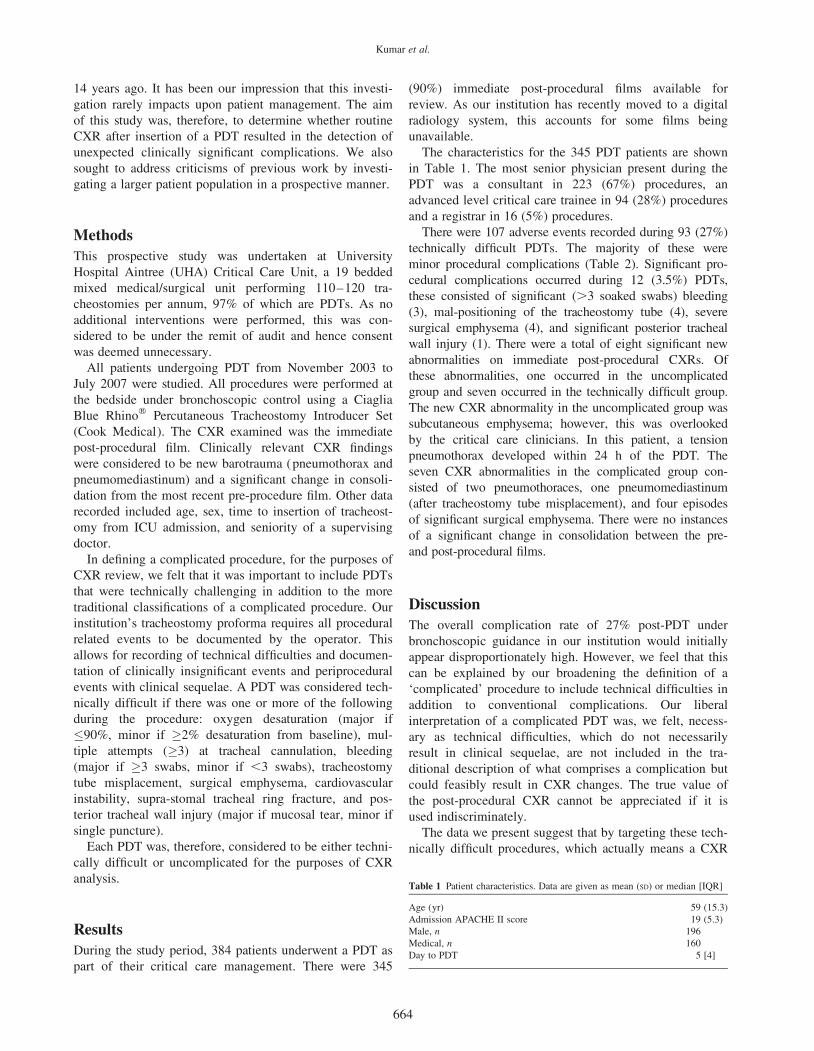
14 years ago. It has been our impression that this investi-gation rarely impacts upon patient management. The aimof this study was, therefore, to determine whether routineCXR after insertion of a PDT resulted in the detection ofunexpected clinically significant complications. We alsosought to address criticisms of previous work by investi-gating a larger patient population in a prospective manner.
MethodsThis prospective study was undertaken at UniversityHospital Aintree (UHA) Critical Care Unit, a 19 beddedmixed medical/surgical unit performing 110–120 tra-cheostomies per annum, 97% of which are PDTs. As noadditional interventions were performed, this was con-sidered to be under the remit of audit and hence consentwas deemed unnecessary.
All patients undergoing PDT from November 2003 toJuly 2007 were studied. All procedures were performed atthe bedside under bronchoscopic control using a CiagliaBlue Rhinow Percutaneous Tracheostomy Introducer Set(Cook Medical). The CXR examined was the immediatepost-procedural film. Clinically relevant CXR findingswere considered to be new barotrauma (pneumothorax andpneumomediastinum) and a significant change in consoli-dation from the most recent pre-procedure film. Other datarecorded included age, sex, time to insertion of tracheost-omy from ICU admission, and seniority of a supervisingdoctor.
In defining a complicated procedure, for the purposes ofCXR review, we felt that it was important to include PDTsthat were technically challenging in addition to the moretraditional classifications of a complicated procedure. Ourinstitution’s tracheostomy proforma requires all proceduralrelated events to be documented by the operator. Thisallows for recording of technical difficulties and documen-tation of clinically insignificant events and periproceduralevents with clinical sequelae. A PDT was considered tech-nically difficult if there was one or more of the followingduring the procedure: oxygen desaturation (major if!90%, minor if "2% desaturation from baseline), mul-tiple attempts ("3) at tracheal cannulation, bleeding(major if "3 swabs, minor if ,3 swabs), tracheostomytube misplacement, surgical emphysema, cardiovascularinstability, supra-stomal tracheal ring fracture, and pos-terior tracheal wall injury (major if mucosal tear, minor ifsingle puncture).
Each PDT was, therefore, considered to be either techni-cally difficult or uncomplicated for the purposes of CXRanalysis.
ResultsDuring the study period, 384 patients underwent a PDT aspart of their critical care management. There were 345
(90%) immediate post-procedural films available forreview. As our institution has recently moved to a digitalradiology system, this accounts for some films beingunavailable.
The characteristics for the 345 PDT patients are shownin Table 1. The most senior physician present during thePDT was a consultant in 223 (67%) procedures, anadvanced level critical care trainee in 94 (28%) proceduresand a registrar in 16 (5%) procedures.
There were 107 adverse events recorded during 93 (27%)technically difficult PDTs. The majority of these wereminor procedural complications (Table 2). Significant pro-cedural complications occurred during 12 (3.5%) PDTs,these consisted of significant (.3 soaked swabs) bleeding(3), mal-positioning of the tracheostomy tube (4), severesurgical emphysema (4), and significant posterior trachealwall injury (1). There were a total of eight significant newabnormalities on immediate post-procedural CXRs. Ofthese abnormalities, one occurred in the uncomplicatedgroup and seven occurred in the technically difficult group.The new CXR abnormality in the uncomplicated group wassubcutaneous emphysema; however, this was overlookedby the critical care clinicians. In this patient, a tensionpneumothorax developed within 24 h of the PDT. Theseven CXR abnormalities in the complicated group con-sisted of two pneumothoraces, one pneumomediastinum(after tracheostomy tube misplacement), and four episodesof significant surgical emphysema. There were no instancesof a significant change in consolidation between the pre-and post-procedural films.
DiscussionThe overall complication rate of 27% post-PDT underbronchoscopic guidance in our institution would initiallyappear disproportionately high. However, we feel that thiscan be explained by our broadening the definition of a‘complicated’ procedure to include technical difficulties inaddition to conventional complications. Our liberalinterpretation of a complicated PDT was, we felt, necess-ary as technical difficulties, which do not necessarilyresult in clinical sequelae, are not included in the tra-ditional description of what comprises a complication butcould feasibly result in CXR changes. The true value ofthe post-procedural CXR cannot be appreciated if it isused indiscriminately.
The data we present suggest that by targeting these tech-nically difficult procedures, which actually means a CXR
Table 1 Patient characteristics. Data are given as mean (SD) or median [IQR]
Age (yr) 59 (15.3)Admission APACHE II score 19 (5.3)Male, n 196Medical, n 160Day to PDT 5 [4]
Kumar et al.
664
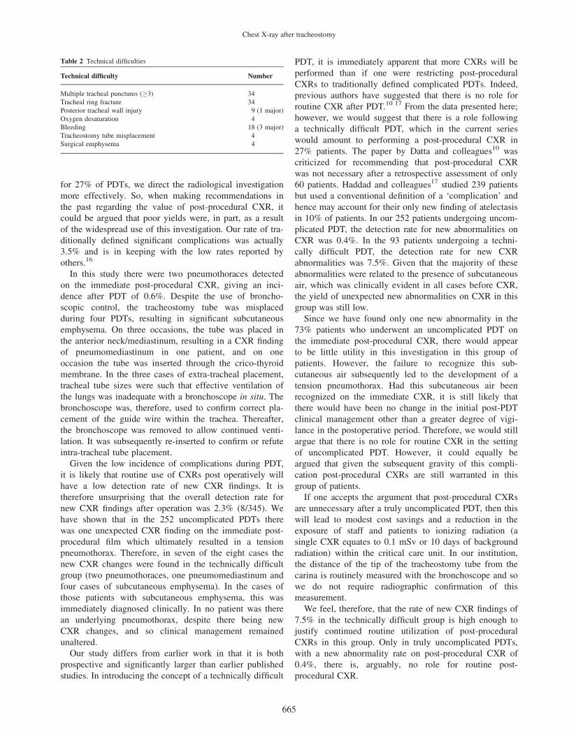
for 27% of PDTs, we direct the radiological investigationmore effectively. So, when making recommendations inthe past regarding the value of post-procedural CXR, itcould be argued that poor yields were, in part, as a resultof the widespread use of this investigation. Our rate of tra-ditionally defined significant complications was actually3.5% and is in keeping with the low rates reported byothers.16
In this study there were two pneumothoraces detectedon the immediate post-procedural CXR, giving an inci-dence after PDT of 0.6%. Despite the use of broncho-scopic control, the tracheostomy tube was misplacedduring four PDTs, resulting in significant subcutaneousemphysema. On three occasions, the tube was placed inthe anterior neck/mediastinum, resulting in a CXR findingof pneumomediastinum in one patient, and on oneoccasion the tube was inserted through the crico-thyroidmembrane. In the three cases of extra-tracheal placement,tracheal tube sizes were such that effective ventilation ofthe lungs was inadequate with a bronchoscope in situ. Thebronchoscope was, therefore, used to confirm correct pla-cement of the guide wire within the trachea. Thereafter,the bronchoscope was removed to allow continued venti-lation. It was subsequently re-inserted to confirm or refuteintra-tracheal tube placement.
Given the low incidence of complications during PDT,it is likely that routine use of CXRs post operatively willhave a low detection rate of new CXR findings. It istherefore unsurprising that the overall detection rate fornew CXR findings after operation was 2.3% (8/345). Wehave shown that in the 252 uncomplicated PDTs therewas one unexpected CXR finding on the immediate post-procedural film which ultimately resulted in a tensionpneumothorax. Therefore, in seven of the eight cases thenew CXR changes were found in the technically difficultgroup (two pneumothoraces, one pneumomediastinum andfour cases of subcutaneous emphysema). In the cases ofthose patients with subcutaneous emphysema, this wasimmediately diagnosed clinically. In no patient was therean underlying pneumothorax, despite there being newCXR changes, and so clinical management remainedunaltered.
Our study differs from earlier work in that it is bothprospective and significantly larger than earlier publishedstudies. In introducing the concept of a technically difficult
PDT, it is immediately apparent that more CXRs will beperformed than if one were restricting post-proceduralCXRs to traditionally defined complicated PDTs. Indeed,previous authors have suggested that there is no role forroutine CXR after PDT.10 17 From the data presented here;however, we would suggest that there is a role followinga technically difficult PDT, which in the current serieswould amount to performing a post-procedural CXR in27% patients. The paper by Datta and colleagues10 wascriticized for recommending that post-procedural CXRwas not necessary after a retrospective assessment of only60 patients. Haddad and colleagues17 studied 239 patientsbut used a conventional definition of a ‘complication’ andhence may account for their only new finding of atelectasisin 10% of patients. In our 252 patients undergoing uncom-plicated PDT, the detection rate for new abnormalities onCXR was 0.4%. In the 93 patients undergoing a techni-cally difficult PDT, the detection rate for new CXRabnormalities was 7.5%. Given that the majority of theseabnormalities were related to the presence of subcutaneousair, which was clinically evident in all cases before CXR,the yield of unexpected new abnormalities on CXR in thisgroup was still low.
Since we have found only one new abnormality in the73% patients who underwent an uncomplicated PDT onthe immediate post-procedural CXR, there would appearto be little utility in this investigation in this group ofpatients. However, the failure to recognize this sub-cutaneous air subsequently led to the development of atension pneumothorax. Had this subcutaneous air beenrecognized on the immediate CXR, it is still likely thatthere would have been no change in the initial post-PDTclinical management other than a greater degree of vigi-lance in the postoperative period. Therefore, we would stillargue that there is no role for routine CXR in the settingof uncomplicated PDT. However, it could equally beargued that given the subsequent gravity of this compli-cation post-procedural CXRs are still warranted in thisgroup of patients.
If one accepts the argument that post-procedural CXRsare unnecessary after a truly uncomplicated PDT, then thiswill lead to modest cost savings and a reduction in theexposure of staff and patients to ionizing radiation (asingle CXR equates to 0.1 mSv or 10 days of backgroundradiation) within the critical care unit. In our institution,the distance of the tip of the tracheostomy tube from thecarina is routinely measured with the bronchoscope and sowe do not require radiographic confirmation of thismeasurement.
We feel, therefore, that the rate of new CXR findings of7.5% in the technically difficult group is high enough tojustify continued routine utilization of post-proceduralCXRs in this group. Only in truly uncomplicated PDTs,with a new abnormality rate on post-procedural CXR of0.4%, there is, arguably, no role for routine post-procedural CXR.
Table 2 Technical difficulties
Technical difficulty Number
Multiple tracheal punctures (!3) 34Tracheal ring fracture 34Posterior tracheal wall injury 9 (1 major)Oxygen desaturation 4Bleeding 18 (3 major)Tracheostomy tube misplacement 4Surgical emphysema 4
Chest X-ray after tracheostomy
665
References1 Delaney A, Bagshaw SM, Nalos M. Percutaneous dilatational tra-
cheostomy versus surgical tracheostomy in critically ill patients: asystematic review and meta-analysis. Crit Care 2006; 10: R55
2 Ciaglia P, Graniero KD. Percutaneous dilatational tracheostomy.Results and long term follow up. Chest 1992; 101: 464–7
3 Grant CA, Dempsey G, Harrison J, Jones T. Tracheo-innominateartery fistula after percutaneous tracheostomy: three casereports and a clinical review. Br J Anaesth 2006; 96: 127–31
4 Brander L, Takala J. Tracheal tear and tension pneumothoraxcomplicating bronchoscopy-guided percutaneous tracheostomy.Heart Lung 2006; 35: 144–5
5 Friedman Y, Mayer AD. Bedside percutaneous tracheostomy incritically ill patients. Chest 1993; 104: 532–5
6 Powell DM, Price PD, Forrest LA. Review of percutaneoustracheostomy. Laryngoscope 1998; 108: 170–7
7 Silvester W, Goldsmith D, Uchino S, et al. Percutaneous versussurgical tracheostomy: A randomized controlled study with long-term follow-up. Crit Care Med 2006; 34: 2145–52
8 Winkler WB, Karnik R, Seelmann O, Havlicek J, Slany J. Bedsidepercutaneous dilational tracheostomy with endoscopic guidance:experience with 71 ICU patients. Intensive Care Med 1994; 20:476–9
9 Barba CA, Angood PB, Kauder DR, et al. Bronchoscopic guidancemakes percutaneous tracheostomy a safe, cost-effective, andeasy-to-teach procedure. Surgery 1995; 118: 879–83
10 Datta D, Onyirimba F, McNamee MJ. The utility of chest radio-graphs following percutaneous dilatational tracheostomy. Chest2003; 123: 1603–6
11 Donaldson DR, Emami AJ, Wax MK. Chest radiographs afterdilatational percutaneous tracheotomy: are they necessary?Otolaryngol Head Neck Surg 2000; 123: 236–9
12 Swanson GJ, Meleca RJ, Bander J, Stachler RJ. The utility of chestradiography following percutaneous dilational tracheotomy. ArchOtolaryngol Head Neck Surg 2002; 128: 1253–4
13 Gonzalez I, Davis A, Schriner S, Rajarajan S, Bonner S. Thevalue of routine chest X-ray post endoscopically guidedpercutaneous dilatational tracheostomy. Intensive Care Med 2002;28: S29
14 Gonzalez I, Bonner S. Routine chest radiographs afterendoscopically guided percutaneous dilatational tracheostomy.Chest 2004; 125: 1173–4
15 Tarnoff M, Moncure M, Jones F, Ross S, Goodman M. The valueof routine posttracheostomy chest radiography. Chest 1998; 113:1647–9
16 Higgins KM, Punthakee X. Meta-analysis comparison of openversus percutaneous tracheostomy. Laryngoscope 2007; 117:447–54
17 Haddad SH, Aldawood AS, Arabi YM. The diagnostic yield andclinical impact of a chest X-ray after percutaneous dilatationaltracheostomy: a prospective cohort study. Anaesth Intensive Care2007; 35: 393–7
Kumar et al.
666
British Journal of Anaesthesia 96 (1): 127–31 (2006)
doi:10.1093/bja/aei282 Advance Access publication November 18, 2005
RESPIRATION AND THE AIRWAY
Case Report
Tracheo-innominate artery fistula after percutaneoustracheostomy: three case reports and a clinical review
C. A. Grant1*, G. Dempsey1, J. Harrison1 and T. Jones2
1Critical Care Unit and 2Department of Head and Neck Surgery, University Hospital,Aintree, Liverpool, UK
*Corresponding author. E-mail: [email protected]
Tracheo-innominate artery fistula (TIF) is an uncommon yet life threatening complication after atracheostomy. Rates of 0.1–1% after surgical tracheostomy have been reported, with a peakincidence at 7–14 days post procedure. It is usually fatal unless treatment is instituted immediately.Initial case reports of TIF resulted from surgically performed tracheostomies. We present threefatalities attributable to TIF, confirmed by histopathology, after percutaneous dilatational tracheo-stomy (PDT). The use of PDT has resulted in tracheostomies being performed by specialists fromdifferent backgrounds and the incidence of this complication may be increasing. Pressure necrosisfrom high cuff pressure, mucosal trauma from malpositioned cannula tip, low tracheal incision,radiotherapy and prolonged intubation are all implicated in TIF formation. Massive haemorrhageoccurring 3 days to 6 weeks after tracheostomy is a result of TIF until proven otherwise. Wepresent a simple algorithm for management of this situation. The manoeuvres outlined willcontrol bleeding in more than 80% of patients by a direct tamponade effect. Surgical stasis isobtained by debriding the innominate artery proximally, then transecting and closing the lumen.Neurological sequelae are few. Post-mortem diagnosis of TIF may be difficult, but specificpathology request should be made to assess innominate artery abnormalities.
Br J Anaesth 2006; 96: 127–31
Keywords: complications, death; complications, fistula; complications, haemorrhage; surgery,tracheostomy; surgery, tracheotomy
Accepted for publication: October 17, 2005
Percutaneous dilatational tracheostomy (PDT) has becomea standard technique in critical care medicine. As a result ofits widespread use, it continues to attract controversy anddebate. The UK Intensive Care Society recently launchedthe TracMan trial which concluded in its preface that PDTis ‘a common procedure but with limited evidence base tosupport its use.’ We present three fatalities resulting fromtracheo-innominate artery fistulae (TIF) after PDT.
Although possible to perform under local anaesthesia, themajority of PDTs are performed under general anaesthesiawith neuromuscular block on Critical Care Units by non-surgically trained operators. Complications of PDT aretraditionally divided into early and late. The majorityof publications centre on early complications includingbleeding, pneumothorax, technical failure and perioperativehypoxia.1 The major reported late complications include
tracheo-oesophageal fistula, tracheomalacia and trachealstenosis.2
TIF is an uncommon yet life threatening complicationthat can occur after a tracheostomy. Reported incidenceis 0.1–1% after surgical tracheostomy, with peak incidence7–14 days post procedure.3 4 It is usually fatal unlesstreatment is instituted immediately.5 6 We present a case-series of tracheo-innominate arterial fistulae after PDT withhistopathological confirmation.
Case 1A 43-yr-old female with a community-acquired pneumoniapresented to the Critical Care Unit requiring invasiveventilatory support. Subsequent management included anuneventful PDT 11 days from her critical care admission.
! The Board of Management and Trustees of the British Journal of Anaesthesia 2005. All rights reserved. For Permissions, please e-mail: [email protected]
by guest on April 3, 2015
http://bja.oxfordjournals.org/D
ownloaded from
Two subsequent uneventful tracheostomy tube exchangeswere performed during the admission. Thirty-two daysafter this procedure, a small haemorrhage developed fromthe tracheostomy site. This was self-limiting and broncho-scopic investigation did not identify a bleeding site withinthe respiratory tract. Similarly, no signs of inflammation orinfection were present in the tracheostomy wound. How-ever, within 3 h of this initial bleed, massive oral and trach-eal haemorrhage occurred. Bleeding did not initially appearpulsatile but the volume of blood prevented effectiveoxygenation. The trachea was intubated translaryngeally,allowing removal of the tracheostomy tube, inspection ofthe stoma and digital compression of the suspected bleedingsite. Failure to terminate the bleeding and persistentsuboptimal ventilation resulted in a fatal cardiac arrest.
Case 2A 57-year-old male was admitted to the Critical Care Unitfollowing a road traffic accident. His injuries consisted ofa fractured pelvis, which required external fixation, and anextensive retroperitoneal haemorrhage. Inability to with-draw mechanical ventilation and an associated pneumonialed to an uneventful PDT on Day 11. Twelve days after thePDT, his respiratory function had improved sufficiently toallow consideration of decannulation. However, prior tothis, sudden haemorrhage developed via the tracheostomytube. Adequate ventilation failed because of the amountof blood in the major airways. Tracheal intubation wasachieved via the translaryngeal route. It was not possibleto identify a bleeding point and a double lumen endobron-chial tube and bilateral intercostal drains were inserted.Unsuccessful attempts, using fibreoptic bronchoscopy, tosuction the aspirated blood from the tracheobronchial treefollowed. It proved impossible to re-establish effectiveventilation, resulting in a fatal hypoxic cardiac arrest.
Case 3A 69-yr-old female was admitted to the Critical Care Unitafter an elective oesophagogastrectomy for oesophagealcarcinoma. Her initial postoperative course was uncompli-cated until Day 5 when she developed bronchopneumonia.On Day 8, inspiratory stridor and acute respiratory failureled to emergency tracheal intubation and mechanical vent-ilation. A PDT followed on the same day. Her subsequentmanagement was unremarkable and artificial ventilation wasdiscontinued by Day 10. She was discharged to the wardwith a tracheostomy tube in situ. On Day 17, she developedmassive haemorrhage into the airway resulting in desatura-tion and cardiac arrest. Despite translaryngeal intubation andprolonged attempts to clear the airway, resuscitation was notpossible.
Each procedure was bronchoscopically guided andperformed in the presence of an intensive care consultant.Ciaglia Blue Rhino! (Cook, Letchworth, UK) tracheostomyset was used and a size 8.0 mm Crystal Clear! (Rusch,
Lurgan, UK) tracheostomy tube was used in each case.Each PDT was considered to have been between trachealrings I and IV. All cuff pressures were monitored regularly,as is our standard practice.The occurrence of these three cases within 18 months of
each other aroused clinical suspicion of a potential link inunderlying pathology. After direction from the cliniciansinvolved, the subsequent post-mortem examinations lookedspecifically at both the tracheostomy site and the tracheo-innominate trunk.As a result of their small size, post-mortem diagnosis of
TIF can be difficult. For all three patients, sections from thetracheo-innominate artery at the level of the tracheostomysite revealed a small focus of active chronic inflammationextending through the full thickness of the arterial wall intothe luminal surface. Histological evidence of necrosis wasidentified extending through the adjacent wall of the tracheawith consequent focal disruption of the tracheal wall. Thehistopathological confirmation of a TIF was reported on thesubsequent death certificates.
Tracheo-innominate fistula
The true incidence of this rare complication is difficult toassess. After surgical tracheostomy, the incidence has beenestimated to be 0.1–1%.4 Pooled data of 5530 tracheostom-ies recorded the incidence of delayed massive haemorrhageas 0.3%.7 Initial case reports of TIF resulted from surgicallyperformed tracheostomies. However, the incidence mayhave declined because of advances in tracheostomy tubetechnology and the introduction of PDT. The increasedpopularity of PDT has resulted in tracheostomies being per-formed by an increasing number of specialists from differentbackgrounds. It is conceivable that the rate of such a rarecomplication is perceived by different clinical groups, to belower than it actually is. Consequently we suggest that TIF,although rare, should be borne in mind by all those involvedin tracheostomy management. In our institution more than1000 PDT have been performed since 1994, giving a crudeincidence of 0.3%.
Pathophysiology
Knowledge of the anatomy of the innominate (brachioceph-alic) artery (Fig. 1) and its relationship to a tracheostomytube is essential in understanding the pathophysiologyof TIF. The tracheo-innominate artery (or trunk) is thefirst branch of the aortic arch. It divides into the right com-mon carotid and right subclavian artery, 3–4 cm lateral to thetrachea, behind the right sternoclavicular joint. In its inferiorproximal portion, its relations include:
! anterior: left tracheo-innominate vein and thymus;! posterior: trachea ("6–10th ring);! posterior and left: left common carotid artery;! right: right tracheo-innominate vein, superior vena cava
and pleura.
Grant et al.
128
by guest on April 3, 2015
http://bja.oxfordjournals.org/D
ownloaded from
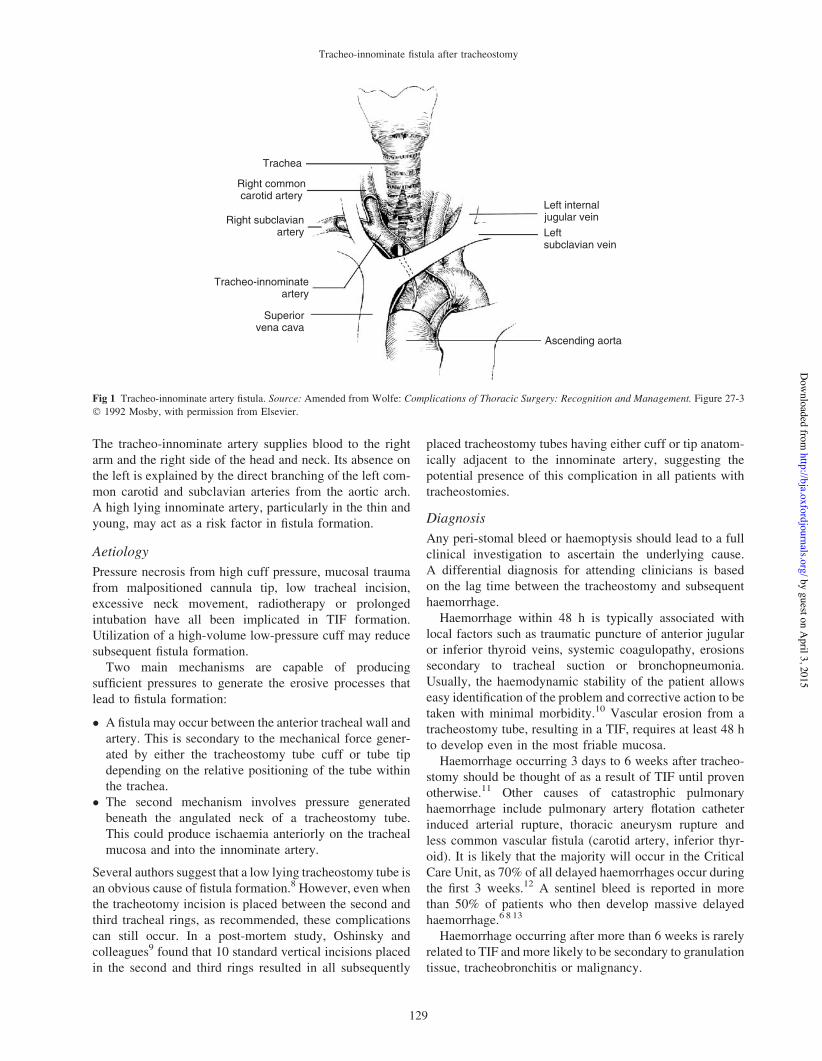
The tracheo-innominate artery supplies blood to the rightarm and the right side of the head and neck. Its absence onthe left is explained by the direct branching of the left com-mon carotid and subclavian arteries from the aortic arch.A high lying innominate artery, particularly in the thin andyoung, may act as a risk factor in fistula formation.
Aetiology
Pressure necrosis from high cuff pressure, mucosal traumafrom malpositioned cannula tip, low tracheal incision,excessive neck movement, radiotherapy or prolongedintubation have all been implicated in TIF formation.Utilization of a high-volume low-pressure cuff may reducesubsequent fistula formation.
Two main mechanisms are capable of producingsufficient pressures to generate the erosive processes thatlead to fistula formation:
! A fistula may occur between the anterior tracheal wall andartery. This is secondary to the mechanical force gener-ated by either the tracheostomy tube cuff or tube tipdepending on the relative positioning of the tube withinthe trachea.
! The second mechanism involves pressure generatedbeneath the angulated neck of a tracheostomy tube.This could produce ischaemia anteriorly on the trachealmucosa and into the innominate artery.
Several authors suggest that a low lying tracheostomy tube isan obvious cause of fistula formation.8 However, even whenthe tracheotomy incision is placed between the second andthird tracheal rings, as recommended, these complicationscan still occur. In a post-mortem study, Oshinsky andcolleagues9 found that 10 standard vertical incisions placedin the second and third rings resulted in all subsequently
placed tracheostomy tubes having either cuff or tip anatom-ically adjacent to the innominate artery, suggesting thepotential presence of this complication in all patients withtracheostomies.
Diagnosis
Any peri-stomal bleed or haemoptysis should lead to a fullclinical investigation to ascertain the underlying cause.A differential diagnosis for attending clinicians is basedon the lag time between the tracheostomy and subsequenthaemorrhage.
Haemorrhage within 48 h is typically associated withlocal factors such as traumatic puncture of anterior jugularor inferior thyroid veins, systemic coagulopathy, erosionssecondary to tracheal suction or bronchopneumonia.Usually, the haemodynamic stability of the patient allowseasy identification of the problem and corrective action to betaken with minimal morbidity.10 Vascular erosion from atracheostomy tube, resulting in a TIF, requires at least 48 hto develop even in the most friable mucosa.
Haemorrhage occurring 3 days to 6 weeks after tracheo-stomy should be thought of as a result of TIF until provenotherwise.11 Other causes of catastrophic pulmonaryhaemorrhage include pulmonary artery flotation catheterinduced arterial rupture, thoracic aneurysm rupture andless common vascular fistula (carotid artery, inferior thyr-oid). It is likely that the majority will occur in the CriticalCare Unit, as 70% of all delayed haemorrhages occur duringthe first 3 weeks.12 A sentinel bleed is reported in morethan 50% of patients who then develop massive delayedhaemorrhage.6 8 13
Haemorrhage occurring after more than 6 weeks is rarelyrelated to TIF and more likely to be secondary to granulationtissue, tracheobronchitis or malignancy.
Trachea
Right commoncarotid artery
Left internaljugular veinLeftsubclavian vein
Ascending aorta
Right subclavianartery
Tracheo-innominateartery
Superiorvena cava
Fig 1 Tracheo-innominate artery fistula. Source: Amended from Wolfe: Complications of Thoracic Surgery: Recognition and Management. Figure 27-3! 1992 Mosby, with permission from Elsevier.
Tracheo-innominate fistula after tracheostomy
129
by guest on April 3, 2015
http://bja.oxfordjournals.org/D
ownloaded from
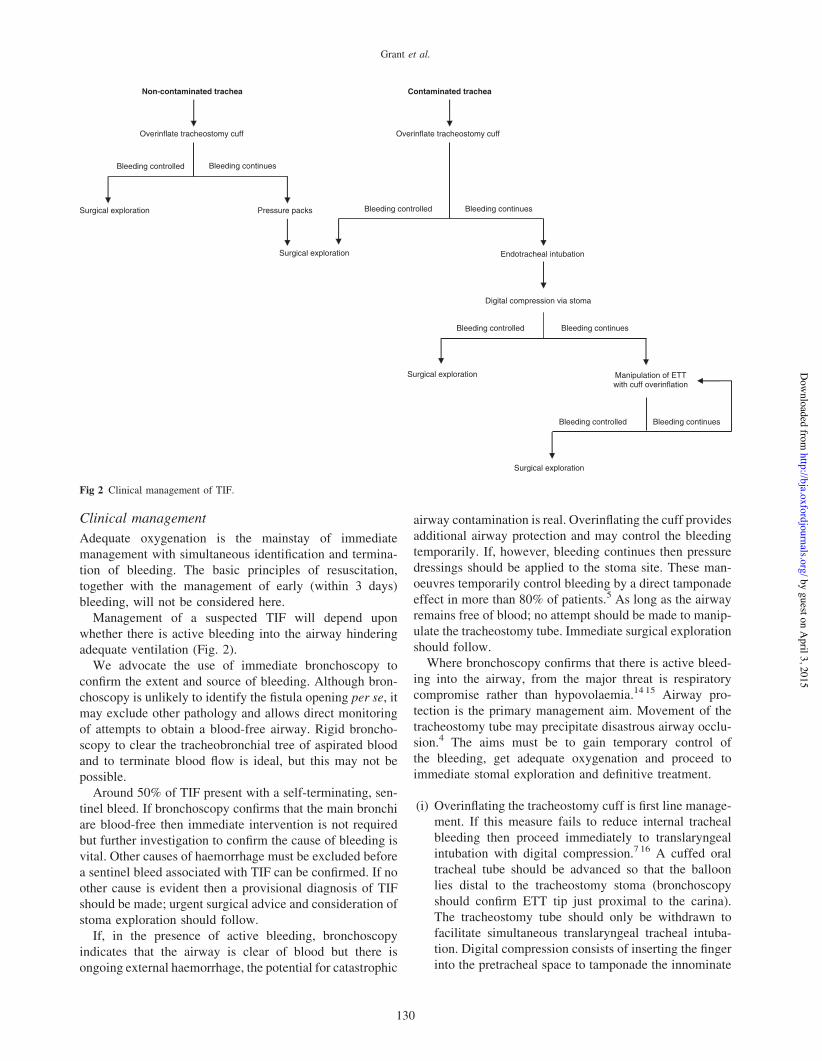
Clinical management
Adequate oxygenation is the mainstay of immediatemanagement with simultaneous identification and termina-tion of bleeding. The basic principles of resuscitation,together with the management of early (within 3 days)bleeding, will not be considered here.
Management of a suspected TIF will depend uponwhether there is active bleeding into the airway hinderingadequate ventilation (Fig. 2).
We advocate the use of immediate bronchoscopy toconfirm the extent and source of bleeding. Although bron-choscopy is unlikely to identify the fistula opening per se, itmay exclude other pathology and allows direct monitoringof attempts to obtain a blood-free airway. Rigid broncho-scopy to clear the tracheobronchial tree of aspirated bloodand to terminate blood flow is ideal, but this may not bepossible.
Around 50% of TIF present with a self-terminating, sen-tinel bleed. If bronchoscopy confirms that the main bronchiare blood-free then immediate intervention is not requiredbut further investigation to confirm the cause of bleeding isvital. Other causes of haemorrhage must be excluded beforea sentinel bleed associated with TIF can be confirmed. If noother cause is evident then a provisional diagnosis of TIFshould be made; urgent surgical advice and consideration ofstoma exploration should follow.
If, in the presence of active bleeding, bronchoscopyindicates that the airway is clear of blood but there isongoing external haemorrhage, the potential for catastrophic
airway contamination is real. Overinflating the cuff providesadditional airway protection and may control the bleedingtemporarily. If, however, bleeding continues then pressuredressings should be applied to the stoma site. These man-oeuvres temporarily control bleeding by a direct tamponadeeffect in more than 80% of patients.5 As long as the airwayremains free of blood; no attempt should be made to manip-ulate the tracheostomy tube. Immediate surgical explorationshould follow.Where bronchoscopy confirms that there is active bleed-
ing into the airway, from the major threat is respiratorycompromise rather than hypovolaemia.14 15 Airway pro-tection is the primary management aim. Movement of thetracheostomy tube may precipitate disastrous airway occlu-sion.4 The aims must be to gain temporary control ofthe bleeding, get adequate oxygenation and proceed toimmediate stomal exploration and definitive treatment.
(i) Overinflating the tracheostomy cuff is first line manage-ment. If this measure fails to reduce internal trachealbleeding then proceed immediately to translaryngealintubation with digital compression.7 16 A cuffed oraltracheal tube should be advanced so that the balloonlies distal to the tracheostomy stoma (bronchoscopyshould confirm ETT tip just proximal to the carina).The tracheostomy tube should only be withdrawn tofacilitate simultaneous translaryngeal tracheal intuba-tion. Digital compression consists of inserting the fingerinto the pretracheal space to tamponade the innominate
Manipulation of ETTwith cuff overinflation
Surgical exploration
Surgical exploration
Endotracheal intubation
Overinflate tracheostomy cuff
Bleeding controlled
Non-contaminated trachea
Overinflate tracheostomy cuff
Surgical exploration Pressure packs
Surgical exploration
Bleeding continues
Bleeding continues Bleeding controlled
Contaminated trachea
Digital compression via stoma
Bleeding continues Bleeding controlled
Bleeding continues Bleeding controlled
Fig 2 Clinical management of TIF.
Grant et al.
130
by guest on April 3, 2015
http://bja.oxfordjournals.org/D
ownloaded from
artery against the posterior surface of the manu-brium.11 12 This procedure should terminate bleedingin >90% patients and if maintained, allows transferto the operating theatres.6 17
(ii) If digital compression fails to stem bleed, slow with-drawal of the tracheal tube and cuff over-inflationshould follow. Manipulating the ETT tube, and its cuffto produce tamponade is the only other manoeuvre avail-able in this situation and attempts should persist.
If the bleeding stops temporarily, and ventilation is accept-able, then immediate surgical intervention should follow.
Management of TIF is a surgical emergency. Mortality is!100% without operative intervention. The paucity of evi-dence to support any imaging leads us to recommend theimmediate surgical exploration. The index of clinical sus-picion precludes delaying surgical intervention and anyimaging, we feel, should only be undertaken within thetheatre suite in conjunction with surgical management.
A standard median sternotomy approach is appropriatefor access. It has been suggested that in specialist units,two separate incisions involving a right anterior thora-cotomy and neck approach may be advantageous in termsof prevention of mediastinitis and sternal dehiscence.13
Successful surgical interventions have included the use ofsaphenous vein and innominate vein grafts, sternocleido-mastoid patches or pedicled pericardial grafts.17 However,the mainstay of surgical treatment is to terminate flowwithinthe innominate artery by debriding the innominate arteryproximally until healthy tissue is obtained, then transectingand closing the lumen. There is no convincing evidence tosuggest that this leads to significant neurological or vascularcompromise.18 19 Arterial reconstruction should no longerbe considered, as arterial tie-off presents significantly bettermortality and morbidity results.
In a patient presenting with fresh haemorrhage beyond72 h after tracheostomy, the overriding concern should beof an underlying TIF. The simple algorithm that we havepresented may be a useful guide. Our understanding of thelong-term sequelae of the PDT is poor. We recommend thatany death that results from, or is associated with massivehaemorrhage from the respiratory tract in a patient who hasreceived a PDT should have a post-mortem looking specif-ically at innominate artery pathology.
AcknowledgementThe authors thank Ms E. Walker for her help with preparation of themanuscript.
References1 Ciaglia P, Graniero KD. Percutaneous dilatational tracheostomy.
Results and long term follow up. Chest 1992; 101: 464–6722 Wood DE, Mathisen DJ. Late complications of tracheotomy.
Clin Chest Med 1991; 12: 597–6093 Schaefer OP, Irwin RS. Tracheoarterial fistula: an unusual
complication of tracheostomy. J Intensive Care Med 1995; 10:64–7
4 Allan JS, Wright CD. Tracheoinnominate fistula: diagnosis andmanagement. Chest Surg Clin N Am 2003; 13: 331–41
5 Bloss RS, Ward RE. Survival after tracheoinnominate arteryfistula. Am J Surg 1980; 139: 251–3
6 Courcy PA, Rodriguez A, Garrett HE. Operative technique forrepair of tracheoinnominate artery fistula. J Vasc Surg 1985; 2:332–4
7 Dyer RK, Fisher SR. Tracheal-innominate and tracheal-esophagealfistula. In: Wolfe WG, ed. Complications in Thoracic Surgery:Recognition & Management. St Louis: Mosby Year Book, 1992;288–306
8 Grillo CG. Tracheal Fistula to Brachiocephalic Artery. In:Grillo CG, ed. Surgery of the Trachea and Bronchi. Hamilton: BCDecker, 2003; ch. 13, 1–9
9 Oshinsky AE, Rubin JS, Gwozdz CS. The anatomical basis forpost-tracheotomy innominate artery rupture. Laryngoscope1988; 98: 1061–4
10 Muhammad JK, Major E, Wood A, Patton DW.Percutaneous dilatational tracheostomy: haemorrhagic complica-tions and the vascular anatomy of the anterior neck. Areview based on 497 cases. Int J Oral Maxillofac Surg 2000; 29:217–22
11 Nelems JM. Tracheo-innominate artery fistula. Am J Surg 1981;141: 526–7
12 Bertelsen S, Jensen NM. Innominate artery rupture. A fatalcomplication of tracheostomy. Ann Chir Gynaecol 1987; 76:230–3
13 Jones JW, Reynolds M, Hewitt RL, Drapanas T. Tracheo-innominate artery erosion: successful surgical management of adevastating complication. Ann Surg 1976; 184: 194–204
14 Hafez A, Couraud L, Velly JF, Bruneteau A. Late cataclysmichemorrhage from the innominate artery after tracheostomy.Thorac Cardiovasc Surg 1984; 32: 315–9
15 Gelman JJ, Aro M, Weiss SM. Tracheo-innominate artery fistula.J Am Coll Surg 1994; 179: 626–34
16 Keceligil HT, Erk MK, Kolbakir F, Yildirim A, Yilman M, Unal R.Tracheoinnominate artery fistula following tracheostomy.Cardiovasc Surg 1995; 3: 509–10
17 Utley JR, Singer MM, Roe BB, Fraser DG, Dedo HH. Definitivemanagement of innominate artery hemorrhage complicatingtracheostomy. JAMA 1972; 24: 577–9
18 Brewster DC, Moncure AC, Darling RC, Ambrosino JJ,Abbott WM. Innominate artery lesions: problems encounteredand lessons learned. J Vasc Surg 1985; 2: 99–112
19 Deslauriers J, Ginsberg RJ, Nelems JM, Pearson FG. Innominateartery rupture. A major complication of tracheal surgery. AnnThorac Surg 1975; 20: 671–7
Tracheo-innominate fistula after tracheostomy
131
by guest on April 3, 2015
http://bja.oxfordjournals.org/D
ownloaded from
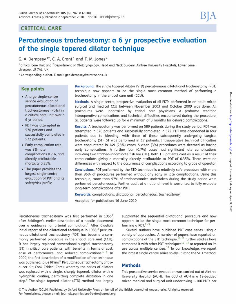
CRITICAL CARE
Percutaneous tracheostomy: a 6 yr prospective evaluationof the single tapered dilator techniqueG. A. Dempsey 1*, C. A. Grant 1 and T. M. Jones 2
1 Critical Care Unit and 2 Department of Otolaryngology, Head and Neck Surgery, Aintree University Hospitals, Lower Lane,Liverpool L9 7AL, UK
* Corresponding author. E-mail: [email protected]
Key points
† A large single-centreservice evaluation ofpercutaneous dilatationaltracheostomies (PDTs) ina critical care unit over a6 yr period.
† PDT was attempted in576 patients andsuccessfully completed in572 patients.
† Early complication ratewas 3%, latecomplications 0.7%, anddirectly attributablemortality 0.35%.
† The paper provides thelargest single-centreevaluation of PDT and itssafety/risk profile.
Background. The single tapered dilator (STD) percutaneous dilatational tracheostomy (PDT)technique now appears to be the single most common method of performing atracheostomy in the critical care unit (CCU).
Methods. A single-centre, prospective evaluation of all PDTs performed in an adult mixedsurgical and medical CCU between November 2003 and October 2009 was done. Allprocedures were undertaken by critical care physicians. A proforma recordedintraoperative complications and technical difficulties encountered during the procedure;all patients were followed up for a minimum of 3 months for delayed complications.
Results. A tracheostomy was performed on 589 patients during the study period. PDT wasattempted in 576 patients and successfully completed in 572. PDT was abandoned in fourpatients due to bleeding, with three of these subsequently undergoing surgicaltracheostomy (ST). ST was performed in 17 patients. Intraoperative technical difficultieswere encountered in 149 (26%) cases. Sixteen (3%) procedures were deemed as havingearly complications. A further four (0.7%) cases had significant late complicationsincluding two tracheo-innominate fistulae (TIF). Both TIF patients died as a result of theircomplications giving a mortality directly attributable to PDT of 0.35%. There were nodifferences with respect to the occurrence of complications according to grade of operator.
Conclusions. PDT performed by the STD technique is a relatively safe procedure with morethan 96% of procedures performed without any early or late complications. Using thistechnique, more than 97% of tracheostomies undertaken during the study period wereperformed percutaneously. Further audit at a national level is warranted to fully evaluatelong-term complications after PDT.
Keywords: complications; dilatational; percutaneous; tracheostomy
Accepted for publication: 16 June 2010
Percutaneous tracheostomy was first performed in 19551
after Seldinger’s earlier description of a needle placementover a guidewire for arterial cannulation.2 After Ciaglia’sinitial report of the dilatational technique in 1985,3 percuta-neous dilatational tracheostomy (PDT) has become a com-monly performed procedure in the critical care unit (CCU).It has largely replaced conventional surgical tracheostomy(ST) in critical care patients, with benefits in terms of cost,ease of performance, and reduced complications.4 5 In2000, the first description of a modification of the techniquewas published (Blue Rhinow PercutaneousTracheotomy Intro-ducer Kit; Cook Critical Care), whereby the series of dilatorswas replaced with a single, sharply tapered, dilator with ahydrophilic coating, permitting complete dilatation in onestep.6 The single tapered dilator (STD) method has largely
supplanted the sequential dilatational procedure and nowappears to be the single most common technique for per-forming a PDT.7 – 9
Several authors have published PDT case series using avariety of approaches. A number of papers have reported oncomplications of the STD technique;10 11 further studies havecompared it with other PDT techniques12–14 or reported on itsuse across multiple centres.15 To our knowledge, we reportthe largest single-centre series solely utilizing the STD method.
MethodsThis prospective service evaluation was carried out at AintreeUniversity Hospital (AUH). The CCU at AUH is a 19-beddedmixed medical and surgical unit undertaking !100 PDTs per
British Journal of Anaesthesia 105 (6): 782–8 (2010)Advance Access publication 2 September 2010 . doi:10.1093/bja/aeq238
& The Author [2010]. Published by Oxford University Press on behalf of the British Journal of Anaesthesia. All rights reserved.For Permissions, please email: [email protected]
at Liverpool University Library on A
pril 8, 2012http://bja.oxfordjournals.org/
Dow
nloaded from
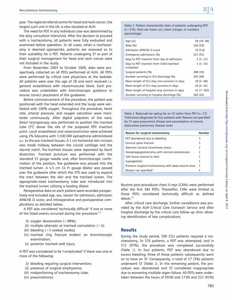
year. The regional referral centre for head and neck cancer, thelargest such unit in the UK, is also situated at AUH.
The need for PDT in any individual case was determined bythe duty consultant intensivist. After the decision to proceedwith a tracheostomy, all patients were fully evaluated andexamined before operation. In all cases, when a tracheost-omy is deemed appropriate, patients are assessed as totheir suitability for a PDT. Patients undergoing ST as part oftheir surgical management for head and neck cancer werenot included in the study.
From November 2003 to October 2009, data were pro-spectively collected on all PDTs performed at AUH. All PDTswere performed by critical care physicians at the bedside.All patients were over the age of 18 and each received i.v.general anaesthesia with neuromuscular block. Each pro-cedure was undertaken with bronchoscopic guidance toensure correct placement of the guidewire.
Before commencement of the procedure, the patient waspositioned with the head extended and the lungs were ven-tilated with 100% oxygen. Throughout the procedure, heartrate, arterial pressure, and oxygen saturation were moni-tored continuously. After digital palpation of the neck,direct laryngoscopy was performed to position the trachealtube (TT) above the site of the proposed PDT insertionpoint. Local anaesthesia and vasoconstriction were achievedusing 2% lidocaine with 1:100 000 epinephrine administereds.c. to the pre-tracheal tissues. A 1 cm horizontal skin incisionwas made midway between the cricoid cartilage and thesternal notch. Pre-tracheal tissues were separated by bluntdissection. Tracheal puncture was performed with thestandard 15 gauge needle and, after bronchoscopic confir-mation of the position, the guidewire was passed into thetracheal lumen. A 4.5 cm 14 Fr gauge dilator was passedover the guidewire after which the STD was used to expandthe tract between the skin and the tracheal lumen. Theappropriate-sized tracheostomy tube was introduced intothe tracheal lumen utilizing a loading dilator.
Perioperative data on each patient were recorded prospec-tively and included age, sex, reason for admission, admissionAPACHE II score, and intraoperative and postoperative com-plications as detailed below.
A PDT was considered ‘technically difficult’ if one or moreof the listed events occurred during the procedure:16
(i) oxygen desaturation (,88%);(ii) multiple attempts at tracheal cannulation (.3);(iii) bleeding (.3 soaked swabs);(iv) tracheal ring fracture evident on bronchoscopic
examination;(v) posterior tracheal wall injury.
A PDT was considered to be ‘complicated’ if there was one ormore of the following:
(i) bleeding requiring surgical intervention;(ii) presence of surgical emphysema;(iii) malpositioning of tracheostomy tube;(iv) pneumothorax.
Routine post-procedural chest X-rays (CXRs) were performedafter the first 384 PDTs. Thereafter, CXRs were limited tothose PDTs considered technically difficult as definedabove.16
After critical care discharge, further surveillance was pro-vided by the AUH Critical Care Outreach Service and afterhospital discharge by the critical care follow-up clinic allow-ing identification of late complications.
ResultsDuring the study period, 590 CCU patients required a tra-cheostomy. In 576 patients, a PDT was attempted, and in572 (97%), the procedure was completed successfully(Table 1). In four patients, PDT was abandoned due toexcess bleeding; three of these patients subsequently wenton to have an ST. Consequently, a total of 17 (3%) patientsunderwent ST (Table 2). In the remaining patient, the pro-cedure was abandoned and ST considered inappropriatedue to worsening multiple organ failure. All PDTs were under-taken between the hours of 09:00 and 17:00 and 522 (91%)
Table 2 Rationale for opting for an ST rather than PDT (n!17).*Admission diagnoses for four patients with ‘Reason not specified’for ST were pneumonia (three) and exacerbation of chronicobstructive pulmonary disease (one)
Reason for surgical tracheostomy Number
PDT abandoned due to bleeding 3
Cervical spine fracture 3
Aberrant cervical innominate artery 2
Oesophagogastrectomy with cervical anastomosis 2
Soft tissue trauma to neck 1
Supraglottitis 1
Previous surgical tracheostomy with deep wound sinus 1
Reason not specified* 4
Table 1 Patient characteristic data of patients undergoing PDT(n!576). Data are mean (SD), mean (range), or numbers(percentage)
Age (yr) 58 (18–86)
Male (%) 340 (59)
Admission APACHE II score 19 (5.6)
Emergency admissions (%) 521 (91)
Days to PDT insertion from day of admission 5 (1–21)
Days to PDT insertion from initial trachealintubation
4 (1–14)
Surgical patients (%) 288 (50)
Number surviving to ICU discharge (%) 395 (69)
Mean length of ICU stay non-survivors in days 18 (3–68)
Mean length of ICU stay survivors in days 20 (4–92)
Mean length of hospital stay survivors in days 42 (7–382)
Number surviving to hospital discharge (%) 360 (63)
Percutaneous tracheostomy BJA
783
at Liverpool University Library on A
pril 8, 2012http://bja.oxfordjournals.org/
Dow
nloaded from
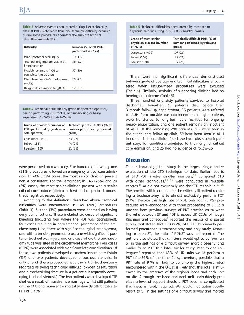
were performed on a weekday. Five hundred and twenty-one(91%) procedures followed an emergency critical care admis-sion. In 406 (71%) cases, the most senior clinician presentwas a consultant. For the remainder, in 146 (26%) and 20(3%) cases, the most senior clinician present was a seniorcritical care trainee (clinical fellow) and a specialist anaes-thetic registrar, respectively.
According to the definitions described above, technicaldifficulties were encountered in 149 (26%) procedures(Table 3). Sixteen (3%) procedures were deemed as havingearly complications. These included six cases of significantbleeding (including four where the PDT was abandoned),four cases resulting in para-tracheal placement of the tra-cheostomy tube, three with significant surgical emphysema,one with a tension pneumothorax, one with significant pos-terior tracheal wall injury, and one case where the tracheost-omy tube was sited in the cricothyroid membrane. Four cases(0.7%) were associated with significant late complications. Ofthese, two patients developed a tracheo-innominate fistula(TIF) and two patients developed a tracheal stenosis. Inonly one of these procedures was the initial tracheostomyregarded as being technically difficult (oxygen desaturationand a tracheal ring fracture in a patient subsequently devel-oping tracheal stenosis). The two patients who developed TIFdied as a result of massive haemorrhage whilst still patientson the CCU and represent a mortality directly attributable toPDT of 0.35%.
There were no significant differences demonstratedbetween grade of operator and technical difficulties encoun-tered when unsupervised procedures were excluded(Table 4). Similarly, seniority of supervising clinician had nobearing on outcome (Table 5).
Three hundred and sixty patients survived to hospitaldischarge. Thereafter, 25 patients died before their3 month follow-up appointment, 36 patients were referredto AUH from outside our catchment area, eight patientswere transferred to long-term care facilities for ongoingneuro-rehabilitation, and one patient remains an inpatientat AUH. Of the remaining 290 patients, 202 were seen inthe critical care follow-up clinic, 59 have been seen in AUHin non-critical care clinics, four have had subsequent inpati-ent stays for conditions unrelated to their original criticalcare admission, and 25 had no evidence of follow-up.
DiscussionTo our knowledge, this study is the largest single-centreevaluation of the STD technique to date. Earlier reportsof STD PDT involve smaller numbers,10 compared STDwith other techniques,12 – 14 were conducted in multiplecentres,15 or did not exclusively use the STD technique.11 15
The practice within our unit, for the critically ill patient requir-ing a tracheostomy, is to almost exclusively perform PDT(97%). Despite this high rate of PDT, only four (0.7%) pro-cedures were abandoned with three proceeding to ST. It isunclear from previous surveys of PDT practice as to whatthe ratio between ST and PDT is across UK CCUs. AlthoughKrishnan and colleagues7 reported the results of a postalsurvey that stated that 173 (97%) of UK ICUs primarily per-formed percutaneous tracheostomy and only rarely, resort-ing to open ST, the ratio of PDT:ST was not reported. Theauthors also stated that clinicians would opt to perform anST in the settings of a difficult airway, morbid obesity, andearlier failed PDT. In a later, similar study, Veenith and col-leagues8 reported that 43% of UK units would perform aPDT of .95% of the time. It is, therefore, possible that aPDT rate of 97% is likely to be among the highest ratesencountered within the UK. It is likely that this rate is influ-enced by the presence of the regional head and neck uniton site. Although the head and neck unit undoubtedly pro-vides a level of support should a PDT become complicatedthis input is rarely required. We would not automaticallyresort to ST in the settings of a difficult airway and morbid
Table 3 Adverse events encountered during 149 technicallydifficult PDTs. Note more than one technical difficulty occurredduring some procedures, therefore the sum of technicaldifficulties exceeds 149
Difficulty Number (% of all PDTsperformed, n5576)
Minor posterior wall injury 9 (1.6)
Tracheal ring fracture visible atbronchoscopy
56 (9.7)
Multiple attempts (!3) tocannulate the trachea
57 (10)
Minor bleeding (3–5 small soakedswabs)
25 (4.3)
Oxygen desaturation to "88% 17 (2.9)
Table 4 Technical difficulties by grade of operator; operator,person performing PDT, that is, not supervising or beingsupervised. P.0.05 Kruskal–Wallis
Grade of operator (number ofPDTs performed by grade as asole operator)
Technically difficult PDTs (% ofnumber performed by relevantgrade)
Consultant (149) 33 (22)
Fellow (151) 44 (29)
Registrar (120) 31 (26)
Table 5 Technical difficulties encountered by most seniorphysician present during PDT. P.0.05 Kruskal–Wallis
Grade of most seniorphysician present (numberof PDTs)
Technically difficult PDTs (% ofnumber performed by relevantgrade)
Consultant (406) 107 (26)
Fellow (146) 38 (26)
Registrar (20) 4 (20)
BJA Dempsey et al.
784
at Liverpool University Library on A
pril 8, 2012http://bja.oxfordjournals.org/
Dow
nloaded from
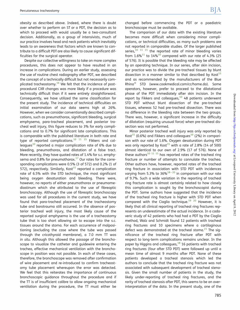
obesity as described above. Indeed, where there is doubtover whether to perform an ST or a PDT, the decision as towhich to proceed with would usually be a two-consultantdecision. Additionally, as a group of intensivists, much ofour practice involves head and neck patients which inevitablyleads to an awareness that factors which are known to con-tribute to a difficult PDT are also likely to cause significant dif-ficulties for the surgical operator.
Despite our collective willingness to take on more complexprocedures, this does not appear to have resulted in anincrease in complications. During an earlier study, assessingthe use of routine chest radiography after PDT, we describedthe concept of a technically difficult but not necessarily com-plicated tracheostomy.16 We felt that the incidence of post-procedural CXR changes was more likely if a procedure wastechnically difficult than if it were entirely straightforward.Consequently, we have utilized the same classification forthe present study. The incidence of technical difficulties oninitial examination of our data seems high at 26%.However, when we consider conventionally described compli-cations, such as pneumothorax, significant bleeding, surgicalemphysema, para-tracheal placement, and posterior tra-cheal wall injury, this figure reduces to 3% for early compli-cations and to 0.7% for significant late complications. Thisis comparable with the published literature in both rate andtype of reported complications.10 11 15 Fikkers and col-leagues10 reported a major complication rate of 6% due tobleeding, pneumothorax, and dilatation of a false tract.More recently, they have quoted rates of 1.4% for s.c. emphy-sema and 0.8% for pneumothorax.17 Our rates for the corre-sponding complications were 0.5% (3 of 572) and 0.2% (1 of572), respectively. Similarly, Kost15 reported a complicationrate of 6.5% with the STD technique, the most significantbeing oxygen desaturation and bleeding. There were,however, no reports of either pneumothorax or pneumome-diastinum which she attributed to the use of fibreopticbronchoscopy. Although the use of fibreoptic bronchoscopywas used for all procedures in the current study, we havefound that para-tracheal placement of the tracheostomytube and barotrauma still occurred. In the absence of pos-terior tracheal wall injury, the most likely cause of thereported surgical emphysema is the use of a tracheostomytube that is too short allowing air to escape into the s.c.tissues around the stoma. For each occurrence of malposi-tioning (excluding the case where the tube was passedthrough the cricothyroid membrane), a 7.0 mm TT wasin situ. Although this allowed the passage of the broncho-scope to visualize the catheter and guidewire entering thetrachea, effective mechanical ventilation with the broncho-scope in position was not possible. In each of these cases,therefore, the bronchoscope was removed after confirmationof wire placement and re-introduced to confirm tracheost-omy tube placement whereupon the error was detected.We feel that this reiterates the importance of continuousbronchoscopic guidance throughout the procedure and ifthe TT is of insufficient calibre to allow ongoing mechanicalventilation during the procedure, the TT must either be
changed before commencing the PDT or a paediatricbronchoscope must be available.
The comparison of our data with the existing literaturebecomes more difficult when considering minor compli-cations, or technical difficulties, as many such problems arenot reported in comparable studies. Of the larger publishedseries,10 11 15 the reported rate of minor bleeding variesfrom 1.6%11 to 14%10 compared with our rate of 4.3% (25of 576). It is possible that the bleeding rate may be affectedby an operating technique. In our series, after skin incision,our practice was to divide the pre-tracheal tissues by bluntdissection in a manner similar to that described by Kost15
and as recommended by the manufacturers of the BlueRhinow STD (www.cookmedical.com/cc/home.do). Someoperators, however, prefer to proceed to the dilatationalphase of the PDT immediately after skin incision. In thereport by Fikkers and colleagues,10 48 patients underwentSTD PDT without blunt dissection of the pre-trachealtissues, whereas 52 had pre-tracheal dissection. There wasno difference in the bleeding rate between the two groups.There was, however, a significant increase in the difficultyof dilatation (requiring unusual force) when pre-tracheal dis-section was not performed.
Minor posterior tracheal wall injury was only reported byKost15 (0.6%) and Fikkers and colleagues10 (2%) in compari-son with our rate of 1.6%. Oxygen desaturation during PDTwas only reported by Kost15 with a rate of 2.8% (14 of 500)almost identical to our own of 2.9% (17 of 576). None ofthese authors10 11 15 has reported rates of the tracheal ringfracture or number of attempts to cannulate the trachea.Other authors have, however, reported rates of the trachealring fracture in association with STD PDT with incidencesvarying from 5.3% to 36%18 – 20 in comparison with our rateof 9.7%. Such a wide variation in the reporting of trachealring fracture rate is almost certainly related to how closelythis complication is sought by the bronchoscopist duringthe PDT. Some authors have suggested that the incidenceof the tracheal ring fracture is higher with STD PDT whencompared with the Ciaglia technique.19 20 However, it islikely that all clinical reporting of tracheal ring fractures rep-resents an underestimate of the actual incidence. In a cada-veric study of 42 patients who had had a PDT by the Ciagliamethod, Walz and Schmidt found 12 patients with trachealring fractures and 10 specimens where a cartilaginousdefect was demonstrated at the tracheal stoma.21 The sig-nificance of the tracheal ring fracture after PDT withrespect to long-term complications remains unclear. In thepaper by Higgins and colleagues,18 16 patients with trachealring fractures (four after STD PDT) were followed up until amean time of almost 9 months after PDT. None of thesepatients developed a tracheal stenosis which led theauthors to conclude that the tracheal ring fracture was notassociated with subsequent development of tracheal steno-sis. Given the small number of patients in the study, thelikely under-reporting of tracheal ring fractures, and therarity of tracheal stenosis after PDT, this seems to be an over-interpretation of the data. In the present study, one of the
Percutaneous tracheostomy BJA
785
at Liverpool University Library on A
pril 8, 2012http://bja.oxfordjournals.org/
Dow
nloaded from
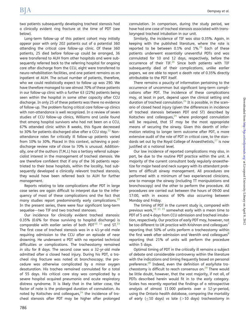
two patients subsequently developing tracheal stenosis hada clinically evident ring fracture at the time of PDT (seebelow).
Long-term follow-up of this patient cohort may initiallyappear poor with only 202 patients out of a potential 360attending the critical care follow-up clinic. Of these 360patients, 25 died before follow-up could be arranged, 36were transferred to AUH from other hospitals and were sub-sequently referred back to the referring hospital for ongoingcare after discharge from the CCU, eight were transferred toneuro-rehabilitation facilities, and one patient remains as aninpatient at AUH. The actual number of patients, therefore,who we could realistically expect to follow up was 290. Wehave therefore managed to see almost 70% of these patientsin our follow-up clinic with a further 63 (22%) patients beingseen within the hospital in some other capacity after CCUdischarge. In only 25 of these patients was there no evidenceof follow-up. The problem facing critical care follow-up clinicswith non-attendance is well recognized. In a review of sevenstudies of CCU follow-up clinics, Williams and Leslie foundthat among hospital survivors who had not been on a CCU,67% attended clinic within 8 weeks, this figure decreasedto 30% for patients discharged alive after a CCU stay.22 Non-attendance rates for critically ill follow-up patients variedfrom 10% to 30%. Placed in this context, achieving a post-discharge review rate of close to 70% is unusual. Addition-ally, one of the authors (T.M.J.) has a tertiary referral subspe-cialist interest in the management of tracheal stenosis. Weare therefore confident that if any of the 36 patients repa-triated to their base hospitals, within the locality, had sub-sequently developed a clinically relevant tracheal stenosis,they would have been referred back to AUH for furthermanagement.
Reports relating to late complications after PDT in largecase series are again difficult to interpret due to the infre-quency of most of these complications and the fact thatmany studies report predominantly early complications.15
In the present series, there were four significant long-termsequelae—two TIF and two tracheal stenoses.
Our incidence for clinically evident tracheal stenosis:0.35% (0.6% for those surviving to hospital discharge) iscomparable with earlier series of both PDT10 11 and ST.23
The first case of tracheal stenosis was in a 41-yr-old malerequiring admission to the CCU after an episode of neardrowning. He underwent a PDT with no reported technicaldifficulties or complications. The tracheostomy remainedin situ for 8 days. The second case was a 32-yr-old maleadmitted after a closed head injury. During his PDT, a tra-cheal ring fracture was noted at bronchoscopy; the pro-cedure was otherwise complicated by a minor oxygendesaturation. His trachea remained cannulated for a totalof 55 days. His critical care stay was complicated by asevere hospital acquired pneumonia and acute respiratorydistress syndrome. It is likely that in the latter case, thefactor of note is the prolonged duration of cannulation. Asnoted by Koitschev and colleagues,24 the incidence of tra-cheal stenosis after PDT may be higher after prolonged
cannulation. In comparison, during the study period, wehave had one case of tracheal stenosis associated with trans-laryngeal tracheal intubation in our unit.
Similarly, the incidence of TIF was also 0.35%. Again, inkeeping with the published literature, where the rate isreported to be between 0.1% and 1%.25 Each of thesepatients underwent essentially uneventful PDTs and wascannulated for 10 and 12 days, respectively, before theoccurrence of their TIF.26 Since both patients with TIFsubsequently died of their complications, unlike earlierpapers, we are able to report a death rate of 0.35% directlyattributable to the PDT itself.
There remains a paucity of information pertaining to theoccurrence of uncommon but significant long-term compli-cations after PDT. The incidence of these complicationsmay, at least in part, depend upon the clinical setting andduration of tracheal cannulation.24 It is possible, in the scen-ario of closed head injury (given the differences in incidenceof tracheal stenosis between PDT and ST) described byKoitschev and colleagues,24 where prolonged cannulationwill be required, that ST may be the most appropriatemeans of securing the airway. Given this dearth of infor-mation relating to longer term outcome after PDT, a moreextensive audit of the role of PDT in critical care, to the stan-dards set out by the Royal College of Anaesthetists,27 is nowjustified at a national level.
Our low incidence of reported complications may also, inpart, be due to the routine PDT practice within the unit. Amajority of the current consultant body regularly anaesthe-tize for major head and neck surgery with its attendant prob-lems of difficult airway management. All procedures areperformed with a minimum of two experienced clinicians,one to manage the airway (including TT manipulations andbronchoscopy) and the other to perform the procedure. Allprocedures are carried out between the hours of 09:00 and17:00, with in excess of 90% also occurring betweenMonday and Friday.
The timing of PDT in the current study is, compared withprevious work,10 11 15 somewhat early with a mean time toPDT of 5 and 4 days from CCU admission and tracheal intuba-tion, respectively. Our practice of early PDT may, however, notbe too different to UK practice with Krishnan and colleagues7
reporting that 50% of units perform a tracheostomy withinthe first week after admission and Veenith and colleagues8
reporting that 21% of units will perform the procedurewithin 5 days.
Optimal timing of PDT in the critically ill remains a subjectof debate and considerable controversy within the literaturewith the indications and timing frequently based on personalpreference.28 Indeed, even the definition of early/late tra-cheostomy is difficult to reach consensus on.29 There wouldbe little doubt, however, that the vast majority, if not all, ofPDTs described herein would fit in to the early category.Scales has recently reported the findings of a retrospectiveanalysis of almost 11 000 patients over a 12-yr-period,using the Ontario health database, comparing the mortalityof early (!10 days) vs late (.10 days) tracheostomy in
BJA Dempsey et al.
786
at Liverpool University Library on A
pril 8, 2012http://bja.oxfordjournals.org/
Dow
nloaded from
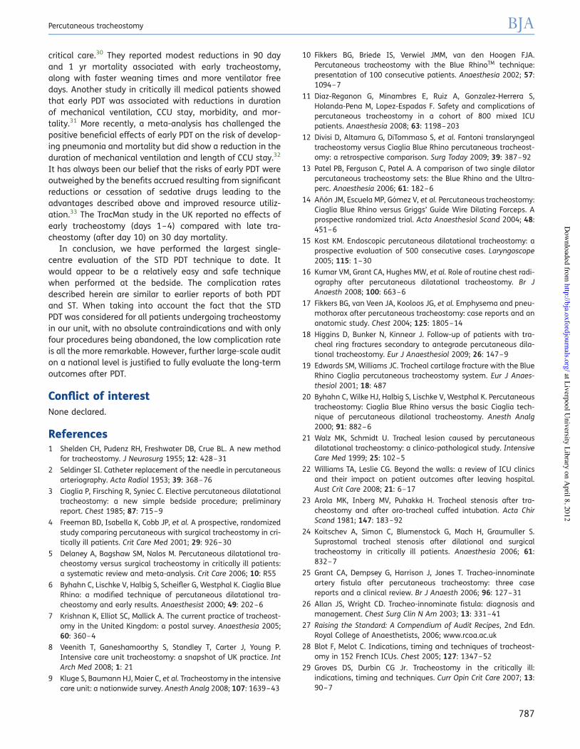
critical care.30 They reported modest reductions in 90 dayand 1 yr mortality associated with early tracheostomy,along with faster weaning times and more ventilator freedays. Another study in critically ill medical patients showedthat early PDT was associated with reductions in durationof mechanical ventilation, CCU stay, morbidity, and mor-tality.31 More recently, a meta-analysis has challenged thepositive beneficial effects of early PDT on the risk of develop-ing pneumonia and mortality but did show a reduction in theduration of mechanical ventilation and length of CCU stay.32
It has always been our belief that the risks of early PDT wereoutweighed by the benefits accrued resulting from significantreductions or cessation of sedative drugs leading to theadvantages described above and improved resource utiliz-ation.33 The TracMan study in the UK reported no effects ofearly tracheostomy (days 1–4) compared with late tra-cheostomy (after day 10) on 30 day mortality.
In conclusion, we have performed the largest single-centre evaluation of the STD PDT technique to date. Itwould appear to be a relatively easy and safe techniquewhen performed at the bedside. The complication ratesdescribed herein are similar to earlier reports of both PDTand ST. When taking into account the fact that the STDPDT was considered for all patients undergoing tracheostomyin our unit, with no absolute contraindications and with onlyfour procedures being abandoned, the low complication rateis all the more remarkable. However, further large-scale auditon a national level is justified to fully evaluate the long-termoutcomes after PDT.
Conflict of interestNone declared.
References1 Shelden CH, Pudenz RH, Freshwater DB, Crue BL. A new method
for tracheostomy. J Neurosurg 1955; 12: 428–312 Seldinger SI. Catheter replacement of the needle in percutaneous
arteriography. Acta Radiol 1953; 39: 368–763 Ciaglia P, Firsching R, Syniec C. Elective percutaneous dilatational
tracheostomy: a new simple bedside procedure; preliminaryreport. Chest 1985; 87: 715–9
4 Freeman BD, Isabella K, Cobb JP, et al. A prospective, randomizedstudy comparing percutaneous with surgical tracheostomy in cri-tically ill patients. Crit Care Med 2001; 29: 926–30
5 Delaney A, Bagshaw SM, Nalos M. Percutaneous dilatational tra-cheostomy versus surgical tracheostomy in critically ill patients:a systematic review and meta-analysis. Crit Care 2006; 10: R55
6 Byhahn C, Lischke V, Halbig S, Scheifler G, Westphal K. Ciaglia BlueRhino: a modified technique of percutaneous dilatational tra-cheostomy and early results. Anaesthesist 2000; 49: 202–6
7 Krishnan K, Elliot SC, Mallick A. The current practice of tracheost-omy in the United Kingdom: a postal survey. Anaesthesia 2005;60: 360–4
8 Veenith T, Ganeshamoorthy S, Standley T, Carter J, Young P.Intensive care unit tracheostomy: a snapshot of UK practice. IntArch Med 2008; 1: 21
9 Kluge S, Baumann HJ, Maier C, et al. Tracheostomy in the intensivecare unit: a nationwide survey. Anesth Analg 2008; 107: 1639–43
10 Fikkers BG, Briede IS, Verwiel JMM, van den Hoogen FJA.Percutaneous tracheostomy with the Blue RhinoTM technique:presentation of 100 consecutive patients. Anaesthesia 2002; 57:1094–7
11 Diaz-Reganon G, Minambres E, Ruiz A, Gonzalez-Herrera S,Holanda-Pena M, Lopez-Espadas F. Safety and complications ofpercutaneous tracheostomy in a cohort of 800 mixed ICUpatients. Anaesthesia 2008; 63: 1198–203
12 Divisi D, Altamura G, DiTommaso S, et al. Fantoni translaryngealtracheostomy versus Ciaglia Blue Rhino percutaneous tracheost-omy: a retrospective comparison. Surg Today 2009; 39: 387–92
13 Patel PB, Ferguson C, Patel A. A comparison of two single dilatorpercutaneous tracheostomy sets: the Blue Rhino and the Ultra-perc. Anaesthesia 2006; 61: 182–6
14 Anon JM, Escuela MP, Gomez V, et al. Percutaneous tracheostomy:Ciaglia Blue Rhino versus Griggs’ Guide Wire Dilating Forceps. Aprospective randomized trial. Acta Anaesthesiol Scand 2004; 48:451–6
15 Kost KM. Endoscopic percutaneous dilatational tracheostomy: aprospective evaluation of 500 consecutive cases. Laryngoscope2005; 115: 1–30
16 Kumar VM, Grant CA, Hughes MW, et al. Role of routine chest radi-ography after percutaneous dilatational tracheostomy. Br JAnaesth 2008; 100: 663–6
17 Fikkers BG, van Veen JA, Kooloos JG, et al. Emphysema and pneu-mothorax after percutaneous tracheostomy: case reports and ananatomic study. Chest 2004; 125: 1805–14
18 Higgins D, Bunker N, Kinnear J. Follow-up of patients with tra-cheal ring fractures secondary to antegrade percutaneous dila-tional tracheostomy. Eur J Anaesthesiol 2009; 26: 147–9
19 Edwards SM, Williams JC. Tracheal cartilage fracture with the BlueRhino Ciaglia percutaneous tracheostomy system. Eur J Anaes-thesiol 2001; 18: 487
20 Byhahn C, Wilke HJ, Halbig S, Lischke V, Westphal K. Percutaneoustracheostomy: Ciaglia Blue Rhino versus the basic Ciaglia tech-nique of percutaneous dilational tracheostomy. Anesth Analg2000; 91: 882–6
21 Walz MK, Schmidt U. Tracheal lesion caused by percutaneousdilatational tracheostomy: a clinico-pathological study. IntensiveCare Med 1999; 25: 102–5
22 Williams TA, Leslie CG. Beyond the walls: a review of ICU clinicsand their impact on patient outcomes after leaving hospital.Aust Crit Care 2008; 21: 6–17
23 Arola MK, Inberg MV, Puhakka H. Tracheal stenosis after tra-cheostomy and after oro-tracheal cuffed intubation. Acta ChirScand 1981; 147: 183–92
24 Koitschev A, Simon C, Blumenstock G, Mach H, Graumuller S.Suprastomal tracheal stenosis after dilational and surgicaltracheostomy in critically ill patients. Anaesthesia 2006; 61:832–7
25 Grant CA, Dempsey G, Harrison J, Jones T. Tracheo-innominateartery fistula after percutaneous tracheostomy: three casereports and a clinical review. Br J Anaesth 2006; 96: 127–31
26 Allan JS, Wright CD. Tracheo-innominate fistula: diagnosis andmanagement. Chest Surg Clin N Am 2003; 13: 331–41
27 Raising the Standard: A Compendium of Audit Recipes, 2nd Edn.Royal College of Anaesthetists, 2006; www.rcoa.ac.uk
28 Blot F, Melot C. Indications, timing and techniques of tracheost-omy in 152 French ICUs. Chest 2005; 127: 1347–52
29 Groves DS, Durbin CG Jr. Tracheostomy in the critically ill:indications, timing and techniques. Curr Opin Crit Care 2007; 13:90–7
Percutaneous tracheostomy BJA
787
at Liverpool University Library on A
pril 8, 2012http://bja.oxfordjournals.org/
Dow
nloaded from
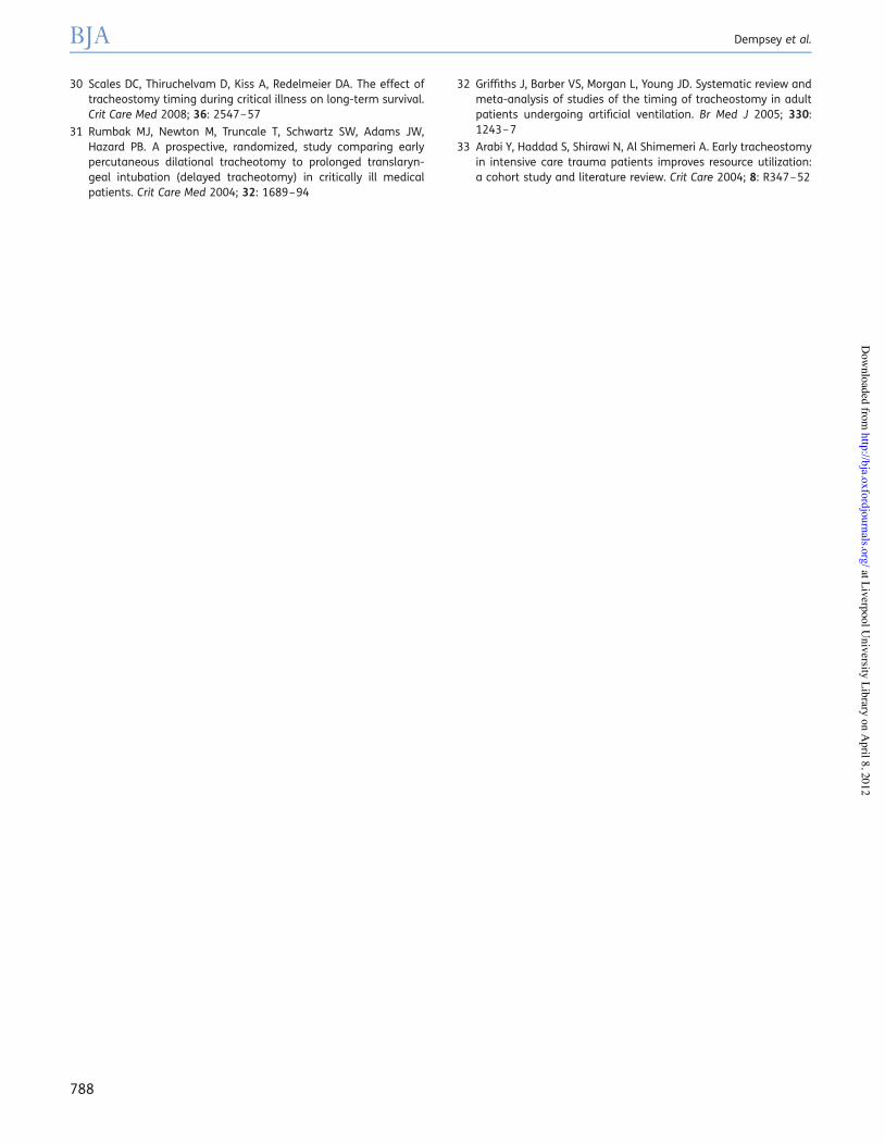
30 Scales DC, Thiruchelvam D, Kiss A, Redelmeier DA. The effect oftracheostomy timing during critical illness on long-term survival.Crit Care Med 2008; 36: 2547–57
31 Rumbak MJ, Newton M, Truncale T, Schwartz SW, Adams JW,Hazard PB. A prospective, randomized, study comparing earlypercutaneous dilational tracheotomy to prolonged translaryn-geal intubation (delayed tracheotomy) in critically ill medicalpatients. Crit Care Med 2004; 32: 1689–94
32 Griffiths J, Barber VS, Morgan L, Young JD. Systematic review andmeta-analysis of studies of the timing of tracheostomy in adultpatients undergoing artificial ventilation. Br Med J 2005; 330:1243–7
33 Arabi Y, Haddad S, Shirawi N, Al Shimemeri A. Early tracheostomyin intensive care trauma patients improves resource utilization:a cohort study and literature review. Crit Care 2004; 8: R347–52
BJA Dempsey et al.
788
at Liverpool University Library on A
pril 8, 2012http://bja.oxfordjournals.org/
Dow
nloaded from
Anaesthesia and Intensive Care, Vol. 42, No. 6, November 2014
Despite widespread adoption of the single tapered dilator (STD) technique for percutaneous dilatational tracheostomy (PDT) in critical care, there is uncertainty about longer-term complications, such as tracheal stenosis (TS). Whilst the incidence of TS is evident from a number of long-term cohort studies1–3, the issue of missed or subclinical stenoses KDV�QRW�EHHQ�IXOO\�DGGUHVVHG��1RQ�VSHFLÀF�V\PSWRPV�are common following critical care admission and PDT, with frequent reports of voice changes, VZDOORZLQJ� GLIÀFXOWLHV�� FRXJK� DQG� VKRUWQHVV� RI�breath4,5. We hypothesised that in this cohort of patients, there were a number of patients with undetected TS.
Earlier studies have attempted to identify the prevalence of TS following PDT using computed tomography (CT)4,6, magnetic resonance imaging
(MRI)7, plain linear tomography8, spirometry with ÀEUHRSWLF� ODU\QJRVFRS\9�� RU�ÀEUHRSWLF� ODU\QJRVFRS\�alone10. Norwood’s study4 using CT evaluation of tracheal anatomy after sequential dilatation PDT and correlation with symptoms found an incidence of TS of 31%, 20% of whom were symptomatic, but could not determine whether voice changes or respiratory problems were a result of critical illness or PDT itself. Spirometry has been variably described as straightforward but with unproven utility in detecting stenosis11 and, as unhelpful in detection of TS in patients after critical illness, ZLWK� SRRU� FRUUHODWLRQ� EHWZHHQ� VSLURPHWU\� ÀQGLQJV�and endoscopic features9. The forced expiratory YROXPH� LQ�RQH� VHFRQG� WR�SHDN� H[SLUDWRU\�ÁRZ� UDWH�ratio (FEV1:PEFR) was demonstrated by Empey to be useful in distinguishing upper airway obstruction from lower airway disease but has not been widely adopted12.
Despite the above attempts to delineate tracheal post-PDT changes, there are limited data pertaining to STD PDT. Although Fikkers et al used MRI to evaluate long-term outcome, only 14 STD patients were evaluated7. We believe the present study is the largest to exclusively radiographically evaluate tracheal changes following STD PDT in an attempt WR�EHWWHU�GHÀQH�WKH�SUHYDOHQFH�RI�VXEFOLQLFDO�76�
* MB ChB, FRCA, Clinical Fellow† MB ChB, FRCA, FFICM, Clinical Fellow‡ MB ChB, FRCR, Consultant Radiologist, Department of Radiology,
Aintree University Hospital NHS Foundation Trust, Liverpool, UK§ MB ChB, FRCA, FFICM, Consultant** MB ChB, FRCA, FFICM, Clinical Fellow†† MB ChB, FRCA, FFICM, Clinical Fellow‡‡ BSc, FRCS(ORL-HNS), MD, Professor of Head & Neck Surgery§§ MB ChB, FRCA, FFICM, Clinical Fellow and Institute of Translational
Medicine, Liverpool CR-UK Centre, Liverpool, UK
Address for correspondence: Dr Ged Dempsey. Email: [email protected]
Accepted for publication on July 28, 2014
Tracheal stenosis following percutaneous dilatational trache-ostomy using the single tapered dilator: an MRI studyE. YOUNG*, R. PUGH†, R. HANLON‡, E. O'CALLAGHAN§, C. WRIGHT**, P. JEANRENAUD††, T. M. JONES‡‡, G. A. DEMPSEY§§Department of Critical Care, Aintree University Hospital NHS Foundation Trust, Liverpool, United Kingdom
SUMMARYDespite widespread adoption of percutaneous dilatational tracheostomy within the critical care setting, there is still uncertainty regarding long-term complications, particularly in relation to missed or subclinical tracheal stenosis. In this study, all patients underwent tracheostomy using a single tapered dilator � three months prior to enrolment and were evaluated using magnetic resonance imaging, spirometry and questionnaire. Tracheal area was recorded and deemed to be stenotic if a reduction of �10% was found. Fifty patients underwent magnetic resonance imaging and 49 attended for interview. Five patients were diagnosed with tracheal stenosis—none were symptomatic. Six of the 50 tracheostomies were technically difficult. Spirometry was not predictive of stenosis. A post critical care exercise tolerance of less than 100 metres was found in four tracheal stenosis patients. The prevalence of subclinical tracheal stenosis following percutaneous tracheostomy is low, with limited clinical significance. No patients required corrective surgery for tracheal stenosis. Routine airway follow-up in asymptomatic patients appears to be unwarranted.
Key Words: tracheostomy, percutaneous, dilatational, complications, critical care
Anaesth Intensive Care 2014; 42: 745-751
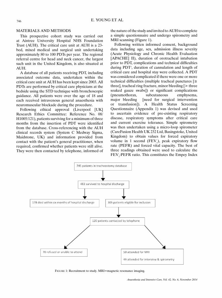
746 E. YOUNG ET AL
Anaesthesia and Intensive Care, Vol. 42, No. 6, November 2014
MATERIALS AND METHODSThis prospective cohort study was carried out
at Aintree University Hospital NHS Foundation Trust (AUH). The critical care unit at AUH is a 23-bed, mixed medical and surgical unit undertaking approximately 80 to 100 PDTs per year. The regional referral centre for head and neck cancer, the largest such unit in the United Kingdom, is also situated at AUH.
A database of all patients receiving PDT, including associated outcome data, undertaken within the critical care unit at AUH has been kept since 2003. All PDTs are performed by critical care physicians at the bedside using the STD technique with bronchoscopic guidance. All patients were over the age of 18 and each received intravenous general anaesthesia with neuromuscular blockade during the procedure.
Following ethical approval (Liverpool [UK] Research Ethics Committee: Reference No. 08/H1005/121), patients surviving for a minimum of three PRQWKV� IURP� WKH� LQVHUWLRQ� RI� 3'7� ZHUH� LGHQWLÀHG�from the database. Cross-referencing with the AUH clinical records system (System C Medway Sigma, Maidstone, UK) and information provided from contact with the patient’s general practitioner, when UHTXLUHG��FRQÀUPHG�ZKHWKHU�SDWLHQWV�ZHUH�VWLOO�DOLYH��They were then contacted by telephone, informed of
the nature of the study and invited to AUH to complete a simple questionnaire and undergo spirometry and MRI scanning (Figure 1).
Following written informed consent, background data including age, sex, admission illness severity (Acute Physiology and Chronic Health Evaluation [APACHE] II), duration of orotracheal intubation SULRU�WR�3'7��FRPSOLFDWLRQV�DQG�WHFKQLFDO�GLIÀFXOWLHV�during PDT3, duration of cannulation and length of critical care and hospital stay were collected. A PDT was considered complicated if there were one or more WHFKQLFDO�GLIÀFXOWLHV��PXOWLSOH�WUDFKHDO�SXQFWXUHV�>��three], tracheal ring fracture, minor bleeding [> three VRDNHG� JDX]H� VZDEV@�� RU� VLJQLÀFDQW� FRPSOLFDWLRQV�(pneumothorax, subcutaneous emphysema, major bleeding [need for surgical intervention or transfusion]). A Health Status Screening Questionnaire (Appendix 1) was devised and used to ascertain evidence of pre-existing respiratory disease, respiratory symptoms after critical care and current exercise tolerance. Simple spirometry was then undertaken using a micro-loop spirometer (CareFusion Health UK 232 Ltd, Basingstoke, United Kingdom) to obtain values for forced expiratory volume in 1 second (FEV1��� SHDN� H[SLUDWRU\� ÁRZ�rate (PEFR) and forced vital capacity. The best of three readings obtained were used to calculate the FEV1:PEFR ratio. This constitutes the Empey Index
FiGuRe 1: Recruitment to study. MRI=magnetic resonance imaging.
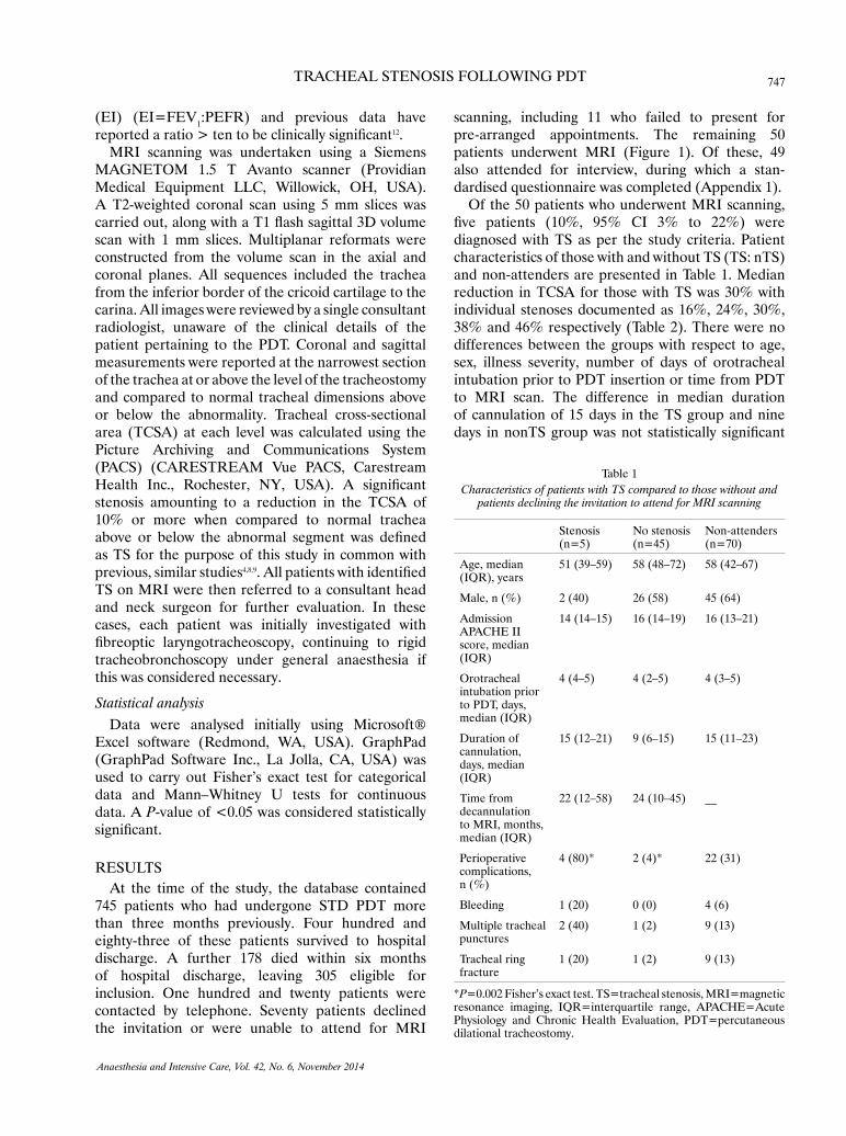
747TRACHEAL STENOSIS FOLLOWING PDT
Anaesthesia and Intensive Care, Vol. 42, No. 6, November 2014
(EI) (EI=FEV1:PEFR) and previous data have UHSRUWHG�D�UDWLR�!�WHQ�WR�EH�FOLQLFDOO\�VLJQLÀFDQW12.
MRI scanning was undertaken using a Siemens MAGNETOM 1.5 T Avanto scanner (Providian Medical Equipment LLC, Willowick, OH, USA). A T2-weighted coronal scan using 5 mm slices was FDUULHG�RXW��DORQJ�ZLWK�D�7��ÁDVK�VDJLWWDO��'�YROXPH�scan with 1 mm slices. Multiplanar reformats were constructed from the volume scan in the axial and coronal planes. All sequences included the trachea from the inferior border of the cricoid cartilage to the carina. All images were reviewed by a single consultant radiologist, unaware of the clinical details of the patient pertaining to the PDT. Coronal and sagittal measurements were reported at the narrowest section of the trachea at or above the level of the tracheostomy and compared to normal tracheal dimensions above or below the abnormality. Tracheal cross-sectional area (TCSA) at each level was calculated using the Picture Archiving and Communications System (PACS) (CARESTREAM Vue PACS, Carestream +HDOWK� ,QF��� 5RFKHVWHU�� 1<�� 86$��� $� VLJQLÀFDQW�stenosis amounting to a reduction in the TCSA of 10% or more when compared to normal trachea DERYH� RU� EHORZ� WKH� DEQRUPDO� VHJPHQW� ZDV� GHÀQHG�as TS for the purpose of this study in common with previous, similar studies4,8,9��$OO�SDWLHQWV�ZLWK�LGHQWLÀHG�TS on MRI were then referred to a consultant head and neck surgeon for further evaluation. In these cases, each patient was initially investigated with ÀEUHRSWLF� ODU\QJRWUDFKHRVFRS\�� FRQWLQXLQJ� WR� ULJLG�tracheobronchoscopy under general anaesthesia if this was considered necessary.
Statistical analysisData were analysed initially using Microsoft®
Excel software (Redmond, WA, USA). GraphPad (GraphPad Software Inc., La Jolla, CA, USA) was used to carry out Fisher’s exact test for categorical data and Mann–Whitney U tests for continuous data. A P-value of <0.05 was considered statistically VLJQLÀFDQW���
RESULTSAt the time of the study, the database contained
745 patients who had undergone STD PDT more than three months previously. Four hundred and eighty-three of these patients survived to hospital discharge. A further 178 died within six months of hospital discharge, leaving 305 eligible for inclusion. One hundred and twenty patients were contacted by telephone. Seventy patients declined the invitation or were unable to attend for MRI
scanning, including 11 who failed to present for pre-arranged appointments. The remaining 50 patients underwent MRI (Figure 1). Of these, 49 also attended for interview, during which a stan- dardised questionnaire was completed (Appendix 1).
Of the 50 patients who underwent MRI scanning, ÀYH� SDWLHQWV� ������ ���� &,� ��� WR� ����� ZHUH�diagnosed with TS as per the study criteria. Patient characteristics of those with and without TS (TS: nTS) and non-attenders are presented in Table 1. Median reduction in TCSA for those with TS was 30% with individual stenoses documented as 16%, 24%, 30%, 38% and 46% respectively (Table 2). There were no differences between the groups with respect to age, sex, illness severity, number of days of orotracheal intubation prior to PDT insertion or time from PDT to MRI scan. The difference in median duration of cannulation of 15 days in the TS group and nine GD\V� LQ�QRQ76�JURXS�ZDV�QRW� VWDWLVWLFDOO\� VLJQLÀFDQW�
Table 1Characteristics of patients with TS compared to those without and
patients declining the invitation to attend for MRI scanning
Stenosis (n=5)
No stenosis (n=45)
Non-attenders (n=70)
Age, median (IQR), years
51 (39–59) 58 (48–72) 58 (42–67)
Male, n (%) 2 (40) 26 (58) 45 (64)
Admission APACHE II score, median (IQR)
14 (14–15) 16 (14–19) 16 (13–21)
Orotracheal intubation prior to PDT, days, median (IQR)
4 (4–5) 4 (2–5) 4 (3–5)
Duration of cannulation, days, median (IQR)
15 (12–21) 9 (6–15) 15 (11–23)
Time from decannulation to MRI, months, median (IQR)
22 (12–58) 24 (10–45) __
Perioperative complications, n (%)
4 (80)* 2 (4)* 22 (31)
Bleeding 1 (20) 0 (0) 4 (6)
Multiple tracheal punctures
2 (40) 1 (2) 9 (13)
Tracheal ring fracture
1 (20) 1 (2) 9 (13)
*P=0.002 Fisher’s exact test. TS=tracheal stenosis, MRI=magnetic resonance imaging, IQR=interquartile range, APACHE=Acute Physiology and Chronic Health Evaluation, PDT=percutaneous dilational tracheostomy.
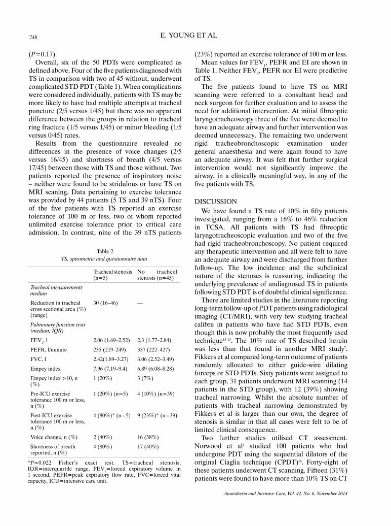
748 E. YOUNG ET AL
Anaesthesia and Intensive Care, Vol. 42, No. 6, November 2014
(P=0.17).Overall, six of the 50 PDTs were complicated as
GHÀQHG�DERYH��)RXU�RI�WKH�ÀYH�SDWLHQWV�GLDJQRVHG�ZLWK�TS in comparison with two of 45 without, underwent complicated STD PDT (Table 1). When complications were considered individually, patients with TS may be more likely to have had multiple attempts at tracheal puncture (2/5 versus 1/45) but there was no apparent difference between the groups in relation to tracheal ring fracture (1/5 versus 1/45) or minor bleeding (1/5 versus 0/45) rates.
Results from the questionnaire revealed no differences in the presence of voice changes (2/5 versus 16/45) and shortness of breath (4/5 versus 17/45) between those with TS and those without. Two patients reported the presence of inspiratory noise – neither were found to be stridulous or have TS on MRI scaning. Data pertaining to exercise tolerance was provided by 44 patients (5 TS and 39 nTS). Four RI� WKH� ÀYH� SDWLHQWV� ZLWK� 76� UHSRUWHG� DQ� H[HUFLVH�tolerance of 100 m or less, two of whom reported unlimited exercise tolerance prior to critical care admission. In contrast, nine of the 39 nTS patients
(23%) reported an exercise tolerance of 100 m or less. Mean values for FEV1, PEFR and EI are shown in
Table 1. Neither FEV1, PEFR nor EI were predictive of TS. 7KH� ÀYH� SDWLHQWV� IRXQG� WR� KDYH� 76� RQ� 05,�
scanning were referred to a consultant head and neck surgeon for further evaluation and to assess the QHHG�IRU�DGGLWLRQDO�LQWHUYHQWLRQ��$W�LQLWLDO�ÀEUHRSWLF�ODU\QJRWUDFKHRVFRS\�WKUHH�RI�WKH�ÀYH�ZHUH�GHHPHG�WR�have an adequate airway and further intervention was deemed unnecessary. The remaining two underwent rigid tracheobronchoscopic examination under general anaesthesia and were again found to have an adequate airway. It was felt that further surgical LQWHUYHQWLRQ� ZRXOG� QRW� VLJQLÀFDQWO\� LPSURYH� WKH�airway, in a clinically meaningful way, in any of the ÀYH�SDWLHQWV�ZLWK�76�
DISCUSSION:H�KDYH� IRXQG�D�76� UDWH�RI� ���� LQ�ÀIW\�SDWLHQWV�
investigated, ranging from a 16% to 46% reduction LQ� 7&6$�� $OO� SDWLHQWV� ZLWK� 76� KDG� ÀEUHRSWLF�ODU\QJRWUDFKHRVFRSLF� HYDOXDWLRQ� DQG� WZR� RI� WKH� ÀYH�had rigid tracheobronchoscopy. No patient required any therapeutic intervention and all were felt to have an adequate airway and were discharged from further follow-up. The low incidence and the subclinical nature of the stenoses is reassuring, indicating the underlying prevalence of undiagnosed TS in patients IROORZLQJ�67'�3'7�LV�RI�GRXEWIXO�FOLQLFDO�VLJQLÀFDQFH��
There are limited studies in the literature reporting long-term follow-up of PDT patients using radiological imaging (CT/MRI), with very few studying tracheal calibre in patients who have had STD PDTs, even though this is now probably the most frequently used technique13–15. The 10% rate of TS described herein was less than that found in another MRI study7. Fikkers et al compared long-term outcome of patients randomly allocated to either guide-wire dilating forceps or STD PDTs. Sixty patients were assigned to each group, 31 patients underwent MRI scanning (14 patients in the STD group), with 12 (39%) showing tracheal narrowing. Whilst the absolute number of patients with tracheal narrowing demonstrated by Fikkers et al is larger than our own, the degree of stenosis is similar in that all cases were felt to be of limited clinical consequence.
Two further studies utilised CT assessment. Norwood et al4 studied 100 patients who had undergone PDT using the sequential dilators of the original Ciaglia technique (CPDT)16. Forty-eight of these patients underwent CT scanning. Fifteen (31%) patients were found to have more than 10% TS on CT
Table 2TS, spirometric and questionnaire data
Tracheal stenosis (n=5)
No tracheal stenosis (n=45)
Tracheal measurements median
Reduction in tracheal cross sectional area (%) (range)
30 (16–46) —
Pulmonary function tests (median, IQR)
FEV1, l 2.06 (1.69–2.52) 2.3 (1.77–2.84)
PEFR, l/minute 235 (219–249) 337 (222–427)
FVC, l 2.42(1.89–3.27) 3.06 (2.52–3.49)
Empey index 7.96 (7.19–9.4) 6.89 (6.08–8.28)
Empey index >10, n (%)
1 (20%) 3 (7%)
Pre-ICU exercise tolerance 100 m or less, n (%)
1 (20%) (n=5) 4 (10%) (n=39)
Post-ICU exercise tolerance 100 m or less, n (%)
4 (80%)* (n=5) 9 (23%)* (n=39)
Voice change, n (%) 2 (40%) 16 (38%)
Shortness of breath reported, n (%)
4 (80%) 17 (40%)
*P=0.022 Fisher’s exact test. TS=tracheal stenosis, IQR=interquartile range, FEV1=forced expiratory volume in 1 second, PEFR=peak expiratory flow rate, FVC=forced vital capacity, ICU=intensive care unit.
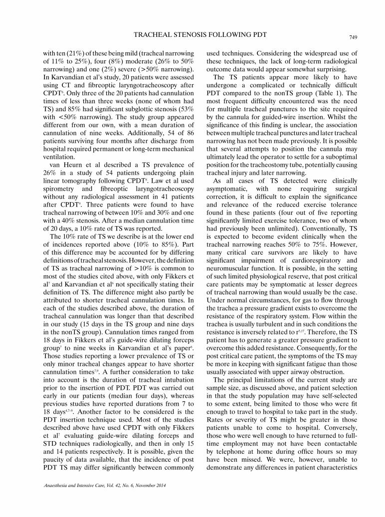
749TRACHEAL STENOSIS FOLLOWING PDT
Anaesthesia and Intensive Care, Vol. 42, No. 6, November 2014
with ten (21%) of these being mild (tracheal narrowing of 11% to 25%), four (8%) moderate (26% to 50% narrowing) and one (2%) severe (>50% narrowing). In Karvandian et al’s study, 20 patients were assessed XVLQJ� &7� DQG� ÀEUHRSWLF� ODU\QJRWUDFKHRVFRS\� DIWHU�CPDT6. Only three of the 20 patients had cannulation times of less than three weeks (none of whom had 76��DQG�����KDG�VLJQLÀFDQW�VXEJORWWLF�VWHQRVLV������with <50% narrowing). The study group appeared different from our own, with a mean duration of cannulation of nine weeks. Additionally, 54 of 86 patients surviving four months after discharge from hospital required permanent or long-term mechanical ventilation.
van Heurn et al described a TS prevalence of 26% in a study of 54 patients undergoing plain linear tomography following CPDT8. Law et al used VSLURPHWU\� DQG� ÀEUHRSWLF� ODU\QJRWUDFKHRVFRS\�without any radiological assessment in 41 patients after CPDT9. Three patients were found to have tracheal narrowing of between 10% and 30% and one with a 40% stenosis. After a median cannulation time of 20 days, a 10% rate of TS was reported.
The 10% rate of TS we describe is at the lower end of incidences reported above (10% to 85%). Part of this difference may be accounted for by differing GHÀQLWLRQV�RI�WUDFKHDO�VWHQRVLV��+RZHYHU��WKH�GHÀQLWLRQ�of TS as tracheal narrowing of >10% is common to most of the studies cited above, with only Fikkers et al7 and Karvandian et al6�QRW�VSHFLÀFDOO\�VWDWLQJ�WKHLU�GHÀQLWLRQ�RI�76��7KH�GLIIHUHQFH�PLJKW�DOVR�SDUWO\�EH�attributed to shorter tracheal cannulation times. In each of the studies described above, the duration of tracheal cannulation was longer than that described in our study (15 days in the TS group and nine days in the nonTS group). Cannulation times ranged from 18 days in Fikkers et al’s guide-wire dilating forceps group7 to nine weeks in Karvandian et al’s paper6. Those studies reporting a lower prevalence of TS or only minor tracheal changes appear to have shorter cannulation times7,9. A further consideration to take into account is the duration of tracheal intubation prior to the insertion of PDT. PDT was carried out early in our patients (median four days), whereas previous studies have reported durations from 7 to 18 days4,7–9. Another factor to be considered is the PDT insertion technique used. Most of the studies described above have used CPDT with only Fikkers et al7 evaluating guide-wire dilating forceps and STD techniques radiologically, and then in only 15 and 14 patients respectively. It is possible, given the paucity of data available, that the incidence of post 3'7�76�PD\�GLIIHU� VLJQLÀFDQWO\� EHWZHHQ� FRPPRQO\�
used techniques. Considering the widespread use of these techniques, the lack of long-term radiological outcome data would appear somewhat surprising.
The TS patients appear more likely to have XQGHUJRQH� D� FRPSOLFDWHG� RU� WHFKQLFDOO\� GLIÀFXOW�PDT compared to the nonTS group (Table 1). The PRVW� IUHTXHQW� GLIÀFXOW\� HQFRXQWHUHG� ZDV� WKH� QHHG�for multiple tracheal punctures to the site required by the cannula for guided-wire insertion. Whilst the VLJQLÀFDQFH�RI�WKLV�ÀQGLQJ�LV�XQFOHDU��WKH�DVVRFLDWLRQ�between multiple tracheal punctures and later tracheal narrowing has not been made previously. It is possible that several attempts to position the cannula may ultimately lead the operator to settle for a suboptimal position for the tracheostomy tube, potentially causing tracheal injury and later narrowing.
As all cases of TS detected were clinically asymptomatic, with none requiring surgical FRUUHFWLRQ�� LW� LV� GLIÀFXOW� WR� H[SODLQ� WKH� VLJQLÀFDQFH�and relevance of the reduced exercise tolerance IRXQG� LQ� WKHVH� SDWLHQWV� �IRXU� RXW� RI� ÀYH� UHSRUWLQJ�VLJQLÀFDQWO\�OLPLWHG�H[HUFLVH�WROHUDQFH��WZR�RI�ZKRP�had previously been unlimited). Conventionally, TS is expected to become evident clinically when the tracheal narrowing reaches 50% to 75%. However, many critical care survivors are likely to have VLJQLÀFDQW� LPSDLUPHQW� RI� FDUGLRUHVSLUDWRU\� DQG�neuromuscular function. It is possible, in the setting of such limited physiological reserve, that post critical care patients may be symptomatic at lesser degrees of tracheal narrowing than would usually be the case. 8QGHU�QRUPDO�FLUFXPVWDQFHV��IRU�JDV�WR�ÁRZ�WKURXJK�the trachea a pressure gradient exists to overcome the resistance of the respiratory system. Flow within the trachea is usually turbulent and in such conditions the resistance is inversely related to r5,17. Therefore, the TS patient has to generate a greater pressure gradient to overcome this added resistance. Consequently, for the post critical care patient, the symptoms of the TS may EH�PRUH�LQ�NHHSLQJ�ZLWK�VLJQLÀFDQW�IDWLJXH�WKDQ�WKRVH�usually associated with upper airway obstruction.
The principal limitations of the current study are sample size, as discussed above, and patient selection in that the study population may have self-selected WR� VRPH� H[WHQW�� EHLQJ� OLPLWHG� WR� WKRVH�ZKR�ZHUH� ÀW�enough to travel to hospital to take part in the study. Rates or severity of TS might be greater in those patients unable to come to hospital. Conversely, those who were well enough to have returned to full-time employment may not have been contactable E\� WHOHSKRQH� DW� KRPH� GXULQJ� RIÀFH� KRXUV� VR� PD\�have been missed. We were, however, unable to demonstrate any differences in patient characteristics
750 E. YOUNG ET AL
Anaesthesia and Intensive Care, Vol. 42, No. 6, November 2014
between those attending for scans and those unable to do so (Table 1). Additionally, we are unaware of the cause of death in those patients surviving their critical care stay but dying prior to an invitation to attend for scan. It is possible some of these patients may have had some degree of upper airway obstruction. Given that our institution houses the regional head and neck service, and such patients would have been referred back should they develop airway obstruction, this seems less likely.
Our initial intention was to assess 60 patients IROORZLQJ� 67'�3'7�� VSHFLÀFDOO\� UHFUXLWLQJ� ��� DIWHU�uncomplicated procedures, 20 after PDT complicated by tracheal ring fracture and a further 20 after more than 14 days cannulation. The incidences of ring fracture and of cannulation for more than 14 days were too low for the original aim to be achievable. Consequently, we recruited 50 eligible patients from the database. Whilst we were unable to study these factors as initially intended, analysis of the limited data obtained would appear to suggest that tracheal ring fracture is not obviously implicated in TS. Data pertaining to the length of tracheal cannulation may indicate a possible relationship between prolonged cannulation and TS (nTS nine days versus 15 days 76�� EXW� WKLV�ZDV� QRW� VWDWLVWLFDOO\� VLJQLÀFDQW��:KLOVW�ZH� FRXOG� QRW� GHPRQVWUDWH� D� VWDWLVWLFDOO\� VLJQLÀFDQW�association between duration of cannulation and TS, further studies with larger numbers may be able to do so. Such studies are likely to be confounded by differences in timing of PDT between institutions. The long follow-up required to detect post tracheostomy tracheal stenosis would be another limiting factor, as would cost—particularly if only 10% of cases prove positive for TS which then may EH�UHJDUGHG�WR�KDYH�OLPLWHG�FOLQLFDO�VLJQLÀFDQFH�
We have demonstrated a lower rate of subclinical stenosis in patients following STD PDT than has previously been reported. Whilst we considered that functional limitation in such patients following critical care discharge may be in part due to an underlying prevalence of TS, this would appear not to be the case. Those stenoses found were felt to be RI� GRXEWIXO� VLJQLÀFDQFH� ZLWK� QR� SDWLHQWV� UHTXLULQJ�corrective surgery, and all ultimately discharged from further follow-up. Routine radiological imaging and spirometry following STD PDT would appear to be unwarranted in asymptomatic patients.
FUNDINGThe study was funded by a £9600 Young
Investigators Award from the Intensive Care Society UK.
REFERENCES 1. Kost KM. Endoscopic percutaneous dilatational tracheotomy: a
prospective evaluation of 500 consecutive cases. Laryngoscope 2005; 115:1-30.
2. Diaz-Reganon G, Minambres E, Ruiz A, Gonzalez-Herrera S, Holanda-Pena M, Lopez-Espadas F. Safety and complications of percutaneous tracheostomy in a cohort of 800 mixed ICU patients. Anaesthesia 2008; 63:1198-1203.
3. Dempsey GA, Grant CA, Jones TM. Percutaneous tracheos-tomy: a 6 yr prospective evaluation of the single tapered dilator technique. Br J Anaesth 2010; 105:782-788.
4. Norwood S, Vallina VL, Short K, Sigusa M, Fernandez LG, McLarty JW. Incidence of tracheal stenosis and other late com-plications after percutaneous tracheostomy. Ann Surg 2000; 232:233-241.
5. Needham DM, Davidson J, Cohen H, Hopkins RO et al. Improving long-term outcomes after discharge from intensive care unit: report from a stakeholders' conference. Crit Care Med 2012; 40:502 -509.
6. Karvandian K, Jafarzadeh A, Hajipour A, Zolfaghari N. Subglottic stenosis following percutaneous tracheostomy: a sin-gle centre report as a descriptive study. Acta Otorhinolaryngol Ital 2011; 31:239-242.
7. Fikkers BG, Staatsen M, van den Hoogen FJA, van der Hoeven JG. Early and late outcome after single step dilatational tra-cheostomy versus the guide wire dilating forceps technique: a prospective randomized clinical trial. Intensive Care Med 2011; 37:1103-1109.
8. van Heurn LW, Goei R, de Ploeg I, Ramsay G, Brink PR. Late complications of percutaneous dilatational tracheotomy. Chest 1996; 110:1572-1576.
9. Law RC, Carney, AS, Manara AR. Long-term outcome after percutaneous dilatational tracheostomy. Anaesthesia 1997; 52:51-56.
10. Rosenbower TJ, Morris JA, Eddy VA, Ries WR. The long-term complications of percutaneous dilatational tracheostomy. Am Surg 1998; 64:82-87.
11. Leonard RC, Lewis RH, Singh B, van Heerden PV. Late out-come from percutaneous tracheostomy using the Portex kit. Chest 1999; 115:1070-1075.
12. Empey DW. Assessment of upper airways obstruction. Br Med J 1972; 3:503-505.
13. Krishnan K, Elliot SC, Mallick A. The current practice of tracheostomy in the United Kingdom: a postal survey. Anaesthesia 2005; 60:360-364.
14. Veenith T, Ganeshamoorthy S, Standley T, Carter J, Young P. Intensive care unit tracheostomy: a snapshot of UK practice. Int Arch Med 2008; 1:21.
15. Kluge S, Baumann HJ, Maier C, Klose H, Meyer A, Nierhaus A et al. Tracheostomy in the intensive care unit: a nationwide survey. Anesth Analg 2008; 107:1639-1643.
16. Ciaglia P, Firsching R, Syniec C. Elective percutaneous dilata-tional tracheostomy: a new simple bedside procedure; prelimi-nary report. Chest 1985; 87:715-719.
17. Bock KR, Silver P, Rom M, Sagy M. Reduction in tracheal lumen due to endotracheal intubation and its calculated clinical significance. Chest 2000; 118:468-472.

PaƟent sƟcker Outpatient appointment date: __/___/__
Date of critical care admission: __/___/__
Date first intubated: __/___/__
No. extubations pre-tracheostomy: ________
Date of percutaneous tracheostomy: __/___/__
Date of decannulation: __/___/__
Dates of re-cannulation (if applicable):
______________________________________________________________
Complications of
tracheostomy:
___________________________________________________________
____
Pre-existing respiratory
disease
Smoking history: _________ pack years Still smoking: Y/N
Established COPD: Y/N
Other respiratory disease:
_______________________________________
Pre-ITU spirometry: FEV1_____________l FVC________________l
Pre-ITU Exercise tolerance on flat:_____m Limiting
factor:
Current symptoms
Change in voice : Y/N
Short of breath: Y/N Exertion/rest
Stridor Y/N Exertion/rest
Exercise tolerance on flat: ______m Limiting factor:
Other (difficulty expectorating? difficulty swallowing? problematic scar?):
Examination
Scar: Not visible/Acceptable/Disfiguring/Tethering
Short of breath: Y/N Exertion/rest
Stridor: Y/N Exertion/rest
Spirometry
Measured values Calculated values
1. 2. 3. Best
FIF50
FEF50 FEF50/ FIF50
FEV1 FEV1/PEFR
PEFR
FVC FEV1/FVC
NB. Any one of: FIF50 <100 l/minute, FEF50/FIF50 �1, FEV1/PEFR >10l/ml per minute.
appeNdix 1: assessMeNt oF tRaCheal steNosis FolloWiNG peRCutaNeous tRaCheostoMy

Copyright of Anaesthesia & Intensive Care is the property of Australian Society ofAnaesthetists and its content may not be copied or emailed to multiple sites or posted to alistserv without the copyright holder's express written permission. However, users may print,download, or email articles for individual use.
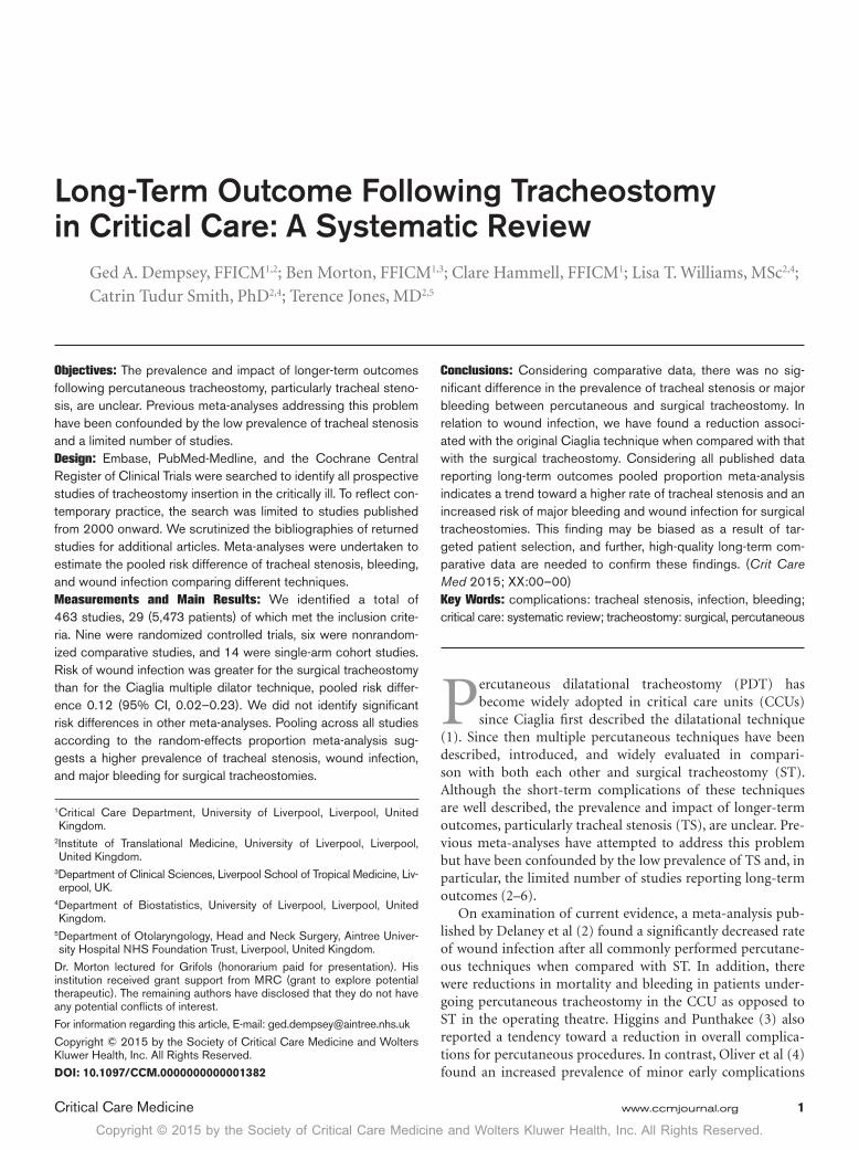
Copyright © 2015 by the Society of Critical Care Medicine and Wolters Kluwer Health, Inc. All Rights Reserved.
Critical Care Medicine www.ccmjournal.org 1
Objectives: The prevalence and impact of longer-term outcomes following percutaneous tracheostomy, particularly tracheal steno-sis, are unclear. Previous meta-analyses addressing this problem have been confounded by the low prevalence of tracheal stenosis and a limited number of studies.Design: Embase, PubMed-Medline, and the Cochrane Central Register of Clinical Trials were searched to identify all prospective studies of tracheostomy insertion in the critically ill. To reflect con-temporary practice, the search was limited to studies published from 2000 onward. We scrutinized the bibliographies of returned studies for additional articles. Meta-analyses were undertaken to estimate the pooled risk difference of tracheal stenosis, bleeding, and wound infection comparing different techniques.Measurements and Main Results: We identified a total of 463 studies, 29 (5,473 patients) of which met the inclusion crite-ria. Nine were randomized controlled trials, six were nonrandom-ized comparative studies, and 14 were single-arm cohort studies. Risk of wound infection was greater for the surgical tracheostomy than for the Ciaglia multiple dilator technique, pooled risk differ-ence 0.12 (95% CI, 0.02–0.23). We did not identify significant risk differences in other meta-analyses. Pooling across all studies according to the random-effects proportion meta-analysis sug-gests a higher prevalence of tracheal stenosis, wound infection, and major bleeding for surgical tracheostomies.
Conclusions: Considering comparative data, there was no sig-nificant difference in the prevalence of tracheal stenosis or major bleeding between percutaneous and surgical tracheostomy. In relation to wound infection, we have found a reduction associ-ated with the original Ciaglia technique when compared with that with the surgical tracheostomy. Considering all published data reporting long-term outcomes pooled proportion meta-analysis indicates a trend toward a higher rate of tracheal stenosis and an increased risk of major bleeding and wound infection for surgical tracheostomies. This finding may be biased as a result of tar-geted patient selection, and further, high-quality long-term com-parative data are needed to confirm these findings. (Crit Care Med 2015; XX:00–00)Key Words: complications: tracheal stenosis, infection, bleeding; critical care: systematic review; tracheostomy: surgical, percutaneous
Percutaneous dilatational tracheostomy (PDT) has become widely adopted in critical care units (CCUs) since Ciaglia first described the dilatational technique
(1). Since then multiple percutaneous techniques have been described, introduced, and widely evaluated in compari-son with both each other and surgical tracheostomy (ST). Although the short-term complications of these techniques are well described, the prevalence and impact of longer-term outcomes, particularly tracheal stenosis (TS), are unclear. Pre-vious meta-analyses have attempted to address this problem but have been confounded by the low prevalence of TS and, in particular, the limited number of studies reporting long-term outcomes (2–6).
On examination of current evidence, a meta-analysis pub-lished by Delaney et al (2) found a significantly decreased rate of wound infection after all commonly performed percutane-ous techniques when compared with ST. In addition, there were reductions in mortality and bleeding in patients under-going percutaneous tracheostomy in the CCU as opposed to ST in the operating theatre. Higgins and Punthakee (3) also reported a tendency toward a reduction in overall complica-tions for percutaneous procedures. In contrast, Oliver et al (4) found an increased prevalence of minor early complications
1Critical Care Department, University of Liverpool, Liverpool, United Kingdom.
2Institute of Translational Medicine, University of Liverpool, Liverpool, United Kingdom.
3Department of Clinical Sciences, Liverpool School of Tropical Medicine, Liv-erpool, UK.
4Department of Biostatistics, University of Liverpool, Liverpool, United Kingdom.
5Department of Otolaryngology, Head and Neck Surgery, Aintree Univer-sity Hospital NHS Foundation Trust, Liverpool, United Kingdom.
Dr. Morton lectured for Grifols (honorarium paid for presentation). His institution received grant support from MRC (grant to explore potential therapeutic). The remaining authors have disclosed that they do not have any potential conflicts of interest.For information regarding this article, E-mail: [email protected] © 2015 by the Society of Critical Care Medicine and Wolters Kluwer Health, Inc. All Rights Reserved.DOI: 10.1097/CCM.0000000000001382
Long-Term Outcome Following Tracheostomy in Critical Care: A Systematic Review
Ged A. Dempsey, FFICM1,2; Ben Morton, FFICM1,3; Clare Hammell, FFICM1; Lisa T. Williams, MSc2,4; Catrin Tudur Smith, PhD2,4; Terence Jones, MD2,5
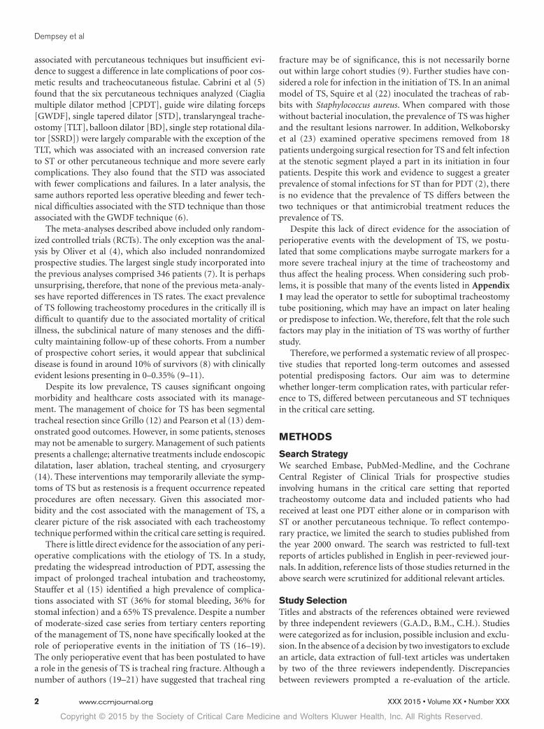
Copyright © 2015 by the Society of Critical Care Medicine and Wolters Kluwer Health, Inc. All Rights Reserved.
Dempsey et al
2 www.ccmjournal.org
associated with percutaneous techniques but insufficient evi-dence to suggest a difference in late complications of poor cos-metic results and tracheocutaneous fistulae. Cabrini et al (5) found that the six percutaneous techniques analyzed (Ciaglia multiple dilator method [CPDT], guide wire dilating forceps [GWDF], single tapered dilator [STD], translaryngeal trache-ostomy [TLT], balloon dilator [BD], single step rotational dila-tor [SSRD]) were largely comparable with the exception of the TLT, which was associated with an increased conversion rate to ST or other percutaneous technique and more severe early complications. They also found that the STD was associated with fewer complications and failures. In a later analysis, the same authors reported less operative bleeding and fewer tech-nical difficulties associated with the STD technique than those associated with the GWDF technique (6).
The meta-analyses described above included only random-ized controlled trials (RCTs). The only exception was the anal-ysis by Oliver et al (4), which also included nonrandomized prospective studies. The largest single study incorporated into the previous analyses comprised 346 patients (7). It is perhaps unsurprising, therefore, that none of the previous meta-analy-ses have reported differences in TS rates. The exact prevalence of TS following tracheostomy procedures in the critically ill is difficult to quantify due to the associated mortality of critical illness, the subclinical nature of many stenoses and the diffi-culty maintaining follow-up of these cohorts. From a number of prospective cohort series, it would appear that subclinical disease is found in around 10% of survivors (8) with clinically evident lesions presenting in 0–0.35% (9–11).
Despite its low prevalence, TS causes significant ongoing morbidity and healthcare costs associated with its manage-ment. The management of choice for TS has been segmental tracheal resection since Grillo (12) and Pearson et al (13) dem-onstrated good outcomes. However, in some patients, stenoses may not be amenable to surgery. Management of such patients presents a challenge; alternative treatments include endoscopic dilatation, laser ablation, tracheal stenting, and cryosurgery (14). These interventions may temporarily alleviate the symp-toms of TS but as restenosis is a frequent occurrence repeated procedures are often necessary. Given this associated mor-bidity and the cost associated with the management of TS, a clearer picture of the risk associated with each tracheostomy technique performed within the critical care setting is required.
There is little direct evidence for the association of any peri-operative complications with the etiology of TS. In a study, predating the widespread introduction of PDT, assessing the impact of prolonged tracheal intubation and tracheostomy, Stauffer et al (15) identified a high prevalence of complica-tions associated with ST (36% for stomal bleeding, 36% for stomal infection) and a 65% TS prevalence. Despite a number of moderate-sized case series from tertiary centers reporting of the management of TS, none have specifically looked at the role of perioperative events in the initiation of TS (16–19). The only perioperative event that has been postulated to have a role in the genesis of TS is tracheal ring fracture. Although a number of authors (19–21) have suggested that tracheal ring
fracture may be of significance, this is not necessarily borne out within large cohort studies (9). Further studies have con-sidered a role for infection in the initiation of TS. In an animal model of TS, Squire et al (22) inoculated the tracheas of rab-bits with Staphylococcus aureus. When compared with those without bacterial inoculation, the prevalence of TS was higher and the resultant lesions narrower. In addition, Welkoborsky et al (23) examined operative specimens removed from 18 patients undergoing surgical resection for TS and felt infection at the stenotic segment played a part in its initiation in four patients. Despite this work and evidence to suggest a greater prevalence of stomal infections for ST than for PDT (2), there is no evidence that the prevalence of TS differs between the two techniques or that antimicrobial treatment reduces the prevalence of TS.
Despite this lack of direct evidence for the association of perioperative events with the development of TS, we postu-lated that some complications maybe surrogate markers for a more severe tracheal injury at the time of tracheostomy and thus affect the healing process. When considering such prob-lems, it is possible that many of the events listed in Appendix 1 may lead the operator to settle for suboptimal tracheostomy tube positioning, which may have an impact on later healing or predispose to infection. We, therefore, felt that the role such factors may play in the initiation of TS was worthy of further study.
Therefore, we performed a systematic review of all prospec-tive studies that reported long-term outcomes and assessed potential predisposing factors. Our aim was to determine whether longer-term complication rates, with particular refer-ence to TS, differed between percutaneous and ST techniques in the critical care setting.
METHODS
Search StrategyWe searched Embase, PubMed-Medline, and the Cochrane Central Register of Clinical Trials for prospective studies involving humans in the critical care setting that reported tracheostomy outcome data and included patients who had received at least one PDT either alone or in comparison with ST or another percutaneous technique. To reflect contempo-rary practice, we limited the search to studies published from the year 2000 onward. The search was restricted to full-text reports of articles published in English in peer-reviewed jour-nals. In addition, reference lists of those studies returned in the above search were scrutinized for additional relevant articles.
Study SelectionTitles and abstracts of the references obtained were reviewed by three independent reviewers (G.A.D., B.M., C.H.). Studies were categorized as for inclusion, possible inclusion and exclu-sion. In the absence of a decision by two investigators to exclude an article, data extraction of full-text articles was undertaken by two of the three reviewers independently. Discrepancies between reviewers prompted a re-evaluation of the article.
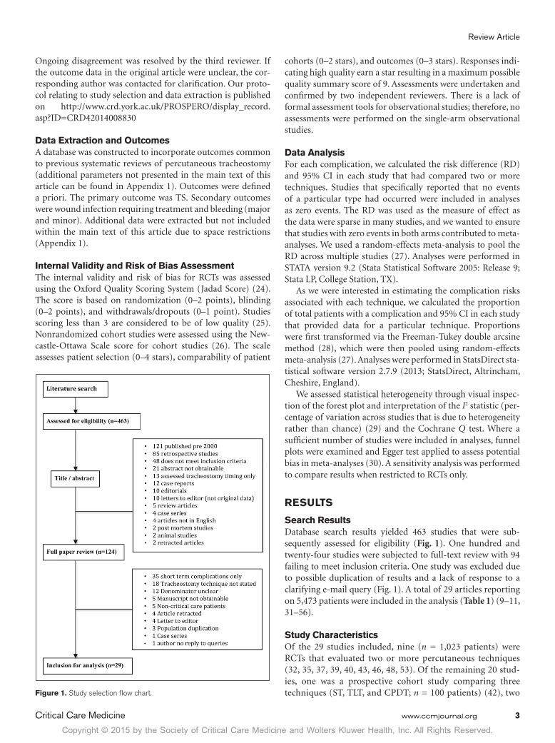
Copyright © 2015 by the Society of Critical Care Medicine and Wolters Kluwer Health, Inc. All Rights Reserved.
Review Article
Critical Care Medicine www.ccmjournal.org 3
Ongoing disagreement was resolved by the third reviewer. If the outcome data in the original article were unclear, the cor-responding author was contacted for clarification. Our proto-col relating to study selection and data extraction is published on http://www.crd.york.ac.uk/PROSPERO/display_record.asp?ID=CRD42014008830
Data Extraction and OutcomesA database was constructed to incorporate outcomes common to previous systematic reviews of percutaneous tracheostomy (additional parameters not presented in the main text of this article can be found in Appendix 1). Outcomes were defined a priori. The primary outcome was TS. Secondary outcomes were wound infection requiring treatment and bleeding (major and minor). Additional data were extracted but not included within the main text of this article due to space restrictions (Appendix 1).
Internal Validity and Risk of Bias AssessmentThe internal validity and risk of bias for RCTs was assessed using the Oxford Quality Scoring System (Jadad Score) (24). The score is based on randomization (0–2 points), blinding (0–2 points), and withdrawals/dropouts (0–1 point). Studies scoring less than 3 are considered to be of low quality (25). Nonrandomized cohort studies were assessed using the New-castle-Ottawa Scale score for cohort studies (26). The scale assesses patient selection (0–4 stars), comparability of patient
cohorts (0–2 stars), and outcomes (0–3 stars). Responses indi-cating high quality earn a star resulting in a maximum possible quality summary score of 9. Assessments were undertaken and confirmed by two independent reviewers. There is a lack of formal assessment tools for observational studies; therefore, no assessments were performed on the single-arm observational studies.
Data AnalysisFor each complication, we calculated the risk difference (RD) and 95% CI in each study that had compared two or more techniques. Studies that specifically reported that no events of a particular type had occurred were included in analyses as zero events. The RD was used as the measure of effect as the data were sparse in many studies, and we wanted to ensure that studies with zero events in both arms contributed to meta-analyses. We used a random-effects meta-analysis to pool the RD across multiple studies (27). Analyses were performed in STATA version 9.2 (Stata Statistical Software 2005: Release 9; Stata LP, College Station, TX).
As we were interested in estimating the complication risks associated with each technique, we calculated the proportion of total patients with a complication and 95% CI in each study that provided data for a particular technique. Proportions were first transformed via the Freeman-Tukey double arcsine method (28), which were then pooled using random-effects meta-analysis (27). Analyses were performed in StatsDirect sta-tistical software version 2.7.9 (2013; StatsDirect, Altrincham, Cheshire, England).
We assessed statistical heterogeneity through visual inspec-tion of the forest plot and interpretation of the I2 statistic (per-centage of variation across studies that is due to heterogeneity rather than chance) (29) and the Cochrane Q test. Where a sufficient number of studies were included in analyses, funnel plots were examined and Egger test applied to assess potential bias in meta-analyses (30). A sensitivity analysis was performed to compare results when restricted to RCTs only.
RESULTS
Search ResultsDatabase search results yielded 463 studies that were sub-sequently assessed for eligibility (Fig. 1). One hundred and twenty-four studies were subjected to full-text review with 94 failing to meet inclusion criteria. One study was excluded due to possible duplication of results and a lack of response to a clarifying e-mail query (Fig. 1). A total of 29 articles reporting on 5,473 patients were included in the analysis (Table 1) (9–11, 31–56).
Study CharacteristicsOf the 29 studies included, nine (n = 1,023 patients) were RCTs that evaluated two or more percutaneous techniques (32, 35, 37, 39, 40, 43, 46, 48, 53). Of the remaining 20 stud-ies, one was a prospective cohort study comparing three techniques (ST, TLT, and CPDT; n = 100 patients) (42), two Figure 1. Study selection flow chart.
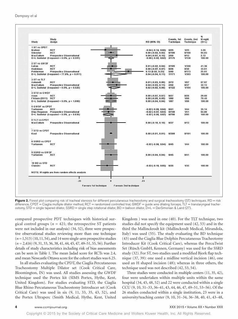
Copyright © 2015 by the Society of Critical Care Medicine and Wolters Kluwer Health, Inc. All Rights Reserved.
Dempsey et al
4 www.ccmjournal.org
compared prospective PDT techniques with historical sur-gical control groups (n = 421; the retrospective ST patients were not included in our analysis) (34, 52), three were prospec-tive observational studies reviewing more than one technique (n = 1,513) (10, 11, 54), and 14 were single-arm prospective studies (n = 2,416) (9, 31, 33, 36, 38, 41, 44, 45, 47, 49–51, 55, 56). Further details of study characteristics including risk of bias assessments can be seen in Table 1. The mean Jadad score for RCTs was 2.4, and mean Newcastle Ottawa score for the cohort studies was 6.25.
In all studies evaluating the CPDT, the Ciaglia Percutaneous Tracheostomy Multiple Dilator set (Cook Critical Care, Bloomington, IN) was used. All studies assessing the GWDF technique used the Portex kit (SIMS Portex, Hythe, Kent, United Kingdom). For studies evaluating STD, the Ciaglia Blue Rhino Percutaneous Tracheostomy Introducer set (Cook Critical Care) was used in six (9, 11, 33, 35, 43, 52), and the Portex Ultraperc (Smith Medical, Hythe, Kent, United
Kingdom ) was used in one (40). For the TLT technique, two studies did not specify the equipment used (42, 53) and in the third the Mallinckrodt kit (Mallinckrodt Medical, Mirandola, Italy) was used (55). The study evaluating the BD technique (43) used the Ciaglia Blue Dolphin Percutaneous Tracheotomy Introducer Kit (Cook Critical Care), whereas the PercuTwist Set (Rüsch GmbH, Kennen, Germany) was used for the SSRD study (32). For ST, two studies used a modified Bjork flap tech-nique (37, 39): one used a midline vertical incision (46), one used an H shaped incision (48); whereas in three others, the technique used was not described (42, 53, 54).
Three studies were conducted in multiple centers (11, 35, 42), four were undertaken within multiple units within the same hospital (34, 45, 48, 52) and 22 were conducted within a single CCU (9, 10, 31–33, 36–41, 43, 44, 46, 47, 49–51, 53–56). Of the 26 studies conducted within a single institution, 23 were in a university/teaching center (9, 10, 31–34, 36–38, 40, 41, 43–48,
Figure 2. Forest plot comparing risk of tracheal stenosis for different percutaneous tracheostomy and surgical tracheostomy (ST) techniques. RD = risk difference, CPDT = Ciaglia multiple dilator method, RCT = randomized controlled trial, GWDF = guide wire dilating forceps, TLT = translaryngeal trache-ostomy, STD = single tapered dilator, SSRD = single step rotational dilator, BD = balloon dilator, D+L = DerSimonian & Laird (27).
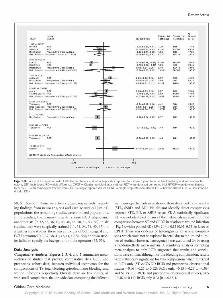
Copyright © 2015 by the Society of Critical Care Medicine and Wolters Kluwer Health, Inc. All Rights Reserved.
Review Article
Critical Care Medicine www.ccmjournal.org 5
50, 51, 53–56). There were two studies, respectively, report-ing findings from neuro (31, 55) and cardiac surgical (49, 51) populations; the remaining studies were of mixed populations. In 12 studies, the primary operators were CCU physicians/anaesthetists (9, 32, 35, 36, 40, 43, 46, 48, 50, 52, 53, 56); in six studies, they were surgically trained (11, 31, 34, 39, 45, 47); in a further nine studies, there was a mixture of both surgical and CCU personnel (10, 37, 38, 41, 42, 44, 49, 51, 54); and two stud-ies failed to specify the background of the operator (33, 55).
Data AnalysisComparative Analyses. Figures 2, 3, 4, and 5 summarize meta-analyses of studies that provide comparative data (RCT and prospective cohort data) between individual techniques for the complications of TS, total bleeding episodes, major bleeding, and wound infections, respectively. Overall, there are few studies, all with small sample sizes, that provide data comparing the different
techniques, particularly in relation to those described more recently (STD, SSRD, and BD). We did not identify direct comparisons between STD, BD, or SSRD versus ST. A statistically significant RD was not identified for any of the meta-analyses, apart from the comparison between ST and CPDT in relation to wound infection (Fig. 5), with a pooled RD (95% CI) of 0.12 (0.02–0.23) in favor of CPDT. There was evidence of heterogeneity for several compari-sons, which could not be explored in detail due to the limited num-ber of studies. However, heterogeneity was accounted for by using a random-effects meta-analysis. A sensitivity analysis restricting meta-analyses to only RCTs suggested that results and conclu-sions were similar, although for the bleeding complication, results were statistically significant for two comparisons when restricted to RCTs only (ST vs GWDF: RCTs and prospective observational studies, –0.06 [–0.25 to 0.12]; RCTs only, –0.15 [–0.23 to –0.08] and ST vs TLT: RCTs and prospective observational studies, 0.05 (–0.02 to 0.11); RCTs only, 0.08 [0 to 0.16]).
Figure 3. Forest plot comparing risk of all bleeding (major and minor) episodes reported for different percutaneous tracheostomy and surgical trache-ostomy (ST) techniques. RD = risk difference, CPDT = Ciaglia multiple dilator method, RCT = randomized controlled trial, GWDF = guide wire dilating forceps, TLT = translaryngeal tracheostomy, STD = single tapered dilator, SSRD = single step rotational dilator, BD = balloon dilator, D+L = DerSimonian & Laird (27).
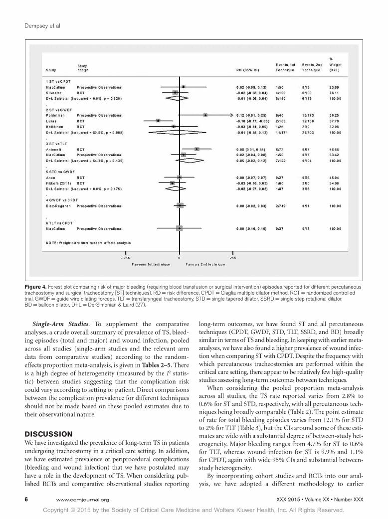
Copyright © 2015 by the Society of Critical Care Medicine and Wolters Kluwer Health, Inc. All Rights Reserved.
Dempsey et al
6 www.ccmjournal.org
Single-Arm Studies. To supplement the comparative analyses, a crude overall summary of prevalence of TS, bleed-ing episodes (total and major) and wound infection, pooled across all studies (single-arm studies and the relevant arm data from comparative studies) according to the random-effects proportion meta-analysis, is given in Tables 2–5. There is a high degree of heterogeneity (measured by the I2 statis-tic) between studies suggesting that the complication risk could vary according to setting or patient. Direct comparisons between the complication prevalence for different techniques should not be made based on these pooled estimates due to their observational nature.
DISCUSSIONWe have investigated the prevalence of long-term TS in patients undergoing tracheostomy in a critical care setting. In addition, we have estimated prevalence of periprocedural complications (bleeding and wound infection) that we have postulated may have a role in the development of TS. When considering pub-lished RCTs and comparative observational studies reporting
long-term outcomes, we have found ST and all percutaneous techniques (CPDT, GWDF, STD, TLT, SSRD, and BD) broadly similar in terms of TS and bleeding. In keeping with earlier meta-analyses, we have also found a higher prevalence of wound infec-tion when comparing ST with CPDT. Despite the frequency with which percutaneous tracheostomies are performed within the critical care setting, there appear to be relatively few high-quality studies assessing long-term outcomes between techniques.
When considering the pooled proportion meta-analysis across all studies, the TS rate reported varies from 2.8% to 0.6% for ST and STD, respectively, with all percutaneous tech-niques being broadly comparable (Table 2). The point estimate of rate for total bleeding episodes varies from 12.1% for STD to 2% for TLT (Table 3), but the CIs around some of these esti-mates are wide with a substantial degree of between-study het-erogeneity. Major bleeding ranges from 4.7% for ST to 0.6% for TLT, whereas wound infection for ST is 9.9% and 1.1% for CPDT, again with wide 95% CIs and substantial between-study heterogeneity.
By incorporating cohort studies and RCTs into our anal-ysis, we have adopted a different methodology to earlier
Figure 4. Forest plot comparing risk of major bleeding (requiring blood transfusion or surgical intervention) episodes reported for different percutaneous tracheostomy and surgical tracheostomy [ST] techniques). RD = risk difference, CPDT = Ciaglia multiple dilator method, RCT = randomized controlled trial, GWDF = guide wire dilating forceps, TLT = translaryngeal tracheostomy, STD = single tapered dilator, SSRD = single step rotational dilator, BD = balloon dilator, D+L = DerSimonian & Laird (27).
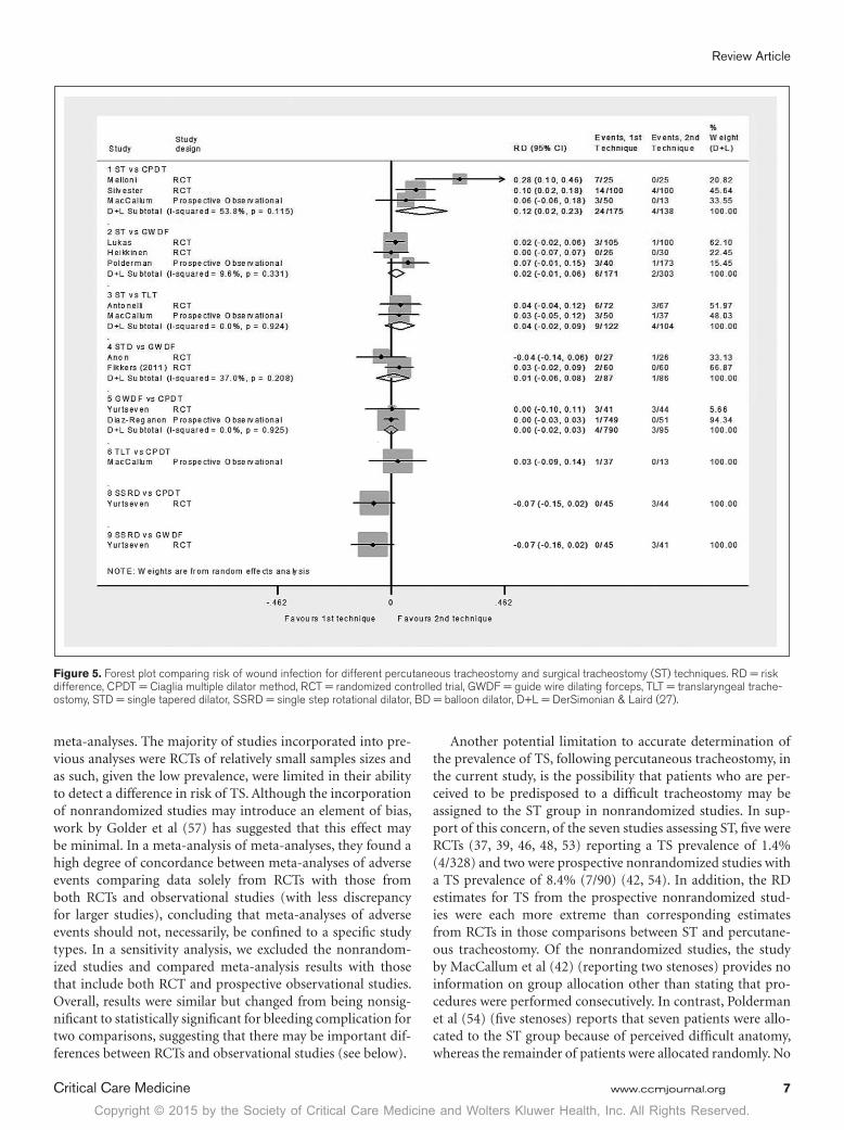
Copyright © 2015 by the Society of Critical Care Medicine and Wolters Kluwer Health, Inc. All Rights Reserved.
Review Article
Critical Care Medicine www.ccmjournal.org 7
meta-analyses. The majority of studies incorporated into pre-vious analyses were RCTs of relatively small samples sizes and as such, given the low prevalence, were limited in their ability to detect a difference in risk of TS. Although the incorporation of nonrandomized studies may introduce an element of bias, work by Golder et al (57) has suggested that this effect may be minimal. In a meta-analysis of meta-analyses, they found a high degree of concordance between meta-analyses of adverse events comparing data solely from RCTs with those from both RCTs and observational studies (with less discrepancy for larger studies), concluding that meta-analyses of adverse events should not, necessarily, be confined to a specific study types. In a sensitivity analysis, we excluded the nonrandom-ized studies and compared meta-analysis results with those that include both RCT and prospective observational studies. Overall, results were similar but changed from being nonsig-nificant to statistically significant for bleeding complication for two comparisons, suggesting that there may be important dif-ferences between RCTs and observational studies (see below).
Another potential limitation to accurate determination of the prevalence of TS, following percutaneous tracheostomy, in the current study, is the possibility that patients who are per-ceived to be predisposed to a difficult tracheostomy may be assigned to the ST group in nonrandomized studies. In sup-port of this concern, of the seven studies assessing ST, five were RCTs (37, 39, 46, 48, 53) reporting a TS prevalence of 1.4% (4/328) and two were prospective nonrandomized studies with a TS prevalence of 8.4% (7/90) (42, 54). In addition, the RD estimates for TS from the prospective nonrandomized stud-ies were each more extreme than corresponding estimates from RCTs in those comparisons between ST and percutane-ous tracheostomy. Of the nonrandomized studies, the study by MacCallum et al (42) (reporting two stenoses) provides no information on group allocation other than stating that pro-cedures were performed consecutively. In contrast, Polderman et al (54) (five stenoses) reports that seven patients were allo-cated to the ST group because of perceived difficult anatomy, whereas the remainder of patients were allocated randomly. No
Figure 5. Forest plot comparing risk of wound infection for different percutaneous tracheostomy and surgical tracheostomy (ST) techniques. RD = risk difference, CPDT = Ciaglia multiple dilator method, RCT = randomized controlled trial, GWDF = guide wire dilating forceps, TLT = translaryngeal trache-ostomy, STD = single tapered dilator, SSRD = single step rotational dilator, BD = balloon dilator, D+L = DerSimonian & Laird (27).
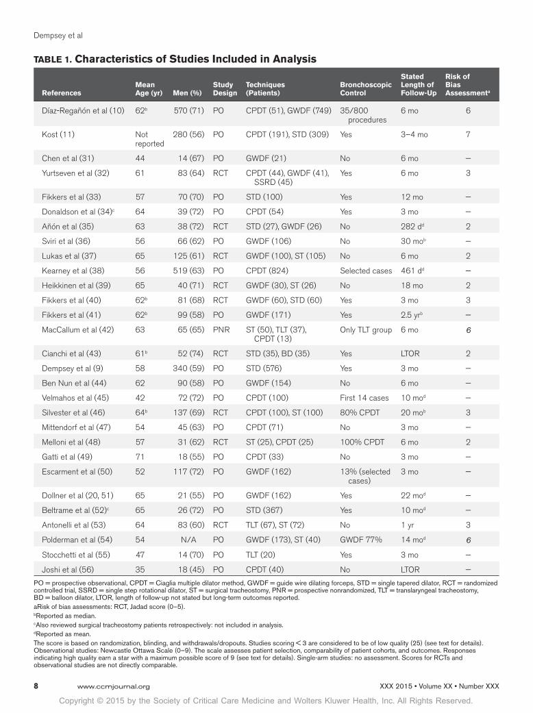
Copyright © 2015 by the Society of Critical Care Medicine and Wolters Kluwer Health, Inc. All Rights Reserved.
Dempsey et al
8 www.ccmjournal.org
TABLE 1. Characteristics of Studies Included in Analysis
ReferencesMean Age (yr) Men (%)
Study Design
Techniques (Patients)
Bronchoscopic Control
Stated Length of Follow-Up
Risk of Bias Assessmenta
Díaz-Regañón et al (10) 62b 570 (71) PO CPDT (51), GWDF (749) 35/800 procedures
6 mo 6
Kost (11) Not reported
280 (56) PO CPDT (191), STD (309) Yes 3–4 mo 7
Chen et al (31) 44 14 (67) PO GWDF (21) No 6 mo —
Yurtseven et al (32) 61 83 (64) RCT CPDT (44), GWDF (41), SSRD (45)
Yes 6 mo 3
Fikkers et al (33) 57 70 (70) PO STD (100) Yes 12 mo —
Donaldson et al (34)c 64 39 (72) PO CPDT (54) Yes 3 mo —
Añón et al (35) 63 38 (72) RCT STD (27), GWDF (26) No 282 dd 2
Sviri et al (36) 56 66 (62) PO GWDF (106) No 30 mob —
Lukas et al (37) 65 125 (61) RCT GWDF (100), ST (105) No 6 mo 2
Kearney et al (38) 56 519 (63) PO CPDT (824) Selected cases 461 dd —
Heikkinen et al (39) 65 40 (71) RCT GWDF (30), ST (26) No 18 mo 2
Fikkers et al (40) 62b 81 (68) RCT GWDF (60), STD (60) Yes 3 mo 3
Fikkers et al (41) 62b 99 (58) PO GWDF (171) Yes 2.5 yrb —
MacCallum et al (42) 63 65 (65) PNR ST (50), TLT (37), CPDT (13)
Only TLT group 6 mo 6
Cianchi et al (43) 61b 52 (74) RCT STD (35), BD (35) Yes LTOR 2
Dempsey et al (9) 58 340 (59) PO STD (576) Yes 3 mo —
Ben Nun et al (44) 62 90 (58) PO GWDF (154) No 6 mo —
Velmahos et al (45) 42 72 (72) PO CPDT (100) First 14 cases 10 mod —
Silvester et al (46) 64b 137 (69) RCT CPDT (100), ST (100) 80% CPDT 20 mob 3
Mittendorf et al (47) 54 45 (63) PO CPDT (71) No 3 mo —
Melloni et al (48) 57 31 (62) RCT ST (25), CPDT (25) 100% CPDT 6 mo 2
Gatti et al (49) 71 18 (55) PO CPDT (33) No 3 mo —
Escarment et al (50) 52 117 (72) PO GWDF (162) 13% (selected cases)
3 mo —
Dollner et al (20, 51) 65 21 (55) PO GWDF (162) Yes 22 mod —
Beltrame et al (52)c 65 26 (72) PO STD (367) Yes 10 mod —
Antonelli et al (53) 64 83 (60) RCT TLT (67), ST (72) No 1 yr 3
Polderman et al (54) 54 N/A PO GWDF (173), ST (40) GWDF 77% 14 mod 6
Stocchetti et al (55) 47 14 (70) PO TLT (20) Yes 3 mo —
Joshi et al (56) 35 18 (45) PO CPDT (40) No LTOR —
PO = prospective observational, CPDT = Ciaglia multiple dilator method, GWDF = guide wire dilating forceps, STD = single tapered dilator, RCT = randomized controlled trial, SSRD = single step rotational dilator, ST = surgical tracheostomy, PNR = prospective nonrandomized, TLT = translaryngeal tracheostomy, BD = balloon dilator, LTOR, length of follow-up not stated but long-term outcomes reported.aRisk of bias assessments: RCT, Jadad score (0–5).b Reported as median.c Also reviewed surgical tracheostomy patients retrospectively: not included in analysis.d Reported as mean.The score is based on randomization, blinding, and withdrawals/dropouts. Studies scoring < 3 are considered to be of low quality (25) (see text for details). Observational studies: Newcastle Ottawa Scale (0–9). The scale assesses patient selection, comparability of patient cohorts, and outcomes. Responses indicating high quality earn a star with a maximum possible score of 9 (see text for details). Single-arm studies: no assessment. Scores for RCTs and observational studies are not directly comparable.
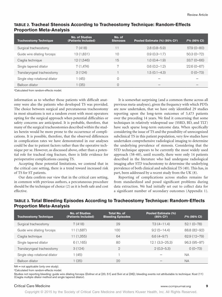
Copyright © 2015 by the Society of Critical Care Medicine and Wolters Kluwer Health, Inc. All Rights Reserved.
Review Article
Critical Care Medicine www.ccmjournal.org 9
information as to whether those patients with difficult anat-omy were also the patients who developed TS was provided. The choice between surgical and percutaneous tracheostomy in most situations is not a random event with most operators opting for the surgical approach when potential difficulties or safety concerns are anticipated. It is probable, therefore, that many of the surgical tracheostomies described within the stud-ies herein would be more prone to the occurrence of compli-cations. It is possible, therefore, that the observed differences in complication rates we have demonstrated in our analyses could be due to patient factors rather than the operative tech-nique per se. However, as discussed above, other than a poten-tial role for tracheal ring fracture, there is little evidence for perioperative complications causing TS.
Accepting these potential limitations, we contend that in the critical care setting, there is a trend toward increased risk of TS for ST patients.
Our data confirm our view that in the critical care setting, in common with previous authors, a percutaneous procedure should be the technique of choice (2) as it is both safe and cost effective.
It is somewhat surprising (and a common theme across all previous meta-analyses), given the frequency with which PDTs are now undertaken, that we have only identified 29 studies reporting upon the long-term outcomes of 5,473 patients over the preceding 14 years. We find it concerning that some techniques in relatively widespread use (SSRD, BD, and TLT) have such sparse long-term outcome data. When specifically considering the issue of TS and the possibility of unrecognized subclinical TS in this patient population, very few studies have undertaken comprehensive radiological imaging to determine the underlying prevalence of stenosis. Considering that the STD technique appears to be currently the most widely used approach (58–60), until recently, there were only 14 patients described in the literature who had undergone radiological imaging after STD tracheostomy to determine the underlying prevalence of both clinical and subclinical TS (40). This has, in part, been addressed by a recent study from the UK (8).
Reporting of complications across studies remains far from standardized and posed significant problems during data extraction. We had initially set out to collect data for a significant number of secondary outcomes (Appendix 1).
TABLE 2. Tracheal Stenosis According to Tracheostomy Technique: Random-Effects Proportion Meta-Analysis
Tracheostomy TechniqueNo. of Studies
(Patients Included)No. of
Stenoses Pooled Estimate (%) (95% CIa) I2% (95% CI)
Surgical tracheostomy 7 (418) 11 2.8 (0.8–5.9) 57.9 (0–80)
Guide wire dilating forceps 13 (1,831) 10 0.9 (0.3–1.7) 50.3 (0–72)
Ciaglia technique 12 (1,546) 15 1.0 (0.4–1.9) 33.7 (0–66)
Single tapered dilator 7 (1,474) 7 0.6 (0.2–1.2) 22.6 (0–67)
Translaryngeal tracheostomy 3 (124) 1 1.5 (0.1–4.3) 0 (0–73)
Single step rotational dilator 1 (45) 0 — —
Balloon dilator 1 (35) 0 — —a Calculated from random-effects model.
TABLE 3. Total Bleeding Episodes According to Tracheostomy Technique: Random-Effects Proportion Meta-Analysis
Tracheostomy TechniqueNo. of Studies
(Patients Included)Total No. of
Bleeding EpisodesPooled Estimate (%)
(95% CIa) I2% (95% CI)
Surgical tracheostomy 7 (418) 29 7.3 (4–11.4) 52.1 (0–78)
Guide wire dilating forceps 11 (1,687) 100 9.2 (5–14.4) 88.8 (82–92)
Ciaglia technique 11 (1,355) 64 6.6 (4–9.7) 62.9 (12–79)
Single tapered dilator 6 (1,165) 80 12.1 (3.3–25.3) 96.3 (95–97)
Translaryngeal tracheostomy 3 (124) 2 2 (0.3–5.3) 0 (0–73)
Single step rotational dilator 1 (45) 1 — NA
Balloon dilator 1 (35) 20 — NA
NA = not applicable (only one study).a Calculated from random-effects model.Studies not reporting bleeding: guide wire dilating forceps (Dollner et al [20, 51] and Sviri et al [36]); bleeding events not attributable to technique: Kost (11) (Ciaglia multiple dilator method and single tapered dilator).
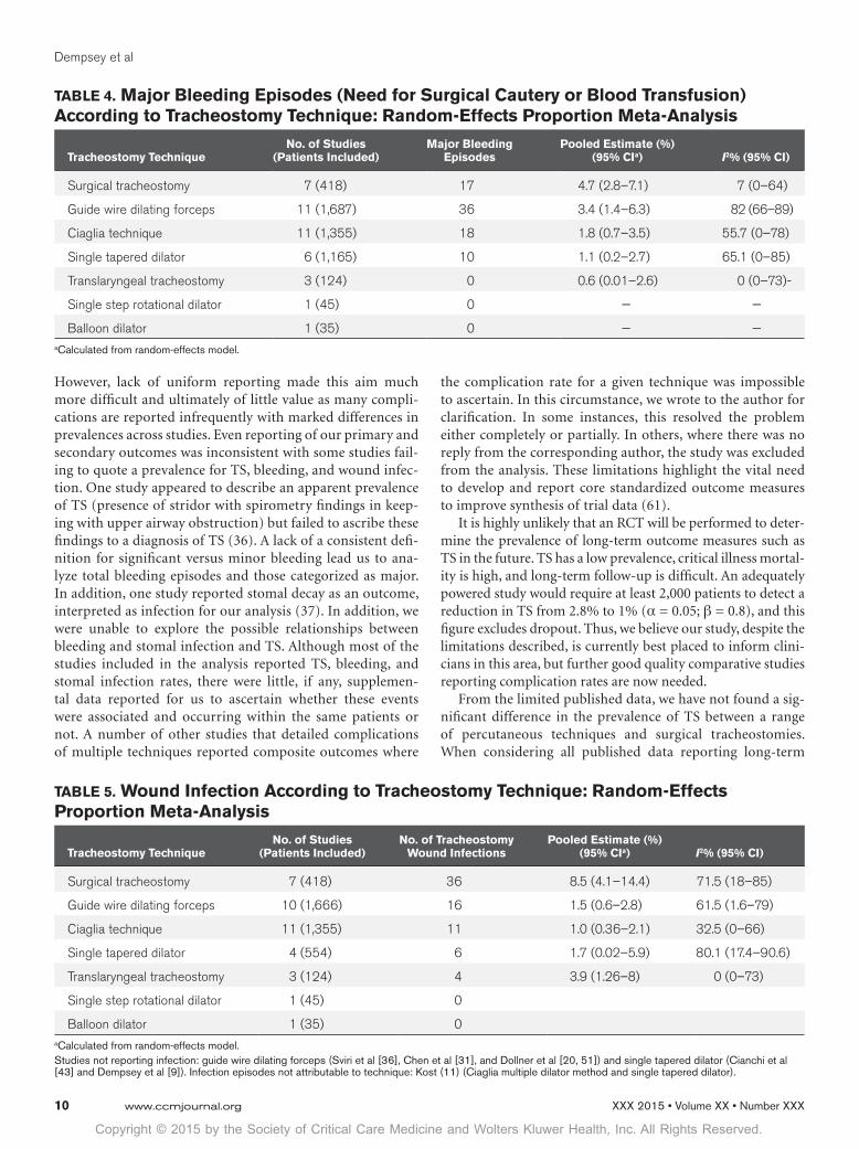
Copyright © 2015 by the Society of Critical Care Medicine and Wolters Kluwer Health, Inc. All Rights Reserved.
Dempsey et al
10 www.ccmjournal.org
However, lack of uniform reporting made this aim much more difficult and ultimately of little value as many compli-cations are reported infrequently with marked differences in prevalences across studies. Even reporting of our primary and secondary outcomes was inconsistent with some studies fail-ing to quote a prevalence for TS, bleeding, and wound infec-tion. One study appeared to describe an apparent prevalence of TS (presence of stridor with spirometry findings in keep-ing with upper airway obstruction) but failed to ascribe these findings to a diagnosis of TS (36). A lack of a consistent defi-nition for significant versus minor bleeding lead us to ana-lyze total bleeding episodes and those categorized as major. In addition, one study reported stomal decay as an outcome, interpreted as infection for our analysis (37). In addition, we were unable to explore the possible relationships between bleeding and stomal infection and TS. Although most of the studies included in the analysis reported TS, bleeding, and stomal infection rates, there were little, if any, supplemen-tal data reported for us to ascertain whether these events were associated and occurring within the same patients or not. A number of other studies that detailed complications of multiple techniques reported composite outcomes where
the complication rate for a given technique was impossible to ascertain. In this circumstance, we wrote to the author for clarification. In some instances, this resolved the problem either completely or partially. In others, where there was no reply from the corresponding author, the study was excluded from the analysis. These limitations highlight the vital need to develop and report core standardized outcome measures to improve synthesis of trial data (61).
It is highly unlikely that an RCT will be performed to deter-mine the prevalence of long-term outcome measures such as TS in the future. TS has a low prevalence, critical illness mortal-ity is high, and long-term follow-up is difficult. An adequately powered study would require at least 2,000 patients to detect a reduction in TS from 2.8% to 1% (! = 0.05; " = 0.8), and this figure excludes dropout. Thus, we believe our study, despite the limitations described, is currently best placed to inform clini-cians in this area, but further good quality comparative studies reporting complication rates are now needed.
From the limited published data, we have not found a sig-nificant difference in the prevalence of TS between a range of percutaneous techniques and surgical tracheostomies. When considering all published data reporting long-term
TABLE 4. Major Bleeding Episodes (Need for Surgical Cautery or Blood Transfusion) According to Tracheostomy Technique: Random-Effects Proportion Meta-Analysis
Tracheostomy TechniqueNo. of Studies
(Patients Included)Major Bleeding
EpisodesPooled Estimate (%)
(95% CIa) I2% (95% CI)
Surgical tracheostomy 7 (418) 17 4.7 (2.8–7.1) 7 (0–64)
Guide wire dilating forceps 11 (1,687) 36 3.4 (1.4–6.3) 82 (66–89)
Ciaglia technique 11 (1,355) 18 1.8 (0.7–3.5) 55.7 (0–78)
Single tapered dilator 6 (1,165) 10 1.1 (0.2–2.7) 65.1 (0–85)
Translaryngeal tracheostomy 3 (124) 0 0.6 (0.01–2.6) 0 (0–73)-
Single step rotational dilator 1 (45) 0 — —
Balloon dilator 1 (35) 0 — —a Calculated from random-effects model.
TABLE 5. Wound Infection According to Tracheostomy Technique: Random-Effects Proportion Meta-Analysis
Tracheostomy TechniqueNo. of Studies
(Patients Included)No. of Tracheostomy
Wound InfectionsPooled Estimate (%)
(95% CIa) I2% (95% CI)
Surgical tracheostomy 7 (418) 36 8.5 (4.1–14.4) 71.5 (18–85)
Guide wire dilating forceps 10 (1,666) 16 1.5 (0.6–2.8) 61.5 (1.6–79)
Ciaglia technique 11 (1,355) 11 1.0 (0.36–2.1) 32.5 (0–66)
Single tapered dilator 4 (554) 6 1.7 (0.02–5.9) 80.1 (17.4–90.6)
Translaryngeal tracheostomy 3 (124) 4 3.9 (1.26–8) 0 (0–73)
Single step rotational dilator 1 (45) 0
Balloon dilator 1 (35) 0a Calculated from random-effects model.Studies not reporting infection: guide wire dilating forceps (Sviri et al [36], Chen et al [31], and Dollner et al [20, 51]) and single tapered dilator (Cianchi et al [43] and Dempsey et al [9]). Infection episodes not attributable to technique: Kost (11) (Ciaglia multiple dilator method and single tapered dilator).
Copyright © 2015 by the Society of Critical Care Medicine and Wolters Kluwer Health, Inc. All Rights Reserved.
Review Article
Critical Care Medicine www.ccmjournal.org 11
outcomes, our pooled proportion meta-analysis may indicate a tendency toward a higher rate of TS for ST, but this is likely to be prone to selection bias. Similarly, in relation to major bleeding, we have not found a difference between the tech-niques commonly used. Again the pooled proportions meta-analysis may indicate a tendency toward more major bleeding for ST. In relation to wound infection, we have found a reduc-tion (RD, –6% [95% CI, –12% to 1%]) associated with CPDT when compared with ST in keeping with earlier work. Across all percutaneous techniques for the primary and secondary outcomes studied, complication rates appear to be broadly similar, but CIs for pooled RDs are wide and include clini-cally important differences in both directions.
REFERENCES 1. Ciaglia P, Firsching R, Syniec C: Elective percutaneous dilatational
tracheostomy. A new simple bedside procedure; preliminary report. Chest 1985; 87:715–719
2. Delaney A, Bagshaw SM, Nalos M: Percutaneous dilatational trache-ostomy versus surgical tracheostomy in critically ill patients: A system-atic review and meta-analysis. Crit Care 2006; 10:R55
3. Higgins KM, Punthakee X: Meta-analysis comparison of open versus percutaneous tracheostomy. Laryngoscope 2007; 117:447–454
4. Oliver ER, Gist A, Gillespie MB: Percutaneous versus surgical tracheotomy: An updated meta-analysis. Laryngoscope 2007; 117:1570–1575
5. Cabrini L, Monti G, Landoni G, et al: Percutaneous tracheostomy, a systematic review. Acta Anaesthesiol Scand 2012; 56:270–281
6. Cabrini L, Landoni G, Greco M, et al: Single dilator vs. guide wire dilating forceps tracheostomy: A meta-analysis of randomised trials. Acta Anaesthesiol Scand 2014; 58:135–142
7. Khalili TM, Koss W, Margulies DR, et al: Percutaneous dilatational tra-cheostomy is as safe as open tracheostomy. Am Surg 2002; 68:92–94
8. Young E, Pugh R, Hanlon R, et al: Tracheal stenosis following percu-taneous dilatational tracheostomy using the single tapered dilator: An MRI study. Anaesth Intensive Care 2014; 42:745–751
9. Dempsey GA, Grant CA, Jones TM: Percutaneous tracheostomy: A 6 yr prospective evaluation of the single tapered dilator technique. Br J Anaesth 2010; 105:782–788
10. Díaz-Regañón G, Miñambres E, Ruiz A, et al: Safety and complica-tions of percutaneous tracheostomy in a cohort of 800 mixed ICU patients. Anaesthesia 2008; 63:1198–1203
11. Kost KM: Endoscopic percutaneous dilatational tracheotomy: A pro-spective evaluation of 500 consecutive cases. Laryngoscope 2005; 115:1–30
12. Grillo HC: Primary reconstruction of airway after resection of subglot-tic laryngeal and upper tracheal stenosis. Ann Thorac Surg 1982; 33:3–18
13. Pearson FG, Cooper JD, Nelems JM, et al: Primary tracheal anastomo-sis after resection of the cricoid cartilage with preservation of recur-rent laryngeal nerves. J Thorac Cardiovasc Surg 1975; 70:806–816
14. Bacon JL, Patterson CM, Madden BP: Indications and interventional options for non-resectable tracheal stenosis. J Thorac Dis 2014; 6:258–270
15. Stauffer JL, Olson DE, Petty TL: Complications and consequences of endotracheal intubation and tracheotomy. A prospective study of 150 critically ill adult patients. Am J Med 1981; 70:65–76
16. Herrak L, Ahid S, Abouqal R, et al: Tracheal stenosis after intubation and/or tracheostomy. Egypt J Chest Dis Tub 2014;63:233–237
17. Nair S, Mohan S, Mandal G, et al: Tracheal stenosis: Our experience at a tertiary care centre in India with special regard to cause and man-agement. Indian J Otolaryngol Head Neck Surg 2014; 66:51–56
18. Zias N, Chroneou A, Tabba MK, et al: Post tracheostomy and post intubation tracheal stenosis: Report of 31 cases and review of the literature. BMC Pulm Med 2008; 8:18
19. Raghuraman G, Rajan S, Marzouk JK, et al: Is tracheal stenosis caused by percutaneous tracheostomy different from that by surgical tracheostomy? Chest 2005; 127:879–885
-ings in long-term follow-up after Griggs tracheostomy. Chest 2002; 122:206–212
21. van Heurn LW, Theunissen PH, Ramsay G, et al: Pathologic changes of the trachea after percutaneous dilatational tracheotomy. Chest 1996; 109:1466–1469
22. Squire R, Brodsky L, Rossman J: The role of infection in the pathogenesis of acquired tracheal stenosis. Laryngoscope 1990; 100:765–770
23. Welkoborsky HJ, Hinni ML, Moebius H, et al: Microscopic examination of iatrogenic subglottic tracheal stenosis: Observations that may elucidate its histopathologic origin. Ann Otol Rhinol Laryngol 2014; 123:25–31
24. Jadad AR, Moore RA, Carroll D, et al: Assessing the quality of reports of randomized clinical trials: Is blinding necessary? Control Clin Trials 1996; 17:1–12
and discrepancies between large and small randomized trials in meta-analyses. Ann Intern Med 2001; 135:982–989
26. Wells GA, Shea B, O'Connell D, et al: The Newcastle-Ottawa Scale (NOS) for assessing the quality of nonrandomized studies in meta-analyses. Ottawa, ON, Ottawa Hospital Research Institute, 2011. Available at: http://www.ohri.ca/programs/clinical_epidemiology/oxford.htm. Accessed September 30, 2015
27. DerSimonian R, Laird N: Meta-analysis in clinical trials. Control Clin Trials 1986; 7:177–188
28. Miller J. Inverse of the Freeman-Tukey Double Arcsine Transformation. Amer Stat 1978; 32:138
29. Higgins JP, Thompson SG: Quantifying heterogeneity in a meta-anal-ysis. Stat Med 2002; 21:1539–1558
30. Egger M, Davey Smith G, Schneider M, et al: Bias in meta-analysis detected by a simple, graphical test. BMJ 1997; 315:629–634
31. Chen Y, Wang Y, Sun W, et al: Implementation of percutaneous dila-tational tracheostomy on neurosurgical coma patients. Chin Med J (Engl) 2002; 115:1345–1347
32. Yurtseven N, Aydemir B, Karaca P, et al: PercuTwist: A new alterna-tive to Griggs and Ciaglia’s techniques. Eur J Anaesthesiol 2007; 24:492–497
with the Blue Rhino trade mark technique: Presentation of 100 con-secutive patients. Anaesthesia 2002; 57:1094–1097
34. Donaldson DR, Emami AJ, Wax MK: Endoscopically monitored percu-taneous dilational tracheotomy in a residency program. Laryngoscope 2000; 110:1142–1146
Ciaglia Blue Rhino versus Griggs’ Guide Wire Dilating Forceps. A pro-spective randomized trial. Acta Anaesthesiol Scand 2004; 48:451–456
36. Sviri S, Samie R, Roberts BL, et al: Long-term outcomes following percutaneous tracheostomy using the Griggs technique. Anaesth Intensive Care 2003; 31:401–407
37. Lukas J, Duskova J, Lukas D, et al: Standard surgical versus percuta-neous dilatational tracheostomy in intensive care patients. Saudi Med J 2007; 28:1529–1533
38. Kearney PA, Griffen MM, Ochoa JB, et al: A single-center 8-year expe-rience with percutaneous dilational tracheostomy. Ann Surg 2000; 231:701–709
39. Heikkinen M, Aarnio P, Hannukainen J: Percutaneous dilational tra-cheostomy or conventional surgical tracheostomy? Crit Care Med 2000; 28:1399–1402
40. Fikkers BG, Staatsen M, van den Hoogen FJ, et al: Early and late outcome after single step dilatational tracheostomy versus the guide wire dilating forceps technique: A prospective randomized clinical trial. Intensive Care Med 2011; 37:1103–1109
41. Fikkers BG, van Heerbeek N, Krabbe PF, et al: Percutaneous trache-ostomy with the guide wire dilating forceps technique: Presentation of 171 consecutive patients. Head Neck 2002; 24:625–631
42. MacCallum PL, Parnes LS, Sharpe MD, et al: Comparison of open, percutaneous, and translaryngeal tracheostomies. Otolaryngol Head Neck Surg 2000; 122:686–690
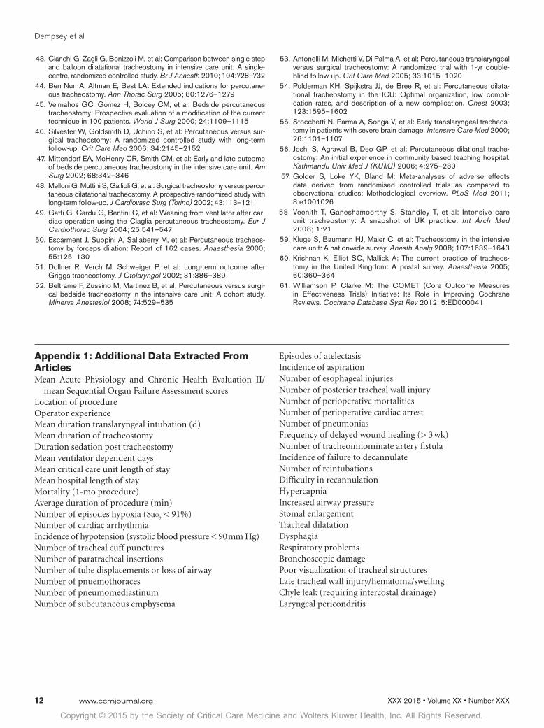
Copyright © 2015 by the Society of Critical Care Medicine and Wolters Kluwer Health, Inc. All Rights Reserved.
Dempsey et al
12 www.ccmjournal.org
43. Cianchi G, Zagli G, Bonizzoli M, et al: Comparison between single-step and balloon dilatational tracheostomy in intensive care unit: A single-centre, randomized controlled study. Br J Anaesth 2010; 104:728–732
44. Ben Nun A, Altman E, Best LA: Extended indications for percutane-ous tracheostomy. Ann Thorac Surg 2005; 80:1276–1279
tracheostomy: Prospective evaluation of a modification of the current technique in 100 patients. World J Surg 2000; 24:1109–1115
46. Silvester W, Goldsmith D, Uchino S, et al: Percutaneous versus sur-gical tracheostomy: A randomized controlled study with long-term follow-up. Crit Care Med 2006; 34:2145–2152
47. Mittendorf EA, McHenry CR, Smith CM, et al: Early and late outcome of bedside percutaneous tracheostomy in the intensive care unit. Am Surg 2002; 68:342–346
48. Melloni G, Muttini S, Gallioli G, et al: Surgical tracheostomy versus percu-taneous dilatational tracheostomy. A prospective-randomized study with long-term follow-up. J Cardiovasc Surg (Torino) 2002; 43:113–121
49. Gatti G, Cardu G, Bentini C, et al: Weaning from ventilator after car-diac operation using the Ciaglia percutaneous tracheostomy. Eur J Cardiothorac Surg 2004; 25:541–547
50. Escarment J, Suppini A, Sallaberry M, et al: Percutaneous tracheos-tomy by forceps dilation: Report of 162 cases. Anaesthesia 2000; 55:125–130
Griggs tracheostomy. J Otolaryngol 2002; 31:386–389 52. Beltrame F, Zussino M, Martinez B, et al: Percutaneous versus surgi-
cal bedside tracheostomy in the intensive care unit: A cohort study. Minerva Anestesiol 2008; 74:529–535
versus surgical tracheostomy: A randomized trial with 1-yr double-blind follow-up. Crit Care Med 2005; 33:1015–1020
54. Polderman KH, Spijkstra JJ, de Bree R, et al: Percutaneous dilata-tional tracheostomy in the ICU: Optimal organization, low compli-cation rates, and description of a new complication. Chest 2003; 123:1595–1602
-tomy in patients with severe brain damage. Intensive Care Med 2000; 26:1101–1107
56. Joshi S, Agrawal B, Deo GP, et al: Percutaneous dilational trache-ostomy: An initial experience in community based teaching hospital. Kathmandu Univ Med J (KUMJ) 2006; 4:275–280
57. Golder S, Loke YK, Bland M: Meta-analyses of adverse effects data derived from randomised controlled trials as compared to observational studies: Methodological overview. PLoS Med 2011; 8:e1001026
unit tracheostomy: A snapshot of UK practice. Int Arch Med 2008; 1:21
59. Kluge S, Baumann HJ, Maier C, et al: Tracheostomy in the intensive care unit: A nationwide survey. Anesth Analg 2008; 107:1639–1643
60. Krishnan K, Elliot SC, Mallick A: The current practice of tracheos-tomy in the United Kingdom: A postal survey. Anaesthesia 2005; 60:360–364
61. Williamson P, Clarke M: The COMET (Core Outcome Measures in Effectiveness Trials) Initiative: Its Role in Improving Cochrane Reviews. Cochrane Database Syst Rev 2012; 5:ED000041
Appendix 1: Additional Data Extracted From ArticlesMean Acute Physiology and Chronic Health Evaluation II/ mean Sequential Organ Failure Assessment scoresLocation of procedureOperator experienceMean duration translaryngeal intubation (d)Mean duration of tracheostomyDuration sedation post tracheostomyMean ventilator dependent daysMean critical care unit length of stayMean hospital length of stayMortality (1-mo procedure)Average duration of procedure (min)Number of episodes hypoxia (SaO
2 < 91%)
Number of cardiac arrhythmiaIncidence of hypotension (systolic blood pressure < 90 mm Hg)Number of tracheal cuff puncturesNumber of paratracheal insertionsNumber of tube displacements or loss of airwayNumber of pnuemothoracesNumber of pneumomediastinumNumber of subcutaneous emphysema
Episodes of atelectasisIncidence of aspirationNumber of esophageal injuriesNumber of posterior tracheal wall injuryNumber of perioperative mortalitiesNumber of perioperative cardiac arrestNumber of pneumoniasFrequency of delayed wound healing (> 3 wk)Number of tracheoinnominate artery fistulaIncidence of failure to decannulateNumber of reintubationsDifficulty in recannulationHypercapniaIncreased airway pressureStomal enlargementTracheal dilatationDysphagiaRespiratory problemsBronchoscopic damagePoor visualization of tracheal structuresLate tracheal wall injury/hematoma/swellingChyle leak (requiring intercostal drainage)Laryngeal pericondritis