process development of an acellular dermal matrix (adm) for biomedical applications
TRANSCRIPT

Biomaterials 25 (2004) 2679–2686
ARTICLE IN PRESS
*Correspondin
E-mail addres
0142-9612/$ - see
doi:10.1016/j.bio
Process development of an acellular dermal matrix (ADM) forbiomedical applications
Ray-Neng Chen, Hsiu-O Ho, Yu-Ting Tsai, Ming-Thau Sheu*
Graduate Institute of Pharmaceutical Sciences, College of Pharmacy, Taipei Medical University, 250 Wu-Hsing Street, Taipei 110, Taiwan, ROC
Received 20 January 2003; accepted 5 September 2003
Abstract
The object of this study was to compare the extent of decellularization at each critical step of processing porcine skin to produce
an acellular dermal matrix (ADM) for biomedical applications. The results demonstrated that the removal of epidermis using
treatment with 0.25% trypsin for 18 h and 0.1% sodium dodecyl sulfate (SDS) for 12 h at room temperature was beneficial for the
subsequent treatment to remove cells in the dermal structure. Lengthy incubation in 0.25% trypsin (12 h) and then 560 units/l
Dispase (12 h) at 25�C of small pieces of porcine skin from which the epidermis had been removed efficiently removed cells and
cellular components from the skin. Histological examinations revealed that the epidermis, dermal fibroblasts, and epidermal
appendages were completely removed by these treatments, and the basic dermal architecture of collagen bundles was that of a loose
meshwork. Examinations by TEM showed that the characteristics of collagen fibers in the ADM were retained after complete
removal of cells present under optimal conditions defined in this study. SDS-PAGE and size-exclusion HPLC revealed that collagen
fibers in the ADM were mostly type I and showed two typical component peaks identified as oligomers and monomers, respectively.
r 2003 Elsevier Ltd. All rights reserved.
Keywords: Acellular dermal matrix; Trypsin; Sodium dodecyl sulfate; Dispase
1. Introduction
Currently, due to excellent achievements in a varietyof different biomedical applications, the performance ofacellular dermal matrices, which are derived from full-thickness skin treated to remove cells and cellularcomponents but which retain the native dermal struc-ture, has drawn the attention of researchers in manyfields [1–3]. Many recent studies have presented severalmethods for producing an acellular dermal matrix(ADM) from porcine skin [4] and the submucosal layerof the small intestine [5]. Walter et al. commented thatthese methods using treatment with trypsin, freeze-thawing, and prolonged incubations with enzymesproduced ADMs which were too highly antigenic whenimplanted into recipients, where they induced immunereactions resulting in poor graft survival [6]. Two moreeffective and controlled extraction methods have beenreported for producing ADMs which exhibit very lowantigenicity and excellent stability, while retaining thenative dermal structure. One method uses hypertonic
g author. Tel./fax: +886-2-23771942.
s: [email protected] (M.-T. Sheu).
front matter r 2003 Elsevier Ltd. All rights reserved.
materials.2003.09.070
NaCl followed by sodium dodecyl sulfate (SDS) andfreeze-drying [1,7], while the other uses sequentialtreatments with Dispase followed by Triton-100 [8,9].Walter et al. compared these two methods andconcluded that both methods of ADM preparationresulted in extensive extraction of both cellular andextracellular components of the skin but retained thebasic dermal architecture. Lee et al. also publishedanother alternative method to produce a truly cell-freeporcine dermal matrix [10]. The optimal aseptic methodfor preparing a cell-free dermal matrix with a loosercollagen structure required the treatment of porcine skinof 0.03 in thickness with 0.25% trypsin at 4�C for 24 hfollowed sequentially by treatments with 0.1% TritonX-100 for 8 h at room temperature, Dispase (560 units/l)at 4�C for 24 h, and a final extensive washing with R.O.water.Recently, scaffolds derived from a xenogenic extra-
cellular matrix (ECM) have been shown to be effectivein the repair and reconstruction of several body tissuesincluding the lower urinary tract, dura mater, esopha-gus, musculotendinous structures, and blood vessels[11]. The characteristic of these scaffolds recognized asimportant for their effectiveness is their ability to induce

ARTICLE IN PRESSR.-N. Chen et al. / Biomaterials 25 (2004) 2679–26862680
a host cellular response that supports constructiveremodeling rather than default scar tissue formation.In a study comparing purified collagen, naturallyoccurring ECM scaffolds, and synthetic scaffold materi-als for in vitro endothelial cell attachment [12], it wasfound that ECM possessed the ability to recruitcirculating marrow-derived progenitor cells and attractmature endothelial cells from selected organs such as theheart and liver to promote successful vascularization ofengineered tissue structures. These studies reveal thatextracellular components in a cell-free or ADM arecritical for success in biomedical applications as scaf-folds. The object of this study, therefore, was tocompare the extent of decellularization at each criticalstep in the processing of porcine skin into an ADM forbiomedical applications.
2. Experimental methods
2.1. Materials
2.1.1. Acellular dermal matrix
Fresh porcine skin was obtained from a localslaughterhouse. After a complete cleaning, excision ofthe subdermal fat tissue, and removal of hair, theresulting skin was kept at �20�C until use. The skin wasdivided into four groups for comparison, and theoptimal conditions for processing porcine skin into anADM were defined in the final results.Group A was processed as follows to compare the
influence of treatment conditions of the trypsin solutionon the extent of decellularization. Porcine skin was cutin pieces which were 0.5� 0.5� 0.3 cm3. They werefurther divided into four subgroups (A1–A4), and fivepieces of this size of porcine skin were allocated to eachsubgroup. Porcine skin in the four subgroups wassoaked in a 0.25% trypsin solution at 4�C for 12 h(A1), at 4�C for 24 h (A2), at 25�C for 12 h (A3), and at25�C for 24 h (A4), respectively. This was followed bytreatment with a 0.1% SDS solution at room tempera-ture for 6 h, then by 560 units/l of a Dispase solution at4�C for 12 h, and finally in a 0.1% SDS solution at roomtemperature for 6 h before being washed twice with PBSbuffer for 15min each. Samples were preserved in PBSbuffer. Gentamicin at 10 mg/ml was added to allsolutions to prevent bacterial growth.Porcine skin in the four subgroups (B1–B4) of
group B was processed in similar respective conditionsto that of group A except that both treatments with the0.1% SDS solution were extended from 6 to 12 h. Thiswas designed to compare the influence of washingtime with 0.1% SDS solution on the extent ofdecellularization.Group C was processed as follows to compare the
influence of treatment conditions of both trypsin and
Dispase on the extent of decellularization after removingthe epidermis of porcine skin. Porcine skin was cutinto pieces which were 10� 7� 0.3 cm3. The epidermisof the skin was removed after treating with a 0.25%trypsin solution at 25�C for 18 h, and then thedermal part was cut into pieces which were0.5� 0.5� 0.3 cm3. These were divided into two sub-groups (C1–C2) with 10 pieces of skin in each subgroup.This skin was treated with a 0.25% trypsin solutionfollowed by shaking at 25�C for 12 h for one subgroupand at 25�C for 24 h for the other. Porcine skin inboth subgroups was then washed with a 0.1% SDSsolution at room temperature for 12 h, followed by560 units/l of a Dispase solution at 4�C for 12 h (C11and C21) or at 25�C for 12 h (C12 and C22),respectively. Sequentially, porcine skin in the foursubgroups was washed with 0.1% SDS at roomtemperature for 12 h and then washed with PBS buffertwice for 15min each. Samples were preserved in PBSbuffer. Gentamicin at 10 mg/ml was added to allsolutions to prevent bacterial growth.Porcine skin in group D was processed in similar
respective conditions as those of subgroup C2 exceptthat treatment with the Dispase solution and SDSsolution in the final step was shortened from 12 to 6 h.This was designed to compare the influence of washingtime of the Dispase solution and SDS solution on theextent of decellularization.
2.1.2. Histological examinations
ADM samples were first dehydrated with an increas-ing series of alcohol concentrations and then embeddedin paraffin. Paraffin-embedded ADMs were sectionedat a thickness of 5 mm. After removing the paraffin,samples were stained with hematoxylin and eosin.After sealing, samples were examined by lightmicroscopy at a magnification of 200� to inspectfibroblast cells (stained by hematoxylin to a bluish-purple color) and collagen fibers (stained by eosin to apink color).
2.1.3. Scanning electron microscopy (SEM) and
transmission electron microscopy (TEM) examinations
ADM samples after treatment with various conditionsor those without treatment were freeze-dried for SEMand TEM examinations. For SEM examinations,samples were loaded onto aluminum studs and coatedwith gold for 3min at 8mA under a pressure of 0.1 Torr.Collagen morphologies were examined under a scanningelectron microscope (Hitachi model S-2400, Departmentof Pathology, Taipei Medical University). Samples werescanned, and the micrographs were recorded. Compar-isons were made of morphological changes to collagenfibers before and after treatment under various condi-tions. For TEM examinations, samples were scannedusing a transmission electron microscope (Hitachi model

ARTICLE IN PRESSR.-N. Chen et al. / Biomaterials 25 (2004) 2679–2686 2681
H-600). Characteristics of the sequences of black andwhite bands of collagen fibers of samples treated withvarious conditions were compared using the TEMexamination.
(A)
(C)
Fig. 2. Histological photographs of ADM samples fro
(A)
(C)
(E)
Fig. 1. Histological photographs of ADM samples from grou
2.1.4. Sodium dodecyl sulfate-polyacrylamide gel
electrophoresis (SDS-PAGE)
Separation of collagen types and their a-chains wasperformed on SDS-polyacrylamide slab gels, using 5%
(B)
(D)
m group B. (A) B1, (B) B2, (C) B3 and (D) B4.
(B)
(D)
p A. (A) Control, (B) A1, (C) A2, (D) A3 and (E) A4.

ARTICLE IN PRESS
Fig. 4. Histological photographs of ADM samples from group D.
(A) (B)
(C) (D)
(E) (F)
Fig. 3. Histological photographs of ADM samples from group C. (A) C1, (B) C2, (C) C11, (D) C12, (E) C21 and (F) C22.
R.-N. Chen et al. / Biomaterials 25 (2004) 2679–26862682
(w/v) polyacrylamide for the separating gel and 6%(w/v) polyacrylamide for the stacking gel. ADMsamples, purified samples of ADM prepared by theisoelectric point method [6], and a standard of a collagensample (type I) were prepared in 1.5m Tris-HCl buffer(pH 6.8) containing 10% SDS, 11.14% 2-mercaptoetha-nol, 40% glycerol, and 0.02% bromophenol blue at aconcentration of 0.5mg/ml and heated to 95�C for5min. Then 20 ml of each sample was loaded andelectrophoresed at 85–100V on vertical slab gels untilthe bromophenol blue had moved out of the gel.Polyacrylamide gels were stained for 2 h in 0.1%Coomassie blue R-250 in methanol/acetic acid/water5:2:5 (v/v/v) and destained in 15% methanol/7.5%acetic acid.
2.1.5. HPLC size exclusion
Prior to HPLC analysis, ADM samples contain-ing collagen were diluted to 0.6mg/ml with an HClsolution (pH 2.0) and filtered through a 0.45-mmmembrane filter. The size-exclusion column was aNucleogel aqua-OH 50-8 with dimensions 7.5�300mm and a 13-mm particle diameter. The mobilephase was 5mm acetic acid with the addition of 0.25msodium chloride, and the flow rate was set at 0.2ml/min.The injection volume was 20 ml. The UV absorbance wasmonitored at 220 nm.
2.1.6. Biocompatibility test
The biocompatibility of injectable ADM wasconfirmed by evaluation using an in vitro cytotoxicitytest with fibroblasts (3T3) as a qualitative indicatorbased on a morphological examination of cell damageand growth rates when in direct contact with thematerials.

ARTICLE IN PRESSR.-N. Chen et al. / Biomaterials 25 (2004) 2679–2686 2683
3. Results and discussion
Histological examinations of ADM samples fromgroup A are shown in Fig. 1. A histological photographof porcine skin with no treatment as the control isshown in Fig. 1A. It shows a dense structure of collagen
(A)
(C)
Fig. 6. TEM photograph
Fig. 5. SEM photograph
fiber (a gray color) with the presence of many fibroblastcells (dark granules) among them. The histologicalphotographs of the dermal matrix for samples of groupsA1–A4 are in Fig. 1B–E, respectively. Apparently, noepidermis is shown, and the appendages and basicdermal architecture of the collagen were in a looser
(B)
(D)
s of ADM samples.
s of ADM samples.
ARTICLE IN PRESS
mV
minute0 20 40 60 80
0
20
40
60
mV
minute0 20 40 60 80
0
20
40
60
mV
minute0 20 40 60 80
0
20
40
60
mV
minute
0 20 40 60 80
0
20
40
60
(A)
(B)
(C)
(D)
Fig. 8. Size-exclusion HPLC patterns. (A) Standard, (B) ADM
sample, (C) ADM sample further purified by pH precipitation and
(D) solvent peak.
α1α2
β12β11 250
150
100
50
D C B A
Fig. 7. SDS-PAGE electrophoresis patterns. (A) Marker, (B) stan-
dard, (C) ADM sample and (D) ADM sample further purified by pH
precipitation.
R.-N. Chen et al. / Biomaterials 25 (2004) 2679–26862684
meshwork with the presence of a fewer number offibroblast cells among them than that with no treatment.However, cells were not completely removed in thesefour samples. In a comparison of treatment A4 (trypsinat 25�C for 24 h) with treatments A1–A3, a longer timeof treatment at a higher temperature with trypsin led tomore-complete removal of cells and a looser meshworkof the collagen fiber.Since the above conditions were not able to com-
pletely remove cells from the skin structure, extension ofthe treatment time with 0.1% SDS from 6 to 12 h inboth steps of the procedure was first tested at the sameconditions for trypsin and Dispase treatments. Histolo-gical photographs of the results for groups B1–B4 areshown in Fig. 2. Obviously, cell removal under thesefour conditions was much greater than before, but stillincomplete. This confirmed that extending the treatmenttime with the SDS solution was able to improve theefficiency of cell removal. Further, the dermal matrixobtained by the treatment conditions of B4 appeared tohave a fewer number of cells present than that for theremaining treatment conditions (B1–B3).Since the epidermis of porcine skin used in groups A
and B was retained during treatments, it was thoughtthat it would be less efficient for enzymes to remove cellspresent in the dermis. With treatment conditions ofgroup C, removal of the epidermis was preceded bytreatment with trypsin and the SDS solution. Resultsdemonstrated in Fig. 3A and B are photographs forADM samples of groups C1 and C2 after de-epidermis.Both still show the incomplete removal of cells in thedermal matrix. This indicates that treatment withDispase and SDS in the subsequent steps is necessary.Fig. 3C–F comprises photographs of ADM samples oftreatments C11, C12, C21, and C22, respectively.Complete removal of cells originally present in thedermal matrix can be observed in all four samples. Thisreveals that removal of the epidermis before treatment isnecessary to produce a truly cell-free dermal matrix.Fig. 4 further compares the influence of shortening
treatment time with Dispase and SDS in the final step ofthe process on the efficiency of removal of cellsoriginally present in the dermal matrix. Obviously, cellremoval with a shortened treatment time with Dispaseand SDS in the final step of the process was incomplete.To achieve the complete removal of cells originallypresent in the dermal matrix, the process cannot besimplified by shortening the treatment time. Finally inthis work, it was found that de-epidermis usingtreatment with 0.25% Trypsin at 25�C for 18 h wasbeneficial for efficiently removing those cells present inthe dermal matrix. When followed by incubation with0.25% Trypsin at 25�C for 12 h, washing with 0.1%SDS at room temperature for 12 h, treatment with560 units/l Dispase at room temperature for 12 h, and afinal washing with 0.1% SDS at room temperature for

ARTICLE IN PRESSR.-N. Chen et al. / Biomaterials 25 (2004) 2679–2686 2685
12 h, the removal of cells and cellular components wasefficient and complete. Ultimately, the process toproduce ADMs was achievable at room temperaturewith a yield of about 55%.Histological comparisons using SEM examination of
ADM samples at each major step following theprocessing conditions defined above are shown inFig. 5. The structure of the dermal matrix in the originalsample with no treatment is shown to be dense andintegral (Fig. 5A). After treatment with trypsin for 18 hto remove the epidermis, the dermal structure as shownin Fig. 5B is a loosened meshwork. Further washingwith 0.1% SDS and incubation with 0.25% trypsin for12 h produced an even looser meshwork (Fig. 5C). The
(A)
(C)
(E)
(G)
Fig. 9. Biocompatibility tests of ADM and collagen samples co-cultured with
further purified by pH precipitation, 24 h; (D) ADM further purified by p
(F) collagen isolated by pH precipitation, 48 h; (G) control, 24 h and (H) co
SEM photograph for ADM produced with the processconditions defined above is shown in Fig. 5D. Althoughthe sample underwent a long period of processing withtrypsin, SDS, and Dispase, the structural pattern ofcollagen fiber still remained. It seems to reveal thatmodification of collagen fibers or loss of other extra-cellular components might be minimal.Fig. 6 shows the TEM photographs for ADM samples
for each major step following the processing conditionsdefined above. Fig. 6A is the structural pattern ofcollagen fiber in the dermal matrix of the original ADMsample. Apparently, a sequential arrangement of blackand white bands at a bandwidth of 64 nm is the typicalpattern of collagen fibers under TEM examination. A
(B)
(D)
(F)
(H)
fibroblasts for 24 and 48 h. (A) ADM, 24 h; (B) ADM, 48 h; (C) ADM
H precipitation, 48 h; (E) collagen isolated by pH precipitation, 24 h;
ntrol, 48 h.

ARTICLE IN PRESSR.-N. Chen et al. / Biomaterials 25 (2004) 2679–26862686
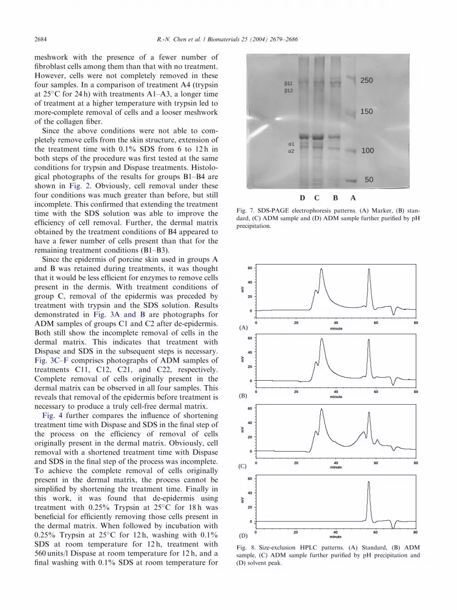
similar structural pattern of collagen fibers for ADMsamples obtained at each major step of the processingconditions can be observed in Fig. 6B–D in sequencewith that of no treatment. This indicates that character-istics of collagen fiber in ADM samples obtained at eachmajor step of the processing conditions was preserved oraltered to a small extent.Fig. 7 shows the SDS-PAGE electrophoresis patterns
of the standard (B) and two collagen samples (ADMobtained from treatment C2 (C) and collagen furtherpurified by pH precipitation from ADM obtained fromtreatment C2 (D)). The collagen standard and the twoADM samples display the same bands, two a bands (a1and a2; MW ¼ 95; 000) and two b bands (b11 and b12;MW ¼ 19; 000). These results are consistent with thefact that the known helical structure of type I collagen iscomposed of three polypeptide chains of a1; a1; and a2;and the ADM samples contain type I collagen in greatabundance.Compared to the standard (A) using the size-
exclusion HPLC method, both collagen in the ADMobtained from treatment C2 (B) and that in the ADMfurther purified by pH precipitation of ADM obtainedfrom treatment C2 (C) showed two typical componentpeaks eluting serially from the column, which wereidentified as oligomers and monomers (Fig. 8). Collagenoligomers represent intermolecular crosslinking of na-tive collagen molecules. After processing of the skin,some of the crosslinks of collagen still remain. Hence,the ADM preparations generally contain a mixture ofmonomers, dimers, and higher oligomers.The biocompatibility tests were carried out for ADMs
obtained from treatment C2 by co-culture with fibro-blasts (3T3) for 24 and 48 h (Fig. 9). For all materials,microscopic observations show that fibroblasts adheredas rapidly to all materials as they did to the control. Nosignificant morphologic changes were observed in thosecells in contact with ADM obtained from treatment C2for all studied time periods.
4. Conclusions
Optimum process conditions were defined with theability to completely remove all cellular components andmaintain the ECM structure of porcine dermis. Exam-inations by SEM and TEM and analysis by SDS-PAGEand HPLC size-exclusion revealed that the ADMobtained with optimal processing conditions retainedmost of the original type I collagen. In the in vitro
biocompatibility test, there were no significant morpho-logical changes observed for those fibroblast cells (3T3)in contact with the ADM obtained from these optimalprocessing conditions. For safety issues in biomedicalapplications, this material should be further subjected toin vivo biocompatibility and biodegradability tests.
Acknowledgements
Financial support from the National Science Councilof the ROC is highly appreciated (NSC 91-2320-B038-035).
References
[1] Wainwright DJ. Use of an acellular allograft dermal matrix
(Alloderm) in the management of full-thickness burns. Burns
1995;21:243–8.
[2] Chaplin JM, Costantino PD, Wolpoe ME, Bederson JB, Griffey
ES, Zhang WX. Use of an acellular dermal allograft for dural
replacement: an experimental study. Neurosurgery 1999;45:320–7.
[3] Sclafani AP, Jacono AA, McCormick S, Cocker R, Parker A.
Evaluation of acellular dermal graft in sheet (Alloderm) and
injectable (micronized Alloderm) forms for soft tissue augmenta-
tion. Arch Facial Plast Surg 2000;2:130–6.
[4] Srivastava A, DeSagun EZ, Jennings LJ, Sethi S, Phuangsab A,
Hanumadass M, Reyes HM, Walter RJ. Use of porcine acellular
dermal matrix as a dermal substitute in rates. Ann Surg
2000;233:400–8.
[5] Abraham GA, Murray J, Billiar K, Sullivan SJ. Evaluation of the
porcine intestinal collagen layer as a biomaterial. J Biomed Mater
Res 2000;51:442–52.
[6] Walter RJ, Matsuda T, Reyes HM, Walter JM, Hanumadass M.
Characterization of acellular dermal matrices (ADMs) prepared
by two different methods. Burns 1998;24:104–13.
[7] Livesey SA, Herndon DN, Hollyoak MA, Atkinson YH, Nag A.
Transplanted acellular allograft dermal matrix. Potential for the
reconstruction of viable dermis. Transplantation 1995;60:1–9.
[8] Takami Y, Matsuda T, Yoshitake M, Hanumadass M, Walter
RJ. Dispase/detergent treated dermal matrix as a dermal
substitute. Burns 1996;22:182–90.
[9] Takami Y, Matsuda T, Hanumadass M, Sakurai M, Walter RJ.
Acellular allogenic dermal matrix as a dermal substitute. Proc Int
Conf Plast Reconstruct Aesthet Surg 1995;55:212–5.
[10] Lee Y, Chen MY, Kuo CY, Chang HC. Preparation of porcine
dermal matrix graft. Global Chinese Symposium on Biomaterials
and Controlled Release; 1999. p. 584–7.
[11] Badylak SF, Matheny R, Obermiller J, Geddes LA. ECM scaffold
for myocardial reconstruction. Symposium on Tissue Engineering
Science. Aegean Conferences Series, vol. 4, 2002. p. 65.
[12] Badylak SF, Ling F, Record R, Hodde J. Vascularization of
3-dimensional scaffolds. Symposium on Tissue Engineering
Science. Aegean Conferences Series, vol. 4, 2002. p. 63.