prevalence, vascular distribution and multi-territorial extent of subclinical atherosclerosis in a...
TRANSCRIPT
DOI: 10.1161/CIRCULATIONAHA.114.014310
1
Prevalence, Vascular Distribution and Multi-territorial Extent of
Subclinical Atherosclerosis in a Middle-Aged Cohort:
The PESA (Progression of Early Subclinical Atherosclerosis) Study
Running title: Fernández-Friera et al.; Multi-territorial extent of early atherosclerosis
Leticia Fernández-Friera, MD, PhD1,2; José L. Peñalvo, PhD1; Antonio Fernández-Ortiz, MD, PhD1,3; Borja Ibañez, MD, PhD1,3; Beatriz López-Melgar, MD1,2; Martín Laclaustra, MD, PhD1; Belén Oliva, MSc1; Agustín Mocoroa, MD4; José Mendiguren, MD4; Vicente Martínez de Vega,
MD5; Laura García, BSc1; Jesús Molina, BSc1; Javier Sanchez-Gonzalez, PhD6; Gabriela Guzman, MD, PhD1,7; Juan C. Alonso-Farto, MD, PhD8; Eliseo Guallar, MD, PhD9; Fernando Civeira, MD, PhD10; Henrik Sillesen, MD, DMSc1,11; Stuart Pocock, PhD1,12; José M. Ordovás, PhD1,13; Ginés
Sanz, MD, PhD1; Luis Jesús Jiménez-Borreguero, MD1,14; Valentín Fuster, MD, PhD1,15
1Centro Nacional de Investigaciones Cardiovasculares Carlos III (CNIC), Madrid, Spain; 2Hospital Universitario Montepríncipe, Madrid, Spain; 3Hospital Clínico San Carlos, Universidad
Complutense, Madrid, Spain; 4Banco de Santander, Madrid, Spain; 5Hospital Universitario Quirón, Madrid, Spain; 6Philips Healthcare, Madrid, Spain; 7Hospital Universitario La Paz, Madrid, Spain;
8Hospital General Universitario Gregorio Marañón, Madrid, Spain; 9Johns Hopkins University Bloomberg School of Public Health, Baltimore, MD; 10Lipids Unit and Molecular Research Laboratory, Hospital Universitario Miguel Servet, Instituto Aragonés de Ciencias de Salud,
Zaragoza, Spain; 11Rigshospitalet, University of Copenhaguen, Copenhaguen, Denmark; 12London School of Hygiene & Tropical Medicine, London, UK; 13US Department of Agriculture Human Nutrition Research Center on Aging, Tufts University, Boston, MA; 14Hospital Universitario La
Princesa, Madrid, Spain; 15Mount Sinai School of Medicine, New York, NY
Correspondence:
Valentin Fuster, MD, PhD
Icahn School of Medicine at Mount Sinai
One Gustave L. Levy Place, Box 1030
New York, NY 10029’
Tel: 212-241-3852
Fax: 212-423-9488
E-mail: [email protected].
Journal Subject Codes: Atherosclerosis:[135] Risk factors, Diagnostic testing:[124] Cardiovascular imaging agents/techniques
Sanz, MD, PhD ; Luis Jesús Jiménez Borreguero, MD ; Valentín Fuster, MD,,, PPhD
Centro Nacional de Investigaciones Cardiovasculares Carlos III (CNIC), Madrid, SSSpapapaininin;;; 222HoHoHospspspitititaaaaUniversitario Montepríncipe, Madrid, Spain; 3Hospital Clínico San Carlos, Universidad
Complutense, Madrid, Spain; 4Banco de Santander, Madrid, Spain; 5Hospital Universitario QuirónMaadrdrdrididid, SpSpSpaiaiain; 666PhPhPhilips Healthcare, Madrid, Spaiaiainnn; 777Hospital Universitarararioioio La Paz, Madrid, Spain;a
888HHoHospital GGGeneral Universitario Gregorio Maraññónnn, Madrid, Spapapaiiin; 9Johns Hopkpp ins University Bloomberg SSSchhooololol ofofof PPPububublililiccc HeHeHealala ththth, Baltltltimmmoree, MMMD;D;D; 101010LLiL pppidsdsd UUU iniit t ananandd MoMoMolelelecuuulalalar r r ReReReseseearararchchch Laboratory, HoHHospiittaal Uniniivvversitario MMigueeel Servveeet, Innnstststitititutuu ooo Arrraggonésss dde CiCiCienciaaas ddde Saaalud,,,
ZaZaZarararagogogoza, SpSpSpaain;;; 11Rigsgsgshhoh sppititi alalaletete ,,, UnUU ivvvererersitytyty ooof CCoC pppennnhagagagueueuen,n,n, CCopopopenennhaaaguuuennn, ,, DDDenmmmaaark; 1222LonnndoonoSchohohoololol ooofff Hygigiene e & && TTrTropicaalll MMeMediciiinenene, LoLoLondndono , UKUKUK;; 13133USUSUS DDepparaartmt entt ofo AAAgrgrgriciciculturer HHHumuman NNNutttritititiiion RRResear hhch CCCenttter on AAA iiging, TTT ffufttts UUU iiniversititity, BBBosttton, MMMAAA; 141414HHHospititit llal UUU iiniversitititariiio LLLa
15
by RAFAEL PEINADO on May 15, 2015http://circ.ahajournals.org/Downloaded from by RAFAEL PEINADO on May 15, 2015http://circ.ahajournals.org/Downloaded from by RAFAEL PEINADO on May 15, 2015http://circ.ahajournals.org/Downloaded from by RAFAEL PEINADO on May 15, 2015http://circ.ahajournals.org/Downloaded from by RAFAEL PEINADO on May 15, 2015http://circ.ahajournals.org/Downloaded from by RAFAEL PEINADO on May 15, 2015http://circ.ahajournals.org/Downloaded from by RAFAEL PEINADO on May 15, 2015http://circ.ahajournals.org/Downloaded from by RAFAEL PEINADO on May 15, 2015http://circ.ahajournals.org/Downloaded from by RAFAEL PEINADO on May 15, 2015http://circ.ahajournals.org/Downloaded from by RAFAEL PEINADO on May 15, 2015http://circ.ahajournals.org/Downloaded from by RAFAEL PEINADO on May 15, 2015http://circ.ahajournals.org/Downloaded from by RAFAEL PEINADO on May 15, 2015http://circ.ahajournals.org/Downloaded from by RAFAEL PEINADO on May 15, 2015http://circ.ahajournals.org/Downloaded from by RAFAEL PEINADO on May 15, 2015http://circ.ahajournals.org/Downloaded from by RAFAEL PEINADO on May 15, 2015http://circ.ahajournals.org/Downloaded from by RAFAEL PEINADO on May 15, 2015http://circ.ahajournals.org/Downloaded from
DOI: 10.1161/CIRCULATIONAHA.114.014310
2
Abstract
Background—Data are limited regarding the presence, distribution and extent of subclinical
atherosclerosis in middle-aged populations.
Methods and Results—The PESA (Progression of Early Subclinical Atherosclerosis) study
prospectively enrolled 4184 asymptomatic participants aged 40-54 years (mean age 45.8 years,
63% male) to evaluate the systemic extent of atherosclerosis in the carotid, abdominal aortic and
ilio-femoral territories by 2D/3D ultrasound and coronary artery calcification (CAC) by
computed tomography. The extent of subclinical atherosclerosis, defined as presence of plaque
or CAC 1, was classified as focal (one site affected), intermediate (2-3 sites) or generalized (4-6
sites) after exploring each vascular site (right/left carotids, aorta, right/left ilio-femorals and
coronary arteries). Subclinical atherosclerosis was present in 63% of participants (71% of men;
48% of women). Intermediate and generalized atherosclerosis was identified in 41%. Plaques
were most common in the ilio-femorals (44%), followed by carotids (31%) and aorta (25%),
while CAC was present in 18%. Among participants with low Framingham Heart Study (FHS)
10-year risk, subclinical disease was detected in 58%, with intermediate or generalized disease in
36%. When assessing longer-term risk (30-year FHS), 83% of participants at high-risk had
atherosclerosis, with 66% classified as intermediate or generalized.
Conclusions—Subclinical atherosclerosis was highly prevalent in this middle-aged cohort, with
nearly half the participants classified as having intermediate or generalized disease. Most
participants at high FHS risk had subclinical disease; nonetheless, extensive atherosclerosis was
also present in a substantial number of low-risk individuals, suggesting added value of imaging
for diagnosis and prevention.
Clinical Trial Registration Information—ClinicalTrials.gov. Identifier: NCT01410318.
Key words: atherosclerosis, imaging, plaque, risk stratification
coronary arteries). Subclinical atherosclerosis was present in 63% of participants (((717171%% ofofof mmmenenen;
48% of women). Intermediate and generalized atherosclerosis was identified in 41411%.%.%. PPPlalalaquququeseses
were most common in the ilio-femorals (44%), followed by carotids (31%) and aorta (25%),
whilllee e CACACACCC waww s prprprese ent in 18%. Among participppananantstt with low Framinggghahaham Heart Study (FHS)
1000-yyyear risk, susubcbcbclill ninicacacal ll dididisess asasseee wawawas dededetetet ctcttededed in 558%%%, wwwititith h h inininteeermrmrmededediaiatetete ooor geeenenenerararalilil zed d d dididiseseeasasase ee in
3633 %%.% When asseesssing loongerrr-ttterm riskkk (330-yyyeaar FHHHS), 838383%%% ooof paaarttticipaaannts aaat hhigh---risssk hhaaad
atheheherororoscscsclelelerororosssis,,, wwwititith h 666666% %% clasasassisisifififiededed as s ininntetetermrmrmededediaaiateee oor rr gegegeneneneraaalililizezeed.dd d
CoCConcnclullusiisiononss—SuSSubcbcbclililinininicacacalll atatatheheherororoscscsclelelerororosisisisss waawasss hihihighghghlylly ppprererevalalalenenenttt ininin ttthihihisss mimimiddddddlelele aa-agegegeddd cococohohohortrtrt, wiiwiththth
by RAFAEL PEINADO on May 15, 2015http://circ.ahajournals.org/Downloaded from
DOI: 10.1161/CIRCULATIONAHA.114.014310
3
The natural history of atherosclerosis involves a protracted subclinical phase, with disease often
detected only at an advanced stage or following a cardiovascular (CV) event. This is of particular
importance because CV events are often fatal, and many deaths attributable to coronary artery
disease are sudden1. There is thus a clear need to identify disease at an early stage, and as a result
primary prevention forms the cornerstone of management. Currently, risk stratification scores rely
on the presence of identifiable risk factors and levels of biochemical markers. However,
conventional risk assessment has well-recognized limitations, notably in lower-risk groups such as
women and younger people2, 3. Detection of atherosclerosis in its subclinical stage may help
identify strategies to arrest disease development. Indeed, the significance of subclinical carotid
atherosclerosis and coronary artery calcification (CAC) in relation to clinical outcomes has been
established in the MESA study4-6 and in the recently published US High Risk Plaque Study7.
The introduction of noninvasive imaging techniques has unlocked the potential to evaluate
atherosclerosis in asymptomatic populations. Specific imaging modalities include vascular
ultrasound, computed tomography (CT), and magnetic resonance imaging8, 9. Many imaging
studies evaluated individual vascular territories, but given the systemic nature of atherosclerosis a
multi-territory analysis has the potential to provide a more comprehensive overview of the
distribution and burden of atherosclerosis.
The PESA (Progression of Early Subclinical Atherosclerosis) study evaluates atherosclerosis
in the carotid, aortic, coronary and ilio-femoral territories using accessible noninvasive imaging
techniques in asymptomatic middle-aged individuals10. Through the evaluation of multiple vascular
beds in relatively young adults we aim to improve understanding of the origin and progression of
atherosclerosis. Here, we present the prevalence, vascular distribution and extent of subclinical
atherosclerosis in the PESA cohort and their relation to CV risk algorithms.
atherosclerosis and coronary artery calcification (CAC) in relation to clinical outcomomomeseses hhhasasas bbbeeeeeen nn
established in the MESA study4-6 and in the recently published US High Risk Plaque Study7.
ThThTheee ininintrodododuuuction of noninvasive imaging tetetechchchniques has unlockededed ttthe potential to evaluate
atatatheeerosclerosiis ininin aasyyympmpmptototomamamatititicc c popopopupupulalalations.s SSSpep ciifiiic immmaaaginininggg momomodadadalililititiieese incllludududee vavavascscscululularaa
ullltrtrtrasasasound, cococomppputtted tttoomo oggrrraphphphy yy (C(C(CT)T)), ana d d d mmmagnnetttic rreesesonnnannncecc imamamagigg nnng88, 9. MMaManyy iiimmmagiinngng
tttuddudieieiesss eveevalalaluaauateteteddd ininindididiviividuddualalal vasasascucculalalarrr teteterrrrrritititorororieieiesss, bbbuttt gigigiveevennn thththeee syssystststemememicicic nnnatataturrureee ofofof aaathththerererosososclclclerererosososisisis aaa uu
by RAFAEL PEINADO on May 15, 2015http://circ.ahajournals.org/Downloaded from
DOI: 10.1161/CIRCULATIONAHA.114.014310
4
Methods
Study sample
The rationale and design of the PESA study has been described10. Briefly, PESA-CNIC
Santander is a prospective cohort study of asymptomatic employees of the Santander Bank in
Madrid, aged 40 to 54 years and consecutively recruited between June 2010 and February 2014.
Participants with prior CV disease and any condition reducing life expectancy or affecting study
adherence were not included. Participants were examined at baseline by ankle-brachial index
(ABI), vascular ultrasound and non-contrast CT, and will be followed up at 3- and 6-years. In
addition, each visit includes clinical interviews, physical examination, fasting blood draw, urine
sample, and a 12-lead electrocardiogram. The study protocol has been approved by the Instituto
de Salud Carlos III Ethics Committee and all eligible participants have provided written
informed consent.
Traditional CV risk factors were determined from blood samples and interviews, as
follows: 1) diabetes: fasting plasma glucose 126 mg/dL, or treatment with insulin or oral
hypoglycemic medication11; 2) arterial hypertension: systolic blood pressure 140 mmHg,
diastolic blood pressure 90 mmHg, or use of antihypertensive medication11; 3) dyslipidemia:
total cholesterol 240 mg/dL, LDL-cholesterol 160 mg/dL, HDL-cholesterol <40 mg/dL, or use
of lipid-lowering drugs12; 4) smoking: current smoking status, or a lifetime consumption of >100
cigarettes8, 13; 5) family history of CV disease: first degree-relative diagnosed with
atherosclerosis before the age of 55 years in men and 65 in women14. Obesity, defined as body
mass index 30 kg/m2, was also assessed14. CV risk was evaluated by the 10-year risk of
coronary heart disease and the 30-year risk of CV disease from the Framingham Heart Study
(FHS)13, 15, and additionally by the European Society of Cardiology’s SCORE (Systematic
ample, and a 12-lead electrocardiogram. The study protocol has been approved bybyby ttthehehe InInInstststitititutututo
de Salud Carlos III Ethics Committee and all eligible I participants have provided written
nfoormrmrmededed ccconononsentntnt.
Traditiooonnnall CVCVCV rrrisisiskkk fafafactctctororrs ss wewewerrer detettererermmim neddd frooommm blblb ooood dd sasaampmpmpleleles and d d ininintetetervrvrvieieiewswsws, asasas
fooollll owowows: 1) dididiaba eete eese : ffafassts ing plplplasaa mamaa gluuuccoc seee 126 mmmg/d/d/dLL,L, ooor ttrtreaeaeatmtmtmenennt wiwwith iiinsnsnsulinnn ooor orral
hyhhypopopoglglglyccycemememicicic mmmedededicicicatatatioioionnn1111;;; 2)2)2) aaartrtrterereriaiaialll hyhhypepepertrtrtenenensisisiononon::: sssyssystototolililiccc blblblooooooddd prprpresesessussurerere 141414000 mmmmmmHgHgHg,
by RAFAEL PEINADO on May 15, 2015http://circ.ahajournals.org/Downloaded from
DOI: 10.1161/CIRCULATIONAHA.114.014310
5
COronary Risk Evaluation), which calculates 10-year risk of fatal CV disease14, using the low
CV risk charts applicable to Spain. FHS scores were classified as low (<10%), moderate (10 to
20%) or high risk (<20%) and the SCORE risks as low (<1%) and moderate-high ( 1%). In light
of recent US guidelines for statin therapy16, we also calculated 10-year risk using the
atherosclerotic cardiovascular disease (ASCVD) algorithm, an atherosclerotic risk calculator
based on Pooled Cohort Equations9, and cut-off values were defined as <5%, 5 to <7.5% and
7.5 risk.
Vascular ultrasound imaging
The 2D/3D vascular ultrasound protocol has been described7. Presence of atherosclerotic plaques
was assessed by cross-sectional sweep of carotids, infra-renal abdominal aorta and ilio-femoral
arteries. Plaque was defined as a focal protrusion into the arterial lumen of thickness >0.5 mm or
>50% of the surrounding intima-media thickness (IMT), or a diffuse thickness >1.5 mm
measured between the media-adventitia and intima-lumen interfaces7, 17. Semi-automated
detection of carotid and femoral IMT was also assessed (details in supplemental material).
Ultrasound studies were analyzed with QLab9 (Philips Healthcare, Bothel, WA, USA) at the
CNIC Core Imaging Laboratory7. Imaging quality was evaluated as optimal, suboptimal or non-
interpretable, and inclusion of studies was determined by consensus. Good reproducibility was
found for presence of plaque in all territories (kappa = 0.75 for carotids, 0.89 for aorta, 0.88 for
ilio-femorals). The ABI was calculated as the ratio of systolic blood pressure in the posterior
tibial artery to systolic blood pressure in the brachial artery using Doppler ultrasound and a
standard sphygmomanometer. ABI values <0.9 were considered abnormal18.
Coronary artery calcification by computed tomography
CAC was detected with a 16-slice CT scanner (Philips Brilliance, Philips Healthcare, Andover,
was assessed by cross-sectional sweep of carotids, infra-renal abdominal aorta andndnd iiilililio-o-o-fefefemomomorararal
arteries. Plaque was defined as a focal protrusion into the arterial lumen of thickness >0.5 mm or
>50%0%% ooof ff thththeee sus rrrrououunding intima-media thicknesssss (I(I(IMTMM ), or a diffuse thhhicicicknkk ess >1.5 mm
mmmeaasa ured betweweweenene thehehe mmmededediaiaia-a-a-advdvdvenenentitititititia anddd innntimma---lummmeenen iiintntnterrfafafacececess7, 7, 7, 171717. Semimimi--a-autututomomomatatatededed
deeetetetectctc ion offf cccarooto iddd andndnd femmoororalala IMTMTMT wwwaasa aalllsooo asssessseeded (((deeetaaaillss ininin supupu pppleemeeenttatal mmamatteteriaal))).
UlUlUltrtrtrasasasouooundndnd ssstuttudididieseses wererereee anananalalalyzededed wititithhh QLQLQLababab999 (P(P(Phihihilililipspsps HHHeaeaealtltlthchchcararareee, BBBototothehehelll, WWWAAA, UUUSASASA))) atatat ttthehehe
by RAFAEL PEINADO on May 15, 2015http://circ.ahajournals.org/Downloaded from

DOI: 10.1161/CIRCULATIONAHA.114.014310
6
MA, USA) using non-contrast prospective electrocardiography-gated acquisition. Estimated
absorbed radiation was 0.6-1.2 mSv. CAC score (CACS) was calculated by the Agatston method
and graded as 1-99, 100-399 and 4004. Three trained technicians blinded to other imaging
results quantified CACS supervised by experienced physicians.
Definition of subclinical atherosclerosis
Subclinical atherosclerosis was defined as the presence of atherosclerotic plaques in the carotid,
aortic or ilio-femoral territories or CACS 1. The multi-territorial extent of subclinical
atherosclerosis was defined according to the number of vascular sites affected (right carotid, left
carotid, abdominal aorta, right ilio-femoral, left ilio-femoral and coronary arteries). Participants
were classified as disease-free (0 vascular sites affected) or having focal (1 site), intermediate (2-
3 sites) or generalized atherosclerosis (4-6 sites).
Statistical analysis
Baseline characteristics were calculated using mean and standard deviation for continuous
variables and count and proportions for categorical variables. Differences between continuous
variables and categorical variables were tested with unpaired t tests and chi-square tests,
respectively. Non-normally distributed variables (triglycerides, fasting glucose, HbA1c, CACS
and CV risk scales) were log-transformed before analysis to normalize the distribution. Age and
gender-adjusted associations between vascular disease in each territory were examined using
logistic regression models. The reproducibility of ultrasound measurements was studied by
replicating the analysis of a random sample of 60 studies 3 months after the initial assessment
and Cohen’s Kappa was used for the agreement analysis. Statistical analyses were conducted
using Stata 12 (StataCorp, College Station, TX, USA).
were classified as disease-free (0 vascular sites affected) or having focal (1 site), iiintntntererermememedididiatatateee (2-
3 sites) or generalized atherosclerosis (4-6 sites).
Stattisisistititicacacal l l anananalyyysisii
BBBasses line charactctcterristititicscsc wwwererereee cacacalclclculululatata edee usiiingngng meanann andndnd sstatatandndndara d dd dededeviviviaaatioon fofofor r r cooontntntinii uououoususus
vaaaririr ababables annnd d d counununt annnd dd proppporrrtitt onnnsss fooor cccateeegoooricaal vararriaaableees.. DiDiD ffffffeererenee cccess beeetwwween n n cococontininnuous
vaavariririababablelelesss anananddd cacacatetetegogogoririricacacalll vaavariririababablelelesss weewererere tttesesesteteteddd wiiwiththth unpnpnpaiaiairerereddd ttt tttesesestststs aaandndnd ccchihihi ss-squqquararareee tetetestststsss, ttt
by RAFAEL PEINADO on May 15, 2015http://circ.ahajournals.org/Downloaded from
DOI: 10.1161/CIRCULATIONAHA.114.014310
7
Results
The PESA cohort comprised 4184 participants (78% of the eligible population). At the time of
publication, 34 individuals (0.8%) had discontinued the study and 84 (2%) were either excluded
for missing data or were pending evaluation, resulting in 4066 participants available for analysis.
After excluding non-interpretable images (64 participants, 1.5%), the sample available for
imaging analysis was 4002 (98% of the cohort). Only 1 case of abnormal ABI was detected in
the first 2536 participants, and therefore the protocol was amended to discontinue measurement
of ABI in subsequent examinations. Baseline demographic characteristics and CV risk factors
are summarized in Table 1. The average age of participants was 45.8 years, 63% were male and
99.9% Caucasian. The most prevalent traditional risk factor was dyslipidemia (42%), followed
by smoking (21%), family history (16%), hypertension (12%), and diabetes (2%). Additionally,
obesity was found in 15% of PESA participants. The presence of traditional risk factors and
obesity was higher in men, with the exception of smoking (23% women and 19% men) and
family history (17% women and 16% men). Most participants (62%) had at least one traditional
risk factor, 18% had two and 5% had three or more. Aside from family history, the prevalence of
traditional risk factors increased with age except for smoking and diabetes in women (Table S1).
Risk-factor distribution was not significantly different in the participants not included in the
imaging analysis (1.5%), thus excluding systematic bias.
Prevalence, vascular distribution and extent of subclinical atherosclerosis
Prevalence of subclinical atherosclerosis (presence of plaque or CACS 1) was 63%. Plaques
were detected by ultrasound in 60% of participants (31% in the carotids, 25% in the aorta and
44% in the ilio-femoral arteries) and 18% had CAC (CACS 1-99 in 14%, 100-399 in 3% and
400 in 0.7%). In men, subclinical atherosclerosis was more prevalent (71% vs. 48% in women)
99.9% Caucasian. The most prevalent traditional risk factor was dyslipidemia (4222%)%)%), fofofollllllowowowededed
by smoking (21%), family history (16%), hypertension (12%), and diabetes (2%). Additionally,
obesssititity y y wawawasss foff unnnddd in 15% of PESA participants... TTThehh presence of tradititiiononnal risk factors and f
obobobeese ity was hih ghghgherr in nn mememen,n,n, wwwititithhh thththeee exxxceepttiiiononon of smsmmokkkininingg (2(2(23%3%3% wwwomomomenenen aandd 1119%9%9% mmmenenen))) annnd dd
faaamimimilylyly histoooryryry (171 %% % wowowomen n annnd dd 1666%%% mememen)))... MMMostt pppartititiccicipapaanntnts (6(662%%%) ) ) haaadd att lleaeaeast ooonenene traaddditionnnaal
iiisksksk fffacacactototorrr, 1118%8%8% hhhadadad tttwoowo aaandndnd 555%%% hahahaddd thththrerereeee ororor mmmorororeee. AAAsisisidedede fffrororommm fafafamimimilylly hhhisisistototoryrry, thththeee prprpreveevalalalenenencecece oooffff
by RAFAEL PEINADO on May 15, 2015http://circ.ahajournals.org/Downloaded from
DOI: 10.1161/CIRCULATIONAHA.114.014310
8
across all vascular territories, with differences most pronounced in the ilio-femoral and coronary
arteries (Figure 1). Of the 23 participants with CACS 400, only one was female (Table S1).
Atherosclerosis prevalence increased with age for both genders and across all vascular territories,
and only the presence of aortic disease was found to be independent of gender (Figure 2).
Associations between subclinical atherosclerosis in different territories and each individual risk
factor are shown in Table S2 (details in supplemental material).
The presence of ilio-femoral disease was more strongly correlated with aortic disease and
CAC than with carotid disease (Table 2). Furthermore, having disease in the ilio-femorals
corresponds to a 70% probability of finding disease in any other territory explored. Conversely,
the absence of plaque in the ilio-femorals confers a 67% probability of being disease-free in the
other vascular territories. In participants with carotid disease, the odds ratio for co-existing
plaque was slightly higher in the aortic and ilio-femoral territories compared with CAC, and the
probability of subclinical atherosclerosis in any other territory was 72%, with a negative
predictive value of 55%. Regarding participants with CACS 1, 87% had plaques present at other
vascular sites, whereas 54% of participants with CACS=0 had plaques in other territories (37%
in ilio-femorals, 27% in carotids and 20% in aorta).
Classification of participants according to the extent of atherosclerosis showed focal
disease in 21%, intermediate in 28% and generalized in 13%. The prevalence of generalized
atherosclerosis was greater in men, increased with age, and was related to the presence of obesity
and traditional risk factors (Figure S1, Table S3). Notably, the extent of subclinical
atherosclerosis in men aged 40-44 years was similar to that in women 5 to 10 years older (Figure
S1). Among those participants having focal disease, the ilio-femoral was the most likely territory
to be affected, independently of age, gender or risk profile (Table S4). In an attempt to highlight
he absence of plaque in the ilio-femorals confers a 67% probability of being diseaeaeasesese-f-f-frereree e e ininin ttthehh
other vascular territories. In participants with carotid disease, the odds ratio for co-existing
plaqqqueueue wwwasasas ssslill ghhhtltltlyyy higher in the aortic and ilio-fefefemomm ral territories commmpapapared with CAC, and the
ppprobbbability off sssubububcclininniiicalalal aaathththerereroososclclcleererosossiss in ananany ottheer teteterrrrrritititororory yy wwwasss 727272%%%, witth h h aaa nenenegagagatiiveveve
prrredededicici tive vvalalalue off f 55%.%% Regggarrrdidd ngngng paaartititicipapapannnts wwittth CCCAACACSSS 1,11 8887%7%7% hadaad plaaaququques pppreeesennt at oothher
vaavascscscullulararar sssitititeseses, whhwhererereaeaeasss 545454%%% ofofof pppararartititicicicipapapantntntsss wiiwiththth CCCACACACSSS=000 hahahaddd plplplaqaqaqueeuesss ininin otototheheherrr teteterrrrrritititorororieieiesss (3(3(37%7%7%
by RAFAEL PEINADO on May 15, 2015http://circ.ahajournals.org/Downloaded from
DOI: 10.1161/CIRCULATIONAHA.114.014310
9
differences between participants with atherosclerosis in a single territory (focal disease) and
those with multiple territories affected (intermediate or generalized disease) we performed a
subgroup analysis, finding a significantly higher prevalence for all traditional risk factors among
those participants with multi-territorial atherosclerosis (Table 3).
CV risk scales and subclinical atherosclerosis
Mean FHS 10-year score in the PESA cohort was 6%, and most participants (85%) were at low-
risk, compared with 14% at moderate risk and 1% at high risk. Similarly, most individuals (85%)
were at low-risk by the European SCORE compared with 15% at moderate-high risk. To assess
longer-term risk, FHS 30-year score was calculated, yielding a mean value of 18% and higher
proportions of participants at moderate and high-risk (30% and 35%, respectively).
The relationship between CV risk scales and subclinical atherosclerosis is shown in
Figures 3 and 4. Among participants at low 10-year FHS risk, 58% had subclinical
atherosclerosis, including 36% with intermediate or generalized disease. Most FHS high-risk
participants (95%) had atherosclerosis, with intermediate or generalized disease in 86%.
Similarly, among participants with a low European SCORE, 58% had subclinical atherosclerosis,
including 35% with intermediate or generalized disease, and almost 80% of moderate-high risk
participants had intermediate or generalized disease. Given the low numbers of individuals at
high-risk using the 10-year FHS scale (55 participants), we also examined the relationship
between atherosclerosis and 30-year FHS score. Among individuals at high 30-year FHS risk,
83% had subclinical atherosclerosis, with intermediate or generalized disease in 66%, whereas
among low-risk individuals, 43% had atherosclerosis and 22% had intermediate or generalized
disease. Such associations were also observed for each vascular territory analyzed separately
(Figures S2, S3 and S4).
proportions of participants at moderate and high-risk (30% and 35%, respectively)y)y).
The relationship between CV risk scales and subclinical atherosclerosis is shown in
Figuguurereres ss 333 ananand 44. AmAA ong participants at low 10-y-y-yeaeaear FHS risk, 58% haaad d d suss bclinical
atatatheeerosclerosiis, iiinccluuudddingngng 3336%6%% wwititithhh inininteermedee iiai te orrr geeeneneneraraalililizzzed dd didiiseseseasasase.ee Mososst t t FHFHFHSSS hihih ghghgh-rrrisisiskk k
paaartrtr icicicippantss (((9599 %)%)%) haddd aaatherrrosososccclerooosiss s,,, wwwith h h innntermmeeediaiaiattete oorrr gegegeneneeraaaliiizezz ddd ddiseeeassse inn 88866%6%.
SiSiSimimimilalalarlrlrly, aaamomomongngng pppararartititicicicipapapantntntsss wiiwiththth aaa lllowoow EEEurruropopopeaeaeannn SSSCOCOCORERERE, 585858%%% hahahaddd sussubcbcbclililinininicacacalll atatatheheherororoscscsclelelerororosisisissss
by RAFAEL PEINADO on May 15, 2015http://circ.ahajournals.org/Downloaded from

DOI: 10.1161/CIRCULATIONAHA.114.014310
10
A further analysis was performed to explore subclinical atherosclerosis according to the
10-year ASCVD risk algorithm16. The prevalence of the ASCVD risk subgroups across the
PESA sample was 11%, 79% and 10% for the <5%, 5 to <7.5% and 7.5 risk groups. The <5%
risk group had a significantly lower prevalence of subclinical atherosclerosis compared to the 5
to <7.5% and 7.5% risk groups (57% vs 80% vs 92%, p<0.001). Comparison of the <5%, 5 to
<7.5% and 7.5% risk groups by vascular territory revealed carotid disease in 29% vs 42% vs
57%, aortic disease in 21% vs 35% vs 42%, CACS 1 in 12% vs 33% vs 45% and ilio-femoral
disease in 37% vs 64% vs 79% (p<0.001 for all comparisons). Figure 5 shows the relation
between the extent of atherosclerosis and the groups who meet the AHA/ACC criteria for statin
treatment ( 7.5% ASCVD risk, diabetes or LDL 190 mg/dL), those considered for statin (5 to
<7.5% ASCVD risk) and those not considered for statin (<5% ASCVD risk). Interestingly, the
prevalence of generalized disease increased with higher ASCVD risk score (8% vs 24% vs 37%
in the <5%, 5 to <7.5% and 7.5% risk groups, respectively).
Discussion
The main findings from the PESA cohort are as follows: 1) Subclinical atherosclerosis is highly
prevalent in this middle-aged asymptomatic sample; 2) The ilio-femoral territory is the most
frequently affected vascular site in the early stages of atherosclerosis; 3) Most individuals
classified at high-risk by traditional scales (FHS and European SCORE) had subclinical
atherosclerosis, but atherosclerosis is also present in nearly 60% of participants classified at low
risk, with intermediate or generalized disease in one-third. Ongoing PESA follow-up over at
least 6 years will enable study of associations between subclinical disease evaluated at baseline
and subsequent CV events.
reatment ( 7.5% ASCVD risk, diabetes or LDL 190 mg/dL), those considered fffororor ssstatatatititin n n (5(5(5 ttto oo
<7.5% ASCVD risk) and those not considered for statin (<5% ASCVD risk). Interestingly, the
prevvvalalalenenencecece ooof ff geeeneneneralized disease increased withhh hhhigigigher ASCVD risk sscococore (8% vs 24% vs 37%
nnn thhhe <5%, 5 tooo <77.5%5%5% aaandndnd 7.7.7.5%5%5% rrrisisi k kk ggrouuupsss, reespppectititivvevelylyly).).).
DiDiDiscscscussussisisiononon
by RAFAEL PEINADO on May 15, 2015http://circ.ahajournals.org/Downloaded from

DOI: 10.1161/CIRCULATIONAHA.114.014310
11
Subclinical atherosclerosis is prevalent in middle-aged individuals
Few population studies have investigated the prevalence and extent of subclinical atherosclerosis
across multiple vascular sites in a middle-aged sample, despite atherosclerosis being a systemic
disease with a long latent subclinical phase. In the MESA and CARDIA studies, evaluation of
atherosclerosis was limited to carotid IMT and CAC3, 8. The ARIC study focused only on carotid
and popliteal territories assessed by ultrasound19. The Heinz Nixdorf Recall, High Risk Plaque,
and Rotterdam studies recruited older individuals with prior CV disease or a high-risk profile1, 7,
20. Using a multi-territory evaluation, the PESA study detects a high prevalence of subclinical
disease, with nearly half the participants classified as having intermediate or generalized disease,
despite being predominantly at low-risk according to traditional scales. This finding is probably
due to the examination of several territories, including vascular areas more susceptible to disease
such as the ilio-femoral arteries, which were not explored in earlier studies. Other studies that did
investigate multiple vascular sites included only men with at least one risk factor21, examined
participants at higher risk22, or explored fewer territories23.
An innovation of PESA is the early detection of atherosclerosis in other vascular
territories even in the absence of CAC. A zero CAC could be considered as indicative of absence
of disease, but among PESA participants with CAC=0, nearly 60% had plaques at other vascular
sites. Therefore, in this low-risk sample, absence of CAC does not necessarily indicate that a
participant is disease free. We hypothesize that by studying other vascular territories in subjects
with CAC=0, we could identify those who will develop CAC in the future and who would likely
benefit from more intensive preventive management. Although follow-up will be needed to
confirm this hypothesis, multi-territorial assessment using vascular ultrasound—a safe, cost-
efficient, reproducible and simple technique—might be especially important at early stages of
despite being predominantly at low-risk according to traditional scales. This findinining g g isisis ppprororobababablblbly
due to the examination of several territories, including vascular areas more susceptible to disease
uchhh aaas s s thththe e e ilililioii -fffememmoral arteries, which were not t t exexexplpp ored in earlier stududdieieies. Other studies that did
nnnveeestigate multltltiipiplle vvvasasascucuculalalar r r sisisitetetesss inininclclcluduuded dd onononly mmeeen wwwititithhh atatat leaeastss ooonenene rrrissk faaactctctoror212121, exexexamamaminininededed
paaartrtr icicicippantss aaatt t hiiighhher rrrisssk22, ororor eeexpxpxplololoreeed fef wwwerrr terrritttoriiiesss23.
AnAnAn iiinnnnnnovoovatatatioioionnn ofofof PPPESESESAAA isisis ttthehehe eaeaearlrlrly dededetetetectctctioioionnn ofofof aaathththerererosososccclelelerororosisisisss ininin ooothththererer vasasascucculalalarrr
by RAFAEL PEINADO on May 15, 2015http://circ.ahajournals.org/Downloaded from
DOI: 10.1161/CIRCULATIONAHA.114.014310
12
atherosclerosis in younger people with a high probability of zero CAC.
The added clinical value of a multi-territory vascular evaluation is supported by the
CAFES-CAVE study, showing that scanning only carotids or only femorals predicts 15% and
13% fewer events than examining both territories in a 10-year follow up24. This finding supports
the view that the wider sampling that comes from exploring several territories overcomes the
problem of not detecting a lesion when examining only one territory. The predictive value of
multi-territory imaging will be assessed in detail with the appearance of events during PESA
follow-up. The prognostic relevance of subclinical atherosclerosis is also supported by the
MESA study4-6 and the recently published US High Risk Plaque Study7, where strong
associations have been shown between CV events and subclinical carotid and coronary disease.
Similarly, the Northern Manhattan Study demonstrated that subclinical carotid plaque is a
precursor of CV events25. These studies highlight the potential value of evaluating subclinical
atherosclerosis in multiple territories for CV event prediction.
The association of increased subclinical atherosclerosis prevalence with male gender and
age is consistent with previous reports26, and may be related to the natural history of the disease.
In fact, the risk of atherosclerosis in men is similar to that in women aged five to ten years older
in this cohort. This discovery may assist in determining the most appropriate time window for
atherosclerosis screening and intensification of primary CV prevention. We interestingly found
that individuals at 7.5% ASCVD risk had substantial atherosclerosis compared with lower-risk
individuals, especially regarding CAC, and had a 4-fold higher prevalence of generalized
disease. This interesting finding is in line with the proposed intensification of statin treatment in
the most recent guidelines on the treatment of blood cholesterol16.
associations have been shown between CV events and subclinical carotid and corrronononararary y y dididiseseseasasasee.e
Similarly, the Northern Manhattan Study demonstrated that subclinical carotid plaque is a
preccururursososor r r ofofof CCCV evvvents25. These studies highlighhhtt t thththe potential value offf eeevavv luating subclinical
atatatheeerosclerosiis ininin mmululultttiplplplee e tteterrrrrritititooriririeseses fffororo CCCVVV eeevennt pppredddicicictititiononon.
The asasassoccic aata ionnn of incncncrereeasa ededed suuubcclc innniiicaaal attheeeroooscccleroroosssis ss pppreeevalaa ennncce wwwittth hh maaaleee geenddder anaand
agagageee isisis ccconononsisisistststenenenttt wiiwiththth ppprerereviiviouoousss rererepopoportrtrtsss2626, anananddd mamamay bebebe rrrelelelatatatededed tttooo thththeee nananatutturararalll hihihistststororory ofofof ttthehehe dddisisiseaeaeasesese.
by RAFAEL PEINADO on May 15, 2015http://circ.ahajournals.org/Downloaded from
DOI: 10.1161/CIRCULATIONAHA.114.014310
13
The ilio-femoral territory is the most frequently affected vascular site
The clear predominance of disease in the ilio-femoral arteries is possibly related to specific
patterns of shear stress and disturbed flow caused by the vessel curvature27. In PESA
participants, the presence of ilio-femoral disease increases the risk of concurrent CAC and is
predictive of disease elsewhere. Moreover, the absence of ilio-femoral disease is strongly
associated with the absence of atherosclerosis at other vascular sites. Thus, imaging of peripheral
arteries may be a useful population-wide screening tool for detecting atherosclerosis in its early
stages. Follow-up will be extremely valuable to clarify the impact of early detection of ilio-
femoral disease on the primary prevention of peripheral arterial disease, since advanced stages
are associated with higher risk of myocardial infarction and stroke28.
The ilio-femoral territory has traditionally not been examined as extensively as the
carotids and CAC. By including this territory in PESA, comparisons with other more commonly
examined vascular sites are available. Indeed, evaluation of the ilio-femoral arteries appears to
be more valuable than CAC for detecting subclinical atherosclerosis, given the high prevalence
(82%) of a zero CACS in this low-risk middle-aged sample, suggesting that CAC represents a
more advanced stage of disease. Notably, prevalence of abnormal ABI is low in PESA,
consistent with previous studies that found low prevalence in middle-aged individuals29-31. This
finding supports the idea that ABI adds little valuable information to the screening of subclinical
atherosclerosis in early stages of the disease.
Subclinical atherosclerosis and traditional CV risk scales
Current risk stratification strategies have successfully identified individuals at risk of CV events.
Traditional risk scales include the widely used FHS and SCORE, an adaptation of FHS that
avoids risk overestimation in European populations with less coronary heart disease14, 32.
are associated with higher risk of myocardial infarction and strokef 28.
The ilio-femoral territory has traditionally not been examined as extensively as the
carootititidsdsds aaandndnd CCCACCC.. By including this territory inn PPPESESE A, comparisons wwititith hh other more commonly nn
exexexamamamined vascuuulllarr sisiitetetess arararee e avavavaiaiailalalablblble.e IInndeeeed,d, evaaluuuationonon oof ff thththee ilii iooo ff-femememooral aaartrtrtererrieieiesss appppepp arararsss tototo
beee mmmoroo e valuluuabaa leee ttthannn CCAC C fofofor r r detetetectiiingg g suuubcbccliniiicaaal aaathhheroooscccleeeroroosssis,s,s, giiiveen ththt eee highghgh pprevaaalencccee
8882%2%2%))) ofofof aaa zerererooo CACACACSCSCS iiinnn thththisisis lllowoow rr-risisiskkk mimimiddddddlelele-agagagededed sssamamamplplpleee, sssugguggegegestststinininggg thththatatat CCCACACAC rrrepepeprereresesesentntntsss aaa
by RAFAEL PEINADO on May 15, 2015http://circ.ahajournals.org/Downloaded from
DOI: 10.1161/CIRCULATIONAHA.114.014310
14
However, the impact of these scales in younger, low-risk populations is limited, with many
individuals still suffering CV events and little success in promoting lifestyle changes3. In a
cohort of 122,458 patients with coronary disease, 9%-13% of those aged <55 years had no
conventional risk factors33, indicating disparity between traditional risk factors and the presence
of disease in younger populations. The limited value of CV risk assessment by FHS alone in
young healthy adults was raised in a recent study by Armstrong et al., which demonstrated
improved discrimination when risk classification includes left ventricular mass as an additional
independent predictor of CV disease34.
Although the FHS and the European SCORE scales were designed to assess risk of CV
events derived from atherosclerosis, and not the presence of subclinical atherosclerosis, we
aimed to complement these predictive models by comparing the presence and extent of
subclinical disease across different risk categories. Most individuals classified at high-risk have
subclinical atherosclerosis, with a high proportion having intermediate or generalized disease.
Nonetheless, subclinical atherosclerosis is also present in nearly 60% of PESA participants at
low risk, with one third having at least two sites affected. In this regard, a sub-study of the
MESA and CARDIA participants detected higher carotid IMT and higher CAC in individuals
with low 10-year but high lifetime risk compared with individuals with low 10-year and low
lifetime risk3. Together these results strongly suggest an association of atherosclerosis with
characteristics not considered in standard risk scales, and that will be the basis for further
investigation in PESA. We also propose that individuals presenting multi-territorial
atherosclerosis, despite being classified at low-risk, will be more likely to develop clinical
events. Future confirmation of this hypothesis in longitudinal follow-up could support the
broader application of multi-territorial imaging, a reasonable expectation given the systemic
events derived from atherosclerosis, and not the presence of subclinical atheroscllerererosososisisis, , , wewewe
aimed to complement these predictive models by comparing the presence and extent of
ubccclililinininicacacalll dididiseaasasee e across different risk categoriieseses. MMMost individuals claaassiss fied at high-risk have
uuubbcbclinical atheheerror ssclelelerororosisisis,s,s, wwwititithhh a a a hihihighghgh pproppporrrtionnn hhhavvinininggg ininintetetermmmedddiiaiatetee orr geenenenerraralililizezezed dididiseeeasasase.ee
NoNoNonenenetheless,s,s, subbbclllinicccalll athheeerosososclerereroosiiis iis alalalsooo prp esessenttt ininin nneaeaearllly y 6000%%% of PPESESSAAA partrtrticiccipananntts attt
ooow riririsksksk, wiiwiththth ooonenene ttthihihirdrdrd hhhavaavinininggg atatat llleaeaeaststst tttwoowo sssitititeseses aaaffffffecececteteteddd. IIInnn thththisisis rrregegegarararddd, aaa sssubbub ss-stuttudyddy ooofff thththeee
by RAFAEL PEINADO on May 15, 2015http://circ.ahajournals.org/Downloaded from

DOI: 10.1161/CIRCULATIONAHA.114.014310
15
nature of atherosclerosis. Multi-territory vascular imaging therefore appears to have the potential
to help identify new factors and thus complement traditional risk scales, helping to achieve the
goal of individualized risk assessment.
Limitations
This study presents a cross-sectional analysis of the PESA cohort at baseline, and therefore
cannot yet evaluate clinical events, precluding the possibility of establishing causality; these
current findings will be complemented by long-term monitoring of atherosclerosis progression.
Follow-up data from PESA will help to clarify the clinical significance of early detection of non-
obstructive disease, including ilio-femoral atherosclerosis, and the predictive value of multi-
territorial atherosclerosis in low-risk individuals. The present analysis of the PESA baseline
cohort sets the basis for understanding the relationship between the extent and progression of
subclinical disease and future CV events. The PESA sample consists of middle-aged,
predominantly male white-collar workers, which may limit the generalizability of the results.
Although the prevalence of disease might not be universally representative given the specific
characteristics of our participants, the observed associations between CV risk profile and the
presence and extent of atherosclerosis could be extrapolated to other cohorts. It is challenging to
assess whether the distribution of risk factors in PESA is similar to that in an age-gender
matched representative population because we included younger individuals than in most
previous population-based studies. The present results, however, will complement ongoing
studies on atherosclerosis by giving considerable insight into the early stages of atherosclerosis.
Detection of plaques in the iliac arteries may be limited by the penetration of the vascular probe
used and the presence of air (23% of iliac studies were suboptimal, compared with 2% for
obstructive disease, including ilio-femoral atherosclerosis, and the predictive valuuueee ofofof mmmululultititi--
erritorial atherosclerosis in low-risk individuals. The present analysis of the PESA baseline
cohooortrtrt sssetetets s s thththe baaasisisis for understanding the relatiionononshshs ip between the exttenenent and progression of
uuubbcbclinical diiseeeaaasee anananddd fufufutututureree CCCVV V evevevennntss. ThThThe PESASAA samamamplplple ee cococonsnn isssttsts ooofff mmiddddlelele a-aagegeged,dd
prrredededomoo inannntltltlyyy mamamalel wwwhhih te-ccololo lalalar wowoworkkkerrrs,s wwwhhiich mamm y lilimmmit t t thhheee gggennenerarr liiizaabiilill tytyy of thththeee reesuuults.
AlAlAlthththouooughghgh ttthehehe ppprererevaavalelelencncnceee ofofof dddisisiseaeaeassseee mimimighghghttt nononottt bebebe unininiveeversrsrsalalallylly rrrepepeprrresesesenenentatatatititiveeve gggiviivenenen ttthehehe ssspepepecicicififificcc
by RAFAEL PEINADO on May 15, 2015http://circ.ahajournals.org/Downloaded from
DOI: 10.1161/CIRCULATIONAHA.114.014310
16
carotids, 10% for aorta, and 6% for femorals). However, a further evaluation of variability in the
iliac arteries showed good results (kappa = 0.84), and only 1% of iliac studies were non-
interpretable. When classifying the extent of subclinical atherosclerosis, aortic and coronary sites
were each considered as single territories, with greater weight therefore given to carotid and ilio-
femoral territories; however, the multi-territorial extent of disease includes the concept of
laterality and introduces a novel evaluation of atherosclerosis. Although CAC is a well-
established evaluation of subclinical coronary disease, it is not suitable for non-calcified plaques.
In the interest of clarity and ease of clinical application, atherosclerotic plaques and CAC were
considered as dichotomized variables (presence or absence) to evaluate the extent of subclinical
atherosclerosis.
In conclusion, subclinical atherosclerosis is highly prevalent in this middle-aged
asymptomatic cohort, with nearly half the participants presenting with intermediate or
generalized disease. Prevalence is higher in men and in the ilio-femoral arteries, highlighting the
value of screening this territory. As a substantial proportion of low-risk participants had
subclinical atherosclerosis, imaging of early atherosclerosis may be particularly valuable in this
setting. Long-term follow up will determine whether detection of early atherosclerosis has any
impact on predicting and preventing CV events.
Acknowledgments: We thank our imaging technicians (Alberto Ávila, Aurora del Barrio, Ángel
Macías, Rosario Pérez, Braulio Pérez) and radiologists (Ana Álvarez, Estefanía Fernández, and
Susana Linares) for outstanding high-quality image acquisition and analysis. We also thank our
nurse (Maite Rodríguez) and the epidemiology technicians (María José Diego, Carolina Rojas,
Estrella Rubio, and Natalia Serrano) for excellent work on inclusion and special care of
participants. Laboratory data were available thanks to the notable work of Juan José Alvarez,
Ricardo Ponce, and Marta Rodríguez. Special thanks are extended to the medical team at Banco
atherosclerosis.
In conclusion, subclinical atherosclerosis is highly prevalent in this middle-aged
asymmmptptptomomomatataticicic cohohohoroo t, with nearly half the particcipipipananants presenting with iiintntntermediate or
gggennen ralized did seseeasasa e. PPPrererevavavalelelencncnceee isisis hhhigigighehh rr inn mmmen aannd innn thththe ee ilililioioi -f-f-femmmorororalalal aarteriririeseses, hihihighghghliliiggghtititingngng ttthehhrr
vaaalululue ee of scrreeeeeeningngng thisss ttterritotooryyy.. Asss aa sssubbbstaananttitial ppprooopooortrtrtionnn ooof ff looowww-riririskkk pppartititiciiipapp nttts hhhadd
uubcbcbclililinininicacacalll atatatheheherororoscscsclelelerororosisisisss, iiimamamagigigingngng ooofff eaeaearlrlrly atatatheheherororoscscsclelelerororosisisisss mamamay bebebe pppararartititicucculalalarlrlrly vaavalulluababablelele iiinnn thththisisis
by RAFAEL PEINADO on May 15, 2015http://circ.ahajournals.org/Downloaded from

DOI: 10.1161/CIRCULATIONAHA.114.014310
17
Santander (Baltasar Furones, Laura Gómez, Alicia Muñoz, Juan Muñoz, Catalina Ortega and
María Antonia Serrano), Sergio Cárdenas for PESA database development, Simon Bartlett for
text revision, and the administrative teams at Banco Santander (Esther Graciano, Magdalena
Inmaculada, José Ignacio Leirana and Emilio Marín) and CNIC (Ana Gutiérrez) for optimal
study coordination. We thank Alberto Sanz (CNIC General Manager) and the CNIC
administrative team for their continuous support. Finally, we are extremely grateful to all the
participants in the PESA study.
Funding Sources: This study was supported by a non-competitive unrestricted grant shared
equally between the National Center for Cardiovascular Research (CNIC) and the Fundación
Botín of Banco Santander. The PESA study is a noncommercial study independent of the health
care and pharmaceutical industry. The CNIC is supported by the Spanish Ministry of Economy
and Competetiveness and the Pro-CNIC Foundation.
Conflict of Interest Disclosures: None.
References: 1. Erbel R, Delaney JA, Lehmann N, McClelland RL, Mohlenkamp S, Kronmal RA, Schmermund A, Moebus S, Dragano N, Stang A, Jockel KH, Budoff MJ. Signs of subclinical coronary atherosclerosis in relation to risk factor distribution in the Multi-Ethnic Study of Atherosclerosis (MESA) and the Heinz Nixdorf Recall Study (HNR). Eur Heart J. 2008;29:2782-2791. 2. Sibley C, Blumenthal RS, Merz CN, Mosca L. Limitations of current cardiovascular disease risk assessment strategies in women. J Womens Health (Larchmt). 2006;15:54-56. 3. Berry JD, Liu K, Folsom AR, Lewis CE, Carr JJ, Polak JF, Shea S, Sidney S, O'Leary DH, Chan C, Lloyd-Jones DM. Prevalence and progression of subclinical atherosclerosis in younger adults with low short-term but high lifetime estimated risk for cardiovascular disease: the coronary artery risk development in young adults study and multi-ethnic study of atherosclerosis. Circulation. 2009;119:382-389. 4. Martin SS, Blaha MJ, Blankstein R, Agatston A, Rivera JJ, Virani SS, Ouyang P, Jones SR, Blumenthal RS, Budoff MJ, Nasir K. Dyslipidemia, coronary artery calcium, and incident atherosclerotic cardiovascular disease: implications for statin therapy from the multi-ethnic study of atherosclerosis. Circulation. 2014;129:77-86.
and Competetiveness and the Pro-CNIC Foundation.
Conflict of Interest Disclosures: None.
RRReffef rences:
1. EEErbrbrbel R, DeDeDelaneneney JAAA,,, Lehmhmhmaanann N,NN MMMccCc leeellaand RRRL, MMMohhhllelenknknkamamampp S,SS KKKronmnmnmalaa RA,A,A, Schmmmererermumumunnd AAA, MoMoMoebebebusus S, DrDrDragagagaaano N,NN SSStatatangngng AAA, JJocockekekelll KHKHKH, BBuB dododofffff MMMJJ. SSSiggnsnsns ooofff subcbcbclilinininicaccall cococorororonananaryrry aaathththerererosososclclclerererosososisisis iiinnn rererelalalatititiononon tttooo riririsksksk fffacacactototorrr dididistststririribubbutititiononon iiinnn thththffff eee MuMMultltltiii-EtEtEthnhnhnicicic SSStuttudyddy ooofff
by RAFAEL PEINADO on May 15, 2015http://circ.ahajournals.org/Downloaded from

DOI: 10.1161/CIRCULATIONAHA.114.014310
18
5. Zavodni AE, Wasserman BA, McClelland RL, Gomes AS, Folsom AR, Polak JF, Lima JA, Bluemke DA. Carotid artery plaque morphology and composition in relation to incident cardiovascular events: the Multi-Ethnic Study of Atherosclerosis (MESA). Radiology. 2014;271:381-389. 6. Gibson AO, Blaha MJ, Arnan MK, Sacco RL, Szklo M, Herrington DM, Yeboah J. Coronary Artery Calcium and Incident Cerebrovascular Events in an Asymptomatic Cohort: The MESA Study. JACC Cardiovasc Imaging. 2014;7:1108-1115. 7. Baber U, Mehran R, Sartori S, Schoos MM, Sillesen H, Muntendam P, Garcia M, Gregson J, Pocock S, Falk E, Fuster V. Prevalence, Impact and Predictive Value of Detecting Subclinical Coronary and Carotid Atherosclerosis In Asymptomatic Adults: the BioImage Study. J Am Coll Cardiol. 2015;65:1065-1074. 8. Bild DE, Bluemke DA, Burke GL, Detrano R, Diez Roux AV, Folsom AR, Greenland P, Jacob DR, Jr., Kronmal R, Liu K, Nelson JC, O'Leary D, Saad MF, Shea S, Szklo M, Tracy RP. Multi-ethnic study of atherosclerosis: objectives and design. Am J Epidemiol. 2002;156:871-881.
9. Goff DC, Jr., Lloyd-Jones DM, Bennett G, Coady S, D'Agostino RB, Sr., Gibbons R, Greenland P, Lackland DT, Levy D, O'Donnell CJ, Robinson JG, Schwartz JS, Shero ST, Smith SC, Jr., Sorlie P, Stone NJ, Wilson PW, American College of Cardiology/American Heart Association Task Force on Practice G. 2013 ACC/AHA guideline on the assessment of cardiovascular risk: a report of the American College of Cardiology/American Heart Association Task Force on Practice Guidelines. J Am Coll Cardiol. 2014;63:2935-2959. 10. Fernandez-Ortiz A, Jimenez-Borreguero LJ, Penalvo JL, Ordovas JM, Mocoroa A, Fernandez-Friera L, Laclaustra M, Garcia L, Molina J, Mendiguren JM, Lopez-Melgar B, de Vega VM, Alonso-Farto JC, Guallar E, Sillesen H, Rudd JH, Fayad ZA, Ibanez B, Sanz G, Fuster V. The Progression and Early detection of Subclinical Atherosclerosis (PESA) study: rationale and design. Am Heart J. 2013;166:990-998. 11. Pearson TA, Palaniappan LP, Artinian NT, Carnethon MR, Criqui MH, Daniels SR, Fonarow GC, Fortmann SP, Franklin BA, Galloway JM, Goff DC, Jr., Heath GW, Frank AT, Kris-Etherton PM, Labarthe DR, Murabito JM, Sacco RL, Sasson C, Turner MB. American Heart Association Guide for Improving Cardiovascular Health at the Community Level, 2013 update: a scientific statement for public health practitioners, healthcare providers, and health policy makers. Circulation. 2013;127:1730-1753. 12. Executive Summary of The Third Report of The National Cholesterol Education Program (NCEP) Expert Panel on Detection, Evaluation, And Treatment of High Blood Cholesterol In Adults (Adult Treatment Panel III). JAMA. 2001;285:2486-2497. 13. Ford ES, Giles WH, Mokdad AH. The distribution of 10-Year risk for coronary heart disease among US adults: findings from the National Health and Nutrition Examination Survey III. J Am Coll Cardiol. 2004;43:1791-1796.
9. Goff DC, Jr., Lloyd-Jones DM, Bennett G, Coady S, D'Agostino RB, Sr., Gibbobobonsnsns RRR, , , Greenland P, Lackland DT, Levy D, O'Donnell CJ, Robinson JG, Schwartz JS, Shhherererooo STSTST,,, SmSmSmititith h h SC, Jr., Sorlie P, Stone NJ, Wilson PW, American College of Cardiology/American Heart Association Task Force on Practice G. 2013 ACC/AHA guideline on the assessment of carddioioiovavavascscscululularaa risisisk:kk a report of the American Cooollllllegegege of Cardiology/Ammmeeerican Heart AssociationTaaasksksk Force ooonnn Practice Guidelines. J Am Coll Cardrdrdiol. 2014;63:29292935-2959.
1011 . Fernandez-OOOrttiz AAA, Jimeeneneez-Borreeeguuerooo LLLJ, PPennnalvvo o o JLJLJL, Ordododovvas JMJJM, MoMoMocorooaa a AAA, Feeernrnrnanaa dez-FrFrFriei raaa LLL, LLLacclc austststraaa M, , , GaGG rrrciiai LLL, , MMMoliinnaa JJ, MMMendndndigigigururreenen JJJMMM, LLopppezzz-Meeelgggar B,, de Vegaaa VVVM,MM AAAlonsnnsoo-FaFaFartrtoo JCJJ , GuGuGualalalllalar E, SSSililillelelessesen HHH, RuRRudddddd JJJHH,H FFFayyadadad ZZZA,AA IIIbabb neeezzz BB,B Sannnzz GGG, FuFFustststererer VVV. ThThTheee PrPrProgogogrereressssssioioionnn anananddd EaEaEarlrlrly dededetetetectctctioioionnn ofofof SSSubbubclclclinininicicicalalal AtAtAtheheherororoscscsclelelerororosisisisss (P(P(PESESESA)A)A) ssstuttudyddy:::
by RAFAEL PEINADO on May 15, 2015http://circ.ahajournals.org/Downloaded from
DOI: 10.1161/CIRCULATIONAHA.114.014310
19
14. Perk J, De Backer G, Gohlke H, Graham I, Reiner Z, Verschuren M, Albus C, Benlian P, Boysen G, Cifkova R, Deaton C, Ebrahim S, Fisher M, Germano G, Hobbs R, Hoes A, Karadeniz S, Mezzani A, Prescott E, Ryden L, Scherer M, Syvanne M, Scholte op Reimer WJ, Vrints C, Wood D, Zamorano JL, Zannad F, European Association for Cardiovascular P, Rehabilitation and Guidelines ESCCfP. European Guidelines on cardiovascular disease prevention in clinical practice (version 2012). The Fifth Joint Task Force of the European Society of Cardiology and Other Societies on Cardiovascular Disease Prevention in Clinical Practice (constituted by representatives of nine societies and by invited experts). Eur Heart J. 2012;33:1635-1701. 15. Pencina MJ, D'Agostino RB, Sr., Larson MG, Massaro JM, Vasan RS. Predicting the 30-year risk of cardiovascular disease: the framingham heart study. Circulation. 2009;119:3078-3084. 16. Stone NJ, Robinson JG, Lichtenstein AH, Bairey Merz CN, Blum CB, Eckel RH, Goldberg AC, Gordon D, Levy D, Lloyd-Jones DM, McBride P, Schwartz JS, Shero ST, Smith SC, Jr., Watson K, Wilson PW, American College of Cardiology/American Heart Association Task Force on Practice G. 2013 ACC/AHA guideline on the treatment of blood cholesterol to reduce atherosclerotic cardiovascular risk in adults: a report of the American College of Cardiology/American Heart Association Task Force on Practice Guidelines. J Am Coll Cardiol. 2014;63:2889-2934. 17. Touboul PJ, Hennerici MG, Meairs S, Adams H, Amarenco P, Desvarieux M, Ebrahim S, Fatar M, Hernandez Hernandez R, Kownator S, Prati P, Rundek T, Taylor A, Bornstein N, Csiba L, Vicaut E, Woo KS, Zannad F, Advisory Board of the 3rd Watching the Risk Symposium tESC. Mannheim intima-media thickness consensus. Cerebrovasc Dis. 2004;18:346-349. 18. Heald CL, Fowkes FG, Murray GD, Price JF, Ankle Brachial Index C. Risk of mortality and cardiovascular disease associated with the ankle-brachial index: Systematic review. Atherosclerosis. 2006;189:61-69. 19. Investigators TA. The Atherosclerosis Risk in Communities Study: design and objectives. Am J Epidemiol. 2002;156:871-881. 20. Hofman A, Darwish Murad S, van Duijn CM, Franco OH, Goedegebure A, Ikram MA, Klaver CC, Nijsten TE, Peeters RP, Stricker BH, Tiemeier HW, Uitterlinden AG, Vernooij MW. The Rotterdam Study: 2014 objectives and design update. Eur J Epidemiol. 2013;28:889-926. 21. Simon A, Giral P, Levenson J. Extracoronary atherosclerotic plaque at multiple sites and total coronary calcification deposit in asymptomatic men. Association with coronary risk profile. Circulation. 1995;92:1414-1421. 22. Megnien JL, Sene V, Jeannin S, Hernigou A, Plainfosse MC, Merli I, Atger V, Moatti N, Levenson J, Simon A. Coronary calcification and its relation to extracoronary atherosclerosis in asymptomatic hypercholesterolemic men. The PCV METRA Group. Circulation. 1992;85:1799-1807.
atherosclerotic cardiovascular risk in adults: a report of the American College of Cardiology/American Heart Association Task Force on Practice Guidelines. J Ammm CCColololl l CaCaCardrdrdioioiol. 2014;63:2889-2934.
17. Touboul PJ, Hennerici MG, Meairs S, Adams H, Amarenco P, Desvarieux M, Ebrahim S,Fataarr r M,M,M, HHHererernann ndndndeeez Hernandez R, Kownator S, PrPrPrataa i P, Rundek T, Tayyylololor A, Bornstein N, CsibaL,L,, VVViicicaut E,E, WWWoo KS, Zannad F, Advisory Board dd oof the 3rd Watttchchching the Risk Symposium EEESSCS . Mannheh imimim iintttimimima-a-a-mememedididiaaa thththiciccknknknesese s cooonsssenssusss. CeCeCerrrebrbrbrooovasasasc DDiDis.ss 200044;1;1;18:8:8:3434346-6-6 34344999.
1888. . HHHeald CLCLCL, Fooowwkw esss FFFG, MMMurururray y y GDGDGD, Pricicce JF, AAAnkkklelee Brrarachchchiaiaal IIIndedd xxx CC. RRRisssk k offf mmmorttalllity aaannd cardddioioiovavavascscscuular dddisiseaaeassese aasssssociaateteteddd wwith ttthehehe anannklklkle-bbrbracachihihialalal iiindndndeeex: SySySysststemeemattaticici revevvieieiewww. AtAtAthehherosclellerosiisis. 202020060606;1;1;1898989:6:6:6111-696969.
by RAFAEL PEINADO on May 15, 2015http://circ.ahajournals.org/Downloaded from
DOI: 10.1161/CIRCULATIONAHA.114.014310
20
23. Karim R, Hodis HN, Detrano R, Liu CR, Liu CH, Mack WJ. Relation of Framingham risk score to subclinical atherosclerosis evaluated across three arterial sites. Am J Cardiol. 2008;102:825-830. 24. Belcaro G, Nicolaides AN, Ramaswami G, Cesarone MR, De Sanctis M, Incandela L, Ferrari P, Geroulakos G, Barsotti A, Griffin M, Dhanjil S, Sabetai M, Bucci M, Martines G. Carotid and femoral ultrasound morphology screening and cardiovascular events in low risk subjects: a 10-year follow-up study (the CAFES-CAVE study(1). Atherosclerosis. 2001;156:379-387.
25. Rundek T, Arif H, Boden-Albala B, Elkind MS, Paik MC, Sacco RL. Carotid plaque, a subclinical precursor of vascular events: the Northern Manhattan Study. Neurology. 2008;70:1200-1207. 26. Jain A, McClelland RL, Polak JF, Shea S, Burke GL, Bild DE, Watson KE, Budoff MJ, Liu K, Post WS, Folsom AR, Lima JA, Bluemke DA. Cardiovascular imaging for assessing cardiovascular risk in asymptomatic men versus women: the multi-ethnic study of atherosclerosis (MESA). Circ Cardiovasc Imaging. 2011;4:8-15. 27. Gallino A, Aboyans V, Diehm C, Cosentino F, Stricker H, Falk E, Schouten O, Lekakis J, Amann-Vesti B, Siclari F, Poredos P, Novo S, Brodmann M, Schulte KL, Vlachopoulos C, De Caterina R, Libby P, Baumgartner I. Non-coronary atherosclerosis. Eur Heart J. 2014;35:112-119. 28. Faxon DP, Creager MA, Smith SC, Jr., Pasternak RC, Olin JW, Bettmann MA, Criqui MH, Milani RV, Loscalzo J, Kaufman JA, Jones DW, Pearce WH. Atherosclerotic Vascular Disease Conference: Executive summary: Atherosclerotic Vascular Disease Conference proceeding for healthcare professionals from a special writing group of the American Heart Association. Circulation. 2004;109:2595-2604. 29. Syvanen K, Aarnio P, Jaatinen P and Korhonen P. Effects of age, sex and smoking on ankle-brachial index in a Finnish population at risk for cardiovascular disease. The Int J Angiol. 2007;16:128-130. 30. Allison MA, Ho E, Denenberg JO, Langer RD, Newman AB, Fabsitz RR, Criqui MH. Ethnic-specific prevalence of peripheral arterial disease in the United States. Am J Prev Med. 2007;32:328-333. 31. Ramos R, Quesada M, Solanas P, Subirana I, Sala J, Vila J, Masia R, Cerezo C, Elosua R, Grau M, Cordon F, Juvinya D, Fito M, Isabel Covas M, Clara A, Angel Munoz M, Marrugat J, Investigators R. Prevalence of symptomatic and asymptomatic peripheral arterial disease and the value of the ankle-brachial index to stratify cardiovascular risk. Eur J Vasc Endovasc Surg. 2009;38:305-311. 32. Hense HW, Schulte H, Lowel H, Assmann G, Keil U. Framingham risk function overestimates risk of coronary heart disease in men and women from Germany--results from the MONICA Augsburg and the PROCAM cohorts. Eur Heart J. 2003;24:937-945.
27. Gallino A, Aboyans V, Diehm C, Cosentino F, Stricker H, Falk E, Schouten OOO, LeLeLekakakakikikiss s J,J,J, Amann-Vesti B, Siclari F, Poredos P, Novo S, Brodmann M, Schulte KL, Vlachopoppouououlololosss C,C,C, DDDeeeCaterina R, Libby P, Baumgartner I. Non-coronary atherosclerosis. Eur Heart J. 2014;35:112-JJ119.
2888. FFaFaxon DPDPDP, Creager MA, Smith SC, Jr., Pasterrnaaak RC, Olin JW,WW Bettmann MA, Criqui MH, MMMilllani RV, Lossscacc llzo oo JJJ, KKKauauaufmfmfmananan JJJA,A,A, JJJonnes DDWW, PePeearccece WWWHH.H. AAAthtt erererosososcclcleeroticicic VVVasasascucuculaaarr r DiDiDiseseseasasasee eCoCC nnfn erence: Exeece uutiveee ssummmmaaary: Atheeeroosclerereroto ic VVVasculululararar DDDiseaaassse Connnffereeenccce prroococeeeddinnng fooor heeealalalthththcare ppprororofessss iioi naaalsss frommm a a a spsppecececialll wwrw itititinggg groouuup ooof f f theee AmAmAmeeeriiicaaan n HeHHeart tt AAAssoccciaata ionn. Circulululatatatioioionn. 200004004;11;1090909:2:259595 5-262626040404.
by RAFAEL PEINADO on May 15, 2015http://circ.ahajournals.org/Downloaded from
DOI: 10.1161/CIRCULATIONAHA.114.014310
21
33. Khot UN, Khot MB, Bajzer CT, Sapp SK, Ohman EM, Brener SJ, Ellis SG, Lincoff AM, Topol EJ. Prevalence of conventional risk factors in patients with coronary heart disease. Jama. 2003;290:898-904. 34. Armstrong AC, Jacobs DR, Jr., Gidding SS, Colangelo LA, Gjesdal O, Lewis CE, Bibbins-Domingo K, Sidney S, Schreiner PJ, Williams OD, Goff DC, Jr., Liu K, Lima JA. Framingham score and LV mass predict events in young adults: CARDIA study. Int J Cardiol. 2014;172:350-355.
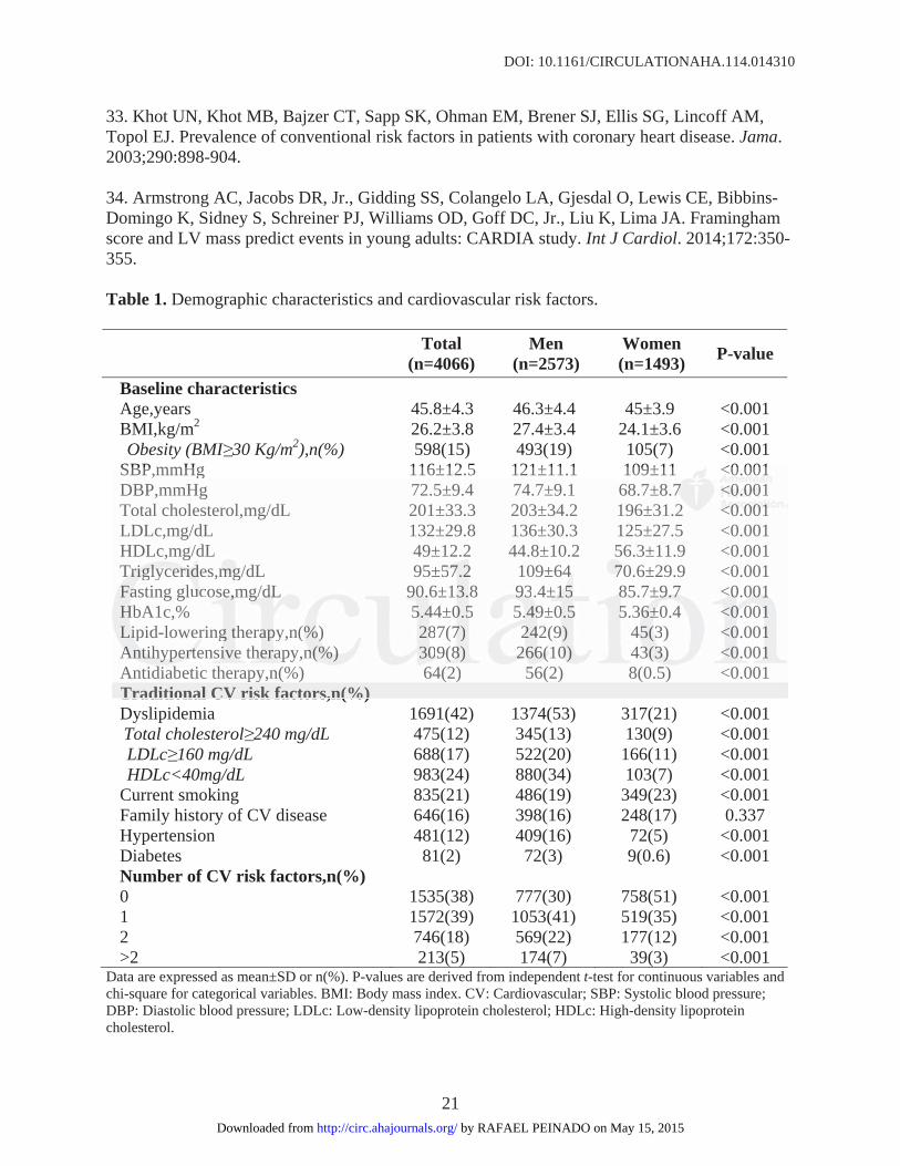
Table 1. Demographic characteristics and cardiovascular risk factors.
Total(n=4066)
Men(n=2573)
Women(n=1493) P-value
Baseline characteristics Age,years 45.8±4.3 46.3±4.4 45±3.9 <0.001 BMI,kg/m2 26.2±3.8 27.4±3.4 24.1±3.6 <0.001 Obesity (BMI 30 Kg/m2),n(%) 598(15) 493(19) 105(7) <0.001
SBP,mmHg 116±12.5 121±11.1 109±11 <0.001 DBP,mmHg 72.5±9.4 74.7±9.1 68.7±8.7 <0.001 Total cholesterol,mg/dL 201±33.3 203±34.2 196±31.2 <0.001 LDLc,mg/dL 132±29.8 136±30.3 125±27.5 <0.001 HDLc,mg/dL 49±12.2 44.8±10.2 56.3±11.9 <0.001 Triglycerides,mg/dL 95±57.2 109±64 70.6±29.9 <0.001 Fasting glucose,mg/dL 90.6±13.8 93.4±15 85.7±9.7 <0.001 HbA1c,% 5.44±0.5 5.49±0.5 5.36±0.4 <0.001 Lipid-lowering therapy,n(%) 287(7) 242(9) 45(3) <0.001 Antihypertensive therapy,n(%) 309(8) 266(10) 43(3) <0.001 Antidiabetic therapy,n(%) 64(2) 56(2) 8(0.5) <0.001 Traditional CV risk factors,n(%) Dyslipidemia 1691(42) 1374(53) 317(21) <0.001 Total cholesterol 240 mg/dL 475(12) 345(13) 130(9) <0.001 LDLc 160 mg/dL 688(17) 522(20) 166(11) <0.001 HDLc<40mg/dL 983(24) 880(34) 103(7) <0.001
Current smoking 835(21) 486(19) 349(23) <0.001 Family history of CV disease 646(16) 398(16) 248(17) 0.337 Hypertension 481(12) 409(16) 72(5) <0.001 Diabetes 81(2) 72(3) 9(0.6) <0.001 Number of CV risk factors,n(%) 0 1535(38) 777(30) 758(51) <0.001 1 1572(39) 1053(41) 519(35) <0.001 2 746(18) 569(22) 177(12) <0.001 >2 213(5) 174(7) 39(3) <0.001
Data are expressed as mean±SD or n(%). P-values are derived from independent t-test for continuous variables and chi-square for categorical variables. BMI: Body mass index. CV: Cardiovascular; SBP: Systolic blood pressure; DBP: Diastolic blood pressure; LDLc: Low-density lipoprotein cholesterol; HDLc: High-density lipoprotein cholesterol.
SBP,mmHg 116±12.5 121±11.1 109±11 <00.0.000101 DBP,mmHg 72.5±9.4 74.7±9.1 68.7±8.777 <0<0<0.0.0.001010 Total cholesterol,mg/dL 201±33.3 203±34.2 196±31.2 <<<00.0 000000111LDLc,mg/dL 132±29.8 136±30.3 125±27.5 <0.001 HDLc,mg/dL 49±12.2 44.8±10.2 56.3±11.9 <0.001TrTrTrigigiglylylyceceeririridedd s,mgmgmg/dL 95±555777.222 109±6444 7770.6±29.9 <0.001 FaFaFasting glucucucose,e,e,mgmgmg/d/ddL L L 909090.6.6. ±13.8 939393.4±1±1±155 8588 .7±9±9±9.7.7.7 <<0.00 000000111HHHbA1c,% 555.4444±00.555 5..4. 999±000.555 55.36366±±±0.44 <<<0.0.0.000000111LiLiipip d-lowerir ngg ttheraaappy,n(%%%))) 28887(77) 2224442(99) 4455(3(3(3) <0<00.00111 AnAnAntititihyhyhypepepertrtrteensiisiveee theheheraraapypp ,nnn(%(%(%))) 30009(9(9(88)8 22266666(1(( 00)0) 43(3(3(3)3)3) <<<00.0.000111Antididiabetic therapypy,n(%( ) ) 64(2( )) 56(2( ) ) 8((0.5)5) <0.001 Traditional CV risk factors,n(%)
by RAFAEL PEINADO on May 15, 2015http://circ.ahajournals.org/Downloaded from

DOI: 10.1161/CIRCULATIONAHA.114.014310
22
Table 2. Association between presence of atherosclerosis in individual vascular territories.
Carotid disease Coronary calcification Aortic disease Ilio-femoral disease OR(95%CI) 2.06(1.73-2.47) 2.80(2.40-3.27) 2.13(1.84-2.46) Carotid disease
p-value (-)
p<0.001 p<0.001 p<0.001 OR(95%CI) 2.06(1.73-2.47) 2.48(2.06-2.99) 3.16(2.60-3.84) Coronary calcification
p-value p<0.001 (-)
p<0.001 p<0.001 OR(95%CI) 2.80(2.40-3.27) 2.48(2.06-2.99) 4.85(4.09-5.75) Aortic disease
p-value p<0.001 p<0.001 (-)
p<0.001 OR(95%CI) 2.13(1.84-2.46) 3.16(2.60-3.84) 4.85(4.09-5.75) Ilio-femoral disease
p-value p<0.001 p<0.001 p<0.001 (-) Data are expressed as odds ratio (OR) and 95% confidence interval (CI) adjusted by age and gender and calculated using logistic regression models. Coronary calcification was defined as CACS 1.
p p p pOR(95%CI) 2.13(1.84-2.46) 3.16(2.60-3.84) 4.85(4.09-5.75)5)5) emoral disease
p-value p<0.001 p<0.001 p<0.001 (-(-(-) ) ) re expressed as odds ratio (OR) and 95% confidence interval (CI) adjusted by age and gender and calculated using logistic reg eresssss ioi n modedd lsl . CCCoronacation was defined as CACS 1.
by RAFAEL PEINADO on May 15, 2015http://circ.ahajournals.org/Downloaded from
DOI: 10.1161/CIRCULATIONAHA.114.014310
23
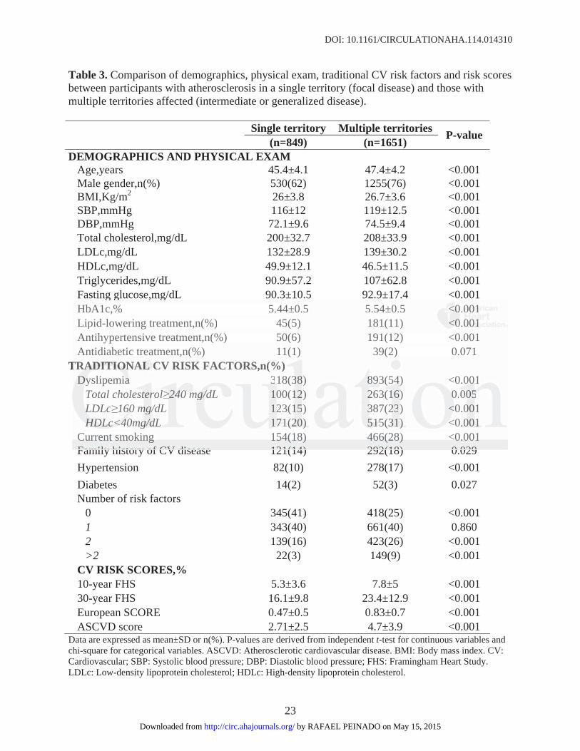
Table 3. Comparison of demographics, physical exam, traditional CV risk factors and risk scores between participants with atherosclerosis in a single territory (focal disease) and those with multiple territories affected (intermediate or generalized disease).
Single territory Multiple territories (n=849) (n=1651) P-value
DEMOGRAPHICS AND PHYSICAL EXAM Age,years 45.4±4.1 47.4±4.2 <0.001 Male gender,n(%) 530(62) 1255(76) <0.001 BMI,Kg/m2 26±3.8 26.7±3.6 <0.001 SBP,mmHg 116±12 119±12.5 <0.001 DBP,mmHg 72.1±9.6 74.5±9.4 <0.001 Total cholesterol,mg/dL 200±32.7 208±33.9 <0.001 LDLc,mg/dL 132±28.9 139±30.2 <0.001 HDLc,mg/dL 49.9±12.1 46.5±11.5 <0.001 Triglycerides,mg/dL 90.9±57.2 107±62.8 <0.001 Fasting glucose,mg/dL 90.3±10.5 92.9±17.4 <0.001 HbA1c,% 5.44±0.5 5.54±0.5 <0.001 Lipid-lowering treatment,n(%) 45(5) 181(11) <0.001 Antihypertensive treatment,n(%) 50(6) 191(12) <0.001 Antidiabetic treatment,n(%) 11(1) 39(2) 0.071
TRADITIONAL CV RISK FACTORS,n(%) Dyslipemia 318(38) 893(54) <0.001
Total cholesterol 240 mg/dL 100(12) 263(16) 0.005 LDLc 160 mg/dL 123(15) 387(23) <0.001 HDLc<40mg/dL 171(20) 515(31) <0.001
Current smoking 154(18) 466(28) <0.001 Family history of CV disease 121(14) 292(18) 0.029 Hypertension 82(10) 278(17) <0.001 Diabetes 14(2) 52(3) 0.027 Number of risk factors
0 345(41) 418(25) <0.001 1 343(40) 661(40) 0.860 2 139(16) 423(26) <0.001 >2 22(3) 149(9) <0.001
CV RISK SCORES,% 10-year FHS 5.3±3.6 7.8±5 <0.001 30-year FHS 16.1±9.8 23.4±12.9 <0.001 European SCORE 0.47±0.5 0.83±0.7 <0.001 ASCVD score 2.71±2.5 4.7±3.9 <0.001
Data are expressed as mean±SD or n(%). P-values are derived from independent t-test for continuous variables and chi-square for categorical variables. ASCVD: Atherosclerotic cardiovascular disease. BMI: Body mass index. CV: Cardiovascular; SBP: Systolic blood pressure; DBP: Diastolic blood pressure; FHS: Framingham Heart Study. LDLc: Low-density lipoprotein cholesterol; HDLc: High-density lipoprotein cholesterol.
HbA1c,% 5.44±0.5 5.54±0.5 <0<0<0.000010101 Lipid-lowering treatment,n(%) 45(5) 181(11) <0<0<0.000010101 Antihypertensive treatment,n(%) 50(6) 191(12) <0.001 Antidiabetic treatment,n(%) 11(1) 39(2) 0.071
TRRRADADADITITITIOIOIONANN LL L CVC RISK FACTORS,n(%)DyDyDyslipemiaiaia 33181 (3388) 889893(3 54544) )) <0.000100
Total cholesttterroll 2444000 mgmgmg/d/d/dL 10000(12122) 22263((1116)) 0.00 00000555LDLL Lc 16660 00 mgmg//dL 12223(1515) 33387(2223)) <<0<0.0001 HDHDHDLcLcLc<4<4<40mmmg/gg/dLdLdL 17771(1(1(202020) )) 5151515(5(5(3331) ) ) <<<0.0.0.00111
CuCuCurrrrrrenenenttt smsmsmokokokinininggg 1515154(4(4(181818))) 4646466(6(6(282828))) <0<00.0.0.0010101 Family history of CV disease 121(14) 292(18) 0 029
by RAFAEL PEINADO on May 15, 2015http://circ.ahajournals.org/Downloaded from

DOI: 10.1161/CIRCULATIONAHA.114.014310
24
Figure Legends:
Figure 1. Prevalence of subclinical atherosclerosis by vascular territory in PESA.
Figure 2. Prevalence of subclinical atherosclerosis by age and gender in each vascular territory.
Figure 3. Distribution of subclinical atherosclerosis detected by noninvasive imaging according
to Framingham Heart Study risk (FHS) categories. Vascular sites examined were the right and
left carotids, abdominal aorta and the right and left ilio-femoral arteries (presence of plaque), and
the coronary vessels (CAC). FHS scores were classified as low (<10%), moderate ( 10 to 20%)
or high risk (>20%)13, 15.
Figure 4. Distribution of subclinical atherosclerosis detected by noninvasive imaging according
to European SCORE categories. European SCORE was classified as low (<1%) and moderate-
high risk ( 1%)14.
Figure 5. Distribution of subclinical atherosclerosis across participants meeting AHA/ACC
criteria for statin treatment ( 7.5% ASCVD risk, diabetes, or LDL 190 mg/dL), those
considered for statin (5 to <7.5% ASCVD risk) and those not considered for statin (<5%
ASCVD risk). ASCVD: Atherosclerotic cardiovascular disease.
he coronary vessels (CAC). FHS scores were classified as low (<10%), moderate ee ((( 101010 ttto oo 202020%)%%
or high risk (>20%)13, 15.
FFFiggugure 4. Distririribubbutiononon ooof f f sususubcbcbclililinininicacacal ll atatathherossscllel rossisss deeetetete tctctededed by y nooonininvnvnvassiveee imimimagagaginining acacaccococordrdrdinining gg
oo EEEuururopean n n SCSS ORORORE cacacattet gorrrieseses. Euuurrropppeaana SSSCCCORRE waaas claasasssififif eed aasss low ww (<<1%1%1%)) annnd d d moodeeerateee-
hihihighghgh rrrisisiskkk ((( 1%1%1%)))1414.
by RAFAEL PEINADO on May 15, 2015http://circ.ahajournals.org/Downloaded from
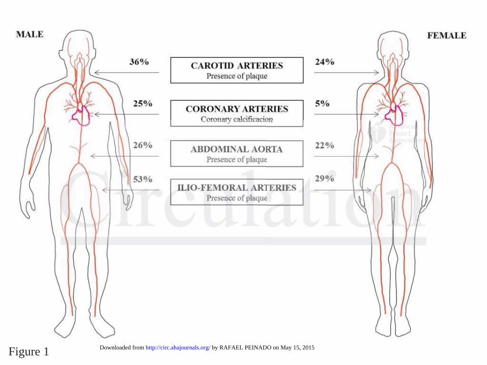
Figure 1 by RAFAEL PEINADO on May 15, 2015http://circ.ahajournals.org/Downloaded from
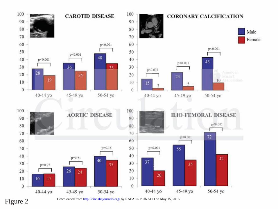
Figure 2 by RAFAEL PEINADO on May 15, 2015http://circ.ahajournals.org/Downloaded from

Figure 3 by RAFAEL PEINADO on May 15, 2015http://circ.ahajournals.org/Downloaded from

Figure 4 by RAFAEL PEINADO on May 15, 2015http://circ.ahajournals.org/Downloaded from

Figure 5 by RAFAEL PEINADO on May 15, 2015http://circ.ahajournals.org/Downloaded from

1
SUPPLEMENTAL MATERIAL
Supplemental Methods
IMT was also assessed by vascular ultrasound at the level of the posterior wall of the
common carotid and femoral arteries in longitudinal views. Values >0.9 were
considered abnormal, as established in previous studies1 and guidelines
2.
Reproducibility of IMT measurements was evaluated by replicating the analysis of a
random sample of 60 studies 3 months after the initial assessment and determining
intraclass correlation coefficients (ICC). Excellent values (ICC>0.95) were found for
both carotid and femoral IMT measurements.
Supplemental Results
Associations between risk factors and subclinical atherosclerosis in different territories
are shown in Table S2. Age and male gender were significantly associated with the
presence of plaque in any territory, and particularly with CAC. All risk factors were
associated with atherosclerosis across all territories, although results did not reach
significance for the association of diabetes or hypertension with aortic disease and of
family history with carotid disease. Diabetes was most strongly associated with carotid
disease and CAC, whereas smoking had a stronger association with aortic and ilio-
femoral disease and family history had a stronger association with CAC.
Regarding IMT measurements, the mean value was 0.59 mm in carotid arteries and 0.61
mm in femoral arteries. Abnormal IMT (>0.9 mm)1 was detected in 9% of participants,
and was more frequent in femoral than in carotid arteries (8% vs. 1%). The prevalence
of abnormal IMT was higher in men and increased with age (Table S1). The sensitivity,
specificity, and positive and negative predictive values for IMT to detect subclinical

2
atherosclerosis were 14%, 99%, 99% and 41%, respectively. Consistent with previous
studies3-5
, IMT measurements did not correlate with the presence of subclinical
atherosclerosis (presence of plaque or CACS≥1). Prevalence of abnormal IMT was low
(9%), especially in the carotid arteries (0.8%), despite the overall high prevalence of
subclinical disease (63%). The increasing rate of abnormal IMT with age suggests a
confounding influence of factors not directly related to atherosclerosis. In addition,
almost all participants with abnormal IMT also had disease (99%), indicating that IMT
provides little information that cannot be derived by directly determining the presence
of plaques. Further evidence of the limited utility of IMT for assessing subclinical
atherosclerosis is provided by its low sensitivity in our sample (14%), the lack of
standard cut-off values for normal age reference intervals6, and its reported poor
predictive value for CV disease compared with plaque-based markers4. In this regard,
the most recent American College of Cardiology/American Heart Association
(ACC/AHA) Guide on the assessment of CV risk no longer recommends carotid IMT
for routine clinical assessment of the risk of a first atherosclerotic CV event7.

3
Supplemental Tables
Table S1. Prevalence of cardiovascular risk factors, risk scale scoring, CACS and IMT
values in PESA participants stratified by age and gender
MEN WOMEN
40-44 yo
(n=1000) 45-49 yo
(n=822) 50-54 yo
(n=702) 40-44 yo
(n=747) 45-49 yo
(n=506) 50-54 yo
(n=225)
Traditional CV risk factors
Dyslipidemia 483 (48) 449 (55) 406 (58) 113 (15) 121 (24) 81 (36)
Smoking 170 (17) 159 (19) 146 (21) 163 (22) 131 (26) 50 (22)NS
Family history 148 (15) 136 (17) 104 (15) NS 123 (17) 91 (18) 32 (14) NS
Hypertension 84 (8) 124 (15) 179 (26) 13 (2) 34 (7) 25 (11)
Diabetes 12 (1) 18 (2) 37 (5) 3 (0.4) 2 (0.4) 2 (0.89)NS
Number of risk factors
0 365 (37) 234 (29) 173 (25) 419 (56) 238 (47) 96 (43)
1 415 (42) 353 (43) 267 (38)NS 249 (33) 180 (36) 81 (36) NS
2 181 (18) 181 (22) 193 (28) 71 (10) 67 (13) 38 (17)
>2 39 (4) 54 (7) 69 (10) 8 (1) 21 (4) 10 (4)
CV risk scales (%)*
10y SCORE 0.39 ± 0.23 0.77 ± 0.37 1.43 ± 0.73 0.06 ± 0.05 0.17 ± 0.09 0.42 ±0.25
10y FHS 5.84 ± 3.07 7.78 ± 3.80 10.3 ± 5.16 1.81 ± 0.96 3.3 ± 1.98 4.9 ± 2.73
30y FHS 17.3 ± 8.37 23.4 ± 10.1 30.4 ± 12.0 6.68 ± 3.49 10.5 ± 6.0 14.4 ± 8.15
10y ASCVD 2.7 ± 2.27 4.3 ± 2.81 6.9 ± 3.97 0.9 ± 0.84 1.4 ± 1.24 2.0 ± 1.57
CACS
CACS*
(Agatston units)
6.73 ± 30.9 18.6 ± 101.0 45.4 ± 152.0 0.36 ± 2.66 1.52 ± 10.9 4.37 ± 30.9 NS
CACS 0 854 (85) 629 (77) 401 (57) 727 (97) 480 (95) 203 (90)
CACS 1-99 126 (13) 159 (19) 227 (32) 20 (3) 24 (5) 20 (9)
CACS 100-399 19 (2) 26 (3) 57 (8) 0 (0) 2 (0.4) 1 (0.4)
CACS ≥400 1 (0.1) 8 (1) 17 (2) 0 (0) 0 (0) 1 (0.4)
IMT (mm)
Carotid IMT* 0.59 ± 0.09 0.61 ± 0.10 0.64± 0.11 0.53 ± 0.06 0.56 ± 0.07 0.60 ± 0.08
Femoral IMT* 0.60 ± 0.21 0.66 ± 0.26 0.73± 0.32 0.50 ± 0.11 0.55 ± 0.16 0.57 ± 0.16
Carotid IMT > 0.9 7 (0.7) 4 (0.5) 18 (3) 0 (0) 0 (0) 2 (0.9)
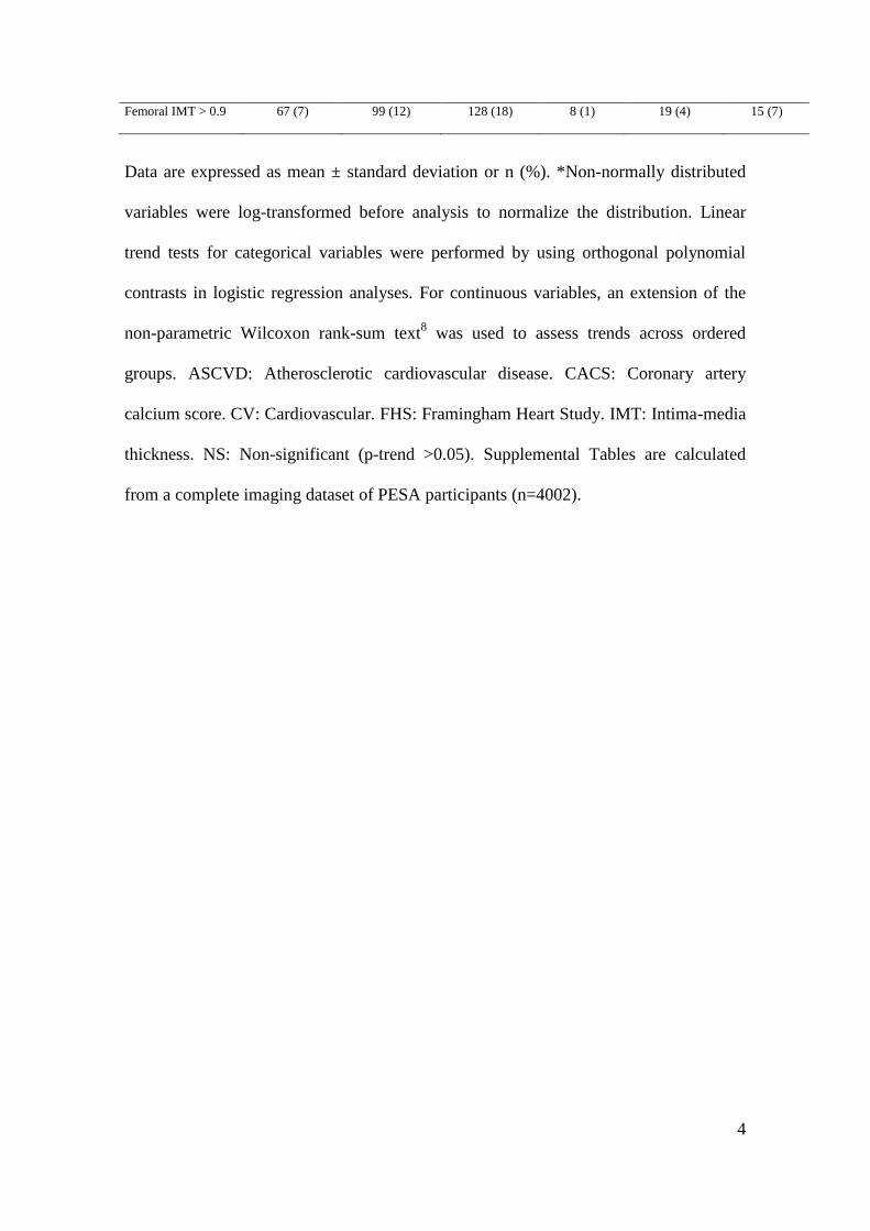
4
Femoral IMT > 0.9 67 (7) 99 (12) 128 (18) 8 (1) 19 (4) 15 (7)
Data are expressed as mean ± standard deviation or n (%). *Non-normally distributed
variables were log-transformed before analysis to normalize the distribution. Linear
trend tests for categorical variables were performed by using orthogonal polynomial
contrasts in logistic regression analyses. For continuous variables, an extension of the
non-parametric Wilcoxon rank-sum text8 was used to assess trends across ordered
groups. ASCVD: Atherosclerotic cardiovascular disease. CACS: Coronary artery
calcium score. CV: Cardiovascular. FHS: Framingham Heart Study. IMT: Intima-media
thickness. NS: Non-significant (p-trend >0.05). Supplemental Tables are calculated
from a complete imaging dataset of PESA participants (n=4002).
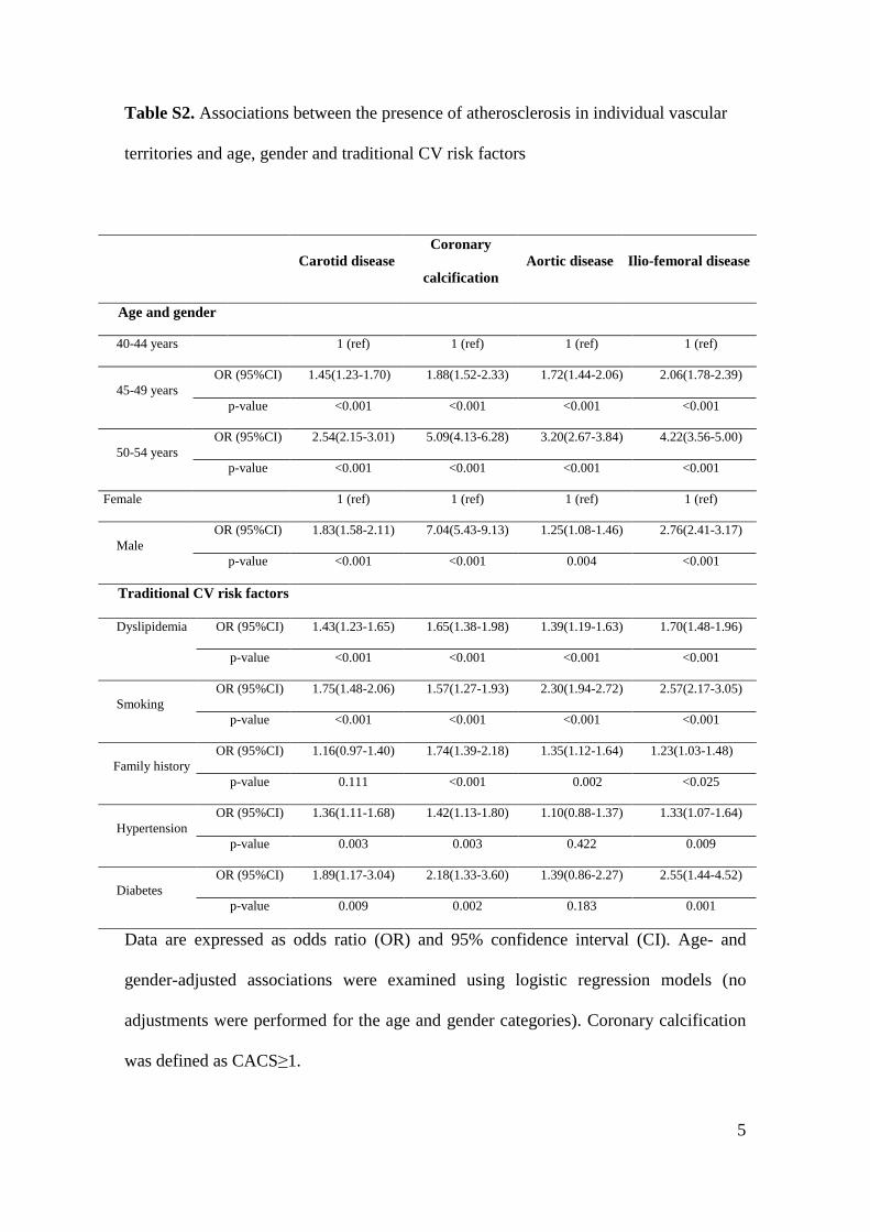
5
Table S2. Associations between the presence of atherosclerosis in individual vascular
territories and age, gender and traditional CV risk factors
Data are expressed as odds ratio (OR) and 95% confidence interval (CI). Age- and
gender-adjusted associations were examined using logistic regression models (no
adjustments were performed for the age and gender categories). Coronary calcification
was defined as CACS≥1.
Carotid disease
Coronary
calcification
Aortic disease Ilio-femoral disease
Age and gender
40-44 years 1 (ref) 1 (ref) 1 (ref) 1 (ref)
45-49 years
OR (95%CI) 1.45(1.23-1.70) 1.88(1.52-2.33) 1.72(1.44-2.06) 2.06(1.78-2.39)
p-value <0.001 <0.001 <0.001 <0.001
50-54 years
OR (95%CI) 2.54(2.15-3.01) 5.09(4.13-6.28) 3.20(2.67-3.84) 4.22(3.56-5.00)
p-value <0.001 <0.001 <0.001 <0.001
Female 1 (ref) 1 (ref) 1 (ref) 1 (ref)
Male
OR (95%CI) 1.83(1.58-2.11) 7.04(5.43-9.13) 1.25(1.08-1.46) 2.76(2.41-3.17)
p-value <0.001 <0.001 0.004 <0.001
Traditional CV risk factors
Dyslipidemia OR (95%CI) 1.43(1.23-1.65) 1.65(1.38-1.98) 1.39(1.19-1.63) 1.70(1.48-1.96)
p-value <0.001 <0.001 <0.001 <0.001
Smoking
OR (95%CI) 1.75(1.48-2.06) 1.57(1.27-1.93) 2.30(1.94-2.72) 2.57(2.17-3.05)
p-value <0.001 <0.001 <0.001 <0.001
Family history
OR (95%CI) 1.16(0.97-1.40) 1.74(1.39-2.18) 1.35(1.12-1.64) 1.23(1.03-1.48)
p-value 0.111 <0.001 0.002 <0.025
Hypertension
OR (95%CI) 1.36(1.11-1.68) 1.42(1.13-1.80) 1.10(0.88-1.37) 1.33(1.07-1.64)
p-value 0.003 0.003 0.422 0.009
Diabetes
OR (95%CI) 1.89(1.17-3.04) 2.18(1.33-3.60) 1.39(0.86-2.27) 2.55(1.44-4.52)
p-value 0.009 0.002 0.183 0.001
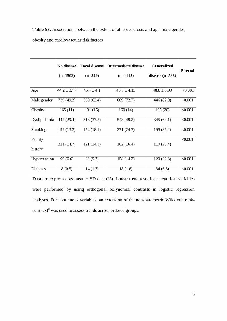
6
Table S3. Associations between the extent of atherosclerosis and age, male gender,
obesity and cardiovascular risk factors
No disease
(n=1502)
Focal disease
(n=849)
Intermediate disease
(n=1113)
Generalized
disease (n=538)
P-trend
Age 44.2 ± 3.77 45.4 ± 4.1 46.7 ± 4.13 48.8 ± 3.99 <0.001
Male gender 739 (49.2) 530 (62.4) 809 (72.7) 446 (82.9) <0.001
Obesity 165 (11) 131 (15) 160 (14) 105 (20) <0.001
Dyslipidemia 442 (29.4) 318 (37.5) 548 (49.2) 345 (64.1) <0.001
Smoking 199 (13.2) 154 (18.1) 271 (24.3) 195 (36.2) <0.001
Family
history
221 (14.7) 121 (14.3) 182 (16.4) 110 (20.4)
<0.001
Hypertension 99 (6.6) 82 (9.7) 158 (14.2) 120 (22.3) <0.001
Diabetes 8 (0.5) 14 (1.7) 18 (1.6) 34 (6.3) <0.001
Data are expressed as mean ± SD or n (%). Linear trend tests for categorical variables
were performed by using orthogonal polynomial contrasts in logistic regression
analyses. For continuous variables, an extension of the non-parametric Wilcoxon rank-
sum text8 was used to assess trends across ordered groups.
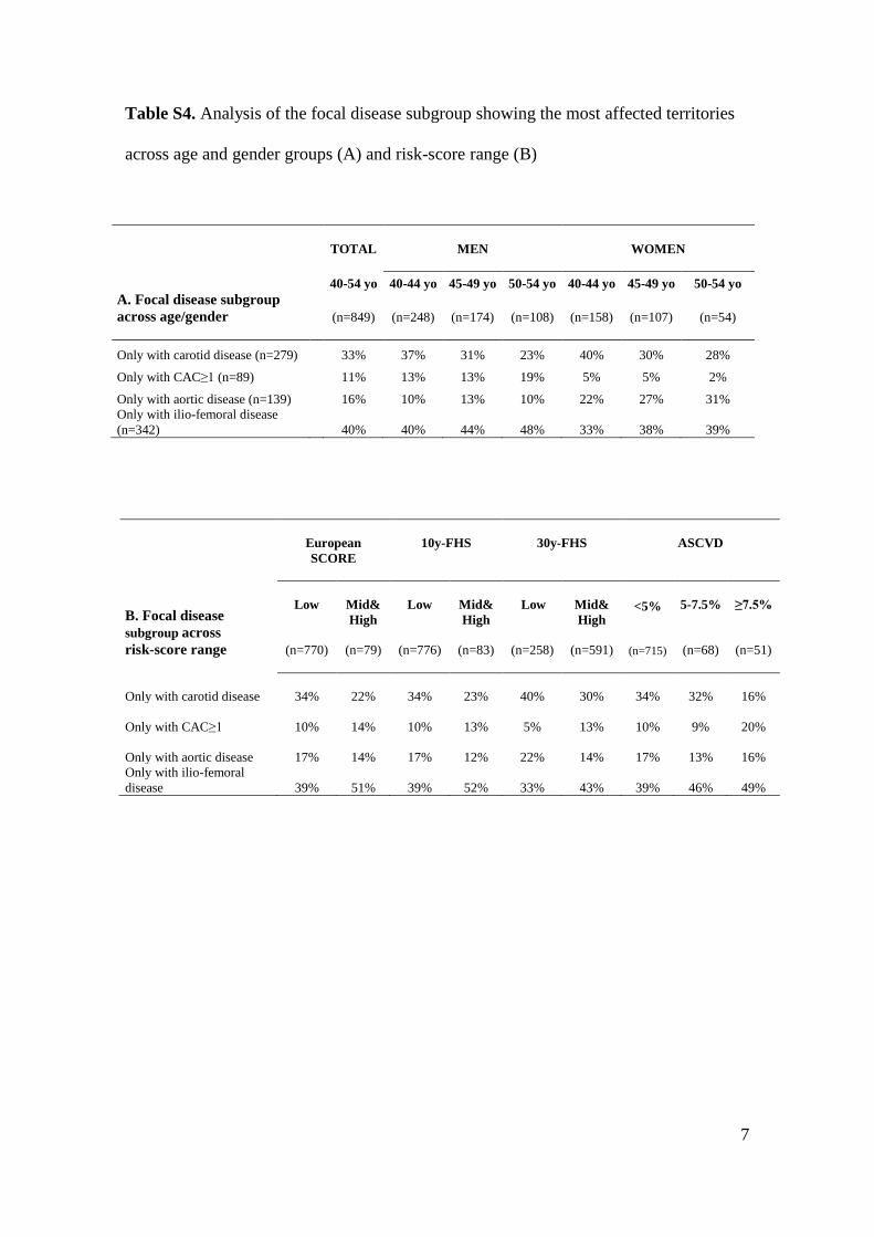
7
Table S4. Analysis of the focal disease subgroup showing the most affected territories
across age and gender groups (A) and risk-score range (B)
A. Focal disease subgroup
across age/gender
TOTAL
MEN WOMEN
40-54 yo 40-44 yo 45-49 yo 50-54 yo 40-44 yo 45-49 yo 50-54 yo
(n=849)
(n=248) (n=174) (n=108) (n=158) (n=107) (n=54)
Only with carotid disease (n=279) 33% 37% 31% 23% 40% 30% 28%
Only with CAC≥1 (n=89) 11% 13% 13% 19% 5% 5% 2%
Only with aortic disease (n=139) 16% 10% 13% 10% 22% 27% 31%
Only with ilio-femoral disease
(n=342) 40% 40% 44% 48% 33% 38% 39%
B. Focal disease
subgroup across
risk-score range
European
SCORE
10y-FHS
30y-FHS
ASCVD
Low
(n=770)
Mid&
High
(n=79)
Low
(n=776)
Mid&
High
(n=83)
Low
(n=258)
Mid&
High
(n=591)
<5%
(n=715)
5-7.5%
(n=68)
≥7.5%
(n=51)
Only with carotid disease 34% 22%
34%
23%
40%
30%
34%
32%
16%
Only with CAC≥1 10% 14%
10%
13%
5%
13%
10%
9%
20%
Only with aortic disease 17% 14%
17%
12%
22%
14%
17%
13%
16%
Only with ilio-femoral
disease 39% 51%
39%
52%
33%
43%
39%
46%
49%
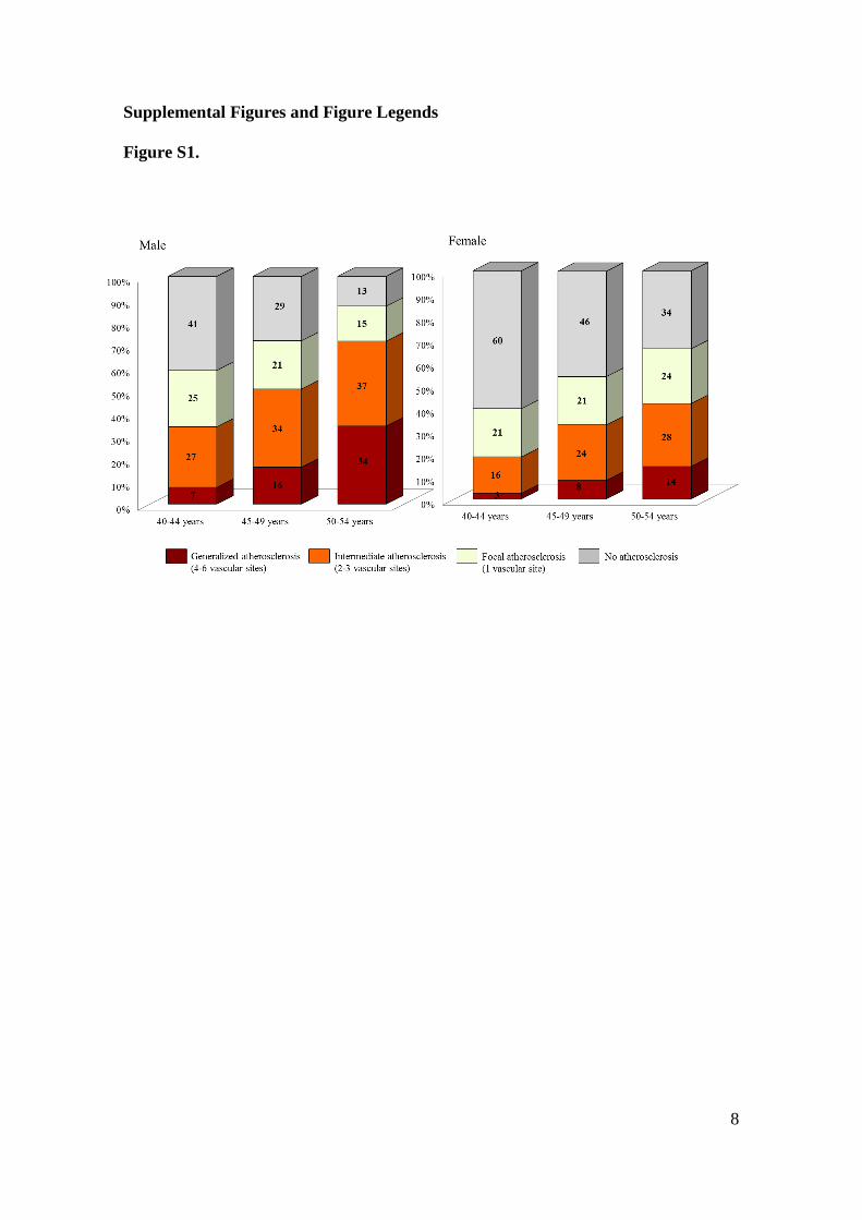
8
Supplemental Figures and Figure Legends
Figure S1.
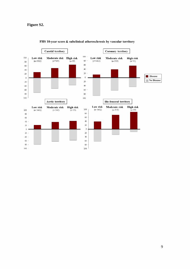
9
Figure S2.
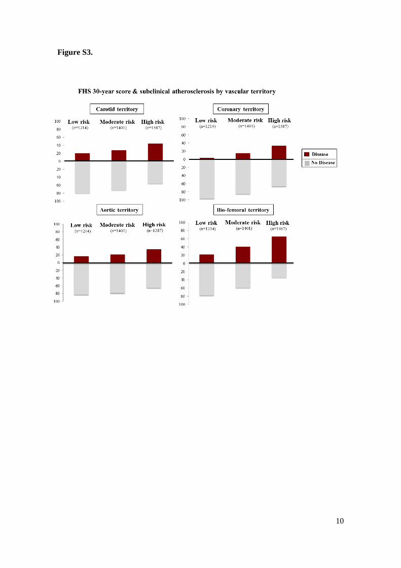
10
Figure S3.
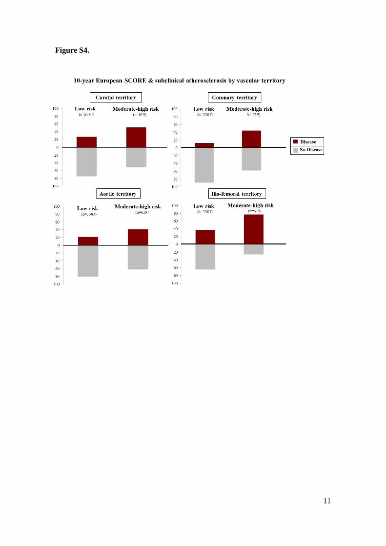
11
Figure S4.

12
Figure Legends
Figure S1. Distribution of multi-territorial extent of subclinical atherosclerosis.
Figure S2. Association of subclinical atherosclerosis with the 10-year Framingham
Heart Study risk score according to vascular territory.
Figure S3. Association of subclinical atherosclerosis with the 30-year Framingham
Heart Study risk score according to vascular territory.
Figure S4. Association of subclinical atherosclerosis with European SCORE risk
according to vascular territory.
13
Supplemental References
1. Touboul PJ, Hennerici MG, Meairs S, Adams H, Amarenco P, Desvarieux M,
Ebrahim S, Fatar M, Hernandez Hernandez R, Kownator S, Prati P, Rundek T, Taylor
A, Bornstein N, Csiba L, Vicaut E, Woo KS, Zannad F and Advisory Board of the 3rd
Watching the Risk Symposium tESC. Mannheim intima-media thickness consensus.
Cerebrovascular diseases. 2004;18:346-9.
2. Graham I, Atar D, Borch-Johnsen K, Boysen G, Burell G, Cifkova R,
Dallongeville J, De Backer G, Ebrahim S, Gjelsvik B, Herrmann-Lingen C, Hoes A,
Humphries S, Knapton M, Perk J, Priori SG, Pyorala K, Reiner Z, Ruilope L, Sans-
Menendez S, Op Reimer WS, Weissberg P, Wood D, Yarnell J, Zamorano JL, Walma
E, Fitzgerald T, Cooney MT, Dudina A, Vahanian A, Camm J, De Caterina R, Dean V,
Dickstein K, Funck-Brentano C, Filippatos G, Hellemans I, Kristensen SD, McGregor
K, Sechtem U, Silber S, Tendera M, Widimsky P, Zamorano JL, Altiner A, Bonora E,
Durrington PN, Fagard R, Giampaoli S, Hemingway H, Hakansson J, Kjeldsen SE,
Larsen m L, Mancia G, Manolis AJ, Orth-Gomer K, Pedersen T, Rayner M, Ryden L,
Sammut M, Schneiderman N, Stalenhoef AF, Tokgozoglu L, Wiklund O and Zampelas
A. European guidelines on cardiovascular disease prevention in clinical practice: full
text. Fourth Joint Task Force of the European Society of Cardiology and other societies
on cardiovascular disease prevention in clinical practice (constituted by representatives
of nine societies and by invited experts). Eur J Cardiovasc Prev Rehabil. 2007;14 Suppl
2:S1-113.
3. Inaba Y, Chen JA and Bergmann SR. Carotid plaque, compared with carotid
intima-media thickness, more accurately predicts coronary artery disease events: a
meta-analysis. Atherosclerosis. 2012;220:128-33.
14
4. Yerly P, Rodondi N, Viswanathan B, Riesen W, Vogt P and Bovet P.
Association between conventional risk factors and different ultrasound-based markers
of atherosclerosis at carotid and femoral levels in a middle-aged population. Int J
Cardiovasc Imaging. 2013;29:589-99.
5. Lorenz MW, Schaefer C, Steinmetz H and Sitzer M. Is carotid intima media
thickness useful for individual prediction of cardiovascular risk? Ten-year results from
the Carotid Atherosclerosis Progression Study (CAPS). Eur Heart J. 2010;31:2041-8.
6. Engelen L, Ferreira I, Stehouwer CD, Boutouyrie P, Laurent S and Reference
Values for Arterial Measurements C. Reference intervals for common carotid intima-
media thickness measured with echotracking: relation with risk factors. European heart
journal. 2013;34:2368-80.
7. Goff DC, Jr., Lloyd-Jones DM, Bennett G, Coady S, D'Agostino RB, Sr.,
Gibbons R, Greenland P, Lackland DT, Levy D, O'Donnell CJ, Robinson JG, Schwartz
JS, Shero ST, Smith SC, Jr., Sorlie P, Stone NJ, Wilson PW and American College of
Cardiology/American Heart Association Task Force on Practice G. 2013 ACC/AHA
guideline on the assessment of cardiovascular risk: a report of the American College of
Cardiology/American Heart Association Task Force on Practice Guidelines. J Am Coll
Cardiol. 2014;63:2935-59.
8. Cuzick J. A Wilcoxon-type test for trend. Statistics in medicine. 1985;4:87-90.
Ginés Sanz, Luis Jesús Jiménez-Borreguero and Valentín FusterAlonso-Farto, Eliseo Guallar, Fernando Civeira, Henrik Sillesen, Stuart Pocock, José M. Ordovás,
Martínez de Vega, Laura García, Jesús Molina, Javier Sanchez-Gonzalez, Gabriela Guzman, Juan C.López-Melgar, Martín Laclaustra, Belén Oliva, Agustín Mocoroa, José Mendiguren, Vicente Leticia Fernández-Friera, José L. Peñalvo, Antonio Fernández-Ortiz, Borja Ibañez, Beatriz
a Middle-Aged Cohort: The PESA (Progression of Early Subclinical Atherosclerosis) StudyPrevalence, Vascular Distribution and Multi-territorial Extent of Subclinical Atherosclerosis in
Print ISSN: 0009-7322. Online ISSN: 1524-4539 Copyright © 2015 American Heart Association, Inc. All rights reserved.
is published by the American Heart Association, 7272 Greenville Avenue, Dallas, TX 75231Circulation published online April 16, 2015;Circulation.
http://circ.ahajournals.org/content/early/2015/04/16/CIRCULATIONAHA.114.014310World Wide Web at:
The online version of this article, along with updated information and services, is located on the
http://circ.ahajournals.org/content/suppl/2015/04/16/CIRCULATIONAHA.114.014310.DC1.htmlData Supplement (unedited) at:
http://circ.ahajournals.org//subscriptions/
is online at: Circulation Information about subscribing to Subscriptions:
http://www.lww.com/reprints Information about reprints can be found online at: Reprints:
document. Permissions and Rights Question and Answer available in the
Permissions in the middle column of the Web page under Services. Further information about this process isOnce the online version of the published article for which permission is being requested is located, click Request
can be obtained via RightsLink, a service of the Copyright Clearance Center, not the Editorial Office.Circulation Requests for permissions to reproduce figures, tables, or portions of articles originally published inPermissions:
by RAFAEL PEINADO on May 15, 2015http://circ.ahajournals.org/Downloaded from