lipoic acid effects on established atherosclerosis
TRANSCRIPT
Lipoic acid effects on established atherosclerosis
Zhekang Yinga, Nisharahmed Kheradaa, Britten Farrara, Thomas Kampfratha, YiuchoChunga, Orlando Simonettia, Jeffrey Deiuliisa, Rajagopal Desikana, Bobby Khana,Frederick Villamenaa, Qinghua Suna,b, Sampath Parthasarathya, and SanjayRajagopalan.aaDavis Heart Lung Research Institute, Colleges of Medicine and Public Health, the Ohio StateUniversitybDivision of Environmental Health Sciences, Colleges of Medicine and Public Health, the OhioState University
AbstractAims—Alpha-lipoic acid (LA) is a commonly used dietary supplement that exerts anti-oxidantand anti-inflammatory effects in vivo and in vitro. We investigated the mechanisms by which LAmay confer protection in models of established atherosclerosis.
Main Methods—Watanabe heritable hyperlipidemic (WHHL) rabbits were fed with highcholesterol chow for 6 weeks and then randomized to receive either high cholesterol diet alone orcombined with LA (20 mg/kg/day) for 12 weeks. Vascular function was analyzed by myography.The effects of LA on T cell migration to chemokine gradients was assessed by Boyden chamber.NF-κB activation was determined by measuring translocation and electrophoresis migration shiftassay (EMSA).
Key findings—LA decreased body weight by 15 ± 5% without alterations in lipid parameters.Magnetic Resonance Imaging (MRI) analysis demonstrated that LA reduced atheroscleroticplaques in the abdominal aorta, with morphological analysis revealing reduced lipid andinflammatory cell content. Consistent with its effect on atherosclerosis, LA improved vascularreactivity (decreased constriction to angiotensin II and increased relaxation to acetylcholine andinsulin), inhibited NF-κB activation, and decreased oxidative stress and expression of keyadhesion molecules in the vasculature. LA reduced T cell content in atherosclerotic plaque inconjunction with decreasing ICAM and CD62L (L-Selectin) expression. These effects wereconfirmed by demonstration of a direct effect of LA in reducing T cell migration in response toCCL5 and SDF-1 and decreasing T cell adhesion to the endothelium by intra-vital microscopy
Significance—The present findings offer a mechanistic insight into the therapeutic effects of LAon atherosclerosis.
KeywordsLipoic acid; NF-kappaB; Atheosclerosis; T cells; Innate immunity; Superoxide
© 2009 Elsevier Inc. All rights reserved.Correspondence should be addressed to: Sanjay Rajagopalan, M.D., Davis Heart Lung Research Institute, The Ohio State University,Room 110, 473 W 12th Avenue, Columbus, OH 43210-1252, Tel: 614-247-2532, Fax: 614-293-5614,[email protected]'s Disclaimer: This is a PDF file of an unedited manuscript that has been accepted for publication. As a service to ourcustomers we are providing this early version of the manuscript. The manuscript will undergo copyediting, typesetting, and review ofthe resulting proof before it is published in its final citable form. Please note that during the production process errors may bediscovered which could affect the content, and all legal disclaimers that apply to the journal pertain.
NIH Public AccessAuthor ManuscriptLife Sci. Author manuscript; available in PMC 2011 April 13.
Published in final edited form as:Life Sci. 2010 January 16; 86(3-4): 95–102. doi:10.1016/j.lfs.2009.11.009.
NIH
-PA Author Manuscript
NIH
-PA Author Manuscript
NIH
-PA Author Manuscript
Introductionα-Lipoic acid (LA; 1,2-dithiolane-3-pentanoic acid) is a naturally occurring disulfidecompound commonly found in diet and is a necessary cofactor for mitochondrial enzymesα-ketoglutarate and pyruvate dehydrogenases in mammals (Reed, et al., 1951). Variousstudies have shown that LA exerts powerful anti-inflammatory and anti-oxidant effects invitro (Packer, et al., 1995). Treatment of cultured monocytes with LA inhibitslipopolysaccharide-mediated induction of pro-inflammatory factors through up-regulation ofthe PI3 kinase-Akt pathway (Zhang, et al., 2007). Consistent with these effects, LA has beenshown to be protective in human diseases associated with abnormal oxidative stress andenergy metabolism such as diabetes mellitus (Estrada, et al., 1996; Henriksen, et al., 1997),hyperhomocysteinemia (Baydas, et al., 2002) and hypertension (Vasdev, et al., 2000;Vasdev, et al., 2000). Previous studies performed by our group, have revealed that 4 weeksof dietary supplementation with LA in humans was sufficient to improve endothelialfunction in patients with metabolic syndrome (Sola, et al., 2005). Prior studies with LA andatherosclerosis have tested the effects of the agent very early on in the life cycle ofatherosclerosis development (Zhang, et al., 2008) and have not tested the effects onceatherosclerosis is established. In this study, we tested the hypothesis that oralsupplementation of LA in the diet would inhibit established atherosclerosis and furtherinvestigated the mechanisms by which LA exerts these effects by parallel investigations onvascular inflammation and in particular the infiltration of T cells.
MethodsOrgan chamber experiments were performed in the thoracic aorta of WHHL rabbits asdetailed in online methods. In-vivo magnetic resonance imaging of the abdominal aorta wasperformed at various time points as shown in Supplementary Figure 1. Quantitativemorphometry, lipid profiles, oxidative stress measures, quantitative real time PCR western,NF-κB activation assays, Intra-vital microscopy, T-cell migration assays, are all described inthe online Supplemental Methods Section.
StatisticsResults are expressed as means ± SD. The unpaired Student’s t test was used to compareparameters in the LA and control treated groups. P values < 0.05 were reported assignificant. With multiple comparisons, a Bonferroni correction was used for multiplecomparisons. In-vitro experiments comparing LA with other anti-oxidants involvingmultiple groups were analyzed using 1-way ANOVA with a Bonferroni post-hoc correction.
ResultsThe study design of rabbit experiments is illustrated in Supplementary Figure 1. All rabbitswere fed with high cholesterol chow for a period of 6 weeks prior to randomization to LA/control groups in order to hasten the development of plaque in this pre-disposed animalmodel. During this period the rabbit gained weight (2.62 ± 0.4 kg baseline and 2.96 ± 0.38kg after 6 weeks of high cholesterol chow; n = 9; P<0.05, paired student’s t-test for 6 weeksversus baseline). Dietary supplementation with LA started at the end of 6 weeks for aduration of 12 weeks, reduced rabbit weight by 15 ± 4 %, in contrast control rabbits did notchange their weights (Supplementary Figure 2). Compared to the baseline lipoprotein levels,6 weeks of high cholesterol feeding in the control group significantly increased total, HDL,LDL and VLDL cholesterol. LA did not significantly affect the level of any of the plasmalipoprotein sub-fractions (Supplementary Table 1). LA markedly reduced fastinghyperglycemia and increased insulin level at the end of the treatment period compared to the
Ying et al. Page 2
Life Sci. Author manuscript; available in PMC 2011 April 13.
NIH
-PA Author Manuscript
NIH
-PA Author Manuscript
NIH
-PA Author Manuscript
control group (Supplementary Table 1). LA treatment did not alter the triglyceride levelsafter 12 weeks of treatment.
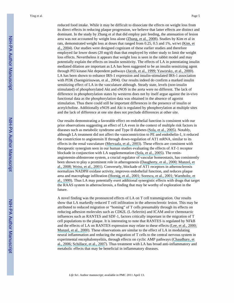
The wall volume in the rabbit abdominal aorta, an indicator of atherosclerotic plaqueburden, was analyzed by serial in vivo MRI scanning. Comparison with age and sex-matched New Zealand White rabbits confirmed a significantly higher wall volume ofabdominal aorta in WHHL rabbits after 6 weeks of feeding with high cholesterol chow(Supplementary Figure 3). After 6 weeks of dietary supplementation with LA, wall volumein the abdominal aorta was significantly lower compared to the control arm (Figure 1A andB). These effects of LA were more pronounced at the end-point (12 weeks). At the end of 12weeks, there was a significant slowing of the rate of progression of plaque in the LA group,while abdominal aorta wall volume continued to increase in the control group (Figure 1Aand B). Morphometric assessment of plaque burden in the thoracic aorta at sacrifice,corroborated the anti-atherosclerotic effects of LA on the abdominal aorta (Figure 1C andD). Immunohistochemical analysis of thoracic aortic sections revealed a reduction in CD68+
cells in atherosclerotic plaque and increased smooth muscle content (Figure 2A-D). LA alsodecreased lipid accumulation and fibrillar collagen deposition (Figure 2E-H).
Figure 3 and Supplementary Table 2 illustrates the responses to various agonists in thethoracic aorta of WHHL rabbits. Figure 3A and B depict LA effects on acetylcholine andinsulin mediated responses. In contrast to significant effects on acetylcholine and insulinmediated dilation, LA had no effects in response to SNP, an endothelium-independentvasodilator (Supplementary Table 2). LA reduced vasoconstrictor responses to angiotensinII (Figure 3C) but did not alter responses to phenylephrine or endothelin-1 (SupplementaryTable 2). In view of decreased contractility to angiotensin II in the aortic segments, weexamined the expression of the angiotensin Type 1 (AT1) receptor. Real time PCR analysisof revealed that AT1 receptor was reduced by LA treatment (Supplementary Figure 4).
LA reduced oxidative stress as evidenced by lower 8-isoprostane levels in plasma and aortichomogenates (Figure 4A and B). These results were confirmed by in situ aortic O2
•-
measurement using dihydroethidium (DHE) staining (Figure 4C and D). Because variousstudies have indicated that NADPH oxidase is the major source of vascular O2
•-, its activityin aortic homogenates was analyzed.(Mehta and Griendling, 2006) Consistent with theresults of 8-isoprostane measurement and DHE staining, basal O2
•- production in aortichomogenates was significantly lower in the LA group compared to the control groups(Figure 4E). In response to exogenously added NADPH, there was a 1.75 fold lower O2
•-
generation in LA treated aortas compared to controls (Figure 4E).
As vascular inflammation plays a pivotal role in atherosclerosis we assessed pro-inflammatory gene expression and cellular correlates of inflammation. LA decreased mRNAexpression of VCAM-1, ICAM-1 and L-selectin (CD62L) in thoracic aorta (Figure 5A). ThemRNA expression of P-selectin and MCP-1 (CCL2) showed non-significant decreases in theLA group (Figure 5A). We corroborated the effect of LA on VCAM-1 and ICAM-1expression by measuring expression of these adhesion molecules at the protein level (Figure5B and C). Since NF-κB regulates expression of pro-inflammatory genes, we examined thecontent of the p65 sub-unit of NF-κB in the nuclear and cytosol as a surrogate for itsactivation. LA decreased the translocation of the p65 sub-unit of NF-κB from cytosol tonucleus (Figure 5D and 5E). The inhibition of NF-κB signaling by LA was confirmed byassessing the DNA binding activity of NF-κB with electrophoretic mobility shift assay(EMSA. Figure 5F and G). Consistent with these effects on NF-κB, acute treatment with LAinhibited adhesion of leukocytes in the cremasteric circulation when acutely administered tomice 24 hours prior to an inflammatory challenge with TNFα (Supplemental Figure 5).
Ying et al. Page 3
Life Sci. Author manuscript; available in PMC 2011 April 13.
NIH
-PA Author Manuscript
NIH
-PA Author Manuscript
NIH
-PA Author Manuscript
Since T lymphocytes are critical components of atherosclerotic inflammation, we examinedwhether supplementation with LA chronically decreased T cell infiltration in aortic wall.Figures 6A and B revealed that LA significantly decreased T cell content (CD3+ cells) at thelevel of the thoracic aorta. Adhesion to endothelium is a very important early step for T cellinfiltration. To investigate the effect of LA on the interaction between T cells andendothelial cells, T cells labeled with the fluorescent dye CFSE were injected into high fatfed ApoE-/- mice with or without LA pre-treatment over the preceding 8 weeks. LA pre-treatment significantly decreased the rolling and adhesion of labeled T cells to thecremasteric endothelium (Supplementary Figure 7A and B).
To further investigate the mechanism whereby LA reduces T cell recruitment toatherosclerotic lesions, the effect of LA on transmigration of jurkat T cells was evaluatedusing Boyden chamber assays. We assessed the effect of various concentrations of LA onmigration of T cells towards a RANTES and SDF-1 gradient. Figures 6C and D demonstratethat LA dose-dependently inhibits transmigration of T cells to RANTES and SDF-1. Finally,we isolated leukocytes from control and LA fed rabbits and examined their migration inresponse to RANTES and SDF-1 in vitro. Fig. 6E and F demonstrated that supplementationwith LA decreased the migration of leukocytes in response to RANTES and SDF-1.
DISCUSSIONThe present study has multiple findings that may have important implications for thetreatment of human atherosclerosis using LA, a commonly used dietary supplement, at lowdoses. 1) LA decreases atherosclerotic plaque burden within a 12 week period with changesseen as early as 6 weeks; 2) Decreased plaque burden was paralleled by markedimprovements in vascular function: 3) LA reduces NADPH oxidase dependent O2
•- and NF-κB-mediated inflammatory responses, offering a molecular basis for the anti-atheroscleroticeffects of LA; 4) LA anti-inflamatory effects involves reduction in key adhesion andchemokine molecules involved in T cell trafficking to atherosclerotic plaque; effects thatwere confirmed by dose dependent effects of LA in reducing T cell migration across CCL5/SDF-1 gradients and prevention of T cells to the endothelium.
MRI scanning demonstrated that 6 weeks of dietary supplementation with LA was sufficientto inhibit atherosclerotic lesion development in WHHL rabbits. We induced rapidprogression of plaque by high cholesterol feeding for 6 weeks prior to randomization. LAtreatment inhibited progression of plaque in the absence of notable changes in plasmalipoprotein levels. Our data are consistent with the effects noted by Zhang et al but differ indosing and timing of LA treatment (Zhang et al fed mice with 0.2% (wt/wt) LA for a periodof 10 weeks.). In contrast, our studies were in established atherosclerosis with much lowerdoses (Zhang, et al., 2008). Zhang et al demonstrated an effect of LA in reducing TGconcentrations in the ApoE-/- mouse model while total cholesterol increased in the ApoE-/-/LDLR-/-strain. These disparate effects on lipoprotein metabolism raise the question ofcontext dependent effects of LA that may relate to degree and genesis of lipoproteinabnormalities in the animal model studied. In our studies, high fat diet administration in thesetting of additional genetic predisposition lead to extremely high values of most lipidparameters and precluded detection of any changes that may have been induced by LA.
An additional important observation in this study pertained to the weight loss noted withLA. LA has been described to have appetite suppressant effect through an AMP kinasemechanism in the hypothalamus (Kim, et al., 2004). The only definitive way to discount aneffect on food intake is to perform pair-feeding experiments. Our experiments were verysimilar to a pair fed experimental situation, in that the LA and control groups consumedequal amount of pre-apportioned diet. Thus the weight loss effects are unlikely to relate to
Ying et al. Page 4
Life Sci. Author manuscript; available in PMC 2011 April 13.
NIH
-PA Author Manuscript
NIH
-PA Author Manuscript
NIH
-PA Author Manuscript
reduced food intake. While it may be difficult to dissociate the effects on weight loss fromits direct effects in reducing plaque progression, we believe that latter effects are distinct anddominant. In the study by Zhang et al that did employ pair feeding, the attenuation of lesionarea was not accounted by weight loss alone (Zhang, et al., 2008). Studies by Kim et al inrats, demonstrated weight loss at doses that ranged from 0.25, 0.5 and 1%, wt/wt (Kim, etal., 2004). Our studies were designed cognizant of these earlier studies and thereforeemployed far lower doses (20 mg/d) than that employed by either study to limit the weightloss effects. Nevertheless it appears that weight loss is seen in the rabbit model and maypotentially explain the effects on insulin sensitivity. The effects of LA in potentiating insulinmediated dilation are important as LA has been suggested to be an insulin sensitizing agentthrough PI3 kinase/Akt dependent pathways (Jacob, et al., 1999; Yaworsky, et al., 2000).LA has been shown to enhance IRS-1 expression and insulin-stimulated IRS-1 associationwith PI3K (Saengsirisuwan, et al., 2004). Our results indeed do confirm a marked insulinsensitizing effect of LA in the vasculature although. Steady state, levels (non-insulinstimulated) of phosphorylated Akt and eNOS in the aorta were no different. The lack ofdifference in phosphorylation states by westerns does not by itself argue against the in-vivofunctional data as the phosphorylation data was obtained in the absence of agoniststimulation. Thus there could still be important differences in the presence of insulin oracetylcholine. Additionally eNOS and Akt is regulated by phosphorylation at multiple sitesand the lack of difference at one site does not preclude differences at other site.
Our results demonstrating a favorable effect on endothelial function is consistent with ourprior observations suggesting an effect of LA even in the context of multiple risk factors indiseases such as metabolic syndrome and Type II diabetes (Sola, et al., 2005). Notably,although LA treatment did not affect the vasoconstriction to PE and endothelin-1, it reducedthe constriction to angiotensin II through down-regulation of AT1 mRNA, similar to itseffects in the renal vasculature (Mervaala, et al., 2003). These effects are consistent withtherapeutic synergism seen in our human studies evaluating the effects of AT-1 receptorblockade in conjunction with LA supplementation (Sola, et al., 2005). The renin-angiotensin-aldosterone system, a crucial regulator of vascular homeostasis, has consistentlybeen shown to play a prominent role in atherogenesis (Daugherty, et al., 2000; Munzel, etal., 2008; Weiss, et al., 2001). Conversely, blockade of AT1 receptors in atherosclerosisnormalizes NADPH oxidase activity, improves endothelial function, and reduces plaquearea and macrophage infiltration (Hornig, et al., 2001; Sorescu, et al., 2001; Warnholtz, etal., 1999). Thus LA may potentially exert additional synergistic effects with drugs that targetthe RAAS system in atherosclerosis, a finding that may be worthy of exploration in thefuture.
A novel finding was the pronounced effects of LA on T cell transmigration. Our resultsshow that LA markedly reduced T cell infiltration in the atherosclerotic lesion. This may beattributed to reduced migration or “homing” of T cells presumably through its effects onreducing adhesion molecules such as CD62L (L-Selectin) and ICAM and/or chemotacticinfluences such as RANTES and SDF-1, factors critically important in the migration of Tcell populations to the plaque. It is interesting to note that RANTES is regulated by NFkBand the effects of LA on RANTES expression may relate to these effects (Lee, et al., 2000;Munzel, et al., 2000). These observations are similar to the effect of LA in modulatingneural inflammation and reducing the migration of T cells to the central nervous system inexperimental encephalomyelitis, through effects on cyclic AMP pathways (Chaudhary, etal., 2006; Schillace, et al., 2007). Thus treatment with LA has broad anti-inflammatory andmetabolic effects that may be beneficial in inflammatory diseases.
Ying et al. Page 5
Life Sci. Author manuscript; available in PMC 2011 April 13.
NIH
-PA Author Manuscript
NIH
-PA Author Manuscript
NIH
-PA Author Manuscript
In conclusion, LA exerts important metabolic and vascular protective effects in models ofestablished atherosclerosis. These findings provide an important impetus to designappropriate human trials with LA.
Supplementary MaterialRefer to Web version on PubMed Central for supplementary material.
AcknowledgmentsWe are grateful to Aixia Wang for her valuable technique assistance. This study is supported by NIH grantsR01ES013406 and R01ES015146 (S.R.). Qinghua Sun is supported by NIH KO1ES016588.
ReferencesBaydas G, Yilmaz O, Celik S, Yasar A, Gursu MF. Effects of certain micronutrients and melatonin on
plasma lipid, lipid peroxidation, and homocysteine levels in rats. Archives of Medical Research.2002; 33(6):515–9. [PubMed: 12505094]
Chaudhary P, Marracci GH, Bourdette DN. Lipoic acid inhibits expression of ICAM-1 and VCAM-1by CNS endothelial cells and T cell migration into the spinal cord in experimental autoimmuneencephalomyelitis. Journal of Neuroimmunology. 2006; 175(1-2):87–96. [PubMed: 16644024]
Daugherty A, Manning MW, Cassis LA. Angiotensin II promotes atherosclerotic lesions andaneurysms in apolipoprotein E-deficient mice. The Journal of Clinical Investigation. 2000; 105(11):1605–12. [PubMed: 10841519]
Estrada DE, Ewart HS, Tsakiridis T, Volchuk A, Ramlal T, Tritschler H, Klip A. Stimulation ofglucose uptake by the natural coenzyme alpha-lipoic acid/thioctic acid: participation of elements ofthe insulin signaling pathway. Diabetes. 1996; 45(12):1798–804. [PubMed: 8922368]
Henriksen EJ, Jacob S, Streeper RS, Fogt DL, Hokama JY, Tritschler HJ. Stimulation by alpha-lipoicacid of glucose transport activity in skeletal muscle of lean and obese Zucker rats. Life Sciences.1997; 61(8):805–12. [PubMed: 9275010]
Hornig B, Landmesser U, Kohler C, Ahlersmann D, Spiekermann S, Christoph A, Tatge H, Drexler H.Comparative Effect of ACE Inhibition and Angiotensin II Type 1 Receptor Antagonism onBioavailability of Nitric Oxide in Patients With Coronary Artery Disease : Role of SuperoxideDismutase. Circulation. 2001; 103(6):799–805. [PubMed: 11171786]
Jacob S, Ruus P, Hermann R, Tritschler HJ, Maerker E, Renn W, Augustin HJ, Dietze GJ, Rett K. Oraladministration of RAC-alpha-lipoic acid modulates insulin sensitivity in patients with type-2diabetes mellitus: a placebo-controlled pilot trial. Free Radical Biology & Medicine. 1999; 27(3-4):309–14. [PubMed: 10468203]
Kim MS, Park JY, Namkoong C, Jang PG, Ryu JW, Song HS, Yun JY, Namgoong IS, Ha J, Park IS,Lee IK, Viollet B, Youn JH, Lee HK, Lee KU. Anti-obesity effects of alpha-lipoic acid mediated bysuppression of hypothalamic AMP-activated protein kinase. Nature Medicine. 2004; 10(7):727–33.
Lee YC, Cheon KT, Lee HB, Kim W, Rhee YK, Kim DS. Gene polymorphisms of endothelial nitricoxide synthase and angiotensin-converting enzyme in patients with asthma. Allergy. 2000; 55(10):959–63. [PubMed: 11030378]
Mehta PK, Griendling KK. Angiotensin II Cell Signaling: Physiological and Pathological Effects inthe Cardiovascular System. American Journal of Physiology-Cell Physiology. 2006
Mervaala E, Finckenberg P, Lapatto R, Muller DN, Park JK, Dechend R, Ganten D, Vapaatalo H, LuftFC. Lipoic acid supplementation prevents angiotensin II-induced renal injury. KidneyInternational. 2003; 64(2):501–8. [PubMed: 12846745]
Munzel T, Li H, Mollnau H, Hink U, Matheis E, Hartmann M, Oelze M, Skatchkov M, Warnholtz A,Duncker L, Meinertz T, Forstermann U. Effects of long-term nitroglycerin treatment onendothelial nitric oxide synthase (NOS III) gene expression, NOS III-mediated superoxideproduction, and vascular NO bioavailability. Circulation Research. 2000; 86(1):E7–E12. Online.[PubMed: 10625313]
Ying et al. Page 6
Life Sci. Author manuscript; available in PMC 2011 April 13.
NIH
-PA Author Manuscript
NIH
-PA Author Manuscript
NIH
-PA Author Manuscript
Munzel T, Sinning C, Post F, Warnholtz A, Schulz E. Pathophysiology, diagnosis and prognosticimplications of endothelial dysfunction. Annals of Internal Medicine. 2008; 40(3):180–96.
Packer L, Witt EH, Tritschler HJ. alpha-Lipoic acid as a biological antioxidant. Free Radical Biology& Medicine. 1995; 19(2):227–50. [PubMed: 7649494]
Reed LJ, De BB, Gunsalus IC, Hornberger CS Jr. Crystalline alpha-lipoic acid; a catalytic agentassociated with pyruvate dehydrogenase. Science. 1951; 114(2952):93–4. [PubMed: 14854913]
Saengsirisuwan V, Perez FR, Sloniger JA, Maier T, Henriksen EJ. Interactions of exercise training andalpha-lipoic acid on insulin signaling in skeletal muscle of obese Zucker rats. American Journal ofPhysiology-Endocrinology and Metabolism. 2004; 287(3):E529–36. [PubMed: 15068957]
Schillace RV, Pisenti N, Pattamanuch N, Galligan S, Marracci GH, Bourdette DN, Carr DW. Lipoicacid stimulates cAMP production in T lymphocytes and NK cells. Biochemical and BiophysicalResearch Communications. 2007; 354(1):259–64. [PubMed: 17210133]
Sola S, Mir MQ, Cheema FA, Khan-Merchant N, Menon RG, Parthasarathy S, Khan BV. Irbesartanand lipoic acid improve endothelial function and reduce markers of inflammation in the metabolicsyndrome: results of the Irbesartan and Lipoic Acid in Endothelial Dysfunction (ISLAND) study.Circulation. 2005; 111(3):343–8. [PubMed: 15655130]
Sorescu D, Szocs K, Griendling KK. NAD(P)H oxidases and their relevance to atherosclerosis. Trendsin Cardiovascular Medicine. 2001; 11(3-4):124–31. [PubMed: 11686001]
Vasdev S, Ford CA, Parai S, Longerich L, Gadag V. Dietary alpha-lipoic acid supplementation lowersblood pressure in spontaneously hypertensive rats. Journal of Hypertension. 2000; 18(5):567–73.[PubMed: 10826559]
Vasdev S, Ford CA, Parai S, Longerich L, Gadag V. Dietary lipoic acid supplementation preventsfructose-induced hypertension in rats. Nutrition, Metabolism & Cardiovascular Diseases. 2000;10(6):339–46.
Warnholtz A, Nickenig G, Schulz E, Macharzina R, Brasen JH, Skatchkov M, Heitzer T, Stasch JP,Griendling KK, Harrison DG, Bohm M, Meinertz T, Munzel T. Increased NADH-oxidase-mediated superoxide production in the early stages of atherosclerosis: evidence for involvement ofthe renin-angiotensin system. Circulation. 1999; 99(15):2027–33. [PubMed: 10209008]
Weiss D, Kools JJ, Taylor WR. Angiotensin II-induced hypertension accelerates the development ofatherosclerosis in apoE-deficient mice. Circulation. 2001; 103(3):448–54. [PubMed: 11157699]
Yaworsky K, Somwar R, Ramlal T, Tritschler HJ, Klip A. Engagement of the insulin-sensitivepathway in the stimulation of glucose transport by alpha-lipoic acid in 3T3-L1 adipocytes.Diabetologia. 2000; 43(3):294–303. [PubMed: 10768090]
Zhang WJ, Wei H, Hagen T, Frei B. Alpha-lipoic acid attenuates LPS-induced inflammatory responsesby activating the phosphoinositide 3-kinase/Akt signaling pathway. Proceedings of the NationalAcademy of Sciences. 2007; 104(10):4077–82.
Zhang WJ, Bird KE, McMillen TS, LeBoeuf RC, Hagen TM, Frei B. Dietary alpha-lipoic acidsupplementation inhibits atherosclerotic lesion development in apolipoprotein E-deficient andapolipoprotein E/low-density lipoprotein receptor-deficient mice. Circulation. 2008; 117(3):421–8.[PubMed: 18158360]
Ying et al. Page 7
Life Sci. Author manuscript; available in PMC 2011 April 13.
NIH
-PA Author Manuscript
NIH
-PA Author Manuscript
NIH
-PA Author Manuscript
Figure 1.LA decreases atherosclerosis burden in WHHL rabbits. A and B, Atherosclerotic burden inabdominal aorta of WHHL rabbits were analyzed by serial MRI at the indicated time points.A representative picture (A) and the summary of the whole group (B) are presented. **,p<0.01 vs control animals. C and D, The descending thoracic aorta of WHHL rabbits weresubject to H&E staining, and en-face atherosclerotic area analyzed as described in methods.Two representative pictures (C) and the summary (D) are presented. *, p<0.05 vs controlanimals.
Ying et al. Page 8
Life Sci. Author manuscript; available in PMC 2011 April 13.
NIH
-PA Author Manuscript
NIH
-PA Author Manuscript
NIH
-PA Author Manuscript
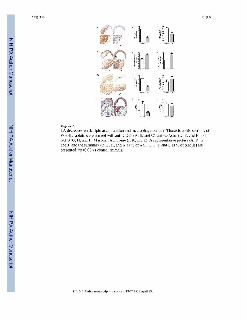
Figure 2.LA decreases aortic lipid accumulation and macrophage content. Thoracic aortic sections ofWHHL rabbits were stained with anti-CD68 (A, B, and C); anti-α-Actin (D, E, and F); oilred O (G, H, and I); Masson’s trichrome (J, K, and L). A representative picture (A, D, G,and J) and the summary (B, E, H, and K as % of wall; C, F, J, and L as % of plaque) arepresented. *p<0.05 vs control animals.
Ying et al. Page 9
Life Sci. Author manuscript; available in PMC 2011 April 13.
NIH
-PA Author Manuscript
NIH
-PA Author Manuscript
NIH
-PA Author Manuscript
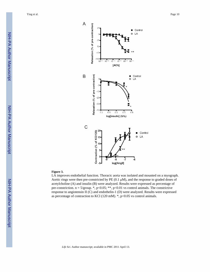
Figure 3.LA improves endothelial function. Thoracic aorta was isolated and mounted on a myograph.Aortic rings were then pre-constricted by PE (0.1 μM), and the response to graded doses ofacetylcholine (A) and insulin (B) were analyzed. Results were expressed as percentage ofpre-constriction. n = 5/group. *, p<0.05; **, p<0.01 vs control animals. The constrictiveresponse to angiotensin II (C) and endothelin-1 (D) were analyzed. Results were expressedas percentage of contraction to KCl (120 mM). *, p<0.05 vs control animals.
Ying et al. Page 10
Life Sci. Author manuscript; available in PMC 2011 April 13.
NIH
-PA Author Manuscript
NIH
-PA Author Manuscript
NIH
-PA Author Manuscript
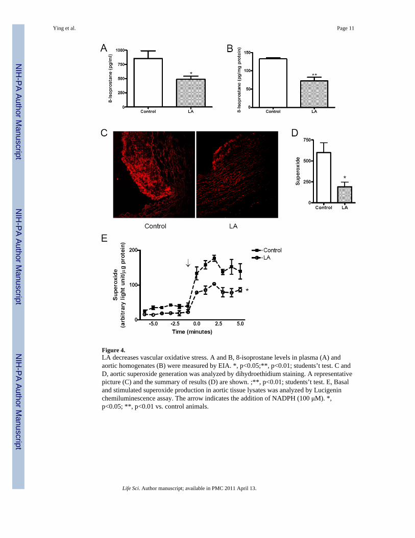
Figure 4.LA decreases vascular oxidative stress. A and B, 8-isoprostane levels in plasma (A) andaortic homogenates (B) were measured by EIA. *, p<0.05;**, p<0.01; students’t test. C andD, aortic superoxide generation was analyzed by dihydroethidium staining. A representativepicture (C) and the summary of results (D) are shown. ;**, p<0.01; students’t test. E, Basaland stimulated superoxide production in aortic tissue lysates was analyzed by Lucigeninchemiluminescence assay. The arrow indicates the addition of NADPH (100 μM). *,p<0.05; **, p<0.01 vs. control animals.
Ying et al. Page 11
Life Sci. Author manuscript; available in PMC 2011 April 13.
NIH
-PA Author Manuscript
NIH
-PA Author Manuscript
NIH
-PA Author Manuscript
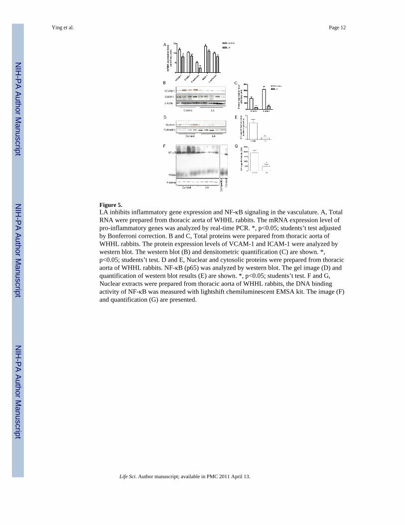
Figure 5.LA inhibits inflammatory gene expression and NF-κB signaling in the vasculature. A, TotalRNA were prepared from thoracic aorta of WHHL rabbits. The mRNA expression level ofpro-inflammatory genes was analyzed by real-time PCR. *, p<0.05; students’t test adjustedby Bonferroni correction. B and C, Total proteins were prepared from thoracic aorta ofWHHL rabbits. The protein expression levels of VCAM-1 and ICAM-1 were analyzed bywestern blot. The western blot (B) and densitometric quantification (C) are shown. *,p<0.05; students’t test. D and E, Nuclear and cytosolic proteins were prepared from thoracicaorta of WHHL rabbits. NF-κB (p65) was analyzed by western blot. The gel image (D) andquantification of western blot results (E) are shown. *, p<0.05; students’t test. F and G,Nuclear extracts were prepared from thoracic aorta of WHHL rabbits, the DNA bindingactivity of NF-κB was measured with lightshift chemiluminescent EMSA kit. The image (F)and quantification (G) are presented.
Ying et al. Page 12
Life Sci. Author manuscript; available in PMC 2011 April 13.
NIH
-PA Author Manuscript
NIH
-PA Author Manuscript
NIH
-PA Author Manuscript
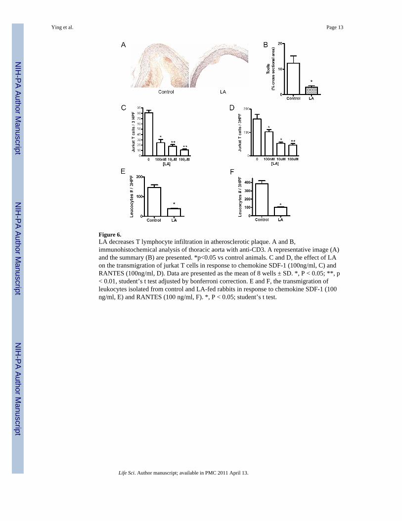
Figure 6.LA decreases T lymphocyte infiltration in atherosclerotic plaque. A and B,immunohistochemical analysis of thoracic aorta with anti-CD3. A representative image (A)and the summary (B) are presented. *p<0.05 vs control animals. C and D, the effect of LAon the transmigration of jurkat T cells in response to chemokine SDF-1 (100ng/ml, C) andRANTES (100ng/ml, D). Data are presented as the mean of 8 wells ± SD. *, P < 0.05; **, p< 0.01, student’s t test adjusted by bonferroni correction. E and F, the transmigration ofleukocytes isolated from control and LA-fed rabbits in response to chemokine SDF-1 (100ng/ml, E) and RANTES (100 ng/ml, F). *, P < 0.05; student’s t test.
Ying et al. Page 13
Life Sci. Author manuscript; available in PMC 2011 April 13.
NIH
-PA Author Manuscript
NIH
-PA Author Manuscript
NIH
-PA Author Manuscript