alternative c3 complement system: lipids and atherosclerosis
TRANSCRIPT
International Journal of
Molecular Sciences
Article
Alternative C3 Complement System: Lipids and Atherosclerosis
Maisa Garcia-Arguinzonis 1, Elisa Diaz-Riera 1 , Esther Peña 1,2 , Rafael Escate 1,2, Oriol Juan-Babot 1,Pedro Mata 3, Lina Badimon 1,2,4,† and Teresa Padro 1,2,*,†
�����������������
Citation: Garcia-Arguinzonis, M.;
Diaz-Riera, E.; Peña, E.; Escate, R.;
Juan-Babot, O.; Mata, P.; Badimon, L.;
Padro, T. Alternative C3 Complement
System: Lipids and Atherosclerosis.
Int. J. Mol. Sci. 2021, 22, 5122.
https://doi.org/10.3390/ijms22105122
Academic Editors: Maria I.
Dorobantu and Maya Simionescu
Received: 17 April 2021
Accepted: 6 May 2021
Published: 12 May 2021
Publisher’s Note: MDPI stays neutral
with regard to jurisdictional claims in
published maps and institutional affil-
iations.
Copyright: © 2021 by the authors.
Licensee MDPI, Basel, Switzerland.
This article is an open access article
distributed under the terms and
conditions of the Creative Commons
Attribution (CC BY) license (https://
creativecommons.org/licenses/by/
4.0/).
1 Cardiovascular Program-ICCC, Research Institute-Hospital Santa Creu i Sant Pau, IIB-Sant Pau,08025 Barcelona, Spain; [email protected] (M.G.-A.); [email protected] (E.D.-R.);[email protected] (E.P.); [email protected] (R.E.); [email protected] (O.J.-B.);[email protected] (L.B.)
2 Centro de Investigación Biomédica en Red Cardiovascular (CIBERCV), Instituto de Salud Carlos III,28029 Madrid, Spain
3 Fundación Hipercolesterolemia Familiar, 28010 Madrid, Spain; [email protected] Cardiovascular Research Chair, UAB, 08025 Barcelona, Spain* Correspondence: [email protected]; Tel.: +34-935-565-886; Fax: +34-935-565-559† Both authors contributed equally.
Abstract: Familial hypercholesterolemia (FH) is increasingly associated with inflammation, a pheno-type that persists despite treatment with lipid lowering therapies. The alternative C3 complementsystem (C3), as a key inflammatory mediator, seems to be involved in the atherosclerotic process;however, the relationship between C3 and lipids during plaque progression remains unknown. Theaim of the study was to investigate by a systems biology approach the role of C3 in relation tolipoprotein levels during atherosclerosis (AT) progression and to gain a better understanding onthe effects of C3 products on the phenotype and function of human lipid-loaded vascular smoothmuscle cells (VSMCs). By mass spectrometry and differential proteomics, we found the extracellularmatrix (ECM) of human aortas to be enriched in active components of the C3 complement system,with a significantly different proteomic signature in AT segments. Thus, C3 products were moreabundant in AT-ECM than in macroscopically normal segments. Furthermore, circulating C3 levelswere significantly elevated in FH patients with subclinical coronary AT, evidenced by computedtomographic angiography. However, no correlation was identified between circulating C3 levels andthe increase in plaque burden, indicating a local regulation of the C3 in AT arteries. In cell culturestudies of human VSMCs, we evidenced the expression of C3, C3aR (anaphylatoxin receptor) andthe integrin αMβ2 receptor for C3b/iC3b (RT-PCR and Western blot). C3mRNA was up-regulatedin lipid-loaded human VSMCs, and C3 protein significantly increased in cell culture supernatants,indicating that the C3 products in the AT-ECM have a local vessel-wall niche. Interestingly, C3a andiC3b (C3 active fragments) have functional effects on VSMCs, significantly reversing the inhibition ofVSMC migration induced by aggregated LDL and stimulating cell spreading, organization of F-actinstress fibers and attachment during the adhesion of lipid-loaded human VSMCs. This study, by usinga systems biology approach, identified molecular processes involving the C3 complement system invascular remodeling and in the progression of advanced human atherosclerotic lesions.
Keywords: atherosclerosis; cardiovascular disease; complement system; proteomics; mass spectrom-etry
1. Introduction
Familial hypercholesterolemia (FH), an autosomal-dominant disorder mainly causedby the loss-of-function mutations in the low-density lipoprotein (LDL) receptor, is asso-ciated with an increased risk of atherosclerosis and ultimately premature cardiovascularevent presentation, resulting in lifelong exposure to high-LDL cholesterol levels [1–5].Increasing evidence suggests that FH patients recurrently present an inflammatory phe-notype that is maintained despite treatment with lipid lowering therapies according to
Int. J. Mol. Sci. 2021, 22, 5122. https://doi.org/10.3390/ijms22105122 https://www.mdpi.com/journal/ijms

Int. J. Mol. Sci. 2021, 22, 5122 2 of 17
guidelines [6–9]. We and others have demonstrated that adult FH patients have higher lev-els of extracellular microvesicles originating from inflammatory cells in plasma [6] as wellas circulating mononuclear cells and monocyte-derived macrophages with inflammatoryphenotypes [7,10].
The complement system is an important component of the innate immunity andplays a key role in the regulation of inflammation. Particularly relevant is the activationof the alternative C3 system, in which the different pathways of the complement systemconverge, leading to the formation of active C3 proteolytic products, C5 convertases andeventually the activation of the terminal complement proteins C5 to C9 and the formationof the membrane attack complex (MAC) [11,12]. The C3 system is tightly regulated bya cascade of components, including activators (Factor B), inhibitors (Factor H) and cellsurface proteins acting as receptors (CR1, C3aR and αMβ2 integrin).
Atherosclerosis is widely recognized as a lipid-induced chronic inflammatory diseaseof the arterial wall with the activation of resident cells and recruitment of circulatingleukocytes [13]. The complement system has been repeatedly associated with vascularremodeling [14] and atherosclerosis [15]. Results from experimental animal models andhuman samples suggest that complement activation may exert dual atheroprotectiveand proatherogenic effects mainly associated with the initial and terminal stages of thecomplement cascade, respectively [16,17]. However, the impact of the C3 complementsystem in atherogenesis is not fully understood.
C3 complement products and their cell receptors have been detected by immunohisto-chemistry in areas with atherosclerotic lesions of different severity in human arteries [18,19],which has led to the hypothesis that local activation of the alternative-complement systemis involved in atherosclerotic plaque progression and complication [15]. In contrast, priorstudies in mice models of atherosclerosis (Ldlr-/- or ApoE-/- Ldlr-/- background) andknock-out C3 expression (C3-/-) evidenced that atherosclerotic lesions developed in theabsence of C3 have a lower content of vascular smooth muscle cells (VSMCs) and collagen,hallmark of vulnerable plaques, and are of a larger size than those plaques developed inanimals with a sufficient content of C3 [20,21]. A potential effect of C3 on the proliferationof VSMCs during atherogenesis was suggested by a recent study, in ApoE-/- mice fed ahigh-fat Western diet, in which dedifferentiated clonally expanding vascular SMC showedan up-regulated C3 expression [22] and also by prior results linking C3a with the increasingproliferation of mouse VSMCs [23].
Results from two recent studies evidenced a noticeable increase in arterial wall inflam-mation, assessed by fluorodeoxyglucose positron emission tomography imaging, in FHpatients with high LDL levels with healthy controls [24,25]. The up-regulation of compo-nents of the complement cascade, including C3-derived products, have been reported intwo studies in FH patients with no clinical evidence of coronary artery disease [23,26]. Todate, however, we do not know whether circulating C3 levels relate to the intensity andprofile of atherosclerotic plaque burden. Moreover, little is known regarding the interplayamong LDL, C3 complement products and VSMCs, although VSMCs are the key cellularcomponents in the development and complication of atherosclerotic lesions.
Therefore, the present study was conducted to investigate the relationship betweencirculating C3 complement, lipids and atherosclerotic plaque burden in FH patients withsubclinical atherosclerosis. In addition, using a mass spectrometry-based proteomic ap-proach, combined with transcriptomic analysis and in vitro functional assays, we analyzedthe pattern of the C3 complement components in the extracellular matrix (ECM) of hu-man atherosclerotic plaques and investigated C3 complement expression and effects onmigration kinetics of lipid-loaded VSMCs.
2. Results2.1. Characteristics of the FH Patient Population
FH patients from the SAFEHEART cohort were included (N = 49; 31 men and18 women). The mean age was 44.7 ± 10.5 years (men: 45.4 ± 11.4 years; women

Int. J. Mol. Sci. 2021, 22, 5122 3 of 17
43.7 ± 9.2 years). The baseline demographic and clinical characteristics of the studiedFH-population are shown in Table 1. All subjects were on lipid-lowering treatment (LLT)and treated with statins as per the guidelines for > 1 year (mean treated years beforeinclusion in the study were 14.9 ± 6.7 years). The mean LDL-cholesterol (LDL-C) in the FHgroup was 136.3 ± 36.0 mg/dL.
Table 1. Demographic, biochemical and clinical variables: Familial hypercholesterolemia and healthy subject groups.
Familial Hyperchlesterolemian = 49
Healthy Subjectsn = 28
Demographic Characteristics; mean ± SDFemale/male, n 18/31 16/12Age, years 38.6 ± 11.3 24.5 ± 4.6Risk Factors; n (%)Smokers 14 (29) 12 (43)Hypertension 1 (2) 0 (0)Diabetes mellitus 0 (0) 0 (0)Dyslipidaemia 48 (98) 0 (0)Biochemical Data, Mean ± SDTotal cholesterol, mg/dL 282 ± 72 170 ± 20Triglycerides, mg/dL 104 ± 67 77 ± 35HDL cholesterol, mg/dL 47 ± 11 56 ± 15LDL cholesterol, mg/dL 221 ± 78 99 ± 15Apo AI, mg/dL 135 ± 20 139 ± 29Apo B, mg/dL 134 ± 41 61 ± 10Lipoprotein(a), mg/dL 42 ± 35 18 ± 21Glucose, mg/dL 89 ± 9 78 ± 9C-reactive protein 1.86 ± 2.6 0.73 ± 0.2Subclinical Atherosclerotic Disease; (%)Plaque burden, % 23.5 ± 6.3 -Calcium burden, % 2.2 ± 2.5 -Non-calcium burden 21.3 ± 5.3 -BAckground Medication; n (%)Angiotensin-converting-enzyme inhibitors 0 (0) 0 (0)Angiotensin II receptor blockers 1 (2) 0 (0)Beta-blockers 0 (0) 0 (0)Diuretics 2 (4) 0 (0)Statins * 39 (80) 0 (0)
SD: standard deviation * Includes: rosuvastatin, ezetimibe, atorvastatin, simvastatin, lovastatin, pravastatin, Fluvastatin, pitavastatin,resins, and fibrates. Healthy subject population was used to establish the C3 range in a healthy group.
None of the FH patients had a clinical history of cardiovascular disease (CVD). Lessthan 5% of patients with FH presented hypertension or Type-2 diabetes. Eleven FH patients(22%) were active tobacco smokers. The mean value for 5- and 10-year CVD risk in the FHpatients, according the SAFEHEART-risk score (SAFEHEART-RS), was 1.00 ± 0.76 % and2.13 ± 1.6 %, respectively.
All FH subjects included in the study presented subclinical atherosclerosis, assessedby computed tomographic angiography (CTA) and quantified by SAPC software [27]. Themean value for the total plaque burden was 23.5 ± 6.3% and, specifically, the mediancalcified plaque burden was 2.2 ± 2.5%, and that of non-calcified-plaque burden was21.3 ± 5.3%. FH patients with plaque burden above the median values (high plaqueburden) had a significantly higher estimated cardiovascular risk based on SAFEHEART-RS, both at 5 (0.77 ± 0.76% vs. 1.24 ± 0.15%; p = 0.03) and 10 years (1.65 ± 1.59% vs.2.60 ± 0.15%; p = 0.03).
2.2. C3 Complement in Patients with Hypercholesterolemia and Subclinical Atherosclerosis
Circulating levels of C3 complement were significantly elevated (p < 0.001) in subjectswith a genetic diagnosis of FH and subclinical coronary atherosclerosis, when comparedto the plasma levels of C3 in young healthy subjects at low atherosclerotic risk (subjectswithout CV risk factors and age between 18 and 35 years) (Figure 1A). Plasma C3 comple-ment levels were significantly correlated with LDL-C levels (Spearman correlation: Rhovalue = 0.412, p < 0.001), ApoB levels (Spearman correlation: Rho = 0.562, p < 0.001) and
Int. J. Mol. Sci. 2021, 22, 5122 4 of 17
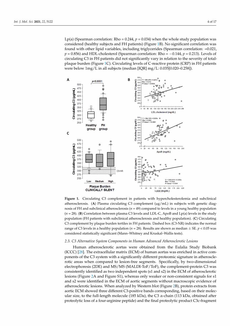
Lp(a) (Spearman correlation: Rho = 0.244, p = 0.034) when the whole study population wasconsidered (healthy subjects and FH patients) (Figure 1B). No significant correlation wasfound with other lipid variables, including triglycerides (Spearman correlation: =0.021,p = 0.856) and HDL-cholesterol (Spearman correlation: Rho = −0.144, p = 0.213). Levels ofcirculating C3 in FH patients did not significantly vary in relation to the severity of total-plaque burden (Figure 1C). Circulating levels of C-reactive protein (CRP) in FH patientswere below 1mg/L in all subjects (median [IQR] mg/L: 0.035[0.020–0.258]).
Int. J. Mol. Sci. 2021, 22, x FOR PEER REVIEW 4 of 18
All FH subjects included in the study presented subclinical atherosclerosis, assessed by computed tomographic angiography (CTA) and quantified by SAPC software [27]. The mean value for the total plaque burden was 23.5 ± 6.3% and, specifically, the median cal-cified plaque burden was 2.2 ± 2.5%, and that of non-calcified-plaque burden was 21.3 ± 5.3%. FH patients with plaque burden above the median values (high plaque burden) had a significantly higher estimated cardiovascular risk based on SAFEHEART-RS, both at 5 (0.77 ± 0.76% vs. 1.24 ± 0.15%; p = 0.03) and 10 years (1.65 ± 1.59% vs. 2.60 ± 0.15%; p = 0.03).
2.2. C3 Complement in Patients with Hypercholesterolemia and Subclinical Atherosclerosis Circulating levels of C3 complement were significantly elevated (p < 0.001) in subjects
with a genetic diagnosis of FH and subclinical coronary atherosclerosis, when compared to the plasma levels of C3 in young healthy subjects at low atherosclerotic risk (subjects without CV risk factors and age between 18 and 35 years) (Figure 1A). Plasma C3 comple-ment levels were significantly correlated with LDL-C levels (Spearman correlation: Rho value = 0.412, p < 0.001), ApoB levels (Spearman correlation: Rho = 0.562, p < 0.001) and Lp(a) (Spearman correlation: Rho = 0.244, p = 0.034) when the whole study population was considered (healthy subjects and FH patients) (Figure 1B). No significant correlation was found with other lipid variables, including triglycerides (Spearman correlation: =0.021, p = 0.856) and HDL-cholesterol (Spearman correlation: Rho = −0.144, p = 0.213). Levels of circulating C3 in FH patients did not significantly vary in relation to the severity of total-plaque burden (Figure 1C). Circulating levels of C-reactive protein (CRP) in FH patients were below 1mg/L in all subjects (median [IQR] mg/L: 0.035[0.020-0.258]).
Figure 1. Circulating C3 complement in patients with hypercholesterolemia and subclinical athero-sclerosis. (A) Plasma circulating C3 complement (µg/mL) in subjects with genetic diagnosis of FH and subclinical atherosclerosis (n = 49) compared to levels in a young healthy population (n = 28). (B) Correlation between plasma C3 levels and LDL-C, ApoB and Lp(a) levels in the study popula-tion (FH patients with subclinical atherosclerosis and healthy population). (C) Circulating C3 com-plement by plaque burden tertiles in FH patients. Dashed box (C3-NR) indicates the normal range
Figure 1. Circulating C3 complement in patients with hypercholesterolemia and subclinicalatherosclerosis. (A) Plasma circulating C3 complement (µg/mL) in subjects with genetic diag-nosis of FH and subclinical atherosclerosis (n = 49) compared to levels in a young healthy population(n = 28). (B) Correlation between plasma C3 levels and LDL-C, ApoB and Lp(a) levels in the studypopulation (FH patients with subclinical atherosclerosis and healthy population). (C) CirculatingC3 complement by plaque burden tertiles in FH patients. Dashed box (C3-NR) indicates the normalrange of C3 levels in a healthy population (n = 28). Results are shown as median ± SE. p < 0.05 wasconsidered statistically significant (Mann–Whitney and Kruskal–Wallis tests).
2.3. C3 Alternative System Components in Human Advanced Atherosclerotic Lesions
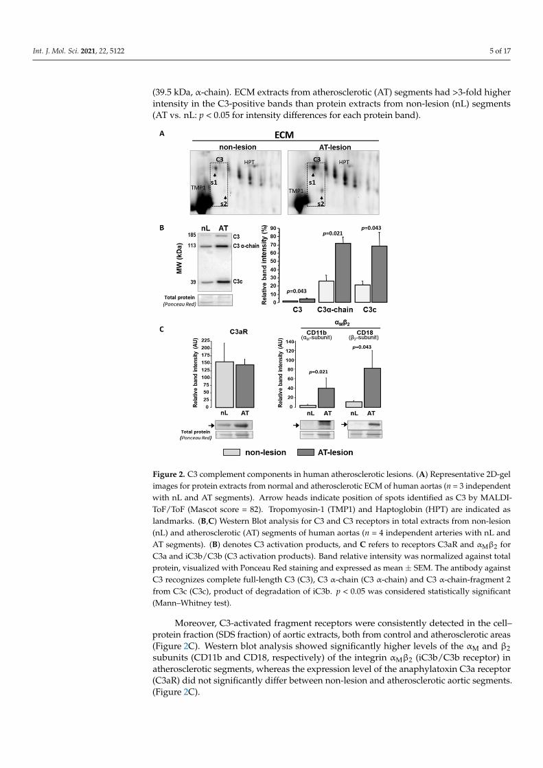
Human atherosclerotic aortas were obtained from the Eulalia Study Biobank(ICCC) [28]. The extracellular matrix (ECM) of human aortas was enriched in active com-ponents of the C3 system with a significantly different proteomic signature in atheroscle-rotic areas when compared to lesion-free segments. Specifically, by two-dimensionalelectrophoresis (2DE) and MS/MS (MALDI-ToF/ToF), the complement-protein C3 wasconsistently identified as two independent spots (s1 and s2) in the ECM of atheroscleroticlesions (Figure 2A and Figure S1), whereas only weaker or non-consistent signals for s1and s2 were identified in the ECM of aortic segments without macroscopic evidence ofatherosclerotic lesions. When analyzed by Western blot (Figure 2B), protein extracts fromaortic ECM showed three different C3-positive bands corresponding, based on their molec-ular size, to the full-length molecule (185 kDa), the C3 α-chain (113 kDa, obtained afterproteolytic loss of a four-arginine peptide) and the final proteolytic product C3c-fragment
Int. J. Mol. Sci. 2021, 22, 5122 5 of 17
(39.5 kDa, α-chain). ECM extracts from atherosclerotic (AT) segments had >3-fold higherintensity in the C3-positive bands than protein extracts from non-lesion (nL) segments(AT vs. nL: p < 0.05 for intensity differences for each protein band).
Int. J. Mol. Sci. 2021, 22, x FOR PEER REVIEW 5 of 18
of C3 levels in a healthy population (n = 28). Results are shown as median ± SE. p < 0.05 was consid-ered statistically significant (Mann–Whitney and Kruskal–Wallis tests).
2.3. C3 Alternative System Components in Human Advanced Atherosclerotic Lesions Human atherosclerotic aortas were obtained from the Eulalia Study Biobank (ICCC)
[28]. The extracellular matrix (ECM) of human aortas was enriched in active components of the C3 system with a significantly different proteomic signature in atherosclerotic areas when compared to lesion-free segments. Specifically, by two-dimensional electrophoresis (2DE) and MS/MS (MALDI-ToF/ToF), the complement-protein C3 was consistently iden-tified as two independent spots (s1 and s2) in the ECM of atherosclerotic lesions (Figures 2A and S1), whereas only weaker or non-consistent signals for s1 and s2 were identified in the ECM of aortic segments without macroscopic evidence of atherosclerotic lesions. When analyzed by Western blot (Figure 2B), protein extracts from aortic ECM showed three different C3-positive bands corresponding, based on their molecular size, to the full-length molecule (185 kDa), the C3 α-chain (113 kDa, obtained after proteolytic loss of a four-arginine peptide) and the final proteolytic product C3c-fragment (39.5 kDa, α-chain). ECM extracts from atherosclerotic (AT) segments had >3-fold higher intensity in the C3-positive bands than protein extracts from non-lesion (nL) segments (AT vs. nL: p < 0.05 for intensity differences for each protein band).
Figure 2. C3 complement components in human atherosclerotic lesions. (A) Representative 2D-gel images for protein extracts from normal and atherosclerotic ECM of human aortas (n = 3 independ-ent with nL and AT segments). Arrow heads indicate position of spots identified as C3 by MALDI-ToF/ToF (Mascot score = 82). Tropomyosin-1 (TMP1) and Haptoglobin (HPT) are indicated as land-marks. (B,C) Western Blot analysis for C3 and C3 receptors in total extracts from non-lesion (nL)
Figure 2. C3 complement components in human atherosclerotic lesions. (A) Representative 2D-gelimages for protein extracts from normal and atherosclerotic ECM of human aortas (n = 3 independentwith nL and AT segments). Arrow heads indicate position of spots identified as C3 by MALDI-ToF/ToF (Mascot score = 82). Tropomyosin-1 (TMP1) and Haptoglobin (HPT) are indicated aslandmarks. (B,C) Western Blot analysis for C3 and C3 receptors in total extracts from non-lesion(nL) and atherosclerotic (AT) segments of human aortas (n = 4 independent arteries with nL andAT segments). (B) denotes C3 activation products, and C refers to receptors C3aR and αMβ2 forC3a and iC3b/C3b (C3 activation products). Band relative intensity was normalized against totalprotein, visualized with Ponceau Red staining and expressed as mean ± SEM. The antibody againstC3 recognizes complete full-length C3 (C3), C3 α-chain (C3 α-chain) and C3 α-chain-fragment 2from C3c (C3c), product of degradation of iC3b. p < 0.05 was considered statistically significant(Mann–Whitney test).
Moreover, C3-activated fragment receptors were consistently detected in the cell–protein fraction (SDS fraction) of aortic extracts, both from control and atherosclerotic areas(Figure 2C). Western blot analysis showed significantly higher levels of the αM and β2subunits (CD11b and CD18, respectively) of the integrin αMβ2 (iC3b/C3b receptor) inatherosclerotic segments, whereas the expression level of the anaphylatoxin C3a receptor(C3aR) did not significantly differ between non-lesion and atherosclerotic aortic segments.(Figure 2C).
Int. J. Mol. Sci. 2021, 22, 5122 6 of 17
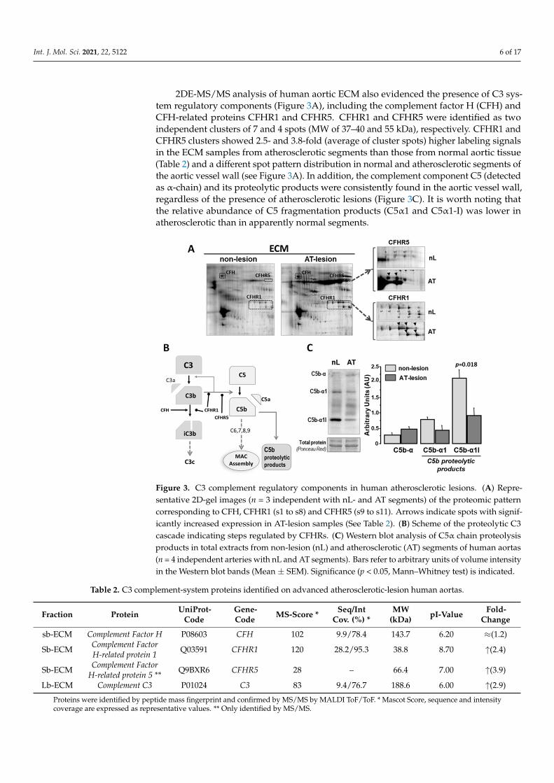
2DE-MS/MS analysis of human aortic ECM also evidenced the presence of C3 sys-tem regulatory components (Figure 3A), including the complement factor H (CFH) andCFH-related proteins CFHR1 and CFHR5. CFHR1 and CFHR5 were identified as twoindependent clusters of 7 and 4 spots (MW of 37–40 and 55 kDa), respectively. CFHR1 andCFHR5 clusters showed 2.5- and 3.8-fold (average of cluster spots) higher labeling signalsin the ECM samples from atherosclerotic segments than those from normal aortic tissue(Table 2) and a different spot pattern distribution in normal and atherosclerotic segments ofthe aortic vessel wall (see Figure 3A). In addition, the complement component C5 (detectedas α-chain) and its proteolytic products were consistently found in the aortic vessel wall,regardless of the presence of atherosclerotic lesions (Figure 3C). It is worth noting thatthe relative abundance of C5 fragmentation products (C5α1 and C5α1-I) was lower inatherosclerotic than in apparently normal segments.
Int. J. Mol. Sci. 2021, 22, x FOR PEER REVIEW 6 of 18
and atherosclerotic (AT) segments of human aortas (n = 4 independent arteries with nL and AT segments). (B) denotes C3 activation products, and C refers to receptors C3aR and αMβ2 for C3a and iC3b/C3b (C3 activation products). Band relative intensity was normalized against total protein, visualized with Ponceau Red staining and expressed as mean ± SEM. The antibody against C3 rec-ognizes complete full-length C3 (C3), C3 α-chain (C3 α-chain) and C3 α-chain-fragment 2 from C3c (C3c), product of degradation of iC3b. p < 0.05 was considered statistically significant (Mann–Whit-ney test).
Moreover, C3-activated fragment receptors were consistently detected in the cell–protein fraction (SDS fraction) of aortic extracts, both from control and atherosclerotic ar-eas (Figure 2C). Western blot analysis showed significantly higher levels of the αM and β2 subunits (CD11b and CD18, respectively) of the integrin αMβ2 (iC3b/C3b receptor) in atherosclerotic segments, whereas the expression level of the anaphylatoxin C3a receptor (C3aR) did not significantly differ between non-lesion and atherosclerotic aortic segments. (Figure 2C).
2DE-MS/MS analysis of human aortic ECM also evidenced the presence of C3 system regulatory components (Figure 3A), including the complement factor H (CFH) and CFH-related proteins CFHR1 and CFHR5. CFHR1 and CFHR5 were identified as two inde-pendent clusters of 7 and 4 spots (MW of 37–40 and 55 kDa), respectively. CFHR1 and CFHR5 clusters showed 2.5- and 3.8-fold (average of cluster spots) higher labeling signals in the ECM samples from atherosclerotic segments than those from normal aortic tissue (Table 2) and a different spot pattern distribution in normal and atherosclerotic segments of the aortic vessel wall (see Figure 3A). In addition, the complement component C5 (de-tected as α-chain) and its proteolytic products were consistently found in the aortic vessel wall, regardless of the presence of atherosclerotic lesions (Figure 3C). It is worth noting that the relative abundance of C5 fragmentation products (C5α1 and C5α1-I) was lower in atherosclerotic than in apparently normal segments.
Figure 3. C3 complement regulatory components in human atherosclerotic lesions. (A) Representa-tive 2D-gel images (n = 3 independent with nL- and AT segments) of the proteomic pattern corre-sponding to CFH, CFHR1 (s1 to s8) and CFHR5 (s9 to s11). Arrows indicate spots with significantly increased expression in AT-lesion samples (See Table 2). (B) Scheme of the proteolytic C3 cascade indicating steps regulated by CFHRs. (C) Western blot analysis of C5α chain proteolysis products in total extracts from non-lesion (nL) and atherosclerotic (AT) segments of human aortas (n = 4
Figure 3. C3 complement regulatory components in human atherosclerotic lesions. (A) Repre-sentative 2D-gel images (n = 3 independent with nL- and AT segments) of the proteomic patterncorresponding to CFH, CFHR1 (s1 to s8) and CFHR5 (s9 to s11). Arrows indicate spots with signif-icantly increased expression in AT-lesion samples (See Table 2). (B) Scheme of the proteolytic C3cascade indicating steps regulated by CFHRs. (C) Western blot analysis of C5α chain proteolysisproducts in total extracts from non-lesion (nL) and atherosclerotic (AT) segments of human aortas(n = 4 independent arteries with nL and AT segments). Bars refer to arbitrary units of volume intensityin the Western blot bands (Mean ± SEM). Significance (p < 0.05, Mann–Whitney test) is indicated.
Table 2. C3 complement-system proteins identified on advanced atherosclerotic-lesion human aortas.
Fraction Protein UniProt-Code
Gene-Code MS-Score * Seq/Int
Cov. (%) *MW
(kDa) pI-Value Fold-Change
sb-ECM Complement Factor H P08603 CFH 102 9.9/78.4 143.7 6.20 ≈(1.2)
Sb-ECM Complement FactorH-related protein 1 Q03591 CFHR1 120 28.2/95.3 38.8 8.70 ↑(2.4)
Sb-ECM Complement FactorH-related protein 5 ** Q9BXR6 CFHR5 28 – 66.4 7.00 ↑(3.9)
Lb-ECM Complement C3 P01024 C3 83 9.4/76.7 188.6 6.00 ↑(2.9)
Proteins were identified by peptide mass fingerprint and confirmed by MS/MS by MALDI ToF/ToF. * Mascot Score, sequence and intensitycoverage are expressed as representative values. ** Only identified by MS/MS.

Int. J. Mol. Sci. 2021, 22, 5122 7 of 17
2.4. C3 Alternative Pathway Components Expression in Vascular Wall Resident Cells
Complement factor C3 was consistently transcribed by human VSMCs (hVSMCs), andC3mRNA levels were up-regulated (1.7-fold, p < 0.05; Figure 4A) in lipid-loaded hVSMCs(24 h exposure to 100 µg/mL aggregated LDL agLDL). The significant protein expressionof C3, receptor C3aR (anaphylatoxin receptor) and integrin αMβ2 (CD11b/CD18) receptorfor C3b/iC3b was also observed in hVSMCs (Figure 4B). In contrast, agLDL did notsignificantly affect the protein expression levels of the cell membrane receptors C3aRand integrin αMβ2 (Figure 4C). Interestingly, the protein levels of C3 (C3 α-chain) weresignificantly increased in cell culture supernatants when hVSMCs were incubated in thepresence of agLDL (Figure 4B). All together, these results indicate that there is a localsynthesis of C3 components that were released to the ECM of atherosclerotic plaques.
Int. J. Mol. Sci. 2021, 22, x FOR PEER REVIEW 7 of 18
independent arteries with nL and AT segments). Bars refer to arbitrary units of volume intensity in the Western blot bands (Mean ± SEM). Significance (p < 0.05, Mann–Whitney test) is indicated.
Table 2. C3 complement-system proteins identified on advanced atherosclerotic-lesion human aortas.
Fraction Protein UniProt-
Code Gene-Code
MS-Score *
Seq/Int Cov. (%) *
MW (kDa) pI-Value
Fold-Change
sb-ECM Complement Factor H P08603 CFH 102 9.9/78.4 143.7 6.20 ≈(1.2)
Sb-ECM Complement Factor H-
related protein 1 Q03591 CFHR1 120 28.2/95.3 38.8 8.70 ↑ (2.4)
Sb-ECM Complement Factor H-
related protein 5 ** Q9BXR6 CFHR5 28 -- 66.4 7.00 ↑ (3.9)
Lb-ECM Complement C3 P01024 C3 83 9.4/76.7 188.6 6.00 ↑ (2.9) Proteins were identified by peptide mass fingerprint and confirmed by MS/MS by MALDI ToF/ToF. * Mascot Score, se-quence and intensity coverage are expressed as representative values. ** Only identified by MS/MS.
2.4. C3 Alternative Pathway Components Expression in Vascular Wall Resident Cells Complement factor C3 was consistently transcribed by human VSMCs (hVSMCs),
and C3mRNA levels were up-regulated (1.7-fold, p < 0.05; Figure 4A) in lipid-loaded hVSMCs (24 h exposure to 100 µg/mL aggregated LDL agLDL). The significant protein expression of C3, receptor C3aR (anaphylatoxin receptor) and integrin αMβ2 (CD11b/CD18) receptor for C3b/iC3b was also observed in hVSMCs (Figure 4B). In con-trast, agLDL did not significantly affect the protein expression levels of the cell membrane receptors C3aR and integrin αMβ2 (Figure 4C). Interestingly, the protein levels of C3 (C3 α-chain) were significantly increased in cell culture supernatants when hVSMCs were in-cubated in the presence of agLDL (Figure 4B). All together, these results indicate that there is a local synthesis of C3 components that were released to the ECM of atherosclerotic plaques.
Figure 4. C3 alternative pathway components expression in human VSMCs. (A) mRNA quantifica-tion by real-time PCR using specific primers for human C3 in human VSMCs treated with or without agLDL (100 µg/mL). (B) Protein levels of C3 and C3-derived products in the supernatant of hVSMCs treated with or without agLDL (100 µg/mL). Human serum (Serum) was used as a positive control for C3 (n = 3 independent experiments). (C) Western blot analysis for C3a receptor (C3aR) and αMβ2 (C11b/CD18) integrin (receptor for C3b/iC3b) in lysates of hVSMCs incubated with or without agLDL (100 µg/mL). Band intensity is given in arbitrary units as mean ± SEM and statistical signifi-cance (p < 0.05, Mann–Whitney test) is indicated (n = 4 independent experiments).
Figure 4. C3 alternative pathway components expression in human VSMCs. (A) mRNA quantifi-cation by real-time PCR using specific primers for human C3 in human VSMCs treated with orwithout agLDL (100 µg/mL). (B) Protein levels of C3 and C3-derived products in the supernatant ofhVSMCs treated with or without agLDL (100 µg/mL). Human serum (Serum) was used as a positivecontrol for C3 (n = 3 independent experiments). (C) Western blot analysis for C3a receptor (C3aR)and αMβ2 (C11b/CD18) integrin (receptor for C3b/iC3b) in lysates of hVSMCs incubated with orwithout agLDL (100 µg/mL). Band intensity is given in arbitrary units as mean ± SEM and statisticalsignificance (p < 0.05, Mann–Whitney test) is indicated (n = 4 independent experiments).
2.5. Exogenous C3 Proteolytic Products, AgLDL and VSMC Function
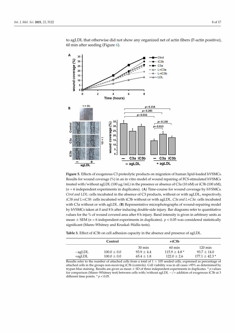
Lipid-loaded VSMCs have an impaired migration rate and cell attachment dynam-ics [29–31]. As shown in Figure 5, exogenously added C3 proteolytic products (10 nM C3aor 100 nM iC3b) partially reversed the impairment of the human VSMC (hVSMC) repairfunction induced by aggregated LDL (agLDL) to levels that did not differ significantly fromthe wound repairing capacity of hVSMC control cells. C3a induced a significant increasein the migrating capacity of lipid-loaded hVSMCs into the wound area. A similar trend,although non-significant, was observed with iC3b. C3 proteolytic products did not affectthe wound-repairing capacity of control VSMCs (not exposed to agLDL).
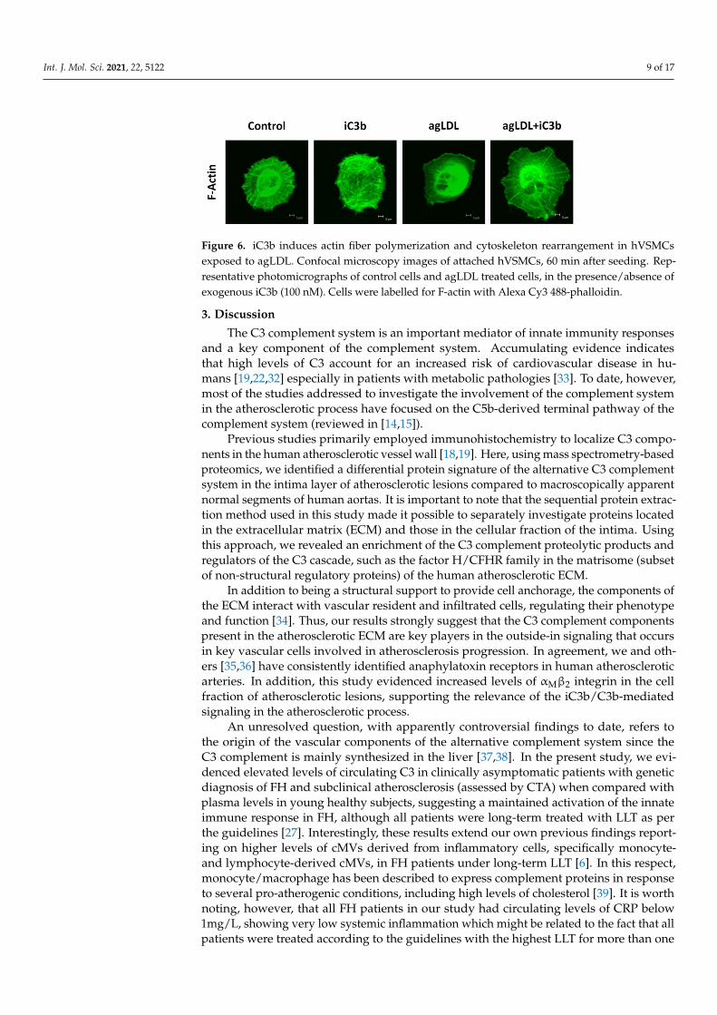
Moreover, the addition of iC3b (100nM) to hVSMCs in adhesion experiments induced asignificant increase in the attachment capacity of the cells, both in the absence and presenceof agLDL. The effect was more evident (higher percentage of increase) and more prolongedin time (up to 2 h after seeding) in the lipid-loaded hVSMCs compared to cells not exposedto agLDL (Table 3). In addition, exogenously added iC3b enhanced the organization of theF-actin cytoskeleton during cell adhesion. This was especially evident in hVSMCs exposed
Int. J. Mol. Sci. 2021, 22, 5122 8 of 17
to agLDL that otherwise did not show any organized net of actin fibers (F-actin positive),60 min after seeding (Figure 6).
Int. J. Mol. Sci. 2021, 22, x FOR PEER REVIEW 8 of 18
2.5. Exogenous C3 Proteolytic Products, AgLDL and VSMC Function Lipid-loaded VSMCs have an impaired migration rate and cell attachment dynamics
[29–31]. As shown in Figure 5, exogenously added C3 proteolytic products (10 nM C3a or 100 nM iC3b) partially reversed the impairment of the human VSMC (hVSMC) repair function induced by aggregated LDL (agLDL) to levels that did not differ significantly from the wound repairing capacity of hVSMC control cells. C3a induced a significant in-crease in the migrating capacity of lipid-loaded hVSMCs into the wound area. A similar trend, although non-significant, was observed with iC3b. C3 proteolytic products did not affect the wound-repairing capacity of control VSMCs (not exposed to agLDL).
Figure 5. Effects of exogenous C3 proteolytic products on migration of human lipid-loaded hVSMCs. Results for wound coverage (%) in an in vitro model of wound repairing of FCS-stimu-lated hVSMCs treated with/without agLDL (100 µg/mL) in the presence or absence of C3a (10 nM) or iC3b (100 nM), (n = 6 independent experiments in duplicates). (A) Time-course for wound cover-age by hVSMCs. Ctrol and LDL: cells incubated in the absence of C3 products, without or with agLDL, respectively. iC3b and L+iC3b: cells incubated with iC3b without or with agLDL. C3a and L+C3a: cells incubated with C3a without or with agLDL. (B) Representative microphotographs of wound-repairing model by hVSMCs taken at 0 and 8 h after inducing double-side injury. Bar dia-grams refer to quantitative values for the % of wound covered area after 8 h injury. Band intensity is given in arbitrary units as mean ± SEM (n = 6 independent experiments in duplicates). p < 0.05 was considered statistically significant (Mann–Whitney and Kruskal–Wallis tests).
Moreover, the addition of iC3b (100nM) to hVSMCs in adhesion experiments in-duced a significant increase in the attachment capacity of the cells, both in the absence and presence of agLDL. The effect was more evident (higher percentage of increase) and more prolonged in time (up to 2 h after seeding) in the lipid-loaded hVSMCs compared to cells not exposed to agLDL (Table 3). In addition, exogenously added iC3b enhanced the or-ganization of the F-actin cytoskeleton during cell adhesion. This was especially evident in hVSMCs exposed to agLDL that otherwise did not show any organized net of actin fibers (F-actin positive), 60 min after seeding (Figure 6).
Figure 5. Effects of exogenous C3 proteolytic products on migration of human lipid-loaded hVSMCs.Results for wound coverage (%) in an in vitro model of wound repairing of FCS-stimulated hVSMCstreated with/without agLDL (100 µg/mL) in the presence or absence of C3a (10 nM) or iC3b (100 nM),(n = 6 independent experiments in duplicates). (A) Time-course for wound coverage by hVSMCs.Ctrol and LDL: cells incubated in the absence of C3 products, without or with agLDL, respectively.iC3b and L+iC3b: cells incubated with iC3b without or with agLDL. C3a and L+C3a: cells incubatedwith C3a without or with agLDL. (B) Representative microphotographs of wound-repairing modelby hVSMCs taken at 0 and 8 h after inducing double-side injury. Bar diagrams refer to quantitativevalues for the % of wound covered area after 8 h injury. Band intensity is given in arbitrary units asmean ± SEM (n = 6 independent experiments in duplicates). p < 0.05 was considered statisticallysignificant (Mann–Whitney and Kruskal–Wallis tests).
Table 3. Effect of iC3b on cell adhesion capacity in the absence and presence of agLDL.
Control +iC3b
30 min 60 min 120 min−agLDL 100.0 ± 0.0 93.9 ± 4.4 115.9 ± 4.8 * 93.7 ± 14.0+agLDL 100.0 ± 0.0 65.4 ± 1.8 122.0 ± 2.6 177.1 ± 42.3 *
Results refer to the number of attached cells from a total of 1 × 10⁵ seeded cells, expressed as percentage ofattached cells in the groups non-receiving iC3b (controls). Cell viability was in all cases >95% as determined bytrypan blue staining. Results are given as mean ± SD of three independent experiments in duplicates. * p-valuesfor comparison (Mann–Whitney test) between cells with/without agLDL −/+ addition of exogenous iC3b at 3different time points. * p < 0.05.
Int. J. Mol. Sci. 2021, 22, 5122 9 of 17
Int. J. Mol. Sci. 2021, 22, x FOR PEER REVIEW 9 of 18
Table 3. Effect of iC3b on cell adhesion capacity in the absence and presence of agLDL.
Control +iC3b 30 min 60 min 120 min
−agLDL 100.0 ± 0.0 93.9 ± 4.4 115.9 ± 4.8 * 93.7 ± 14.0 +agLDL 100.0 ± 0.0 65.4 ± 1.8 122.0 ± 2.6 177.1 ± 42.3 *
Results refer to the number of attached cells from a total of 1 × 10⁵ seeded cells, expressed as percentage of attached cells in the groups non-receiving iC3b (controls). Cell viability was in all cases >95% as determined by trypan blue staining. Results are given as mean ± SD of three independent experiments in duplicates. * P-values for comparison (Mann–Whitney test) between cells with/without agLDL −/+ addition of exogenous iC3b at 3 different time points. * P < 0.05.
Figure 6. iC3b induces actin fiber polymerization and cytoskeleton rearrangement in hVSMCs exposed to agLDL. Confocal microscopy images of attached hVSMCs, 60 min after seeding. Repre-sentative photomicrographs of control cells and agLDL treated cells, in the presence/absence of exogenous iC3b (100 nM). Cells were labelled for F-actin with Alexa Cy3 488-phalloidin.
3. Discussion The C3 complement system is an important mediator of innate immunity responses
and a key component of the complement system. Accumulating evidence indicates that high levels of C3 account for an increased risk of cardiovascular disease in humans [19,22,32] especially in patients with metabolic pathologies [33]. To date, however, most of the studies addressed to investigate the involvement of the complement system in the atherosclerotic process have focused on the C5b-derived terminal pathway of the comple-ment system (reviewed in [14,15]).
Previous studies primarily employed immunohistochemistry to localize C3 compo-nents in the human atherosclerotic vessel wall [18,19]. Here, using mass spectrometry-based proteomics, we identified a differential protein signature of the alternative C3 com-plement system in the intima layer of atherosclerotic lesions compared to macroscopically apparent normal segments of human aortas. It is important to note that the sequential protein extraction method used in this study made it possible to separately investigate proteins located in the extracellular matrix (ECM) and those in the cellular fraction of the intima. Using this approach, we revealed an enrichment of the C3 complement proteolytic products and regulators of the C3 cascade, such as the factor H/CFHR family in the matrisome (subset of non-structural regulatory proteins) of the human atherosclerotic ECM.
In addition to being a structural support to provide cell anchorage, the components of the ECM interact with vascular resident and infiltrated cells, regulating their phenotype and function [34]. Thus, our results strongly suggest that the C3 complement components present in the atherosclerotic ECM are key players in the outside-in signaling that occurs in key vascular cells involved in atherosclerosis progression. In agreement, we and others [35,36] have consistently identified anaphylatoxin receptors in human atherosclerotic ar-teries. In addition, this study evidenced increased levels of αMβ2 integrin in the cell frac-tion of atherosclerotic lesions, supporting the relevance of the iC3b/C3b-mediated signal-ing in the atherosclerotic process.
An unresolved question, with apparently controversial findings to date, refers to the origin of the vascular components of the alternative complement system since the C3
Figure 6. iC3b induces actin fiber polymerization and cytoskeleton rearrangement in hVSMCsexposed to agLDL. Confocal microscopy images of attached hVSMCs, 60 min after seeding. Rep-resentative photomicrographs of control cells and agLDL treated cells, in the presence/absence ofexogenous iC3b (100 nM). Cells were labelled for F-actin with Alexa Cy3 488-phalloidin.
3. Discussion
The C3 complement system is an important mediator of innate immunity responsesand a key component of the complement system. Accumulating evidence indicatesthat high levels of C3 account for an increased risk of cardiovascular disease in hu-mans [19,22,32] especially in patients with metabolic pathologies [33]. To date, however,most of the studies addressed to investigate the involvement of the complement systemin the atherosclerotic process have focused on the C5b-derived terminal pathway of thecomplement system (reviewed in [14,15]).
Previous studies primarily employed immunohistochemistry to localize C3 compo-nents in the human atherosclerotic vessel wall [18,19]. Here, using mass spectrometry-basedproteomics, we identified a differential protein signature of the alternative C3 complementsystem in the intima layer of atherosclerotic lesions compared to macroscopically apparentnormal segments of human aortas. It is important to note that the sequential protein extrac-tion method used in this study made it possible to separately investigate proteins locatedin the extracellular matrix (ECM) and those in the cellular fraction of the intima. Usingthis approach, we revealed an enrichment of the C3 complement proteolytic products andregulators of the C3 cascade, such as the factor H/CFHR family in the matrisome (subsetof non-structural regulatory proteins) of the human atherosclerotic ECM.
In addition to being a structural support to provide cell anchorage, the components ofthe ECM interact with vascular resident and infiltrated cells, regulating their phenotypeand function [34]. Thus, our results strongly suggest that the C3 complement componentspresent in the atherosclerotic ECM are key players in the outside-in signaling that occursin key vascular cells involved in atherosclerosis progression. In agreement, we and oth-ers [35,36] have consistently identified anaphylatoxin receptors in human atheroscleroticarteries. In addition, this study evidenced increased levels of αMβ2 integrin in the cellfraction of atherosclerotic lesions, supporting the relevance of the iC3b/C3b-mediatedsignaling in the atherosclerotic process.
An unresolved question, with apparently controversial findings to date, refers tothe origin of the vascular components of the alternative complement system since theC3 complement is mainly synthesized in the liver [37,38]. In the present study, we evi-denced elevated levels of circulating C3 in clinically asymptomatic patients with geneticdiagnosis of FH and subclinical atherosclerosis (assessed by CTA) when compared withplasma levels in young healthy subjects, suggesting a maintained activation of the innateimmune response in FH, although all patients were long-term treated with LLT as perthe guidelines [27]. Interestingly, these results extend our own previous findings report-ing on higher levels of cMVs derived from inflammatory cells, specifically monocyte-and lymphocyte-derived cMVs, in FH patients under long-term LLT [6]. In this respect,monocyte/macrophage has been described to express complement proteins in responseto several pro-atherogenic conditions, including high levels of cholesterol [39]. It is worthnoting, however, that all FH patients in our study had circulating levels of CRP below1mg/L, showing very low systemic inflammation which might be related to the fact that allpatients were treated according to the guidelines with the highest LLT for more than one

Int. J. Mol. Sci. 2021, 22, 5122 10 of 17
year, and they had a well-compensated lipid profile. Further studies might help to gain abetter understanding of the association between immune cell activation and inflammatorymarkers in heterozygous FH patients and their relevance for disease progression.
Plasmatic C3 was positively correlated with levels of LDL-C and Apo-B in the wholepopulation under study (healthy subjects and FH patients), whereas no correlation wasfound with other lipid components, such as triglycerides and HDL-C. In agreement, aprevious study based on two-dimensional electrophoresis analysis of serum samples fromfour FH patients and four healthy subjects identified a protein spot as C3 in the serum ofFH patients and reported a positive correlation between levels of the C3a product and totalplasma cholesterol [23]. From our results in FH patients, we could not exclude that thehigher content of C3 in the atherosclerotic ECM results from the LDL flux into the intimalarterial wall and the retention of ApoB-rich lipoprotein particles in the intimal ECM spaceby binding to proteoglycans, which in turn favors their modification to form aggregates(agLDL) [40,41]. However, in the present study, although we found a local C3 accumulationin the atherosclerotic ECM of human aortas, the levels of C3 in the systemic circulationdo not seem to be a sensitive measure of the plaque burden severity measured by CTAimaging in FH patients with subclinical coronary atherosclerosis. This finding prompts usto hypothesize that vascular resident cells may represent a main source of C3 in humanatherosclerotic plaques.
Supporting our view, experimental studies on hyperlipidemic Apolipoprotein-Eknockout (ApoE-KO) mice fed a high-fat diet evidenced C3mRNA up-regulation in aortictissue, even before atherosclerotic plaque formation [23]. Interestingly, clonally expand-ing SMC, recently linked to neointimal formation and atherosclerotic plaque pathogene-sis [42,43], overexpressed C3 complement factor in ApoE-/- mice fed a high-fat Westerndiet [22]. Due to the post-mortem condition of the aortic samples, we could not ana-lyze mRNA expression in the human atherosclerotic lesions in the present study, but weevidenced that cultured human VSMCs express C3mRNA and that C3 complement isconsistently over-expressed when cells are exposed to atherogenic agLDL for 24 h periods.In agreement with this, supernatants of lipid-loaded cells were enriched in the C3α chain, aC3 form that was not present in the agLDL, but significantly increased in the atheroscleroticECM of human aortas, supporting the relevance of VSMCs as a source of C3 complementactive forms in the lesions. In contrast, the C3c fragments detected in the atheroscleroticintimal ECM seem mainly to have a systemic origin, entering the arterial intima throughthe LDL. Indeed, the serum showed a strong signal for C3c fragments, the final proteolysisproduct derived from C3 complement activation, when analyzed by Western blot, and asimilar fragment was also found in purified agLDL (Figure S2). C3c has been described asa biomarker of heart failure [44], periodontitis [45] or amyotrophic lateral sclerosis [46], butits role and function needs to be further investigated.
As shown by Wang et al., C3 expressed by the clonal SMC induced proatherogeniceffects, including the paracrine regulation of macrophage inflammation and autocrine-induced SMC proliferation [22]. Among the C3 proteolytic fragments resulting from the C3cascade activation, C3a and iC3b/C3b are widely recognized as highly bioactive moleculesdirectly involved in the regulation of cell phenotype and function [47]. In particular, C3ahas been described to act as a chemoattractant for neural crest cells [48] and, throughits receptor C3aR, to control leukocyte recruitment and endothelial activation in cerebralmicrovessel inflammation [49].
We previously described that agLDL, resembling the LDL retained and aggregatedin the ECM of the intimal layer in areas with atherosclerosis [40,50,51], is internalized byhuman VSMCs, inducing changes in their phenotype and impairing cell functions, suchas adhesion and migration, mainly mediated by effects on actin-cytoskeleton dynamicsand organization [31,52,53]. In the present study, we demonstrate that the inhibitoryeffect of agLDL on VSMC migration is ameliorated by the presence of exogenous C3a toa level that did not significantly differ from the migration capacity of the control group(non-exposed to agLDL) in an in vitro wound healing assay. Interestingly, C3a did not

Int. J. Mol. Sci. 2021, 22, 5122 11 of 17
show a similar enhancing effect of cell migration in the control hVSMCs. Similarly, wefound that exogenously added- iC3b promoted attachment during the cell adhesion andreorganization of the F-actin cytoskeleton network. This effect was more evident andmaintained in lipid-loaded cells that otherwise would not present any organized actincytoskeleton shortly after seeding.
In summary, our results demonstrated for the first time the presence and differentialabundance of active products of the C3 system in the ECM of human atherosclerotic lesions.In addition, we provided evidence of the capacity of C3-derived products, beyond theirwell-known role in inflammation and immunity, to modulate the migratory and repairfunction of VSMCs that is impaired by LDL. These results suggest the C3 complementpathway is a novel player in vascular remodeling and in the progression of advancedhuman atherosclerotic lesions.
4. Materials and Methods4.1. Human Samples4.1.1. Subjects with Familial Hypercholesterolemia and Healthy Volunteers
The present study included 49 subjects with a genetic diagnosis of heterozygousfamilial hypercholesterolemia (FH) and thus lifelong exposure to high LDL plasma levelsand high risk of premature atherosclerosis from the SAFEHEART cohort. A group of younghealthy volunteers (non-FH subjects, N = 28) from the same cohort was used as referencegroup to establish the normal plasma range of C3 levels in a healthy population, at verylow risk of presenting subclinical atherosclerosis. Demographic and clinical data of the FHpatients and the healthy volunteers are provided in Table 1 and Table S1. Neither the FHnor the healthy group included pregnant subjects. Cases of sepsis or infections and withhistory of cancer or suspected clinical cardiovascular events were excluded. This part ofthe study was approved by the Local Ethics Committee for Clinical Investigation in theFundación Jimenez Diaz (CEIC-FJD; Madrid, Spain) (protocol number: 01/09) and wasconducted according to the Declaration of Helsinki (2013), and written informed consentwas obtained from all participants [54].
Coronary atherosclerotic plaque characterization was performed by computed tomo-graphic angiography (CTA), as previously described [27]. Coronary atherosclerotic-plaqueburden was characterized and quantified using the SAPC (QAngio CT (Research EditionV2.1.16.1; Medis Specials, Leiden, The Netherlands) software. SAPC measurements wereperformed by a blinded operator, unaware of any clinical or biochemical data. [27].
4.1.2. Aortas and Coronary Arteries
Abdominal aortic tissue was obtained from the Biobank of the Eulalia Study onout-of-hospital sudden death [28]. Autopsy was performed within 18 h after death (age34–79 years old) following the established forensic protocol [55,56], and the study wasapproved by the Institutional Ethical Committee for Clinical Investigation (Hospital SantaCreu i Sant Pau; Barcelona, Spain). The samples were processed immediately. Afterthe removal of connective tissue and adherent blood, the specimens were divided intogrossly homogeneous parts. Aortic wall segments were classified by their macroscopicappearance according to the presence and severity of atherosclerotic lesions. In this study,we compared macroscopically normal-appearing areas with atherosclerotic plaques (raisedwhite or yellow-white plaques) obtained from the same artery (N = 4 individual aortas).From all segments, the intima layer was dissected from the media, snap-frozen in liquidN2 and stored at −80 ◦C.
To confirm the validity of the macroscopic classification, representative samples of eachtype of segment were examined histologically. To this aim, segments from each aortic tissuewere embedded with paraffin, and 5µM sections were stained with Massons’s trichromic toidentify cellular areas (Figure S3). The images were captured with an Olympus microscopeVanox AHBT3 (Hamburg, Germany) coupled with a Sony 3CCD color video camera andprocessed using Visilog (Sony ESPAC, San Jose, CA, USA) software (version 4.1.5).
Int. J. Mol. Sci. 2021, 22, 5122 12 of 17
4.1.3. VSMC Culture and LDL Preparation
Primary human VSMCs (Cell Application, Inc., San Diego, CA, USA) were cultured inM199 medium containing 20% FBS and used between passage four and seven, as previouslydescribed [30]. Unless otherwise indicated, experiments were performed in subconfluentmonolayers after incubation without or with aggregated LDL (agLDL; 100 µg/mL) for16 h.
Human LDLs (density 1.019–1.063 g/mL) were purified by ultracentrifugation frompooled sera of normocholesterolemic volunteers, and agLDLs were generated by vortexingLDL (1 mg/mL), according to the initial method described by Guyton et al. [57] and aspreviously performed in our group [58]. This method has been shown to produce similarLDL aggregation as LDL versican incubation [59].
LDL protein concentration was determined using the bicinchoninic acid (BCA)-method (ThermoFisher, Rockford, IL, USA) and LDL purity assessed by agarose gelelectrophoresis (SAS-MX Lipo-kit, Helena Biosciences, Gateheads, UK). LDL prepara-tions were tested to exclude the presence of endotoxin (Limulus amebocyte lysate test,BioWhittaker, Walkersville, MD, USA) and potential bacterial contamination that couldderive in confounding results, and this proved to be negative in all cases. LDLs usedin the experiments were less than 48 h old. LDL oxidation in all LDL preparations wasexcluded by assessing thiobarbituric-acid-reactive substance (TBARS) formation, accordingto Ohkawa et al. [60] with slight modifications [61].
4.2. Tissue Processing and Extraction of ECM Proteins
Aortic ECM proteins were extracted according to Didangelos et al. [62]. Briefly, 300 mgsegments of aortic tissue (intima layer) were diced in 8–10 pieces (approximately 2× 2 mm2
size) and washed 5 times with PBS containing 25 mM EDTA. ECM-soluble proteins wereobtained by incubating the aortic samples for 4 h at room temperature (RT) under mildagitation at 800 rpm, with 0.5 M NaCl, 10 mM Tris (pH 7.5), supplemented with 25 mMEDTA (10:1 buffer volume to tissue weight). Tissue pieces were left to drop. Then, thesupernatant was collected, cleaned up with desalting Zeba-Spin columns (ThermoFisher,Rockford, IL, USA) and precipitated overnight with 5 volumes of chilled 100% acetone at−20 ◦C. Proteins were re-dissolved in deglycosilation buffer (NaCl 150 mM; sodium acetate50 mM; EDTA 10 mM, supplemented with 0.05 units of HeparinaseII; Chondroitinase ABCand Endo-β-Galactosidase). Then, tissue samples were incubated with 0.08% SDS (10:1buffer volume to tissue weight) and 25 mM EDTA for a further 4 h (RT, mild agitation at800 rpm). SDS supernatant was collected. Thereafter, tissue pieces were incubated for 48 hin guanidine-HCl buffer (4 M guanidine-HCl, 50 mM sodium acetate, pH 5.8 supplementedwith 25 mM EDTA). After removing the guanidine with 100% ethanol, samples werecentrifuged at 16,000× g (10 min) and stored at −80 ◦C. All buffers contained proteaseinhibitors (1 tablet/50 mL, Complete-EDTA free Roche) and phosphatase inhibitors (1%).Reagents were obtained from Sigma-Aldrich ((Merck KGaA, Darmstadt, Germany). Thepurity of each fraction was confirmed by Western blot with specific antibodies againstβ-actin (ab8228, Abcam, 1/1000); ColI (ab6308, Abcam, 1/1000) and AEBP1 (#250461,Antibodies Online, 1/500; Aachen, Germany), that specifically partitioned in the NaCl-,SDS- and guanidin-HCl-fractions (Figure S4).
4.3. 2D Electrophoresis/Mass Spectrometry Analysis
Proteins (150 µg) were identified by matrix-assisted laser desorption/ionization timeof flight (MALDI-ToF/ToF) mass spectrometry (Bruker Daltonics Autoflex III Smartbeam;Bruker Daltonik GmbH, Leipzig, Germany) after separation by two-dimensional elec-trophoresis (2-DE) [63]. Differential protein pattern analysis by 2DE was performed witharterial segments (apparently normal and atherosclerotic) obtained from 3 independentaortas. To guarantee the highest homogeneity and ensure better comparability, protein ex-tracts from all arterial segments (N = 6) were run simultaneously in an Ettan-Dalt-6 Device(GE-Healthcare, Uppsala, Sweden), and analysis was performed in duplicate. Protein spots
Int. J. Mol. Sci. 2021, 22, 5122 13 of 17
in the gels were labeled by fluorescence (Flamingo labeling, Bio-Rad), scanned (Typhoon,GE-Healthcare, Uppsala, Sweden) and analyzed for differences in the protein pattern be-tween groups with PD-Quest 8.0.1 software (Bio-Rad, Hercules, CA, USA). Quantificationof the protein spot volume was performed with the PD-Quest 8.0.1 software (Bio-Rad,Hercules, CA, USA) as previously described [29,63]. Briefly, the software created a singlemaster that included all gels included in the analysis. A relative value that corresponds tothe single spot volume compared to the total volume of the spots in each gel was assignedto each spot in the gels. Afterwards, this value was subjected to background extraction,and the final intensity value was then normalized by the local regression model (LOESS)method included in the software [29,63]. For protein identification, mass spectrometry(MS) spectra were submitted to a MASCOT (Matrix Science Ltd, London, UK) search onSwiss-Prot 57.15 database using the following parameters: taxonomy Homo sapiens, masstolerance 50–100, up to 2 missed cleavages; carbamidomethyl (C) as global modificationand oxidation (M) as variable modification. Identification was accepted with a score higherthan 56 for peptide mass fingerprint and 20 for MS/MS.
4.4. Western Blot and ELISA Assays
Protein antigen levels in total lysates, obtained with RIPA buffer (50 mM Tris HClpH 8.0, 150 mM NaCl, 0.5% triton X-100, 0.5% Sodium deoxyclodate, 0.1% SDS) as wepreviously described [30], and from ECM extracts (see above), were analyzed by Westernblot, as described previously [52] using the following primary antibodies: C3 (ab200199,dilution 1/2000, Abcam, Cambridge, UK ); C5 (Abcam ab11876, dilution 1/500); C3aR(Abcam ab126250, dilution 1/1000); CD11b (Abcam ab133357, dilution 1/1000); CD18(Abcam ab119830, dilution 1/500); Human β-actin (Abcam ab8226, dilution 1/5000) andtotal protein (Ponceau staining) were used as loading controls. Western blot bands werevisualized by chemiluminescence using a peroxidase enzymatic reaction (Supersignal,ThermoFisher, Rockford, IL, USA)) and quantified with a ChemiDoc™ XRS system usingImage Lab software (Bio-Rad, Hercules, CA, USA).
Quantitative plasma analysis of C3 was performed by a commercial double antibodysandwich enzyme-linked immunosorbent assay (AssayPro EC2101-1, St Charles MO, USA)with a lower limit of detection of 83 ng/mL calculated by 2SD from the mean of a zerostandard. The intra-assay and inter-assay precision were CV < 5.2 and <8.9%, respectively.
4.5. RNA Extraction and Real-Time PCR Analysis
Total RNA was extracted from areas with migrating VSMCs (wound border) or non-migrating cells after 6 h of wounding (Figure S3) or from growth-arrested cells (maintained18 h in M199 without FBS supplementation) using an RNeasy Mini Kit (Qiagen, ref. 74104),according to the manufacturer’s instructions. RNA concentration was determined with aNanoDrop ND-1000 spectrophotometer (NanoDrop Technologies), and purity was checkedwith the A260/A280 ratio.
mRNA levels were analyzed by real-time PCR [59] using an RT2 Profiler PCR targetedarray for human cell motility (Qiagen; Cat. no. 330231 PAHS-128ZA) to compare geneexpression profiles between migrating and non-migrating hVSMCs and with TaqManfluorescent real-time PCR probes (ThermoFischer, Rockford, IL, USA)) to quantify C3(Hs00163811-m1). Human GAPDH (4326317E) was used as an endogenous control. Sam-ples were analyzed in duplicate, and only mRNAs with expression levels below 32 cycleswere accepted.
4.6. Cell Adhesion and Wound-Healing Assays
Migration studies were performed with human VSMCs seeded in a culture insert(ibidi 2-well culture insert, ibidi GmbH, Martinsried, Germany) and left in M-199 with10% FCS with or without 100 µg/mL agLDL until cell confluence was achieved. Whenindicated, C3a (10 nM) or iC3b (100 nM) were added to the culture medium 1 h beforestimulation with agLDL and maintained during the assay. After removing the culture

Int. J. Mol. Sci. 2021, 22, 5122 14 of 17
inserts, cells were washed with PBS and maintained in M199 migration medium (10% FCS)with its corresponding treatment for a total of 8 h. Migration on the cell-depleted areawas controlled using an inverted microscope (Leica DMIRE2, Wetzlar, Germay) with a10× lens magnification. Images were taken at 2 h intervals. During migration, cells weremaintained at 37 ◦C in a humidified atmosphere of 5% CO2. The cell-free area of each fieldwas quantitatively determined using ImageJ software. Changes in the viability of the cellsdue to agLDL had been previously excluded [64].
Cell attachment studies were performed as previously described [53]. Briefly, sub-confluent cultures of VSMCs were incubated with or without iC3b (100 nM), in the pres-ence/absence of agLDL (100 µg/mL) for 16 h. Cells were then harvested with trypsin,suspended in 5% FBS-containing medium and seeded (1 × 10⁵ cells) on FBS-coated glassbottom dishes in the presence or absence of iC3b (100 nM) and/or agLDL (100 µg/mL). Atdifferent time periods (30 min, 1 h and 3 h), attached cells were released by trypsination,stained with trypan blue for determination of cell viability and counted in a Neubauerchamber. Alternatively, at these time periods, cells were fixed with 4% paraformaldehydefor immunolabeling and confocal microscopy.
4.7. Confocal Focal Microscopy
Cells fixed with 4% paraformaldehyde were permeabilized (0.5% Tween-PBS), blockedwith 1% bovine serum albumin (BSA), immunolabeled for F-actin and analyzed by confocalmicroscopy as previously described [30,31,52] using Alexa Fluor 633 or 488 phalloidin(Molecular Probes) on a Leica TCS SP2-AOBS inverted fluorescence microscope (LeicaMicrosystems Heidelberg GmbH, Mannheim, Germany). Fluorescent images were acquiredin a scan format of 1024 × 1024 pixels at intervals of 0.1 mm (20 slides) and processed withthe TCS-AOBS software (Leica). Maximal intensity projection values were calculated usingthe LASAF Leica Software and given as AU/mm2.
4.8. Statistical Analysis
Results are presented as the mean ± SEM (standard error of the mean), except whenindicated. Outlier expressions were excluded by Chauvenet’s criterion. Sample distributionwas verified by the Shapiro–Wilk test. Statistical differences between groups were analyzedby non-parametric tests (Kruskal–Wallis or Mann–Whitney), as indicated. StatView soft-ware (Abacus Concepts) and SPSS Statistics Version 21.0.0 (SPSS, Chicago, IL, USA) wereused for statistical analysis, and a p-value < 0.05 was considered statistically significant.
Supplementary Materials: The following are available online at https://www.mdpi.com/article/10.3390/ijms22105122/s1.
Author Contributions: Conceptualization, T.P. and L.B.; methodology, M.G.-A. and E.P.; validation,M.G.-A., E.D.-R. and T.P.; formal analysis, T.P., L.B., M.G.-A., E.D.-R. and R.E.; investigation, M.G.-A.,E.D.-R., E.P., R.E., P.M., O.J.-B. and T.P.; resources, L.B. and T.P.; writing—original draft preparation,M.G.-A., T.P. and L.B.; writing—review and editing, M.G.-A., E.D.-R., E.P., R.E., O.J.-B., P.M., L.B.,T.P.; visualization, M.G.-A., L.B., T.P.; supervision, T.P. and L.B.; project administration, T.P. andL.B.; funding acquisition, T.P. and L.B. All authors have read and agreed to the published version ofthe manuscript.
Funding: This work was supported by the Institute of Health Carlos III, ISCIII [FIS PI19/01687, toT.P.; Red Terapia Celular TerCel-RD16/0011/0018 to L.B.; CIBERCV to L.B.; Spanish Ministry ofEconomy and Competitiveness of Science-PID2019-107160RB-I00 to L.B.; and cofounded by FEDER“Una Manera de Hacer Europa”. Secretaria d’Universitats i Recerca del Departament d’Empresa iConeixement de la Generalitat de Catalunya [2017 SGR 1480]. We thank Fundación Jesus Serra andFundación de Investigación Cardiovascular, Barcelona, for their continuous support.
Institutional Review Board Statement: The study in humans (SAFEHEART Cohort) was approvedby the Local Ethics Committee of the Investigación Clínica Fundación Jimenez Diaz (CEIC-FJD;Madrid, Spain) (protocol number: 01/09) and was conducted according to the Declaration of Helsinki(2013). Post-mortem studies in autopsy material were performed following an established forensic

Int. J. Mol. Sci. 2021, 22, 5122 15 of 17
protocol [56,57], and the study was approved by the Institutional Ethical Committee for ClinicalInvestigation of the Hospital Santa Creu i Sant Pau (Barcelona, Spain).
Informed Consent Statement: Written informed consent was obtained from all subjects involved inthe study.
Data Availability Statement: Not Applicable.
Acknowledgments: Authors are indebted to Roberta Lugano, Montse Gomez-Pardo and EstherGerbolès for their technical support. R.E. is a CIBERCV investigator. E.DR. is a recipient of apredoctoral research fellowship from the Cardiovascular Program-ICCC (IR-HSCSP).
Conflicts of Interest: The authors declare no conflict of interest.
References1. Ridker, P.M. LDL cholesterol: Controversies and future therapeutic directions. Lancet 2014, 384, 607–617. [CrossRef]2. Vogt, A. The genetics of familial hypercholesterolemia and emerging therapies. Appl. Clin. Genet. 2015, 8, 27–36. [CrossRef]3. Brown, M.S.; Goldstein, J.L. Familial hypercholesterolemia: Defective binding of lipoproteins to cultured fibroblasts associated
with impaired regulation of 3-hydroxy-3-methylglutaryl coenzyme a reductase activity. Proc. Natl. Acad. Sci. USA 1974, 71,788–792. [CrossRef]
4. De Isla, L.P.; Alonso, R.; Watts, G.F.; Mata, N.; Cerezo, A.S.; Muñiz, O.; Fuentes, F.; Diaz-Diaz, J.L.; de Andrés, R.; Zambón, D.;et al. Attainment of LDL-Cholesterol treatment goals in patients with familial hypercholesterolemia. J. Am. Coll. Cardiol. 2016, 67,1278–1285. [CrossRef]
5. Neefjes, L.A.; Kate, G.-J.R.T.; Alexia, R.; Nieman, K.; Galema-Boers, A.J.; Langendonk, J.G.; Weustink, A.C.; Mollet, N.R.;Sijbrands, E.J.; Krestin, G.P.; et al. Accelerated subclinical coronary atherosclerosis in patients with familial hypercholesterolemia.Atherosclerosis 2011, 219, 721–727. [CrossRef]
6. Suades, R.; Padro, T.; Alonso, R.; López-Miranda, J.; Mata, P.; Badimon, L. Circulating CD45+/CD3+ lymphocyte-derivedmicroparticles map lipid-rich atherosclerotic plaques in familial hypercholesterolaemia patients. Thromb. Haemost. 2014, 111,111–121. [CrossRef]
7. Escate, R.; Mata, P.; Cepeda, J.M.; Padreó, T.; Badimon, L. miR-505-3p controls chemokine receptor up-regulation in macrophages:Role in familial hypercholesterolemia. FASEB J. 2017, 32, 601–612. [CrossRef]
8. Bahrami, A.; Liberale, L.; Reiner, Ž. Inflammatory biomarkers for cardiovascular risk stratification in familial hy-percholesterolemia. Rev. Physiol. Biochem. Pharmacol. 2020, 177, 25–52.
9. Holven, K.B.; Narverud, I.; Lindvig, H.W.; Halvorsen, B.; Langslet, G.; Nenseter, M.S.; Ulven, S.M.; Ose, L.; Aukrust, P.; Retterstøl,K. Subjects with familial hypercholesterolemia are characterized by an inflammatory phenotype despite long-term intensivecholesterol lowering treatment. Atherosclerosis 2014, 233, 561–567. [CrossRef]
10. Real, J.T.; Martinez-Hervas, S.; Garcia-Garcia, A.-B.; Civera, M.; Pallardo, F.V.; Ascaso, J.F.; Vina, J.R.; Chaves, F.J.; Carmena, R.;Garcia-Garcia, A.-B. Circulating mononuclear cells nuclear factor-kappa B activity, plasma xanthine oxidase, and low gradeinflammatory markers in adult patients with familial hypercholesterolaemia. Eur. J. Clin. Investig. 2010, 40, 89–94. [CrossRef][PubMed]
11. Merle, N.S.; Church, S.E.; Fremeaux-Bacchi, V.; Roumenina, L.T. Complement system Part I—Molecular mechanisms of activationand regulation. Front. Immunol. 2015, 6, 262. [CrossRef]
12. Merle, N.S.; Noe, R.; Halbwachs-Mecarelli, L.; Fremeaux-Bacchi, V.; Roumenina, L.T. Complement system part II: Role inimmunity. Front. Immunol. 2015, 6, 257. [CrossRef]
13. Poston, R.N. Atherosclerosis: Integration of its pathogenesis as a self-perpetuating propagating inflammation: A review.Cardiovasc. Endocrinol. Metab. 2019, 8, 51–61. [CrossRef]
14. Martin-Ventura, J.L.; Martinez-Lopez, D.; Roldan-Montero, R.; Gomez-Guerrero, C.; Blanco-Colio, L.M. Role of complementsystem in pathological remodeling of the vascular wall. Mol. Immunol. 2019, 114, 207–215. [CrossRef] [PubMed]
15. Vlaicu, S.I.; Tatomir, A.; Rus, V.; Mekala, A.P.; Mircea, P.A.; Niculescu, F.; Rus, H. The role of complement activation inatherogenesis: The first 40 years. Immunol. Res. 2016, 64, 1–13. [CrossRef] [PubMed]
16. Speidl, W.S.; Kastl, S.P.; Huber, K.; Wojta, J. Complement in atherosclerosis: Friend or foe? J. Thromb. Haemost. 2010, 9, 428–440.[CrossRef] [PubMed]
17. Wezel, A.; De Vries, M.R.; Lagraauw, H.M.; Foks, A.C.; Kuiper, J.; Quax, P.H.; Bot, I. Complement factor C5a induces atheroscleroticplaque disruptions. J. Cell. Mol. Med. 2014, 18, 2020–2030. [CrossRef] [PubMed]
18. Hansson, G.K.; Holm, J.; Kral, J.G. accumulation of igg and complement factor c3 in human arterial endothelium and atheroscle-rotic lesions. Acta Pathol. Microbiol. Scand. Ser. A Pathol. 1984, 92, 429–435. [CrossRef]
19. Ge, X.; Xu, C.; Liu, Y.; Zhu, K.; Zeng, H.; Su, J.; Huang, J.; Ji, Y.; Tan, Y.; Hou, Y. Complement activation in the arteries of patientswith severe atherosclerosis. Int. J. Clin. Exp. Pathol. 2018, 11, 1–9.
20. Buono, C.; Come, C.E.; Witztum, J.L.; Maguire, G.F.; Connelly, P.W.; Carroll, M.; Lichtman, A.H. Influence of C3 deficiency onatherosclerosis. Circulation 2002, 105, 3025–3031. [CrossRef] [PubMed]
Int. J. Mol. Sci. 2021, 22, 5122 16 of 17
21. Persson, L.; Borén, J.; Robertson, A.-K.L.; Wallenius, V.; Hansson, G.K.; Pekna, M. Lack of complement factor C3, but not factor B,increases hyperlipidemia and atherosclerosis in apolipoprotein E−/− low-density lipoprotein receptor−/− mice. Arter. Thromb.Vasc. Biol. 2004, 24, 1062–1067. [CrossRef] [PubMed]
22. Wang, Y.; Nanda, V.; DiRenzo, D.; Ye, J.; Xiao, S.; Kojima, Y.; Howe, K.L.; Jarr, K.-U.; Flores, A.M.; Tsantilas, P.; et al. Clonallyexpanding smooth muscle cells promote atherosclerosis by escaping efferocytosis and activating the complement cascade. Proc.Natl. Acad. Sci. USA 2020, 117, 15818–15826. [CrossRef] [PubMed]
23. Verdeguer, F.; Kubicek, M.; Pla, D.; Vila-Caballer, M.; Civeira, F.; Castro, C.; Vinué, Á.; Pocoví, M.; Calvete, J.J.; Andrés, V.Complement regulation in murine and human hypercholesterolemia and role in the control of macrophage and smooth musclecell proliferation. Cardiovasc. Res. 2007, 76, 340–350. [CrossRef]
24. Van Wijk, D.F.; Sjouke, B.; Figueroa, A.; Emami, H.; van der Valk, F.M.; MacNabb, M.H.; Hemphill, L.C.; Schulte, D.M.;Koopman, M.G.; Lobatto, M.E.; et al. Nonpharmacological lipoprotein apheresis reduces arterial inflammation in familialhypercholesterolemia. J. Am. Coll. Cardiol. 2014, 64, 1418–1426. [CrossRef] [PubMed]
25. Toutouzas, K.; Skoumas, J.; Koutagiar, I.; Benetos, G.; Pianou, N.; Georgakopoulos, A.; Galanakos, S.; Antonopoulos, A.;Drakopoulou, M.; Oikonomou, E.K.; et al. Vascular inflammation and metabolic activity in hematopoietic organs and liver infamilial combined hyperlipidemia and heterozygous familial hypercholesterolemia. J. Clin. Lipidol. 2018, 12, 33–43. [CrossRef]
26. Sampietro, T.; Bigazzi, F.; Rossi, G.; Pino, B.D.; Puntoni, M.R.; Sbrana, F.; Chella, E.; Bionda, A. Upregulation of the immunesystem in primary hypercholesterolaemia: Effect of atorvastatin therapy. J. Intern. Med. 2005, 257, 523–530. [CrossRef] [PubMed]
27. De Isla, L.P.; Alonso, R.; De Diego, J.J.G.; Muñiz-Grijalvo, O.; Díaz-Díaz, J.L.; Zambón, D.; Miramontes, J.P.; Fuentes, F.; DeAndrés, R.; Werenitzky, J.; et al. Coronary plaque burden, plaque characterization and their prognostic implications in familialhypercholesterolemia: A computed tomographic angiography study. Atherosclerosis 2021, 317, 52–58. [CrossRef]
28. Subirana, M.T.; Juan-Babot, J.O.; Puig, T.; Lucena, J.; Rico, A.; Salguero, M.; Borondo, J.C.; Ordóñez, J.; Arimany, J.; Vázquez,R.; et al. Specific characteristics of sudden death in a mediterranean spanish population. Am. J. Cardiol. 2011, 107, 622–627.[CrossRef] [PubMed]
29. García-Arguinzonis, M.; Padró, T.; Lugano, R.; Llorente-Cortes, V.; Badimon, L. Low-Density lipoproteins induce heat shockprotein 27 dephosphorylation, oligomerization, and subcellular relocalization in human vascular smooth muscle cells. Arter.Thromb. Vasc. Biol. 2010, 30, 1212–1219. [CrossRef]
30. Lugano, R.; Peña, E.; Casani, L.; Badimon, L.; Padró, T. UPA promotes lipid-loaded vascular smooth muscle cell migrationthrough LRP-1. Cardiovasc. Res. 2013, 100, 262–271. [CrossRef]
31. Padró, T.; Peña, E.; García-Arguinzonis, M.; Llorente-Cortes, V.; Badimon, L. Low-density lipoproteins impair migration ofhuman coronary vascular smooth muscle cells and induce changes in the proteomic profile of myosin light chain. Cardiovasc. Res.2007, 77, 211–220. [CrossRef]
32. Engström, G.; Hedblad, B.; Janzon, L.; Lindgärde, F. Complement C3 and C4 in plasma and incidence of myocardial infarctionand stroke: A population-based cohort study. Eur. J. Cardiovasc. Prev. Rehabil. 2007, 14, 392–397. [CrossRef]
33. Van Greevenbroek, M.M.; Arts, I.C.; Van Der Kallen, C.J.; Geijselaers, S.L.; Feskens, E.J.; Jansen, E.H.; Schalkwijk, C.G.; Stehouwer,C.D.; Hertle, E. Distinct associations of complement C3a and its precursor C3 with atherosclerosis and cardiovascular disease.Thromb. Haemost. 2014, 111, 1102–1111. [CrossRef]
34. Imanaka-Yoshida, K. Extracellular Matrix Remodeling in Vascular Development and Disease. In Etiology and Morphogenesis ofCongenital Heart Disease; Nakanishi, T., Markwald, R.R., Baldwin, H.S., Keller, B.B., Srivastava, D., Yamagishi, H., Eds.; Springer:Tokyo, Japan, 2016; Chapter 29.
35. Oksjoki, R.; Kovanen, P.T.; Pentikäinen, M.O. Role of complement activation in atherosclerosis. Curr. Opin. Lipidol. 2003, 14,477–482. [CrossRef] [PubMed]
36. Vijayan, S.; Asare, Y.; Grommes, J.; Soehnlein, O.; Lutgens, E.; Shagdarsuren, G.; Togtokh, A.; Jacobs, M.J.; Fischer, J.W.; Bernhagen,J.; et al. High expression of C5L2 correlates with high proinflammatory cytokine expression in advanced human atheroscleroticplaques. Am. J. Pathol. 2014, 184, 2123–2133. [CrossRef]
37. Hertle, E.; Van Greevenbroek, M.; Arts, I.; Van Der Kallen, C.; Feskens, E.; Schalkwijk, C.; Stehouwer, C. Complement activationproducts C5a and sC5b-9 are associated with low-grade inflammation and endothelial dysfunction, but not with atherosclerosisin a cross-sectional analysis: The CODAM study. Int. J. Cardiol. 2014, 174, 400–403. [CrossRef] [PubMed]
38. Martínez-López, D.; Roldan-Montero, R.; García-Marqués, F.; Nuñez, E.; Jorge, I.; Camafeita, E.; Minguez, P.; de Cordoba, S.R.;López-Melgar, B.; Lara-Pezzi, E.; et al. Complement C5 protein as a marker of subclinical atherosclerosis. J. Am. Coll. Cardiol.2020, 75, 1926–1941. [CrossRef] [PubMed]
39. Suzuki, M.; Becker, L.; Pritchard, D.K.; Gharib, S.A.; Wijsman, E.M.; Bammler, T.K.; Beyer, R.P.; Vaisar, T.; Oram, J.F.; Heinecke,J.W. Cholesterol accumulation regulates expression of macrophage proteins implicated in proteolysis and complement activation.Arter. Thromb. Vasc. Biol. 2012, 32, 2910–2918. [CrossRef] [PubMed]
40. Camejo, G.; Hurt-Camejo, E.; Wiklund, O.; Bondjers, G. Association of apo B lipoproteins with arterial proteoglycans: Pathologicalsignificance and molecular basis. Atherosclerosis 1998, 139, 205–222. [CrossRef]
41. Williams, K.J. Arterial wall chondroitin sulfate proteoglycans: Diverse molecules with distinct roles in lipoprotein retention andatherogenesis. Curr. Opin. Lipidol. 2001, 12, 477–487. [CrossRef]

Int. J. Mol. Sci. 2021, 22, 5122 17 of 17
42. Shankman, L.S.; Gomez, D.; Cherepanova, O.A.; Salmon, M.; Alencar, G.F.; Haskins, R.M.; Swiatlowska, P.; Newman, A.A.C.;Greene, E.S.; Straub, A.C. KLF4-dependent phenotypic modulation of smooth muscle cells has a key role in atherosclerotic plaquepathogenesis. Nat. Med. 2015, 21, 628–637. [CrossRef] [PubMed]
43. Chappell, J.; Harman, J.L.; Narasimhan, V.M.; Yu, H.; Foote, K.; Simons, B.D.; Bennett, M.R.; Jorgensen, H.F. Extensive proliferationof a subset of differentiated, yet plastic, medial vascular smooth muscle cells contributes to neointimal formation in mouse injuryand atherosclerosis models. Circ. Res. 2016, 119, 1313–1323. [CrossRef]
44. Frey, A.; Ertl, G.; Angermann, C.E.; Hofmann, U.; Störk, S.; Frantz, S. Complement C3c as a biomarker in heart failure. Mediat.Inflamm. 2013, 2013, 1–7. [CrossRef] [PubMed]
45. Grande, M.A.; Belstrøm, D.; Damgaard, C.; Holmstrup, P.; Thangaraj, S.S.; Nielsen, C.H.; Palarasah, Y. Complement split productC3c in saliva as biomarker for periodontitis and response to periodontal treatment. J. Periodontal Res. 2021, 56, 27–33. [CrossRef]
46. Goldknopf, I.L.; Sheta, E.A.; Bryson, J.; Folsom, B.; Wilson, C.; Duty, J.; Yen, A.A.; Appel, S.H. Complement C3c and relatedprotein biomarkers in amyotrophic lateral sclerosis and Parkinson’s disease. Biochem. Biophys. Res. Commun. 2006, 342, 1034–1039.[CrossRef]
47. Pagano, M.B.; Zhou, H.-F.; Ennis, T.L.; Wu, X.; Lambris, J.D.; Atkinson, J.P.; Thompson, R.W.; Hourcade, D.E.; Pham, C.T.Complement-dependent neutrophil recruitment is critical for the development of elastase-induced abdominal aortic aneurysm.Circulation 2009, 119, 1805–1813. [CrossRef]
48. Carmona-Fontaine, C.; Theveneau, E.; Tzekou, A.; Tada, M.; Woods, M.; Page, K.M.; Parsons, M.; Lambris, J.D.; Mayor, R.Complement Fragment C3a Controls Mutual Cell Attraction during Collective Cell Migration. Dev. Cell 2011, 21, 1026–1037.[CrossRef]
49. Wu, F.; Zou, Q.; Ding, X.; Shi, D.; Zhu, X.; Hu, W.; Liu, L.; Zhou, H. Complement component C3a plays a critical role in endothelialactivation and leukocyte recruitment into the brain. J. Neuroinflamm. 2016, 13, 1–14. [CrossRef]
50. Öörni, K.; Pentikäinen, M.O.; Ala-Korpela, M.; Kovanen, P.T. Aggregation, fusion, and vesicle formation of modified low densitylipoprotein particles: Molecular mechanisms and effects on matrix interactions. J. Lipid Res. 2000, 41, 1703–1714. [CrossRef]
51. Badimon, L.; Martinez-Gonzalez, J.; Llorente-Cortes, V.; Rodriguez, C.; Padro, T. Cell biology and lipoproteins in atherosclerosis.Curr. Mol. Med. 2006, 6, 439–456. [CrossRef]
52. Padró, T.; Lugano, R.; García-Arguinzonis, M.; Badimon, L. LDL-Induced impairment of human vascular smooth muscle cellsrepair function is reversed by HMG-CoA reductase inhibition. PLoS ONE 2012, 7, e38935. [CrossRef] [PubMed]
53. Lugano, R.; Pena, E.; Badimon, L.; Padro, T. Aggregated low-density lipoprotein induce impairment of the cytoskeleton dynamicsthrough urokinase-type plasminogen activator/urokinase-type plasminogen activator receptor in human vascular smooth musclecell. J. Thromb. Haemost. 2012, 10, 2158–2167. [CrossRef]
54. Mata, N.; Alonso, R.; Badimón, L.; Padró, T.; Fuentes, F.; Muñiz, O.; Perez-Jiménez, F.; López-Miranda, J.; Díaz, J.L.; Vidal, J.I.; et al.Clinical characteristics and evaluation of LDL-cholesterol treatment of the Spanish Familial Hypercholesterolemia LongitudinalCohort Study (SAFEHEART). Lipids Health Dis. 2011, 10, 94. [CrossRef] [PubMed]
55. Brinkmann, B. Harmonisation of Medico-Legal Autopsy Rules. Int. J. Leg. Med. 1999, 113, 1–14. [CrossRef] [PubMed]56. Basso, C.; Burke, M.; Fornes, P.; Gallagher, P.J.; De Gouveia, R.H.; Sheppard, M.; Thiene, G.; Van Der Wal, A.; On Behalf of the
Association for European Cardiovascular Pathology. Guidelines for autopsy investigation of sudden cardiac death. VirchowsArchiv 2007, 452, 11–18. [CrossRef] [PubMed]
57. Guyton, J.R.; Klemp, K.F.; Mims, M.P. Altered ultrastructural morphology of self-aggregated low density lipoproteins: Coalescenceof lipid domains forming droplets and vesicles. J. Lipid. Res. 1991, 32, 953–962. [CrossRef]
58. Llorente-Cortés, V.; Martínez-González, J.; Badimon, L. Esterified cholesterol accumulation induced by aggregated LDL uptake inhuman vascular smooth muscle cells is reduced by HMG-CoA reductase inhibitors. Arter. Thromb. Vasc. Biol. 1998, 18, 738–746.[CrossRef]
59. Llorente-Cortés, V.; Otero-Viñas, M.; Badimon, L. Differential role of heparan sulfate proteoglycans on aggregated LDL uptake inhuman vascular smooth muscle cells and mouse embryonic fibroblasts. Arter. Thromb. Vasc. Biol. 2002, 22, 1905–1911. [CrossRef]
60. Ohkawa, H.; Ohishi, N.; Yagi, K. Assay for lipid peroxides in animal tissues by thiobarbituric acid reaction. Anal. Biochem. 1979,95, 351–358. [CrossRef]
61. Escate, R.; Padro, T.; Badimon, L. LDL accelerates monocyte to macrophage differentiation: Effects on adhesion and anoikis.Atherosclerosis 2016, 246, 177–186. [CrossRef]
62. Didangelos, A.; Yin, X.; Mandal, K.; Saje, A.; Smith, A.; Xu, Q.; Jahangiri, M.; Mayr, M. Extracellular matrix composition andremodeling in human abdominal aortic aneurysms: A proteomics approach. Mol. Cell. Proteom. 2011, 10. [CrossRef] [PubMed]
63. Cubedo, J.; Padró, T.; García-Arguinzonis, M.; Vilahur, G.; Miñambres, I.; Pou, J.M.; Ybarra, J.; Badimon, L. A novel truncatedform of apolipoprotein A-I transported by dense LDL is increased in diabetic patients. J. Lipid Res. 2015, 56, 1762–1773. [CrossRef][PubMed]
64. Llorente-Cortés, V.; Otero-Viñas, M.; Camino-López, S.; Llampayas, O.; Badimon, L. Aggregated low-density lipoproteinuptake induces membrane tissue factor procoagulant activity and microparticle release in human vascular smooth muscle Cells.Circulation 2004, 110, 452–459. [CrossRef] [PubMed]