phosphoinositide lipids as signaling molecules
TRANSCRIPT
P1: KKK/spd P2: KKK/ary QC: KKK/tkj T1: KKK
September 16, 1998 11:49 Annual Reviews AR066-09
Annu. Rev. Cell Dev. Biol. 1998. 14:231–64Copyright c© 1998 by Annual Reviews. All rights reserved
PHOSPHOINOSITIDE LIPIDSAS SIGNALING MOLECULES:Common Themes for SignalTransduction, Cytoskeletal Regulation,and Membrane Trafficking
T. F. J. MartinDepartment of Biochemistry, University of Wisconsin, 420 Henry Mall, Madison,Wisconsin 53706; e-mail: [email protected]
KEY WORDS: phosphatidylinositol phosphates, lipid kinases, phosphoinositide-binding pro-teins, protein phosphorylation, actin assembly, membrane budding, membranefusion
ABSTRACT
Signaling roles for phosphoinositides that involve their regulated hydrolysis togenerate second messengers have been well characterized. Recent work has re-vealed additional signaling roles for phosphoinositides that do not involve theirhydrolysis. PtdIns 3-P, PtdIns 3,4,5-P3, and PtdIns 4,5-P2 function as site-specificsignals on membranes that recruit and/or activate proteins for the assembly ofspatially localized functional complexes. A large number of phosphoinositide-binding proteins have been identified as the potential effectors for phospho-inositide signals. Common themes of localized signal generation and the spatiallylocalized recruitment of effector proteins appear to underlie mechanisms em-ployed in signal transduction, cytoskeletal, and membrane trafficking events.
CONTENTS
CRITERIA FOR PHOSPHOINOSITIDE SIGNALING. . . . . . . . . . . . . . . . . . . . . . . . . . . . . . 232
A DIVERSITY OF SIGNALING MOLECULES IS GENERATED BY LIPIDPHOSPHORYLATION. . . . . . . . . . . . . . . . . . . . . . . . . . . . . . . . . . . . . . . . . . . 234
LIPID KINASES AND THEIR REGULATION . . . . . . . . . . . . . . . . . . . . . . . . . . . . . . . . . . . . 235
2311081-0706/98/1115-0231$08.00
P1: KKK/spd P2: KKK/ary QC: KKK/tkj T1: KKK
September 16, 1998 11:49 Annual Reviews AR066-09
232 MARTIN
PHOSPHOINOSITIDE-BINDING PROTEINS: EFFECTORS FORPHOSPHOINOSITIDE SIGNALS. . . . . . . . . . . . . . . . . . . . . . . . . . . . . . . . . . 238
MOLECULAR BASIS FOR PROTEIN-PHOSPHOINOSITIDE INTERACTIONS. . . . . . . . 239Basic and Hydrophobic Sequences. . . . . . . . . . . . . . . . . . . . . . . . . . . . . . . . . . . . . . . . . . . 239PH Domains. . . . . . . . . . . . . . . . . . . . . . . . . . . . . . . . . . . . . . . . . . . . . . . . . . . . . . . . . . . . . 240
MEMBRANE MICRODOMAINS OF PHOSPHOINOSITIDESAND SITE-SPECIFIC PROTEIN COMPLEX ASSEMBLY. . . . . . . . . . . . . . 243
PROTEIN KINASES AS EFFECTORS FOR PHOSPHOINOSITIDESIN SIGNAL TRANSDUCTION . . . . . . . . . . . . . . . . . . . . . . . . . . . . . . . . . . . . 243
PROTEIN EFFECTORS FOR CYTOSKELETAL REGULATION. . . . . . . . . . . . . . . . . . . . . 246
PROTEIN EFFECTORS FOR MEMBRANE COAT FORMATIONAND BUDDING REACTIONS . . . . . . . . . . . . . . . . . . . . . . . . . . . . . . . . . . . . 247
PROTEIN EFFECTORS FOR MEMBRANE FUSION AND RETRIEVAL. . . . . . . . . . . . . . 252
PERSPECTIVES. . . . . . . . . . . . . . . . . . . . . . . . . . . . . . . . . . . . . . . . . . . . . . . . . . . . . . . . . . . . 256
CRITERIA FOR PHOSPHOINOSITIDE SIGNALING
A signaling role for inositol phospholipids was established in the 1980s when itbecame clear that phospholipase C-mediated hydrolysis of PtdIns 4,5-P2 gener-ates the intracellular signals Ins 1,4,5-P3 and diacylglycerol for regulating Ca2+
mobilization and protein phosphorylation mechanisms, respectively (Berridge& Irvine 1984). More recently the inositol phospholipids have been found tohave signaling roles that do not require their hydrolysis. In this role, phospho-inositides serve as site-specific signals on membranes that recruit and regulateprotein complexes at the interface with the cytosol. Phosphoinositide signalsare used in this way for signal transduction, cytoskeletal assembly, and mem-brane budding and fusion processes that are spatially restricted to specific mem-brane domains.
The concept of phosphoinositides as spatially localized membrane signalsis relatively recent, and there remain many gaps in our understanding of thedetailed mechanisms involved. There are considerable obstacles to establishingthe full outline of membrane-based signaling processes. Many of the phospho-lipids are in low abundance in cells but at high local concentrations in membranedomains that are not readily detected by conventional biochemical approaches.Establishing the identity of the effector proteins regulated by phosphoinositidebinding is also challenging because of the difficulty of extending in vitro find-ings into the intracellular environment. As was true for cytosol-based signalingpathways, it is useful to describe criteria for establishing a signaling role forphosphoinositides in a cellular process.
Firstly, it is essential to establish the nature of the lipid involved in the cel-lular process (e.g. PtdIns 3,4,5-P3, PtdIns 4,5-P2, or others). In some cases
P1: KKK/spd P2: KKK/ary QC: KKK/tkj T1: KKK
September 16, 1998 11:49 Annual Reviews AR066-09
PHOSPHOINOSITIDE SIGNALING 233
it may be possible to detect changes in the levels of the phosphoinositide inresponse to cellular activation. In other cases, it may be necessary to usetechniques for detecting localized phosphoinositides in membrane domains(e.g. immunocytochemistry with phosphoinositide antibodies; Voorhout et al1992, Tran et al 1993). For some cellular processes, phosphoinositides may beessential constitutively produced cofactors rather than regulated signals whoselevels change. The introduction of phosphoinositide phosphatases (Zhanget al 1998) and phospholipases (Rhee & Bae 1997) of defined specificity or ofphosphoinositide-specific antibodies (Fukami et al 1988) and phosphoinositide-binding peptides (Hartwig et al 1995) into cells should inhibit cellular responsesmediated by phosphoinositide signals and help to establish the identity of thelipid involved. Cell-permeant lipid kinase inhibitors with better specificitiesthan those currently available will simplify this identification.
Secondly, overexpressing wild-type or constitutively active phosphoinosi-tide kinases of defined substrate specificity (Shibasaki et al 1997) or sometimeseven introducing the phosphoinositide itself (Franke et al 1997) should mimicthe effects of cellular activation or enhance a phosphoinositide-dependent pro-cess. With the increasingly complete characterization of lipid kinases, the identi-fication of their upstream activators, and the elucidation of the targeting mecha-nisms responsible for their membrane localization, a fuller repertoire of methodsto selectively increase phosphoinositide levels will become available.
Lastly, it is important to identify the effector apparatus that is regulated bythe phosphoinositide signal and is responsible for mediating changes in cellu-lar events. Numerous phosphoinositide-binding proteins have been identifiedas candidate effector proteins for phosphoinositide signals (see below). Animproved understanding of the molecular basis for phosphoinositide-proteininteractions will facilitate the generation of mutations that abrogate effector-phosphoinositide binding to critically assess the role of such interactions incellular events (Salim et al 1996). The possibility that localized phosphoinosi-tide synthesis directly imparts new properties to the membrane leaflet (such ascurvature for budding) will require testing by reconstituting cellular processesin artificial membranes.
In only a limited number of cases has the nature of the lipid, the role ofits regulated synthesis by a defined lipid kinase, and the identity of a phys-iological downstream effector for the lipid been characterized for a cellularprocess. This article reviews the diversity of known phosphoinositide signals,the lipid kinases responsible for their synthesis, and the potential effectors ofthe phosphoinositide signals involved in signal transduction, cytoskeletal andmembrane trafficking events. For related reviews of these topics see DeCamilliet al 1996 and Martin 1997.
P1: KKK/spd P2: KKK/ary QC: KKK/tkj T1: KKK
September 16, 1998 11:49 Annual Reviews AR066-09
234 MARTIN
A DIVERSITY OF SIGNALING MOLECULESIS GENERATED BY LIPID PHOSPHORYLATION
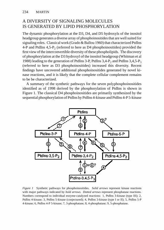
The dynamic phosphorylation at the D3, D4, and D5 hydroxyls of the inositolheadgroup generates a diverse array of phosphoinositides that are well suited forsignaling roles. Classical work (Grado & Ballou 1960) that characterized PtdIns4-P and PtdIns 4,5-P2 (referred to here as D4 phosphoinositides) provided thefirst view of the interconvertible diversity of these phospholipids. The discoveryof phosphorylation at the D3 hydroxyl of the inositol headgroup (Whitman et al1988) leading to the generation of PtdIns 3-P, PtdIns 3,4-P2 and PtdIns 3,4,5-P3(referred to here as D3 phosphoinositides) increased this diversity. Recentfindings have uncovered additional phosphoinositides generated by novel ki-nase reactions, and it is likely that the complete cellular complement remainsto be be characterized.
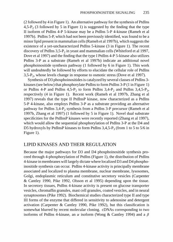
A summary of the synthetic pathways for the seven polyphosphoinositidesidentified as of 1998 derived by the phosphorylation of PtdIns is shown inFigure 1. The classical D4 phosphoinositides are primarily synthesized by thesequential phosphorylation of PtdIns by PtdIns 4-kinase and PtdIns 4-P 5-kinase
Figure 1 Synthetic pathways for phosphoinositides.Solid arrowsrepresent kinase reactionswith major pathways indicated bybold arrows. Dotted arrowsrepresent phosphatase reactions.Numbers correspond to individual enzyme-catalyzed reactions: 1, PtdIns 3-kinase (type III); 2,PtdIns 4-kinase; 3, PtdIns 5-kinase (conjectured); 4, PtdIns 3-kinase (type I or II); 5, PtdIns 5-P4-kinase; 6, PtdIns 4-P 5-kinase; 7, 3-phosphatase; 8, 4-phosphatase; 9, 5-phosphatase.
P1: KKK/spd P2: KKK/ary QC: KKK/tkj T1: KKK
September 16, 1998 11:49 Annual Reviews AR066-09
PHOSPHOINOSITIDE SIGNALING 235
(2 followed by 4 in Figure 1). An alternative pathway for the synthesis of PtdIns4,5-P2 (3 followed by 5 in Figure 1) is suggested by the finding that the typeII isoform of PtdIns 4-P 5-kinase may be a PtdIns 5-P 4-kinase (Rameh et al1997b). PtdIns 5-P, which had not been previously identified, was found to be aminor lipid present in mammalian cells (Rameh et al 1997b), which suggests theexistence of a yet-uncharacterized PtdIns 5-kinase (3 in Figure 1). The recentdiscovery of PtdIns 3,5-P2 in yeast and mammalian cells (Whiteford et al 1997,Dove et al 1997) and the finding that the type I PtdIns 4-P 5-kinase also utilizesPtdIns 3-P as a substrate (Rameh et al 1997b) indicate an additional novelphosphoinositide synthesis pathway (1 followed by 6 in Figure 1). This workwill undoubtedly be followed by efforts to elucidate the cellular role of PtdIns3,5-P2, whose levels change in response to osmotic stress (Dove et al 1997).
Synthesis of D3 phosphoinositides is catalyzed by several classes of PtdIns 3-kinases (see below) that phosphorylate PtdIns to form PtdIns 3-P (1 in Figure 1)or PtdIns 4-P and PtdIns 4,5-P2 to form PtdIns 3,4-P2 and PtdIns 3,4,5-P3,respectively (4 in Figure 1). Recent work (Rameh et al 1997b, Zhang et al1997) reveals that the type II PtdInsP kinase, now characterized as a PtdIns5-P 4-kinase, also employs PtdIns 3-P as a substrate providing an alternativepathway for PtdIns 3,4-P2 synthesis from a PtdIns 3-P precursor (Rameh et al1997b, Zhang et al 1997) (1 followed by 5 in Figure 1). Novel dual substratespecificities for the PtdInsP kinases were recently reported (Zhang et al 1997),which would allow the sequential phosphorylation of PtdIns 3-P at the D4 andD5 hydroxyls by PtdInsP kinases to form PtdIns 3,4,5-P3 (from 1 to 5 to 5/6 inFigure 1).
LIPID KINASES AND THEIR REGULATION
Because the major pathways for D3 and D4 phosphoinositide synthesis pro-ceed through 4-phosphorylation of PtdIns (Figure 1), the distribution of PtdIns4-kinase in membranes will largely dictate where localized D3 and D4 phospho-inositide synthesis can occur. PtdIns 4-kinase activity is principally membraneassociated and localized to plasma membrane, nuclear membrane, lysosomes,Golgi, endoplasmic reticulum and constitutive secretory vesicles (Carpenter& Cantley 1990, Pike 1992, Olsson et al 1995) depending upon the tissue.In secretory tissues, PtdIns 4-kinase activity is present on glucose transportervesicles, chromaffin granules, mast cell granules, coated vesicles, and in neuralsynaptosomes (Pike 1992). Biochemical studies characterized type II and typeIII forms of the enzyme that differed in sensitivity to adenosine and detergentactivation (Carpenter & Cantley 1990, Pike 1992), but this classification issomewhat blurred by recent molecular cloning. cDNAs corresponding to twoisoforms of PtdIns 4-kinase, anα isoform (Wong & Cantley 1994) and aβ
P1: KKK/spd P2: KKK/ary QC: KKK/tkj T1: KKK
September 16, 1998 11:49 Annual Reviews AR066-09
236 MARTIN
isoform (Meyers & Cantley 1997), have been characterized. Theα isoform,homologous to a yeast STT4 enzyme, exists as long (≈200 kDa) and short(≈100 kDa) forms derived from alternative splicing (Wong & Cantley 1994,Nakagawa et al 1996a, Gehrmann et al 1996, Balla et al 1997). Theβ isoform(≈100 kDa), homologous to the yeast PIK1 enzyme ( Nakagawa et al 1996b,Meyers & Cantley 1997, Balla et al 1997), is inhibited by both wortmanninand LY294002 (Downing et al 1996), which were previously thought to bediagnostic for certain PtdIns 3-kinase enzymes (Carpenter & Cantley 1996).
Wortmannin inhibits the synthesis of PtdIns 4-P that is required for hormonereceptor-mediated phosphoinositide turnover, which initially suggested thatthe wortmannin-sensitiveβ isoform of PtdIns 4-kinase had a dedicated rolein phosphoinositide synthesis for signal transduction (Downing et al 1996).Recent studies, however, found that splicing isoforms of the typeα PtdIns 4-kinase are also inhibited by wortmannin (Nakagawa et al 1996a, Balla et al1997). Nonetheless, it is likely that theα andβ isoforms play distinct cellularroles because they exhibit distinct intracellular distributions (Wong et al 1997).Additional isoforms of PtdIns 4-kinase, possibly with dedicated membranetrafficking roles, will likely be characterized. Neither theα nor β cDNAsencode the enzyme classified as the type II enzyme, a 55 kDa protein with akinase activity inhibited by the 4C5G antibody (Carpenter & Cantley 1990). A4C5G-sensitive PtdIns 4-kinase, a resident of glucose transporter vesicles (DelVecchio & Pilch 1991), is involved in the phosphoinositide synthesis essentialfor ARF activation of phospholipase D (Liscovitch et al 1994) and differs inmembrane localization and wortmannin-insensitivity fromα andβ isoforms(Wong et al 1997).
The characterized cDNAs for PtdIns 4-kinases encode hydrophilic proteinsthat lack transmembrane domains. The basis for the membrane associationand targeting of PtdIns 4-kinase isoforms to specific membranes is poorly un-derstood although several potential mechanisms for localization have emerged.Theα isoform contains a PH domain that could mediate its membrane asso-ciation (Wong & Cantley 1994, Nakagawa et al 1996a).β isoforms associatewith the cytoplasmic domains of several transmembrane and receptor proteins(Kauffmann-Zeh et al 1994, Pertile & Cantley 1995, Berditchevski et al 1997).Additional studies are needed to determine the basis for the membrane recruit-ment of the PtdIns 4-kinases in order to understand how localized membranedomains of D3 and D4 phosphoinositides are formed.
PtdIns 4-P 5-kinase activity is principally cytosolic and is enriched in neuralsynaptosomes (Stubbs et al 1988). Two isoforms of PtdIns 4-P 5-kinase thatare related in sequence and differ from members of the phosphoinositide 3-and 4-kinase families have been characterized (Boronenkov & Anderson 1995,Loijens & Anderson 1996, Ishihara et al 1996). The type I isoform (Loijens
P1: KKK/spd P2: KKK/ary QC: KKK/tkj T1: KKK
September 16, 1998 11:49 Annual Reviews AR066-09
PHOSPHOINOSITIDE SIGNALING 237
& Anderson 1996, Ishihara et al 1996) is a true PtdIns 4-P 5-kinase and alsophosphorylates the D5 hydroxyl of PtdIns 3-P (Rameh et al 1997b). The type IIisoform, a PtdInsP 4-kinase, phosphorylates the D4 hydroxyl of PtdIns 5-P andPtdIns 3-P (Zhang et al 1997, Rameh et al 1997b). Although the type I isoformis likely to be a bona fide constituent in the classical biosynthetic pathway forformation of PtdIns 4,5-P2 from PtdIns 4-P, the type II isoform may catalyze analternative route of PtdIns 4,5-P2 synthesis from PtdIns 5-P or catalyze PtdIns3,4-P2 synthesis from PtdIns 3-P (see Figure 1).
The central importance of PtdIns 4-P 5-kinase for the synthesis of PtdIns4,5-P2 and PtdIns 3,4,5-P3 in specific membrane compartments implies thatregulatory and targeting mechanisms can operate on this enzyme, but onlypreliminary accounts of this have been reported. The type II isoform contains aproline-rich domain that may be an SH3-binding site and thus able to mediate thecoupling of type I PtdIns 3-kinases for the channeled synthesis of PtdIns 3,4,5-P3(Boronenkov & Anderson 1995, Zhang et al 1997). A splicing isoform of thetype II enzyme associates with a TNF receptor (Castellino et al 1997). The bonafide PtdIns 4-P 5-kinase (type I) is uniquely stimulated by phosphatidic acid(Jenkins et al 1994), which is speculated to be an important element of a positivefeedback circuit with PLD (phospholipase D) that generates increased PtdIns4,5-P2 and may operate in membrane budding or fusion events (Liscovitch& Cantley 1995) (see below). Nonhydrolyzable guanine nucleotides stimulatePtdIns 4-P phosphorylation, implying a G protein regulation of 5-kinase activity(Smith & Chang 1989). Rho stimulates PtdIns 4-P 5-kinase activity in celllysates (Chong et al 1994) and associates with a type I enzyme, although thisassociation may be indirect (Ren et al 1996). Rac1 also associates directlyor indirectly with the type I PtdIns 4-P 5-kinase (Tolias et al 1995). It willbe important to further characterize Rac/Rho protein regulation of the type IPtdIns 4-P 5-kinase because of the role of phosphoinositides and Rho familymembers in the regulation of the actin cytoskeleton (see below).
The characteristics of PtdIns 3-kinases have been discussed in several re-cent reviews (Carpenter & Cantley 1996, Vanhaesebroeck et al 1997, Domin &Waterfield 1997). These enzymes are classified into three groups according tostructure and activity. Class I enzymes are heterodimers of catalytic and adap-tor subunits that utilize PtdIns, PtdIns 4-P, and PtdIns 4,5-P2 as substrates. Theadaptor subunits of these enzymes contain SH2 domains that mediate recruit-ment to phosphotyrosine residues on the cytoplasmic domains of receptors,which results in activation of the 3-kinase. Class II enzymes preferentiallyphosphorylate PtdIns and PtdIns 4-P but not PtdIns 4,5-P2 in vitro and containC2 domains that could mediate membrane interactions. Class III enzymes,which phosphorylate only PtdIns in vitro, consist of heterodimers of a cat-alytic subunit (such as yeast Vps34p) associated with a serine/threonine protein
P1: KKK/spd P2: KKK/ary QC: KKK/tkj T1: KKK
September 16, 1998 11:49 Annual Reviews AR066-09
238 MARTIN
kinase adaptor subunit (such as yeast Vps15p) that is required for membranerecruitment (Stack et al 1993).
PHOSPHOINOSITIDE-BINDING PROTEINS:EFFECTORS FOR PHOSPHOINOSITIDE SIGNALS
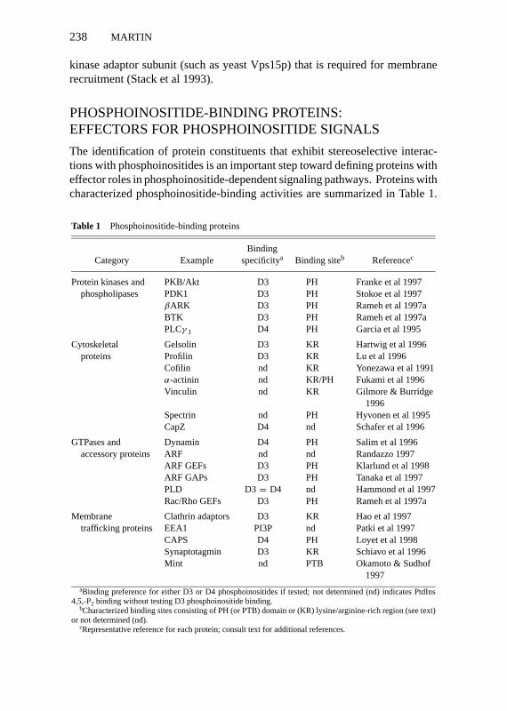
The identification of protein constituents that exhibit stereoselective interac-tions with phosphoinositides is an important step toward defining proteins witheffector roles in phosphoinositide-dependent signaling pathways. Proteins withcharacterized phosphoinositide-binding activities are summarized in Table 1.
Table 1 Phosphoinositide-binding proteins
BindingCategory Example specificitya Binding siteb Referencec
Protein kinases and PKB/Akt D3 PH Franke et al 1997phospholipases PDK1 D3 PH Stokoe et al 1997
βARK D3 PH Rameh et al 1997aBTK D3 PH Rameh et al 1997aPLCγ 1 D4 PH Garcia et al 1995
Cytoskeletal Gelsolin D3 KR Hartwig et al 1996proteins Profilin D3 KR Lu et al 1996
Cofilin nd KR Yonezawa et al 1991α-actinin nd KR/PH Fukami et al 1996Vinculin nd KR Gilmore & Burridge
1996Spectrin nd PH Hyvonen et al 1995CapZ D4 nd Schafer et al 1996
GTPases and Dynamin D4 PH Salim et al 1996accessory proteins ARF nd nd Randazzo 1997
ARF GEFs D3 PH Klarlund et al 1998ARF GAPs D3 PH Tanaka et al 1997PLD D3= D4 nd Hammond et al 1997Rac/Rho GEFs D3 PH Rameh et al 1997a
Membrane Clathrin adaptors D3 KR Hao et al 1997trafficking proteins EEA1 PI3P nd Patki et al 1997
CAPS D4 PH Loyet et al 1998Synaptotagmin D3 KR Schiavo et al 1996Mint nd PTB Okamoto & Sudhof
1997aBinding preference for either D3 or D4 phosphoinositides if tested; not determined (nd) indicates PtdIns
4,5,-P2 binding without testing D3 phosphoinositide binding.bCharacterized binding sites consisting of PH (or PTB) domain or (KR) lysine/arginine-rich region (see text)
or not determined (nd).cRepresentative reference for each protein; consult text for additional references.
P1: KKK/spd P2: KKK/ary QC: KKK/tkj T1: KKK
September 16, 1998 11:49 Annual Reviews AR066-09
PHOSPHOINOSITIDE SIGNALING 239
Until recently, only D4 phosphoinositides were employed in protein bindingstudies (Janmey 1994) but the availability of D3 phosphoinositides has led tothe characterization of phosphoinositide-binding specificity for a number ofproteins. Many proteins exhibit a binding specificity that favors D3 over D4phosphoinositides (Table 1). Proteins in this category are potential effectorsthat function downstream of PtdIns 3-kinases and may mediate the effects ofPtdIns 3,4-P2 and PtdIns 3,4,5-P3 on protein phosphorylation, cytoskeletal reg-ulation, GTP exchange reactions, and membrane coat recruitment. Proteinsthat bind PtdIns 3-P that may act downstream of type III PtdIns 3-kinase in-volved in membrane trafficking have recently been identified, such as AP-2 andEEA1 (Rapoport et al 1997, Patki et al 1997). A small group of proteins exhibita specificity for binding D4 phosphoinositides in preference to D3 phospho-inositides (Table 1). This group includes proteins that function in cytoskeletalregulation, exocytosis, and endocytosis and are the potential effectors for thecellular roles of D4 phosphoinositides.
MOLECULAR BASIS FOR PROTEIN-PHOSPHOINOSITIDE INTERACTIONS
Basic and Hydrophobic SequencesTwo general classes of binding sites for the stereoselective interaction of phos-phoinositides with proteins have been identified: The first consists of short(≈10–20 residues) colinear sequences that are rich in basic and hydrophobicresidues, and the second consists of longer sequences (≈120 residues) thatshare a well-defined tertiary structure of the PH domain. Studies on gelsolin(Yu et al 1992) resulted in the assignment of phosphoinositide binding to twoseparate regions near the N terminus (CKSGLKYKKGGVASGF and KHVVP-NEVVVQRLFQVKGRR). Peptides corresponding to these sequences exhibitPtdIns 4,5-P2–binding similar to that of gelsolin (Janmey et al 1992). The firstof these lysine/arginine-rich sequences was suggested to constitute a motif(K/RXXXXKXK/RK/R) that was present in other phosphoinositide-bindingcytoskeletal proteins such as gCap39, villin, cofilin, and profilin (Yu et al 1992).A simple electrostatic interaction between lysine/arginine and phosphoinositolphosphates cannot fully account for the phosphoinositide-binding properties ofgelsolin, which does not bind deacylated or deacylated/deglycerinated phos-phoinositides (Janmey 1994). However, these short sequences contain a highpercentage of hydrophobic amino acids that may form the part of the bindingsite that interacts with the diacylglycerol moiety.
The phosphoinositide-binding sites for several other actin-associated proteinsare not homologous to those of gelsolin. It was suggested that a short peptidesequence (FSMDLRTKST) in profilin was responsible for phosphoinositide
P1: KKK/spd P2: KKK/ary QC: KKK/tkj T1: KKK
September 16, 1998 11:49 Annual Reviews AR066-09
240 MARTIN
binding (Sohn et al 1995). A 12 residue sequence (WAPECAPLKSKM) ofcofilin was reported to bind PtdIns 4,5-P2 (Yonezawa et al 1991). A linear se-quence inα-actinin (TAPYRNVNIQNFHLSWK) accounted for PtdIns 4,5-P2binding, which was eliminated by mutagenesis of the two arginine and lysineresidues (Fukami et al 1996). Although a diverse array of nonhomologous se-quences constitute phosphoinositide-binding sites in cytoskeletal proteins, eachcontains at least two basic residues in the context of a high percentage (≈50%)of hydrophobic residues. These short sequences may provide hydrophobiccontacts with phospholipid acyl chains as well as charge interactions betweenthe basic residues and the phosphate groups on the inositol headgroup. A hex-adecapeptide from neurogranin exhibiting these hallmark features (WAAK-IQASFRGHMARKK) interacts with high affinity and specificity with phos-phoinositides preferring PtdIns 3,4,5-P3 over other phospholipids, and acquiresa structure upon binding to phosphoinositides (Lu & Chen 1997).
Several coat proteins have been isolated in an effort to identify inositolpolyphosphate-binding proteins. Clathrin adaptor proteins AP-2 and AP-3,as well as coatomer protein, interact with inositol phosphates and with phos-phoinositides (Beck & Keen 1991, Fleischer et al 1994, Ye et al 1995, Norriset al 1995). Phosphoinositide interactions with AP-2 are mediated by N-terminalsequences of theα subunit (Beck & Keen 1991, Gaidarov et al 1996) and thephosphoinositide-binding site of a homologous subunit of AP-3 was suggestedto consist of PKKKHLDYLIQATNE (Ye et al 1995, Hao et al 1997), a sequencealso rich in basic and hydrophobic residues.
Several inositol phosphate-binding proteins such as centaurin-α and synap-totagmin II were purified based on inositol polyphosphate-binding (Fukudaet al 1994, Cullen et al 1995, Theibert et al 1997). However, phosphoinosi-tides are probably the natural ligands for these proteins (Hammonds-Odie et al1996, Schiavo et al 1996, Mehrotra et al 1997). The inositol phosphate-bindingdomain on synaptotagmin consists of the highly basic sequence GKRLKKKK-TTVKKK in the C2B domain (Fukuda et al 1994, 1995a). This portion of theC2 domain is disordered (Sutton et al 1995) but adopts an ordered structureupon binding inositol phosphates (Mehrotra et al 1997). Phosphoinositidesbind to the same or an overlapping site in the C2B domain (Schiavo et al 1996).These lysine-rich sequences probably represent the inositol phosphate-bindingportions of larger phosphoinositide-binding domains. In synaptotagmin, thisbasic sequence is flanked by regions rich in hydrophobic residues that couldmediate acyl chain interactions.
PH DomainsPH (and related PTB) domains are the best-characterized and possibly mostwidespread of phosphoinositide-binding motifs. PH domains, termed pleckstrin
P1: KKK/spd P2: KKK/ary QC: KKK/tkj T1: KKK
September 16, 1998 11:49 Annual Reviews AR066-09
PHOSPHOINOSITIDE SIGNALING 241
homology from the sequences initially identified in the platelet protein kinase Csubstrate pleckstrin, comprise≈120 amino acid colinear regions identified bysequence comparison in nearly 100 proteins (Musacchio et al 1993, Gibson et al1994). PH domains are present in cytoskeletal components (spectrin,α-actinin),guanine nucleotide exchange proteins or GTPase-regulating proteins (Ras-GRF,Dbl, VAV, cdc24, SOS, Ras-GAP, Tiam-1, ARNO, GRP1/cytohesin-1) andGTPases (dynamin), phosphoinositide-regulated protein kinases (Akt/PKB,PDK1), other protein kinases (BTK,βARK), and phospholipases (PLC). Al-though not well conserved at the primary sequence level, NMR and X-raydiffraction studies reveal a remarkable similarity between the structures forPH domains from pleckstrin, spectrin, PLCδ1, and dynamin (Yoon et al 1994,Macias et al 1994, Hyvonen et al 1995, Ferguson et al 1995, Zheng et al 1996a,Salim et al 1996). The PH domain consists of two nearly orthogonalβ sheetsof three or four strands that form aβ sandwich closed off near its C terminusby an amphipathicα helix.
Harlan et al (1994) originally reported that the PH domains of pleckstrin, ras-GAP,βARK, and T cell kinase interacted with PtdIns-4,5-P2. Subsequent stud-ies confirmed that PH domains from a large number of proteins (PLCδ1, spec-trin, dynamin, BTK, SOS, Tiam-1, OSBP, Akt/PKB, SOS, GRP1/cytohesin-1,ARNO) bind phosphoinositides but with different affinities and specificities(Zheng et al 1996a, Salim et al 1996, Rameh et al 1997b, Frech et al 1997,Franke et al 1997, Kubiseski et al 1997, Klarlund et al 1997, Paris et al 1997).A study of six PH domains (Rameh et al 1997a) found that four exhibit a se-lectivity for PtdIns 3,4,5-P3 over other phosphoinositides, whereas two othersbound PtdIns 3,4,5-P3 and PtdIns-4,5-P2 with similar affinities. The PH domainof Akt/PKB has a high affinity and specificity for PtdIns 3,4-P2 over PtdIns4,5-P2 and PtdIns 3,4,5-P3 (Franke et al 1997, Klippel et al 1997, Frech et al1997), whereas the PH domain of a kinase that phosphorylates PKB/Akt, PDK1,prefers PtdIns 3,4,5-P3 over PtdIns 3,4-P2 (Stokoe et al 1997). A similar prefer-ential selectivity for binding PtdIns 3,4,5-P3 was characterized for a number ofPH domains including ARF nucleotide exchange factors and GTPase-activatingproteins (Tanaka et al 1997, Klarlund et al 1998). The PH domains of PLCδ,CAPS, and dynamin are distinct in preferentially binding PtdIns 4,5-P2 ratherthan D3 phosphoinositides (Salim et al 1996, Loyet et al 1998). The majority ofPH domains where binding specificity has been determined have characteristicsexpected for effector proteins for PtdIns 3-kinase signaling.
Phosphoinositide binding to PH domains occurs at an electrostatically po-larized (basic) face of the domain with critical residues residing on the loopsbetween theβ sheets (Lemmon et al 1996, 1997). This is a highly variableregion between individual PH domains, which accounts for the diversity ofphosphoinositide-binding specificity and affinity. For the PH domain of PLCδ1,
P1: KKK/spd P2: KKK/ary QC: KKK/tkj T1: KKK
September 16, 1998 11:49 Annual Reviews AR066-09
242 MARTIN
which exhibits the highest affinity for phosphoinositides among the PH do-mains, binding is mediated by a network of interactions between the 4 and5 position phosphates of the inositol headgroup and amino acid side chains,mainly lysine and arginine, of the variable loops of the PH domain (Fergusonet al 1995). Weaker binding of phosphoinositides to other PH domains cor-responds to a more superficial interaction of the inositol headgroup with PHdomain loops establishing many fewer interactions (Lemmon et al 1997).
There is evidence that phosphoinositides are physiological ligands for thePH domains of some of the above proteins. Mutation of critical loop residuesrequired for phosphoinositide binding by the BTK PH domain results in a loss-of-function agammaglobinaemia phenotype (Salim et al 1996). Increasing ev-idence implicates D3 phosphoinositides as physiological regulators of Akt/PKBactivity (see below). Mutagenesis studies on many of the other phosphoinositide-binding proteins will be required to convincingly establish the role of suchinteractions in cell function. Whether phosphoinositides constitute the soleligands for all PH domains is an unresolved issue. The fact that the structurallyhomologous PTB domain (Lemmon et al 1996) interacts with phosphotyro-syl peptides via a binding site that overlaps with that for phosphoinositides(Rameh et al 1997a) suggests that phosphoamino acid peptides could be theligands, or co-ligands, for some PH domains. For certain PH domains, coop-erative interactions with multiple ligands may occur such as forβARK, wherephosphoinositides andβγ subunits exhibit a synergism in binding (Pitcheret al 1995). The ability of the dynamin-1 PH domain, but not other PH domainsincluding that of PLCδ1, to interfere with rapid endocytosis in chromaffin cells(Artalejo et al 1997) suggests that phosphoinositides are not the endogenousligand for this PH domain despite the fact that direct phosphoinositide bind-ing (Salim et al 1996) as well as phosphoinositide activation of the dynaminGTPase (Lin & Gilman 1996) have been demonstrated.
PH domains are essential for the membrane recruitment of several pro-teins that contain them, such as PKB/Akt (Andjelkovic et al 1997), GAP1IP4BP
(Lockyer et al 1997), Dbl (Zheng et al 1996b), andβARK (Pitcher et al 1995).It is possible that other phospholipids, including minor novel phosphoinositidessuch as PtdIns 3,5-P2, are the ligands that mediate membrane recruitment ofPH domain-containing proteins found to bind D3 or D4 phosphoinositides onlyweakly. The active pace of research on the PH domain will soon clarify thegeneral role of this motif and lead to the identification of additional physio-logical ligands that mediate membrane recruitment. At present, the concept ofthe PH domain as a signal-dependent membrane adapter that functions in thefocal assembly of protein complexes at the membrane interface is very useful(Hemmings 1997). The fact that PH domains are frequently accompanied byother motifs (SH2, SH3, proline-rich, GTP exchange) in proteins is consistent
P1: KKK/spd P2: KKK/ary QC: KKK/tkj T1: KKK
September 16, 1998 11:49 Annual Reviews AR066-09
PHOSPHOINOSITIDE SIGNALING 243
with the notion that the recruitment of the PH domain protein would nucleatesites on the membrane for protein complex assembly.
MEMBRANE MICRODOMAINSOF PHOSPHOINOSITIDES AND SITE-SPECIFICPROTEIN COMPLEX ASSEMBLY
It is likely that phosphoinositides and the proteins they recruit are spatiallylocalized at focal sites in specific membranes. Phosphoinositides and thephosphoinositide kinases are not uniformly distributed in cellular membranes(Pike 1992), and the phosphoinositides are metabolically compartmentalized(Monaco & Gershengorn 1992). Biochemical evidence for membrane domainsenriched in receptor-regulated pools of PtdIns 4,5-P2 and enzymes has beenreported (Pike & Casey 1996, Hope & Pike 1996). Studies employing fluores-cent PtdIns 4,5-P2 in liposomes revealed that phosphoinositide-binding peptidescan stabilize membrane microdomains in which both protein and phospholipidconstituents are segregated (Glaser et al 1996).
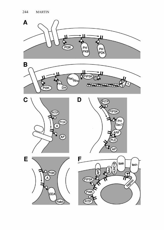
Immunocytochemical studies with phosphoinositide-specific antibodies pro-vide a preview of what may be a general concept of spatially localized domainsof phosphoinositides in cellular membranes. PtdIns 4,5-P2 colocalizes withα-actinin in focal contacts and membrane ruffles (Fukami et al 1994). A local-ized synthesis of PtdIns 4,5-P2 on dense-core vesicles in neuroendocrine cellsduring a priming step for exocytosis can be detected by immunocytochemistry(K Loyet, personal communication). Spatially segregated membrane domainsenriched for polyphosphoinositides would exhibit a positive membrane cur-vature (Chernomordik 1996) that could contribute to the remodeling of thebilayer for events such as membrane budding and fusion. The recruitment ofphosphoinositide-binding proteins to such sites could allow for the assemblyof signal transduction complexes, cytoskeletal-membrane attachments, coatedmembrane domains for bud formation, and scaffolds for membrane fusion reac-tions. Current efforts to define such mechanisms involving phosphoinositide-binding effector proteins are discussed below and summarized in Figure 2.
PROTEIN KINASES AS EFFECTORS FORPHOSPHOINOSITIDES IN SIGNAL TRANSDUCTION
PtdIns 3,4,5-P3 serves an essential signaling role in mediating the effects ofa wide range of extracellular stimuli on cell proliferation, cell survival, andmetabolism (Toker & Cantley 1997). Recent studies elucidated an importanteffector pathway that involves D3 phosphoinositide-mediated membrane re-cruitment and activation of several protein kinases (Figure 2a). The PKB/Akt
P1: KKK/spd P2: KKK/ary QC: KKK/tkj T1: KKK
September 16, 1998 11:49 Annual Reviews AR066-09
244 MARTIN

P1: KKK/spd P2: KKK/ary QC: KKK/tkj T1: KKK
September 16, 1998 11:49 Annual Reviews AR066-09
PHOSPHOINOSITIDE SIGNALING 245
kinase is activated downstream of receptor-regulated PtdIns 3-kinase in partby direct interaction of PtdIns 3,4-P2 with its PH domain (Franke et al 1997,Klippel et al 1997, Frech et al 1997, Cohen et al 1997). Recruitment of PKB/Aktto the membrane requires its PH domain (Andejkovic et al 1997), and possi-ble dimerization of PKB/Akt at the membrane has been suggested (Frankeet al 1997). PKB/Akt undergoes phosphorylation and activation by additionalphosphoinositide-dependent protein kinases such as PDK1 (Alessi et al 1997,Stephens et al 1998). The phosphorylation of PKB/Akt by PDK1 requiresD3 phosphoinositide binding to the PH domain of PKB/Akt (Stokoe et al 1997,Cohen et al 1997). PDK1 also contains a PH domain that binds PtdIns 3,4,5-P3;however, its kinase activity may be constitutive, and phosphoinositide bindingmay serve to recruit PDK1 to a membrane site near its membrane bound sub-strate (Alessi et al 1997). This system represents the clearest example of phos-phoinositide signaling where the identity of the lipid(s) involved, the basis ofits regulated synthesis by a defined kinase, and the role of a physiologically rel-evant phosphoinositide-binding effector are relatively well defined (Figure 2a).
Several substrates of PKB/Akt that act downstream in signaling pathwaysregulating metabolism (Cohen et al 1997) and cell survival (del Peso et al1997) have been identified. Expression of a constitutively active PtdIns 3-kinase
←−−−−−−−−−−−−−−−−−−−−−−−−−−−−−−−−−−−−−−−−−−−−−−−−−−−−−−Figure 2 Common themes in phosphoinositide signaling involving site-specific membrane recruit-ment. (A) Signal transduction mechanism employing receptor-regulated PtdIns 3-kinase (PI3K) toform D3 phosphoinositides for the recruitment of PH domain-containing kinases, PKB and PDK.(B) Cytoskeleton assembly mechanisms employing receptor-regulated PtdIns 3-kinase to form D3phosphoinositides for the activation of PH domain-containing Dbl family members that promoteguanine nucleotide exchange on Rho family members. Rho/Rac are depicted as potential activatorsof PtdIns 4-P 5-kinase (PIP5K) to form PtdIns 4,5-P2, which uncaps gelsolin (G) from actin to allowfilament elongation, or promotes a conformational change in vinculin (V) to allow it to tether actinto talin (T). (C) Golgi membrane-budding reaction is depicted to proceed through cargo receptoractivation of a PtdIns 3-kinase (PI3K) containing an adaptor (A) subunit. PtdIns transfer protein(PITP) enhances PtdIns 3-kinase-catalyzed phosphorylation to form PtdIns 3-P, which promotesthe binding of clathrin adaptor proteins (AP) to cargo receptor tail. (D) Golgi membrane-buddingreaction is depicted as proceeding through PITP enhancement of PtdIns 4-kinase phosphoryla-tion along with PIP5K to form PtdIns 4,5-P2. This phosphoinositide recruits and activates thePH domain and Sec7 homology-containing exchange factors that promote guanine nucleotide ex-change on ARF. ARF activates phospholipase D, which promotes further synthesis of PtdIns 4,5-P2via phosphatidic acid stimulation of PIP5K. Adaptor proteins (AP) are shown binding to acidicphospholipids. (E) Endosome fusion mechanism is depicted to proceed through activation of anadaptor-containing PtdIns 3-kinase (A and PI3K) that forms PtdIns 3-P, which recruits EEA forbinding to Rab5. (F) Exocytotic fusion mechanism is depicted to involve PITP, PtdIns 4-kinase(PI4K), and PIP5K to promote formation of PtdIns 4,5-P2 on the vesicle membrane. Potential effec-tors of this phosphoinositide are CAPS, Mint-mUNC18 complexes, and synaptotagmin (tagmin).Potential interactions of mUNC18 and tagmin with syntaxin (taxin) are depicted.
P1: KKK/spd P2: KKK/ary QC: KKK/tkj T1: KKK
September 16, 1998 11:49 Annual Reviews AR066-09
246 MARTIN
(Martin et al 1996, Frevert & Kahn 1997) or a PKB/Akt (Kohn et al 1996)enhances Glut4 translocation to the plasma membrane of 3T3-L1 adipocytes,suggesting that the PKB/Akt-catalyzed phosphorylation of an unidentifiedprotein substrate regulates an aspect of the exocytotic pathway of glucose trans-porter vesicles. Delineation of this downstream pathway will provide interest-ing insights into the regulation of membrane trafficking by phosphoinositide-dependent protein phosphorylation.
PROTEIN EFFECTORS FOR CYTOSKELETALREGULATION
Phosphoinositides have been implicated in the regulation of actin cytoskeletonassembly at several levels. Extensive connections between phosphoinositidesand the Rho family of GTP-binding proteins, which mediate extracellular sig-nal regulation of cytoskeletal rearrangements, have been identified (Hall 1998).A large family of Rho guanine nucleotide exchange factors has been charac-terized whose members contain Dbl homology (DH) domain as well as a PHdomain that mediates the targeting of these proteins to the membrane (Cerione& Zheng 1996, Zheng et al 1996b). Constitutively active PtdIns 3-kinase ac-tivates Rac-dependent lamellipodia and Rho-dependent stress fiber assemblyin fibroblasts (Reif et al 1996). The PH domain of some Dbl family membersexhibits selectivity for binding D3 phosphoinositides (Rameh et al 1997a). Thisindicates that D3 phosphoinositide synthesis could promote recruitment of ex-change factors to specific sites on the membrane for activation of Rho familyGTPases (Carpenter et al 1997) (Figure 2b).
With the identification of numerous Rho/Rac-binding proteins (Tapon & Hall1997), there are many options for downstream signaling to the cytoskeleton.One pathway for the Rho-dependent induction of stress fiber formation in-volves the activation of a Rho kinase (ROK) that phosphorylates both myosinlight chain (Amano et al 1996) and myosin phosphatase (Kimura et al 1996),which leads to increased binding of myosin and the bundling of actin filaments.Another mechanism that promotes de novo actin polymerization involves D4phosphoinositides as mediators of Rho/Rac regulation of the cytoskeleton(Figure 2b). In platelets, Rac activation results in increased actin polymer-ization for lamellae formation, which arises from an increase in the number offree actin barbed-ends available for filament elongation (Hartwig et al 1995).Rac activation causes an increased synthesis of PtdIns 4,5-P2, which may bemediated by Rac stimulation of a type I PtdIns 4-P 5-kinase. The downstreameffector proteins in this system are gelsolin and other capping proteins whoseactin barbed-end capping activities are inhibited by phosphoinositides (Janmey1994, Schafer et al 1996) (Figure 2b).
P1: KKK/spd P2: KKK/ary QC: KKK/tkj T1: KKK
September 16, 1998 11:49 Annual Reviews AR066-09
PHOSPHOINOSITIDE SIGNALING 247
There are other potential downstream effectors among the phosphoinositide-binding proteins that regulate actin assembly (Table 1) (Janmey 1994). Poly-phosphoinositides induce conformational changes in vinculin that allow vin-culin to mediate the cross-linking of talin to actin, which plays a role in focaladhesion assembly (Gilmore & Burridge 1996) (Figure 2b). Consistent witha role for vinculin or other phosphoinositide-binding cytoskeletal proteins asimportant effectors, the Rho-dependent stimulation of focal adhesion forma-tion in fibroblasts is blocked by microinjection of antibodies to PtdIns 4,5-P2(Gilmore & Burridge 1996).
In related efforts to assess physiological roles for phosphoinositides in regu-lating actin assembly, overexpression of the type I PtdIns 4-P 5-kinase in COS-7cells was found to cause a dramatic increase in the assembly of short actin fil-aments (Shibasaki et al 1997). This depends upon a functional kinase domainand is counteracted by coexpression of a type II 5-phosphatase that decreasescellular PtdIns 4,5-P2 levels (Zhang et al 1995). Conversely, overexpression ofa type II 5-phosphatase in COS-7 cells reduces the number of actin stress fibers(Sakasaki et al 1997). Expression of a dominant-negative form of RhoA fails tosuppress the formation of actin filaments induced by 5-kinase overexpression,consistent with a role for PtdIns 4-P 5-kinase downstream of Rho (Shibasakiet al 1997). These studies to alter cellular levels of polyphosphoinositides incells provide an important link between in vitro studies of phosphoinositide-binding by cytoskeletal proteins and the regulation of actin assembly in vivo.Additional studies are required to determine which of the many candidate cy-toskeletal proteins (Table 1) serve as essential effectors for phosphoinositidesignaling in cytoskeletal rearrangements.
PROTEIN EFFECTORS FOR MEMBRANE COATFORMATION AND BUDDING REACTIONS
Many intracellular transport events require the assembly of vesicle coats atspecific sites on the membrane during budding (Rothman & Wieland 1996).Coat components bind to the cytoplasmic tails of transmembrane proteins thatassociate with transported cargo in the lumen of the budding vesicle, whichserves to concentrate and sort cargo for transport (Kirchhausen et al 1997).Non-clathrin COPII- and COPI-coated vesicles mediate transport between theendoplasmic reticulum and Golgi and within the Golgi, respectively (Schekman& Orci 1996). Clathrin-coated vesicles mediate the endocytic trafficking ofproteins to early endosomes and the trafficking of proteins from thetrans-Golgi network to late endosomes. Clathrin assembly at the site of vesiclebudding utilizes adaptor proteins, AP-1 for the Golgi and AP-2/AP-3 for theplasma membrane (Robinson 1997). Protein sorting at thetrans-Golgi network
P1: KKK/spd P2: KKK/ary QC: KKK/tkj T1: KKK
September 16, 1998 11:49 Annual Reviews AR066-09
248 MARTIN
to multiple destinations involves the generation of numerous distinct transportvesicles for which coats and mechanisms of assembly have yet to be fullycharacterized (Traub & Kornfeld 1997).
As for other spatially localized, membrane-associated events requiring pro-tein recruitment, there is evidence for an essential role for phosphoinositidesin the assembly of vesicle coats. It remains to be determined whether phospho-inositides are required as constitutive cofactors for assembly or whether theyact as regulatory signals that specify the site and timing of assembly. Withinthe variety of budding processes examined, coat components and adaptor pro-teins, as well as the enzyme PLD, have suggested roles as phosphoinositideeffectors.
The initial indication that D3 phosphoinositides were critical for aspects ofmembrane trafficking was the discovery that a yeast vacuolar protein sortinggeneVPS34encoded a PtdIns 3-kinase (Schu et al 1993). TheVPS34geneproduct phosphorylates PtdIns, but not other phosphoinositides, and mam-malian homologues exhibit a similar substrate specificity (Volinia et al 1995).Loss-of-function mutants ofVPS34exhibit a defect in sorting newly synthe-sized proteins, as well as endocytosed proteins, to the vacuole, and it wassuggested that the Vps34p protein executes a function essential for the forma-tion of vesicles in the Golgi or their subsequent trafficking (Stack et al 1995).A serine/threonine-specific protein kinase (VPS15p) mediates the membranerecruitment and activation of the Vps34p lipid kinase (Stack et al 1993). TheVps15p/Vps34p complex has been suggested to be regulated by cargo receptorsfor vacuolar hydrolases (Marcusson et al 1994). In this model, cargo accumu-lation would specify the timing and location of budding by stimulating PtdIns3-P formation (Horazdovsky et al 1995).
The immediate downstream effector for PtdIns 3-P remains to be identified.PtdIns 3-P could alter the structure of the bilayer to facilitate budding, recruitcoat proteins or adaptors to the site of budding, or function as a vesicle mem-brane component essential for subsequent docking or fusion reactions (Stacket al 1995). The role of a dynamin homologue (Vps1p) in vacuolar sorting sug-gests that clathrin coat proteins may be involved (Conibear & Stevens 1995),and the sorting defects of conditional clathrin mutants are consistent with this(Stack et al 1995). Whether PtdIns 3-P functions to recruit clathrin adaptorproteins for bud formation remains to be determined.
In yeasts, PtdIns 3,5-P2 synthesis was found to be dependent upon the Vps34pPtdIns 3-kinase (Dove et al 1997). This could indicate that the role of PtdIns3-P in vacuolar sorting is to serve as a precursor to PtdIns 3,5-P2. The loss-of-function phenotype forFAB1, which encodes one of two yeast PtdInsP 5-kinases (Yamamoto et al 1995, Loijens & Anderson 1996), includes a vacuolarmorphology defect, which suggests a role for a 5-phosphorylated inositide in
P1: KKK/spd P2: KKK/ary QC: KKK/tkj T1: KKK
September 16, 1998 11:49 Annual Reviews AR066-09
PHOSPHOINOSITIDE SIGNALING 249
aspects of vacuole membrane cycling. Genetic studies, however, did not revealthe epistatic interaction betweenVPS34andFAB1alleles that is anticipated fora conversion of PtdIns 3-P to PtdIns 3,5-P2 as essential for vacuolar targeting(Yamamoto et al 1995).
To investigate potential roles for the products of PtdIns 3-kinases in mem-brane trafficking in mammalian cells, extensive use has been made of wortman-nin, a characterized irreversible inhibitor of some but not all PtdIns 3-kinases(Carpenter & Cantley 1996). The Vps34p lipid kinase is relatively insensi-tive to wortmannin (Stack & Emr 1994), whereas mammalian class III PtdIns3-kinases that include Vps34p homologues exhibit a range of sensitivities tothe drug (Stephens et al 1994, Volinia et al 1995). Wortmannin is not highlyspecific and is known to inhibit myosin light chain kinase, several phospho-lipases, and several PtdIns 4-kinases, albeit at higher concentrations (Crosset al 1995, Wong et al 1997). A chemically distinct inhibitor of PtdIns 3-kinasesLY290042 (Vlahos et al 1994) is also used in membrane trafficking studies.Several mammalian PtdIns 3-kinase isoforms are inhibited by LY290042, butunfortunately so are isoforms of PtdIns 4-kinase. Thus studies that rely exclu-sively on wortmannin and LY294002 are difficult to interpret, and it is necessaryto employ other methods such as dominant-negative mutants to assess the roleof PtdIns 3-kinase in membrane trafficking reactions (Haruta et al 1995).
Wortmannin treatment of mammalian cells results in a limited repertoireof membrane trafficking defects that is restricted to the late Golgi-lysosomal-endosomal pathway (Brown et al 1995, Clague et al 1995, Davidson 1995,Reaves et al 1996, Shpetner et al 1996). Wortmannin treatment causes a strikingswelling of late endosomal-prelysosomal compartments (Brown et al 1995,Reaves et al 1996); the inhibition of transport of endocytosed receptors fromlate endosomes to lysosomes (Shpetner et al 1996); the mis-sorting of cathepsinD, a lysosomal protease that is sorted via the mannose 6-phosphate receptor inthetrans-Golgi (Brown et al 1995, Davidson 1995); an inhibition of transferrinreceptor recycling (Shepherd et al 1996); the inhibition of GLUT4 transporterrecycling (Yang et al 1996); and a partial inhibition of fluid phase endocytosis(G Li et al 1995).
In vitro reconstitution studies in mammalian systems of vesicle biogenesisfrom thetrans-Golgi have identified several molecular requirements for bud-ding and have begun to elucidate the role of phosphoinositides in buddingreactions (Figure 2c,d). The formation of constitutive and regulated vesiclesfrom the trans-Golgi of neuroendocrine cells in vitro depends upon at leasttwo cytosolic protein fractions as well as ATP (Ohashi et al 1995). One of therequired cytosolic factors is PtdIns transfer protein, which binds and transportsPtdIns and PtdChol in vitro (Wirtz 1997). Mammalian PtdIns transfer pro-teins accelerate the phosphorylation of PtdIns through 4-phosphorylation and
P1: KKK/spd P2: KKK/ary QC: KKK/tkj T1: KKK
September 16, 1998 11:49 Annual Reviews AR066-09
250 MARTIN
3-phosphorylation reactions by providing PtdIns to PtdIns 4-kinase and PtdIns3-kinase (Martin 1995, Wirtz 1997). A requirement for PtdIns transfer proteinin Golgi budding reactions likely indicates a role for phosphoinositides in someaspect of vesicle formation.
In vitro reactions in which the formation of constitutive exocytic vesicles con-taining TGN38 are formed are inhibited by high concentrations of wortmannin,which correspond to the range that inhibits a Golgi-associated PtdIns 3-kinase(Jones et al 1993, Jones & Howell 1997). The PtdIns 3-kinase resembles theVPS34 lipid kinase in substrate specificity and low sensitivity to wortmannin,which led to the suggestion of a required role for PtdIns 3-P in vesicle formation(Jones & Howell 1997; see also Hickinson et al 1997). The enzyme resides in amembrane-bound complex with an adaptor protein (p62) that is related to, butimmunologically distinct from, the p85a subunit of a type I PtdIns 3-kinase.Rab6 and several unidentified GTP-binding proteins co-reside in the complexin association with the cytoplasmic domain of TGN38 (Jones et al 1993, Jones& Howell 1997). The properties of a membrane and cytosolic p62-containingcomplex suggest a model in which a p62-Rab6 complex is recruited to TGN38in the Golgi, thereby mediating the binding of PtdIns 3-kinase for the essen-tial formation of PtdIns 3-P (Figure 2c). The effector system for PtdIns 3-Premains to be identified for this Golgi budding reaction, but may involve thep200 lace-like coats described for these vesicles (Ladinsky et al 1994).
Other experimental work on the biogenesis of vesicles in thetrans-Golgiand the formation of COPI-coated vesicles within the Golgi suggests alter-native or additional roles for the phosphoinositides (PtdIns 4,5-P2 or PtdIns3,4,5-P3) in budding reactions (Figure 2d). ADP ribosylation factors (ARFs)comprise a family of proteins that play essential roles in Golgi membrane trafficby mediating the recruitment of coat proteins to the membrane (Rothman &Wieland 1996). COPI vesicle formation can be reconstituted in vitro with ARFand coatomer as the sole cytosolic factors (Orci et al 1993). An ARF gua-nine nucleotide exchange factor (ARF GEF) catalyzes GTP binding to ARF,and ARF-GTP promotes coatomer binding. An ARF GTPase-activating factor(ARF GAP) catalyzes uncoating prior to fusion of vesicles with an acceptormembrane. The recruitment of AP-1 clathrin adaptors to the Golgi membrane(Stamness & Rothman 1993) is ARF dependent, as is recruitment of AP-1 toimmature secretory granules (Dittie et al 1996) and AP-2 to endosomal mem-branes (West et al 1997).
A potential key role for phosphoinositides in coat protein recruitment medi-ated by ARF is indicated by the dependence of ARF regulators and effectorson the presence of phosphoinositides in the membrane. ARF GEFs that con-tain Sec7 homology domains and PH domains that mediate phosphoinositide-stimulated exchange activity have been characterized (Paris et al 1997, Klarlund
P1: KKK/spd P2: KKK/ary QC: KKK/tkj T1: KKK
September 16, 1998 11:49 Annual Reviews AR066-09
PHOSPHOINOSITIDE SIGNALING 251
et al 1997, 1998). ARF itself interacts with polyphosphoinositides (Randazzo1997), which promote its nucleotide exchange (Terui et al 1994, Paris et al1997) and its interaction with ARF GAP (Randazzo 1997). ARF GAPs alsocontain PH domains and exhibit phosphoinositide-regulated activity (Tanakaet al 1997). These observations suggest that phosphoinositides could play acentral role as cofactors or regulators in recruiting ARF to the membrane andregulating its cycle of activity by affecting ARF GEFs and GAPs. Consistentwith an essential role for phosphoinositides in membrane-coating reactions,the charged antibiotic neomycin, which binds polyphosphoinositides, inhibitstrans-Golgi budding of regulated secretory granules and the membrane recruit-ment of AP-2 in vitro (Ohashi et al 1995, West et al 1997).
The finding that ARF is a GTP-dependent activator of PLD (Brown et al 1993,Cockcroft et al 1994) led to the suggestion that PLD functions as an effectorfor ARF in coat protein recruitment (Ktistakis et al 1996, Roth & Sternweis1997). For COPI vesicle formation from the Golgi (Ktistakis et al 1996), for thebudding of regulated secretory granules from thetrans-Golgi involving an AP-1adaptor (Chen et al 1996, 1997), and for AP-2 adaptor recruitment to endosomalmembranes (West et al 1997), the ARF requirement can be bypassed by directprovision of PLD to the in vitro budding reaction. Diverting endogenous PLDcatalysis with primary alcohols inhibits budding (Ktistakis et al 1996, Bi et al1997, Chen et al 1997). The data are consistent with the possibility that PLDserves an essential effector role for ARF in budding.
PLD itself is an additional potential effector for phosphoinositides in mem-brane budding reactions because PLD activity and its stimulation by ARF strin-gently require polyphosphoinositides (Brown et al 1993, Liscovitch et al 1994).Either D4 or D3 polyphosphoinositides function equally effectively as cofac-tors for PLD. The phosphoinositide dependence of enzyme activity is mediatedby direct interactions with the PLD, although the phosphoinositide-binding siteon the enzyme has not been identified (Hammond et al 1997). The basis for anautocatalytic cycle for the ARF-dependent activation of PLD has been proposed(Liscovitch & Cantley 1995), which suggests that production of phosphatidicacid by PLD could result in the enhanced production of PtdIns 4,5-P2 by ac-tivation of the type I PtdIns 4-P 5-kinase (Jenkins et al 1994). The PtdIns4,5-P2 formed would further enhance PLD activity, as well as ARF activationby ARF GEFs. Termination of this cycle might occur by deactivation of ARFthrough phosphoinositide stimulation of an ARF-GAP or by a phosphoinositide5-phosphatase (Chung et al 1997). This mechanism encompasses some of theknown in vitro properties of participant proteins and provides a basis for generat-ing a spatially restricted membrane budding site (Figure 2d). The requirementfor phosphoinositides in all aspects of the ARF activation/deactivation cycleand for PLD activation suggests a critical role for phosphoinositides in Golgi
P1: KKK/spd P2: KKK/ary QC: KKK/tkj T1: KKK
September 16, 1998 11:49 Annual Reviews AR066-09
252 MARTIN
budding and membrane coat recruitment; however, there is presently no evi-dence for this role in vivo and future work will need to address this.
The precise steps in late Golgi trafficking that are affected by wortmanninand presumed to require D3 phosphoinositides are uncertain. Such steps mayinvolve sorting of proteins to appropriate vesicles, the formation of vesicles andthe exit of a sorted protein from thetrans-Golgi, the fusion of transport vesicleswith endosomal intermediates, or recycling of sorted proteins back to thetrans-Golgi. A recent study reported that wortmannin did not affect the recyclingof the mannose 6-phosphate receptor to thetrans-Golgi (Nakajima & Pfeffer1997). However, there is evidence that wortmannin affects the sorting in thetrans-Golgi of pro-cathepsin D by the mannose 6-phosphate receptor to clathrin-coated post-Golgi vesicles (Gaffet et al 1997). Wortmannin had little effect onlevels of clathrin-coated vesicles; however, the vesicles from drug-treated cellsexhibited reduced levels of the mannose 6-phosphate receptor, suggesting thatcargo sorting rather than budding was being affected by wortmannin (Gaffetet al 1997).
A mechanism by which 3-phosphorylated inositides could regulate the sort-ing of cargo in the endosomal/lysosomal pathway was suggested by the obser-vation that PtdIns 3-P enhances the affinity of the adaptor protein AP-2 complexfor binding tyrosine-based signals present on membrane receptor cytoplasmictails (Rapoport et al 1997, Kirchhausen et al 1997) (Figure 2c). In this mecha-nism, PtdIns 3-P would be a co-ligand in a protein complex that would modulatean interaction. By affecting the affinity of cytoplasmic tail interactions withadaptor proteins AP-1 and AP-2, D3 phosphoinositides could influence therouting of internalized proteins to recycling endosomes or to late endosomesand lysosomes (Marks et al 1997).
PROTEIN EFFECTORS FOR MEMBRANEFUSION AND RETRIEVAL
In addition to budding and membrane coat recruitment, phosphoinositides havebeen implicated in membrane fusion reactions. D3 and D4 phosphoinositidesappear to be involved in endosome and exocytotic membrane fusion reactions,respectively. The nature of the effector proteins that mediate the requirementfor phosphoinositides in fusion are beginning to be identified.
There is evidence for additional steps in the endosomal pathway at whichPtdIns 3-kinases are required. Wortmannin and LY294002 inhibit the homo-typic fusion between early endosomes that can be reconstituted in vitro (Jones& Clague 1995, G Li et al 1995). A clue to the underlying mechanism was pro-vided by the observation that a constitutively active but not wild-type Rab5reversed the inhibition by wortmannin (G Li et al 1995). A constitutively
P1: KKK/spd P2: KKK/ary QC: KKK/tkj T1: KKK
September 16, 1998 11:49 Annual Reviews AR066-09
PHOSPHOINOSITIDE SIGNALING 253
active PtdIns 3-kinase also stimulated in vitro endosome fusion. These studiesprovide a convincing link between early endosome fusion and PtdIns 3-kinaseactivity and suggest that lipid kinases or their D3 phosphoinositide productsfunction upstream of a fusion mechanism that involves activation of Rab5(G Li et al 1995). An indication for a direct effect of D3 phosphoinositides anda tangible clue on a potential effector for D3 phosphoinositide regulation ofRab5 function were provided by the identification of EEA1 (early endosomalantigen) as a PtdIns 3-P-binding protein that resides in part on endosomes (Patkiet al 1997). Treatment of cells with wortmannin caused a translocation of EEA1from an endosomal to cytosolic distribution. Moreover, EEA1 was found to bindliposomes that contain PtdIns-3-P (Patki et al 1997), although its specificity forbinding other D3 and D4 phosphoinositides is unknown. The sequence of theprotein is homologous to that of Vps19p and contains a Zn2+-binding domainthat mediates interactions with Rab5 (Mu et al 1995). This work provides amodel (Figure 2e) for the mechanism of action of D3 phosphoinositides inmembrane fusion acting as a signal that recruits EEA1 to endosomes. EEA1would then recruit or bind resident Rab5 to promote fusion. A number ofcomponents of the membrane-bound machinery that interact with activatedRab5, such as nucleotide exchange factors and effectors, have been identified(Horiuchi et al 1997), and this complex may constitute a protein scaffold that,acting with EEA1, allows membrane ligation and fusion.
That phosphoinositides are essential for exocytic fusion reactions was sug-gested by the discovery that PtdIns transfer protein is required for the reconsti-tution of Ca2+-dependent neurotransmitter secretion in permeable PC12 cells(Hay & Martin 1993). PtdIns transfer protein was purified as one of three cy-tosolic factors required for an ATP-dependent priming step that precedes a Ca2+-triggered membrane fusion step (Hay & Martin 1993). The ATP-dependence ofpriming and the role of PtdIns transfer protein were clarified by the finding thata type I PtdIns 4-P 5-kinase was one of the other cytosolic priming factors (Hayet al 1995) and that these two proteins synergistically stimulate priming. Theseresults indicate a role for phosphoinositide phosphorylation in some aspect ofregulated exocytic membrane fusion, which had been suggested by earlier stud-ies of Holz and coworkers (Eberhard et al 1990). PtdIns 4-kinase is a residenton secretory vesicles from adrenal chromaffin, mast, and pancreaticβ cells(Pike 1992), and this enzyme was shown to be essential for priming exocytosis(Wiedemann et al 1996). Priming is a reversible process, and the reversal ofpriming appears to be catalyzed by a phosphoinositide 5-phosphatase (Martinet al 1997).
These results indicate that synthesis of PtdIns 4,5-P2 during priming is es-sential for regulated exocytosis. That additional phosphorylation by a PtdIns3-kinase is not involved in priming was suggested by the failure of wortmannin
P1: KKK/spd P2: KKK/ary QC: KKK/tkj T1: KKK
September 16, 1998 11:49 Annual Reviews AR066-09
254 MARTIN
and LY294002 to interfere with priming (Martin et al 1997) and by the ability ofphospholipase Cδ to inhibit exocytosis from ATP-primed cells (Hay et al 1995).In addition, introduction of PtdIns 4,5-P2 but not PtdIns 3,4,5-P3 micelles intopermeable cells selectively interfered with regulated exocytosis presumably bycompeting with endogenous effectors for the phosphoinositides (Martin et al1997). The essential role of the intact PtdIns 4,5-P2 lipid, rather than a derivedmetabolite, was suggested by the failure of all potential metabolites tested tosignificantly alter exocytosis in the absence or presence of Ca2+ (Hay et al1995, Martin et al 1997) and the ability of a PtdIns 4,5-P2 antibody to inhibitexocytosis from ATP-primed cells (Hay et al 1995).
A requirement for PtdIns 4,5-P2 in regulated exocytosis raises a numberof questions concerning the identity of the membrane where critical pools ofphosphoinositides are generated and the function of these phosphoinositides inexocytosis. Recent immunocytochemical studies with PtdIns 4,5-P2 specificantibodies conducted with permeable PC12 cells indicate that high concentra-tions of PtdIns 4,5-P2 are synthesized on secretory granule membranes duringATP-dependent priming (K Loyet, personal communication). There have beenfew direct studies of the biophysical effects of PtdIns 4,5-P2 in membrane bi-layers, but it is likely that the highly charged hydrophilic headgroup wouldconfer properties on a membrane that are antagonistic to fusion. The strongpositive curvature imparted to the membrane might destabilize a stalk interme-diate envisioned for bilayer fusion (Chernomordik 1996). It is likely that PtdIns4,5-P2 in the granule membrane would need to be segregated into domains inorder to allow fusion to proceed. Such segregation could be mediated by pro-tein binding to PtdIns 4,5-P2, as was demonstrated for a lysine-rich peptide inliposomes (Glaser et al 1996). An analogous process on the secretory granulecould recruit a PtdIns 4,5-P2-binding protein to the membrane that is essentialfor subsequent Ca2+-triggered fusion events.
A small number of proteins, including dynamin, CapZ, and CAPS, havebeen characterized that exhibit a specificity for binding D4 phosphoinositidesover D3 phosphoinositides (Table 1). The CAPS protein was discovered as afactor that reconstitutes regulated secretion in permeable PC12 cells at the Ca2+-dependent fusion step that follows ATP-dependent priming (Walent et al 1992,Ann et al 1997). Liposome binding and proteolysis studies indicate that CAPSinteracts with D4 but not D3 phosphoinositides and that binding of PtdIns 4,5-P2promotes a conformational change (Loyet et al 1998). These observations sug-gest that CAPS may be an effector for PtdIns 4,5-P2 in regulated exocytosisin neural and endocrine cells. CAPS copurifies with dense-core vesicles andplasma membrane from brain tissue (Berwin et al 1998). A model in whichCAPS on vesicles and plasma membrane undergoes a conformational change inresponse to PtdIns 4,5-P2 synthesis on the vesicle has been proposed (Loyet et al
P1: KKK/spd P2: KKK/ary QC: KKK/tkj T1: KKK
September 16, 1998 11:49 Annual Reviews AR066-09
PHOSPHOINOSITIDE SIGNALING 255
1998). This could serve the purpose of segregating PtdIns 4,5-P2 on the vesicleand promoting the close apposition of the vesicle to the plasma membrane forfusion (Figure 2f ).
PtdIns 4,5-P2 functions in the regulated exocytosis of granules in cells ofhemapoietic origin as well. GTPγS-stimulated hexosaminidase secretion in per-meable HL60 cells requires the cytosolic factors ARF and PtdIns transfer pro-tein (Fensome et al 1996). It was suggested that the synthesis of PtdIns 4,5-P2,either activated by PtdIns transfer protein through a PtdIns 4-kinase/PtdIns 4-P5-kinase pathway, or by ARF via PLD production of phosphatidic acid andstimulation of PtdIns 4-P 5-kinase, was the common factor essential for ex-ocytosis. Phosphoinositide-binding proteins that mediate the essential role ofPtdIns 4,5-P2 in regulated exocytosis in hemapoietic cells, which do not expressCAPS, have not been identified.
Based on in vitro phosphoinositide-binding studies, additional potential ef-fectors for PtdIns 4,5-P2 in regulated exocytosis have been suggested. Geneticand biochemical evidence indicates that synaptotagmin I, an abundant secre-tory vesicle protein, is an essential component of the Ca2+-sensing mecha-nism in the regulated exocytosis of synaptic vesicles in nerve cells (Sudhof1995). Synaptotagmin I interacts with phosphoinositides via its membrane dis-tal C2B domain, and Ca2+ increases binding to PtdIns 4,5-P2 but decreasesbinding to PtdIns 3,4,5-P3 (Schiavo et al 1996). It was suggested that the Ca2+-dependent switching of synaptotagmin from a preference for binding PtdIns3,4,5-P3 to binding PtdIns 4,5-P2 may be part of a vesicle docking mech-anism that is activated by Ca2+ at concentrations below those required forfusion (Schiavo et al 1996). The distribution of PtdIns 3,4,5-P3 and PtdIns4,5-P2 in synaptic terminals will need to be established to evaluate this model.If synaptic vesicles, like dense-core vesicles, synthesize high concentrationsof PtdIns 4,5-P2, the effect of Ca2+ may be to promote the association of themembrane distal C2B domain of synaptotagmin with the vesicle membrane(Figure 2f ). This folding of synaptotagmin might unmask the C2A domain forCa2+-dependent interactions that occur with the presynaptic membrane pro-teins syntaxin and SNAP-25, which may be essential for fusion (Chapmanet al 1995, C Li et al 1995, Schiavo et al 1997). The effects of InsP6 indicatean important role for the C2B domain of synaptotagmin in evoked neurotrans-mitter release. InsP6 inhibits evoked neurotransmitter release, but this can beprevented by C2B domain antibodies (Fukuda et al 1995b, Ohara-Imaizumiet al 1997). Synaptotagmin I binds InsP6 via its C2B domain (Fukuda et al1994, 1995b) and InsP6 competitively inhibits C2B domain interactions withphosphoinositides (Schiavo et al 1996). It is possible that InsP6 inhibits evokedneurotransmitter release by interfering with synaptotagmin-phosphoinositideinteractions.
P1: KKK/spd P2: KKK/ary QC: KKK/tkj T1: KKK
September 16, 1998 11:49 Annual Reviews AR066-09
256 MARTIN
An additional component of the exocytotic apparatus suggested to inter-act with phosphoinositides are the Mint proteins (Okamoto & Sudhof 1997).mUNC18 is a major soluble syntaxin-interacting protein required for neuro-transmitter secretion. mUNC18-interacting proteins termed Mints, identifiedby yeast two-hybrid screening, contain a PTB domain that interacts with Pt-dIns 4,5-P2. It was proposed that Mint complexed with mUNC18 may mediatevesicle docking through the binding of Mint to PtdIns 4,5-P2 on the vesicle andof mUNC18 with syntaxin on the plasma membrane (Figure 2f ).
PtdIns 4,5-P2 formed on secretory vesicles during priming in neuroendocrinecells may also play a role in the endocytic retrieval of the vesicle membrane.The clathrin adaptors AP-2 and AP-3 and dynamin bind polyphosphoinositides(Beck & Keen 1991, Ye et al 1995, Norris et al 1995, Salim et al 1996, Gaidarovet al 1996, Hao et al 1997). Synaptojanin, a phosphoinositide 5-phosphatase,has been suggested to function in endocytosis but its role is unclear (Cremona& DeCamilli 1997). The dynamin GTPase is stimulated by PtdIns 4,5-P2 bind-ing (Lin & Gilman 1996), suggesting that phosphoinositide hydrolysis mightoccur following clathrin coating and scission of the coated vesicle by dynamin.Alternatively, because phosphoinositide binding to AP-2 and AP-3 inhibitsadaptor-mediated clathrin coat assembly (Beck & Keen 1991, Ye et al 1995,Norris et al 1995), dephosphorylation of phosphoinositides could be a positivesignal for clathrin recruitment if it occurred following AP-2 or AP-3 recruitmentto the membrane. At present there is no direct evidence for either of these mod-els, and additional studies are required to clarify the roles of phosphoinositidesin endocytosis.
PERSPECTIVES
There are now many examples where phosphoinositides have been implicatedas essential cofactors or regulators involved in signal transduction, cytoskele-tal, and membrane trafficking events. A theme common to each is the role ofphosphoinositides as site-specific signals that recruit and/or activate proteineffectors to assemble a spatially localized machinery on the membrane thatalters a cellular function. Critical gaps in our knowledge, particularly for mem-brane trafficking events, concern the mechanisms that activate phosphoinositidekinases that specify the site and timing of phosphoinositide signals. Cellularlocalization studies need to be undertaken to reveal what is predicted to bethe highly localized properties of phosphoinositide signaling mechanisms.In vitro studies of protein-phosphoinositide binding and membrane-associatedreactions have contributed heavily to our current view of phosphoinositides assite-specific membrane signals, and the major future challenge is to test thevalidity of these models on the role of phosphoinositides in vivo. Particularly
P1: KKK/spd P2: KKK/ary QC: KKK/tkj T1: KKK
September 16, 1998 11:49 Annual Reviews AR066-09
PHOSPHOINOSITIDE SIGNALING 257
important will be the determination of whether phosphoinositide binding is crit-ical for the function of numerous effector proteins that are proposed to functiondownstream of phosphoinositide kinase reactions.
Visit the Annual Reviews home pageathttp://www.AnnualReviews.org
Literature Cited
Alessi DR, Deak M, Casamyor A, Caudwell FB,Morrice N, et al. 1997. 3-phosphoinositide-dependent protein kinase-1 (PDK1): struc-tural and functional homology with theDrosophila DSTPK61 kinase.Curr. Biol.7:776–89
Amano M, Ito M, Kimura K, Fukata Y, ChiharaK, et al. 1996. Phosphorylation and activationof myosin by Rho-associated kinase.J. Biol.Chem.271:20246–49
Andjelkovic M, Alessi DR, Meier R, FernandezA, Lamb NJC, et al. 1997. Role of transloca-tion in the activation and function of proteinkinase B.J. Biol. Chem.272:31515–24
Ann K, Kowalchyk JA, Loyet KM, Martin TFJ.1997. Novel Ca2+-binding protein (CAPS)related to UNC-31 required for Ca2+-acti-vated exocytosis.J. Biol. Chem.272:19637–40
Artalejo CR, Lemmon MA, Schlessinger J,Palfrey HC. 1997. Specific role for the PHdomain of dynamin 1 in the regulation ofrapid endocytosis in adrenal chromaffin cells.EMBO J.16:1565–74
Balla T, Downing GJ, Jaffe H, Kim S, Zoly-omi A, Catt KJ. 1997. Isolation and molecularcloning of wortmannin-sensitive bovine typeIII phosphatidylinositol 4-kinases.J. Biol.Chem.272:18358–66
Beck KA, Keen JH. 1991. Interaction of phos-phoinositide cycle intermediates with theplasma membrane-associated clathrin assem-bly protein AP-2.J. Biol. Chem.266:4442–47
Berditchevski F, Tolias KF, Wong K, CarpenterCL, Hemler ME. 1997. A novel link betweenintegrins, transmembrane 4 superfamily pro-teins and phosphatidylinositol 4-kinase.J.Biol. Chem.272:2595–98
Berridge MJ, Irvine RF. 1984. Inositol trisphos-phate, a novel second messenger in cellularsignal transduction.Nature312:315–21
Berwin B, Floor E, Martin TFJ. 1998. CAPS(mammalian UNC-31) protein localizes tomembranes involved in dense-core vesicleexocytosis.Neuron.In press
Bi K, Roth MG, Ktistakis NT. 1997. Phospha-tidic acid formation by phospholipase D isrequired for transport from the endoplasmic
reticulum to the Golgi complex.Curr. Biol.7:301–7
Boronenkov IV, Anderson RA. 1995. The se-quence of phosphatidylinositol 4-phosphate5-kinase defines a novel family of lipid ki-nases.J. Biol. Chem.270:2881–84
Brown HA, Gutowski S, Moomaw CR, Slaugh-ter C, Sternweis PC. 1993. ADP-ribosylationfactor, a small GTP-dependent regulatoryprotein, stimulates phospholipase D activity.Cell 75:1137–44
Brown WJ, DeWald DB, Emr SD, Plutner H,Balch WE. 1995. Role for phosphatidyli-nositol 3-kinase in the sorting and transportof newly synthesized lysosomal enzymes inmammalian cells.J. Cell Biol.130:781–96
Carpenter CL, Cantley LC. 1990. Phosphoinosi-tide kinases.Biochemistry29:11147–56
Carpenter CL, Cantley LC. 1996. Phosphoinosi-tide kinases.Curr. Opin. Cell Biol.8:153–58
Carpenter CL, Tolias KF, Couvillon AC,Hartwig JH. 1997. Signal transduction path-ways involving the small G proteins Rac andCdc42 and phosphoinositide kinases.Adv.Enzyme Regul.37:377–90
Castellino AM, Parker GJ, Boronenkov IV, An-derson RA, Chao MV. 1997. A novel inter-action between the juxtamembrane region ofthe p55 tumor necrosis factor receptor andphosphatidylinositol 4-phosphate 5-kinase.J. Biol. Chem.272:5861–70
Cerione RA, Zheng Y. 1996. The Dbl family ofoncogenes.Curr. Opin. Cell Biol.8:216–22
Chapman ER, Hanson PI, An S, Jahn R. 1995.Ca2+ regulates the interaction between syn-aptotagmin and syntaxin I.J. Biol. Chem.270:23667–71
Chen Y-G, Shields D. 1996. ADP-ribosylationfactor-1 stimulates formation of nascent se-cretory vesicles from thetrans-Golgi networkof endocrine cells.J. Biol. Chem.271:5297–300
Chen Y-G, Siddhanta A, Austin CD, HammondSM, Sung T-C, et al. 1997. Phospholipase Dstimulates release of nascent secretory vesi-cles from thetrans-Golgi network.J. CellBiol. 138:495–504
Chernomordik L. 1996. Non-bilayer lipids and
P1: KKK/spd P2: KKK/ary QC: KKK/tkj T1: KKK
September 16, 1998 11:49 Annual Reviews AR066-09
258 MARTIN
biological fusion intermediates.Chem. Phys.Lipids81:203–13
Chong LD, Traynor-Kaplan A, Bokoch GM,Schwartz MA. 1994. The small GTP-bindingprotein rho regulates a phosphatidylinositol4-phosphate 5-kinase in mammalian cells.Cell 79:507–13
Chung J-K, Sekiya F, Kang H-S, Lee C, HanJ-S, et al. 1997. Synaptotagmin inhibitionof phospholipase D activity by hydrolysisof phosphatidylinositol 4,5-bisphosphate.J.Biol. Chem.272:15980–85
Clague MJ, Thorpe C, Jones AT. 1995. Phos-phatidylinositol 3-kinase regulation of fluidphase endocytosis.FEBS Lett.367:272–74
Cockcroft S, Thomas GM, Fensome A, GenyB, Cunningham E, et al. 1994. PhospholipaseD: a downstream effector of ARF in granulo-cytes.Science263:523–26
Cohen P, Alessi DR, Cross DAE. 1997. PDK1,one of the missing links in insulin signaltransduction?FEBS Lett.410:3–10
Conibear E, Stevens TH. 1995. Vacuolar bio-genesis in yeast: sorting out the sorting pro-teins.Cell 83:513–16
Cullen PJ, Hsuan JJ, Truong O, Letcher AJ,Jackson TR, et al. 1995. Identification of aspecific Ins(1,3,4,5)P4-binding protein as amember of the GAP1 family.Nature 376:527–30
Cremona O, DeCamilli P. 1997. Synaptic ve-sicle endocytosis.Curr. Opin. Neurobiol.7:323–30
Cross MJ, Stewart A, Hodgkin MN, KerrDJ, Wakelam MJO. 1995. Wortmannin andits structural analogue demethoxviridin in-hibit stimulated phospholipase A2 activity inSwiss 3T3 cells.J. Biol. Chem.270:25352–55
Davidson HW. 1995. Wortmannin causes mis-targeting of procathepsin D. Evidence forthe involvement of a phosphatidylinositol 3-kinase in vesicular transport to lysosomes.J.Cell Biol. 130:797–805
DeCamilli P, Emr SD, McPherson PS, NovickP. 1996. Phosphoinositides as regulators inmembrane traffic.Science271:1533–39
del Peso L, Gonzales-Garcia M, Page C, Her-rera R, Nunez G. 1997. Interleukin 3-inducedphosphorylation of BAD through the proteinkinase Akt.Science278:687–89
Del Vecchio RL, Pilch PF. 1991. Phosphatidyli-nositol 4-kinase is a component of glucosetransporter (GLUT 4)-containing vesicles.J.Biol. Chem.266:13278–83
Dittie AS, Hajibagheri N, Tooze SA. 1996. TheAP-1 adaptor complex binds to immature se-cretory granules from PC12 cells and is regu-lated by ADP-ribosylation factor.J. Cell Biol.132:523–36
Domin J, Waterfield MD. 1997. Using structure
to define the function of phosphoinositide 3-kinase family members.FEBS Lett.410:91–95
Dove SK, Cooke FT, Douglas MR, Sayers LG,Parker PJ, Michell RH. 1997. Osmotic stressactivates phosphatidylinositol 3,5-bisphos-phate synthesis.Nature390:187–92
Downing GJ, Kim S, Nakanishi S, Catt KJ, BallaT. 1996. Characterization of a soluble adrenalphosphatidylinositol 4-kinase reveals wort-mannin sensitivity of type III phosphatidyli-nositol kinases. Biochemistry 35:3587–94
Eberhard DA, Cooper CL, Low MG, Holz RW.1990. Evidence that the inositol phospho-lipids are necessary for exocytosis.Biochem.J. 268:15–25
Fensome A, Cunningham E, Prosser S, TanSK, Swigart P, et al. 1996. ARF and PITPrestore GTPγS-stimulated protein secretionfrom granulocytic HL60 cells depleted ofcytosol by promoting PIP2 synthesis.Curr.Biol. 6:730–38
Ferguson KM, Lemmon MA, Schlessinger J,Sigler PB. 1995. Structure of the high affin-ity complex of inositol trisphosphate witha phospholipase C pleckstrin homology do-main.Cell 83:1037–46
Fleischer B, Xie J, Mayrleitner M, ShearsSB, Palmer DJ, et al. 1994. Golgi coatomerbinds and forms K+-selective channels gatedby inositol polyphosphates.J. Biol. Chem.269:17826–32
Franke TF, Kaplan DR, Cantley LC, Toker A.1997. Direct regulation of the Akt proto-oncogene product by phosphatidylinositol-3,4-bisphosphate.Science275:665–68
Frech M, Andjelkovic M, Ingley E, Reddy KK,Falck JR, Hemmings BA. 1997. High affinitybinding of inositol phosphates and phospho-inositides to the pleckstrin homology domainof RAC/protein kinase B and their influenceon kinase activity.J. Biol. Chem.272:8474–81
Frevert EU, Kahn BB. 1997. Differential ef-fects of constitutively active PI 3-kinase onglucose transport, glycogen synthase activ-ity and DNA synthesis in 3T3-L1 adipocytes.Mol. Cell. Biol.17:190–98
Fukami K, Endo T, Imamura M, TakenawaT. 1994. α-actinin and vinculin are PIP2-binding proteins involved in signaling by ty-rosine kinase.J. Biol. Chem.269:1518–22
Fukami K, Matsuoka K, Nakanishi O, Yama-kawa A, Kawai S, Takenawa T. 1988. Anti-body to phosphatidylinositol 4,5-bisphos-phate inhibits oncogene-induced mitogene-sis.Proc. Natl. Acad. Sci. USA85:9057–61
Fukami K, Sawada N, Endo T, Takenawa T.1996. Identification of a phosphatidylinosi-tol 4,5-bisphosphate binding site in chicken
P1: KKK/spd P2: KKK/ary QC: KKK/tkj T1: KKK
September 16, 1998 11:49 Annual Reviews AR066-09
PHOSPHOINOSITIDE SIGNALING 259
skeletal muscleα-actinin.J. Biol. Chem.271:2646–50
Fukuda M, Aruga J, Niinobe M, AimotoS, Mikoshiba K. 1994. Inositol 1,3,4,5-tetrakisphosphate binding to C2B domainof IP4BP/synaptotagmin II.J. Biol. Chem.269:29206–11
Fukuda M, Kojima T, Aruga J, Niinobe M,Mikoshiba K. 1995a. Functional diversity ofC2 domains of synaptotagmin family.J. Biol.Chem.270:26523–27
Fukuda M, Moreira JE, Lewis FMT, SugimoriM, Niinobe M, et al. 1995b. Role of theC2B domain of synaptotagmin in vesicularrelease and recycling as determined by spe-cific antibody injection into the squid giantsynapse preterminal.Proc. Natl. Acad. Sci.USA92:10708–12
Gaffet P, Jones AT, Clague MJ. 1997. Inhi-bition of calcium-independent mannose 6-phosphate receptor incorporation intotrans-Golgi network-derived clathrin-coated vesi-cles by wortmannin.J. Biol. Chem.272:24170–75
Gaidarov I, Chen Q, Falck JR, Reddy KK, KeenJH. 1996. A functional phosphatidylino-sitol 3,4,5-trisphosphate/phosphoinositide-binding domain in the clathrin adaptor AP-2α subunit.J. Biol. Chem.271:20922–9
Garcia P, Gupta R, Shah S, Morris AJ, RudgeSA, et al. 1995. The pleckstrin homologydomain of phospholipase C-δ1 binds withhigh affinity to phosphatidylinositol 4,5-bisphosphate in bilayer membranes.Bio-chemistry34:16228–34
Gehrmann T, Vereb G, Schmidt M, Klix D,Meyer HE, et al. 1996. Identification of a200 kD polypeptide as type 3 phosphatidyli-nositol 4-kinase from bovine brain by partialprotein and cDNA sequencing.Biochim. Bio-phys. Acta1311:53–63
Gibson TJ, Hyvonen M, Musacchio A, SarasteM, Birney E. 1994. PH domain: the firstanniversary.Trends Biochem. Sci.19:349–53
Gilmore AP, Burridge K. 1996. Regulationof vinculin binding to talin and actin byphosphatidylinositol 4,5-bisphosphate.Na-ture381:531–35
Glaser M, Wanaski S, Buser CA, BoguslavskyV, Rashidzada W, et al. 1996. Myristoylatedalanine-rich C kinase substrate (MARCKS)produces reversible inhibition of phospholi-pase C by sequestering phosphatidylinositol4,5-bisphosphate in lateral domains.J. Biol.Chem.271:26187–93
Grado C, Ballou CE. 1960. Myo-inositol phos-phates from beef brain phosphoinositide.J.Biol. Chem.235:PC23–24
Hall A. 1998. Rho GTPases and the actin cy-toskeleton.Science279:509–14
Hammond SM, Jenco JM, Nakashima S, Cad-wallader K, Gu Q, et al. 1997. Characteri-zation of two alternatively spliced forms ofphospholipase D1.J. Biol. Chem.272:3860–68
Hammonds-Odie LP, Jackson TR, Profit AA,Blader IJ, Turck CW, et al. 1996. Identifi-cation and cloning of centaurin-α. J. Biol.Chem.271:18859–68
Hao W, Tan Z, Prasad K, Reddy KK, Chen J,et al. 1997. Regulation of AP-3 functionby inositides. Identification of phosphatidyli-nositol 3,4,5-trisphosphate as a potent ligand.J. Biol. Chem.272:6393–98
Harlan JE, Hajduk PJ, Yoon HS, Fesik SW.1994. Pleckstrin homology domains bind tophosphatidylinositol 4,5-bisphosphate.Na-ture371:168–70
Hartwig JH, Bokoch GM, Carpenter CL, Jan-mey PA, Taylor LA, et al. 1995. Throm-bin receptor ligation and activated Rac uncapactin filament barbed ends through phospho-inositide synthesis in permeabilized humanplatelets.Cell 82:643–53
Hartwig JH, Kung S, Kovacsovics T, JanmeyPA, Cantley LC, et al. 1996. D3 phospho-inositides and outside-in integrin signaling byglycoprotein IIb-IIIa mediate platelet actinassembly and filopodial extension inducedby phorbol 12-myristate 13-acetate.J. Biol.Chem.271:32986–93
Haruta T, Morris AJ, Rose DW, Nelson JG,Meuckler M, Olefsky JM. 1995. Insulin-stimulated GLUT4 translocation is mediatedby a divergent intracellular signaling path-way.J. Biol. Chem.270:27991–94
Hay JC, Fisette PL, Jenkin GH, Fukami K, Take-nawa T. 1995. ATP-dependent inositide phos-phorylation is required for Ca2+-activated ex-ocytosis.Nature374:173–77
Hay JC, Martin TFJ. 1992. Resolution ofregulated secretion into sequential MgATP-dependent and calcium-dependent stages me-diated by distinct cytosolic proteins.J. CellBiol. 119:139–51
Hay JC, Martin TFJ. 1993. Phosphatidylinositoltransfer protein required for ATP-dependentpriming of Ca2+-activated secretion.Nature366:572–75
Hemmings BA. 1997. PH domains-a universalmembrane adapter.Science275:1899
Hickinson DM, Lucocq JM, Towler MC,Clough S, James J, et al. 1997. Association ofa phosphatidylinositol-specific 3-kinase witha humantrans-Golgi network resident pro-tein.Curr. Biol. 7:987–90
Hope HR, Pike LJ. 1996. Phosphoinositidesand phosphoinositide-utilizing enzymes indetergent-insoluble lipid domains.Mol. Biol.Cell 7:843–51
Horazdovsky BF, DeWald DB, Emr SD. 1995.
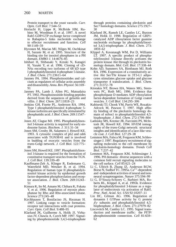
P1: KKK/spd P2: KKK/ary QC: KKK/tkj T1: KKK
September 16, 1998 11:49 Annual Reviews AR066-09
260 MARTIN
Protein transport to the yeast vacuole.Curr.Opin. Cell Biol.7:544–51
Horiuchi H, Lippe R, McBride HM, Ru-bino M, Woodman P, et al. 1997. A novelRab5 GDP/GTP exchange factor complexedto Rabaptin-5 links nucleotide exchangeto effector recruitment and function.Cell90:1149–59
Hyvonen M, Macias MJ, Nilges M, OschkinatH, Saraste M, et al. 1995. Structure of thebinding site for inositol phosphates in a PHdomain.EMBO J.14:4676–85
Ishihari H, Shibasaki Y, Kizuki N, KatagiriH, Yazaki Y, et al. 1996. Cloning of cD-NAs encoding two isoforms of 68 kD typeI phosphatidylinositol 4-phosphate 5-kinase.J. Biol. Chem.271:23611–64
Janmey PA. 1994. Phosphoinositides and cal-cium as regulators of cellular actin assemblyand disassembly.Annu. Rev. Physiol.56:169–91
Janmey PA, Lamb J, Allen PG, MatsudairaPT. 1992. Phosphoinositide-binding peptidesderived from the sequences of gelsolin andvillin. J. Biol. Chem.267:11818–23
Jenkins GH, Fisette PL, Anderson RA. 1994.Type I phosphatidylinositol 4-phosphate 5-kinase isoforms are specifically stimulated byphosphatidic acid.J. Biol. Chem.269:11547–54
Jones AT, Clague MJ. 1995. Phosphatidylinos-itol 3-kinase activity is required for early en-dosome fusion.Biochem. J.311:31–34
Jones SM, Crosby JR, Salamero J, Howell KE.1993. A cytosolic complex of p62 and rab6associates with TGN38/41 and is involvedin budding of exocytic vesicles from thetrans-Golgi network.J. Cell Biol. 122:775–88
Jones SM, Howell KE. 1997. Phosphatidylinos-itol 3-kinase is required for the formation ofconstitutive transport vesicles from the TGN.J. Cell Biol.139:339–49
Kauffmann-Zeh A, Klinger R, Endemann G,Waterfield MD, Wetzker R, et al. 1994.Regulation of human type II phosphatidyli-nositol kinase activity by epidermal growthfactor-dependent phosphorylation and recep-tor association.J. Biol. Chem.269:31243–51
Kimura K, Ito M, Amano M, Chihara K, FukataY, et al. 1996. Regulation of myosin phos-phatase by Rho and Rho-associated kinase.Science273:245–48
Kirchhausen T, Bonifacino JS, Riezman H.1997. Linking cargo to vesicle formation:receptor tail interactions with coat proteins.Curr. Opin. Cell Biol.9:488–95
Klarlund JK, Guilherme A, Holik JJ, Virba-sius JV, Chawla A, Czech MP. 1997. Signal-ing by phosphoinositide 3,4,5-trisphosphate
through proteins containing pleckstrin andSec7 homology domains.Science275:1927–30
Klarlund JK, Rameh LE, Cantley LC, BuxtonJM, Holik JJ. 1998. Regulation of GRP1-catalyzed ADP ribosylation factor guaninenucleotide exchange by phosphatidylinosi-tol 3,4,5-trisphosphate.J. Biol. Chem.273:1859–62
Klippel A, Kavanaugh WM, Pot D, WilliamsLT. 1997. A specific product of phospha-tidylinositol 3-kinase directly activates theprotein kinase Akt through its pleckstrin ho-mology domain.Mol. Cell. Biol.17:338–44
Kohn AD, Summers SA, Birnbaum MJ, RothRA. 1996. Expression of a constitutively ac-tive Akt Ser/Thr kinase in 3T3-L1 adipo-cytes stimulates glucose uptake and glucosetransporter 4 translocation.J. Biol. Chem.271:31372–78
Ktistakis NT, Brown HA, Waters MG, Stern-weis PC, Roth MG. 1996. Evidence thatphospholipase D mediates ADP ribosylationfactor-dependent formation of Golgi coatedvesicles.J. Cell Biol.134:295–306
Kubiseski TJ, Chook YM, Parris WE, Rozakis-Adcock M, Pawson T. 1997. High affin-ity binding of the pleckstrin homology do-main of mSos1 to phosphatidylinositol 4,5-bisphosphate.J. Biol. Chem.272:1799–804
Ladinsky MS, Kremer JR, Furcinitti PS, McIn-tosh JR, Howell KE. 1994. HVEM tomog-raphy of thetrans-Golgi network: structuralinsights and identification of a lace-like vesi-cle coat.J. Cell Biol.127:29–38
Lemmon MA, Falsca M, Ferguson KM, Schles-singer J. 1997. Regulatory recruitment of sig-nalling molecules to the cell membrane bypleckstrin-homology domains.Trends CellBiol. 7:237–42
Lemmon MA, Ferguson KM, Schlessinger J.1996. PH domains: diverse sequences with acommon fold recruit signaling molecules tothe cell surface.Cell 85:621–24
Li C, Ullrich B, Zhang JZ, Anderson RGW,Borse N, Sudhof TC. 1995a. Ca2+-dependentand -independent activities of neural and non-neural synaptotagmins.Nature375:594–99
Li G, D’Souza-Schorey C, Barbieri MA, Ro-berts RL, Klippel A, et al. 1995b. Evidencefor phosphatidylinositol 3-kinase as a regu-lator of endocytosis via activation of Rab5.Proc. Natl. Acad. Sci. USA92:10207–11
Lin HC, Gilman AG. 1996. Regulation ofdynamin I GTPase activity by G proteinβγ subunits and phosphatidylinositol 4,5-bisphosphate.J. Biol. Chem.271:27979–82
Liscovitch M, Cantley LC. 1995. Signal trans-duction and membrane traffic: the PITP/phosphoinositide connection.Cell 81:659–62
P1: KKK/spd P2: KKK/ary QC: KKK/tkj T1: KKK
September 16, 1998 11:49 Annual Reviews AR066-09
PHOSPHOINOSITIDE SIGNALING 261
Liscovitch M, Chalifa V, Pertile P, Chen C-S,Cantley LC. 1994. Novel functions of phos-phatidylinositol 4,5-bisphosphate as a cofac-tor for brain membrane phospholipase D.J.Biol. Chem.269:21403–6
Lockyer PJ, Bottomley JR, Reynolds JS, Mc-Nulty TJ, Venkateswarlu K, et al. 1997.Distinct subcellular localizations of the pu-tative inositol 1,3,4,5-tetrakisphosphate re-ceptors GAP1IP4BP and GAP1m result fromthe GAP1IP4BPPH domain directing plasmamembrane targeting.Curr. Biol. 7:1007–10
Loijens JC, Anderson RA. 1996. Type I phos-phatidylinositol 4-phosphate 5-kinases aredistinct members of this novel lipid kinasefamily. J. Biol. Chem.271:32937–43
Loyet KM, Kowalchyk JA, Chaudhary A, ChenJ, Prestwich GD, Martin TFJ. 1998. Spe-cific binding of phosphatidylinositol 4,5-bisphosphate to CAPS, a potential phospho-inositide effector protein for regulated exo-cytosis.J. Biol. Chem.273:8337–43
Lu P-J, Chen C-S. 1997. Selective recognitionof phosphatidylinositol 3,4,5-trisphosphateby a synthetic peptide.J. Biol. Chem.272:466–72
Lu P-J, Shieh W-R, Rhee SG, Yin HL, ChenC-S. 1996. Lipid products of phosphoinosi-tide 3-kinase bind human profilin with highaffinity. Biochemistry35:14027–34
Macias MJ, Musacchio A, Ponstingl H, NilgesM, Saraste M, et al. 1994. Structure of thepleckstrin homology domain fromβ-spec-trin. Nature369:675–77
Marcusson EG, Horazdovsky BF, Cereghino JL,Gharakhanian E, Emr SD. 1994. The sort-ing receptor for yeast vacuolar carboxypep-tidase Y is encoded by the VPS10 gene.Cell77:579–86
Marks MS, Ohno H, Kirchhausen T, BonifacinoJS. 1997. Protein sorting by tyrosine-basedsignals: adapting to the Ys and wherefores.Trends Cell Biol.7:124–28
Martin SS, Haruta T, Morris AJ, Klippel A,Williams LT, Olefsky JM. 1996. Activatedphosphatidylinositol 3-kinase is sufficient tomediate actin rearrangement and GLUT4translocation in adipocytes.J. Biol. Chem.271:17605–8
Martin TFJ. 1995. New directions for phos-phatidylinositol transfer.Curr. Biol. 5:990–92
Martin TFJ. 1997. Phosphoinositides as spatialregulators of membrane traffic.Curr. Opin.Neurobiol.7:331–38
Martin TFJ, Loyet KM, Barry VA, KowalchykJA. 1997. The role of PtdIns 4,5-bisphosphatein exocytotic membrane fusion.Biochem.Soc. Trans.25:1137–41
Mehrotra B, Elliott JT, Chen J, Olszewski JD,Profit AA, et al. 1997. Selective photoaffinity
labeling of the inositol polyphosphate bind-ing C2B domains of synaptotagmins.J. Biol.Chem.272:4237–44
Meyers R, Cantley LC. 1997. Cloning and char-acterization of a wortmannin-sensitive hu-man phosphatidylinositol 4-kinase.J. Biol.Chem.272:4384–90
Monaco ME, Gershengorn MC. 1992. Subcel-lular organization of receptor-mediated phos-phoinositide turnover.Endocr. Rev.13:707–18
Mu F-T, Callaghan JM, Steele-Mortimer O,Stenmark H, Parton RG, et al. 1995. EEA1, anearly endosome-associated protein.J. Biol.Chem.270:13503–11
Musacchio A, Gibson T, Rice P, Thompson J,Saraste M. 1993. The PH domain: a commonpiece in the structural patchwork of signallingproteins.Trends Biochem. Sci.18:343–48
Nakagawa T, Goto K, Kondo H. 1996a. Cloning,expression and localization of 230 kD phos-phatidylinositol 4-kinase.J. Biol. Chem.271:12088–94
Nakagawa T, Goto K, Kondo H. 1996b. Cloningand characterization of a 92 kD solublephosphatidylinositol 4-kinase.Biochem. J.320:643–49
Nakajima Y, Pfeffer SR. 1997. Phosphatidyli-nositol 3-kinase is not required for recyclingof mannose 6-phosphate receptors from lateendosomes to thetrans-Golgi network.Mol.Biol. Cell 8:577–82
Norris FA, Ungewickell E, Majerus PW. 1995.Inositol hexakisphosphate binds to clathrinassembly protein 3 and inhibits clathrin cageassembly in vitro.J. Biol. Chem.270:214–17
Ohara-Imaizumi M, Fukuda M, Niinobe M,Misonou H, Ikeda K, et al. 1997. Distinctroles of C2A and C2B domains of synap-totagmin in the regulation of exocytosis inadrenal chromaffin cells.Proc. Natl. Acad.Sci. USA94:287–91
Ohashi M, DeVries KJ, Frank R, Snoek GT,Bankaitis VA, et al. 1995. A role for the phos-phatidylinositol transfer protein in secretoryvesicle formation.Nature377:544–47
Okamoto M, Sudhof TC. 1997. Mints, Munc18-interacting proteins in synaptic vesicle exo-cytosis.J. Biol. Chem.272:31459–64
Olsson H, Martinez-Arias W, Drobak BK, JergilB. 1995. Presence of a novel form of phos-phatidylinositol 4-kinase in rat liver.FEBSLett.361:282–86
Orci L, Palmer DJ, Amherdt M, Rothman JE.1993. Coated vesicle assembly in the Golgirequires only coatomer and ARF proteinsfrom the cytosol.Nature364:732–34
Paris S, Beraud-Dufour S, Robineau S, BigayJ, Antonny B, et al. 1997. Role of protein-phospholipid interactions in the activation of
P1: KKK/spd P2: KKK/ary QC: KKK/tkj T1: KKK
September 16, 1998 11:49 Annual Reviews AR066-09
262 MARTIN
ARF1 by the guanine nucleotide exchangefactor Arno.J. Biol. Chem.272:22221–26
Patki V, Virbasius J, Lane WS, Toh BH, Shpet-ner HS, Corvera S. 1997. Identification of anearly endosomal protein regulated by phos-phatidylinositol 3-kinase.Proc. Natl. Acad.Sci. USA94:7326–30
Pertile P, Cantley LC. 1995. Type 2 phos-phatidylinositol 4-kinase is recruited to CD4in response to CD4 cross-linking.Biochim.Biophys. Acta1248:129–40
Pike L. 1992. Phosphatidylinositol 4-kinasesand the role of polyphosphoinositides in cel-lular regulation.Endocr. Rev.13:692–706
Pike LJ, Casey L. 1996. Localization and turn-over of phosphatidylinositol 4,5-bisphos-phate in caveolin-enriched membrane do-mains.J. Biol. Chem.271:26453–56
Pitcher JA, Touhara K, Payne ES, Lefkowitz RJ.1995. Pleckstrin homology domain-mediatedmembrane association and activation of theβ-adrenergic receptor kinase requires co-ordinate interaction with the Gβγ sub-units and lipid.J. Biol. Chem.270:11707–10
Rameh LE, Arvidsson A, Carraway KL, Couvil-lon AD, Rathbun G, et al. 1997a. A compara-tive analysis of the phosphoinositide bindingspecificity of pleckstrin homology domains.J. Biol. Chem.272:22059–66
Rameh LE, Tolias KF, Duckworth BC, Cant-ley LC. 1997b. A new pathway for synthe-sis of phosphatidylinositol 4,5-bisphosphate.Nature390:192–96
Randazzo PA. 1997. Functional interaction ofADP-ribosylation factor 1 with phosphati-dylinositol 4,5-bisphosphate.J. Biol. Chem.272:7688–92
Rapoport I, Miyazaki M, Boll W, DuckworthB, Cantley LC, et al. 1997. Regulatory inter-actions in the recognition of endocytic sort-ing signals by AP-2 complexes.EMBO J.16:2240–50
Reaves B, Bright N, Mullock B, Luzio J. 1996.The effect of wortmannin on the localizationof lysosomal type I integral membrane gly-coproteins suggests a role for phosphoinosi-tide 3-kinase activity in regulating membranetraffic late in the endocytic pathway.J. CellSci.109:749–62
Reif K, Nobes CD, Thomas G, Hall A, CantrellDA. 1996. Phosphatidylinositol 3-kinase sig-nals activate a selective subset of Rac/Rho-dependent effector pathways.Curr. Biol. 6:1445–55
Ren X-D, Bokoch GM, Traynor-Kaplan A,Jenkins GH, Anderson RA, et al. 1996. Phys-ical association of the small GTPase rho witha 68 kD phosphatidylinositol 4-phosphate 5-kinase in Swiss 3T3 cells.Mol. Biol. Cell.7:435–42
Rhee SG, Bae YS. 1997. Regulation of phos-phoinositide-specific phospholipase C iso-zymes.J. Biol. Chem.272:15045–48
Robinson MS. 1997. Coats and vesicle budding.Trends Cell Biol.7:99–102
Roth MG, Sternweis PC. 1997. The role of lipidsignaling in constitutive membrane traffic.Curr. Opin. Cell Biol.9:519–26
Rothman JE, Wieland FT. 1996. Protein sort-ing by transport vesicles.Science272:227–34
Sakisaka T, Itoh T, Miura K, Takenawa T. 1997.Phosphatidylinositol 4,5-bisphosphate phos-phatase regulates the rearrangement of actinfilaments.Mol. Cell. Biol.17:3841–49
Salim K, Bottomley MJ, Querfurth E, ZvelebilMJ, Gout I, et al. 1996. Distinct speci-ficity in the recognition of phosphoinositidesby the pleckstrin homology domains of dy-namin and Bruton’s tyrosine kinase.EMBOJ. 15:6241–50
Schafer DA, Jennings PB, Cooper JA. 1996. Dy-namics of capping protein and actin assemblyin vitro: uncapping barbed ends by polyphos-phoinositides.J. Cell Biol.135:169–79
Schekman R, Orci L. 1996. Coat proteins andvesicle budding.Science271:1526–33
Schiavo G, Gu Q-M, Prestwich GD, SollnerTH, Rothman JE. 1996. Calcium-dependentswitching of the specificity of phosphoinosi-tide binding to synaptotagmin.Proc. Natl.Acad. Sci. USA93:13327–32
Schiavo G, Stenbeck G, Rothman JE, Soll-ner TH. 1997. Binding of the synaptic vesi-cle v-SNARE, synaptotagmin, to the plasmamembrane t-SNARE, SNAP-25, can ex-plain docked vesicles at neurotoxin-treatedsynapses.Proc. Natl. Acad. Sci. USA94:997–1001
Schu PV, Takegawa K, Fry MJ, Stack JH, Water-field MD, Emr SD. 1993. Phosphatidylinos-itol 3-kinase encoded by a yeast Vps34 geneessential for protein sorting.Science260:88–91
Shepherd PR, Reaves BJ, Davidson HW. 1996.Phosphoinositide 3-kinases and membranetraffic. Trends Cell Biol.6:92–97
Shibasaki Y, Ishihara H, Kizuki N, Asano T,Oka Y, Yazaki Y. 1997. Massive actin poly-merization induced by phosphatidylinositol-4-phosphate 5-kinase in vivo.J. Biol. Chem.272:7578–81
Shpetner H, Joly M, Hartley D, Corvera S.1996. Potential sites of PI-3 kinase functionin the endocytic pathway revealed by the PI-3 kinase inhibitor wortmannin.J. Cell Biol.132:595–605
Smith CD, Chang KJ. 1989. Regulation of brainphosphatidylinositol 4-phosphate kinase byGTP analogues.J. Biol. Chem.264:320–10
Sohn RH, Chen J, Koblan KS, Bray PF,
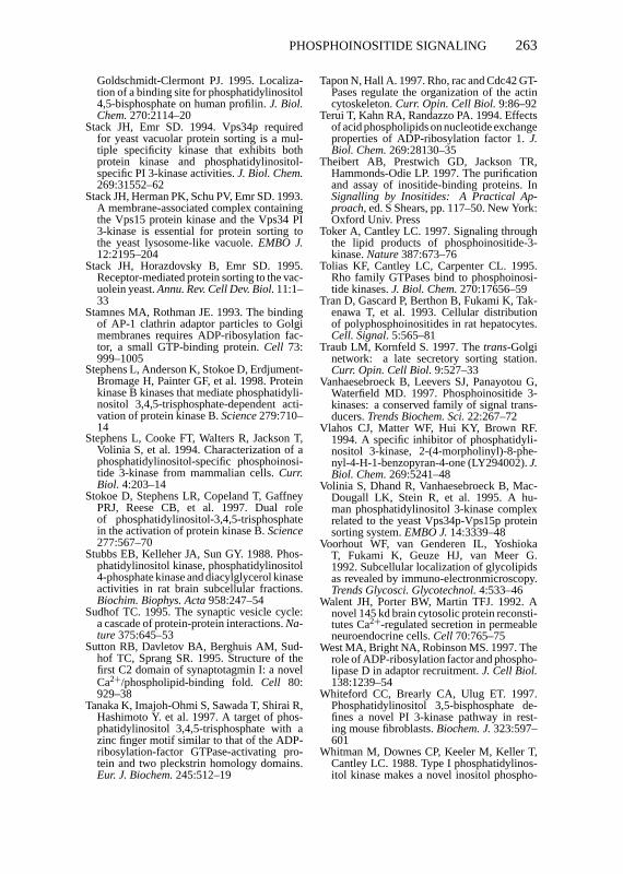
P1: KKK/spd P2: KKK/ary QC: KKK/tkj T1: KKK
September 16, 1998 11:49 Annual Reviews AR066-09
PHOSPHOINOSITIDE SIGNALING 263
Goldschmidt-Clermont PJ. 1995. Localiza-tion of a binding site for phosphatidylinositol4,5-bisphosphate on human profilin.J. Biol.Chem.270:2114–20
Stack JH, Emr SD. 1994. Vps34p requiredfor yeast vacuolar protein sorting is a mul-tiple specificity kinase that exhibits bothprotein kinase and phosphatidylinositol-specific PI 3-kinase activities.J. Biol. Chem.269:31552–62
Stack JH, Herman PK, Schu PV, Emr SD. 1993.A membrane-associated complex containingthe Vps15 protein kinase and the Vps34 PI3-kinase is essential for protein sorting tothe yeast lysosome-like vacuole.EMBO J.12:2195–204
Stack JH, Horazdovsky B, Emr SD. 1995.Receptor-mediated protein sorting to the vac-uolein yeast.Annu. Rev. Cell Dev. Biol.11:1–33
Stamnes MA, Rothman JE. 1993. The bindingof AP-1 clathrin adaptor particles to Golgimembranes requires ADP-ribosylation fac-tor, a small GTP-binding protein.Cell 73:999–1005
Stephens L, Anderson K, Stokoe D, Erdjument-Bromage H, Painter GF, et al. 1998. Proteinkinase B kinases that mediate phosphatidyli-nositol 3,4,5-trisphosphate-dependent acti-vation of protein kinase B.Science279:710–14
Stephens L, Cooke FT, Walters R, Jackson T,Volinia S, et al. 1994. Characterization of aphosphatidylinositol-specific phosphoinosi-tide 3-kinase from mammalian cells.Curr.Biol. 4:203–14
Stokoe D, Stephens LR, Copeland T, GaffneyPRJ, Reese CB, et al. 1997. Dual roleof phosphatidylinositol-3,4,5-trisphosphatein the activation of protein kinase B.Science277:567–70
Stubbs EB, Kelleher JA, Sun GY. 1988. Phos-phatidylinositol kinase, phosphatidylinositol4-phosphate kinase and diacylglycerol kinaseactivities in rat brain subcellular fractions.Biochim. Biophys. Acta958:247–54
Sudhof TC. 1995. The synaptic vesicle cycle:a cascade of protein-protein interactions.Na-ture375:645–53
Sutton RB, Davletov BA, Berghuis AM, Sud-hof TC, Sprang SR. 1995. Structure of thefirst C2 domain of synaptotagmin I: a novelCa2+/phospholipid-binding fold.Cell 80:929–38
Tanaka K, Imajoh-Ohmi S, Sawada T, Shirai R,Hashimoto Y. et al. 1997. A target of phos-phatidylinositol 3,4,5-trisphosphate with azinc finger motif similar to that of the ADP-ribosylation-factor GTPase-activating pro-tein and two pleckstrin homology domains.Eur. J. Biochem.245:512–19
Tapon N, Hall A. 1997. Rho, rac and Cdc42 GT-Pases regulate the organization of the actincytoskeleton.Curr. Opin. Cell Biol.9:86–92
Terui T, Kahn RA, Randazzo PA. 1994. Effectsof acid phospholipids on nucleotide exchangeproperties of ADP-ribosylation factor 1.J.Biol. Chem.269:28130–35
Theibert AB, Prestwich GD, Jackson TR,Hammonds-Odie LP. 1997. The purificationand assay of inositide-binding proteins. InSignalling by Inositides: A Practical Ap-proach, ed. S Shears, pp. 117–50. New York:Oxford Univ. Press
Toker A, Cantley LC. 1997. Signaling throughthe lipid products of phosphoinositide-3-kinase.Nature387:673–76
Tolias KF, Cantley LC, Carpenter CL. 1995.Rho family GTPases bind to phosphoinosi-tide kinases.J. Biol. Chem.270:17656–59
Tran D, Gascard P, Berthon B, Fukami K, Tak-enawa T, et al. 1993. Cellular distributionof polyphosphoinositides in rat hepatocytes.Cell. Signal.5:565–81
Traub LM, Kornfeld S. 1997. Thetrans-Golginetwork: a late secretory sorting station.Curr. Opin. Cell Biol.9:527–33
Vanhaesebroeck B, Leevers SJ, Panayotou G,Waterfield MD. 1997. Phosphoinositide 3-kinases: a conserved family of signal trans-ducers.Trends Biochem. Sci.22:267–72
Vlahos CJ, Matter WF, Hui KY, Brown RF.1994. A specific inhibitor of phosphatidyli-nositol 3-kinase, 2-(4-morpholinyl)-8-phe-nyl-4-H-1-benzopyran-4-one (LY294002).J.Biol. Chem.269:5241–48
Volinia S, Dhand R, Vanhaesebroeck B, Mac-Dougall LK, Stein R, et al. 1995. A hu-man phosphatidylinositol 3-kinase complexrelated to the yeast Vps34p-Vps15p proteinsorting system.EMBO J.14:3339–48
Voorhout WF, van Genderen IL, YoshiokaT, Fukami K, Geuze HJ, van Meer G.1992. Subcellular localization of glycolipidsas revealed by immuno-electronmicroscopy.Trends Glycosci. Glycotechnol.4:533–46
Walent JH, Porter BW, Martin TFJ. 1992. Anovel 145 kd brain cytosolic protein reconsti-tutes Ca2+-regulated secretion in permeableneuroendocrine cells.Cell 70:765–75
West MA, Bright NA, Robinson MS. 1997. Therole of ADP-ribosylation factor and phospho-lipase D in adaptor recruitment.J. Cell Biol.138:1239–54
Whiteford CC, Brearly CA, Ulug ET. 1997.Phosphatidylinositol 3,5-bisphosphate de-fines a novel PI 3-kinase pathway in rest-ing mouse fibroblasts.Biochem. J.323:597–601
Whitman M, Downes CP, Keeler M, Keller T,Cantley LC. 1988. Type I phosphatidylinos-itol kinase makes a novel inositol phospho-
P1: KKK/spd P2: KKK/ary QC: KKK/tkj T1: KKK
September 16, 1998 11:49 Annual Reviews AR066-09
264 MARTIN
lipid, phosphatidylinositol 3-phosphate.Na-ture332:644–46
Wiedemann C, Schafer T, Burger MM. 1996.Chromaffin granule-associated phosphati-dylinositol 4-kinase is required for stimulatedsecretion.EMBO J.15:2094–101
Wirtz KWA. 1997. Phospholipid transfer pro-teins revisited.Biochem. J.324:353–60
Wong K, Cantley LC. 1994. Cloning and char-acterization of a human phosphatidylinositol4-kinase.J. Biol. Chem.269:28878–84
Wong K, Meyers R, Cantley LC. 1997. Sub-cellular locations of phosphatidylinositol 4-kinase isoforms.J. Biol. Chem.272:13236–41
Yamamoto A, DeWald DB, Boronenkov IV,Anderson RA, Emr SD, et al. 1995. NovelPI(4)P 5-kinase homologue, Fab1p, essentialfor normal vacuole function and morphologyin yeast.Mol. Biol. Cell6:525–39
Yang J, Clarke JF, Ester CJ, Young PW, KasugaM, Holman GD. 1996. Phosphatidylinositol3-kinase acts at an intracellular membranesite to enhance GLUT4 exocytosis in 3T3-L1 cells.Biochem. J.313:125–31
Ye W, Ali N, Bembenek ME, Shears SB, LaferEM. 1995. Inhibition of clathrin assembly byhigh affinity binding of specific inositol phos-phates to the synapse-specific clathrin assem-bly protein AP-3.J. Biol. Chem.270:1564–68
Yonezawa N, Homma Y, Yahara I, Sakai H,Nishida E. 1991. A short sequence respon-sible for both phosphoinositide binding andactin binding activities of cofilin.J. Biol.Chem.266:17218–21
Yoon HS, Hajduk PJ, Petros AM, OlejniczakET, Meadows RP, et al. 1994. Solution struc-
ture of a pleckstrin-homology domain.Na-ture369:672–75
Yu F-X, Sun H-Q, Janmey PA, Yin HL. 1992.Identification of a polyphosphoinositide-binding sequence in an actin monomer-bind-ing domain of gelsolin.J. Biol. Chem.267:14616–21
Zhang X, Jefferson AB, Auethavekiat V, Ma-jerus P. 1995. The protein deficient in Lowesyndrome is a phosphatidylinositol 4,5-bis-phosphate 5-phosphatase.Proc. Natl. Acad.Sci. USA92:4853–56
Zhang X, Hartz PA, Philip E, Racusen LC,Majerus PW. 1998. Cell lines from kidneyproximal tubules of a patient with Lowesyndrome lack OCRL inositol polyphos-phate 5-phosphatase and accumulate phos-phatidylinositol 4,5-bisphosphate.J. Biol.Chem.273:1574–82
Zhang X, Loijens JC, Boronenkov IV, ParkerGJ, Norris FA, et al. 1997. Phosphatidylino-sitol-4-phosphate 5-kinase isozymes catalyzethe synthesis of 3-phosphate-containingphosphatidylinositol signaling molecules.J.Biol. Chem.272:17756–61
Zheng J, Cahill SM, Lemmon MA, Fushman D,Schlessinger J, Cowburn D. 1996a. Identifi-cation of the binding site for acidic phospho-lipids on the PH domain of dynamin: impli-cations for stimulation of GTPase activity.J.Mol. Biol. 255:14–21
Zheng Y, Glaven JA, Wu WJ, Cerione RA.1996b. Phosphatidylinositol 4,5-bisphos-phate provides an alternative to guanine nu-cleotide exchange factors by stimulating thedissociation of GDP from Cdc42Hs.J. Biol.Chem.271:23815–19