regulation of phosphoinositide 3-kinase expression in health and disease
TRANSCRIPT

_______________________________________________ Chapter 2x
Regulation of Phosphoinositide 3-kinase Expression in Health and disease Klaartje Kok, Barbara Geering, Bart Vanhaesebroeck

Regulation of PI3K expression
Abstract The biology and therapeutic potential of the phosphoinositide 3-kinase (PI3K) signalling axis have been the subject of intense investigation, yet little is known about the regulation of PI3K expression. Evidence is emerging for alterations in PI3K levels upon cellular stimulation with insulin and nuclear receptor ligands, and during various physiological and pathological processes, including differentiation, regeneration, hypertension and cancer. Recently identified mechanisms that control PI3K production include increased gene copy number in cancer, transcriptional regulation of the PIK3CA/p110α catalytic subunit by FOXO3a, NF-κB and p53, and of the regulatory subunits by STAT3, EBNA-2 and SREBP. In most instances, however, the impact of alterations in PI3K expression on PI3K signalling and disease remains to be established.
26

Chapter 2
Introduction The importance of the PI3K signalling axis in a wide variety of normal and pathological responses is now well established. Most studies to date have focused on the acute alterations in PI3K activity induced by cell stimulation, and its impact on early downstream signalling by protein effectors of the PI3K lipids. However, evidence is emerging for more sustained changes in PI3K gene expression under various conditions, leading to persistent alterations in PI3K activity. Here we summarise and discuss the current knowledge on PI3K tissue distribution, the emerging evidence for dynamic regulation of PI3K expression and its impact on the PI3K subunit ratios and PI3K signalling, as well as the underlying molecular mechanisms that modulate PI3K expression. A better understanding of the regulation of PI3K expression might help to find ways to control PI3K activity in disease conditions where PI3K expression is deregulated. The PI3K family PI3Ks (phosphoinositide 3-kinases) generate 3-phosphorylated phosphoinositide lipids which transmit intracellular signals through binding to various protein effectors (reviewed in [1]). Mammals have genes for 8 catalytic and 6 regulatory subunits (Table 1). The PI3Ks have been divided into 3 classes [1], of which the class I PI3Ks have been studied most extensively and which will be the focus of this review. The currently available information on expression and regulation of class II and III PI3Ks is summarised in Box 1.
Chromosomal localisation Class Subunit Protein Gene namehuman mouse
IA catalytic p110α PIK3CA 3q26.32 3 band A3 p110β PIK3CB 3q22.3 9 band E3.3 p110δ PIK3CD 1p36.22 4 band E1 regulatory (p85s) p85α PIK3R1 5q13.1 13 band D1 p55α PIK3R1 5q13.1 13 band D1 p50α PIK3R1 5q13.1 13 band D1 p85β PIK3R2 19p13.11 8 band B3.3 p55γ PIK3R3 1p34.1 4 band D1 IB catalytic p110γ PIK3CG 7q22.3 12 band A3 regulatory p101 PIK3R5 17p13.1 11 band B3 p84/p87 PIK3R6 17p13.1 11 band B3 II catalytic PI3K‐C2α PIK3C2A 11p15.1 7 band F1 PI3K‐C2β PIK3C2B 1q32.1 1 band E4 PI3K‐C2γ PIK3C2G 12p12.3 6 band G2 III catalytic Vps34 PIK3C3 18q12.3 18 band B2 regulatory Vps15 PIK3R4 3q22.1 9 band F1 Table 1. The catalytic and regulatory PI3K subunits in mammals
27

Regulation of PI3K expression
Class I PI3Ks are heterodimeric enzymes consisting of a 110 kDa catalytic subunit (p110α, β, γ, δ) in complex with a regulatory subunit [1]. They are acutely activated by tyrosine kinase pathways and GPCRs (G protein-coupled receptors), leading to the generation of the PIP3 lipid (phosphatidylinositol-3,4,5-trisphosphate) [1]. Downstream PIP3 effectors include protein kinases, adaptor proteins and regulators of small GTPases, which mediate signalling cascades controlling growth, cell cycle progression and migration [1]. The class IA catalytic subunits (p110α, p110β and p110δ) are bound to a regulatory subunit (p85α, p85β, p55α, p55γ or p50α; collectively referred to as “p85s”) which has two SH2 (Src homology 2) domains which can link these enzymes to tyrosine kinase signalling pathways. By contrast, the class IB catalytic subunit p110γ binds one of two non-p85 regulatory subunits, called p101 [2] and p84 [3,4], and mediates PI3K activity upon GPCR stimulation. Also the class IA PI3K p110β has recently been shown to be activated by GPCR agonists [5]. PI3K tissue distribution Most PI3K subunits appear to have a broad tissue distribution, with the exception of p110γ [6,7] and p110δ [8,9], which are highly expressed in leukocytes (Table 2). At the moment, PI3K expression patterns have only been studied at low resolution using approaches that do not allow discrimination between the different cell types in a tissue. In addition, high quality antibodies to PI3K subunits, especially antibodies that are validated for use in immunohistochemistry, are not available. In the rare instances where immunohistochemistry on tissue sections was used to address PI3K expression, unexpected cellular distribution of PI3K isoforms within tissues was observed. For example, the expression of p110α which is generally thought to be ubiquitous and uniform in various tissues, appears to be markedly enriched in the non-proliferating tumour regions of ovarian cancer in vivo [10]. In normal tissue, the same applies to p85α /p55α /p50α and the class II PI3K isoforms PI3K-C2α and PI3K-C2β, which all were mainly detected in the fully differentiated cells, and not in the proliferating cells [11]. To gain further insight into tissue distribution of class IA PI3Ks, we have created reporter mice with a β-Gal-LacZ reporter gene inserted into endogenous p110 loci by homologous recombination. Such mice are available for p110α [12] and p110δ [13]. These mice have thus far been instrumental in documenting the broad tissue distribution of p110α and the more restricted tissue distribution of p110δ, namely in the hematopoietic and, unexpectedly, also in the nervous system [14]. Reporter mouse strains have also been made for p110γ as part of the generation of p110γ-deficient mice [7]. This allowed the first clear documentation of the high enrichment of p110γ expression in
28

Chapter 2
leukocytes [7]. Reporter mice such as these will also be useful to determine PI3K isoform expression with resolution at the single cell level in tissue sections. Expression profiles of PI3K isoforms can be found in several databases, including https://biogps.gnf.org/, USCS Genome Browser (http://genome.ucsc.edu/index.html), Gene Expression Omnibus (http://www.ncbi.nlm.nih.gov/geo), ArrayExpress (http://www.ebi.ac.uk/microarray-as/aer) and Oncomine (http://www.oncomine.org/). PI3K Expression Refs.
p110α ubiquitous [8,15,16]
p110β ubiquitous [8,16‐18]
p110δ highly enriched in leukocytes, moderate expression in neurons and
cancer cell lines from various origin (including melanoma, breast,
colon) low expression in most other cell types
[8,9,14,16,
19]
p85α ubiquitous, with lowest expression in skeletal muscle [16,20,21]
p55α highest expression in brain and muscle, undetectable in most other
tissues
[21,22]
p50α high in liver, moderate expression in kidney and brain [21,23]
p85β ubiquitous, with lowest expression in skeletal muscle [16,20,21]
p55γ high mRNA expression in brain and testis, moderate mRNA expression
in most tissues, not detectable in liver and muscle. Low protein
expression in liver, muslce, fat, liver, spleen.
[16,20,24]
p110γ high in leukocytes, moderate to low expression in most other tissues [25‐29]
p101 high in leukocytes [3,4]
p84 and
p87
high in leukocytes and heart [3,4]
PI3K‐C2α ubiquitous, highest in heart, placenta, ovary [11,30,31]
PI3K‐C2β ubiquitous, highest in thymus and placenta [11,30,31]
PI3K‐C2γ high in liver and prostate, moderate expression in breast and salivary
gland
[11,32‐34]
Vps34 ubiquitous [35,36]
Vps15 ubiquitous [35,36]
Table 2. Tissue distribution of the mammalian PI3K family members
29

Regulation of PI3K expression
Dynamic regulation of the ratio of p85 to p110 subunits - a mechanism to regulate PI3K activity? Under basal conditions, class IA PI3Ks are thought to exist mainly as obligate heterodimers, with the regulatory and catalytic subunits constitutively bound to each other. This model is based on the notion that the p85 and p110 proteins bind each other extremely tightly, an interaction which can even withstand high concentrations of salt, urea or detergent [37,38]. Furthermore, both the catalytic [39] and the regulatory subunits [40,41] appear to be unstable as monomers. Together, this is expected to lead to the presence of equal amounts of p85 and p110 protein in cells. In addition, there appears to be a good correlation between the total mRNA and total protein levels for all the distinct p85s and p110s, at least in certain cell lines [16], suggesting that the same amounts of regulatory and catalytic subunits are generated under basal conditions. Some researchers have suggested that a constitutive excess of p85 over p110 exists under basal conditions (reviewed in [42]). This hypothesis was put forward to explain the paradoxical observation that p85α and p85β gene knockout mice exhibit enhanced insulin-induced PI3K signalling in insulin-sensitive tissues [43-46]. In wild-type mice, free p85 was thought to compete with p110-bound p85 for binding to signalling complexes, thereby decreasing PI3K lipid-dependent signalling. In p85 knockout cells, the level of free p85 was considered to be preferentially reduced, leading to improved access of the existing p85-p110 dimers to signalling complexes. As discussed recently [47], increasing evidence argues against the presence of excess p85, at least under basal, unstimulated conditions. However, the existence of equal amounts of p85 and p110 subunits under basal conditions does not exclude the possibility that p85 and/or p110 expression can be acutely upregulated in cells. Indeed, p85α mRNA and protein can be induced in diverse cell types upon cellular stimulation ([48-59]; Table 3). Such an increase in p85 might stabilise p110 proteins leading to an increase in p85–p110 heterodimers, possibly enhancing PI3K activity, as might be the case upon stimulation of nuclear receptor RAR (retinoic acid receptor) [56]. Alternatively, increased p85 upon cellular stimulation might exist 'p110-free' - be it temporarily, given that p85 appears to be unstable on its own [40,41]. Such p110-free p85 could be a mechanism for short-term inhibition of PI3K activity and/or might allow p110-independent biological activity of p85 (reviewed in [60]). At the moment, no solid evidence for 'p110-free' p85 is available. Studies to assess the protein expression levels of the whole spectrum of p85 and p110 isoforms would be required to gain insight into this question. Short-term upregulation of p85 levels seems to increase PI3K activity. Thus, increased p85 levels following cellular stimulation with retinoic acid [56], estradiol [61] or cannabinoid receptor type 1 (CB1) antagonist ribonamant [57] correlate with sustained
30

Chapter 2
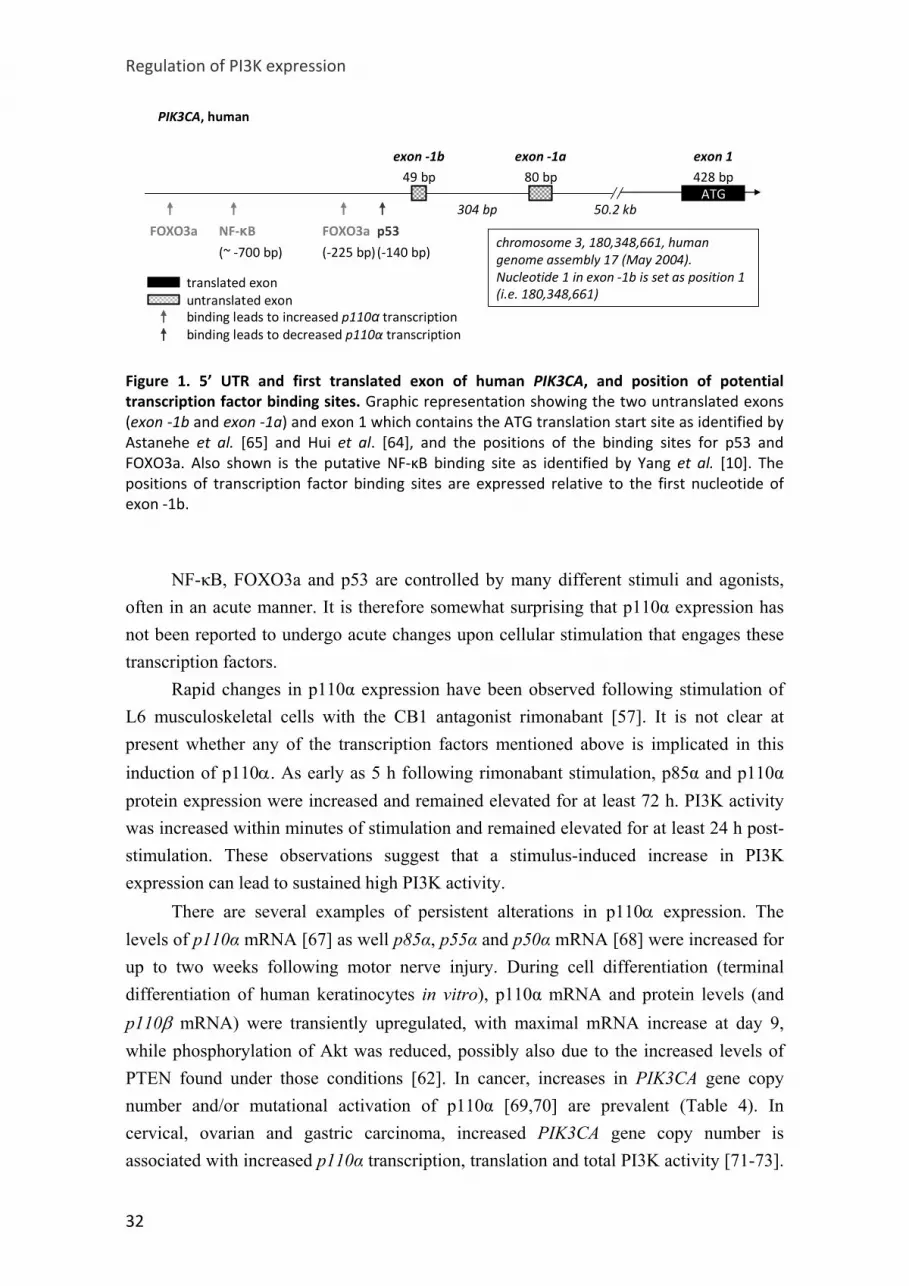
phosphorylation of Akt in cells. Moreover, insulin treatment of cells that express elevated p85 as a consequence of stimulation with nuclear receptors PPAR (peroxisome proliferator-activated receptor) and RAR, gives rise to increased in vitro PI3K activity in p85-immunoprecipitates [50,51]. Similarly, also class IA catalytic subunit expression can be increased upon cellular stimulation (e.g. by poly-2-hydroxyethyl-methacrylate (in keratinocyte differentiation [62]), by 3-isobutyl-1-methylxanthine and dexamethasone (in fibroblast differentiation [63]) and by the CB1 antagonist ribonamant [57]; Table 3). In the only case investigated, the observed increase in p110α appeared to be accompanied by a concomitant increase in p85α protein resulting in increased phosphorylation of Akt in cells [57]. At present, no data are available for physiological conditions where 'p85-free' p110 exists. Regulation of class I PI3K gene expression p110α p110α, encoded by PIK3CA, shows a broad tissue distribution in embryonic [15] and adult tissue [8,15,16]. Recently, three studies have begun to characterise the PIK3CA promoter, indicating that p110α expression can be positively controlled by FOXO3a (forkhead box O3a) [64] and NF-κB (nuclear factor-kappa B) [10] and negatively by p53 [65]. The human PIK3CA locus gives rise to two alternative transcripts, by splicing of one of two distinct 5' untranslated exons [exon -1a (80 bp) or exon -1b (49 bp); Figure 1] onto the ATG-containing exon (defined as exon 1), with the untranslated exons positioned about 50 kb upstream of the translation start site [64,65]. FOXO3a directly induces p110α expression via binding to FOXO3a-responsive elements approximately 792 and 225 bp 5’ upstream of exon -1b [64]. Given that the PI3K-Akt signalling axis inhibits FOXO3a activity (by mediating its cytoplasmic sequestration), p110α has the potential to negatively regulate its own gene expression. NF-κB binds approximately 700 bp 5’ upstream of exon -1b, and TNF-α (tumour necrosis factor alpha) stimulation can increase binding of NF-κB to the PIK3CA promoter and augment p110α mRNA expression in ovarian cancer cells [10]. A p53 binding site is present approximately 140 bp 5’ upstream of exon -1b, leading to PIK3CA transcriptional inhibition [65]. Indeed, p53 overexpression reduces p110α protein levels, whereas suppression of p53 expression increased p110α mRNA and protein levels. These data are in line with earlier reports of negative regulation of PIK3CA expression by p53 [66].
31
Regulation of PI3K expression
FOXO3a
(‐225 bp)
ATG428 bp80 bp49 bp
50.2 kb304 bp
exon ‐1b exon ‐1a exon 1
NF‐κB(~ ‐700 bp)
FOXO3a p53
(‐140 bp)
PIK3CA, human
translated exonuntranslated exonbinding leads to increased p110α transcriptionbinding leads to decreased p110α transcription
chromosome 3, 180,348,661, human genome assembly 17 (May 2004). Nucleotide 1 in exon ‐1b is set as position 1 (i.e. 180,348,661)
Figure 1. 5’ UTR and first translated exon of human PIK3CA, and position of potential transcription factor binding sites. Graphic representation showing the two untranslated exons (exon ‐1b and exon ‐1a) and exon 1 which contains the ATG translation start site as identified by Astanehe et al. [65] and Hui et al. [64], and the positions of the binding sites for p53 and FOXO3a. Also shown is the putative NF‐κB binding site as identified by Yang et al. [10]. The positions of transcription factor binding sites are expressed relative to the first nucleotide of exon ‐1b. NF-κB, FOXO3a and p53 are controlled by many different stimuli and agonists, often in an acute manner. It is therefore somewhat surprising that p110α expression has not been reported to undergo acute changes upon cellular stimulation that engages these transcription factors. Rapid changes in p110α expression have been observed following stimulation of L6 musculoskeletal cells with the CB1 antagonist rimonabant [57]. It is not clear at present whether any of the transcription factors mentioned above is implicated in this induction of p110α. As early as 5 h following rimonabant stimulation, p85α and p110α protein expression were increased and remained elevated for at least 72 h. PI3K activity was increased within minutes of stimulation and remained elevated for at least 24 h post-stimulation. These observations suggest that a stimulus-induced increase in PI3K expression can lead to sustained high PI3K activity. There are several examples of persistent alterations in p110α expression. The levels of p110α mRNA [67] as well p85α, p55α and p50α mRNA [68] were increased for up to two weeks following motor nerve injury. During cell differentiation (terminal differentiation of human keratinocytes in vitro), p110α mRNA and protein levels (and p110β mRNA) were transiently upregulated, with maximal mRNA increase at day 9, while phosphorylation of Akt was reduced, possibly also due to the increased levels of PTEN found under those conditions [62]. In cancer, increases in PIK3CA gene copy number and/or mutational activation of p110α [69,70] are prevalent (Table 4). In cervical, ovarian and gastric carcinoma, increased PIK3CA gene copy number is associated with increased p110α transcription, translation and total PI3K activity [71-73].
32

Chapter 2
Interestingly, immunohistochemistry studies on ovarian cancer sections indicate that p110α levels in the cancer cells are the highest in the non-proliferating regions (as assessed by co-staining with the cell proliferation marker Ki-67), such as in the under-vascularised tumour areas. Interestingly, the proliferating cancer cells expressed relatively low amounts of p110α [10]. p110β This PI3K isoform appears to have a broad tissue distribution, as assessed by Northern and Western blotting across a range of embryonic and adult mouse tissues [8,16-18]. A detailed analysis of the PIK3CB promoter has not been performed, yet in silico analysis of single nucleotide polymorphisms in the putative PIK3CB promoter region (as part of a search for functional polymorphisms associated with the genetics of insulin resistance) revealed the presence of a C>T variant (rs361072; C>T at -359) which could disrupt a putative GATA-binding motif [74]. The T variant decreases GATA transcription factor binding and reduces the transcriptional activity of p110β promoter constructs in vitro and in cell lines. The C variant thus allows higher p110β expression and Akt activation than the T variant [74]. This difference could help to explain why, among obese children, homozygous C/C individuals have a trend for milder insulin resistance than T/T obese patients. The distribution of rs361072 alleles was comparable in obese and non-obese cohorts. p110β mRNA and protein levels are increased upon differentiation of 3T3-L1 mouse embryonic fibroblasts into adipocytes [63] and p110β mRNA is increased upon terminal ex vivo differentiation of human primary keratinocytes (as was shown for p110α in the same study [62]). p110β (and p110δ) protein expression is also upregulated in aorta tissue extracts under conditions of hypertension [75] and in cancer, with PIK3CB gene amplification in primary ovarian tumours [76] and thyroid cancer [77], and increased p110β mRNA expression in prostate cancer [78] and neuroblastoma [79] (Table 4). p110δ p110δ is highly expressed in leukocytes [8,9,16], present at intermediate levels in neurons [14] and found at low levels in most other cell types [8,19], including endothelial cells [80,81]. p110δ has been implicated in endothelial transmigration of leukocytes during inflammation [81] but it is not clear whether this is due to expression of p110δ in leukocytes, endothelial cells or both, although some evidence for endothelial p110δ in leukocyte transmigration has been provided [81]. Other studies on the role of p110δ in blood vessels are also difficult to interpret. Indeed, p110δ expression has been reported to
33

Regulation of PI3K expression
be upregulated in arteries of hypertensive rats [75,82,83], with immunohistochemistry indicating that p110δ is located in the smooth muscle layer of the arteries [82,83]. However, in these studies, vessel homogenates were used to monitor p110δ expression, which does not allow to exclude that increases in p110δ expression are related to increased leukocyte infiltration. p110δ appears to be expressed at similar levels in transformed and non-transformed leukocytes. In acute myeloid leukaemia cells, p110δ is the only class I PI3K isoform that is consistently detected, with the levels of the other class I PI3K being extremely variable [84,85]. p110δ is expressed at moderate levels in some cancer cells of non-leukocyte origin such as melanoma, breast and colon, often with large differences in expression levels in cell lines of the same tissue origin [19], for reasons that are unclear. Increased p110δ mRNA expression without an increase in gene copy number was found in glioblastoma [86]. Both p110δ mRNA and protein levels were shown to be increased in neuroblastoma [79] (Table 4). p110γ This PI3K isoform is highly enriched in leukocytes [6,7] but is also found at lower levels in other cell types, including cardiomyocytes [27,28,87], endothelial cells [81], pancreatic islets [88,89] and smooth muscle cells [90]. Keratinocytes were reported to express high levels of p110γ [29] but others have not been able to confirm this observation (Matthias Wymann and Sabine Werner, personal communication). A putative promoter region in the p110γ gene PIK3CG has been identified and contains several consensus binding sites for leukocyte-specific transcription factors, including c/EBPβ (CCAAT/enhancer binding protein beta) and GATA-1 [91]. Some evidence for a selective upregulation of p110g amongst the PI3K isoforms has been presented. Indeed, treatment of U937 leukaemia cells with ATRA (all-trans retinoic acid), which acts via the RARs and induces cell differentiation, results in an increase in p110γ mRNA and protein levels within 8 h of stimulation, without alterations in the protein levels of p110β, p110δ, p85, or p101 [92] (Table 3). Expression of the BCR-ABL fusion oncoprotein in leukocytes has also been reported to induce p110γ expression at the mRNA and protein level, leading to increased PI3K activity, without affecting p110α, p110β or p110δ mRNA and protein levels [93]. In cancer, increased PIK3CG gene copy number has been documented in ovarian cancer [76] (Table 4). p85α Studies across a panel of mouse and rat tissues indicate that p85α is expressed at moderate to high levels in most tissues, and low levels in skeletal muscle [16,20,21].
34

Chapter 2
Tables 3 and 4 summarise the conditions of altered p85α expression under various conditions of stimulation, and in cancer. p85α expression in insulin signalling and diabetes: an unclear link p85α mRNA levels are rapidly and significantly induced upon insulin stimulation of human muscle derived from healthy donors [58]. This observation, together with the metabolic phenotype (increased insulin sensitivity) of p85α and p85β knockout mice, led several groups to monitor the expression and activity of PI3K pathway components in insulin resistance in humans [52,53,55,94-97] and in mouse models [48,54,98,99]. As described in more detail below, it has not been possible to correlate diabetic conditions with alterations in p85 expression and/or PI3K activity. Although PI3K is a key effector in insulin signalling [12,100], it is possible that factors other than PI3K itself impact on the PI3K signalling axis in diabetes. Indeed, expression and/or phosphorylation of the insulin receptor and insulin receptor substrate have been found to be reduced in insulin-resistant cells compared to control cells [52,53,55,94-96,101], suggesting that PI3K signalling could be reduced due to decreased PI3K recruitment to the plasma membrane [94]. In other words, the primary defect in insulin-resistant cells might not arise from changes in the levels of PI3K itself, but rather as a consequence of defects in upstream signalling pathways. Under basal conditions, human p85α mRNA and protein levels have been found to be unchanged [52,53,55,59,97] or increased [94,97] in primary human skeletal and adipose tissue of diabetic patients, and unchanged [96] or increased [95] in insulin-sensitive tissue of women with gestational diabetes mellitus. In line with these variable results, PI3K activity upon insulin stimulation (assessed by in vitro kinase assay or Akt phosphorylation) was found to be unchanged [94] or reduced [94,97] in insulin-sensitive cells of diabetic patients as compared to control samples. In mouse models of insulin resistance, such as transgenic overexpression of growth hormone or treatment of wild-type mice with GH, the levels of p85α [48,54,98,99] and total p85 [54,99] protein were increased in skeletal muscle and adipose tissue, with unaltered levels of p110 in one of the studies (as analysed by anti-pan-p110 antibody) [98]. Similar to what was observed in diabetic patients, data on PI3K signalling in this mouse model were inconsistent in the different studies. Indeed, under basal conditions in GH-overexpressing muscle cells, in vitro PI3K activity was either unchanged (as assessed in IRS-1 immunoprecipitates) [48] or increased (as assessed in phosphotyrosine immunoprecipitates) [99]. Upon insulin stimulation, however, PI3K activity was always found to be decreased in insulin-sensitive tissue of GH-overexpressing mice, compared to controls [48,98,99].
35

Regulation of PI3K expression
Induction of p85α expression by nuclear receptors: a way to transiently increase PI3K activity? An increase in p85α expression, leading to a concomitant increase in PI3K activity, also occurs upon stimulation of various nuclear receptors, including members of the PPAR, RAR and estrogen receptor (ER) families. Some of these studies originated from investigations into the mechanism of action of the PPARγ ligand Rosiglitazone, known to increase insulin sensitivity and used in the treatment of diabetes [102]. Upon ligand binding, PPARs translocate to the nucleus where they interact with 9-cis-RA-bound RXR (retinoid X receptor) to induce gene transcription. In human adipocytes, which express high levels of PPARγ, p85α is one of the genes induced by Rosiglitazone [49,50]. p85α mRNA was also induced by the RXR activator LG1069, and further enhanced by co-treatment with Rosiglitazone. These data indicate that the increase in p85α expression is mediated by the PPARγ–RXR heterodimer in these cells [49,50]. Rosiglitazone increased p85α mRNA and protein expression without affecting p110α or p110β mRNA levels (the effects on the levels of p85β, p55γ or p110δ mRNA and on protein expression of the PI3K subunits were not investigated [50]). Of greater importance in glucose uptake than adipocytes are muscle cells, and these cells are therefore a more relevant cellular context to study the impact of anti-diabetics. Muscle cells, however, express low levels of PPARγ and indeed, do not show p85α induction upon Rosiglitazone treatment [51]. p85α mRNA and protein are induced in these cells upon activation of PPARα and the PPARα–RXR heterodimer instead [51]. Thus, p85α is a target of PPAR-RXR nuclear receptors. This increased p85α expression by PPAR ligands corresponds with increased PI3K activity upon insulin stimulation, as measured by by in vitro PI3K activity assays in p85α immunoprecipitates. [50,51]. It remains unclear if this effect is due to an increase in p85α alone or whether there is concomitant increase in overall p110 levels. RAR activation can also induce p85 transcription. In F9 mouse embryocarcinoma cells, retinoic acid (RA) transiently induced p85α expression, correlating with a biphasic activation of PI3K and Akt [56]. The increase in PI3K activity is likely due to an increased level of the p85α–p110α heterodimer, as shown by immunoprecipitation with anti-p110α antibody. Interestingly, increased p110 protein levels did not correlate with p110α and p110β mRNA levels, which were not significantly changed upon RA treatment. These data suggest that p110 protein expression might be regulated post-translationally under these conditions, i.e. by stabilisation through p85 binding. Furthermore, the regulation of p85 expression by RA might be tissue-specific, given that treatment of U937 leukaemia cells with ATRA (which induces p110γ expression) does not increase p85 expression [50,51].
36

Chapter 2
p85α expression is also subject to ER stimulation [61]. In several cell types, stimulation with estradiol leads to an induction of PI3K activity, ranging from minutes [103,104] to hours [105]. The immediate increase in PI3K activity upon estradiol stimulation is likely due to a direct interaction between ERα and p85α [103,104], leading to increased PI3K recruitment to the plasma membrane. Yet, in MCF-7 breast cancer cells, Akt phosphorylation was increased up to 72 h after estradiol stimulation and is accompanied by an increase in p85 protein expression [61], suggesting that enhanced p85α expression might result in increased PI3K activity. This effect is achieved through the ERα receptor, and not the ERβ receptor [61]. p85β Based on studies using Northern blotting, the p85β isoform has the highest expression in brain, intermediate expression levels in liver and spleen and is nearly undetectable in muscle cells [16,20,21]. Absolute protein quantification approaches using mass spectrometry, however, revealed high levels of p85β in all tissues analysed, within the same range as those of p85α [16]. Previously, p85α was believed to be more abundant than p85β, a conclusion mainly based on the more severe phenotype of the p85α gene knockout mice (embryonic or perinatal lethality) [44,106] compared to the p85β knockout mice, which are viable and fertile [46]. Furthermore, analysis of PI3K protein expression suggested that p85β contributes only 10% of the total regulatory subunits in mouse embryo fibroblasts [107,108]. The reasons for these inconsistencies between different studies on the ratios of endogenous p85α to p85β protein levels [16,108] are not clear, but might be related to differences in the PI3K antibodies used.
No data are currently available regarding p85β regulation under physiological conditions. Increased PIK3R2 gene copy number has been documented in ovarian cancer, without an associated increase in p85β mRNA expression [76] (Table 3).
Recent findings show that p85β expression is subject to post-transcriptional regulation by the noncoding RNA miRNA-126, which post-transcriptionally downregulates p85β mRNA expression [109]. Likewise, miR-29 miRNA family members have been shown to directly suppress p85α expression [110]. Whereas the effect of miR-29 miRNA on p85β has not been reported, it has been shown that miRNA-126 does not affect p85α [109]. Loss-of-expression of miRNA-126 in colon cancer is thought to contribute to PI3K-Akt pathway activation, possibly by increasing p85β-p110 levels. In endothelial cells, however, increased p85β expression was put forward as a negative regulator of PI3K action, contributing to loss of blood vessel integrity [111]. Such a difference in outcome (up- versus downregulation of PI3K activity) could be
37

Regulation of PI3K expression
related to whether p110 levels change concomitantly with changes in the levels of p85β, which could be cell type-specific. p55α and p50α Like p85α, p55α and p50α are encoded by the PIK3R1 locus. p55α and p50α each possess their own promoters [112,113] and thus are not p85α splice variants as originally speculated [21,23]. Northern blot analysis of rat tissues revealed that p55α is expressed mainly in brain and muscle and is undetectable in most other tissues [21,22], whereas p50α has the highest expression in liver, kidney and brain [21,23]. This distribution pattern could indicate tissue-specific functions of these regulatory subunits. p55α and p50α are transcriptional targets of STAT3 (signal transducer and activator of transcription 3) during mammary gland involution in vivo [112]. STAT3 directly binds the p55α and the p50α promoter (and not to the p85α promoter), leading to upregulation of their expression. Such regulation was not observed in embryonic stem cells, indicating tissue-specific regulation of p50α and p55α expression. The EBNA-2 transcription factor of Epstein-Barr virus can selectively induce p55α expression in the EREB2.5 human B-lymphoblastoid cell line, without alterations in the protein expression of p85α, p55α and p55γ and the class IA p110 catalytic subunits [114]. Expression of p55α is essential for proliferation of EREB2.5 cells, and RNAi-mediated selective knock-down of p55α was accompanied by increased apoptosis [114]. In response to injury of motor neurons in rats, increased expression of mRNA and protein of p55α and to a lesser extent of p50α, lasting up to 28 days has been observed [68]. In a separate study, also p110α was increased upon motor nerve injury [67]. p55α and p50α mRNA levels are increased in skeletal muscle cells upon stimulation with insulin, as was shown for p85a [59]. This study further indicated that PI3K activity itself might regulate transcription of the regulatory subunits by a positive feedback loop, given that treatment of these cells with the PI3K inhibitor LY294002 downregulated the levels of p85α, p55α and p50α mRNA. p55γ PIK3R3 mutant mice have not been reported and the function of p55γ is unknown. Using Northern blotting analysis across a panel of mouse [24] and rat [20] tissues, p55γ was shown to be expressed at high levels in brain and testis, at intermediate levels in several other tissues such as heart and lung, and is undetectable in liver and skeletal muscle. However, protein quantification by mass spectrometry revealed very low levels of p55γ protein in murine muscle, liver, fat, brain and spleen [16]. Good quality antibodies to p55γ are not available, and its tissue expression at the protein level has not been assessed.
38

Chapter 2
PIK3R3 is a direct transcriptional target for sterol-regulatory element binding proteins (SREBPs), which are transcription factors known to regulate genes controlling lipid metabolism. SREBP is ubiquitously expressed as a precursor protein that is processed into active SREBP upon several metabolic stimuli. SREBP-1 activation by stimulation of human fibroblasts with sterols, insulin and platelet derived growth factor triggers p55γ mRNA and protein upregulation [115]. In vivo SREBP activation (by feeding mice with high fat diet) induces p55γ expression in murine liver, which usually expresses low amounts of p55γ [115]. The processing of SREBP itself is dependent on PI3K activity, since inhibition of PI3Ks with LY294002 blocks this activation [116]. Significant DNA copy number gain and mRNA upregulation of PIK3R3 (quantitative PCR in ovarian cell lines) have been documented in ovarian cancer [76]. In the same study, p55γ mRNA up-regulation was also observed in liver, prostate, and breast cancer (Table 4). p101 and p84 p101 and p84 are the regulatory subunits for p110γ. p101 and p84 are encoded by 2 separate genes (located next to each other on chromosome 17), and are highly expressed in leukocytes [3,4]. Northern and Western blotting and RT-PCR analysis showed that p84 is also highly expressed in the heart [4]. p101 expression remains unchanged upon treatment with ATRA or BCR-ABL expression, which both induce p110γ expression [92,93]. The impact of these stimuli on p84 expression has not been investigated. Concluding remarks Under basal, i.e. unstimulated conditions, the expression of the PI3K regulatory and catalytic subunits seems to be mainly under transcriptional control, with mRNA levels correlating well with protein levels, leading to equal amounts of p85 and p110 protein in cells [16]. Stimuli and transcription factors that induce PI3K gene expression have recently been identified. Some stimuli (such as a CB1 antagonist and estradiol) can induce an immediate, initial increase in PI3K activity due to receptor-mediated PI3K activation, as well as a sustained increase in PI3K activity as a result of increased PI3K subunit expression. It is not clear at present how generic this mechanism of PI3K upregulation by PI3K expression will be, given that the long-term impact of most stimuli on PI3K gene expression has not been investigated. In order to understand the functional impact of changes in PI3K subunit alterations, it will be essential to assess more comprehensively the expression levels of PI3K isoforms as well as the impact on cellular PI3K activity. Quantitation of PI3K lipid products, especially in primary tissues, remains a formidable technical challenge
39

Regulation of PI3K expression
[117,118]. It is anticipated that new developments in analytical techniques (such as mass spectrometry [119]) will most likely be critical to move this field forward. In addition, the community should invest in generating high quality antibody reagents against PI3Ks, to enable better monitoring of PI3K expression alterations in health and disease. This is anticipated to be conducive for effective therapeutic interference with PI3K signalling.
40

Chapter 2
41
Box 1: Class II and III PI3Ks
The Class II PI3Ks, which comprise the PI3K‐C2α, PI3K‐C2β and PIK3C2γ isoforms (Table 1),
preferentially phosphorylate phosphatidylinositol to produce phosphatidylinositol‐3‐phosphate.
The importance of class II PI3Ks in cell signalling and biology, relative to that of class I PI3Ks, is
not clear at the moment (reviewed in [123,124]).
Based on Northern blot analysis of human tissues, PI3K‐C2α [31] and PI3K‐C2β [30] are
thought to be ubiquitously expressed. PI3K‐C2γ is present in most tissues but enriched in liver,
breast, prostate and salivary glands [32‐34,125]. A more recent study, however, using
immunohistochemical techniques on normal human tissues, indicated that PI3K‐C2α and PI3K‐
C2β are not ubiquitously distributed, but are restricted to fully differentiated cells (rather than
proliferating cells) of epithelial or mesenchymal origin. Smooth muscle, endothelial and
glomerular epithelium cells only expressed PI3K‐C2α, whereas neurons expressed PI3K‐C2β [11].
All class II PI3K enzymes were further detected in macrophages [11].
PI3K‐C2α and PI3K‐C2β expression is elevated in a large number of human small cell lung
cancer cell lines when compared to normal lung epithelial cells [126]. PIK3C2B gene
amplification and mRNA overexpression has further been documented in a small fraction of
glioblastoma [86] (Table 4). Comparative genomic hybridization in 89 human ovarian cancer
specimens revealed increased gene copy number of PIK3C2B in ovarian tumours. mRNA
expression of PI3Ks was not determined on the same samples but using public micro‐array data
sets that included 2 ovarian cancer studies. In this data set PI3K‐C2β mRNA expression was not
significantly increased [76].
Class III PI3K. vps34, the sole class III PI3K, exists as a heterodimer bound to the vps15
regulatory subunit (formerly called p150 in mammals). Vps34 has been implicated in
endocytosis, autophagy and nutrient signalling [127]. Northern blot analysis indicates that vps34
and vps15 are both ubiquitously expressed [35,36]. Their expression levels are not known to
undergo dynamic changes.

Chapter 2, Table 3
cell type/tissue species stimulus/process mRNA/ Protein
p110α p110β p110δ p85α p55α p50α p85β p55γ p110γ p101 ref.
3T3‐L1 embryonic fibroblasts mouse differentiation into adipocytes Protein = ↑ [63] mRNA ↑ ↑ ↑ ↑ = =
motor neurons in vivo rat regeneration after injury Protein ↑
[67,68]
mRNA ↑ ↑ primary keratinocytes in vitro human differentiation
Protein ↑ [62]
aorta in vivo (homogenate) rat hypertension, DOCA induced Protein = = ↑ = = [75] aorta in vivo (homogenate) rat hypertension, L‐NNA induced Protein = ↑ ↑ = = [75] aorta in vivo (homogenate) rat hypertension, spontaneous Protein = ↑ = = [82] resistance artery in vivo (homogenate)
rat hypertension, DOCA induced Protein = = ↑ = [83]
variou
s stim
uli/processes
L6 skeletal muscle cell line rat Treatment with Cannabinoid receptor type 1 antagonist SR141716 (PKA dependent)
Protein ↑ ↑ [57]
cell type/tissue species receptor stimulus/process p110α p110β p110δ p85α p55α p50α p85β p55γ p110γ p101 ref.mRNA ↑
U937 myelomonocytic cells human RAR ATRA Protein = = = ↑ =
[92]
mRNA ↑ PPARα Wy‐14643
Protein =PPARβ L‐165041 mRNA =PPARγ 15ΔPGJ2 mRNA =PPARγ Rosiglitazone mRNA =
mRNA ↑ RXR 9‐cis‐RA
Protein ↑ mRNA ↑
primary muscle cells
human
PPARα/RXR Wy‐14643/9‐cis‐RA Protein ↑
[51]
mRNA = = ↑ primary adipocytes human
PPARγ or PPARγ/RXR
Rosiglitazone or Rosglitazone/LG1069 Protein ↑
[49,50]
mRNA = = ↑ =
nuclear recep
tors
F9 embryocarcinoma cells mouse RARγ/RXR 9‐cis‐RA Protein ↑
[56]

transcription
cell type/tissue species stimulus/process p110α p110β p110δ p85α p55α p50α p85β p55γ p110γ p101 ref. factor
mRNA = = ↑ ↑ [112] Mammary gland tissue in vivo mouse STAT3 involution
Protein = = = = ↓ ↑ ↑ ↓ [114] EREB2.5 B‐lymphoblasts human EBNA‐2 Protein = = = = = = ↑
mRNA ↑ - AG01518 foreskin fibroblasts - liver tissue in vivo
human mouse
SREBP ‐ PDGF (fibroblasts) ‐ high carbohydrate diet (liver) Protein ↑
[115]
non‐tumorigenic ovarian surface epithelial (OSE) cell
human p53 stimulus unknown ‐ binding to region 5’ of exon ‐1b of PIK3CA
proteinmRNA ↓
[65]
Ovarian cancer xenograft model
human NF‐κB TNF‐α mRNA ↑ [10]
mRNA ↑
transcription factors
[64] K562 chronic myeloid leukaemia cell line
stimulus unknown ‐ binding to region 5’ of exon ‐1b of PIK3CA
human FOXO3a Protein ↑
cell type/tissue species stimulus/process p110α p110β p110δ p85α p55α p50α p85β p55γ p110γ p101 ref.muscle tissue in vivo human insulin mRNA = = [52,55,58 ↑ ↑ ↑ adipocytes in vivo human insulin mRNA [52,55,59↑ ↑ ↑ muscle tissue in vivo human hypocaloric diet mRNA ↑ [53]
muscle cells mouse GH‐transgene expression Protein = = ↑ [54,98,99
mRNA ↑
insulin
[48] adipose cells mouse
GH‐transgene expression
Protein ↑
Table 3. Regulation of class I PI3K isoform expression. =: unaltered levles; ↑: Increased levels; ↑ : increased expression correlates with a reduction in PI3K activity or Akt phosphorylation; DOCA: deoxycorticosterone acetate‐salt; ↑ increase in expression correlates with increase in in vitro PI3K activity and reduction in Akt phosphorylation; L‐NNA: Nω‐nitro‐L‐arginine; ↑: increased levels are associated with an increase in PI3K activity/Akt phosphorylation. For the increase in p85 expression after ligand binding to nuclear receptors, this increase in PI3K activity is measured upon stimulation with insulin; ↓: decreased levels; PDGF: platelet derived growth factor; ↓: decreased levels correlate with a decrease in PI3K activity
43

Chapter 2, Table 4
p110α
PIK3CA
p110β
PIK3CB
p110δ
PIK3CD
p110γ
PIK3CG
PI3K‐C2β
PIIK3C2B
p85α
PIK3R1
p85β
PIK3R2
p55γ
PIK3R3 Refs
ovarian
carcinoma
• ↑ GCN (19/26)
• ↑GCN & ↑mRNA (13/14)
• ↑GCN & ↑PI3K activity (3/3)
• ↑GCN (21/89)
• ↑GCN
(24/89)
• ↑GCN
(23/89)
• ↑GCN
(36/89)
• ↑GCN
(31/89)
• ↑GCN (19/89)
• ↑mRNA,
(18 cancer vs 16 non‐cancer)
[72],
[76];
Neuro‐
blastoma
• ↑mRNA (4/8)
• ↑mRNA
(6/8)
• ↑mRNA (11/20)
• ↑protein & ↑mRNA (10/12)
• ↑mRNA
(10/19)
[79]
prostate
carcinoma
• ↑mRNA
(14/30)
• ↑mRNA
(10/30)
[78]
HNSCC • ↑GCN (12/33)
• ↑mRNA (16/33)
• No correlation ↑GCN & ↑mRNA
• ↑GCN (85/218)
[120],
[121]
lung
cancers
• ↑GCN (44/132)
• ↑pAkt (nuclear) (36/215)
• ↑pAkt (cytoplasmic) (26/215)
[122]
thyroid
cancer
• ↑GCN (33/110)
• ↑pAkt (18/24)
• ↑GCN (n=41/97)
• ↑pAkt (n=13/17)
[77]
Glio‐
blastoma
• ↑mRNA (6/95) • ↑GCN (6/103)
• ↑GCN & ↑mRNA (n =4/94)
[86]
cervical
carcinoma
• ↑GCN (42/55)
• ↑GCN, ↑protein & ↑PI3K
act,ivity (3/3)
[71]
gastric
carcinoma
• ↑GCN (29/70)
• ↑GCN correlates with ↑mRNA
• ↑GCN & ↑pAkt (8/9).
[73]
Table 4. Altered expression of PI3K isoforms in cancer. GCN: Gene copy number. (19/26): number berween brackets indicate the number of positive samples per number of samples tested. HNSCC: Head and neck squamous cell carcinoma. NB: the references are listed for each cell in the same row. NB2: these data are mostly derived from studies using primary human tumours but data using cell lines are included as well.

Chapter 2
References [1] Vanhaesebroeck, B. et al. (2001). Synthesis and function of 3-phosphorylated inositol lipids.
Annual Review of Biochemistry 70, 535-602. [2] Stephens, L.R. et al. (1997). The G[beta][gamma] Sensitivity of a PI3K Is Dependent upon a
Tightly Associated Adaptor, p101. Cell 89, 105-114. [3] Suire, S., Coadwell, J., Ferguson, G.J., Davidson, K., Hawkins, P. and Stephens, L. (2005). p84, a
New G[beta][gamma]-Activated Regulatory Subunit of the Type IB Phosphoinositide 3-Kinase p110[gamma]. Current Biology 15, 566-570.
[4] Voigt, P., Dorner, M.B. and Schaefer, M. (2006). Characterization of p87PIKAP, a novel regulatory subunit of phosphoinositide 3-kinase gamma that is highly expressed in heart and interacts with PDE3B. J Biol Chem 281, 9977-86.
[5] Guillermet-Guibert, J. et al. (2008). The p110beta isoform of phosphoinositide 3-kinase signals downstream of G protein-coupled receptors and is functionally redundant with p110gamma. PNAS in press
[6] Barbier, M. et al. (2001). Tumour biology. Weakening link to colorectal cancer? Nature 413, 796. [7] Li, Z., Jiang, H., Xie, W., Zhang, Z., Smrcka, A.V. and Wu, D. (2000). Roles of PLC-2 and -3 and
PI3K in Chemoattractant-Mediated Signal Transduction. Science 287, 1046-1049. [8] Vanhaesebroeck, B. et al. (1997). p110delta , a novel phosphoinositide 3-kinase in leukocytes.
PNAS 94, 4330-4335. [9] Chantry, D., Vojtek, A., Kashishian, A., Holtzman, D.A., Wood, C., Gray, P.W., Cooper, J.A. and
Hoekstra, M.F. (1997). p110delta, a novel phosphatidylinositol 3-kinase catalytic subunit that associates with p85 and is expressed predominantly in leukocytes. J Biol Chem 272, 19236-41.
[10] Yang, N. et al. (2008). Transcriptional regulation of PIK3CA oncogene by NF-kappaB in ovarian cancer microenvironment. PLoS ONE 3, e1758.
[11] El Sheikh, S.S., Domin, J., Tomtitchong, P., Abel, P., Stamp, G. and Lalani, E.N. (2003). Topographical expression of class IA and class II phosphoinositide 3-kinase enzymes in normal human tissues is consistent with a role in differentiation. BMC Clin Pathol 3, 4.
[12] Foukas, L.C. et al. (2006). Critical role for the p110[alpha] phosphoinositide-3-OH kinase in growth and metabolic regulation. Nature 441, 366-370.
[13] Okkenhaug, K. et al. (2002). Impaired B and T cell antigen receptor signaling in p110delta PI 3-kinase mutant mice. Science 297, 1031-4.
[14] Eickholt, B.J. et al. (2007). Control of Axonal Growth and Regeneration of Sensory Neurons by the p110delta PI 3-Kinase. PLoS ONE 2, e869.
[15] Bi, L., Okabe, I., Bernard, D.J., Wynshaw-Boris, A. and Nussbaum, R.L. (1999). Proliferative defect and embryonic lethality in mice homozygous for a deletion in the p110alpha subunit of phosphoinositide 3-kinase. J Biol Chem 274, 10963-8.
[16] Geering, B., Cutillas, P.R., Nock, G., Gharbi, S.I. and Vanhaesebroeck, B. (2007). Class IA phosphoinositide 3-kinases are obligate p85-p110 heterodimers. Proc Natl Acad Sci U S A 104, 7809-14.
[17] Bi, L., Okabe, I., Bernard, D.J. and Nussbaum, R.L. (2002). Early embryonic lethality in mice deficient in the p110beta catalytic subunit of PI 3-kinase. Mamm Genome 13, 169-72.
[18] Hu, P., Mondino, A., Skolnik, E.Y. and Schlessinger, J. (1993). Cloning of a novel, ubiquitously expressed human phosphatidylinositol 3-kinase and identification of its binding site on p85. Mol Cell Biol 13, 7677-88.
[19] Sawyer, C., Sturge, J., Bennett, D.C., O'Hare, M.J., Allen, W.E., Bain, J., Jones, G.E. and Vanhaesebroeck, B. (2003). Regulation of Breast Cancer Cell Chemotaxis by the Phosphoinositide 3-Kinase p110{delta}. Cancer Res 63, 1667-1675.
[20] Inukai, K. et al. (1996). A novel 55-kDa regulatory subunit for phosphatidylinositol 3-kinase structurally similar to p55PIK Is generated by alternative splicing of the p85alpha gene. J Biol Chem 271, 5317-20.
[21] Inukai, K. et al. (1997). p85alpha gene generates three isoforms of regulatory subunit for phosphatidylinositol 3-kinase (PI 3-Kinase), p50alpha, p55alpha, and p85alpha, with different PI 3-kinase activity elevating responses to insulin. J Biol Chem 272, 7873-82.
[22] Antonetti, D.A., Algenstaedt, P. and Kahn, C.R. (1996). Insulin receptor substrate 1 binds two novel splice variants of the regulatory subunit of phosphatidylinositol 3-kinase in muscle and brain. Mol. Cell. Biol. 16, 2195-2203.
[23] Fruman, D.A., Cantley, L.C. and Carpenter, C.L. (1996). Structural organization and alternative splicing of the murine phosphoinositide 3-kinase p85 alpha gene. Genomics 37, 113-21.
45

Regulation of PI3K expression
[24] Pons, S. et al. (1995). The structure and function of p55PIK reveal a new regulatory subunit for phosphatidylinositol 3-kinase. Mol Cell Biol 15, 4453-65.
[25] Bernstein, H.G., Keilhoff, G., Reiser, M., Freese, S. and Wetzker, R. (1998). Tissue distribution and subcellular localization of a G-protein activated phosphoinositide 3-kinase. An immunohistochemical study. Cell Mol Biol (Noisy-le-grand) 44, 973-83.
[26] Hirsch, E. et al. (2000). Central role for G protein-coupled phosphoinositide 3-kinase gamma in inflammation. Science 287, 1049-53.
[27] Patrucco, E. et al. (2004). PI3Kgamma modulates the cardiac response to chronic pressure overload by distinct kinase-dependent and -independent effects. Cell 118, 375-87.
[28] Stoyanov, B. et al. (1995). Cloning and characterization of a G protein-activated human phosphoinositide-3 kinase. Science 269, 690-3.
[29] Zhao, M. et al. (2006). Electrical signals control wound healing through phosphatidylinositol-3-OH kinase-gamma and PTEN. Nature 442, 457-60.
[30] Brown, R.A., Ho, L.K., Weber-Hall, S.J., Shipley, J.M. and Fry, M.J. (1997). Identification and cDNA cloning of a novel mammalian C2 domain-containing phosphoinositide 3-kinase, HsC2-PI3K. Biochem Biophys Res Commun 233, 537-44.
[31] Domin, J., Pages, F., Volinia, S., Rittenhouse, S.E., Zvelebil, M.J., Stein, R.C. and Waterfield, M.D. (1997). Cloning of a human phosphoinositide 3-kinase with a C2 domain that displays reduced sensitivity to the inhibitor wortmannin. Biochem J 326 ( Pt 1), 139-47.
[32] Misawa, H., Ohtsubo, M., Copeland, N.G., Gilbert, D.J., Jenkins, N.A. and Yoshimura, A. (1998). Cloning and characterization of a novel class II phosphoinositide 3-kinase containing C2 domain. Biochem Biophys Res Commun 244, 531-9.
[33] Ono, F. et al. (1998). A novel class II phosphoinositide 3-kinase predominantly expressed in the liver and its enhanced expression during liver regeneration. J Biol Chem 273, 7731-6.
[34] Rozycka, M., Lu, Y.J., Brown, R.A., Lau, M.R., Shipley, J.M. and Fry, M.J. (1998). cDNA cloning of a third human C2-domain-containing class II phosphoinositide 3-kinase, PI3K-C2gamma, and chromosomal assignment of this gene (PIK3C2G) to 12p12. Genomics 54, 569-74.
[35] Panaretou, C., Domin, J., Cockcroft, S. and Waterfield, M.D. (1997). Characterization of p150, an adaptor protein for the human phosphatidylinositol (PtdIns) 3-kinase. Substrate presentation by phosphatidylinositol transfer protein to the p150.Ptdins 3-kinase complex. J Biol Chem 272, 2477-85.
[36] Volinia, S. et al. (1995). A human phosphatidylinositol 3-kinase complex related to the yeast Vps34p-Vps15p protein sorting system. Embo J 14, 3339-48.
[37] Fry, M.J., Panayotou, G., Dhand, R., Ruiz-Larrea, F., Gout, I., Nguyen, O., Courtneidge, S.A. and Waterfield, M.D. (1992). Purification and characterization of a phosphatidylinositol 3-kinase complex from bovine brain by using phosphopeptide affinity columns. Biochem J 288 ( Pt 2), 383-93.
[38] Kazlauskas, A. and Cooper, J.A. (1990). Phosphorylation of the PDGF receptor beta subunit creates a tight binding site for phosphatidylinositol 3 kinase. EMBO J 9, 3279-86.
[39] Yu, J., Zhang, Y., McIlroy, J., Rordorf-Nikolic, T., Orr, G.A. and Backer, J.M. (1998). Regulation of the p85/p110 phosphatidylinositol 3'-kinase: stabilization and inhibition of the p110alpha catalytic subunit by the p85 regulatory subunit. Mol Cell Biol 18, 1379-87.
[40] Brachmann, S.M., Yballe, C.M., Innocenti, M., Deane, J.A., Fruman, D.A., Thomas, S.M. and Cantley, L.C. (2005). Role of phosphoinositide 3-kinase regulatory isoforms in development and actin rearrangement. Mol Cell Biol 25, 2593-606.
[41] Zhao, J.J., Cheng, H., Jia, S., Wang, L., Gjoerup, O.V., Mikami, A. and Roberts, T.M. (2006). The p110alpha isoform of PI3K is essential for proper growth factor signaling and oncogenic transformation. Proc Natl Acad Sci U S A 103, 16296-300.
[42] Luo, J. and Cantley, L.C. (2005). The negative regulation of phosphoinositide 3-kinase signaling by p85 and it's implication in cancer. Cell Cycle 4, 1309-12.
[43] Chen, D., Mauvais-Jarvis, F., Bluher, M., Fisher, S.J., Jozsi, A., Goodyear, L.J., Ueki, K. and Kahn, C.R. (2004). p50alpha/p55alpha phosphoinositide 3-kinase knockout mice exhibit enhanced insulin sensitivity. Mol Cell Biol 24, 320-9.
[44] Fruman, D.A., Mauvais-Jarvis, F., Pollard, D.A., Yballe, C.M., Brazil, D., Bronson, R.T., Kahn, C.R. and Cantley, L.C. (2000). Hypoglycaemia, liver necrosis and perinatal death in mice lacking all isoforms of phosphoinositide 3-kinase p85 alpha. Nat Genet 26, 379-82.
[45] Terauchi, Y. et al. (1999). Increased insulin sensitivity and hypoglycaemia in mice lacking the p85 alpha subunit of phosphoinositide 3-kinase. Nat Genet 21, 230-5.
46

Chapter 2
[46] Ueki, K., Yballe, C.M., Brachmann, S.M., Vicent, D., Watt, J.M., Kahn, C.R. and Cantley, L.C. (2002). Increased insulin sensitivity in mice lacking p85beta subunit of phosphoinositide 3-kinase. Proc Natl Acad Sci U S A 99, 419-24.
[47] Geering, B., Cutillas, P.R. and Vanhaesebroeck, B. (2007). Regulation of class IA PI3Ks: is there a role for monomeric PI3K subunits? Biochem Soc Trans 35, 199-203.
[48] del Rincon, J.P. et al. (2007). Growth hormone regulation of p85alpha expression and phosphoinositide 3-kinase activity in adipose tissue: mechanism for growth hormone-mediated insulin resistance. Diabetes 56, 1638-46.
[49] Rieusset, J., Auwerx, J. and Vidal, H. (1999). Regulation of Gene Expression by Activation of the Peroxisome Proliferator-Activated Receptor [gamma] with Rosiglitazone (BRL 49653) in Human Adipocytes. Biochemical and Biophysical Research Communications 265, 265-271.
[50] Rieusset, J., Chambrier, C., Bouzakri, K., Dusserre, E., Auwerx, J., Riou, J.P., Laville, M. and Vidal, H. (2001). The expression of the p85alpha subunit of phosphatidylinositol 3-kinase is induced by activation of the peroxisome proliferator-activated receptor gamma in human adipocytes. Diabetologia 44, 544-54.
[51] Rieusset, J., Roques, M., Bouzakri, K., Chevillotte, E. and Vidal, H. (2001). Regulation of p85[alpha] phosphatidylinositol-3-kinase expression by peroxisome proliferator-activated receptors (PPARs) in human muscle cells. FEBS Letters 502, 98-102.
[52] Andreelli, F., Laville, M., Ducluzeau, P.H., Vega, N., Vallier, P., Khalfallah, Y., Riou, J.P. and Vidal, H. (1999). Defective regulation of phosphatidylinositol-3-kinase gene expression in skeletal muscle and adipose tissue of non-insulin-dependent diabetes mellitus patients. Diabetologia 42, 358-64.
[53] Andreelli, F., Laville, M., Vega, N., Riou, J.P. and Vidal, H. (2000). Regulation of gene expression during severe caloric restriction: lack of induction of p85 alpha phosphatidylinositol 3-kinase mRNA in skeletal muscle of patients with type II (non-insulin-dependent) diabetes mellitus. Diabetologia 43, 356-63.
[54] Barbour, L.A. et al. (2005). Increased P85alpha is a potent negative regulator of skeletal muscle insulin signaling and induces in vivo insulin resistance associated with growth hormone excess. J Biol Chem 280, 37489-94.
[55] Ducluzeau, P.H., Perretti, N., Laville, M., Andreelli, F., Vega, N., Riou, J.P. and Vidal, H. (2001). Regulation by insulin of gene expression in human skeletal muscle and adipose tissue. Evidence for specific defects in type 2 diabetes. Diabetes 50, 1134-42.
[56] Bastien, J., Plassat, J.L., Payrastre, B. and Rochette-Egly, C. (2005). The phosphoinositide 3-kinase//Akt pathway is essential for the retinoic acid-induced differentiation of F9 cells. Oncogene 25, 2040-2047.
[57] Esposito, I. et al. (2008). The cannabinoid CB1 receptor antagonist Rimonabant stimulates 2-deoxyglucose uptake in skeletal muscle cells by regulating phosphatidylinositol-3-kinase activity. Mol. Pharm, in press.
[58] Laville, M., Auboeuf, D., Khalfallah, Y., Vega, N., Riou, J.P. and Vidal, H. (1996). Acute Regulation by Insulin of Phosphatidylinositol-3-kinase, Rad, Glut 4, and Lipoprotein Lipase mRNA Levels in Human Muscle. J. Clin. Invest. 98, 43-49.
[59] Lefai, E., Roques, M., Vega, N., Laville, M. and Vidal, H. (2001). Expression of the splice variants of the p85alpha regulatory subunit of phosphoinositide 3-kinase in muscle and adipose tissue of healthy subjects and type 2 diabetic patients. Biochem. J. 360, 117-126.
[60] Okkenhaug, K. and Vanhaesebroeck, B. (2001). New responsibilities for the PI3K regulatory subunit p85 alpha. Sci STKE 2001, PE1.
[61] Lee, Y.R., Park, J., Yu, H.N., Kim, J.S., Youn, H.J. and Jung, S.H. (2005). Up-regulation of PI3K/Akt signaling by 17beta-estradiol through activation of estrogen receptor-alpha, but not estrogen receptor-beta, and stimulates cell growth in breast cancer cells. Biochem Biophys Res Commun 336, 1221-6.
[62] Pankow, S., Bamberger, C., Klippel, A. and Werner, S. (2006). Regulation of epidermal homeostasis and repair by phosphoinositide 3-kinase. J Cell Sci 119, 4033-4046.
[63] Asano, T. et al. (2000). p110beta Is Up-regulated during Differentiation of 3T3-L1 Cells and Contributes to the Highly Insulin-responsive Glucose Transport Activity. J. Biol. Chem. 275, 17671-17676.
[64] Hui, R.C. et al. (2008). The forkhead transcription factor FOXO3a increases PI3K/Akt activity in drug-resistant leukaemic cells through induction of PIK3CA expression. Mol Cell Biol, epub ahead of print
[65] Astanehe, A., Arenillas, D., Wasserman, W.W., Leung, P.C., Dunn, S.E., Davies, B.R., Mills, G.B. and Auersperg, N. (2008). Mechanisms underlying p53 regulation of PIK3CA transcription in ovarian surface epithelium and in ovarian cancer. J Cell Sci 121, 664-74.
47

Regulation of PI3K expression
[66] Singh, B. et al. (2002). p53 regulates cell survival by inhibiting PIK3CA in squamous cell carcinomas. Genes Dev 16, 984-93.
[67] Ito, Y., Sakagami, H. and Kondo, H. (1996). Enhanced gene expression for phosphatidylinositol 3-kinase in the hypoglossal motoneurons following axonal crush. Brain Res Mol Brain Res 37, 329-32.
[68] Okamoto, T., Namikawa, K., Asano, T., Takaoka, K. and Kiyama, H. (2004). Differential regulation of the regulatory subunits for phosphatidylinositol 3-kinase in response to motor nerve injury. Molecular Brain Research 131, 119-125.
[69] Samuels, Y. and Ericson, K. (2006). Oncogenic PI3K and its role in cancer. Curr Opin Oncol 18, 77-82.
[70] Samuels, Y. et al. (2004). High frequency of mutations of the PIK3CA gene in human cancers. Science 304, 554.
[71] Ma, Y.Y. et al. (2000). PIK3CA as an oncogene in cervical cancer. Oncogene 19, 2739-44. [72] Shayesteh, L. et al. (1999). PIK3CA is implicated as an oncogene in ovarian cancer. Nat Genet 21,
99-102. [73] Byun, D.S., Cho, K., Ryu, B.K., Lee, M.G., Park, J.I., Chae, K.S., Kim, H.J. and Chi, S.G. (2003).
Frequent monoallelic deletion of PTEN and its reciprocal associatioin with PIK3CA amplification in gastric carcinoma. Int J Cancer 104, 318-27.
[74] Le Stunff, C. et al. (2008). Association analysis indicates that a variant GATA-binding site in the PIK3CB promoter is a Cis-acting expression quantitative trait locus for this gene and attenuates insulin resistance in obese children. Diabetes 57, 494-502.
[75] Northcott, C.A., Poy, M.N., Najjar, S.M. and Watts, S.W. (2002). Phosphoinositide 3-kinase mediates enhanced spontaneous and agonist-induced contraction in aorta of deoxycorticosterone acetate-salt hypertensive rats. Circ Res 91, 360-9.
[76] Zhang, L. et al. (2007). Integrative genomic analysis of phosphatidylinositol 3'-kinase family identifies PIK3R3 as a potential therapeutic target in epithelial ovarian cancer. Clin Cancer Res 13, 5314-21.
[77] Liu, Z. et al. (2008). Highly prevalent genetic alterations in receptor tyrosine kinases and phosphatidylinositol 3-kinase/akt and mitogen-activated protein kinase pathways in anaplastic and follicular thyroid cancers. J Clin Endocrinol Metab 93, 3106-16.
[78] Zhu, Q. et al. (2008). Phosphoinositide 3-OH kinase p85alpha and p110beta are essential for androgen receptor transactivation and tumor progression in prostate cancers. Oncogene 27, 4569-79.
[79] Boller, D., Schramm, A., Doepfner, K.T., Shalaby, T., von Bueren, A.O., Eggert, A., Grotzer, M.A. and Arcaro, A. (2008). Targeting the Phosphoinositide 3-Kinase Isoform p110{delta} Impairs Growth and Survival in Neuroblastoma Cells. Clin Cancer Res 14, 1172-1181.
[80] Geng, L. et al. (2004). A specific antagonist of the p110delta catalytic component of phosphatidylinositol 3'-kinase, IC486068, enhances radiation-induced tumor vascular destruction. Cancer Res 64, 4893-9.
[81] Puri, K.D., Doggett, T.A., Huang, C.Y., Douangpanya, J., Hayflick, J.S., Turner, M., Penninger, J. and Diacovo, T.G. (2005). The role of endothelial PI3Kgamma activity in neutrophil trafficking. Blood 106, 150-7.
[82] Northcott, C.A., Hayflick, J. and Watts, S.W. (2005). Upregulated function of phosphatidylinositol-3-kinase in genetically hypertensive rats: a moderator of arterial hypercontractility. Clinical and Experimental Pharmacology and Physiology 32, 851-858.
[83] Northcott, C.A., Hayflick, J.S. and Watts, S.W. (2004). PI3-Kinase Upregulation and Involvement in Spontaneous Tone in Arteries From DOCA-Salt Rats: Is p110{delta} the Culprit? Hypertension 43, 885-890.
[84] Billottet, C., Grandage, V.L., Gale, R.E., Quattropani, A., Rommel, C., Vanhaesebroeck, B. and Khwaja, A. (2006). A selective inhibitor of the p110delta isoform of PI 3-kinase inhibits AML cell proliferation and survival and increases the cytotoxic effects of VP16. Oncogene 25, 6648-59.
[85] Sujobert, P. et al. (2005). Essential role for the p110delta isoform in phosphoinositide 3-kinase activation and cell proliferation in acute myeloid leukemia. Blood 106, 1063-6.
[86] Knobbe, C.B. and Reifenberger, G. (2003). Genetic alterations and aberrant expression of genes related to the phosphatidyl-inositol-3'-kinase/protein kinase B (Akt) signal transduction pathway in glioblastomas. Brain Pathol 13, 507-18.
[87] Alloatti, G. et al. (2005). Phosphoinositide 3-kinase gamma controls autonomic regulation of the mouse heart through Gi-independent downregulation of cAMP level. FEBS Lett 579, 133-40.
[88] Li, L.X., MacDonald, P.E., Ahn, D.S., Oudit, G.Y., Backx, P.H. and Brubaker, P.L. (2006). Role of phosphatidylinositol 3-kinasegamma in the beta-cell: interactions with glucagon-like peptide-1. Endocrinology 147, 3318-25.
48

Chapter 2
[89] MacDonald, P.E. et al. (2004). Impaired glucose-stimulated insulin secretion, enhanced intraperitoneal insulin tolerance, and increased beta-cell mass in mice lacking the p110gamma isoform of phosphoinositide 3-kinase. Endocrinology 145, 4078-83.
[90] Vecchione, C. et al. (2005). Protection from angiotensin II-mediated vasculotoxic and hypertensive response in mice lacking PI3Kgamma. J Exp Med 201, 1217-28.
[91] Hirsch, E., Wymann, M.P., Patrucco, E., Tolosano, E., Bulgarelli-Leva, G., Marengo, S., Rocchi, M. and Altruda, F. (2000). Analysis of the murine phosphoinositide 3-kinase [gamma] gene. Gene 256, 69-81.
[92] Baier, R., Bondeva, T., Klinger, R., Bondev, A. and Wetzker, R. (1999). Retinoic Acid Induces Selective Expression of Phosphoinositide 3-Kinase {{gamma}} in Myelomonocytic U937 Cells. Cell Growth Differ 10, 447-456.
[93] Hickey, F.B. and Cotter, T.G. (2006). BCR-ABL Regulates Phosphatidylinositol 3-Kinase-p110{gamma} Transcription and Activation and Is Required for Proliferation and Drug Resistance. J. Biol. Chem. 281, 2441-2450.
[94] Bandyopadhyay, G.K., Yu, J.G., Ofrecio, J. and Olefsky, J.M. (2005). Increased p85/55/50 expression and decreased phosphotidylinositol 3-kinase activity in insulin-resistant human skeletal muscle. Diabetes 54, 2351-9.
[95] Catalano, P.M., Nizielski, S.E., Shao, J., Preston, L., Qiao, L. and Friedman, J.E. (2002). Downregulated IRS-1 and PPARgamma in obese women with gestational diabetes: relationship to FFA during pregnancy. Am J Physiol Endocrinol Metab 282, E522-33.
[96] Friedman, J.E., Ishizuka, T., Shao, J., Huston, L., Highman, T. and Catalano, P. (1999). Impaired glucose transport and insulin receptor tyrosine phosphorylation in skeletal muscle from obese women with gestational diabetes. Diabetes 48, 1807-14.
[97] Tsuchida, H. et al. (2002). Gene expression of the p85alpha regulatory subunit of phosphatidylinositol 3-kinase in skeletal muscle from type 2 diabetic subjects. Pflugers Arch 445, 25-31.
[98] Barbour, L.A., Shao, J., Qiao, L., Leitner, W., Anderson, M., Friedman, J.E. and Draznin, B. (2004). Human placental growth hormone increases expression of the p85 regulatory unit of phosphatidylinositol 3-kinase and triggers severe insulin resistance in skeletal muscle. Endocrinology 145, 1144-50.
[99] Dominici, F.P., Cifone, D., Bartke, A. and Turyn, D. (1999). Alterations in the early steps of the insulin-signaling system in skeletal muscle of GH-transgenic mice. Am J Physiol 277, E447-54.
[100] Shepherd, P.R., Withers, D.J. and Siddle, K. (1998). Phosphoinositide 3-kinase: the key switch mechanism in insulin signalling. Biochem J 333 ( Pt 3), 471-90.
[101] Kerouz, N.J., Horsch, D., Pons, S. and Kahn, C.R. (1997). Differential regulation of insulin receptor substrates-1 and -2 (IRS-1 and IRS-2) and phosphatidylinositol 3-kinase isoforms in liver and muscle of the obese diabetic (ob/ob) mouse. J Clin Invest 100, 3164-72.
[102] Rieusset, J. et al. (1999). Insulin acutely regulates the expression of the peroxisome proliferator-activated receptor-gamma in human adipocytes. Diabetes 48, 699-705.
[103] Castoria, G. et al. (2001). PI3-kinase in concert with Src promotes the S-phase entry of oestradiol-stimulated MCF-7 cells. Embo J 20, 6050-9.
[104] Simoncini, T., Hafezi-Moghadam, A., Brazil, D.P., Ley, K., Chin, W.W. and Liao, J.K. (2000). Interaction of oestrogen receptor with the regulatory subunit of phosphatidylinositol-3-OH kinase. Nature 407, 538-541.
[105] Banerjee, S., Saxena, N., Sengupta, K. and Banerjee, S.K. (2003). 17alpha-estradiol-induced VEGF-A expression in rat pituitary tumor cells is mediated through ER independent but PI3K-Akt dependent signaling pathway. Biochem Biophys Res Commun 300, 209-15.
[106] Fruman, D.A., Snapper, S.B., Yballe, C.M., Davidson, L., Yu, J.Y., Alt, F.W. and Cantley, L.C. (1999). Impaired B cell development and proliferation in absence of phosphoinositide 3-kinase p85alpha. Science 283, 393-7.
[107] Ueki, K., Algenstaedt, P., Mauvais-Jarvis, F. and Kahn, C.R. (2000). Positive and Negative Regulation of Phosphoinositide 3-Kinase-Dependent Signaling Pathways by Three Different Gene Products of the p85alpha Regulatory Subunit. Mol. Cell. Biol. 20, 8035-8046.
[108] Ueki, K., Fruman, D.A., Brachmann, S.M., Tseng, Y.H., Cantley, L.C. and Kahn, C.R. (2002). Molecular balance between the regulatory and catalytic subunits of phosphoinositide 3-kinase regulates cell signaling and survival. Mol Cell Biol 22, 965-77.
[109] Guo, C., Sah, J.F., Beard, L., Willson, J.K., Markowitz, S.D. and Guda, K. (2008). The noncoding RNA, miR-126, suppresses the growth of neoplastic cells by targeting phosphatidylinositol 3-kinase signaling and is frequently lost in colon cancers. Genes Chromosomes Cancer
[110] Park, S.Y., Lee, J.H., Ha, M., Nam, J.W. and Kim, V.N. (2008). miR-29 miRNAs activate p53 by targeting p85alpha and CDC42. Nat Struct Mol Biol
49

Regulation of PI3K expression
[111] Fish, J.E. et al. (2008). miR-126 regulates angiogenic signaling and vascular integrity. Dev Cell 15, 272-84.
[112] Abell, K. et al. (2005). Stat3-induced apoptosis requires a molecular switch in PI(3)K subunit composition. Nat Cell Biol 7, 392-398.
[113] Abell, K. and Watson, C.J. (2005). The Jak/Stat pathway: a novel way to regulate PI3K activity. Cell Cycle 4, 897-900.
[114] Spender, L.C. et al. (2006). Cell target genes of Epstein-Barr virus transcription factor EBNA-2: induction of the p55{alpha} regulatory subunit of PI3-kinase and its role in survival of EREB2.5 cells. J Gen Virol 87, 2859-2867.
[115] Kallin, A. et al. (2007). SREBP1 regulates the expression of heme oxygenase 1 and the phosphatidylinositol-3 kinase regulatory subunit p55gamma. J. Lipid Res., M700136-JLR200.
[116] Demoulin, J.B., Ericsson, J., Kallin, A., Rorsman, C., Ronnstrand, L. and Heldin, C.H. (2004). Platelet-derived growth factor stimulates membrane lipid synthesis through activation of phosphatidylinositol 3-kinase and sterol regulatory element-binding proteins. J Biol Chem 279, 35392-402.
[117] Hawkins, P.T., Anderson, K.E., Davidson, K. and Stephens, L.R. (2006). Signalling through Class I PI3Ks in mammalian cells. Biochem Soc Trans 34, 647-62.
[118] Rusten, T.E. and Stenmark, H. (2006). Analyzing phosphoinositides and their interacting proteins. Nat Methods 3, 251-8.
[119] Pettitt, T.R., Dove, S.K., Lubben, A., Calaminus, S.D. and Wakelam, M.J. (2006). Analysis of intact phosphoinositides in biological samples. J Lipid Res 47, 1588-96.
[120] Fenic, I., Steger, K., Gruber, C., Arens, C. and Woenckhaus, J. (2007). Analysis of PIK3CA and Akt/protein kinase B in head and neck squamous cell carcinoma. Oncol Rep 18, 253-9.
[121] Sticht, C., Hofele, C., Flechtenmacher, C., Bosch, F.X., Freier, K., Lichter, P. and Joos, S. (2005). Amplification of Cyclin L1 is associated with lymph node metastases in head and neck squamous cell carcinoma (HNSCC). Br J Cancer 92, 770-4.
[122] Massion, P.P. et al. (2004). Early Involvement of the Phosphatidylinositol 3-Kinase/Akt Pathway in Lung Cancer Progression. Am. J. Respir. Crit. Care Med. 170, 1088-1094.
[123] Falasca, M. and Maffucci, T. (2007). Role of class II phosphoinositide 3-kinase in cell signalling. Biochem Soc Trans 35, 211-4.
[124] Traer, C.J., Foster, F.M., Abraham, S.M. and Fry, M.J. (2006). Are class II phosphoinositide 3-kinases potential targets for anticancer therapies? Bull Cancer 93, E53-8.
[125] Ho, L.K., Liu, D., Rozycka, M., Brown, R.A. and Fry, M.J. (1997). Identification of four novel human phosphoinositide 3-kinases defines a multi-isoform subfamily. Biochem Biophys Res Commun 235, 130-7.
[126] Arcaro, A., Khanzada, U.K., Vanhaesebroeck, B., Tetley, T.D., Waterfield, M.D. and Seckl, M.J. (2002). Two distinct phosphoinositide 3-kinases mediate polypeptide growth factor-stimulated PKB activation. Embo J 21, 5097-108.
[127] Backer, J.M. (2008). The regulation and function of Class III PI3Ks: novel roles for Vps34. Biochem J 410, 1-17.
50