c3 glomerulopathy: consensus report
TRANSCRIPT

OPEN
C3 glomerulopathy: consensus reportMatthew C. Pickering1, Vivette D. D’Agati2, Carla M. Nester3,4, Richard J. Smith3,4, Mark Haas5,Gerald B. Appel6, Charles E. Alpers7, Ingeborg M. Bajema8, Camille Bedrosian9, Michael Braun10,Mittie Doyle9, Fadi Fakhouri11, Fernando C. Fervenza12, Agnes B. Fogo13, Veronique Fremeaux-Bacchi14,Daniel P. Gale15, Elena Goicoechea de Jorge1, Gene Griffin9, Claire L. Harris16, V. Michael Holers17,Sally Johnson18, Peter J. Lavin19, Nicholas Medjeral-Thomas1, B. Paul Morgan16, Cynthia C. Nast5,Laure-Helene Noel20, D. Keith Peters21, Santiago Rodrıguez de Cordoba22, Aude Servais23,Sanjeev Sethi24, Wen-Chao Song25, Paul Tamburini9, Joshua M. Thurman17, Michael Zavros26 andH. Terence Cook1
1Centre for Complement and Inflammation Research, Imperial College London, London, UK; 2Division of Renal Pathology, Departmentof Pathology, Columbia University Medical Center and the New York Presbyterian Hospital, New York, New York, USA; 3Division ofNephrology, Departments of Internal Medicine and Pediatrics, Carver College of Medicine, University of Iowa, Iowa City, Iowa, USA;4Molecular Otolaryngology and Renal Research Laboratories, Carver College of Medicine, University of Iowa, Iowa City, Iowa, USA;5Department of Pathology and Laboratory Medicine, Cedars-Sinai Medical Center, Los Angeles, California, USA; 6Division of Nephrology,Department of Medicine, Columbia University Medical Center and the New York Presbyterian Hospital, New York, New York, USA;7Department of Pathology, University of Washington, Seattle, Washington, USA; 8Department of Pathology, Leiden University MedicalCenter, Leiden, The Netherlands; 9Alexion Pharmaceuticals, Cheshire, Connecticut, USA; 10Division of Pediatric Nephrology, BaylorCollege of Medicine and Texas Children’s Hospital, Houston, Texas, USA; 11Nephrology Department, Institut de Transplantation Urologieet Nephrologie, CHU de Nantes, Nantes, France; 12Division of Nephrology and Hypertension, Department of Internal Medicine, MayoClinic, Rochester, Minnesota, USA; 13Department of Pathology, Microbiology, and Immunology, Vanderbilt University Medical Center,Nashville, Tennessee, USA; 14Service d’Immunologie Biologique, Hopital George Pompidou, AP-HP, Paris, France; 15Division of Medicine,Centre for Nephrology, University College London, London, UK; 16Institute of Infection and Immunity, School of Medicine, CardiffUniversity, Cardiff, UK; 17Department of Medicine, University of Colorado School of Medicine, Aurora, Colorado, USA; 18Departmentof Paediatric Nephrology, Great North Children’s Hospital, Newcastle Upon Tyne Hospitals NHS Foundation Trust, Newcastle UponTyne, UK; 19Trinity Health Kidney Centre, Tallaght Hospital, Trinity College, Dublin, Ireland; 20INSERM, Service de Nephrologie et detransplantation, Service d’Anatomie Pathologique, Universite Rene Descartes, Hopital Necker, AP-HP, Paris, France; 21Cambridge, UK;22Centro de Investigaciones Biologicas, Consejo Superior de Investigaciones Cientıficas and Centro de Investigacion Biomedica enEnfermedades Raras, Ramiro de Maeztu, Madrid, Spain; 23Service de Nephrologie et de transplantation, Hopital Necker-Enfants Malades,AP-HP, Paris, France; 24Division of Anatomic Pathology, Department of Laboratory Medicine and Pathology, Mayo Clinic, Rochester,Minnesota, USA; 25Department of Pharmacology, Perelman School of Medicine, University of Pennsylvania, Philadelphia, Pennsylvania,USA and 26Department of Nephrology, Nicosia General Hospital, University of Cyprus, Nicosia, Cyprus
C3 glomerulopathy is a recently introduced pathological entity
whose original definition was glomerular pathology
characterized by C3 accumulation with absent or scanty
immunoglobulin deposition. In August 2012, an invited group
of experts (comprising the authors of this document) in renal
pathology, nephrology, complement biology, and
complement therapeutics met to discuss C3 glomerulopathy
in the first C3 Glomerulopathy Meeting. The objectives were to
reach a consensus on: the definition of C3 glomerulopathy,
appropriate complement investigations that should be
performed in these patients, and how complement
therapeutics should be explored in the condition. This meeting
report represents the current consensus view of the group.
Kidney International advance online publication, 30 October 2013;
doi:10.1038/ki.2013.377
KEYWORDS: clinical immunology; clinical nephrology; complement;
glomerulonephritis; immunology and pathology
C3 glomerulopathy is a recently introduced pathologicalentity whose original definition was glomerular pathologycharacterized by C3 accumulation with absent or scantyimmunoglobulin deposition.1 The term was introduced forthree reasons: first, it was recognized that glomerularpathology associated with isolated C3 accumulation is farmore heterogeneous than previously appreciated. Conse-quently, many lesions could not be satisfactorily placedwithin existing pathological descriptors based on morphol-ogy. For example, the absence of membranoproliferative
http://www.kidney-international.org m e e t i n g r e p o r t
& 2013 International Society of Nephrology
Correspondence: Matthew C. Pickering or H. Terence Cook, Centre for
Complement and Inflammation Research, Imperial College London, London
W12 0NN, UK. E-mail: [email protected] or
Received 20 March 2013; revised 30 May 2013; accepted 20 June 2013
Kidney International 1

changes on light microscopy in some cases precluded the useof descriptors like ‘membranoproliferative glomerulone-phritis (MPGN) type II’ or ‘MPGN type I with isolateddeposits of C3’. Second, advances in our understanding ofcomplement-mediated kidney injury have made it possible toidentify the cause of renal disease through specific comple-ment investigations. A renal biopsy report that classified alesion as C3 glomerulopathy should prompt an investigationof the complement system. Third, the existence of a licensedcomplement inhibitor (eculizumab, Alexion Pharmaceu-ticals, Cheshire, CT) together with the many complementinhibitors in clinical development meant that it wastherapeutically relevant to identify patient groups mostlikely to benefit from an anti-complement therapeuticapproach. C3 glomerulopathy, by definition, encompassedcomplement-mediated renal disease, and defined a logicalpatient population in which to test the efficacy ofcomplement inhibitors. C3 glomerulopathy incorporatedrather than replaced existing disease entities where theseterms were considered to be satisfactorily descriptive of thepathology, that is, dense deposit disease and C3 glomeru-lonephritis. The term C3 glomerulonephritis was coined todescribe glomerular lesions in which there is glomerularaccumulation of C3 with little or no immunoglobulin in theabsence of the characteristic highly electron-dense transfor-mation seen in dense deposit disease. C3 glomerulopathy alsoincorporates entities where the presence of a disease-associated complement mutation is causally associated withthe underlying renal pathology. Examples include familialdense deposit disease with C3 mutation2 and familial C3glomerulonephritis with mutations in the CFHR genes.3,4
TOWARD A CONSENSUS
In August 2012, an invited group of experts (comprising theauthors of this document) in renal pathology, nephrology,complement biology, and complement therapeutics met todiscuss C3 glomerulopathy in the first C3 GlomerulopathyMeeting. The meeting was organized by Matthew Pickeringand Terry Cook, hosted at the Wellcome Trust ConferenceCentre, Hinxton, Cambridge, UK, and sponsored by anunconditional educational grant from Alexion Pharmaceu-ticals. The objectives of this working group were: (i) throughexpert-based discussion, to reach a consensus on thedefinition of C3 glomerulopathy; (ii) through expert-baseddiscussion, to reach a consensus on the appropriate complementinvestigations that should be performed in these patients; (iii)through expert-based discussion, to reach a consensus onhow complement therapeutics should be explored in C3glomerulopathy; and (iv) to garner support for an Interna-tional Registry of C3 Glomerulopathy. This documentrepresents the current consensus view of the group.
PATHOLOGYLimitations and difficulties with the original definition
The last decade has seen increasing recognition of a spectrumof glomerular diseases in which the primary pathogenic
process is abnormal control of complement activation,deposition, or degradation leading to deposition of fragmentsof C3 in glomeruli. The C3 fragments can be detected byimmunohistochemistry (IHC)/immunofluorescence (IF) andare associated with electron-dense deposits on electronmicroscopy (EM). The C3 fragments are detected in routineIHC/IF by an antibody directed against C3c and positivitywith this antibody is by convention said to show C3localization (Figure 1). The term C3 glomerulopathy wassuggested to encompass a range of conditions regardless ofthe light or electron microscopic appearances.1 C3 glomeru-lopathy is distinct from atypical hemolytic uremic syn-drome although both diseases are due to abnormal control ofthe alternative pathway. In atypical hemolytic uremic syndrome,activation of complement occurs on glomerular or micro-vascular endothelium causing a thrombotic microangiopathy;in most cases, no electron-dense deposits are seen on EM andglomerular C3 is not detected on IHC/IF.
As the primary process that leads to glomerular C3fragment deposition in C3 glomerulopathy is complementactivation via the alternative pathway, typical cases do notshow any deposition of immunoglobulin or of earlycomponents of the classical or lectin pathways, specificallyC1q and C4c. Therefore, a purist approach would be torestrict the term solely to cases with C3 staining in theabsence of immunoglobulins, C1q and C4c. However, asdiscussed below, well-substantiated cases occur where thepathogenesis and histopathological features are typical for C3glomerulopathy but variable amounts of immunoglobulinare detected on IHC/IF. The converse situation also arises. Incases of post-infectious glomerulonephritis (PIGN), theremay be isolated staining for C3 on IHC/IF, but the clinicalfeatures are consistent with a self-limiting immunecomplex–mediated process. Therefore, the problem in clinicalpractice is to distinguish those patients in whom thepathological process is C3 deposition due to abnormalitiesof complement control from those with another pathogenesissuch as immune complex deposition. We suggest that theterm C3 glomerulopathy be used to designate a diseaseprocess rather than just a set of biopsy appearances. This is,for example, analogous to systemic lupus erythematosus,immunoglobulin A (IgA) nephropathy or diabetic nephrop-athy in which, despite variable morphological appearanceson biopsy, the pathologist can confidently reach a diagnosisbased on the synthesis of light, electron microscopic, IHC/IF,and clinical features.
We will now discuss the range of pathological appearancesseen in C3 glomerulopathy and then make practicalrecommendations for terminology and future research.
Morphology
C3 glomerulopathy may show a range of features on lightmicroscopy and EM. Light microscopic appearances includemesangial proliferative, membranoproliferative, and endo-capillary proliferative; in each case, crescents may also bepresent. In rare cases, glomeruli may be normal by light
m e e t i n g r e p o r t MC Pickering et al.: C3 glomerulopathy
2 Kidney International

microscopy. One of the most variable findings between casesis in the quality of the deposits seen in glomeruli on EM.In many cases, these have a distinctive highly electron-dense,osmiophilic appearance and this has been designated as densedeposit disease (Figure 2a). It is not known why the depositsdevelop this particular morphological appearance. In othercases, the deposits do not have this characteristic density.However, the meeting recognized that, although in typicalcases there is generally good agreement among pathologistson a diagnosis of dense deposit disease, there may be caseswhere the decision to call the deposit ‘dense’ is not clear cut.In addition, the typical dense appearance may only be presentin some segments of glomeruli (Figure 2b) and thereforediagnosis may be affected by EM sampling. In dense depositdisease, the deposits are typically found within the glomer-ular basement membrane, as rounded deposits in themesangium, and, in many cases, in Bowman’s capsule andtubular basement membranes. The glomerular deposits oftenform band-like or sausage-like shapes punctuated by skipareas of more normal-appearing glomerular basementmembrane. These deposits tend to thicken and transformthe lamina densa, but may also involve the subendothelialregion and produce hump-shaped subepithelial deposits thatresemble those seen in acute PIGN. Other than the differencein the morphology of the deposits on EM, which when wellestablished may also lead to characteristic appearances onlight microscopy, there are no other specific histopathologicalor clinical features that allow a distinction between cases ofC3 glomerulopathy with the appearance of dense depositdisease and those without. By light microscopy, cases of densedeposit disease may show a range of appearances. In some,there is a typical membranoproliferative pattern, whereasothers show predominantly mesangial proliferation. Theremay be prominent endocapillary proliferation, leukocyteinfiltration, and/or crescents.5 Glomerular and tubularbasement membrane deposits are often visible by lightmicroscopy with the help of trichrome and silver stains.
C3 glomerulopathies in which the deposits do not fulfillthe criteria for dense deposit disease have been designated as‘C3 glomerulonephritis’.6 By definition, the deposits of C3glomerulonephritis are less electron dense than those seen inclassic dense deposit disease. On EM, there are a range ofappearances. It appears that the changes that have beendesignated as MPGN type III of Strife and Anders oftenrepresent C3 glomerulonephritis. In these cases, there is acomplex pattern of mesangial increase and glomerularbasement membrane thickening with variable combinationsof subendothelial, intramembranous, and subepithelialdeposits associated with fraying of the lamina densa. Ingeneral, the glomerular basement membrane deposits ofthese examples of C3 glomerulonephritis tend to be lessdiscrete, more ill-defined, more confluent, and more likely toblend with the extracellular matrix than those in densedeposit disease (Figure 2c). Other examples of C3 glomer-ulonephritis have more discrete subendothelial depositsresembling MPGN type I. Deposits in the mesangium tend
a
b
c
Figure 1 | Immunohistology for C3c. (a) Immunofluorescence indense deposit disease, (b) immunofluorescence in a case of C3glomerulonephritis showing predominantly capillary wall staining,and (c) immunoperoxidase in a case of C3 glomerulonephritisshowing predominantly mesangial staining.
MC Pickering et al.: C3 glomerulopathy m e e t i n g r e p o r t
Kidney International 3
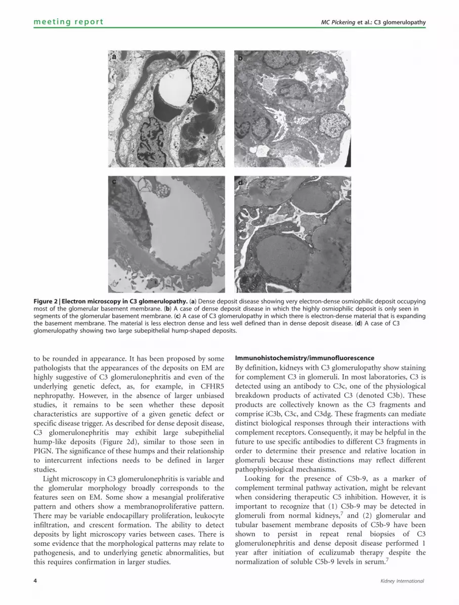
to be rounded in appearance. It has been proposed by somepathologists that the appearances of the deposits on EM arehighly suggestive of C3 glomerulonephritis and even of theunderlying genetic defect, as, for example, in CFHR5nephropathy. However, in the absence of larger unbiasedstudies, it remains to be seen whether these depositcharacteristics are supportive of a given genetic defect orspecific disease trigger. As described for dense deposit disease,C3 glomerulonephritis may exhibit large subepithelialhump-like deposits (Figure 2d), similar to those seen inPIGN. The significance of these humps and their relationshipto intercurrent infections needs to be defined in largerstudies.
Light microscopy in C3 glomerulonephritis is variable andthe glomerular morphology broadly corresponds to thefeatures seen on EM. Some show a mesangial proliferativepattern and others show a membranoproliferative pattern.There may be variable endocapillary proliferation, leukocyteinfiltration, and crescent formation. The ability to detectdeposits by light microscopy varies between cases. There issome evidence that the morphological patterns may relate topathogenesis, and to underlying genetic abnormalities, butthis requires confirmation in larger studies.
Immunohistochemistry/immunofluorescence
By definition, kidneys with C3 glomerulopathy show stainingfor complement C3 in glomeruli. In most laboratories, C3 isdetected using an antibody to C3c, one of the physiologicalbreakdown products of activated C3 (denoted C3b). Theseproducts are collectively known as the C3 fragments andcomprise iC3b, C3c, and C3dg. These fragments can mediatedistinct biological responses through their interactions withcomplement receptors. Consequently, it may be helpful in thefuture to use specific antibodies to different C3 fragments inorder to determine their presence and relative location inglomeruli because these distinctions may reflect differentpathophysiological mechanisms.
Looking for the presence of C5b-9, as a marker ofcomplement terminal pathway activation, might be relevantwhen considering therapeutic C5 inhibition. However, it isimportant to recognize that (1) C5b-9 may be detected inglomeruli from normal kidneys,7 and (2) glomerular andtubular basement membrane deposits of C5b-9 have beenshown to persist in repeat renal biopsies of C3glomerulonephritis and dense deposit disease performed 1year after initiation of eculizumab therapy despite thenormalization of soluble C5b-9 levels in serum.7
Figure 2 | Electron microscopy in C3 glomerulopathy. (a) Dense deposit disease showing very electron-dense osmiophilic deposit occupyingmost of the glomerular basement membrane. (b) A case of dense deposit disease in which the highly osmiophilic deposit is only seen insegments of the glomerular basement membrane. (c) A case of C3 glomerulopathy in which there is electron-dense material that is expandingthe basement membrane. The material is less electron dense and less well defined than in dense deposit disease. (d) A case of C3glomerulopathy showing two large subepithelial hump-shaped deposits.
4 Kidney International
m e e t i n g r e p o r t MC Pickering et al.: C3 glomerulopathy

An important question concerns the presence of immuno-globulins in bona fide cases of C3 glomerulopathy(Figure 3). In many glomerular diseases, small amounts ofimmunoglobulin may become trapped in areas of sclerosis oraccumulate as droplets in podocytes. In dense depositdisease, approximately one-third of patients with eithermesangial proliferative (8 out of 28 cases) or acuteproliferative and exudative (3 out 8 cases) subtypes hadglomerular IgG staining.5 Dr D’Agati and colleagues furtherexplored the effect of applying different ‘cutoff ’ levels ofimmunoglobulin deposition to cases with typical appearanceof dense deposit disease on EM.8 In summary, their datashowed that only 41% of cases had C3 only (withoutimmunoglobulin), 59% had dominant C3 with up to 1þIgM, and 80% of cases had dominant C3 of X2 orders ofmagnitude of intensity by IF greater than any other immunereactant (using a scale of 0 to 3, including 0, trace, 1þ , 2þ ,3þ ). However, even with this liberal interpretation ofdominant C3, 20% of cases of dense deposit disease wouldnot be classified as C3 glomerulopathy. It is likely that IgMstaining has a different significance from staining with IgG orIgA and this should be a subject of further study. Thus, ifcriteria exclude cases with any immunoglobulin deposition, itis very likely that cases in which the pathogenesis isalternative pathway dysregulation will be overlooked.
Some pathologists believe that there are characteristicelectron microscopic appearances in non-dense depositdisease C3 glomerulopathy. These ultrastructural findingswould support the diagnosis of C3 glomerulonephritis evenwhen immunoglobulin is present, but this requires con-firmation in further studies.
It is noteworthy that in a large series of cases classified asidiopathic MPGN type I on the basis of the presence ofimmunoglobulin in addition to complement, there was asignificant incidence of either C3NeF or of genetic mutationsof proteins involved in the alternative pathway.6 There maybe several explanations for this. First, it may be that there isan interaction between immune complex deposition andcomplement dysregulation; it is plausible that in some casesimmune complex deposition may trigger or exacerbatedisease when there is genetic or acquired complementdysregulation.1 Theoretically, an initial trigger of theclassical pathway might uncover a defect in the alternativepathway and then continued complement activation issustained through the alternative pathway. This could resultin an otherwise typically self-limiting illness, for example,post-infectious nephritis, entering a chronic phase. Second, itis important to remember that immunoglobulin may be seennonspecifically in areas of scarring or in podocyte droplets.These possibilities require further study.
Post-infectious glomerulonpehritis
It is not uncommon for typical cases of PIGN, particularlythose beyond the acute stage, to show deposition of C3without immunoglobulin.9 In this case, distinction from C3glomerulopathy will depend on the absence of atypical
features on light microscopy and EM, and also on a typicalclinical course with resolution. However, it is clear that C3glomerulopathy may present following an infectious episode,often a streptococcal infection, and, as noted above,subepithelial humps are often a feature of C3 glomeru-lopathy. Therefore, the presence of any atypical clinical orhistological features in a case of apparent PIGN should raisesuspicion of C3 glomerulopathy.
RESEARCH CONSIDERATIONS
More information is required on the relationship ofmorphological changes in biopsies with clinical features,clinical course, and outcome. Specific questions include:� What features on biopsy are best predictive of clinical course?� Are there correlations between biopsy appearances
and underlying pathogenic processes particularly specificgenetic mutations?
� Are there characteristic electron microscopic features thatare suggestive of C3 glomerulonephritis?
� Which C3 fragments are deposited in glomeruli in C3glomerulopathy and is there any significance to theirrelative locations? Are these C3 fragments different fromthose seen in either ‘MPGN type I with immunoglobulindeposits’ or those seen in typical post-infectious GN?
� What is the etiologic and clinical significance of subepithe-lial hump-shaped deposits in cases of C3 glomerulopathy?
� How can we refine the diagnosis of C3 glomerulopathy todeal with those cases that also have immunoglobulindeposits?
PATHOLOGY—RECOMMENDATIONS
� The term C3 glomerulopathy should be used to designate adisease process due to abnormal control of complementactivation, deposition, or degradation and characterized bypredominant glomerular C3 fragment deposition withelectron-dense deposits on EM.
� The level of suspicion on a renal biopsy for the diseaseprocess of C3 glomerulopathy will depend on the inter-pretation of light microscopy, IHC, EM, and clinical history.
� It is suggested that in renal biopsy diagnosis the use of thedescriptive morphological term ‘glomerulonephritis withdominant C3’ is useful to indicate the likelihood that thecase represents the disease process of C3 glomerulopathy(Figure 4).
� We suggest that in practice ‘glomerulonephritis withdominant C3’ should be used as a morphological termfor those cases with dominant staining for C3c. Dominantis defined as C3c intensityX2 orders of magnitude morethan any other immune reactant on a scale of 0 to 3(including 0, trace, 1þ , 2þ , 3þ ).
� We believe that the use of ‘glomerulonephritis withdominant C3’ in this way will identify most cases of C3glomerulopathy and exclude most cases of immunecomplex disease, but attention must also be paid to theother histological features and clinical history. In particular,there may be EM appearances that are also very helpful in
Kidney International 5
MC Pickering et al.: C3 glomerulopathy m e e t i n g r e p o r t

making a diagnosis of C3 glomerulopathy—particularly,the presence of features of dense deposit disease. Asdiscussed above, some cases of typical PIGN may show C3dominance on IHC/IF.
� As with all biopsies, interpretation of individual casesdepends on integration of information from the biopsytogether with clinical, serological, and genetic features and,at present, no single algorithm can correctly identify allcases of C3 glomerulopathy. The role of the pathologistmust be to draw attention to cases in which there is likely tobe an underlying defect in the complement system.
� We suggest that the term dense deposit disease be appliedto those cases of C3 glomerulopathy in which characteristicvery dense osmiophilic deposits are present and that othercases should be called C3 glomerulonephritis. However, we
recognize that there will be borderline cases. Although thepresence of dense deposit disease may be strongly suspectedon the basis of light microscopy, the gold standard fordiagnosis is EM.
� It should be emphasized that in many cases of glomer-ulonephritis with subepithelial humps on EM and isolatedor dominant C3 deposits, including some formerlyclassified as persistent or resolving PIGN and even somewith a documented infection history, the differentiation oftrue PIGN from C3 glomerulonephritis often cannot bemade on the basis of morphology and clinical andlaboratory data available at the time of biopsy. Accordingto our recommendation, a PIGN patient’s biopsy may beread as ‘glomerulonephritis with dominant C3 (infection-associated)’. However, this does not mean that the patient
Figure 3 | Immunofluorescence in a case of C3 glomerulonephritis illustrating that a small amount of immunoglobulin G (IgG) may bepresent. (a) C3c and (b) IgG.
Morphological appearance
Disease category
Glomerulonephritis with dominant C3
Post-infectious GN OtherC3 glomerulopathy
C3 GNDDD
Specificgenetic forms
and/orautoantibodies
Nototherwisespecified
Specificgenetic formsfor example
CFHR5nephropathy
and/orautoantibodies
Nototherwisespecified
Figure 4 | A schematic diagram showing an approach to the classification of disease in a biopsy showing the morphological changes ofa glomerulonephritis with dominant C3. DDD, dense deposit disease; Post-infectious GN, post-infectious glomerulonephritis.
6 Kidney International
m e e t i n g r e p o r t MC Pickering et al.: C3 glomerulopathy
has C3 glomerulopathy. In these cases, refining thedifferential diagnosis will require following the patientclinically and serologically over several months to deter-mine the course of urinary abnormalities and serum C3levels. If these parameters do not follow a typical course ofPIGN (i.e., normalization of the decreased peripheral C3level in 8–12 weeks), a diagnosis of C3 glomerulopathyshould be reconsidered and additional investigationsperformed as outlined below.
COMPLEMENT INVESTIGATIONS IN C3 GLOMERULOPATHYComplement genetic screening
Genetic factors have been reported in cohorts of patients withdense deposit disease, C3 glomerulonephritis, and MPGNtype 1. These include, but are not limited to, mutations in thecomplement regulatory protein factor H, factor I, and CD46(also termed membrane cofactor protein).6
In a series of 134 patients with idiopathic MPGN type I(n¼ 48), dense deposit disease (n¼ 29), or C3 glomerulone-phritis (n¼ 56), mutation screening of the CFH, CFI, andCD46 genes (encoding factor H, factor I, and CD46,respectively) was performed. Out of the 134 patientsscreened, 17 (12.7%), 6 (4.5%), and 1 (0.7%) had mutationsin the CFH, CFI, and CD46 genes, respectively.6 Conversely,110 out of 134 (82.1%) did not have a mutation in thesegenes, demonstrating that in the majority of patientsmutations in these genes are not present.
Gain-of-function changes in the complement activationproteins, factor B10 and C3,2 are rare but provide importantinformation. For example, one gain-of-function C3 mutationassociated with familial C3 glomerulopathy (reported asdense deposit disease) is resistant to inhibition by factor H.2
Hence, factor H-based therapy would be predicted to beineffective in this instance.
Genomic rearrangements within the complement factorH-related genes, which do not affect the CFH gene, have beenreported in familial C3 glomerulopathy. There are fivecomplement factor H-related genes: CFHR1, CFHR2,CFHR3, CFHR4, and CFHR5.
In CFHR5 nephropathy, familial C3 glomerulopathy of theC3 glomerulonephritis subtype, there is an internal duplica-tion within the CFHR5 gene.3 This specific mutation can bescreened by PCR using genomic DNA.3 In another familial C3glomerulopathy (originally described as MPGN type IIIsubtype), another rearrangement within the CFHR locuswas detected in affected individuals. This was a hybridCFHR3-1 gene, which can also be detected by PCR usinggenomic DNA.4 We are aware of other rearrangementspublished in abstracts that include CFHR2–CFHR5 hybridgene in familial dense deposit disease,11 an internalduplication in CFHR1 associated with dense depositdisease,12 and CFHR5 nephropathy in a family withoutCypriot ancestry.13 To detect rearrangements within the CFH-CFHR locus, copy number assays such as multiplexligation–dependent probe assay, TaqMan qPCR, or genomichybridization assays are needed.
Outside of specifically testing for an established disease-associated variant (e.g., the internal duplication in theCFHR5 gene), complement genetic screening in thesepatients is de facto a candidate gene approach. It is thereforecritical that the detected changes are rigorously analyzed todetermine whether they represent disease-associated changes.Conditions for this could include the following: (i) thedemonstration that the novel variant segregates with diseasein familial cases. The lack of segregation would normallyindicate that the variant is not disease associated; (ii) thedemonstration of rarity of the disease-associated variant/mutation in large population databases in addition to ethnic-specific databases; and (iii) the demonstration that themutation affects protein function. Although this has beenconsidered the gold standard, functional testing will beimpractical for all variants as more and more variants areidentified. In addition, functional experiments are oftencontrived and do not reflect the true biology. A findingsuggestive of pathogenicity is the identification of variants inthe same protein domain in other families/probands with thedisease.
In addition to rare variants (genetic changes o1%frequency in ethnically matched control groups), poly-morphic variation (genetic changes 41% frequency inethnically matched control groups) has been linked to C3glomerulopathy susceptibility.14 As previously, theseassociations have been determined using a candidate geneapproach and, in some cases, verified with functional studies.Expanding these studies to define a more extensive disease-associated complotype requires both appropriately designedand powered association studies and functional studies.
Complement serological tests
C3 glomerulopathy is associated with uncontrolled comple-ment alternative pathway activation, and serological comple-ment assays may be informative in these patients. Theseinclude markers that demonstrate (i) specific activation ofthe alternative pathway (reduced C3, normal C4, reducedfactor B), (ii) C3 turnover (low C3, increased C3 breakdownproduct, e.g., C3d), and (iii) C5 turnover (low C5, increasedsoluble C5b-9 and C5a). These abnormalities may vary overthe disease course.
The reported acquired causes of C3 glomerulopathyinclude (i) C3 nephritic factors (C3NeF, autoantibodies thatstabilize the alternative pathway C3 convertase, C3bBb), (ii)anti-factor B autoantibodies, and (iii) anti-factor H auto-antibodies.
Detection of C3NeF can be done in many differentways.15,16 Some assays use patient-purified immunoglobulinsto screen for autoantibodies that stabilize the alternativepathway C3 convertase.16 Other assays infer the presence ofC3 convertase-stabilizing autoantibodies by detection of C3breakdown products.16 It is possible that patients may bepositive in some but not all of these assays.
More in-depth studies are required to define thesignificance of C3NeF in both the pathophysiology of C3
Kidney International 7
MC Pickering et al.: C3 glomerulopathy m e e t i n g r e p o r t

glomerulopathy and their relationship to disease course andtreatment.
Monoclonal gammopathy in serum has been reported inC3 glomerulopathy without clonal deposits in the kidneytissue. In some cases, the monoclonal protein mediatescomplement dysregulation.17,18 Therefore, it is recommendedthat paraproteinemia be looked for in these patients.19,20
If detected, referral to specialist laboratories to determinewhether the paraprotein is contributing to complementdysregulation should be considered.
COMPLEMENT INVESTIGATIONS—RECOMMENDATIONS
� It is presently recommended that serological investigationsin all patients should include measurement of serum C3,C4, and factor H levels; screening for paraprotein; andscreening for C3 nephritic factor because these havediagnostic value (Table 1).
� It is presently recommended that screening for CFHR5nephropathy be performed, as this is an established disease-associated mutation where comprehensive descriptionsof the clinical course have been reported.3,21 Therefore,the presence or absence of this mutation is clinicallyinformative (Table 1).
� Other investigations can be considered on a case-by-casebasis as they require expert interpretation and/or clinicalvalidation (Table 1).
� The above investigations should be performed regardless ofwhether the diagnosis has been made in the native kidneyor in the kidney transplant.
� The working group recognized that accessing complementassays may require referral to specialist laboratories.Within Europe, examples of laboratories offering some orall of the complement assays are listed on both theInternational Complement Society (www.complement.org)and European Complement Network website (www.ecomplement.org). In North America, such laboratoriesinclude the Molecular Otolaryngology and Renal ResearchLaboratories (www.healthcare.uiowa.edu/labs/morl/) andNational Jewish Health Advanced Diagnostic Laboratories(www.nationaljewish.org/professionals/clinical-services/diagnostics/adx/about-us/lab-expertise/complement/).
� The working group recognized that the interpretation ofthe significance of these tests requires expertise andrecommends that the results be discussed with cliniciansexperienced in genetic and serological complement assays.
� The underlying cause of C3 glomerulopathy in manyindividuals is not known and it is recommended that allpatients be offered the informed opportunity to participatein research studies exploring both causation and responseto novel therapies.
THERAPEUTIC CONSIDERATIONS IN C3 GLOMERULOPATHYExperience with anti-cellular immune suppression
Despite the recent designation of G3 glomerulopathy, theassumption must be that it has always existed (either in theform of dense deposit disease or C3 glomerulonephritis).Identifying potential treatment successes is complicated byterminology, and we have included in our search idiopathicMPGN types I and III (the terms historically used for manycases of C3 glomerulopathy) and MPGN type II, which refersto dense deposit disease. This task is complicated by theheterogeneity of the reported pathology (i.e., often includingimmune complex-mediated disease) and influenced bypublication bias. Publication bias alone, particularly in therare diseases, is likely to color substantially the perception ofthe relative effectiveness of any given agent. Although it isreasonable to review historical cases, they have only limited
Table 1 | Complement investigations in C3 glomerulopathy
Tests recommended in all patientsComment
Measurement of serum C3and C4
Low C3 with normal C4 indicatesalternative pathway activation
Measurement of C3 nephriticfactor
C3 nephritic factors are associated withC3 glomerulopathy; their correlationwith disease course is unclear
Measurement of serumfactor H
Factor H deficiency is associated with C3glomerulopathy and is invariablyassociated with reduction in serum C3
Serum paraprotein detection Paraproteinemia associated with C3glomerulopathy, specialist testsrequired to determine whetherparaprotein is a cause of uncontrolledC3 activation
Screening for CFHR5 mutation CFHR5 nephropathy is a well-character-ized cause of C3 glomerulopathy,3,21
and thus screening for this mutation isclinically informative
Tests that should be considered on a case-by-case basis as they require expertinterpretation and/or clinical validation
CommentMeasurement of serumfactor B
Uncontrolled alternative pathwayactivation may be associated withreduced factor B levels
Measurement of serum C5 May be reduced in terminal pathwayactivation and could indicate groupmost likely to benefit from therapeuticC5 inhibition
Measurement of markers ofC3 activation, e.g., C3d, C3c,C3adesArg
Activated C3 components are moresensitive markers of C3 activation thanantigenic levels of intact C3
Measurement of markers ofC5 activation, e.g., C5adesArg,soluble C5b-9
Activated C5 components are moresensitive markers of C5 activation thanantigenic levels of intact C5
Measurement of anti-factorH autoantibodies
Anti-factor H autoantibodies areassociated with C3 glomerulopathy;correlation with disease course isunclear; especially important to mea-sure in patients with low C3 andnegative C3 nephritic factor
Anti-factor B autoantibodies Anti-factor B autoantibodies areassociated with C3 glomerulopathy;correlation with disease course isunclear
Mutation screening ofcomplement regulatorygenes (e.g., CFH, CFI, CD46),activation protein genes(C3, CFB) and assessment ofcopy number variation acrossthe CFH-CFHR locus
Mutations in these genes associatedwith C3 glomerulopathy; especiallyimportant to screen for CFH mutationsin patients with low C3 and negativeC3 nephritic factor
8 Kidney International
m e e t i n g r e p o r t MC Pickering et al.: C3 glomerulopathy

use for supporting future treatment strategies. Before 2012,treatment has invariably included some type of anti-cellularimmune suppression targeting T and/or B cells (e.g.,cyclophosphamide, mycophenolate, or rituximab) with orwithout plasma therapy.22 More recently, treatment planshave sometimes included anti-complement C5 therapy.
There are no controlled trials to support the use of anti-cellular immune therapy in C3 glomerulopathy. Mycophe-nolate mofetil or rituximab did not alter renal survival in oneretrospective cohort.6 Steroid therapy has not been effectivein dense deposit disease23 and variably so for MPGN.22
Recent KDIGO (Kidney Disease: Improving GlobalOutcomes) clinical guidelines suggest that ‘adults andchildren with presumed idiopathic MPGN accompanied bythe nephritic syndrome and progressive decline in kidneyfunction receive oral cyclophosphamide or MMF plus lowdose daily or alternate day corticosteroids with initial therapylimited to less than 6 months.’ They note that this recom-mendation is based on very low-quality evidence.24 Recently,glucocorticoids failed to establish remission in a densedeposit disease patient despite a 5-year treatment period.25
Strategies to reduce C3Nef with either mycophenolate mofetilor rituximab have not been studied formally, and there arereports of both response and nonresponse in the publishedliterature. The most recent reports suggest that the anti-cellular immunosuppressive approach remains wholly unsati-sfying: McCaughan et al.26 reported a failure to respondto glucocorticoid, mycophenolate mofetil, and rituximabtherapy and Bomback et al.27 reveal the failure of bothprednisone and mycophenolate mofetil treatment despiteappropriate escalation of both agents.
Experience with plasma therapy
There are no data to support the use of plasma therapy in C3glomerulonephritis. As with anti-cellular therapy, the sup-port for plasma therapy in dense deposit disease relies on casereports. Licht et al.28 reported efficacy of plasma therapy in asibling pair with dense deposit disease and factor Hdeficiency. There are reports of recovery of acute kidneyinjury in dense deposit disease patients with plasmapher-esis.29,30 Conversely, McCaughan et al.26 reported an inabilityto establish remission in dense deposit disease despite thedocumented removal of C3Nef via plasmapheresis. As a resultof limited successes and the absence of definitive therapy, it islikely that plasma therapy will continue to be used on a case-by-case basis in C3 glomerulopathy.
Experience with eculizumab
As data have begun to accumulate supporting the causalrelationship between alternative pathway dysregulation andC3 glomerulopathy, it was appropriate to turn to anti-complement C5 therapy as a potential definitive treatmentapproach. This approach has been supported by data fromanimal models,31 and anti-C5 therapy, although not currentlylicensed for use in C3 glomerulopathy, has been used.
Specifically, eculizumab was seen to mitigate disease in threecase reports25,26,32 and in one small trial.27
Vivarelli et al.32 presented the case of a 17-year-old patientwith a 7-year history of dense deposit disease. The patienthad normal renal function, normal blood pressure, and nocomplement gene mutations. Renal biopsy revealed 40%glomerular sclerosis on renal biopsy. Following worsening ofnephrotic-range proteinuria, eculizumab was commenced,and during treatment there was a remarkable improvementin proteinuria. When eculizumab was stopped 18 monthslater, she had a relapse of proteinuria that again remitted withthe restart of eculizumab. Sequential post-treatment renalbiopsies showed a progressive reduction of C3 and C5b-9 byIF and a progressive reduction in mesangial proliferation andglomerular capillary wall thickness.32
Similarly, Daina et al.25 reported the case of a 22-year-oldpatient with a long-standing history of dense deposit diseaseand nephrotic syndrome, nonresponsive to 5 years ofsteroids. The patient had two dense deposit disease ‘at-risk’CFH alleles,14 but no complement gene mutations, low C3,positive C3Nef, elevated soluble C5b-9 (sC5b-9) levels, andnormal renal function. Rituximab treatment was associatedwith reduction in C3Nef but there was no disease response,and 5 months after rituximab the creatinine began to rise.She was then treated with eculizumab for 48 weeks, duringwhich her serum albumin normalized and her creatininedecreased. No post-treatment renal biopsy was reported inthis case.
Finally, McCaughan et al.26 reported the efficacy ofeculizumab in a case of recurrent dense deposit diseasepost-renal transplant. A 29-year-old patient with densedeposit disease developed a recurrence of the disease 4 weekspost transplant, heralded by 6 g of urine protein, despiteprednisone, mycophenolate mofetil, and tacrolimus. Thepatient had a low C3, positive C3Nef, and no complementgene mutation. Despite rituximab and plasmapheresis (with asubsequent normalization of C3Nef), she continued toprogress, and 13 weeks after transplant eculizumab wasstarted (length of therapy is not reported). Creatininerecovered to 1.9 mg/dl from 4.93 mg/dl. As in the precedingcase,26 a post-treatment biopsy was not reported.
A single trial of eculizumab in C3 glomerulopathy exists.27
This was an open-label, proof of concept, efficacy, and safetystudy in which three dense deposit disease patients (one witha renal transplant) and three C3 glomerulonephritis patients(two with a renal transplant) received eculizumab every otherweek for 1 year. All had proteinuria 41 g/day and/or acutekidney injury at enrollment. Genetic and complementfunction testing revealed a mutation in CFH and CD46 inone subject each and C3NeF in three subjects. After 12months of therapy, two subjects showed significantly reducedserum creatinine (dense deposit disease patient 1 and C3glomerulonephritis patient 3), one subject achieved markedreduction in proteinuria (dense deposit disease patient 3),and one subject had stable laboratory parameters buthistopathological improvement (C3 glomerulonephritis
Kidney International 9
MC Pickering et al.: C3 glomerulopathy m e e t i n g r e p o r t

patient 3). Not surprising, given the mechanism of action ofeculizumab, elevated sC5b-9 levels normalized in all assessedpatients while on therapy. The authors concluded that therewas a response to eculizumab in some but not all subjects,and that an elevation of sC5b-9 was potentially a marker ofresponsiveness. Follow-up for these patients is ongoing.
These case reports and the single trial, coupled with ourcurrent understanding of the pathophysiologic underpin-nings of C3 glomerulopathy, suggest that a formal trial ofeculizumab comprising a greater number of well-character-ized patients is warranted (see below).
Transplantation
The risk for recurrence of C3 glomerulopathy is derived fromsmall data sets. In a study that included 18 transplants indense deposit disease, recurrence was reported in 11 kidneys(61%), and there was a trend toward greater likelihood oftransplant recurrence in dense deposit disease compared witheither MPGN type 1 or MPGN type 3 groups.33 In a recentstudy, recurrence in C3 glomerulonephritis (6 out of 10(60%)) and dense deposit disease (6 out of 11 (54.5%)) wassimilar.6 It is not known whether de novo disease in thetransplant kidney behaves in a similar manner to recurrentdisease. Given our current understanding, the definition andevaluation of C3 glomerulopathy should be appliedregardless of whether this pathology is first identified in thenative or the transplant kidney. Notably, some patients havedeveloped thrombotic microangiopathy after renaltransplantation.6
Future therapeutic approaches
The future of therapeutics revolves around four major issues.The first is simply who should be treated and who will have arelatively benign disease course. A related question is whethera particular clinical phenotype or histopathological patternpredicts response to therapy. Alternatively, is it the pathologythat will predict response (i.e., will dense deposit diseaserespond differently to a given therapy than C3 glomerulone-phritis) or will either disease respond similarly depending ongiven associated clinical findings or complement laboratoryfindings? Given the presumption that the C3 glomerulopa-thies are alternative pathway diseases, is it clear that anti-complement therapy is the only treatment option or is itpossible that some patients can achieve remission withstandard anti-cellular immunosuppressive therapy (with theassumption that anti-cellular therapy may have a role inmitigating the effect of C3Nef and other disease-causingautoantibodies or in inhibiting the effect of the anaphylatox-ins)? Finally, once therapy commences, what should thelength of the treatment course be? The corollary to thisquestion is whether there are stable phases of disease that willallow drug-free periods. To answer the first two issues,expanded phenotypes, including robust laboratory character-ization for the individual patient cohorts, must be obtained.The answer to the later questions will in part come fromalready established cohorts. However, it is imperative that
answering these questions should guide the development offuture treatment trials. On the basis of the pathology ofdisease, anti-complement therapy warrants consideration.This could include (1) C3 convertase inhibition, which mayhave its greatest utility in limiting C3 breakdown productdeposition on basement membranes; and, (2) C5 or terminalcomplement pathway inhibition.
Optimizing trial design
Before embarking upon a treatment trial, it would be ideal ifthe association between disease characteristics (clinicalpresentation, laboratory assessments, genetic background,and pathology) and prognosis was well understood. However,this information is not yet available for C3 glomerulopathy.Furthermore, it would be ideal if homogeneous cohorts, forexample, those defined by genetic or antibody-mediatedmechanisms, could be studied. This ideal is also unlikely tobe achieved easily given the rarity of C3 glomerulopathy andthe hitherto heterogeneous case reports. In lieu of achievingthese two ideal precepts, the combination of clinically well-defined cases (i.e., duration of disease, level of renaldysfunction, proteinuria, and pathology) coupled with arobust assessment of complement levels and activity mayallow a post hoc assessment of predictors of not only diseaseseverity but also of response to therapy. It follows then thatany treatment trial will require a broad array of biomarkerassessments (both standard and research).
The potential information that may be obtained from anexamination of renal tissue under various conditions suggeststhat treatment response may also be supported by proto-colized biopsy. In the end, given the rarity of C3 glomerulo-pathy, the ultimate goal of initial trials would be to determinenot only whether a given therapy was effective but also theprecise clinical indicators and biomarkers of disease that maypredict the likely therapeutic response.
Armed with trial biomarker results, it should be possibleto assess for stable disease (a time when no therapy will berequired), times of flare (a time when ideally a marker wouldsupport either early or late complement pathway treatment),or when progressive disease suggests the need for long-termtherapy.
RECOMMENDATIONS ON THERAPEUTIC APPROACHES
� It is timely and necessary to determine the pathologicalspectrum of C3 glomerulopathy. The working group aimsto establish an International Registry of cases of C3glomerulopathy. This will enable us not only to definethe spectrum of glomerular changes, but also allow us todevise appropriate subgroupings and severity scores thatcould have prognostic and therapeutic utility.
� A second aim of the pathology registry would be to linkspecific pathological parameters with laboratory para-meters, treatment history, and clinical response. Thiswould potentially enable us to identify pathologic lesionsthat correlate with response (or non-response) to certaintherapeutic agents or regimens, much as an association
10 Kidney International
m e e t i n g r e p o r t MC Pickering et al.: C3 glomerulopathy

between endocapillary hypercellularity and response tocorticosteroid therapy was identified in the process ofdeveloping the Oxford classification of IgA nephropathy.34
� Consideration should be given to specifically investigatinganti-C5 therapy in patients with C3 glomerulopathy withevidence of C5 activation either in plasma or within thekidney (see ‘Therapeutic considerations’ above).
� Emerging therapies that target C3 convertase activity areclearly of major interest in this condition. Optimal trialdesign with these agents will depend on our understandingof the heterogeneity of this condition. The collective effortof an International Pathology Registry will be invaluabletoward this goal.
DISCLOSUREGBA is a consultant to Alexion Pharmaceuticals and Genentech and aPrincipal Investigator for an Alexion-funded research grant. FF hasreceived fees from Alexion Pharmaceuticals for invited lectures and ismember of an expert board supported by Alexion Pharmaceuticals.VF-B has received fees from Alexion Pharmaceuticals for invitedlectures and is member of an expert board supported by AlexionPharmaceuticals. VMH is a consultant to Alexion Pharmaceuticals, hasreceived sponsored research from Taligen Therapeutics and Alexion,and has received licensing and royalty fees from Taligen and Alexion.SJ is a member of a scientific advisory board for AlexionPhamaceuticals. BPM is a consultant for Baxter International. CMN is aparticipant on the C3 Glomerulopathy Advisory Board, sponsored byAlexion Pharmaceuticals. DKP is a consultant for GlaxoSmithKline.MCP has received fees from Alexion Pharmaceuticals for invitedlectures and funding for pre-clinical studies on experimentalcomplement reagents. JMT is a paid consultant for AlexionPharmaceuticals. CB, MD, GG, and PT are employees of AlexionPharmaceuticals and own stock or stock options. SRdC has receivedfees from Alexion Pharmaceuticals for invited lectures and con-sultancy. CLH has an employment contract with GlaxoSmithKline. Theremaining authors declared no competing interests.
REFERENCES1. Fakhouri F, Fremeaux-Bacchi V, Noel LH et al. C3 glomerulopathy: a new
classification. Nat Rev Nephrol 2010; 6: 494–499.2. Martinez-Barricarte R, Heurich M, Valdes-Canedo F et al. Human C3
mutation reveals a mechanism of dense deposit disease pathogenesisand provides insights into complement activation and regulation. J ClinInvest 2010; 120: 3702–3712.
3. Gale DP, de Jorge EG, Cook HT et al. Identification of a mutation incomplement factor H-related protein 5 in patients of Cypriot origin withglomerulonephritis. Lancet 2010; 376: 794–801.
4. Malik TH, Lavin PJ, Goicoechea de Jorge E et al. A hybrid CFHR3-1 genecauses familial C3 glomerulopathy. J Am Soc Nephrol 2012; 23:1155–1160.
5. Walker PD, Ferrario F, Joh K et al. Dense deposit disease is not amembranoproliferative glomerulonephritis. Mod Pathol 2007; 20:605–616.
6. Servais A, Noel LH, Roumenina LT et al. Acquired and geneticcomplement abnormalities play a critical role in dense deposit diseaseand other C3 glomerulopathies. Kidney Int 2012; 82: 454–464.
7. Herlitz LC, Bomback AS, Markowitz GS et al. Pathology after eculizumab indense deposit disease and C3 GN. J Am Soc Nephrol 2012; 23: 1229–1237.
8. Hou J, Markowitz GS, Herlitz LC et al. Toward a working definition of C3Glomerulopathy by immunofluorescence. Kidney Int 2013 (this issue).doi:10.1038/ki.2013.340.
9. Sethi S, Fervenza FC, Zhang Y et al. Atypical postinfectiousglomerulonephritis is associated with abnormalities in the alternativepathway of complement. Kidney Int 2012; 83: 293–299.
10. Strobel S, Zimmering M, Papp K et al. Anti-factor B autoantibody in densedeposit disease. Mol Immunol 2010; 47: 1476–1483.
11. Chen Q, Wiesner M, Eberhardt H et al. A novel hybrid CFHR2/CFHR5 genedevelops MPGN II and provides insights into disease mechanism andtherapeutic implications. Immunobiology 2012; 217: 1131.
12. Tortajada A, Yebenes H, Abarrategui-Garrido C et al. C3 glomerulopathy-associated CFHR1 mutation alters FHR oligomerization and complementregulation. J Clin Invest 2013; 123: 2434–2446.
13. Medjeral-Thomas N, Malik TH, Patel MP et al. A novel CFHR5 fusionprotein causes C3 glomerulopathy in a family without Cypriot ancestry.Kidney Int 2013 (this issue).
14. Abrera-Abeleda MA, Nishimura C, Frees K et al. Allelic variants ofcomplement genes associated with dense deposit disease. J Am SocNephrol 2011; 22: 1551–1559.
15. Paixao-Cavalcante D, Lopez-Trascasa M, Skattum L et al. Sensitive andspecific assays for C3 nephritic factors clarify mechanisms underlyingcomplement dysregulation. Kidney Int 2012; 82: 1084–1092.
16. Zhang Y, Meyer NC, Wang K et al. Causes of alternative pathwaydysregulation in dense deposit disease. Clin J Am Soc Nephrol 2012; 7:265–274.
17. Jokiranta TS, Solomon A, Pangburn MK et al. Nephritogenic lambda lightchain dimer: a unique human miniautoantibody against complementfactor H. J Immunol 1999; 163: 4590–4596.
18. Nozal P, Strobel S, Ibernon M et al. Anti-factor H antibody affecting factorH cofactor activity in a patient with dense deposit disease. Clin Kidney J2012; 5: 133–136.
19. Sethi S, Sukov WR, Zhang Y et al. Dense deposit disease associated withmonoclonal gammopathy of undetermined significance. Am J Kidney Dis2010; 56: 977–982.
20. Sethi S, Zand L, Leung N et al. Membranoproliferative glomerulonephritissecondary to monoclonal gammopathy. Clin J Am Soc Nephrol 2010; 5:770–782.
21. Athanasiou Y, Voskarides K, Gale DP et al. Familial C3 glomerulopathyassociated with CFHR5 mutations: clinical characteristics of 91 patients in16 pedigrees. Clin J Am Soc Nephrol 2011; 6: 1436–1446.
22. Nester CM, Smith RJ. Treatment options for C3 glomerulopathy. Curr OpinNephrol Hypertens 2013; 22: 231–237.
23. Appel GB, Cook HT, Hageman G et al. Membranoproliferativeglomerulonephritis type II (dense deposit disease): an update. J Am SocNephrol 2005; 16: 1392–1403.
24. Group KDIGOKGW. KDIGO Clinical Practice Guideline forGlomerulonphritis. Kidney Int (Suppl) 2012; 2: 198–199.
25. Daina E, Noris M, Remuzzi G. Eculizumab in a patient with dense-depositdisease. N Engl J Med 2012; 366: 1161–1163.
26. McCaughan JA, O’Rourke DM, Courtney AE. Recurrent dense depositdisease after renal transplantation: an emerging role for complementarytherapies. Am J Transplant 2012; 12: 1046–1051.
27. Bomback AS, Smith RJ, Barile GR et al. Eculizumab for dense depositdisease and C3 glomerulonephritis. Clin J Am Soc Nephrol 2012; 7: 748–756.
28. Licht C, Weyersberg A, Heinen S et al. Successful plasma therapy foratypical hemolytic uremic syndrome caused by factor H deficiency owingto a novel mutation in the complement cofactor protein domain 15. Am JKidney Dis 2005; 45: 415–421.
29. Banks RA, May S, Wallington T. Acute renal failure in dense depositdisease: recovery after plasmapheresis. Br Med J (Clin Res Ed) 1982; 284:1874–1875.
30. Krmar RT, Holtback U, Linne T et al. Acute renal failure in dense depositdisease: complete recovery after combination therapy withimmunosuppressant and plasma exchange. Clin Nephrol 2011;75(Suppl 1): 4–10.
31. Pickering MC, Warren J, Rose KL et al. Prevention of C5 activationameliorates spontaneous and experimental glomerulonephritis in factorH-deficient mice. Proc Natl Acad Sci USA 2006; 103: 9649–9654.
32. Vivarelli M, Pasini A, Emma F. Eculizumab for the treatment ofdense-deposit disease. N Engl J Med 2012; 366: 1163–1165.
33. Little MA, Dupont P, Campbell E et al. Severity of primary MPGN, ratherthan MPGN type, determines renal survival and post-transplantationrecurrence risk. Kidney Int 2006; 69: 504–511.
34. Cattran DC, Coppo R, Cook HT et al. The Oxford classification of IgAnephropathy: rationale, clinicopathological correlations, andclassification. Kidney Int 2009; 76: 534–545.
This work is licensed under a Creative CommonsAttribution-NonCommercial-NoDerivs 3.0 Un-
ported License. To view a copy of this license, visit http://creativecommons.org/licenses/by-nc-nd/3.0/
Kidney International 11
MC Pickering et al.: C3 glomerulopathy m e e t i n g r e p o r t