inducible overexpression of sflt-1 in podocytes ameliorates glomerulopathy in diabetic mice
TRANSCRIPT

Inducible Overexpression of sFlt-1 in PodocytesAmeliorates Glomerulopathy in Diabetic MiceChing-Hsin Ku,
1Kathryn E. White,
2Alessandra Dei Cas,
1Anthea Hayward,
1Zoe Webster,
3
Rudy Bilous,2
Sally Marshall,2
Giancarlo Viberti,1
and Luigi Gnudi1
OBJECTIVE—Podocyte-specific, doxycycline (DOX)-inducibleoverexpression of soluble vascular endothelial growth factor(VEGF) receptor-1 (sFlt-1) in adult mice was used to investigatethe role of the VEGF-A/VEGF receptor (VEGFR) system indiabetic glomerulopathy.
RESEARCH DESIGN AND METHODS—We studied nondia-betic and diabetic transgenic mice and wild-type controls treatedwith vehicle (VEH) or DOX for 10 weeks. Glycemia was mea-sured by a glucose-oxidase method and blood pressure by anoninvasive technique. sFlt-1, VEGF-A, VEGFR2, and nephrinprotein expression in renal cortex were determined by Westernimmunoblotting; urine sFlt-1, urine free VEGF-A, and albumin-uria by enzyme-linked immunosorbent assay; glomerular ultra-structure by electron microscopy; and VEGFR1 and VEGFR2cellular localization with Immunogold techniques.
RESULTS—Nondiabetic DOX-treated transgenic mice showed atwofold increase in cortex sFlt-1 expression and a fourfoldincrease in sFlt-1 urine excretion (P � 0.001). Urine free VEGF-Awas decreased by 50%, and cortex VEGF-A expression wasupregulated by 30% (P � 0.04). VEGFR2 expression was un-changed, whereas its activation was reduced in DOX-treatedtransgenic mice (P � 0.02). Albuminuria and glomerular mor-phology were similar among groups. DOX-treated transgenicdiabetic mice showed a 60% increase in 24-h urine sFlt-1 excre-tion and an �70% decrease in urine free VEGF-A compared withVEH-treated diabetic mice (P � 0.04) and had lower urinealbumin excretion at 10 weeks than VEH-treated diabetic (D)mice: D-VEH vs. D-DOX, geometric mean (95% CI), 117.5 (69–199)vs. 43 (26.8–69) �g/24 h (P � 0.003). Diabetes-induced mesangialexpansion, glomerular basement membrane thickening, podo-cyte foot-process fusion, and transforming growth factor-�1expression were ameliorated in DOX-treated diabetic animals(P � 0.05). Diabetes-induced VEGF-A and nephrin expressionwere not affected in DOX-treated mice.
CONCLUSIONS—Podocyte-specific sFlt-1 overexpression ame-liorates diabetic glomerular injury, implicating VEGF-A in thepathogenesis of this complication. Diabetes 57:2824–2833,
2008
Vascular endothelial growth factor (VEGF)-A isconstitutively expressed in glomerular visceralcells (podocytes). Paracrine VEGF-A signalingoccurs between podocytes and adjacent endothe-
lial and mesangial cells, which express VEGF receptors(VEGFRs) 1 and 2 (1–3), and both autocrine and paracrinesignaling may occur in podocytes themselves (4).
VEGF-A has been implicated in the regulation of glomer-ular barrier properties to protein filtration. In normalanimals (5,6) and in cancer patients (7), inhibition ofVEGF-A results in proteinuria; while in proteinuric condi-tions, which are associated with glomerular VEGF-A up-regulation such as diabetes, systemic inhibition of VEGFameliorates albuminuria (8–10). This evidence suggeststhat a tight regulation of VEGF-A expression level isrequired to maintain the physiological permselective prop-erties of the glomerular filter.
The results of previous studies conducted using eitherVEGF gene targeting techniques (5) or by administrationof inhibitory agents of the VEGF/VEGFR system such asantibodies (6,8,9) or chemicals (10) may have been af-fected by potential interference with animal organ devel-opment and lack of tissue specificity in the mechanisms ofaction of systemic inhibitors. Soluble VEGF receptor-1(sFlt-1), a splice variant of the VEGFR1, lacks the trans-membrane and complete intracellular tyrosine kinase do-main of VEGFR1 but binds to VEGF with the same affinityand specificity as that of the full-length receptor (11,12)and has potent and selective VEGF inhibitory action (11).sFlt-1 acts in two major ways: it can sequester VEGFcompeting for its binding to the VEGF receptors or canform heterodimers with the extracellular region of themembrane spanning VEGFR1 and VEGFR2, thus inhibitingthe activation of downstream signaling pathways (11,12).
To target the action of the podocyte-expressed VEGF-A,we developed a transgenic mouse model to overexpresssFlt-1 specifically at the podocyte level with an inducibleexpression system that is induced only after completedevelopment, in the adult animal, by the administration ofdoxycycline (Tet-on). The aim of this study was to inves-tigate the role of VEGF-A upregulation in the pathogenesisof diabetic glomerulopathy by locally inhibiting podocyte-expressed VEGF-A activity.
RESEARCH DESIGN AND METHODS
All materials and chemicals were purchased from Sigma-Aldrich (Dorset,U.K.). Restriction endonucleases were obtained from Fermentas (St. Leon-Rot, Germany), and the DNA ligase kit was from Roche Applied Science(Sussex, U.K.).Generation of transgenic animals. Construct generation (tetracyclinereponsive element [TRE] construct): Human sFlt-1 cDNA was obtained fromthe plasmid pbb-sFlt-1 (gift from Dr. K.A. Thomas) (12) by BamHI restrictionendonuclease digestion. The obtained cDNA was then cloned into the plasmid
From the 1Cardiovascular Division, King’s College London School of Medi-cine, Guy’s Hospital, King’s College London, London, U.K.; the 2Departmentof Diabetes and Metabolism, School of Clinical Medical Sciences, Universityof Newcastle, Newcastle, U.K.; and the 3Medical Research Council, ImperialCollege School of Medicine, Hammersmith Hospital, Imperial College,London, U.K.
Corresponding author: Luigi Gnudi, [email protected] 15 May 2008 and accepted 12 July 2008.Published ahead of print at http://diabetes.diabetesjournals.org on 22 July
2008. DOI: 10.2337/db08-0647.© 2008 by the American Diabetes Association. Readers may use this article as
long as the work is properly cited, the use is educational and not for profit,and the work is not altered. See http://creativecommons.org/licenses/by-nc-nd/3.0/ for details.
The costs of publication of this article were defrayed in part by the payment of page
charges. This article must therefore be hereby marked “advertisement” in accordance
with 18 U.S.C. Section 1734 solely to indicate this fact.
ORIGINAL ARTICLE
2824 DIABETES, VOL. 57, OCTOBER 2008
pBI-G Tet-Vector (GenBank accession no. U89933) (Takara Bio Europe/Clontech, Saint-Germain-en-Laye, France). The resulting plasmid, pTRE bidi-rectional LacZ/sFlt-1, was studied for the presence and orientation of theinsert (sFlt-1 cDNA) in the final vector by restriction endonuclease digestion.The �8-kb AsnI-AsnI DNA fragment was utilized for microinjection andgeneration of transgenic animals.Generation of transgenic mice. Transgenic mice were generated in theTransgenic Facility of the Medical Research Council, Imperial CollegeLondon.
Animals were kept according to the “Guidelines on the Use of Animals inResearch,” and the number of utilized animals was kept to a minimum. Micewere housed in a pathogen-free environment at 21°C, with a 12-h light-darkcycle, and all received a standard laboratory animal diet (Beekey Feeds) andwater ad libitum.Animal genotyping. Transgene genomic integration was initially studiedusing standard Southern blotting technique. Subsequently, mice were geno-typed by PCR as described (13). For the “pTRE bidirectional LacZ/sFlt-1”plasmid, we used the following set of primers recognizing the 3�-sv40 poly-ADNA sequence: sense 5�-ACCTATAAAAATAGGCGTATCACGA-3�, antisense5�-TGGCTGATTATGATCCTGCA-3�. The genotyping for the podocin-rtTA wasalso studied using PCR, as previously described (14).Induction of podocyte sFlt-1 overexpression in transgenic mice. Exper-iments were conducted in mice obtained from the breeding of homozygousPodocin-rtTA mice (Pod/Pod) with heterozygous pTRE-LacZ/sFlt-1 (sFlt-1/�)mice in order to obtain litters containing animals heterozygous for bothconstructs (Pod/sFlt-1). Expression of sFlt-1 was induced in mice by theadministration of doxycycline (DOX) (2 mg/ml) in the drinking water aspreviously shown (13). Water was supplemented with sucrose (5% wt/vol)(abbreviated as vehicle [VEH]) to enhance palatability. Doxycycline wasreplaced every 3rd day and protected from light at all time. Controls weregiven VEH alone.X-gal staining. Standard histochemical detection of nuclear �-galactosidaseactivity was determined as previously described (13).sFlt-1 expression. sFlt-1 protein levels were assessed by Western immuno-blotting techniques in kidney cortex lysate and by enzyme-linked immunosor-bent assay (ELISA) in 24-h urine collection.Experimental design and determinations. Experiments were conducted in8-week-old nondiabetic or diabetic control mice (Pod/�, only positive for thePodocin-rtTA transgene) and double heterozygote Pod/sFlt-1 mice eitheradministered VEH or treated with DOX for up to 10 weeks.
Twenty four–hour urine collections for each animal (in individual meta-bolic cages) were made at baseline (8-week-old mice, before DOX adminis-tration), between 6 and 8 weeks later, and then 10 weeks thereafter. At the endof the study, mice were weighed and killed, and the cortex was isolated forprotein molecular determinations and electron microscopy studies.Streptozotocin-induced diabetes in transgenic mice. Diabetes was in-duced in �5-week-old mice with daily intraperitoneal streptozotocin injec-tions for 5 days as previously described (15). Control mice were injected withcitrate buffer only. After 2 weeks, diabetes was verified by blood glucosedetermination with the glucose oxidase method. Mice with a glycemia �22mmol/l were not included in the study.Blood pressure. Systolic blood pressure was measured from the mouse tailwith a CODA noninvasive pletismography blood pressure transducer (KentScientific).Creatinine clearance. Blood samples were collected using heparinizedtubes via cardiac puncture at the time of killing of the animals. Urine wascollected as detailed above. Plasma and urine creatinine concentration wasdetermined by isotope dilution electrospray mass spectrometry (16). Creati-nine clearance (�l � min�1 � g�1) was estimated as urinary creatinine urinevolume 1,440 min�1 plasma creatinine�1 body wt (g)�1.Urine albumin excretion. Urine volume was recorded and aliquots (1 ml)were stored at �80°C for subsequent analysis. Albumin concentration in urinewas measured in triplicate by ELISA using an anti-mouse albumin antibody(Bethyl Laboratories, Montgomery, TX). Urine albumin excretion was ex-pressed as the 24-h albumin excretion rate (in micrograms per 24 h).Urine free VEGF-A and sFlt-1 levels. Urine samples were collected andimmediately centrifuged at 13,000g for 5 min, then stored at �80°C. Acommercial ELISA kit was used to measure free VEGF-A165 (R&D SystemsEurope, Abingdon, U.K.). This assay specifically measures unbound VEGF-Aand does not crossreact with VEGF-A bound to sFlt-1. Another ELISA wasused to specifically measure sFlt-1 (RELIATech, Braunschweig, Germany)(17,18). For both determinations, assays were conducted in duplicate andresults expressed as picograms per 24 h for VEGF-A165 and as nanograms per24 h for sFlt-1.Western blotting. The following antibodies (and dilutions) were used: rabbitmonoclonal anti–sFlt-1 (1:500) (Sigma-Aldrich), rabbit polyclonal anti-nephrinintracellular domain (1:500) (donated by H. Holthofer, Helsinki University,
Finland), goat anti-mouse VEGF-A (1:250) (R&D Systems), rat anti-VEGFR2(1:250) (R&D Systems), rabbit anti-VEGFR2[pY (951)] (1:500) (BioSource,Nivelles, Belgium), rabbit polyclonal anti–transforming growth factor (TGF)�-receptor2 (1:400) (Santa Cruz Biotech, Santa Cruz, CA), rabbit polyclonalanti-TGF�1 (1:400) (Santa Cruz Biotech), and anti–�-actin (1:5,000) (Sigma-Aldrich).
Pieces of renal cortex were lysed in modified RIPA buffer containingprotease and phosphatase inhibitor cocktail (Sigma-Aldrich). Equal amountsof total protein lysate (40–200 �g) were run on 7.5 or 10% SDS-polyacrylamidedenaturing gels and transferred to nitrocellulose membranes (AmershamBiosciences, Little Chalfont, U.K.). The membranes were incubated in 5%nonfat milk or 3% bovine serum albumin and 0.1% Tween in PBS for 3 h atroom temperature for sFlt-1, VEGF-A, nephrin, VEGFR2, TGF�-receptor2,TGF�1, �-actin, and VEGFR2[pY (951)] antibodies. Membranes were subse-quently probed with specific horseradish peroxidase–conjugated secondaryantibodies (1:5,000–10,000) (Dako) for 1 h at room temperature. Bands wererevealed using an enhanced chemiluminescence kit (Amersham Biosciences).Quantification of the immunoreactive bands was performed using densitomet-ric scanning with image software (Image J; National Institute of Health,Bethesda, MD).Glomerular morphology. Mesangial volume fraction, as an index of glomer-ular extracellular matrix deposition, glomerular basement membrane (GBM)thickening, podocyte foot process width (FPW), and glomerular capillariesendothelial fenestrae were studied with electron microscopy techniques aspreviously described (19).Glomerular cell VEGF receptor expression. The Immunogold electronmicroscopy technique was used to study the presence of VEGF receptors onglomerular cells in both nondiabetic and diabetic mice. Pieces of corticaltissue 1 mm3 in size were fixed in 2% paraformaldehyde for 4 h, dehydrated inalcohol, and embedded in LR-white resin (TAAB). Ultrathin sections weretaken and mounted on carbon-coated nickel grids. The grids were firstimmersed in 80 mmol/l ammonium chloride in PBS (10 min), then in PBScontaining 0.2 mol/l glycine and 0.5% BSA (10 min), followed by 10% normalrat serum for 30 min, all at room temperature. The grids were then incubatedwith primary antibody (rabbit anti-mouse VEGFR1 and VEGFR2; Abcam)diluted 1:100 in PBS/0.5% BSA overnight at 4°C. After washing in PBS/0.5%BSA, the grids were incubated with 10 nm gold-conjugated goat anti-rabbit IgGdiluted 1:20 in PBS/0.5% BSA for 1 h at room temperature. The primaryantibody was omitted in negative controls. After washing, the grids werestained with 2% aqueous uranyl acetate and examined by transmissionelectron microscopy.Statistics. Results are expressed as the mean SEM unless otherwise stated.Data for albuminuria were log transformed before analysis. Among-groupcomparisons were performed by ANOVA followed by post hoc analysis forbetween-group differences using the least significant difference test. P � 0.05was considered statistically significant.
RESULTS
Generation of transgenic mice. We obtained eight pos-itive founders for the pTRE bidirectional LacZ/sFlt-1 con-struct. Seven of eight founders transmitted the transgeneto the offspring. Mice derived from the F1 generation werethen bred with FVB-N mice homozygous for the constructpodocin-rtTA (gift from Dr. J. Kopp, Bethesda, MD) ex-pressing the rtTA specifically in podocytes (14). First-generation offspring carrying either both transgenes orpodocin rtTA only (mixed background BL6-CBA/FVB-N,abbreviated to Pod/sFlt1 and Pod/� as controls) werestudied for DOX-inducible transgene expression. Of theseseven remaining lines, one showed a DOX-inducible LacZ(nuclear localization) expression in podocytes with nobasal leaky expression of the transgene and was subse-quently used in all experiments.Role of podocyte sFlt-1 overexpression in adult con-trol, nondiabetic mice. Eight-week-old adult doubletransgenic mice (Pod/sFlt-1) or single transgenic mice(Pod/�) were treated with DOX or administered VEH forup to 10 weeks (both sexes showed a similar phenotypeand were analyzed together). X-gal staining revealed nosignal in Pod/� DOX and VEH kidneys, confirming that thestaining protocol did not detect endogenous galactosidaseactivity (not shown). Furthermore, Pod/sFlt-1 mice admin-
C.-H. KU AND ASSOCIATES
DIABETES, VOL. 57, OCTOBER 2008 2825
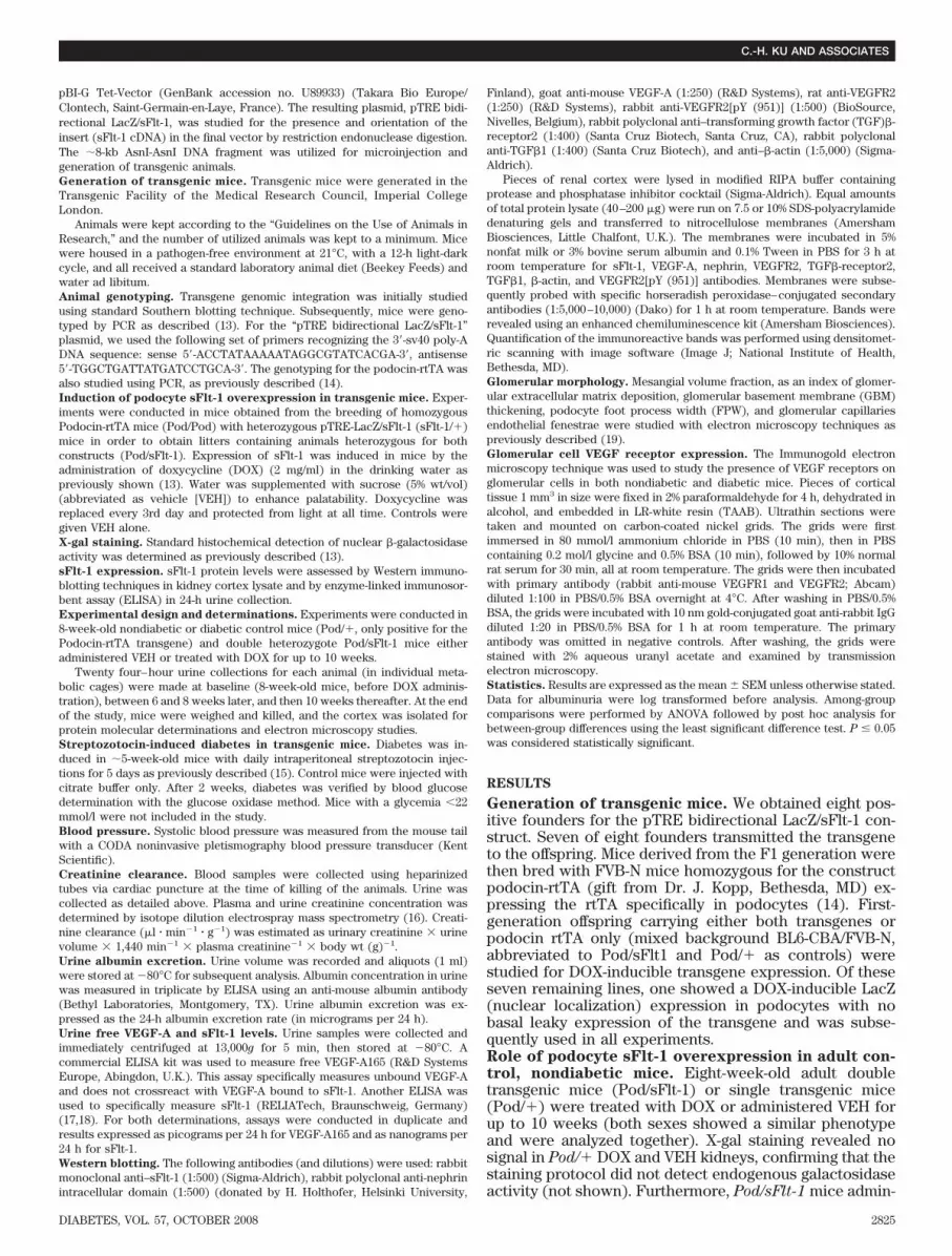
istered with VEH also failed to give a positive signal (Fig.1A), excluding “leakage” of LacZ expression in nonin-duced mice. In contrast, kidneys from adult Pod/sFlt-1mice exposed to DOX showed positive X-gal staining (Fig.1A), consistent with podocyte expression of the transgene(13,14). X-gal staining was analyzed at different time pointsafter DOX administration (10 days, 5–10 weeks), and theresults were superimposable (not shown). After 10 weeks
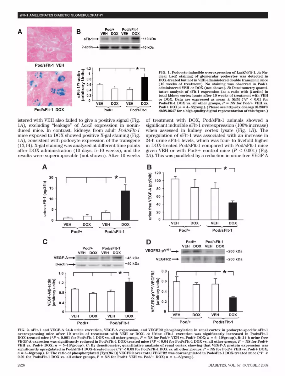
of treatment with DOX, Pod/sFlt-1 animals showed asignificant inducible sFlt-1 overexpression (100% increase)when assessed in kidney cortex lysate (Fig. 1B). Theupregulation of sFlt-1 was associated with an increase in24-h urine sFlt-1 levels, which was four- to fivefold higherin DOX-treated Pod/sFlt-1 compared with Pod/sFlt-1 micegiven VEH or with Pod/� control mice (P � 0.001) (Fig.2A). This was paralleled by a reduction in urine free VEGF-A
~110 kDasFlt-1
?-actin
VEH DOX VEH DOX0
0.2
0.4
0.6
0.8
1
1.2 *
B VEH DOX VEH DOX
Pod/+ Pod/sFlt-1
~40 kDa
A
Pod/sFlt-1 DOX
sF
lt-1
/?-a
cti
n(a
rbit
rary
Un
its
)
Pod/sFlt-1 VEH
Pod/+ Pod/sFlt-1
FIG. 1. Podocyte-inducible overexpression of LacZ/sFlt-1. A: Nu-clear LacZ staining of glomerular podocytes was detected inDOX-treated but not in VEH-administered double transgenic mice(10 weeks of treatment). No staining was observed in Pod/�administered VEH or DOX (not shown). B: Densitometry quanti-tative analysis of sFlt-1 expression (as a ratio with �-actin) intotal kidney cortex lysate after 10 weeks of treatment with VEHor DOX. Data are expressed as mean � SEM (*P < 0.01 forPod/sFlt-1 DOX vs. all other groups, P � NS for Pod/� VEH vs.Pod/� DOX; n � 4–8/group). (Please see http://dx.doi.org/10.2337/db08-0647 for a high-quality digital representation of this figure.)
B *
C
A
0
5
10
15
20 *
*
D
*
DOX
urin
e fre
e VE
GF-
A (p
g/24
h)
0
20
40
60
80
100
120
0
0.4
0.8
1.2
1.6
VEH DOX VEH DOX
Pod/+ Pod/sFlt-1
VEGF-A
β-actin
~45 kDa~40 kDa
VEG
F-A/
β-ac
tin(a
rbitr
ary
units
)
urin
e sF
lt-1
(ng/
24h)
0
5
10
15
20
0
0.2
0.4
0.6
0.8
VEG
FR2-
pY95
1 /VEG
FR2
(arb
itrar
y un
its)
VEGFR2-pY951
VEGFR2
VEH DOX VEH DOX
Pod/+ Pod/sFlt-1
Pod/+ Pod/sFlt-1VEH DOX VEH DOX
~200 kDa
~200 kDa
Pod/+ Pod/sFlt-1VEH DOX VEH DOX
VEH DOX VEH DOX
Pod/+ Pod/sFlt-1VEH DOX VEH
Pod/+ Pod/sFlt-1
FIG. 2. sFlt-1 and VEGF-A 24-h urine excretion, VEGF-A expression, and VEGFR2 phosphorylation in renal cortex in podocyte-specific sFlt-1overexpressing mice after 10 weeks of treatment with VEH or DOX. A: Urine sFlt-1 excretion was significantly increased in Pod/sFlt-1DOX-treated mice (*P < 0.001 for Pod/sFlt-1 DOX vs. all other groups, P � NS for Pod/� VEH vs. Pod/� DOX; n � 6–10/group). B: 24-h urine freeVEGF-A excretion was significantly reduced in Pod/sFlt-1 DOX-treated mice (*P < 0.04 for Pod/sFlt-1 DOX vs. all other groups, P � NS for Pod/�VEH vs. Pod/� DOX; n � 5–10/group). C: By densitometry, quantitative analysis of renal cortex showing that VEGF-A protein expression wassignificantly upregulated in Pod/sFlt-1 DOX-treated mice (*P < 0.03 for Pod/sFlt-1 DOX vs. all other groups, P � NS for Pod/� VEH vs. Pod/� DOX;n � 5–6/group). D: The ratio of phosphorylated [Tyr(951)] VEGFR2 over total VEGFR2 was downregulated in Pod/sFlt-1 DOX-treated mice (*P <0.01 for Pod/sFlt-1 DOX vs. all other groups, P � NS for Pod/� VEH vs. Pod/� DOX; n � 4–8/group).
sFlt-1 AMELIORATES DIABETIC GLOMERULOPATHY
2826 DIABETES, VOL. 57, OCTOBER 2008

in Pod/sFlt-1 DOX-treated mice, suggesting an sFlt-1–medi-ated “sequestration/binding” of VEGF-A (P � 0.04) (Fig. 2B).Renal cortex VEGF-A was upregulated in Pod/sFlt-1 DOXmice in comparison with the other groups (P � 0.03) (Fig.2C). No difference was observed in the renal cortex expres-
sion levels of VEGFR2 among the groups of mice studied(Pod/� VEH, 1.5 0.3; Pod/� DOX, 1.7 0.3; Pod/sFlt-1
VEH, 1.7 0.3; Pod/sFlt-1 DOX, 1.5 0.2 VEGF-R2/�-actinarbitrary units, n � 5–6/group). VEGFR2 activation, deter-mined by phosphorylation of VEGFR2 on Tyr (951) (20,21)was reduced by �50%, but not abolished, in Pod/sFlt-1 micetreated with DOX (Fig. 2D).VEGFR expression in glomerular cells. Immunogoldstaining showed localization of VEGFR1 and VEGFR2 inendothelial cells as previously reported. We detectedexpression of VEGFR2, but not VEGFR1, in podocyte cellbody and foot processes in vivo (Fig. 3).Clinical and biochemical features. There was no differ-ence in creatinine clearance at 10 weeks (Pod/� VEH,9.2 1.5; Pod/� DOX, 10.5 2.2; Pod/sFlt-1 VEH, 9.3 1.6; Pod/sFlt-1 DOX, 10.6 1.7 �l � min�1 � g body wt�1,n � 6–9/group) and in albuminuria throughout the study(n � 10–12/group, geometric mean [95% CI] in �g/24 h, atbaseline: Pod/� VEH, 22.3 [11.9–41.9]; Pod/� DOX, 23.9[16.7–34.1]; Pod/sFlt-1 VEH, 33.3 [17.1–65.0]; and Pod/sFlt-1 DOX, 28.1 [18.8–42.1]; at 10 weeks: Pod/� VEH, 20.6[14–30.4]; Pod/� DOX, 22.7 [15.7–32.9]; Pod/sFlt-1 VEH,28.4 [18.6–43]; and Pod/sFlt-1 DOX, 22.2 [14.1–34.8]).Glomerular morphology was similar among the fourgroups of animals (not shown). DOX administration didnot affect mice behaviors or body weight, which wassimilar in all groups (not shown).Role of podocyte sFlt-1 overexpression in diabeticmice. In these experiments, only Pod/sFlt-1 mice werestudied. Mice were made diabetic at 5 weeks of age.Treatment with VEH or DOX was started in 8-week-oldadult nondiabetic and diabetic Pod/sFlt-1 mice andcontinued for up to 10 weeks (18 weeks of age). (Sexeswere pooled for analysis because they showed a similarphenotype.)Clinical and biochemical features. By the end of 10-week DOX treatment, diabetic animals were lighter, hadraised blood pressure, and had higher creatinine clearancethan control animals. DOX did not affect any of thesevariables in the nondiabetic and diabetic mice (Table 1).
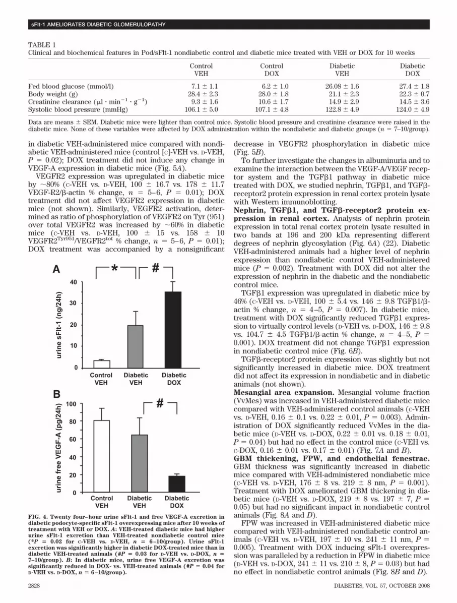
Albumin excretion rate was similar at baseline in thetwo diabetic (D) groups: D-VEH vs. D-DOX, geometric mean(95% CI), in �g/24 h, 28 (21–38) vs. 23 (14–36). By week 10it had risen to 117.5 (69–199) in D-VEH, and this rise wassignificantly blunted in the D-DOX group overexpressingsFlt-1: D-DOX, 43 (26.8–69) (P � 0.003).
To obtain insight in the role of primary podocytes’ sFlt-1overexpression in diabetes, we studied urine sFlt-1 andfree VEGF-A excretion, VEGF-A protein expression, andVEGFR2 expression and phosphorylation in the renalcortex lysate.sFlt-1 and free VEGF-A 24-h urine excretion. UrinesFlt-1 excretion was higher in VEH-administered diabeticmice compared with VEH-administered nondiabetic ani-mals (P � 0.02). In the diabetic group, urine sFlt-1excretion was significantly higher in DOX-treated Pod/sFlt-1 mice compared with VEH-administered animals(D-VEH vs. D-DOX, 19.9 6.3 vs. 35 4.9 ng/24 h, P � 0.03)(Fig. 4A), a pattern similar to that seen in control nondia-betic animals (Fig. 2A). Urine free VEGF-A 24 h excretionwas significantly reduced after 10 weeks in DOX-treateddiabetic mice compared with VEH-administered diabeticmice (P � 0.04) (Fig. 4B).VEGF-A expression, VEGFR2 expression, and phos-phorylation in renal cortex. Renal cortex VEGF-A pro-tein expression was significantly upregulated as expected
A
C
B
GBM
GBM
FIG. 3. Expression of VEGFR1 and VEGFR2 in mouse glomerular cells.Representative transmission electron microscopy images of VEGFR2(A), VEGFR1 (B), and negative control (C). Immunogold staining(arrows on black dots) in mouse glomerular capillaries. PositiveImmunogold staining is seen, as expected, in endothelial cells (EC) (A
and B) and, for the first time, for VEGFR2 in podocyte cell body (P) andfoot processes (Pfp) (A). Scale bars represent 0.5 �m.
C.-H. KU AND ASSOCIATES
DIABETES, VOL. 57, OCTOBER 2008 2827
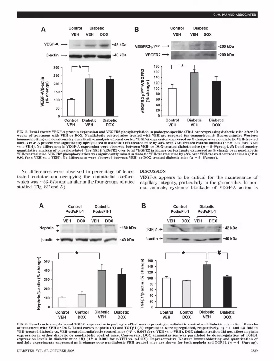
in diabetic VEH-administered mice compared with nondi-abetic VEH-administered mice (control [C]-VEH vs. D-VEH,P � 0.02); DOX treatment did not induce any change inVEGF-A expression in diabetic mice (Fig. 5A).
VEGFR2 expression was upregulated in diabetic miceby �80% (C-VEH vs. D-VEH, 100 16.7 vs. 178 11.7VEGF-R2/�-actin % change, n � 5–6, P � 0.01); DOXtreatment did not affect VEGFR2 expression in diabeticmice (not shown). Similarly, VEGFR2 activation, deter-mined as ratio of phosphorylation of VEGFR2 on Tyr (951)over total VEGFR2 was increased by �60% in diabeticmice (C-VEH vs. D-VEH, 100 15 vs. 158 10VEGFR2Tyr951/VEGFR2tot % change, n � 5–6, P � 0.01);DOX treatment was accompanied by a nonsignificant
decrease in VEGFR2 phosphorylation in diabetic mice(Fig. 5B).
To further investigate the changes in albuminuria and toexamine the interaction between the VEGF-A/VEGF recep-tor system and the TGF�1 pathway in diabetic micetreated with DOX, we studied nephrin, TGF�1, and TGF�-receptor2 protein expression in renal cortex protein lysatewith Western immunoblotting.Nephrin, TGF�1, and TGF�-receptor2 protein ex-
pression in renal cortex. Analysis of nephrin proteinexpression in total renal cortex protein lysate resulted intwo bands at 196 and 200 kDa representing differentdegrees of nephrin glycosylation (Fig. 6A) (22). DiabeticVEH-administered animals had a higher level of nephrinexpression than nondiabetic control VEH-administeredmice (P � 0.002). Treatment with DOX did not alter theexpression of nephrin in the diabetic and the nondiabeticcontrol mice.
TGF�1 expression was upregulated in diabetic mice by46% (C-VEH vs. D-VEH, 100 5.4 vs. 146 9.8 TGF�1/�-actin % change, n � 4–5, P � 0.007). In diabetic mice,treatment with DOX significantly reduced TGF�1 expres-sion to virtually control levels (D-VEH vs. D-DOX, 146 9.8vs. 104.7 4.5 TGF�1/�-actin % change, n � 4–5, P �0.001). DOX treatment did not change TGF�1 expressionin nondiabetic control mice (Fig. 6B).
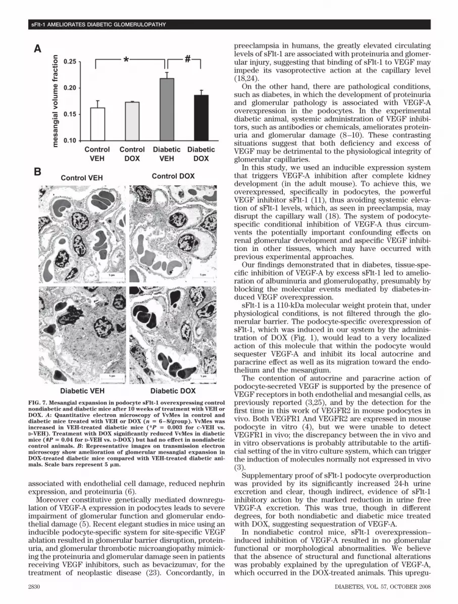
TGF�-receptor2 protein expression was slightly but notsignificantly increased in diabetic mice. DOX treatmentdid not affect its expression in nondiabetic and in diabeticanimals (not shown).Mesangial area expansion. Mesangial volume fraction(VvMes) was increased in VEH-administered diabetic micecompared with VEH-administered control animals (C-VEHvs. D-VEH, 0.16 0.1 vs. 0.22 0.01, P � 0.003). Admin-istration of DOX significantly reduced VvMes in the dia-betic mice (D-VEH vs. D-DOX, 0.22 0.01 vs. 0.18 0.01,P � 0.04) but had no effect in the control mice (C-VEH vs.C-DOX, 0.16 0.01 vs. 0.17 0.01) (Fig. 7A and B).GBM thickening, FPW, and endothelial fenestrae.GBM thickness was significantly increased in diabeticmice compared with VEH-administered nondiabetic mice(C-VEH vs. D-VEH, 176 8 vs. 219 8 nm, P � 0.001).Treatment with DOX ameliorated GBM thickening in dia-betic mice (D-VEH vs. D-DOX, 219 8 vs. 197 7, P �0.05) but had no significant impact in nondiabetic controlanimals (Fig. 8A and D).
FPW was increased in VEH-administered diabetic micecompared with VEH-administered nondiabetic control an-imals (C-VEH vs. D-VEH, 197 10 vs. 241 11 nm, P �0.005). Treatment with DOX inducing sFlt-1 overexpres-sion was paralleled by a reduction in FPW in diabetic mice(D-VEH vs. D-DOX, 241 11 vs. 210 8, P � 0.03) but hadno effect in nondiabetic control animals (Fig. 8B and D).
TABLE 1Clinical and biochemical features in Pod/sFlt-1 nondiabetic control and diabetic mice treated with VEH or DOX for 10 weeks
ControlVEH
ControlDOX
DiabeticVEH
DiabeticDOX
Fed blood glucose (mmol/l) 7.1 1.1 6.2 1.0 26.08 1.6 27.4 1.8Body weight (g) 28.4 2.3 28.0 1.8 21.1 2.3 22.3 0.7Creatinine clearance (�l � min�1 � g�1) 9.3 1.6 10.6 1.7 14.9 2.9 14.5 3.6Systolic blood pressure (mmHg) 106.1 5.0 107.1 4.8 122.8 4.9 124.0 4.9
Data are means SEM. Diabetic mice were lighter than control mice. Systolic blood pressure and creatinine clearance were raised in thediabetic mice. None of these variables were affected by DOX administration within the nondiabetic and diabetic groups (n � 7–10/group).
0
10
20
30
40
urin
e sF
lt-1
(ng/
24h)
DiabeticVEH
DiabeticDOX
A
urin
e fr
ee V
EGF-
A (p
g/24
h)
B
0
20
40
60
80
100
ControlVEH
DiabeticVEH
DiabeticDOX
ControlVEH
* #
#
FIG. 4. Twenty four–hour urine sFlt-1 and free VEGF-A excretion indiabetic podocyte-specific sFlt-1 overexpressing mice after 10 weeks oftreatment with VEH or DOX. A: VEH-treated diabetic mice had higherurine sFlt-1 excretion than VEH-treated nondiabetic control mice(*P � 0.02 for C-VEH vs. D-VEH, n � 6–10/group). Urine sFlt-1excretion was significantly higher in diabetic DOX-treated mice than indiabetic VEH-treated animals (#P � 0.03 for D-VEH vs. D-DOX, n �7–10/group). B: In diabetic mice, urine free VEGF-A excretion wassignificantly reduced in DOX- vs. VEH-treated animals (#P � 0.04 forD-VEH vs. D-DOX, n � 6–10/group).
sFlt-1 AMELIORATES DIABETIC GLOMERULOPATHY
2828 DIABETES, VOL. 57, OCTOBER 2008
No differences were observed in percentage of fenes-trated endothelium occupying the endothelial surface,which was �53–57% and similar in the four groups of micestudied (Fig. 8C and D).
DISCUSSION
VEGF-A appears to be critical for the maintenance ofcapillary integrity, particularly in the glomerulus. In nor-mal animals, systemic blockade of VEGF-A action is
ControlVEH VEH DOX
VEGF-A
β-actin
VEG
F-A
/β-a
ctin
(% c
hang
e)
DiabeticVEH
DiabeticDOX
ControlVEH
~45 kDa
~40 kDa
ControlVEH VEH DOX
Diabetic
0
50
100
150
200
250
300
020406080
100120140160180
DiabeticVEH
DiabeticDOX
ControlVEH
*
VEG
FR2-
pY95
1 /VEG
FR2
(% c
hang
e )
BA ControlVEH VEH DOX
Diabetic
VEGFR2-pY951
VEGFR2
~200 kDa
~200 kDa
*
FIG. 5. Renal cortex VEGF-A protein expression and VEGFR2 phosphorylation in podocyte-specific sFlt-1 overexpressing diabetic mice after 10weeks of treatment with VEH or DOX. Nondiabetic control mice treated with VEH are reported for comparison. A: Representative Westernimmunoblotting and densitometry quantitative analysis of renal cortex VEGF-A expression expressed as % change over nondiabetic VEH-treatedmice. VEGF-A protein was significantly upregulated in diabetic VEH-treated mice by 30% over VEH-treated control animals (*P � 0.02 for C-VEHvs. D-VEH). No differences in VEGF-A expression were observed between VEH- or DOX-treated diabetic mice (n � 5–9/group). B: Densitometryquantitative analysis of phosphorylated [Tyr(951)] VEGFR2 over total VEGFR2 in kidney cortex lysate expressed as % change over nondiabeticVEH-treated mice. VEGFR2 phosphorylation was significantly raised in diabetic VEH-treated mice by 58% over VEH-treated control animals (*P �0.01 for C-VEH vs. D-VEH). No differences were observed between VEH- or DOX-treated diabetic mice (n � 5–6/group).
Nephrin
β-actin
0
100
200
300
400
500
DiabeticVEH
DiabeticDOX
ControlVEH
ControlDOX
Nep
hri
n/β
-acti
n (
% c
han
ge) *
~180 kDa
~40 kDa
ControlPod/sFlt-1
VEH DOX VEH DOX
DiabeticPod/sFlt-1
0
20
40
60
80
100
120
140
160
DiabeticVEH
DiabeticDOX
ControlVEH
ControlDOX
* #
ControlPod/sFlt-1
VEH DOX VEH DOX
DiabeticPod/sFlt-1
β-actin
TGFβ1 ~42 kDa
~40 kDa
BA
TG
Fβ1
/β-a
cti
n (
% c
ha
ng
e)
FIG. 6. Renal cortex nephrin and TGF�1 expression in podocyte sFlt-1 overexpressing nondiabetic control and diabetic mice after 10 weeksof treatment with VEH or DOX. Renal cortex nephrin (A) and TGF�1 (B) expression were upregulated, respectively, by �4- and 1.5-fold inVEH-treated diabetic vs. VEH-treated nondiabetic control mice (*P < 0.007 for C-VEH vs. D-VEH). DOX administration did not affect nephrinexpression in either diabetic or nondiabetic control mice. Conversely, DOX administration was paralleled by downregulation of TGF�1expression levels in diabetic mice (B) (#P � 0.001 for D-VEH vs. D-DOX). Representative Western immunoblotting and quantitation ofmultiple experiments expressed as % change over nondiabetic VEH-treated mice are shown for both nephrin and TGF�1 (n � 4–8/group).
C.-H. KU AND ASSOCIATES
DIABETES, VOL. 57, OCTOBER 2008 2829
associated with endothelial cell damage, reduced nephrinexpression, and proteinuria (6).
Moreover constitutive genetically mediated downregu-lation of VEGF-A expression in podocytes leads to severeimpairment of glomerular function and glomerular endo-thelial damage (5). Recent elegant studies in mice using aninducible podocyte-specific system for site-specific VEGFablation resulted in glomerular barrier disruption, protein-uria, and glomerular thrombotic microangiopathy mimick-ing the proteinuria and glomerular damage seen in patientsreceiving VEGF inhibitors, such as bevacizumav, for thetreatment of neoplastic disease (23). Concordantly, in
preeclampsia in humans, the greatly elevated circulatinglevels of sFlt-1 are associated with proteinuria and glomer-ular injury, suggesting that binding of sFlt-1 to VEGF mayimpede its vasoprotective action at the capillary level(18,24).
On the other hand, there are pathological conditions,such as diabetes, in which the development of proteinuriaand glomerular pathology is associated with VEGF-Aoverexpression in the podocytes. In the experimentaldiabetic animal, systemic administration of VEGF inhibi-tors, such as antibodies or chemicals, ameliorates protein-uria and glomerular damage (8–10). These contrastingsituations suggest that both deficiency and excess ofVEGF may be detrimental to the physiological integrity ofglomerular capillaries.
In this study, we used an inducible expression systemthat triggers VEGF-A inhibition after complete kidneydevelopment (in the adult mouse). To achieve this, weoverexpressed, specifically in podocytes, the powerfulVEGF inhibitor sFlt-1 (11), thus avoiding systemic eleva-tion of sFlt-1 levels, which, as seen in preeclampsia, maydisrupt the capillary wall (18). The system of podocyte-specific conditional inhibition of VEGF-A thus circum-vents the potentially important confounding effects onrenal glomerular development and aspecific VEGF inhibi-tion in other tissues, which may have occurred withprevious experimental approaches.
Our findings demonstrated that in diabetes, tissue-spe-cific inhibition of VEGF-A by excess sFlt-1 led to amelio-ration of albuminuria and glomerulopathy, presumably byblocking the molecular events mediated by diabetes-in-duced VEGF overexpression.
sFlt-1 is a 110-kDa molecular weight protein that, underphysiological conditions, is not filtered through the glo-merular barrier. The podocyte-specific overexpression ofsFlt-1, which was induced in our system by the adminis-tration of DOX (Fig. 1), would lead to a very localizedaction of this molecule that within the podocyte wouldsequester VEGF-A and inhibit its local autocrine andparacrine effect as well as its migration toward the endo-thelium and the mesangium.
The contention of autocrine and paracrine action ofpodocyte-secreted VEGF is supported by the presence ofVEGF receptors in both endothelial and mesangial cells, aspreviously reported (3,25), and by the detection for thefirst time in this work of VEGFR2 in mouse podocytes invivo. Both VEGFR1 And VEGFR2 are expressed in mousepodocyte in vitro (4), but we were unable to detectVEGFR1 in vivo; the discrepancy between the in vivo andin vitro observations is probably attributable to the artifi-cial setting of the in vitro culture system, which can triggerthe induction of molecules normally not expressed in vivo(3).
Supplementary proof of sFlt-1 podocyte overproductionwas provided by its significantly increased 24-h urineexcretion and clear, though indirect, evidence of sFlt-1inhibitory action by the marked reduction in urine freeVEGF-A excretion. This was true, though in differentdegrees, for both nondiabetic and diabetic mice treatedwith DOX, suggesting sequestration of VEGF-A.
In nondiabetic control mice, sFlt-1 overexpression–induced inhibition of VEGF-A resulted in no glomerularfunctional or morphological abnormalities. We believethat the absence of structural and functional alterationswas probably explained by the upregulation of VEGF-A,which occurred in the DOX-treated animals. This upregu-
0.10
0.15
0.20
0.25m
esan
gial
vol
ume
frac
tion
ControlVEH
ControlDOX
DiabeticVEH
DiabeticDOX
A
B Control DOX
Diabetic DOX
Control VEH
Diabetic VEH
* #
FIG. 7. Mesangial expansion in podocyte sFlt-1 overexpressing controlnondiabetic and diabetic mice after 10 weeks of treatment with VEH orDOX. A: Quantitative electron microscopy of VvMes in control anddiabetic mice treated with VEH or DOX (n � 6–8/group). VvMes wasincreased in VEH-treated diabetic mice (*P � 0.003 for C-VEH vs.D-VEH). Treatment with DOX significantly reduced VvMes in diabeticmice (#P � 0.04 for D-VEH vs. D-DOX) but had no effect in nondiabeticcontrol animals. B: Representative images on transmission electronmicroscopy show amelioration of glomerular mesangial expansion inDOX-treated diabetic mice compared with VEH-treated diabetic ani-mals. Scale bars represent 5 �m.
sFlt-1 AMELIORATES DIABETIC GLOMERULOPATHY
2830 DIABETES, VOL. 57, OCTOBER 2008

lation may represent a compensatory response to inter-rupted VEGF-A signaling because of sFlt-1 overexpressionand sequestration of VEGF-A. This might have mitigatedthe effect of sFlt-1–mediated inhibition, and indeed freeVEGF-A in the urine, though reduced by �50%, was stillpresent in substantial amount and was paralleled by a stillpresent although reduced VEGFR2 activation/phosphory-lation. This interpretation is supported by recent evidencethat suggests that in the adult mouse, full ablation of VEGFis required to induce definite glomerular pathologicalchanges (23,26).
Diabetic mice developed the typical renal functional andstructural abnormalities of proteinuria, mesangial expan-sion, GBM thickening, and podocyte foot process fusion.These changes were associated with upregulation ofVEGF-A and VEGFR2 expression in renal cortex as previ-ously described (25). Similar to nondiabetic mice, DOX-induced sFlt-1 overexpression resulted in raised 24-h urineexcretion of sFlt-1 and significant reduction (though notabolition) of urine free VEGF-A excretion, suggestingagain “sequestration” of functional VEGF-A. Of interest,24-h urine sFlt-1 excretion was higher in the diabetic miceadministered VEH compared with that in the controlnondiabetic mice receiving VEH, mimicking similar obser-vations described in diabetic patients (17).
DOX-treated diabetic mice showed a modest nonsignif-icant reduction in VEGFR2 phosphorylation comparedwith VEH-administered animals. The higher degree ofglomerulosclerosis observed in VEH- versus DOX-treateddiabetic mice might explain this observation, as it hasbeen reported that VEGF receptor activation is inverselyrelated to the degree of glomerular injury (27). We focusedour work on VEGFR2 expression and phosphorylationbecause VEGFR2, rather than VEGFR1, is involved in thebiological action of VEGF-A in adult animals (28,29) and isoverexpressed in experimental diabetes (25).
Inhibition of VEGF-A at the podocyte level in diabeticmice improved the albuminuria and morphologicalchanges of the glomerular filtration barrier by mitigatingGBM thickening and preventing foot process fusion andalso by markedly reducing mesangial matrix deposition.This suggests the presence of a podocyte-podocyte as wellas a podocyte-mesangium cellular network, where podo-cyte-specific gene expression can modulate the biology ofother subset of glomerular cells as previously suggestedfor other podocyte secreted cytokines (13,23,26). Indeedthe sFlt-1 overexpression-induced amelioration of GBMthickening and of mesangial extracellular matrix volume islikely to result from an inhibition of the prosclerotic actionof VEGF-A (30,31), the reduction of TGF�1 expression
GB
M w
idth
(nm
)
D
150
200
250
100
A
C
Foot
pro
cess
wid
th (n
m)
0
50
100
150
200
250
300
VEH DOX VEH DOXControl Diabetic
0
10
20
30
40
50
60
70
B
Control DOX
Diabetic DOX
Control VEH
Diabetic VEH
% fi
ltrat
ion
surf
ace
cov
ered
by
endo
thel
ial f
enes
trae
* #
* #
FIG. 8. GBM thickening, FPW, and endothelial fenestrae in podocyte sFlt-1 overexpressing control nondiabetic and diabetic mice after 10 weeksof treatment with VEH or DOX. Quantitative electron microscopy determination of GBM thickening (A), podocyte FPW (B), and endothelialfenestrae (C) in control and diabetic mice treated with VEH or DOX (n � 7–8/group). GBM thickness (A) was higher in VEH-treated diabetic micevs. VEH-treated nondiabetic control animals (*P � 0.001 for C-VEH vs. D-VEH). Administration of DOX reduced GBM thickness in diabetic mice(#P � 0.05 for D-VEH vs. D-DOX) but had no effect in nondiabetic mice. FPW (B) was increased in VEH-treated diabetic mice vs. VEH-treatednondiabetic control animals (*P � 0.005 for C-VEH vs. D-VEH). DOX administration ameliorated foot process fusion in diabetic mice (#P � 0.03for D-VEH vs. D-DOX), but no effect was seen in nondiabetic mice. The endothelial surface covered by endothelial fenestrae was not changedwithin the four groups of animal studied (C). Representative images on transmission electron microscopy show amelioration of GBM thickeningand FPW in diabetic DOX-treated mice. Scale bars represent 2 �m.
C.-H. KU AND ASSOCIATES
DIABETES, VOL. 57, OCTOBER 2008 2831
(32), or both. A potential relationship between VEGF-Aand TGF�1 is suggested by studies reporting a VEGF-A–mediated TGF�1 expression in mouse glomerular endo-thelial cells (33).
Interestingly, in the diabetes sFlt-1 overexpressing mice,no further upregulation of the already overexpressedVEGF-A occurred, suggesting a “ceiling” for renal cortexVEGF-A upregulation in the context of diabetes. In thismouse model, neither diabetes nor the overexpression ofsFlt-1 altered the number of endothelial fenestrae in theglomerular capillary.
Nephrin is a slit diaphragm protein whose deletion orexpression downregulation has been implicated in thepathogenesis of proteinuria in diseases such as the con-genital nephrotic syndrome of the Finnish type or inminimal change nephrotic syndrome (34–36). In experi-mental animal models of diabetes, nephrin expression hasbeen reported to be either upregulated or downregulated(10,37–39). In our diabetic mice, nephrin was upregulatedwhen compared with control nondiabetic animals, and thepodocyte overexpression of sFlt-1 did not affect thisupregulation. These findings, in line with previous reports(10), suggest that nephrin expression downregulation,though it may be sufficient, is not necessary for thedevelopment of proteinuria and concur with the conceptthat albuminuria may occur by a variety of mechanismssuch as redistribution of nephrin at the ultrastructurallevel (10,40,41).
DOX, which was used in this study to induce sFlt-1upregulation, is a nonselective inhibitor of metalloprotein-ases (42,43), enzymes that regulate extracellular matrixdeposition, and may have confounded some of our find-ings. However, our present results in the nondiabeticcontrol mice and previous observations in normal animals(13) and diabetic rodents (44) suggest no direct DOX-mediated effects on albuminuria and extracellular matrixat the dose used in this study. Moreover, DOX-inducedinhibition of metalloproteinases would favor matrix accu-mulation and would result, if anything, in an underesti-mate of the improvement in glomerular extracellularmatrix deposition in the podocyte sFlt-1 overexpressingdiabetic mice.
Our findings provide insight into the direct, local (auto-crine/paracrine) role of VEGF-A in diabetes-induced glo-merular injury, although the experimental design of thiswork makes our results more relevant to the preventionrather than treatment of diabetic glomerular damage.Together with work by others (23), these findings supportthe notion that in order to preserve vascular integrity, afine balance in the regulation of VEGF-A expression isrequired in as much as either too little or too much of thiscytokine would result in capillary vascular pathology.These considerations highlight the potential pitfalls ofusing systemic therapies to tackle tissue-specific changes.
ACKNOWLEDGMENTS
This work was partly founded by a project grant from theBiotechnology and Biological Sciences Research Council(no. S13745), Diabetes UK (RD04/0002860), and the Euro-pean Foundation for the Study of Diabetes/Servier.
We thank the Biomedical Electron Microscopy Unit(Newcastle University) for the electron microscopy study.We are grateful to Dr. J. Kopp for providing the Podocin-rtTA mice and to Dr. N. Dalton and Dr. C. Turner (Guy’s
and St. Thomas Hospital, London, U.K.) for the creatininedetermination.
REFERENCES
1. Eremina V, Cui S, Gerber H, Ferrara N, Haigh J, Nagy A, Ema M, RossantJ, Jothy S, Miner JH, Quaggin SE: Vascular endothelial growth factor asignaling in the podocyte-endothelial compartment is required for mesan-gial cell migration and survival. J Am Soc Nephrol 17:724–735, 2006
2. Ferrara N, Gerber HP, LeCouter J: The biology of VEGF and its receptors.Nat Med 9:669–676, 2003
3. Thomas S, Vanuystel J, Gruden G, Rodriguez V, Burt D, Gnudi L, Hartley B,Viberti G: Vascular endothelial growth factor receptors in human mesan-gium in vitro and in glomerular disease. J Am Soc Nephrol 11:1236–1243,2000
4. Guan F, Villegas G, Teichman J, Mundel P, Tufro A: Autocrine VEGF-Asystem in podocytes regulates podocin and its interaction with CD2AP.Am J Physiol Renal Physiol 291:F422–F428, 2006
5. Eremina V, Sood M, Haigh J, Nagy A, Lajoie G, Ferrara N, Gerber HP,Kikkawa Y, Miner JH, Quaggin SE: Glomerular-specific alterations ofVEGF-A expression lead to distinct congenital and acquired renal diseases.J Clin Invest 111:707–716, 2003
6. Sugimoto H, Hamano Y, Charytan D, Cosgrove D, Kieran M, Sudhakar A,Kalluri R: Neutralization of circulating vascular endothelial growth factor(VEGF) by anti-VEGF antibodies and soluble VEGF receptor 1 (sFlt-1)induces proteinuria. J Biol Chem 278:12605–12608, 2003
7. Sandler AB, Johnson DH, Herbst RS: Anti-vascular endothelial growthfactor monoclonals in non-small cell lung cancer. Clin Cancer Res
10:4258s–4262s, 20048. De Vriese AS, Tilton RG, Elger M, Stephan CC, Kriz W, Lameire NH:
Antibodies against vascular endothelial growth factor improve early renaldysfunction in experimental diabetes. J Am Soc Nephrol 12:993–1000, 2001
9. Flyvbjerg A, Dagnaes-Hansen F, De Vriese AS, Schrijvers BF, Tilton RG,Rasch R: Amelioration of long-term renal changes in obese type 2 diabeticmice by a neutralizing vascular endothelial growth factor antibody. Dia-
betes 51:3090–3094, 200210. Sung SH, Ziyadeh FN, Wang A, Pyagay PE, Kanwar YS, Chen S: Blockade
of vascular endothelial growth factor signaling ameliorates diabetic albu-minuria in mice. J Am Soc Nephrol 17:3093–3104, 2006
11. Kendall RL, Thomas KA: Inhibition of vascular endothelial cell growthfactor activity by an endogenously encoded soluble receptor. Proc Natl
Acad Sci U S A 90:10705–10709, 199312. Kendall RL, Wang G, Thomas KA: Identification of a natural soluble form
of the vascular endothelial growth factor receptor, FLT-1, and its het-erodimerization with KDR. Biochem Biophys Res Commun 226:324–328,1996
13. Davis B, Dei CA, Long DA, White KE, Hayward A, Ku CH, Woolf AS, BilousR, Viberti G, Gnudi L: Podocyte-specific expression of angiopoietin-2causes proteinuria and apoptosis of glomerular endothelia. J Am Soc
Nephrol 18:2320–2329, 200714. Shigehara T, Zaragoza C, Kitiyakara C, Takahashi H, Lu H, Moeller M,
Holzman LB, Kopp JB: Inducible podocyte-specific gene expression intransgenic mice. J Am Soc Nephrol 14:1998–2003, 2003
15. Candido R, Jandeleit-Dahm KA, Cao Z, Nesteroff SP, Burns WC, Twigg SM,Dilley RJ, Cooper ME, Allen TJ: Prevention of accelerated atherosclerosisby angiotensin-converting enzyme inhibition in diabetic apolipoproteinE-deficient mice. Circulation 106:246–253, 2002
16. Lamb EJ, Wood J, Stowe HJ, O’Riordan SE, Webb MC, Dalton RN:Susceptibility of glomerular filtration rate estimations to variations increatinine methodology: a study in older patients. Ann Clin Biochem
42:11–18, 200517. Kim NH, Oh JH, Seo JA, Lee KW, Kim SG, Choi KM, Baik SH, Choi DS, Kang
YS, Han SY, Han KH, Ji YH, Cha DR: Vascular endothelial growth factor(VEGF) and soluble VEGF receptor FLT-1 in diabetic nephropathy. Kidney
Int 67:167–177, 200518. Maynard SE, Min JY, Merchan J, Lim KH, Li J, Mondal S, Libermann TA,
Morgan JP, Sellke FW, Stillman IE, Epstein FH, Sukhatme VP, KarumanchiSA: Excess placental soluble fms-like tyrosine kinase 1 (sFlt1) maycontribute to endothelial dysfunction, hypertension, and proteinuria inpreeclampsia. J Clin Invest 111:649–658, 2003
19. White KE, Bilous RW: Type 2 diabetic patients with nephropathy showstructural-functional relationships that are similar to type 1 disease. J Am
Soc Nephrol 11:1667–1673, 200020. Matsumoto T, Mugishima H: Signal transduction via vascular endothelial
growth factor (VEGF) receptors and their roles in atherogenesis. J
Atheroscler Thromb 13:130–135, 200621. Zeng H, Sanyal S, Mukhopadhyay D: Tyrosine residues 951 and 1059 of
sFlt-1 AMELIORATES DIABETIC GLOMERULOPATHY
2832 DIABETES, VOL. 57, OCTOBER 2008
vascular endothelial growth factor receptor-2 (KDR) are essential forvascular permeability factor/vascular endothelial growth factor-inducedendothelium migration and proliferation, respectively. J Biol Chem 276:32714–32719, 2001
22. Ahola H, Wang SX, Luimula P, Solin ML, Holzman LB, Holthofer H: Cloningand expression of the rat nephrin homolog. Am J Pathol 155:907–913, 1999
23. Eremina V, Jefferson JA, Kowalewska J, Hochster H, Haas M, Weisstuch J,Richardson C, Kopp JB, Kabir MG, Backx PH, Gerber HP, Ferrara N,Barisoni L, Alpers CE, Quaggin SE: VEGF inhibition and renal thromboticmicroangiopathy. N Engl J Med 358:1129–1136, 2008
24. Davison JM, Homuth V, Jeyabalan A, Conrad KP, Karumanchi SA, QuagginS, Dechend R, Luft FC: New aspects in the pathophysiology of preeclamp-sia. J Am Soc Nephrol 15:2440–2448, 2004
25. Cooper ME, Vranes D, Youssef S, Stacker SA, Cox AJ, Rizkalla B, CasleyDJ, Bach LA, Kelly DJ, Gilbert RE: Increased renal expression of vascularendothelial growth factor (VEGF) and its receptor VEGFR-2 in experimen-tal diabetes. Diabetes 48:2229–2239, 1999
26. Eremina V, Baelde HJ, Quaggin SE: Role of the VEGF—a signaling pathwayin the glomerulus: evidence for crosstalk between components of theglomerular filtration barrier. Nephron Physiol 106:32–37, 2007
27. Hohenstein B, Hausknecht B, Boehmer K, Riess R, Brekken RA, Hugo CP:Local VEGF activity but not VEGF expression is tightly regulated duringdiabetic nephropathy in man. Kidney Int 69:1654–1661, 2006
28. Keyt BA, Nguyen HV, Berleau LT, Duarte CM, Park J, Chen H, Ferrara N:Identification of vascular endothelial growth factor determinants forbinding KDR and FLT-1 receptors: generation of receptor-selective VEGFvariants by site-directed mutagenesis. J Biol Chem 271:5638–5646, 1996
29. Waltenberger J, Claesson-Welsh L, Siegbahn A, Shibuya M, Heldin CH:Different signal transduction properties of KDR and Flt1, two receptors forvascular endothelial growth factor. J Biol Chem 269:26988–26995, 1994
30. Amemiya T, Sasamura H, Mifune M, Kitamura Y, Hirahashi J, Hayashi M,Saruta T: Vascular endothelial growth factor activates MAP kinase andenhances collagen synthesis in human mesangial cells. Kidney Int 56:2055–2063, 1999
31. Chen S, Kasama Y, Lee JS, Jim B, Marin M, Ziyadeh FN: Podocyte-derivedvascular endothelial growth factor mediates the stimulation of alpha3(IV)collagen production by transforming growth factor-beta1 in mouse podo-cytes. Diabetes 53:2939–2949, 2004
32. Ziyadeh FN, Hoffman BB, Han DC, Iglesias-De La Cruz MC, Hong SW,Isono M, Chen S, McGowan TA, Sharma K: Long-term prevention of renalinsufficiency, excess matrix gene expression, and glomerular mesangialmatrix expansion by treatment with monoclonal antitransforming growth
factor-beta antibody in db/db diabetic mice. Proc Natl Acad Sci U S A
97:8015–8020, 200033. Li ZD, Bork JP, Krueger B, Patsenker E, Schulze-Krebs A, Hahn EG,
Schuppan D: VEGF induces proliferation, migration, and TGF-beta1 ex-pression in mouse glomerular endothelial cells via mitogen-activatedprotein kinase and phosphatidylinositol 3-kinase. Biochem Biophys Res
Commun 334:1049–1060, 200534. Lahdenkari AT, Lounatmaa K, Patrakka J, Holmberg C, Wartiovaara J,
Kestila M, Koskimies O, Jalanko H: Podocytes are firmly attached toglomerular basement membrane in kidneys with heavy proteinuria. J Am
Soc Nephrol 15:2611–2618, 200435. Tryggvason K, Wartiovaara J: Molecular basis of glomerular permselectiv-
ity. Curr Opin Nephrol Hypertens 10:543–549, 200136. Tryggvason K, Patrakka J, Wartiovaara J: Hereditary proteinuria syn-
dromes and mechanisms of proteinuria. N Engl J Med 354:1387–1401, 200637. Aaltonen P, Luimula P, Astrom E, Palmen T, Gronholm T, Palojoki E,
Jaakkola I, Ahola H, Tikkanen I, Holthofer H: Changes in the expression ofnephrin gene and protein in experimental diabetic nephropathy. Lab
Invest 81:1185–1190, 200138. Bonnet F, Cooper ME, Kawachi H, Allen TJ, Boner G, Cao Z: Irbesartan
normalises the deficiency in glomerular nephrin expression in a model ofdiabetes and hypertension. Diabetologia 44:874–877, 2001
39. Kim JJ, Li JJ, Jung DS, Kwak SJ, Ryu DR, Yoo TH, Han SH, Choi HY, KimHJ, Han DS, Kang SW: Differential expression of nephrin according toglomerular size in early diabetic kidney disease. J Am Soc Nephrol
18:2303–2310, 200740. Luimula P, Ahola H, Wang SX, Solin ML, Aaltonen P, Tikkanen I, Kerjas-
chki D, Holthofer H: Nephrin in experimental glomerular disease. Kidney
Int 58:1461–1468, 200041. Smeets B, Dijkman HB, te Loeke NA, van Son JP, Steenbergen EJ,
Assmann KJ, Wetzels JF, Groenen PJ: Podocyte changes upon induction ofalbuminuria in Thy-1.1 transgenic mice. Nephrol Dial Transplant 18:2524–2533, 2003
42. Bendeck MP, Conte M, Zhang M, Nili N, Strauss BH, Farwell SM:Doxycycline modulates smooth muscle cell growth, migration, and matrixremodeling after arterial injury. Am J Pathol 160:1089–1095, 2002
43. Bouvet C, Gilbert LA, Girardot D, deBlois D, Moreau P: Different involve-ment of extracellular matrix components in small and large arteries duringchronic NO synthase inhibition. Hypertension 45:432–437, 2005
44. Ryan ME, Ramamurthy NS, Sorsa T, Golub LM: MMP-mediated events indiabetes. Ann N Y Acad Sci 878:311–334, 1999
C.-H. KU AND ASSOCIATES
DIABETES, VOL. 57, OCTOBER 2008 2833