prefrontal and striatal activation in elderly subjects during concurrent implicit and explicit...
TRANSCRIPT
Neurobiology of Aging 27 (2006) 741–751
Prefrontal and striatal activation in elderly subjects during concurrentimplicit and explicit sequence learning
Howard J. Aizenstein ∗, Meryl A. Butters, Kristi A. Clark, Jennifer L. Figurski,V. Andrew Stenger, Robert D. Nebes, Charles F. Reynolds III, Cameron S. Carter
Western Psychiatric Institute and Clinic, University of Pittsburgh School of Medicine, Department of Psychiatry,3811 O’Hara Street, Pittsburgh, PA 15213, USA
Received 27 May 2004; received in revised form 2 March 2005; accepted 9 March 2005Available online 1 June 2005
Abstract
Decreased function in the prefrontal cortex (PFC) is regarded as a primary mechanism of cognitive aging. However, despite a strongassociation between the prefrontal cortex and the neostriatum, the role of the neostriatum in cognitive aging is less certain. In the current study,eseclOr©
K
1
fgaatottce
Pc
0d
vent-related functional MRI was used to distinguish the cognitive contributions of neostriatal and prefrontal function in elderly versus youngubjects. Twenty healthy subjects, 9 elderly (mean age 67.6 years), and 11 young (mean age 22 years) performed a concurrent implicit andxplicit sequence learning task while undergoing functional MR imaging. Both groups showed learning in both the implicit and explicit taskonditions. Relative to the young subjects, the elderly subjects showed decreased activation in the left PFC during both implicit and explicitearning, decreased activation in the right putamen during implicit learning, and increased activation in the right PFC during explicit learning.ur results support the theory that changes in a network of brain regions, including the dorsolateral prefrontal cortex and the striatum, are
elated to cognitive aging. Moreover, these changes are observed during an implicit task, and thus do not seem to be mediated by awareness.2005 Elsevier Inc. All rights reserved.
eywords: fMRI; Aging; Implicit learning; Sequence learning; Serial reaction-time; Striatum; Prefrontal cortex
. Introduction
Previous studies have identified that a variety of cognitiveunctions decline with aging. The affected functions compriseeneralized cognitive processes (e.g., speed of processing),s well as specific executive functions, which are generallyttributed to the prefrontal cortex (PFC) and the neostria-um. While much previous work has supported the relationf prefrontal changes and aging [28], the relation of the neos-riatum to cognitive aging is less certain, and is the focus ofhe current study: an event-related fMRI study that uses aoncurrent implicit and explicit sequence learning task andxamines separately the early and late phases of learning.
Neostriatal dysfunction, perhaps in combination withFC dysfunction, might contribute to the decreased executiveontrol associated with aging. In fact, several recent results
∗ Corresponding author. Tel.: +1 412 586 9237; fax: +1 412 246 5880.E-mail address: [email protected] (H.J. Aizenstein).
suggest that elderly subjects have decreased performanceat sequence learning, which is specifically associated withthe striatum. In the classic sequence learning task, the serialreaction-time task (SRTT) [20], individuals become fasterat responding to simple reaction-time events that follow apredetermined repeating sequence. This has been shown tooccur even when the subjects are not explicitly aware that asequential pattern exists [29].
A number of functional imaging studies have identifiedstriatal activation on variants of the SRTT [12,11,4,22].Additionally, several recent studies have contrasted implicitand explicit sequence learning using versions of the SRTT. Inan O15 PET study, Rauch et al. [22] reports striatal activationspecific for the implicit condition and increased frontalactivation in the explicit condition. Using fMRI Willinghamet al. [29], Schendan et al. [26], and Aizenstein et al. [1]have reported a significant overlap between the implicit andexplicit conditions with striatal and frontal activation duringboth implicit and explicit block types. Additionally, fMRI
197-4580/$ – see front matter © 2005 Elsevier Inc. All rights reserved.oi:10.1016/j.neurobiolaging.2005.03.017
742 H.J. Aizenstein et al. / Neurobiology of Aging 27 (2006) 741–751
studies of sequence learning [1,26] have identified differentphases of learning, distinguishing the early learning duringthe first part of the sequence learning task from the laterlearning at the end, and have found more striatal activationlater.
The effect of aging on SRTT performance has also beeninvestigated. In a series of studies, healthy young and el-derly subjects were compared on several different implicitsequence tasks [14]. It was found that on sequences withinterspersed random trials (as opposed to the standard re-peating pattern serial reaction-time task), elderly subjects re-quired more trials to learn the sequence than did youngersubjects. Using a similar paradigm, another study [2] inves-tigated the relation of white-matter hyperintensities on MRI,which are thought to relate to subcortical impairment [16]to sequence learning in the elderly, observing that individ-uals with high white-matter intensity loads had a decreasedimplicit sequence learning effect. This suggests that the sub-cortical structures might be involved in the decreased implicitsequence learning effect observed in the elderly.
Since many of the tasks that engage the prefrontal cor-tex also engage the subcortical structures [3], it is difficultto dissociate their separate contributions to cognitive agingsolely on the basis of cognitive testing. Functional imagingtechniques, such as fMRI, however, may help to dissociatepossible contributions of frontal and subcortical structurestgietos
hfiwo[prtfatwadat
trtPa
of the change in aging is not due to differences in explicitstrategies.
2. Methods
2.1. Research participants
Twenty-three subjects participated in the study. However,data from three subjects were not useable: computer errorsled to the loss of the structural image of one young subjectand unusable data for one elderly subject, and evidence of asubcortical stroke was identified during the structural scan onone elderly subject. Data from the remaining 20 subjects wereanalyzed. These included 11 young healthy controls, 7 males,mean age = 22 years, S.D. = 2.00, and 9 healthy elderly sub-jects, 4 male, mean age = 67.6 years, S.D. = 6.09. All subjectswere right-handed. Any subjects with the following condi-tions (or history of the following conditions) were excluded:alcoholism, depression, schizophrenia, bipolar affective dis-order, stroke, or significant head injury, Alzheimer’s, Parkin-son’s, or Huntington’s disease. Screening was done withstructured interviews based on the Structured Clinical Inter-view for DSM-III (SCID). The mean MiniMental State Exam(MMSE) score for the elderly subjects was 29.1 (S.D. = 1.27)afcaB
matmhtoirme4cc
TD
NAGRHMM
o cognitive aging. A number of fMRI studies have investi-ated regional brain activation associated with cognitive ag-ng. These studies have primarily investigated the PFC usingxplicit cognitive tasks, such as encoding or attentional con-rol, and have found bilateral PFC activation in the elderlyn tasks that show unilateral PFC activation in the youngubjects [25,6].
The effect of aging on functional activation in the striatum,owever, is less clear. While previous studies [24] have identi-ed a decreased size of the neostriatum associated with aging,e are aware of only one previous functional imaging studyf aging with a task that specifically engages the striatum10]. In this previous study, 26 young and 46 elderly subjectserformed the SRTT while undergoing fMRI. Blocks of theepeating sequence were interspersed with blocks in whichhe stimuli appeared in a pseudorandom order. A voxel-wiseull brain analysis of this data was done to identify regionsctive in each group. A test of the interaction of age by block-ype was then done in these selected ROIs, and no differenceas found between groups, suggesting a similar pattern of
ctivation in young and elderly subjects. Our current studyiffers from the previous by focusing on a small number ofnatomically predefined ROIs, and thus may be more sensi-ive to group differences in these regions.
The current study uses event-related fMRI to investigatehe effect of aging on PFC and striatal function using a concur-ent implicit and explicit sequence learning task that is knowno engage PFC and striatal regions [1]. Replicating previousFC findings on an implicit learning task (i.e., when subjectsre not aware of learning) would suggest that the mechanism
nd the mean Mattis Dementia Rating Scale (MDRS) scoreor the elderly subjects was 141.9 (S.D. = 1.17). Informedonsent was obtained prior to scanning through procedurespproved by the University of Pittsburgh Institutional Reviewoard. Each subject was paid US$ 75 (Table 1).
Subjects were excluded if they were taking psychotropicedications. However, other medications were acceptable,
s medication use is common in a typical elderly popula-ion. Two of the elderly subjects were on antihypertensive
edication (quinapril 80 mg QD and diltiazam 240 mg QD;ydrochlorothiazide 25 mg QD and triamterene 37.5 QD),wo were taking aspirin (81 mg QD and 325 mg QD). Onef the elderly subjects was taking thyroid replacement med-cation (levothyroxine .5 mg QD), two were taking hormoneeplacement therapy (Prempro 1 tablet QD), one was takingetformin 500 mg BID, two were taking cholesterol low-
ring medications (fluvastatin 20 mg QD and atorvastatin0 mg QD), and two were taking COX-2 inhibitors (cele-oxib 200 mg QD). Three young subjects were taking birthontrol pills.
able 1emographics
Elderly subjects Young subjects
9 11ge 67.6 ± 6.09 22 ± 2.00ender 4M, 5F 7M, 4Face 8C, 1AA 8C, 1AA, 2Asianandedness 9R 11Rini-mental status exam 29.1 ± 1.27 N/Aattis dementia rating scale 141.9 ± 1.17 N/A
H.J. Aizenstein et al. / Neurobiology of Aging 27 (2006) 741–751 743
2.2. Procedure
This experiment was divided into two different conditions:practice (outside the scanner) and sequence learning, whichoccurred while subjects underwent MR scanning. The se-quence learning task involved concurrent implicit and explicitcomponents. The task is a variation of the serial reaction-timetask. The standard SRTT involves several hundred trials thatfollow a repeating sequence and then a block of randomly or-dered trials. In the current study, the goal was to optimize thecontrast between the learning and random trials; therefore,we used a modification of the SRTT in which the pattern andrandom trials were interspersed, similar to that described byHoward and Howard [14]. Moreover, since the study useda rapid event-related design it was important to randomizethe occurrence of pattern and random trials; thus, we usedprobabilistic sequences as described by Jiminez and Mendez[15].
2.3. Sequence learning stimuli
For each subject eight different sequences of 85 coloredshapes were generated. Three colors (red, green, and blue)and three shapes (circle, square, and triangle) were used.The sequence of colors and shapes were determined inde-ptdif3ocots
FEw
probabilistic sequences; correlations between the color andshapes sequences were low (maximum R2 = .031).
2.4. Practice
Prior to performing the category learning tasks each sub-ject was practiced on the mapping of color to button press.This practice occurred just before entering the MRI machine.The subjects spent 7 min responding to patches of red, green,and blue by pressing the left button, under their index fingerfor red, the middle button, under their third finger, for green,and the right button, under their ring finger, for blue.
2.5. Sequence learning procedure
Each stimulus appeared in the center of the screen. Af-ter 750 ms, the red, green, or blue colored stimulus changedto white, i.e., a white circle, square, or triangle on a blackbackground. The subject was instructed to look at each col-ored shape and respond to the color as quickly and accu-rately as possible by pressing the button corresponding to thecolor. The white shape stayed on the screen for an additional750 ms. A fixation stimulus was then displayed on the screenfor 500 ms, until the onset of the next trial.
Participants were instructed that while they were to re-spond as quickly as possible to the color, they were also toltsbmibaLlapcatsaocsdc
cPpuppt
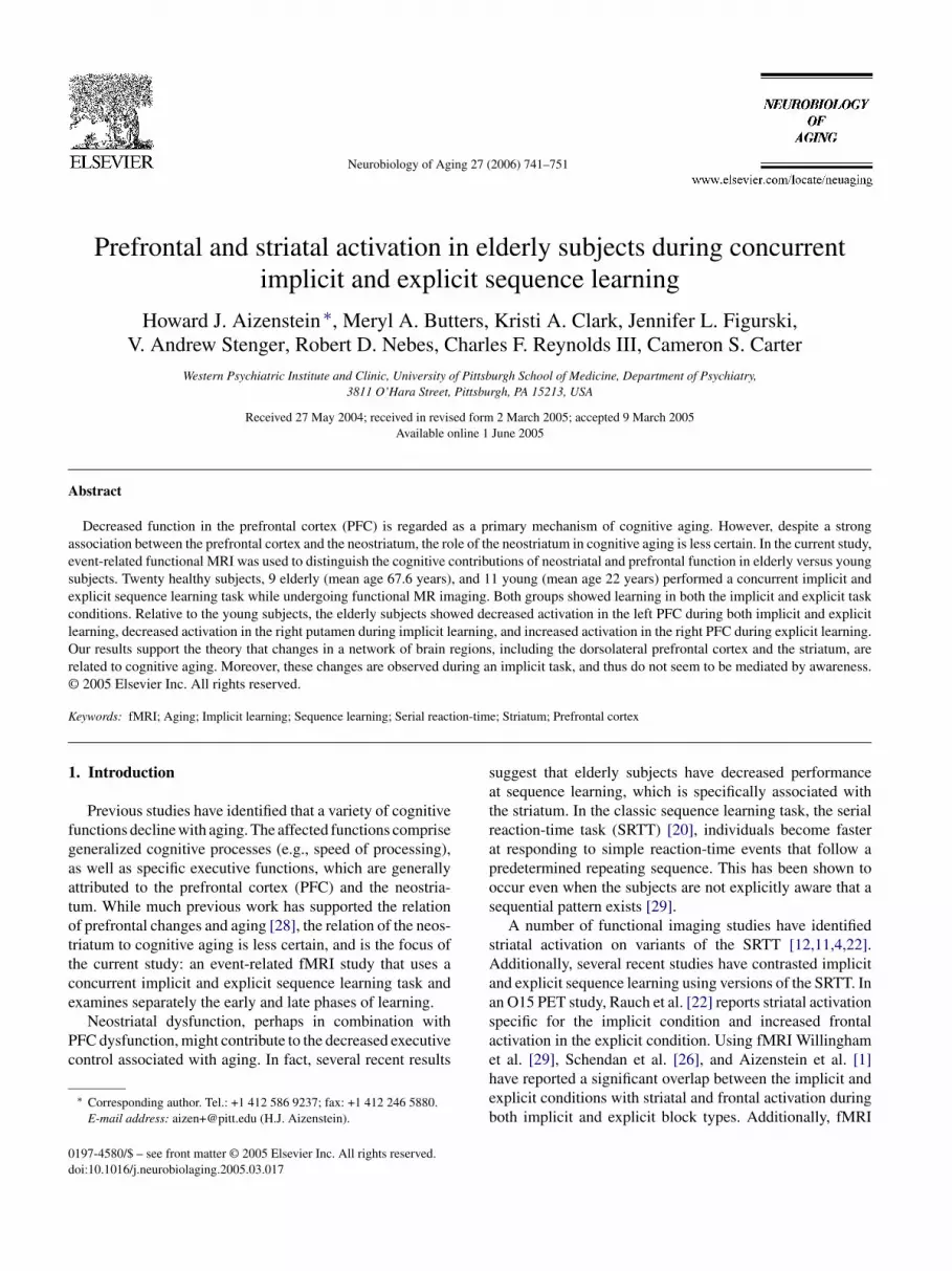
endently using two different first-order Markov chains (i.e.,he probability distribution of states at time i + 1 is completelyetermined by the state at time i, see Fig. 1). At each staten the Markov chain the next color had a 70% chance ofollowing in the set order (e.g., red, green, and blue) and a0% chance of violating the set order (i.e., either repeatingr skipping a position). Similarly, the next shape had a 70%hance of following a set order of shapes and a 30% chancef violating the order. The Markov chains were thus usedo generate a random sequence of colored shapes for eachubject, such that the color and shape followed independent
ig. 1. Markov chains that generate the color (a) and shape (b) sequences.ach stimulus was a colored shape. The color and shape of each stimulusas determined independently using these Markov chains.
ook for a sequential pattern in the shapes. They were toldhat the sequential pattern did not always hold, but that theyhould look for one and report this after each of the learninglocks. As described, the shapes did follow a sequence deter-ined by the Markov chain (Fig. 1b). Learning the sequence
n shapes was the explicit learning component of the task. Un-eknownst to the subjects, however, the colors also followedsequential pattern, independent of the pattern of shapes.earning the sequence of colors was the implicit sequence
earning component. Subjects’ change in reaction-time andccuracy at identifying the color was used to measure im-licit learning. We assessed for explicit knowledge of theolor sequence at the completion of the task. Subjects weresked a series of increasingly specific questions, such as didhey notice anything about the colors and did they notice aequence in the colors. Finally, they were told that there wassequence in the colors and were asked if they were awaref the sequence. The color disappeared after 750 ms to en-ourage subjects to respond quickly to the color. Pilot testinghowed that when the color remained for the entire stimulusuration some subjects would respond more slowly to theolor, apparently as they thought about the shape.
Stimulus presentation and response recording wasontrolled by a Power Macintosh computer, using theSYSCOPE software package [8]. The stimuli subtended ap-roximately 30◦ of the visual field. Responses were measuredsing a fiber optic button box attachment. This equipmentrovided 1 ms temporal resolution. It was strapped to thearticipant’s right hand. Participants were instructed to presshese buttons in response to the stimuli.
744 H.J. Aizenstein et al. / Neurobiology of Aging 27 (2006) 741–751
2.6. Scanning procedures
2.6.1. Data acquisitionImaging data was collected with a 1.5 T Signa scanner
(GE Medical Systems). High-resolution anatomical images(SPGRs) were acquired for each subject. Additionally, T1structural images were acquired with a 3.8 mm thickness(in-plane with the functional images). These had 36 obliqueaxial slices aligned with the anterior commissure–posteriorcommissure (AC–PC) line, with an in-plane resolution of0.9375 mm2 and with a field of view of 240 mm. Functionalimages were acquired using a one-shot spiral pulse sequencewith TE = 35 ms and TR = 2000 ms; 26 oblique axial sliceswere acquired with an in-plane resolution of 64 × 64 with3.75 mm2 pixels and a slice thickness of 3.8 mm, with a fieldof view of 240 mm. The fourth most inferior slice of thefunctional images was aligned with the AC–PC line.
Images were acquired in eight blocks of 85 trials each overthe course of learning. The presentation of stimuli was syn-chronized with the scanning such that one image was acquiredper trial. Since the trials occurred more frequently than the12–14 s hemodynamic response, this is a rapid event-relatedfMRI design [9]. Rapid event-related studies generally usefixation intervals to allow the hemodynamic response toreturn to a baseline, thus improving the estimate of thehemodynamic response for a given trial condition. Becauseficpcawcwgwsmsttrs
2
2
eptpmtwd
2.7.2. Automated labeling pathwayWe used a region-of-interest approach, with the regions
defined anatomically in each individual’s anatomic space.This approach was chosen to minimize the problem of align-ing the elderly and young fMRI maps to the same standardreference. We used an automated approach for defining theROIs. The anatomical regions of interest were chosen basedon our previous experience with this task [1]; bilateral BA9(Dorsolateral Prefrontal Cortex), BA46 (Dorsolateral Pre-frontal Cortex), caudate, putamen, and hippocampus) weredefined in the aal map obtained from the MRIcro softwarepackage [27]. The striatal and PFC regions were chosen to testour primary hypothesis that striatal dysfunction accompaniesthe frontal changes associated with cognitive aging. The hip-pocampus and anterior cingulate were also included as theseregions are strongly associated with PFC and striatal activa-tion. The Anterior Cingulate Cortex ROI was chosen from aprevious functional imaging study [7]. In order to define eachROI for each single subject in his/her own space, the follow-ing steps were performed. First, each subject’s low-resolutionanatomical image was coregistered to his/her high-resolutionanatomical image, using a 6-parameter linear algorithm [30].Then, the MNI single-subject high-resolution anatomical im-age (the Colin brain) was aligned with each subject’s high-resolution anatomical image using a second-order warpingalgorithm with 30 parameters [31]. Each ROI from the aalmmgsmtg
2
edsrbrvsevitapdiscTf
xation trials would disrupt a sequence of stimuli, it is un-lear how the addition of fixations would affect the cognitiverocess of sequence learning. Thus, in the current study wehose to avoid the fixations. An alternative approach is to usepseudo-random jittered stimulus presentation. However,e chose not to jitter the stimulus presentation, because of
oncern that the pseudo-random presentation could interfereith learning of the probabilistic sequence. Instead, to miti-ate against the possibility of saturation of the BOLD signal,e relied on the ‘random’ intermixing of trial types. Previous
tudies [9,5] have shown that the HRF summates approxi-ately linearly at an interval of 2 s in both young and elderly
ubjects. Thus, using selective averaging (i.e., accounting forhe cumulative effects of different prior trials) it is possibleo construct a mean time-series and compare the peak HRFsesulting from different trial types, as was done in the currenttudy.
.7. Imaging analysis
.7.1. Data preprocessingMotion correction was performed using a 6-parameter lin-
ar algorithm [30]. A linear detrending algorithm was alsoerformed, using only data within 3S.D. of the mean to es-imate the linear trend. An outlier correction algorithm waserformed to remove data that was more than 7S.D. from theean. Global normalization was performed multiplicatively
o give each subject a mean intensity of 3000. All analysesere conducted on a single-subject basis on non-coregisteredata.
ap was then put onto each subject’s SPGR. Then, a grayatter mask was applied to each label, using the FAST al-
orithm from the FSL package [31]. The labeling of eachubject’s ROIs was then visually inspected to assure accurateappings (see Fig. 3 for a representative image). Volumes of
he ROIs were also calculated to compare region sizes acrossroups.
.7.3. Time series analysisFor each subject the fMRI time-series was extracted for
ach voxel in all eleven ROIs. Outlier correction was thenone to remove those voxels in the ROI whose mean fMRIignal was more than 1S.D. from the ROI mean signal. Thisemoved voxels near the edges of the ROI, whose ROI mem-ership was least certain. For each ROI, a mean time se-ies was generated by calculating the mean MR signal of theoxel-wise time-series at six successive time points, corre-ponding to each 2 s interval up to 12 s (roughly the temporalxtent of the HRF). To account for the additive effects of pre-ious trials, we used selective averaging one trial back. Thats, to calculate the mean signal for a pattern trial we averagedhe mean signal of a pattern preceded by a non-pattern andpattern trial preceded by a pattern trial. This was done forattern and non-pattern trials in both implicit and explicit con-itions. Because the implicit sequences were independent wegnored the implicit condition when generating the explicitequence time-series, and similarly we ignored the implicitondition when generating the implicit sequence time-series.he resulting values were transformed into a percent change
rom baseline (time point 1) for standard presentation. A t-
H.J. Aizenstein et al. / Neurobiology of Aging 27 (2006) 741–751 745
test analysis was performed on each subject’s data, using trialas a random factor, and contrasting the mean fMRI signal atthe first time-point with the signal at the third time-point, i.e.,4–6 s after the trial onset, when the HRF is expected to peak.The primary analysis thus tests the significance of the meanchange at the HRF peak (scan 3) from the baseline at scan 1.
In previous work with this fMRI paradigm, we have identi-fied two different stages of learning [1] an early stage, whichcan be described as the process of learning the rule, and alater stage, which can be described as using the learned rule.Since these stages have different regional brain activity (morePFC activity early and more ACC activity later), we choseto distinguish these stages in our primary analysis. For earlylearning we look at the first half of the trials (trials 1–340)and for later learning we consider only the trials in the secondhalf of the task (trials 341–680). Another option for analyz-ing early and late learning would have been to split early andlate learning based on each individual’s reaction-time data,to estimate when each individual learned the rule. However,we are not convinced that an individual subject’s behavioraldata can accurately distinguish early and late learning, as itis confounded by noise and fatigue affects.
3. Results
3
(ptaweicseettwld(
insctci
p
quence of shapes. All 20 subjects reported the correct explicitrule at the end of the first block (Table 1).
3.2. ROI identification
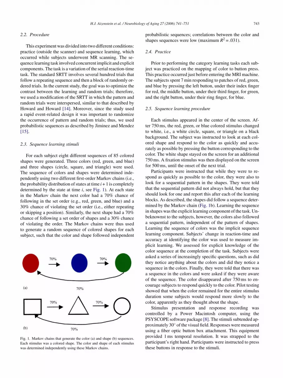
The eleven regions (bilateral BA9, BA46, caudate, puta-men, and hippocampus, and a single midline caudal ACC re-gion) were defined in each subject’s original anatomic space(as opposed to in the transformed template space). The re-gions were visually inspected for each subject to verify ac-curate labeling. Fig. 3 shows all of the labeled regions on arepresentative elderly subject and Fig. 4 shows the labelingof bilateral caudate and putamen for each subject. Table 2shows the mean size (i.e., number of voxels) of the selectedROIs in the young elderly images. As expected, based on pre-vious morphometric studies of aging [21,23], we found thatthe elderly subjects had significantly smaller regions in theprefrontal cortex, ACC, and striatum.
3.3. Estimated movement data
A concern with group comparisons of fMRI data is thatdifferences in head movement may confound true differencein the BOLD response. To address this concern we analyzedmovement parameters that were generated during the move-ment correction stage of image preprocessing. Movementceoatgowt
3
antRttiBmrcb
3
ts
.1. Behavioral performance
To test for implicit learning, we used the reaction-timeRT) and error-rate data that were acquired while subjectserformed the task. On the non-pattern trials, as comparedo the pattern trials, there was a significantly greater RTnd error-rate for both the young and elderly subjects,ithout a significant difference in the change in RT or
rror-rate between groups. These results are illustratedn Fig. 2. For reaction-time, the matched sample t-testsomparing non-pattern and pattern trials were, for the youngubjects, t(10) = 3.84 and p = .002, two-tailed, and for thelderly subjects, t(8) = 4.4 and p = .002, two-tailed. Forrror-rate, for the young subjects, t(10) = 4.12 and p = .002,wo-tailed, and for the elderly subjects, t(8) = 3.0 and p = .02,wo-tailed. As expected, there was a difference in overall RT,ith the elderly subjects overall having a longer response
atency t(18) = 2.54 and p = .02. There was not a significantifference in either RT or accuracy when comparing expliciti.e., shape) pattern versus non-pattern trials.
To test whether there was any explicit awareness of themplicit pattern, subjects were debriefed as to whether theyoticed any sequential pattern in the colors of the stimuli. Allubjects responded ‘no’. Frequently, subjects spontaneouslyommented that they were too busy looking for a pattern inhe shapes to think about a pattern in the colors. Thus, theoncurrent explicit condition may have helped to mask themplicit rule.
After each of the eight learning blocks, we tested for ex-licit learning by asking subjects to report the observed se-
orrection was performed using a 6-parameter rigid body lin-ar algorithm, AIR [30]. This corrected for movement thatccurred in the x, y, and z dimensions, as well as roll, pitch,nd yaw, relative to the first scan in the study. According tohese parameters none of the subjects (elderly or young) hadreater than 1.5 mm of movement in the x, y, or z dimensions,r greater 1.5◦ of roll, pitch, or yaw. None of the parametersere significantly different across groups. Thus, it is unlikely
hat group differences in movement affected our results.
.4. Mean time series
The identified ROIs for each subject were used to generatemean time series associated with each condition (pattern oron-pattern). The peak of these time series (i.e., 4–6 s afterrial onset) was then compared within and across groups.esults are shown in Table 3. As can be seen in the table,
he striatum (right putamen) is significantly more active inhe young subjects, and the PFC shows significant activationn early implicit learning and later explicit learning, with leftA9 being more active in the young subjects and right BA46ore active in the elderly subjects. The time-series for these
egions is shown in Fig. 5. The within- and across-groupomparisons (shown in the table) are described in more detailelow.
.4.1. Implicit LearningThe right putamen showed a significant difference be-
ween the groups. It showed significant activity in the youngubjects (as has previously been reported by Schendan et al.

746 H.J. Aizenstein et al. / Neurobiology of Aging 27 (2006) 741–751
Fig. 2. Behavioral results in young and elderly subjects at responding to the color of the stimulus, reaction-time (a) and accuracy (b). Error bars represent±1S.E.of the difference between condition types (i.e., pattern vs. non-pattern). For both young and elderly subjects there was a significant difference (p < .05)between trial types (pattern vs. non-pattern) in reaction-time and accuracy.
Table 2Region of interest volumes
Size young mean (S.D.) Size elderly mean (S.D.) t(18) p-Value
BA9 right 335.4 (38.9) 264.9 (19.7) 4.9 0.0001BA9 left 338.4 (45.0) 270.0 (33.3) 3.8 .001BA46 right 277.0 (32.6) 232.2 (15.0) 3.8 .001BA46 left 268.5 (38.2) 234.1 (33.5) 2.1 .05ACC 65.3 (8.3) 48.7 (10.11) 4.0 .0008Caudate L 136.1 (12.1) 125.1 (10.7) 2.1 .05Caudate R 140.0 (11.2) 125.0 (12.2) 2.8 .01Hippocampus L 85.8 (26.0) 91.1 −.5 .64Hippocampus R 95.9 (23.3) 99.1 (26.8) −.3 .78Putamen L 140.4 (13.5) 125.4 (16.4) 2.2 .04Putamen R 146.2 (12.0) 135.0 (12.3) 2.0 .05
Note: The table lists the size in mean number of voxels per region in the functional images.

H.J. Aizenstein et al. / Neurobiology of Aging 27 (2006) 741–751 747
Fig. 3. Automatic labeling of gray matter in (a) DLPFC, (b) caudal ACC, (c) striatum, and (d) hippocampus in a representative elderly subject. Regions weremapped using a 30-parameter non-linear registration from identified regions on the MNI (colin) reference image.
[26]) and showed only marginally significant activity in theelderly. The difference in activation across groups was alsosignificant (t(18) = 6.35 and p < .02). The prefrontal cortexshowed a significant difference between the groups duringthe first half of implicit learning. Left BA9 showed signifi-cant activity in the young subjects, whereas right BA9 showedmarginally significant activity in the elderly.
3.4.2. Explicit learningNo significant differences between the groups were iden-
tified during explicit learning. Significant prefrontal cortexactivation, however, was found during the second half of ex-plicit learning. This followed the same pattern as the PFCactivation during implicit learning, with more right sided ac-tivation in the young subjects and more left activation in theelderly subjects.
4. Discussion
This study contrasted the BOLD functional MRI signalin young and elderly subjects during concurrent implicitand explicit sequence learning. Our primary aim was toinvestigate the role of striatal function in cognitive aging. Aspredicted, we did find a significant difference in striatal acti-
vation between the elderly and the young subjects; the elderlysubjects showed diminished activation (relative to youngsubjects) in the right putamen in the later part of implicitlearning. This result differs from the a previous study [10],which did not find a statistical difference associated withaging in the striatal activation during sequence learning. Inthe current study a small number of preselected anatomicallydefined ROIs were delineated (using an automated approach)in each individual’s functional imaging space. The Daselaarstudy, on the other hand, registered each image to standardspace, selected ROIs based on a voxel-wise full-brainanalysis in each group, and then tested in each of theseROIs whether there was a significant interaction of sequencelearning and aging. The ROIs defined in the Daselaar studyare necessarily smaller than the anatomically defined ROIsused in the current study. Previously, we showed how somefindings of the BOLD dynamic response depend on ROIdefinition [1]. We found that with smaller ROIs there was asimilar peak between young and elderly in the visual cortex,whereas in a different study using a similar paradigm, butwith larger ROIs [5] the peaks in the visual cortex weresmaller in the elderly. We wonder if a similar mechanismcould explain the discrepancy between our findings and thoseof Daselaar. The current finding of decreased right putamenactivation in aging supports a role for striatal dysfunction in

748 H.J. Aizenstein et al. / Neurobiology of Aging 27 (2006) 741–751
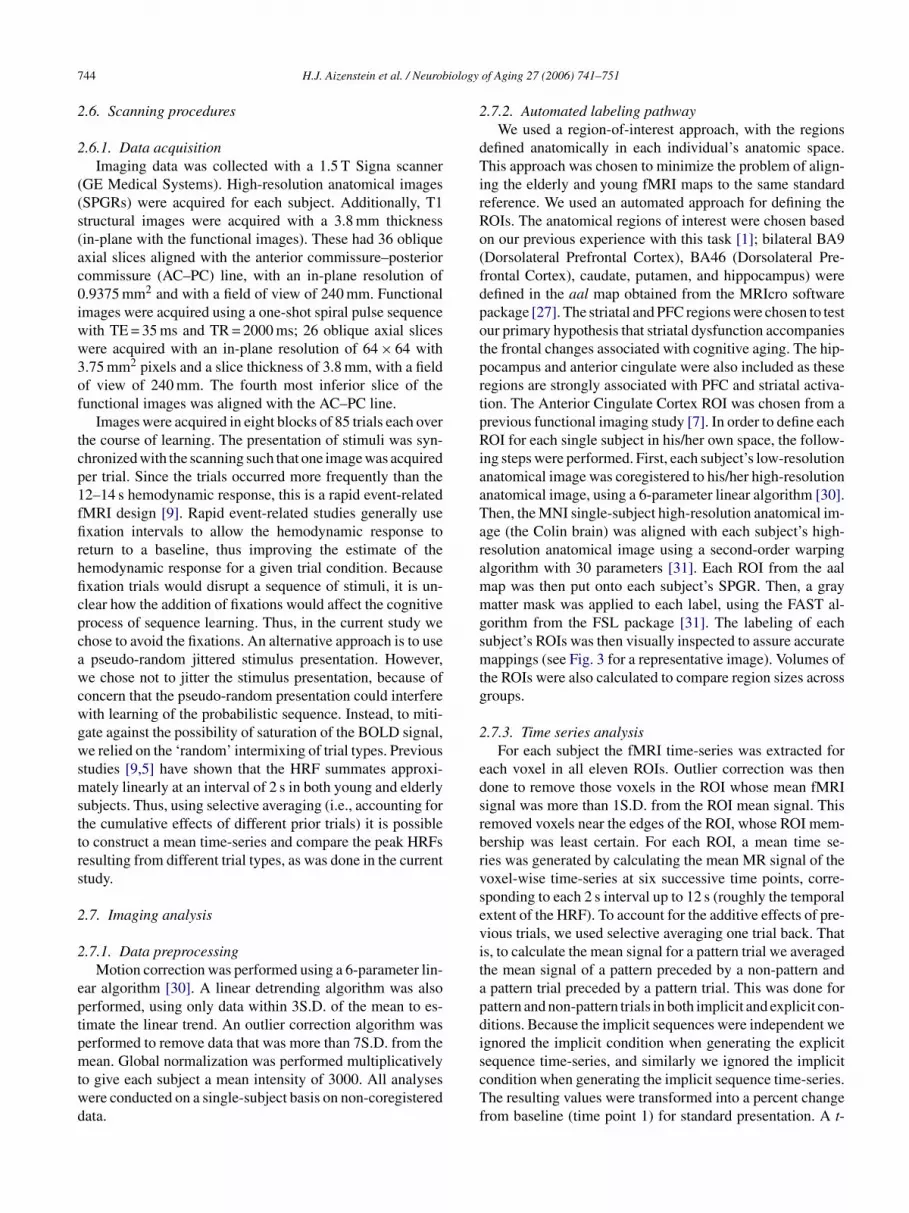
Fig. 4. Labeling of bilateral caudate (green) and putamen (red) in each of the 11 young control subjects (a) and the 9 elderly subjects (b). Oblique axial slicesare shown at the level of the AC–PC line (z = 0). This illustrates the similar accuracy of region identification across groups.
Table 3Areas of fMRI activation
First half Second half
Region p-value Region p-Value
Implicit learningYoung BA9 L* .02 Putamen R** .05
BA46 R .08Hippocampus L** .03Putamen L .07
Elderly BA9 R .08 Putamen R .09Hippocampus R .09Caudate R .07ACC* .02
Explicit learningYoung BA9 L* .007
BA46 L* .04BA46 R .06Putamen L .09
Elderly Hippocampus R* .05 BA9 L* .02BA9 R* .04BA46 R* .009
Note: Regions which were significant at t(8) > 1.86 or t(10) > 1.81, p < .05,in the within-group analysis are indicated by * in the table and shaded.Regions significant for t(18) > 1.73, p < .05, in the across-group comparisonare indicated by **. All regions with t(8) > 1.40 or t(10) > 1.37, p < .1, in thewithin-group comparison, are shown in the table.
cognitive aging, and extend the frontal lobe hypothesis ofcognitive aging [28] to also include subcortical changes.
In addition to our observation of significantly decreasedputamen activation in the elderly (relative to young controls),our data also suggest that the elderly may have increased ac-tivity in other subregions of the striatum. Specifically, wefound that in the right caudate there was a “trend” of acti-vation in the elderly (but not the young). This different spa-tial pattern of activity could be interpreted as non-selectiveactivation [17]. That is, instead of selectively activating theright putamen with implicit sequence learning, the elderlyshow non-specific striatal activation. Alternatively, the dif-ferent pattern of striatal activation across the groups couldreflect compensation, i.e., the elderly recruit additional stri-atal areas to compensate for dysfunction in the right putamen.
Differences in activation across groups were also appar-ent in the prefrontal cortex, with the elderly showing greateractivation in left dorsolateral prefrontal cortex (DLPFC) anddiminished activation in the right DLPFC. Since the task in-volves learning pictures (i.e., colored shapes), the right PFCactivation is expected. Our observation of increased left PFCactivation in the elderly supports previous observations ofsymmetric DLPFC activity in elderly, but not in young sub-jects, during cognitive tasks. The observation of this lossof asymmetry is sometimes referred to with the acronymHAROLD, hemispheric asymmetry reduction in old age [6].

H.J. Aizenstein et al. / Neurobiology of Aging 27 (2006) 741–751 749
Fig. 5. Time-series for fMRI activation during the second half of the explicit condition within (a) right BA46 and (b) left BA9. Graphs show the percent signalchange as a function of scan number.
Our results suggest that the HAROLD pattern of change oc-curred even without awareness (i.e., during an implicit task).This demonstrates that the changes in prefrontal activationof aging are not necessarily mediated by awareness. Thus, ifthe changes in PFC activity in the elderly are a reflection ofcompensation, they are not (in this case) due to an explicitcompensation strategy.
Aside from the changes in the PFC and subcortical areasdiscussed above, we also found significant activation in thewithin group analysis of the ACC and hippocampus. Thesechanges, however, were not significant in the across groupcontrast. We found ACC activation in the later stage of im-plicit learning significant only in the elderly. The hippocam-pal activity was significant in the early part of implicit learn-ing in the young, but did not reach statistical significance inthe elderly subjects. These findings are consistent with pre-vious observations of increased hippocampal activation inyounger subjects [19], and increased ACC activation in theelderly [18].
Certain limitations of the current study should be recog-nized. Although, we do report statistically significant results,we also observed some statistical trends (e.g., in the left puta-men). The modest sample size in the current likely limitedour ability to determine whether significant differences withaging do occur in these other regions. Our most significant
results were found for the PFC activation in the second halfof explicit learning, with p values of .007 for elderly sub-jects in BA9 right and .009 for young subjects in BA46 left.Since four separate PFC regions were tested for each hypoth-esis, the results for explicit learning, withstand a Bonferonnimultiple comparison correction for number of regions, i.e.,alpha < .05/4 = .0125. The other reported results do not satisfythis more stringent statistical test.
Another potential limitation in the current study is thatboth implicit and explicit learning proceeded concurrently,which could have led to a dual-task, or parallel processing,load. Our design, however, was based on tasks by Jiminezand Mandez [15], which do not show significant interfer-ence during concurrent administration. Thus, we do not feelthe parallel processing of the implicit and explicit tasks hada significant effect on the current results. Another possiblelimitation in this study is the difference in the gender distri-bution between the elderly and young cohorts. Specifically,there was a greater proportion of females in the elderly group.
Additionally, there some differences between the elderlyand groups, which may have affected the results. There was agreater proportion of females in the elderly group. Althoughthis difference was not statistically significant, it is relevant,as previous studies [13] have identified differences in lateral-ity between the genders, with loss of asymmetry in men. Our

750 H.J. Aizenstein et al. / Neurobiology of Aging 27 (2006) 741–751
findings of loss of asymmetry in the elder group (which hada higher proportion of women) would suggest that the gen-der difference did not contribute to the current result. Also,as expected, there was a difference in overall reaction-timebetween the groups, with the elderly subjects having a longerlatency of response. This could have led to a decreased im-plicit learning effect in the putamen in elderly subjects, dueto the shorter interval between response and subsequent trial.We feel, however, that this is unlikely as there was not a sig-nificant difference between groups in the behavioral learningeffect. The current study differs from many other variantsof the SRTT in that the Markov chain that was used gener-ates only pair-wise associations and not second-order asso-ciations. Perhaps the striatal activation we found is specificfor the learning pair-wise associations and differs from thelearning of higher order associations, as occurs in the stan-dard SRTT. We feel this is unlikely, as high order associationshave also been associated with striatal activation [26]. More-over, as discussed in Jiminez and Mendez [15], and Howardand Howard [14] probabilistic sequences such as the one usedin the current study, are difficult to identify explicitly, and inthis sense more like higher order dependencies. Our test forexplicit learning during the implicit condition was somewhatlimited. We relied on debriefing, asking subjects whether theyrecognized an explicit pattern during the implicit condition.No subjects were aware of a rule, and several said that theywtdwwbe
sresmawttsOiws
etrpcoc
in PFC with aging by showing that these changes are notnecessarily mediated by explicit ‘top–down’ processes.
Acknowledgments
This research was supported by AAGP Pfizer/Eisai Fel-lowship (HJA), Hartford AFAR Fellowship (HJA), Pitts-burgh Foundation, Burroughs Wellcome Translational Sci-entist Award (CSC), and NIMH Grants T32-MH19986,R25-MH060473, K02-MH064190, K23-MH64678, K01-MH01684, and P30 MH52247. We thank Kate Fissell andthe Clinical Cognitive Neuroscience Laboratory at the Uni-versity of Pittsburgh for their assistance.
References
[1] Aizenstein HJ, Stenger VA, Cochran JL, Clark KA, JohnsonM, Nebes RD, Carter CS. Regional brain activation during con-current implicit and explicit sequence learning. Cereb Cortex2004;14:199–208.
[2] Aizenstein HJ, Nebes RD, Meltzer CC, Fukui MB, Williams RL,Saxton J, Houck PR, Carter CS, Reynolds 3rd CF, DeKosky ST.The relation of white matter hyperintensities to implicit learning inhealthy older adults. Int J Geriatr Psychiatry 2002;17:664–9.
[
[
[
[
[
ere too busy looking for a rule in the explicit condition (i.e.,he shapes) to notice a sequential pattern in the colors. An ad-itional test of explicit learning during the implicit conditionould have been to ask subjects to try to generate the patternith cues (as was done in Schendan et al. [26]). It is possi-le that this more sensitive test would have identified somexplicit learning in the implicit condition.
Functional imaging studies comparing elderly and youngubjects are potentially confounded by differences in neu-oanatomy between elderly and young brains. Since thelderly brains have greater anatomic variability and oftenhow decreased regional size (as was found here), standardethods for aligning the functional MRI data in a common
natomic space may be biased. That is, the elderly brains,ith the greater variability, will have more alignment error
han the young subjects. In the current study, we addressedhis by using a single-subject region-of-interest based analy-is, focusing on regions for which we had initial hypotheses.ur approach limits errors due to poor registration by select-
ng the ROI separately for each subject, using a non-lineararp and segmentation, to define the anatomic ROIs for each
ubject.A primary motivation of this study was to investigate the
ffects in the striatal region on a task that specifically activateshis area. We do find a similar age-related effect in the striatalegion as was identified in the prefrontal areas. This extendsrevious fronto-centric views of cognitive aging to also in-lude changes in the subcortical structure. Additionally, thebservation of PFC changes even in a task without awarenessonstrains the interpretation of previously observed changes
[3] Alexander GE, DeLong MR, Strick PL. Parallel organization of func-tionally segregated circuits linking basal ganglia and cortex. AnnuRev Neurosci 1986;9:357–81.
[4] Berns GS, Cohen JD, Mintun MA. Brain regions responsive to nov-elty in the absence of awareness. Science 1997;276:1272–5.
[5] Buckner RL, Snyder AZ, Sanders AL, Raichle ME, Morris JC. Func-tional brain imaging of young, nondemented, and demented olderadults. J Cogn Neurosci 2000;12:24–34.
[6] Cabeza R, Anderson ND, Locantore JK, McIntosh AR. Aging grace-fully: compensatory brain activity in high-performing older adults.Neuroimage 2002;17:1394–402.
[7] Carter CS, Macdonald AM, Botvinick M. Parsing executive pro-cesses: strategic vs. evaluative functions of the anterior cingulatecortex. Proc Natl Acad Sci USA 2000;97:1944–8.
[8] Cohen JD, MacWhinney B, Flatt M, Provost J. PsyScope: an inter-active graphic system for designing and controlling experiments inthe psychology laboratory using Macintosh computers. Behav ResMethods Instrum Comput 1993;25:257–71.
[9] Dale AM, Buckner RL. Selective averaging of rapidly presentedindividual trials using fMRI. Hum Brain Mapp 1997;5:329–40.
10] Daselaar SM, Rombouts SA, Veltman DJ, Raaijmakers JG, JonkerC. Similar network activated by young and old adults during the ac-quisition of a motor sequence. Neurobiol Aging 2003;24(7):1013–9.
11] Doyon J, Owen AM, Petrides M, Sziklas V, Evans AC. Func-tional anatomy of visuomotor skill learning in human subjectsexamined with positron emission tomography. Eur J Neurosci1996;8(4):637–48.
12] Grafton ST, Hazeltine E, Ivry R. Functional mapping of mo-tor sequence learning in normal humans. J Cogn Neurosci1995;7:497–510.
13] Gur RC, Alsop D, Glahn D, Petty R, Swanson CL, Maldjian JA. AnfMRI study of sex differences in regional activation to a verbal anda spatial task. Brain Lang 2000;74(2):157–70.
14] Howard JH, Howard DV. Age differences in implicit learningof higher-order dependencies in serial patterns. Psychol Aging1997;12:634–56.
H.J. Aizenstein et al. / Neurobiology of Aging 27 (2006) 741–751 751
[15] Jimenez L, Mendez C. Which attention is needed for implicit se-quence learning? J Exp Psychol Learn Mem Cogn 1999;25:236–59.
[16] Kramer-Ginsberg E, Greenwald BS, Krishnan KR, ChristiansenB, Hu J, Ashtari M. Neuropsychological functioning and MRIsignal hyperintensities in geriatric depression. Am J Psychiatry1999;156(3):438–44.
[17] Logan JM, Sanders AL, Snyder AZ, Morris JC, Buckner RL. Under-recruitment and nonselective recruitment: dissociable neural mecha-nisms associated with aging. Neuron 2002;33:827–40.
[18] Milham MP, Erickson KI, Banich MT. Attentional control in theaging brain: insights from an fMRI study of the stroop task. BrainCogn 2002;49:277–96.
[19] Mitchell KJ, Johnson MK, Raye CL, D’Esposito M. fMRI evidenceof age-related hippocampal dysfunction in feature binding in workingmemory. Cogn Brain Res 2000;10:197–206.
[20] Nissen MJ, Bullemer P. Attentional requirements of learning: ev-idence from performance measures. Cogn Psychol 1987;19(1):1–32.
[21] Ohnishi T, Matsuda H, Tabira T, Asada T, Uno M. Changes in brainmorphology in Alzheimer disease and normal aging: is Alzheimerdisease an exaggerated aging process? AJNR Am J Neuroradiol2001;22:1680–5.
[22] Rauch SL, Whalen PJ, Savage CR, Curran T, Kendrick A, BrownHD. Striatal recruitment during an implicit sequence learning taskas measured by functional magnetic resonance imaging. Hum BrainMapp 1997;5(2):124–32.
[23] Raz N, Gunning-Dixon F, Head D, Dupuis J, Acker J. Neuroanatom-ical correlates of cognitive aging: evidence from structural magneticresonance imaging. Neuropsychology 1998;12:95–114.
[24] Raz N, Rodrigue KM, Kennedy KM, Head D, Gunning-Dixon F,Acker JD. Differential aging of the human striatum: longitudinalevidence. AJNR Am J Neuroradiol 2003;24(9):1849–56.
[25] Reuter-Lorenz PA. New visions of the aging mind and brain. TrendsCogn Sci 2002;6:394–400.
[26] Schendan H, Searl M, Melrose R, Stern C. An fMRI study of therole of the medial temporal lobe in implicit and explicit sequencelearning. Neuron 2003;37:1013–25.
[27] Tzourio-Mazoyer N, Landeau B, Papathanassiou D. Automatedanatomical labeling of activations in SPM using a macroscopicanatomical parcellation of the MNI MRI single-subject brain. Neu-roimage 2002;15:273–89.
[28] West RL. An application of prefrontal cortex function theory tocognitive aging. Psychol Bull 1996;120:272–92.
[29] Willingham DB, Nissen MJ, Bullemer P. On the developmentof procedural knowledge. J Exp Psychol Learn Mem Cogn1989;15(6):1047–60.
[30] Woods RP, Grafton ST, Holmes CJ, Cherry SR, Mazziotta JC. Au-tomated image registration. I. General methods and intrasubject, in-tramodality validation. J Comput Assist Tomogr 1998;22:139–52.
[31] Woods RP, Grafton ST, Watson JD, Sicotte NL, Mazziotta JC. Au-tomated image registration. II. Intersubject validation of linear andnonlinear models. J Comput Assist Tomogr 1998;22:153–65.