chronic clozapine selectively decreases prefrontal cortex dopamine as shown by simultaneous...
TRANSCRIPT

Pergamon
Pharmacology Biochemistry and Behavior, Vol. 52, No. 3, pp. 581-589, 1995 Copyright o 1995 Elsevier Science Inc. Printed in the USA. All rights reserved
0091-3057/95 $9.50 + .oo
009%3057(95)00144-1
Chronic Clozapine Selectively Decreases Prefrontal Cortex Dopamine as Shown by Simultaneous Cortical, Accumbens, and
Striatal Microdialysis in Freely Moving Rats
LUIS HERNANDEZ’ AND BARTLEY G. HOEBEL’
Department of Psychology, Princeton University, Princeton, NJ 08544-1010
Received 24 June 1994
HERNANDEZ, L. AND B. 0. HOEBEL. Chronic clozapine selectively decreases prefrontal cortex dopamine as shown by simultaneous cortical, accumbens, and striatal microdialysis in freely moving rats. PHARMACOL BIOCHEM BEHAV 52(3) 581-589, 1995.-We used microdialysis to study the acute and chronic effects of clozapine on the metabolism of dopamine (DA) in terminal areas of the mesocortical, mesolimbic, and nigrostriatal systems simultaneously. In the acute experiment, groups of four rats received the following doses: 0 (vehicle), 10, 20, and 40 mg/kg of clozapine subcutaneously, which resulted in a dose-related increase in extracellular DA, 3,4-dihydroxyphenalacetic acid (DOPAC). and homovanillic acid (HVA) in the prefrontal cortex (PFC). In the nucleus accumbens (NAC) and striatum (STR), no significant changes were observed at any dose. In the chronic experiment, six rats received 20 mg/kg of clozapine and a control group received vehicle daily for 30 days. After 30 days of treatment, DA, DOPAC, and HVA were significantly lower in the PFC, and unchanged in the NAC or STR. The 30th clozapine injection failed to increase DA, DOPAC, or HVA in any of the three regions. We conclude that clozapine acted selectively on the mesocortical system, and that this may underlie clozapine’s therapeutic, antipsychotic effect.
Microdialysis Neuroleptics Clozapine Dopamine Schizophrenia Rats
THE DOPAMINERGIC theory of schizophrenia was pro- posed on the basis of two types of neuropharmacologic evi- dence. First, the antipsychotic drugs that relieved symptoms of schizophrenia increased the synthesis, release, and metabo- lism of dopamine (DA) (1,15,59,85,86) and increased the elec- trophysiologic activity of dopaminergic neurons (13). Second, the clinical potency of these drugs was well correlated with their affinity for dopamine receptors (23,43,69,73). However, not all of the neuroleptics act the same way. Typical neurolep- tics such as haloperidol tend to produce extrapyramidal side- effects of the type seen in Parkinson’s disease (4,74). Eventu- ally, tardive dyskinesia may develop as a result of irreversible damage to the basal ganglia (21,22). In contrast, atypical neu- roleptics such as clozapine do not produce such pronounced extrapyramidal side-effects (29,70) and may bind differen- tially to various dopamine receptors (42). It was also clear that
acute administration of neuroleptics has only a weak antipsy- chotic effect (20,58); prolonged treatment of 2 or more weeks is usually necessary for the antipsychotic effect. This clinical observation underlined the importance of long-term adminis- tration in experimental studies aimed at clarifying the mecha- nism of neuroleptic action.
Refinement of the dopamine theory of schizophrenia has become necessary as these and other new experimental obser- vations have been revealed. Currently, evidence suggests that only selected populations of dopamine neurons are involved in the pathogenesis of schizophrenia. Well-known subdivi- sions of the dopaminergic system respond differently to the typical and the atypical neuroleptics. For example, some cells of the mesocortical and mesolimbic systems in the ventral tegmental area enter a refractory state because of sustained depolarization, as a result of prolonged treatment with atypi-
’ L. Hernandez’s present address is the Laboratory of Behavioral Physiology, Medical School, Los Andes University, Merida 5101, Venezuela. * To whom requests for reprints should be addressed.
581
582 HERNANDEZ AND HOEBEL
cal or typical neuroleptics (18,19,82). The cells of origin of the nigrostriatal system in the pars compacta of the substantia nigra show depolarization blockade after long-term adminis- tration of typical, but not atypical neuroleptics. Neurons of the mesocortical system seem to lack autoreceptors regulating the synthesis and release of dopamine (6,7). In this respect, they are unlike the nigrostriatal and mesolimbic cells.
Biochemical studies in brain homogenates also reveal dif- ferences in the response of dopamine systems to neuroleptics. For instance, systemic acute injections of typical neuroleptics are effective in increasing 3,4-dihydroxyphenalacetic acid (DOPAC) and homovanillic acid (HVA) in homogenates of the striatum (STR), nucleus accumbens (NAC), and prefrontal cortex (PFC). (5,10,67,68). By contrast, atypical neuroleptics only increase DOPAC and HVA in homogenates of the NAC and PFC (2,44). Based on this and other evidence, it was suggested that the mesolimbic system might be more relevant to schizophrenia than the nigrostriatal system (24,50,55,77).
Brain homogenate studies after chronic administration of neuroleptics have yielded mixed results according to the region and the species studied (44,48,52,61). In general, chronic neu- roleptics increase DOPAC or HVA in PFC homogenates but have little effect on the STR or the NAC. These findings strongly suggest that the mesocortical system might have an important role in the genesis of schizophrenia.
The development of in vivo techniques has allowed mea- surement of extracellular DA, DOPAC, and HVA in localized brain regions (26,34,35,78). This complements and extends the information obtained with classical biochemical and elec- trophysiologic techniques.
Acute administration of typical neuroleptics such as halo- peridol increases DA, DOPAC, and HVA in the PFC, NAC, and STR in rats as shown by brain microdialysis (31,39,84). Atypical neuroleptics such as sulpiride and clozapine increase DA, DOPAC, and HVA in the STR (11,38,39&O). In vivo voltammetry confirmed that acute administration of typical and atypical neuroleptics increases dopamine in the STR and NAC (8,49).
Long-term effects of neuroleptics have not been thor- oughly examined with in vivo measurement techniques. In vivo voltammetry studies suggested that long-term administra- tion of haloperidol decreased dopamine in the NAC and STR. Clozapine decreased DA in the NAC but not in the STR (9,46). Microdialysis studies found that chronic haloperidol decreased DA, DOPAC, and HVA in the PFC, and decreased DOPAC and HVA in the STR (31,33,37). In the NAC, how- ever, one study found that chronic haloperidol did not affect DA, DOPAC, or HVA, (31); but in another study, DA and DOPAC decreased (37). The microdialysis study of chronic clozapine has shown negative results [i.e., no effect on DA, DOPAC, or HVA in the STR and NAC (37)l.
In summary, both acute and chronic administration of atypical neuroleptics give disparate results when in vivo mea- surement techniques are used. In an effort to clarify the mech- anism of action of the atypical neuroleptic clozapine, we used triple microdialysis in freely moving rats to test both the acute and long-term effect of clozapine. A dose-response study for the acute effect and a chronic study for the long-term effect revealed that the mesocortical system is the one most affected as reported in an earlier abstract (32).
Subjects and Surgery
METHODS
We individually housed 20 male Sprague-Dawley rats, weighing between 300 and 350 g, with food and water ad lib in
a room with temperature controlled at 25OC and a 15 L : 9 D cycle. On the day of surgery, the rats received an intraperito- neal (IP) injection of atropine followed 30 min later by IP pentobarbital (20 mg/kg) and ketalar (40 mg/kg); then, three guide cannulae were implanted in the brain of each rat. The cannulae were aimed to the PFC, NAC, and posterior part of the STR. The coordinates were A 11.2 mm, L 0.5 mm, and V 1.5 mm for the PFC; A 10.0 mm, L 1.2 mm, and V 4.0 mm for the NAC; and A 8.7 mm, L 3.0 mm, and V 4 mm for the STR relative to the interaural axis, midsagittal suture, and leveled surface of the skull. Microdialysis probes were inserted at least 7 days after recovery from surgery and extended an additional 5 mm beyond the guide shaft.
Microdialysis
Microdialysis probes of the narrow, concentric type were made of fused silica capillary (150 hrn OD x 75 pm ID), in- side 26-ga stainless-steel tubing with a 4-mm reconstituted cel- lulose tubular tip with a molecular weight cutoff of 6000. Probe details and recovery characteristics have been reported elsewhere (35). On the day of microdialysis perfusion, three probes were connected by their inlet tubes to three syringe pumps through a triple swivel joint. This swivel joint allowed free movement to the rat during the perfusion. The outlet of each probe was connected to a vial clipped to the tether line 5 cm above the rat’s head. The three probes were inserted into the brain of the awake rat and the animal placed in the perfu- sion chamber. The perfusion solution, which flowed at 1 $1 min was a modified Ringer’s solution made of 146 mM NaCI, 3.4 mM KCI, and 2.0 mM CaCI, at pH 6.0. Samples were collected from each probe every 20 min and analyzed in three high-performance liquid chromatography (HPLC) systems. All of these systems were equipped with rapid refill pumps (model 222D; SSI, State College, PA), injection valves (model 7125; Rheodine, Cotati, CA) and lo-cm-long, 3.2-mm-bore, 3-pm packing ODS columns (Browlee, Foster City, CA). Two systems were equipped with coulometric detectors (model 5100A; ESA, Chelmsford, MA) and one with an am- perometric detector (model 400; EG&G Princeton Applied Re- search Corp., Princeton, NJ). The mobile phase for the coulo- metric systems was a 40-mM phosphate buffer at pH 3.6, with 238 PM EDTA, 1.3 mM heptane sulfonic acid as an ion- pairing reagent, and 6% v/v methanol. The mobile phase for the amperometric system was a 150 mM acetate buffer at pH 3.1, with 100 PM EDTA, 1.38 mM octanesulfonic acid as an ion-pairing reagent, and 3% v/v acetonitrile. Neurochemicals were measured in the reduction mode on the second detection electrode of the coulometric detectors with the potential set as follows: guard cell: +SOO mV, electrode 1: + 100 mV, and electrode 2: - 350 mV. In the amperometric detector the neu- rochemicals were oxidized at 750 mV against an Ag-AgCI reference electrode. The peaks of the different neurochemicals were identified by their retention time and compared to the height of the peaks of standard solutions which were injected at the beginning and end of the experiment every day.
We conducted two experiments. In the first, a dose- response study was performed to calibrate the acute effect of clozapine. We divided 16 rats into four groups to receive one of the following doses of clozapine: 0 (vehicle injection), 10, 20, and 40 mg/kg, subcutaneously. Clozapine (Sandoz, Basel, Switzerland) was dissolved in 0.1 N hydrochloric acid, and 0.1 N sodium hydroxide was added until the solution became clouded. Then, another drop of 0.1 N HCI was added to redissolve the precipitate. To obtain baseline, samples were collected starting at least 3 h after probe insertion until in
CHRONIC CLOZAPINE DECREASES DA IN THE PFC 583
five consecutive, 20-min samples, the neurochemicals showed < 10% variation. Then, the clozapine or vehicle at the same pH was injected and eight more samples were collected. For the second experiment, two more rats were added to the group that received 20 mg/kg and two more to the group that re- ceived vehicle. This provided two groups of six rats each. After acute measurements, the microdialysis probes were re- moved and saved in refrigerated nanopure bacteria-free water for reinsertion 1 month later after chronic clozapine treat- ment. The rats were returned to their home cage and received daily injections of clozapine or vehicle. After 29 injections (including the first one during the first microdialysis session for the acute study), the probes were reinserted and a microdi- alysis session was performed before, during, and after injec- tion of clozapine or vehicle on the last day.
For histology, after the animals received an overdose of pentobarbital, their brains were perfused with saline and for- malin, frozen, sliced, and photographed as wet, unstained sections. The tracks of the probes were located on the Paxinos and Watson atlas (62).
For data analysis, to minimize interprobe and interanimal variations, data were expressed in the acute as well as the chronic experiment as a percent of the first sample in the acute experiment.
The dose-response relation was calculated by regression analysis, and the significance of fit by analysis of variance (ANOVA). The effect on neurochemicals was assessed by one- way ANOVA followed by Newman-Keuls t-test when appro- priate, and by two-way ANOVA (one within, one between) for the comparisons of the vehicle vs. clozapine effects.
RESULTS
Table 1 shows the basal levels of DA, DOPAC, and HVA expressed as picograms/20 ~1 (mean + SE) and uncorrected for relative recovery.
Acute Effect of Clozapine
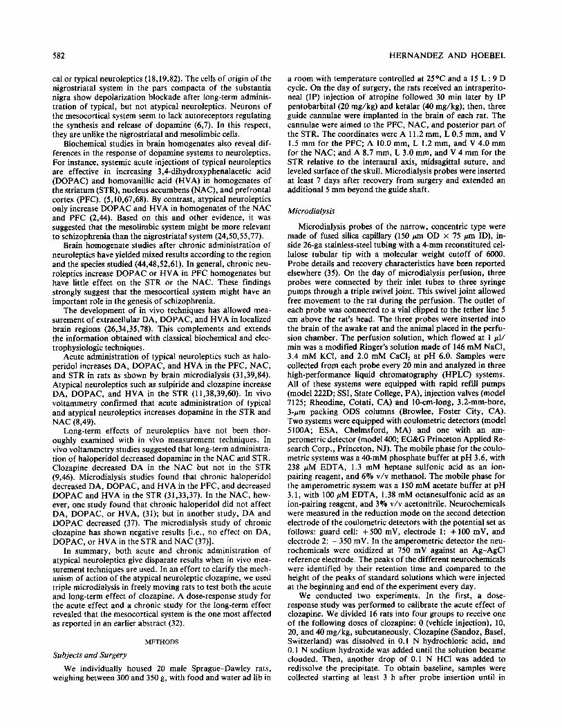
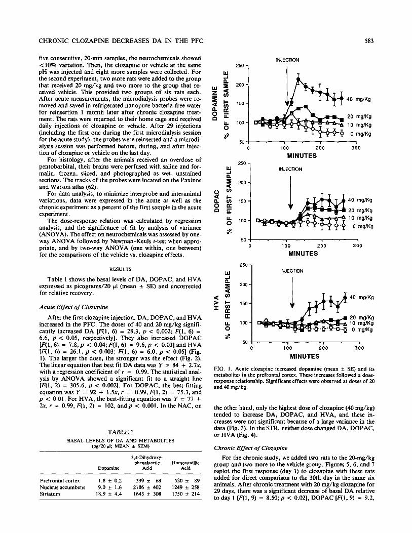
After the first clozapine injection, DA, DOPAC, and HVA increased in the PFC. The doses of 40 and 20 mg/kg signifi- cantly increased DA [F(l, 6) = 28.3, p < 0.002; F(1, 6) = 6.6, p c 0.05, respectively]. They also increased DOPAC [F(l, 6) = 7.8,~ c O.O4;F(l, 6) = 9.6,~ < 0.031 and HVA [F(l, 6) = 26.1, p < 0.003; F(1, 6) = 6.0, p < 0.051 (Fig. 1). The larger the dose, the stronger was the effect (Fig. 2). The linear equation that best fit DA data was Y = 84 + 2.7x, with a regression coefficient of r = 0.99. The statistical anal- ysis by ANOVA showed a significant fit to a straight line [F(l, 2) = 305.6, p < 0.002]. For DOPAC, the best-fitting equation was Y = 92 + 1.5x, r = 0.99, F(l,2) = 75.3, and p < 0.01. For HVA, the best-fitting equation was Y = 77 + 2x, r = 0.99, F(1, 2) = 102, and p c 0.001. In the NAC, on
TABLE 1 BASAL LEVELS OF DA AND METABOLITES
(pgI2Opl; MEAN f SEM)
Dopamine
3,CDihydroxy- phenalacetic
Acid Homovanillic
Acid
Prefrontal cortex 1.8 + 0.2 339 f 68 520 f 89 Nucleus accumbens 9.0 f 1.6 2186 f 402 1249 + 258 Striatum 18.9 + 4.4 1645 + 308 1750 + 214
INJECTION 250-
5
40 mg/Kg
20 mg/Kg IL lOO- 0 10 mg/Kg
S 0 mglKg
50 ! I I I 0 100 200 300
MINUTES 250
Y INJECTION
4 200 I
2 8
cii I- 2 150 40 mg/Kg
0 G 20 mglKg
8 100 10 - mglKg
0 8 mglKg
.x” I
0 40 2;o 3;0
MINUTES
250 - 5 INJECTDN
k 200 -
G!i
$I- 40 mglKg
=z
150 -
C 20 mg/Kg k 100 - 10 0 mglKg
mg/Kg
0 100 200 300
MINUTES
FIG. I. Acute clozapine increased dopamine (mean + SE) and its metabolites in the prefrontal cortex. These increases followed a dose- response relationship. Significant effects were observed at doses of 20 and 40 mg/kg.
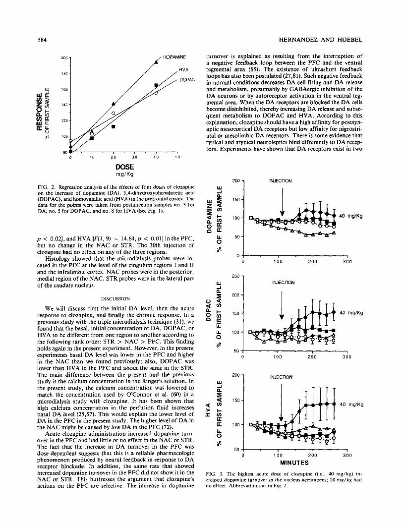
the other hand, only the highest dose of clozapine (40 mg/kg) tended to increase DA, DOPAC, and HVA, and these in- creases were not significant because of a large variance in the data (Fig. 3). In the STR, neither dose changed DA, DOPAC, or HVA (Fig. 4).
Chronic Effect of Clozapine
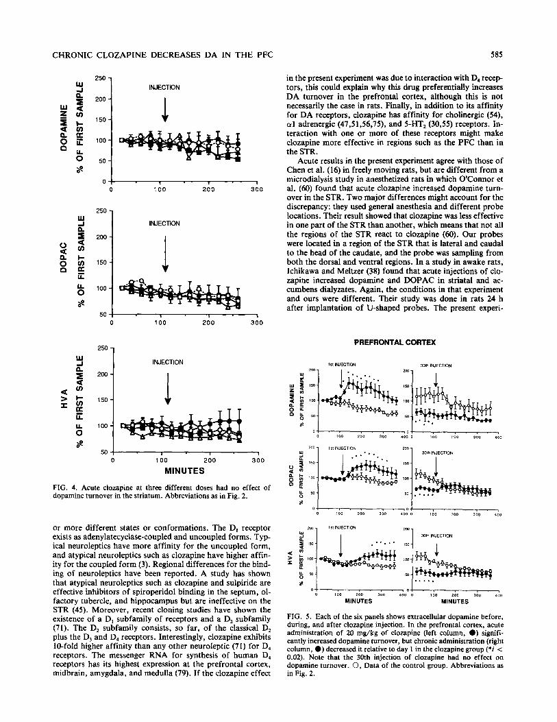
For the chronic study, we added two rats to the 20-mg/kg group and two more to the vehicle group. Figures 5, 6, and 7 replot the first response (day 1) to clozapine with these rats added for direct comparison to the 30th day in the same six animals. After chronic treatment with 20 mg/kg clozapine for 29 days, there was a significant decrease of basal DA relative to day 1 [F(l, 9) = 8.50;~ < 0.021, DOPAC [F(l, 9) = 9.2,
584 HERNANDEZ AND HOEBEL
160-
0 10 20 30 40 50
DOSE w/k7
FIG. 2. Regression analysis of the effects of four doses of clozapine on the increase of dopamine (DA), 3,4-dihydroxyphenalacetic acid (DOPAC), and homovanilhc acid (HVA) in the prefrontal cortex. The data for the points were taken from postinjection samples no. 3 for DA, no. 5 for DOPAC, and no. 8 for HVA (See Fig. 1).
p < 0.021, and HVA [F(l, 9) = 14.64,~ < 0.011 in the PFC, but no change in the NAC or STR. The 30th injection of clozapine had no effect on any of the three regions.
Histology showed that the microdialysis probes were lo- cated in the PFC at the level of the cingulum regions I and II and the infralimbic cortex. NAC probes were in the posterior, medial region of the NAC. STR probes were in the lateral part of the caudate nucleus.
DISCUSSION
We will discuss first the initial DA level, then the acute response to clozapine, and finally the chronic response. In a previous study with the triple microdialysis technique (31), we found that the basal, initial concentration of DA, DOPAC, or HVA to be different from one region to another according to the following rank order: STR > NAC > PFC. This finding holds again in the present experiment. However, in the present experiments basal DA level was lower in the PFC and higher in the NAC than we found previously; also, DOPAC was lower than HVA in the PFC and about the same in the STR. The main difference between the present and the previous study is the calcium concentration in the Ringer’s solution. In the present study, the calcium concentration was lowered to match the concentration used by O’Connor et al. (60) in a microdialysis study with clozapine. It has been shown that high calcium concentration in the perfusion fluid increases basal DA level (25,57). This would explain the lower level of DA in the PFC in the present study. The higher level of DA in the NAC might be caused by low DA in the PFC (72).
Acute clozapine administration increased dopamine turn- over in the PFC and had little or no effect in the NAC or STR. The fact that the increase in DA turnover in the PFC was dose dependent suggests that this is a reliable pharmacologic phenomenon produced by neural feedback in response to DA receptor blockade. In addition, the same rats that showed increased dopamine turnover in the PFC did not show it in the NAC or STR. This buttresses the argument that clozapine’s actions on the PFC are selective. The increase in dopamine
turnover is explained as resulting from the interruption of a negative feedback loop between the PFC and the ventral tegmental area (65). The existence of ultrashort feedback loops has also been postulated (27,81). Such negative feedback in normal conditions decreases DA cell firing and DA release and metabolism, presumably by GABAergic inhibition of the DA neurons or by autoreceptor activation in the ventral teg- mental area. When the DA receptors are blocked the DA cells become disinhibited, thereby increasing DA release and subse- quent metabolism to DOPAC and HVA. According to this explanation, clozapine should have a high affinity for postsyn- aptic mesocortical DA receptors but low affinity for nigrostri- atal or mesolimbic DA receptors. There is some evidence that typical and atypical neuroleptics bind differently to DA recep- tors. Experiments have shown that DA receptors exist in two
200 1 INJECTION
150 -
100 -
50 -
40 mg/Kg
0 ! I I 1
0 100 200 300
250
INJECTION
150 - 40 mg/Kg
100 -
100
INJECTION
200 300
H 40 mg/Kg
0 100 200 300
MINUTES
FIG. 3. The highest acute dose of clozapine (i.e., 40 m&kg) in- creased dopamine turnover in the nucleus accumbens; 20 mg/kg had no effect. Abbreviations as in Fig. 2.
CHRONIC CLOZAPINE DECREASES DA IN THE PFC 585
250 -I W
$
INJECTION
5 200 -
4s s c 150- 1
iis g ii 100 -
b 50-
8
0 , I I 1 0 100 200 300
250 -I
Y INJECTION
!k 200 -
150 -
50 : I 1 1 0 100 200 300
250
W -I 1 INJECTION
“” I
0 IA0 2;o 3;10
MINUTES
FIG. 4. Acute clozapine at three different doses had no effect of dopamine turnover in the striatum. Abbreviations as in Fig. 2.
or more different states or conformations. The D, receptor exists as adenylatecyclase-coupled and uncoupled forms. Typ- ical neuroleptics have more affinity for the uncoupled form, and atypical neuroleptics such as clozapine have higher affin- ity for the coupled form (3). Regional differences for the bind- ing of neuroleptics have been reported. A study has shown that atypical neuroleptics such as clozapine and sulpiride are effective inhibitors of spiroperidol binding in the septum, ol- factory tubercle, and hippocampus but are ineffective on the STR (45). Moreover, recent cloning studies have shown the existence of a D, subfamily of receptors and a D, subfamily (71). The D, subfamily consists, so far, of the classical D, plus the D, and D, receptors. Interestingly, clozapine exhibits lo-fold higher affinity than any other neuroleptic (71) for D, receptors. The messenger RNA for synthesis of human D, receptors has its highest expression at the prefrontal cortex, midbrain, amygdala, and medulla (79). If the clozapine effect
in the present experiment was due to interaction with D4 recep- tors, this could explain why this drug preferentially increases DA turnover in the prefrontal cortex, although this is not necessarily the case in rats. Finally, in addition to its affinity for DA receptors, clozapine has affinity for cholinergic (54), (~1 adrenergic (47,51,56,75), and 5-HT2 (30,55) receptors. In- teraction with one or more of these receptors might make clozapine more effective in regions such as the PFC than in the STR.
Acute results in the present experiment agree with those of Chen et al. (16) in freely moving rats, but are different from a microdialysis study in anesthetized rats in which O’Connor et al. (60) found that acute clozapine increased dopamine turn- over in the STR. Two major differences might account for the discrepancy: they used general anesthesia and different probe locations. Their result showed that clozapine was less effective in one part of the STR than another, which means that not all the regions of the STR react to clozapine (60). Our probes were located in a region of the STR that is lateral and caudal to the head of the caudate, and the probe was sampling from both the dorsal and ventral regions. In a study in awake rats, Ichikawa and Meltzer (38) found that acute injections of clo- zapine increased dopamine and DOPAC in striatal and ac- cumbens dialyzates. Again, the conditions in that experiment and ours were different. Their study was done in rats 24 h after implantation of U-shaped probes. The present experi-
PREFRONTAL CORTEX
od.o. 200 400
MINUTES MINUTES
FIG. 5. Each of the six panels shows extracellular dopamine before, during, and after clozapine injection. In the prefrontal cortex, acute administration of 20 mg/kg of clozapine (left column, 0) signifi- cantly increased dopamine turnover, but chronic administration (right column, 0) decreased it relative to day 1 in the clozapine group (*i < 0.02). Note that the 30th injection of clozapine had no effect on dopamine turnover. 0, Data of the control group. Abbreviations as in Fig. 2.
586 HERNANDEZ AND HOEBEL
NUCLEUS ACCUMBENS
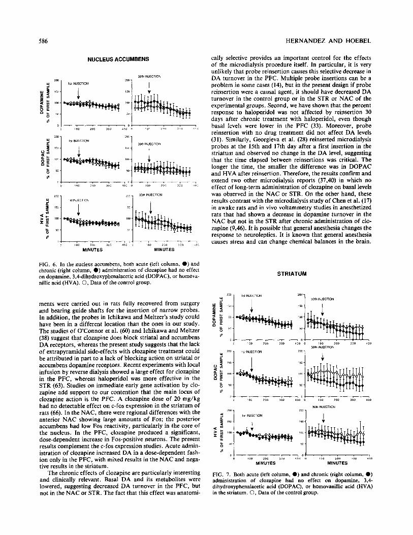
FIG. 6. In the nucleus accumbens, both acute (left column, 0) and chronic (right column, 0) administration of clozapine had no effect on dopamine, 3,4-dihydroxyphenalacetic acid (DOPAC), or homova- nillic acid (HVA). 0, Data of the control group.
ments were carried out in rats fully recovered from surgery and bearing guide shafts for the insertion of narrow probes. In addition, the probes in Ichikawa and Meltzer’s study could have been in a different location than the ones in our study. The studies of O’Connor et al. (60) and Ichikawa and Meltzer (38) suggest that clozapine does block striatal and accumbens DA receptors, whereas the present study suggests that the lack of extrapyramidal side-effects with clozapine treatment could be attributed in part to a lack of blocking action on striatal or accumbens dopamine receptors. Recent experiments with local infusion by reverse dialysis showed a large effect for clozapine in the PFC, whereas haloperidol was more effective in the STR (63). Studies on immediate early gene activation by clo- zapine add support to our contention that the main locus of clozapine action is the PFC. A clozapine dose of 20 mg/kg had no detectable effect on c-fos expression in the striatum of rats (66). In the NAC, there were regional differences with the anterior NAC showing large amounts of Fos; the posterior accumbens had low Fos reactivity, particularly in the core of the nucleus. In the PFC, clozapine produced a significant, dose-dependent increase in Fos-positive neurons. The present results complement the c-fos expression studies. Acute admin- istration of clozapine increased DA in a dose-dependent fash- ion only in the PFC, with mixed results in the NAC and nega- tive results in the striatum.
The chronic effects of clozapine are particularly interesting and clinically relevant. Basal DA and its metabolites were lowered, suggesting decreased DA turnover in the PFC, but not in the NAC or STR. The fact that this effect was anatomi-
cally selective provides an important control for the effects of the microdialysis procedure itself. In particular, it is very unlikely that probe reinsertion causes this selective decrease in DA turnover in the PFC. Multiple probe insertions can be a problem in some cases (14), but in the present design if probe reinsertion were a causal agent, it should have decreased DA turnover in the control group or in the STR or NAC of the experimental groups. Second, we have shown that the percent response to haloperidol was not affected by reinsertion 30 days after chronic treatment with haloperidol, even though basal levels were lower in the PFC (33). Moreover, probe reinsertion with no drug treatment did not affect DA levels (31). Similarly, Georgieva et al. (28) reinserted microdialysis probes at the 15th and 17th day after a first insertion in the striatum and observed no change in the DA level, suggesting that the time elapsed between reinsertions was critical. The longer the time, the smaller the difference was in DOPAC and HVA after reinsertion. Therefore, the results confirm and extend two other microdialysis reports (37,40) in which no effect of long-term administration of clozapine on basal levels was observed in the NAC or STR. On the other hand, these results contrast with the microdialysis study of Chen et al. (17) in awake rats and in vivo voltammetry studies in anesthetized rats that had shown a decrease in dopamine turnover in the NAC but not in the STR after chronic administration of clo- zapine (9.46). It is possible that general anesthesia changes the response to neuroleptics. It is known that general anesthesia causes stress and can change chemical balances in the brain.
STRIATUM
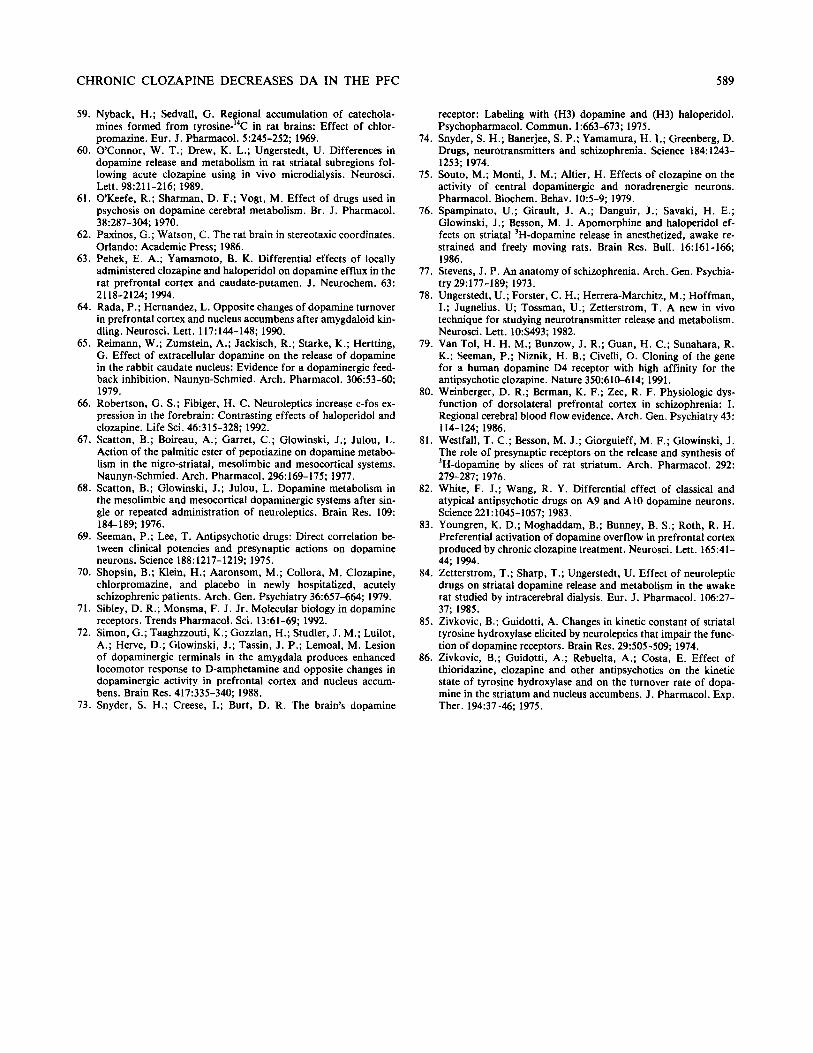
FIG. 7. Both acute (left column, 0) and chronic (right column, 0) administration of clozapine had no effect on dopamine, 3,4- dihydroxyphenalacetic acid (DOPAC), or homovanillic acid (HVA) in the striatum. 0, Data of the control group.

CHRONIC CLOZAPINE DECREASES DA IN THE PFC
Anesthesia is reported to increase serotonin and DA turnover. It also changes the response to haloperidol(41,76).
Microdialysis in the PFC of awake rats revealed a decrease in basal extracellular DA when the clozapine was given subcu- taneously in our study (31), but given IP, Chen et al. (16) found no effect. When clozapine was given orally, Youngren et al. (83) reported an increase, but they did the microdialysis with anesthesia. The route of administration may be a relevant variable in addition to those already mentioned, such as anes- thesia and probe placement. There are also large differences in the calcium concentration used for probe perfusion [e.g., 3.37 mM in the Chen study (17), 2 mM in this study, and 1.2 mM in the Youngren (83) study]. The present results point to the mesocortical DA system as a site of clozapine antipsy- chotic action when given chronically.
The present results with chronic clozapine can be compared to haloperidol. With both drugs, our triple microdialysis stud- ies show the strongest chronic effect on the PFC (31,33). Chronic administration of haloperidol did not affect the DA and DOPAC levels in the NAC; however, the Ichicawa and Meltzer study (37) did show a decrease of DA and DOPAC in the STR and NAC, a result that has been replicated by those authors (38). However, our study and theirs were done with different doses of haloperidol; we used 0.5 mg/kg and they used 2 mg/kg. A recent microdialysis report (12) showed that chronic administration of 0.5 mg/kg of haloperidol had no effect on dopamine levels in the STR, which coincides with our results for the same dose. Therefore, it is likely that the dose of haloperidol is a critical variabIe for the evaluation of the effect of chronic administration.
The consequences for dopaminergic theories of schizophre-
587
nia are interesting. It was postulated that the mesolimbic sys- tem is the locus of action for the antipsychotic effects of neu- roleptics (50). The present findings and those of Ichicawa and Meltzer suggest instead that the antipsychotic effect of atypi- cal neuroleptics is due in part to their action on the PFC. This confirms early speculation of PFC involvement in schizophre- nia based on cognitive and language processing. Later, La- duron et al. (44) found that the PFC was the most affected by chronic neuroleptic treatment based on brain homogenate studies. Other evidence obtained with blood flow measure- ment techniques have shown alterations in the normal blood flow response in the PFC of schizophrenic patients when they execute alternate delayed-response tests (80). However, on a note of caution, we still cannot rule out involvement of the NAC or some of its components (17). A reciprocal relation- ship between the PFC and the NAC has been shown (64,72). When amygdala stimulation increases DA turnover in the PFC, it decreases extracellular DA in the NAC (64); and con- versely, amygdala lesions decrease extracellular DA in the PFC and increase it in the NAC (72). Therefore, the decrease of DA turnover in the PFC, if and when it is caused by clozap- ine, might tend to increase DA turnover in the NAC, thereby counteracting any abnormal tendency toward low DA turn- over in the NAC which might be involved in affective disor- ders.
ACKNOWLEDGEMENTS
This research was supported by the Scottish Rite Foundation Schizophrenia Research Program. Clozapine was donated by Sandoz, Inc. Appreciation is expressed to Dawn Davidson for technical assis- tance.
REFERENCES
I
2.
3.
4.
5.
6.
7.
8.
9.
10.
Anden, N. E.; Butcher, S. G.; Corrodi, H.; Fuxe, K.; Ungerstedt, U. Receptor activity and turnover of dopamine and noradrenaline after neuroleptics. Eur. J. Pharmacol. 11:303-314; 1970. Anden, N. E.; Stock, G. Effect of clozapine on the turnover of dopamine in the corpus striatum and in the limbic system. J. Pharm. Pharmacol. 25:346-348; 1973. Andersen, P. H.; Braestrup, C. Evidence for different states of the D, receptor: Clozapine and fluperlapine may preferentially label an adenylcyclase coupled-state of the D, receptor. J. Neuro- them. 47:1822-1831; 1986. Ayd, F. J. Neuroleptics and extrapyramidal reactions in psychiat- ric patients. In: Bordelau, J. M., ed. Systeme exptrapyramidal et neuroleptiques. Montreal: Editions Psychiatriques; 1%1:355- 363. Bacopoulos, N. G.; Bustos, G.; Redmond, E. Jr.; Roth, R. H. Chronic treatment with haloperidol or fluphenazine decanoate: Regional effects on dopamine and serotonin metabolism in pri- mate brains. J. Pharmacol. Exp. Ther. 221:22-28; 1982. Bannon, M. J.; Michaud, R. L.; Roth, R. H. Mesocortical dopa- mine system: Lack of autoreceptors modulating dopamine syn- thesis. Mol. Pharmacol. 19:270-275; 1981. Bannon, M. J.; Reinhard J. F. Jr.; Roth, R. H. Mesocortical dopamine neurons: Unique response to antipsychotic drugs ex- plained by the absence of terminal autoreceptors. Nature 296: 444-446; 1982. Blaha, C. D.; Lane, R. F. Direct in vivo electrochemical monitor- ing of dopamine release in response to neuroleptic drugs. Eur. J. Pharmacol. 98: 113-119; 1984. Blaha, C. D.; Lane, R. F. Chronic treatment with classical and atypical antipsychotic drugs differentially decreases dopamine re- lease in striatum and nucleus accumbens in vivo. Neurosci. Lett. 78:199-204; 1987. Bowers, M. B. Jr.; Rozitis, A. Regional differences in homovanil-
11.
12.
13.
14.
15.
16.
17.
18.
lit acid concentrations after acute and chronic administration of antipsychotic drugs. J. Pharm. Pharmacol. 26:743-745; 1974. Bunney. B. S.; Moghaddam, B. Regional differences in the effect of typical and atypical neuroleptics on terminal release of dopa- mine: In vivo microdialysis studies. Sot. Neurosci. Abstr. 14:741; 1988. Bunney, B. S.; Moghaddam, B. Some terminal release character- istics of dopamine neurons after chronic haloperidol treatment. Sot. Neurosci. Abstr. 16:532; 1990. Bunney, B. S.; Walters, J. R.; Roth, R. H.; Aghajanian, G. K. Dopaminergic neurons: Effect of antipsychotic drugs and am- phetamine on single cell activity. J. Pharmacol. Exp. Ther. 185: 560-571; 1973. Camp, D. M.; Robinson, T. E. On the use of multiple probe insertions at the same site for repeated intracerebral microdialysis experiments in the nigrostriatal dopamine system of rats. J. Neu- rochem. 58:1706-1715; 1992. Carlsson, A.; Lindquist, M. Effect of chlorpromazine or haloper- idol on formation of 3-methoxytyramine and normetanephrine in mouse brain. Acta Pharmacol. Toxicol. 20:140; 1%3. Chen, J.; Paredes, P.; Gardner, E. L. Chronic treatment with clozapine selectively decreases basal dopamine release in nucleus accumbens but not in caudate-putamen as measured by in vivo brain microdialysis; further evidence for depolarization block. Neurosci. Lett. 122:127-131; 1991. Chen, J.; Ruan, D.; Paredes, W.; Gardner, E. L. Effects of acute and chronic clozapine on dopaminergic function in medial prefrontal cortex of awake, freely moving rats. Brain Res. 571: 235-241; 1992. Chiodo, L. A.; Bunney, B. S. Typical and atypical neuroleptics: Differential effects of chronic administration on the activity of A9 and Al0 midbrain dopaminergic neurons. J. Neurosci. 3: 1607-1619; 1983.

588 HERNANDEZ AND HOEBEL
19.
20.
21.
22.
23.
24.
25.
26.
27.
28.
29.
30.
31.
32.
33.
34.
35.
36.
31.
38.
Chiodo, L. A.; Bunney, B. S. Possible mechanism by which re- peated clozapine administration differentially affects the activity of two subpopulations of midbrain dopamine neurons. J. Neu- rosci. 5:2539-2544; 1985. Chouinard, G.; Annable, L. Clozapine in the treatment of newly admitted schizophrenic patients. J. Clin. Pharmacol. 5-6:289- 297; 1976. Crane, G. E. Persistent dyskinesia. Br. J. Psychiatry 122:395- 405; 1973. Crane, G. E. Tardive dyskinesia in patients treated with major neuroleptics: A review of the literature. Am. J. Psychiatry 124: 40-48; 1968. Creese, I.; Burt, D. R.; Snyder S. H. Dopamine receptor binding predicts clinical and pharmacological potencies of antipsychotic drugs. Science 192:481-483; 1976. Crow, T. J. Molecular pathology of schizophrenia: More than one dimension of pathology? BMJ 280:66-68; 1980. de Boer, P.; Damsma, G.; Fibiger, H. C.; Timmerman, W.; de Vries, J. B.; Westerink, B. H. C. Dopaminergic-cholinergic inter- actions in the striatum: The critical significance of calcium con- centrations in brain microdialysis. Arch. Pharmacol. 342:528- 534; 1990. Delgado, J. M. R.; DeFeudis, F. V.; Roth, R. H.; Ryugo, D. K.; Mitruka, B. M. Dialytrode for long term intracerebral perfusion in awake monkeys. Arch. Int. Pharmacodyn. 198:9-21; 1972. Farnebo, L. 0.; Hamberger, B. Drug induced changes in the release of ‘H-dopamine from field stimulated brain slices. Acta Physiol. Stand. 84:35-44; 1971. Georgieva, J.; Luthman, J.; Mohringe, B.; Magnusson, 0. Tissue and microdialysate changes after repeated and permanent probe implantation in the striatum of freely moving rats. Brain Res. Bull. 31:463-470; 1993. Gerlach, J.; Thorsen, K.; Fog, R. Extrapyramidal reactions and amine metabolites in cerebrospinal fluid during haloperidol and clozapine treatment of schizophrenic patients. Psychopharmaco- logia 40:341-350; 1975. Hartvig, P.; Eckernas, S. A.; Ekblom, B.; Lindstrom, L.; Lund- quist, H.; Axelsson, S.; Fasth, K. J.; Gullberg, P.; Landstrom, B. Receptor binding and selectivity of three carbon-11-labelled dopamine receptor antagonists in the brain of the Rhesus mon- keys studied with positron emission tomography. Acta Neural. Stand. 77:314-321; 1988. Hernandez, L.; Hoebel, B. G. Haloperidol given chronically de- creases basal dopamine in the prefrontal cortex more than the striatum or nucleus accumbens as simultaneously measured by microdialysis. Brain Res. Bull. 22:763-769; 1989. Hernandez, L.; Hoebel, B. G. Selective effect of clozapine on DA turnover on the prefrontal cortex in rats. Sot. Neurosci. Abstr. 16:588; 1990. Hernandez, L.; Baptista, T.; Hoebel, B. G. Neurochemical ef- fects of chronic haloperidol and lithium assessed by brain micro- dialysis in rats. Prog. Neuropsychopharmacol. Biol. Psychiatry 14: s17-s35; 1990. Hernandez, L.; Paez, X.; Hamlin, C. Neurotransmitters extrac- tion by local cerebral dialysis in anesthetized rats. Pharmacol. Biochem. Behav. 18:159-162; 1983. Hernandez, L.; Stanley, B. G.; Hoebel B. G. A small, removable microdialysis probe. Life Sci. 39:2629-2637; 1986. Huger, F. P.; Craig, P. S.; Chiang, Y.; Glamkowski, E. J.; Ellis, D. B. Pharmacological evaluation of HP370, a potential atypical antipsychotic agent: 2. In vivo profile. Drug Dev. Res. ll:169- 176; 1987. Ichikawa, J.; Meltzer, H. Y. The effect of chronic clozapine and haloperidol on basal dopamine release and metabolism in rat stri- atum and nucleus accumbens studied by in vivo microdialysis. Eur. J. Pharmacol. 176:371-374; 1990. Ichikawa, J.; Meltzer, H. Y. Differential effect of repeated treat- ment with haloperidol and clozapine on dopamine release and metabolism in the striatum and the nucleus accumbens. J. Phar- macol. Exp. Ther. 256:348-357; 1991.
39. Imperato, A.; Di Chiara, G. Dopamine release and metabolism in
40.
41.
42.
43.
44.
45.
46.
47.
48.
49.
50.
51.
52.
53.
54.
55.
56.
57.
58.
awake rats after systemic neuroleptics as studied by trans-striatal dialysis. J. Neurosci. 5:297-306; 1985. Invernizzi, R.; Morali, F.; Pozzi, L.; Samanin, R. Effects of acute and chronic clozapine on dopamine release and metabolism in the striatum and nucleus accumbens of conscious rats. Br. J. Pharmacol. 100:774-778; 1990. Kalen, P.; Strecker, R. E.; Rosengren, E.; Bjorklund, A. Endoge- nous release of neuronal serotonin and 5-hydroxyindolacetic acid in the caudate of the rat as revealed by intracerebral dialysis coupled to high performance liquid chromatography with electro- chemical detection. J. Neurochem. 51:1422-1435; 1988. Kebabian, J. W.; Calme, D. B. Multiple receptors for dopamine. Nature 277~93-96; 1979. Kebabian, J. W.; Petzold, G. L.; Greengard, P. Dopamine- sensitive adenylate cyclase in caudate nucleus of rat brain, and its similarity to the “dopamine receptor.” Proc. Nat]. Acad. Sci. 69: 2145-2149; 1972. Laduron, P.; De Bie, K.; Leysen, J. Specific effect of haloperidol on dopamine turnover in the frontal cortex. Naunyn-Schmied. Arch. Pharmacol. 2%:183-185; 1977. Lahti, R. A.; David, D. Cr. In vivo tritiated spiperone binding: The locus of action of antipsychotic agents. Res. Commun. Psy- cho]. Psychiatry Behav. 12:141-150; 1987. Lane, R. F.; Blaha, C. D. Chronic haloperidol decreases dopa- mine release in striatum and nucleus accumbens in vivo: Depolar- ization block as a possible mechanism of action. Brain Res. Bull. 18:135-138; 1987. Lane, R. F.; Blaha, C. D.; Rivet, J. M. Selective inhibition of mesohmbic dopamine release following chronic administration of clozapine: Involvement of (~1 noradrenergic receptors demon- strated by in vivo voltammetry. Brain Res. 460:398-401; 1988. Lerner, P.; Nose, P.; Gordon, E. K.; Lovenberg, W. Haloperi- dol: Effect of long term treatment on rat striatal dopamine syn- thesis and turnover. Science 197:181-183; 1977. Maidment, N. T.; Marsden, C. A. Acute administration of clo- zapine, thioridazine and metoclopramide increases extracellular DOPAC and decreases extracellular 5-HIAA, measured in the nucleus accumbens and striatum of rats using in vivo voltamme- try. Neuropharmacology 26:187-193; 1987. Matthysse, S. Nucleus accumbens and schizophrenia. In: Chro- nister, R. B.; DeFrance, J. F., eds. The neurobiology of the nu- cleus accumbens. Brunswick, ME: Haer Institute Press; 1981: 351-360. McMillen, B. A.; Shore, P. A. Comparative effect of clozapine and alpha-adrenoceptor blocking drugs on regional noradrenaline metabolism in rat brains. Eur. J. Pharmacol. 52:225-230; 1978. Melford, I. N.; Roth, K. A.; Agren, H.; Barchas, J. D. Enhance- ment of dopamine metabolism in rat brain frontal cortex: A com- mon effect of chronically administered antipsychotic drugs. Brain Res. 425:380-384; 1988. Meltzer, H. Y. Biochemical studies in schizophrenia. In: Bellak, L., ed. Disorders of the schizophrenic syndrome. New York: Ba- sic Books; 1979. Meltzer, H. Y.; Chai, B. L.; Thompson, P. A.; Yamamoto, B. K. Effect of scopolamine on the efflux of dopamine and its me- tabolites after clozapine, haloperidol or thioridazine. J. Pharma- col. Exp. Ther. 268:1452-1461; 1994. Meltzer, H. Y.; Zhang, Y.; Stockmeier, C. Effect of typical and atypical antipsychotic drugs (APD) on frontal cortical (FC) sero- tonin (5HT2) and striatal dopamine2 (D2) binding in vivo. Sot. Neurosci. Abstr. 244. 3:586; 1990. Menon, M. K.; Gordon, L. I.; Fitten, J. Interaction between clozapine and a lipophilic (~1 adrenergic agonist. Life Sci. 43: 1791-1804; 1988. Moghaddam, B.; Bunney, B. S. Ion composition in microdialysis perfusing solution alters the pharmacological responsiveness and basal outflow of striatal dopamine. J. Neurochem. 52:652-654; 1989. National Institute of Mental Health Service Center Collaborative Study Group. Phenothiazine treatment in acute schizophrenia. Arch. Gen. Psychiatry 10:246-261; 1964.
CHRONIC CLOZAPINE DECREASES DA IN THE PFC 589
59. Nyback, H.; Sedvall, G. Regional accumulation of catechola- mines formed from tyrosine-‘4C in rat brains: Effect of chlor- promazine. Eur. J. Pharmacol. 5:245-252; 1969.
60. O’Connor, W. T.; Drew, K. L.; Ungerstedt, U. Differences in dopamine release and metabolism in rat striatal subregions fol- lowing acute clozapine using in vivo microdialysis. Neurosci. Lett. 98:211-216; 1989.
61. G’Keefe, R.; Sharman, D. F.; Vogt, M. Effect of drugs used in psychosis on dopamine cerebral metabolism. Br. J. Pharmacol. 38:287-304; 1970.
62. Paxinos, G.; Watson, C. The rat brain in stereotaxic coordinates. Orlando: Academic Press; 1986.
63. Pehek, E. A.; Yamamoto, B. K. Differential effects of locally administered clozapine and haloperidol on dopamine efflux in the rat prefrontal cortex and caudate-putamen. J. Neurochem. 63: 2118-2124; 1994.
64. Rada, P.; Hernandez, L. Opposite changes of dopamine turnover in prefrontal cortex and nucleus accumbens after amygdaloid kin- dling. Neurosci. Lett. 117:144-148; 1990.
65. Reimann, W.; Zumstein, A.; Jackisch, R.; Starke, K.; Hertting, G. Effect of extracellular dopamine on the release of dopamine in the rabbit caudate nucleus: Evidence for a dopaminergic feed- back inhibition. Naunyn-Schmied. Arch. Pharmacol. 306:53-m; 1979.
66. Robertson, G. S.; Fibiger, H. C. Neuroleptics increase c-fos ex- pression in the forebrain: Contrasting effects of haloperidol and clozapine. Life Sci. 46:315-328; 1992.
67. Scatton, B.; Boireau, A.; Garret, C.; Glowinski, J.; Julou, L. Action of the palmitic ester of pepotiazine on dopamine metabo- lism in the nigro-striatal, mesolimbic and mesocortical systems. Naunyn-Schmied. Arch. Pharmacol. 296:169-175; 1977.
68. Scatton, B.; Glowinski, J.; Julou, L. Dopamine metabolism in the mesolimbic and mesocortical dopaminergic systems after sin- gle or repeated administration of neuroleptics. Brain Res. 109: 184-189; 1976.
69. Seeman, P.; Lee, T. Antipsychotic drugs: Direct correlation be- tween clinical potencies and presynaptic actions on dopamine neurons. Science 188:1217-1219; 1975.
70. Shopsin, B.; Klein, H.; Aaronsom, M.; Collora, M. Clozapine, chlorpromazine, and placebo in newly hospitalized, acutely schizophrenic patients. Arch. Gen. Psychiatry 36657-664; 1979.
71. Sibley, D. R.; Monsma, F. J. Jr. Molecular biology in dopamine receptors. Trends Pharmacol. Sci. 13:61-69; 1992.
72. Simon, G.; Taaghzzouti, K.; Gozzlan, H.; Studler, J. M.; Luilot, A.; Herve, D.; Glowinski, J.; Tassin, J. P.; Lemoal, M. Lesion of dopaminergic terminals in the amygdala produces enhanced locomotor response to D-amphetamine and opposite changes in dopaminergic activity in prefrontal cortex and nucleus accum- bens. Brain Res. 417:335-340; 1988.
73. Snyder, S. H.; Creese, I.; Burt, D. R. The brain’s dopamine
receptor: Labeling with (H3) dopamine and (H3) haloperidol. Psychopharmacol. Commun. 1663-673; 1975.
74. Snyder, S. H.; Banerjee, S. P.; Yamamura, H. 1.; Greenberg, D. Drugs, neurotransmitters and schizophrenia. Science 184: 1243- 1253; 1974.
75. Souto, M.; Monti, J. M.; Altier, H. Effects of clozapine on the activity of central dopaminergic and noradrenergic neurons. Pharmacol. Biochem. Behav. 10:5-9; 1979.
76. Spampinato, U.; Girault, J. A.; Danguir, J.; Savaki, H. E.; Glowinski, J.; Besson, M. J. Apomorphine and haloperidol ef- fects on striatal ‘H-dopamine release in anesthetized, awake re- strained and freely moving rats. Brain Res. Bull. 16:161-166; 1986.
77. Stevens, J. P. An anatomy of schizophrenia. Arch. Gen. Psychia- try 29:177-189; 1973.
78. Ungerstedt, U.; Forster, C. H.; Herrera-Marchitz, M.; Hoffman, I.; Jugnelius. U; Tossman, U.; Zetterstrom, T. A new in vivo technique for studying neurotransmitter release and metabolism. Neurosci. Lett. lO:S493; 1982.
79. Van Tol, H. H. M.; Bunzow, J. R.; Guan, H. C.; Sunahara, R. K.; Seeman, P.; Niznik, H. B.; Civelli, 0. Cloning of the gene for a human dopamine D4 receptor with high affinity for the antipsychotic clozapine. Nature 350:610-614; 1991.
80. Weinberger, D. R.; Berman, K. F.; Zec, R. F. Physiologic dys- function of dorsolateral prefrontal cortex in schizophrenia: I. Regional cerebral blood flow evidence. Arch. Gen. Psychiatry 43: 114-124; 1986.
81. Westfall, T. C.; Besson, M. J.; Giorguieff, M. F.; Glowinski, J. The role of presynaptic receptors on the release and synthesis of ‘H-dopamine by slices of rat striatum. Arch. Pharmacol. 292: 279-287; 1976.
82. White, F. J.; Wang, R. Y. Differential effect of classical and atypical antipsychotic drugs on A9 and Al0 dopamine neurons. Science 221:1045-1057; 1983.
83. Youngren, K. D.; Moghaddam, B.; Bunney, B. S.; Roth, R. H. Preferential activation of dopamine overflow in prefrontal cortex produced by chronic clozapine treatment. Neurosci. Lett. 165:41- 44; 1994.
84. Zetterstrom, T.; Sharp, T.; Ungerstedt, U. Effect of neuroleptic drugs on striatal dopamine release and metabolism in the awake rat studied by intracerebral dialysis. Eur. J. Pharmacol. 106:27- 37; 1985.
85. Zivkovic, B.; Guidotti, A. Changes in kinetic constant of striatal tyrosine hydroxylase elicited by neuroleptics that impair the func- tion of dopamine receptors. Brain Res. 29:505-509; 1974.
86. Zivkovic, B.; Guidotti, A.; Rebuelta, A.; Costa, E. Effect of thioridazine, clozapine and other antipsychotics on the kinetic state of tyrosine hydroxylase and on the turnover rate of dopa- mine in the striatum and nucleus accumbens. J. Pharmacol. Exp. Ther. 194:37-46; 1975.