morphine effects on striatal transcriptome in mice
TRANSCRIPT

com
ment
reviews
reports
deposited research
refereed researchinteractio
nsinfo
rmatio
n
Open Access2007Korostynskiet al.Volume 8, Issue 6, Article R128ResearchMorphine effects on striatal transcriptome in miceMichal Korostynski, Marcin Piechota, Dorota Kaminska, Wojciech Solecki and Ryszard Przewlocki
Address: Department of Molecular Neuropharmacology, Institute of Pharmacology PAS, Smetna 12, 31-343, Krakow, Poland.
Correspondence: Ryszard Przewlocki. Email: [email protected]
© 2007 Korostynski et al.; licensee BioMed Central Ltd. This is an open access article distributed under the terms of the Creative Commons Attribution License (http://creativecommons.org/licenses/by/2.0), which permits unrestricted use, distribution, and reproduction in any medium, provided the original work is properly cited.Morphine effects on mouse brains<p>Global transcriptional analysis of mouse striata following acute and chronic exposure to morphine reveals multiple physiological fac-tors which may affect opioid-related phenotypes and implicates a number of gene networks, including glucocorticoid receptor regulated genes, in the response to this opioid.</p>
Abstract
Background: Chronic opiate use produces molecular and cellular adaptations in the nervoussystem that lead to tolerance, physical dependence, and addiction. Genome-wide comparison ofmorphine-induced changes in brain transcription of mouse strains with different opioid-relatedphenotypes provides an opportunity to discover the relationship between gene expression andbehavioral response to the drug.
Results: Here, we analyzed the effects of single and repeated morphine administrations in selectedinbred mouse strains (129P3/J, DBA/2J, C57BL/6J, and SWR/J). Using microarray-based geneexpression profiling in striatum, we found 618 (false discovery rate < 1%) morphine-responsivetranscripts. Through ontologic classification, we linked particular sets of genes to biologic functions,including metabolism, transmission of nerve impulse, and cell-cell signaling. We identified numerousnovel morphine-regulated genes (for instance, Olig2 and Camk1g), and a number of transcripts withstrain-specific changes in expression (for instance, Hspa1a and Fzd2). Moreover, transcriptionalactivation of a pattern of co-expressed genes (for instance, Tsc22d3 and Nfkbia) was identified asbeing mediated via the glucocorticoid receptor (GR). Further studies revealed that blockade of theGR altered morphine-induced locomotor activity and development of physical dependence.
Conclusion: Our results indicate that there are differences between strains in the magnitude oftranscriptional response to acute morphine treatment and in the degree of tolerance in geneexpression observed after chronic morphine treatment. Using whole-genome transcriptionalanalysis of morphine effects in the striatum, we were able to reveal multiple physiological factorsthat may influence opioid-related phenotypes and to relate particular gene networks to thiscomplex trait. The results also suggest the possible involvement of GR-regulated genes in mediatingbehavioral response to morphine.
BackgroundOpioids are considered to be among the most potent drugs forrelieving severe chronic pain. Long-term morphine treatmentis undesirable because of the development of tolerance to its
analgesic effects and physical dependence. On the otherhand, prolonged abuse of opiates leads to drug addiction - achronic, relapsing disorder with a complex mechanism. Accu-mulating evidence is converging to suggest that formation of
Published: 28 June 2007
Genome Biology 2007, 8:R128 (doi:10.1186/gb-2007-8-6-r128)
Received: 3 May 2007Accepted: 28 June 2007
The electronic version of this article is the complete one and can be found online at http://genomebiology.com/2007/8/6/R128
Genome Biology 2007, 8:R128

R128.2 Genome Biology 2007, Volume 8, Issue 6, Article R128 Korostynski et al. http://genomebiology.com/2007/8/6/R128
opioid addiction involves changes in synaptic structure andneuronal plasticity [1]. These long-lasting neuroadaptationsprobably include compound changes in gene expression inthe mesocorticolimbic system of the brain [2]. The major sub-strates of the molecular and cellular mechanisms of opioidaddiction are suggested to be the dorsal and ventral striatum.Morphine administration enhances the release of dopaminein both the dorsal striatum and nucleus accumbens [3]. It iswell established that the nucleus accumbens, a ventral subre-gion that receives dopaminergic projections from the ventraltegmental area, is related to the reward properties of opioids[4]. The dorsal part of the striatum is a brain region that isimplicated in habit learning, which is a fundamental compo-nent of addiction [5].
It is well known that drugs of abuse stimulate the transcrip-tion of numerous genes in several brain regions [6,7]. Moreo-ver, a significant contribution of genetic factors tovulnerability to the addictive action of opiates and otheraddictive drugs is well established [8]. Several other effects ofopioid action, for example analgesia and hypothermia, arealso likely to be determined by combinations of genetic fac-tors [9]. In contrast, the influence of genotype on genomicresponse to opioids and the association between changes ingene expression and development of the rewarding andaddictive effects are poorly characterized. Inbred strains ofmice with well described phenotypes provide valuable modelsin which to analyze interactions between genetic background,and behavioral and transcriptional responses to the drug.
To separate the relationship between the different effects ofmorphine and the gene expression profiles in the striatum, wecompared responses to acute and chronic drug treatmentacross four mouse strains with extreme opioid-related pheno-types (C57BL/6J, DBA/2J, 129P3/J, and SWR/J). Two com-monly used inbred strains of mice, C57BL/6J and DBA/2J,exhibit remarkable differences in morphine-induced locomo-tor activation and conditional place preference [10,11]. Com-pared with the other strains, C57BL/6J mice were found tohave the greatest preference for oral self-administered mor-phine [12]. Furthermore, the 129P3/J strain failed to developphysical dependence and tolerance, whereas extraordinarysensitivity to opioid withdrawal was observed in SWR/J mice[13].
Our prior comparison of gene expression profiles of naïve ani-mals from the selected inbred mouse strains indicated diver-sity at the level of several hundreds of transcripts in thestriatum [14]. Here, we use microarray technology to obtain aprofile of genes that are regulated by acute and chronic mor-phine in the striatum of the four mouse strains. Our ultimategoal is to link particular genes, regulatory elements, and spe-cific signaling pathways with opioid-related traits. To thisend, we have combined gene expression profiling with bioin-formatic approaches and behavioral testing. The results pre-sented here identify several novel morphine-responsive genes
that may modulate the molecular as well as behavioralresponse to morphine and may contribute to development ofmorphine addiction.
ResultsMicroarray analysisA microarray experiment was designed to determine theimpact of genetic background on the transcriptional effects ofmorphine in striatum. Twelve experimental groups werecompared to analyze the transcriptional response to acuteand chronic morphine among the four inbred strains of mice(Figure 1). Three biologic replicates of the microarray wereprepared for each experimental group. The quality of micro-arrays was carefully checked to ensure that hybridization ofthe samples to the arrays was comparable across the datasetcomposed of 36 microarrays (see Materials and methods,below). Correlation coefficients of raw microarray resultswithin the strains (129P3/J: 0.986 to 0.988; C57BL/6J: 0.977to 0.993; DBA/2J: 0.979 to 0.99; and SWR/J: 0.983 to0.987) and within the experimental groups (control: 0.977 to0.987; acute: 0.988 to 0.993; and chronic: 0.981 to 0.987)were very high. The microarray data reported in this manu-script are publicly available at the Gene Expression Omnibusdatabase under accession number GSE7762 [15].
Signals from 21,467 probe sets were detected reproducibly onthe microarrays, representing 46% of all probe sets on themicroarray (23,633 were filtered out). Genes annotated tothese probe sets were considered to be expressed in the exam-ined brain tissue. The list of detected probe sets was used forfurther analyses. Main factors (strain and treatment), as wellas interaction, were calculated by using multivariate analysisof variance (MANOVA).
Protocol of morphine administrationFigure 1Protocol of morphine administration. Acutely treated mice (n = 9) were injected with a single dose of morphine (20 mg/kg, subcutaneously). Chronically treated animals (n = 9) were injected with increasing doses of morphine for 5 days (10, 20, and 40 mg/kg, subcutaneously; see Materials and methods). Control (n = 9) and acute morphine groups received injections of saline in the same time schedule as the chronic group received morphine. Animals were killed by decapitation 4 hours after the last injection. The time point used for tissue collection and gene expression analysis is indicated by the red arrow.
Day 1 Day 2 Day 3 Day 4 Day 59 13 17 9 13 17 9 13 17 9 13 17 9 13Time
Chronic
Acute
Control
Morphinedose
[mg/kg]
Mice: C57BL/6J, DBA/2J, 129P3/J, SWR/J27 animals per each strain in 3 experimental groups (n = 9)
0 0 0 0 0 0 0 0 0 0 0 0 20
0 0 0 0 0 0 0 0 0 0 0 0 0
10 10 10 20 20 20 40 40 40 40 40 40 40
Genome Biology 2007, 8:R128
http://genomebiology.com/2007/8/6/R128 Genome Biology 2007, Volume 8, Issue 6, Article R128 Korostynski et al. R128.3
com
ment
reviews
reports
refereed researchdepo
sited researchinteractio
nsinfo
rmatio
n
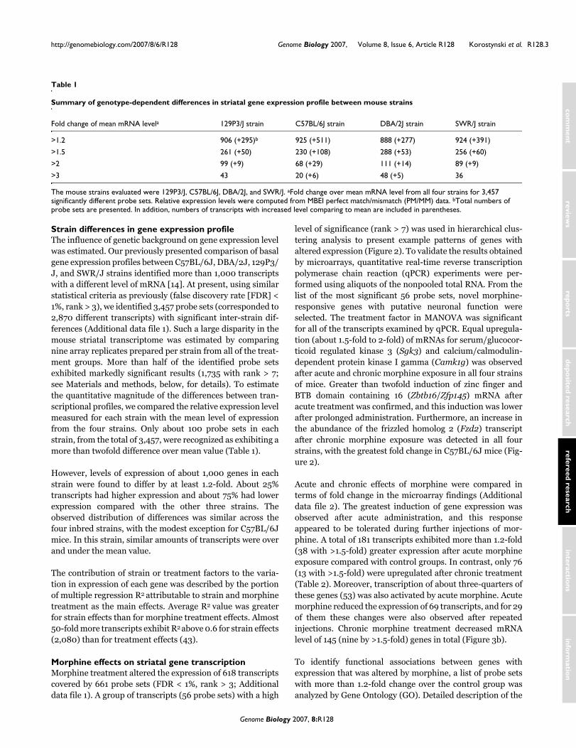
Strain differences in gene expression profileThe influence of genetic background on gene expression levelwas estimated. Our previously presented comparison of basalgene expression profiles between C57BL/6J, DBA/2J, 129P3/J, and SWR/J strains identified more than 1,000 transcriptswith a different level of mRNA [14]. At present, using similarstatistical criteria as previously (false discovery rate [FDR] <1%, rank > 3), we identified 3,457 probe sets (corresponded to2,870 different transcripts) with significant inter-strain dif-ferences (Additional data file 1). Such a large disparity in themouse striatal transcriptome was estimated by comparingnine array replicates prepared per strain from all of the treat-ment groups. More than half of the identified probe setsexhibited markedly significant results (1,735 with rank > 7;see Materials and methods, below, for details). To estimatethe quantitative magnitude of the differences between tran-scriptional profiles, we compared the relative expression levelmeasured for each strain with the mean level of expressionfrom the four strains. Only about 100 probe sets in eachstrain, from the total of 3,457, were recognized as exhibiting amore than twofold difference over mean value (Table 1).
However, levels of expression of about 1,000 genes in eachstrain were found to differ by at least 1.2-fold. About 25%transcripts had higher expression and about 75% had lowerexpression compared with the other three strains. Theobserved distribution of differences was similar across thefour inbred strains, with the modest exception for C57BL/6Jmice. In this strain, similar amounts of transcripts were overand under the mean value.
The contribution of strain or treatment factors to the varia-tion in expression of each gene was described by the portionof multiple regression R2 attributable to strain and morphinetreatment as the main effects. Average R2 value was greaterfor strain effects than for morphine treatment effects. Almost50-fold more transcripts exhibit R2 above 0.6 for strain effects(2,080) than for treatment effects (43).
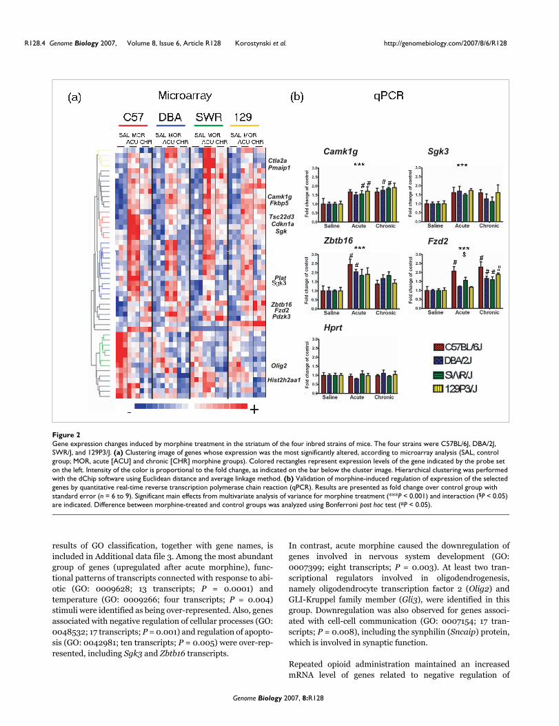
Morphine effects on striatal gene transcriptionMorphine treatment altered the expression of 618 transcriptscovered by 661 probe sets (FDR < 1%, rank > 3; Additionaldata file 1). A group of transcripts (56 probe sets) with a high
level of significance (rank > 7) was used in hierarchical clus-tering analysis to present example patterns of genes withaltered expression (Figure 2). To validate the results obtainedby microarrays, quantitative real-time reverse transcriptionpolymerase chain reaction (qPCR) experiments were per-formed using aliquots of the nonpooled total RNA. From thelist of the most significant 56 probe sets, novel morphine-responsive genes with putative neuronal function wereselected. The treatment factor in MANOVA was significantfor all of the transcripts examined by qPCR. Equal upregula-tion (about 1.5-fold to 2-fold) of mRNAs for serum/glucocor-ticoid regulated kinase 3 (Sgk3) and calcium/calmodulin-dependent protein kinase I gamma (Camk1g) was observedafter acute and chronic morphine exposure in all four strainsof mice. Greater than twofold induction of zinc finger andBTB domain containing 16 (Zbtb16/Zfp145) mRNA afteracute treatment was confirmed, and this induction was lowerafter prolonged administration. Furthermore, an increase inthe abundance of the frizzled homolog 2 (Fzd2) transcriptafter chronic morphine exposure was detected in all fourstrains, with the greatest fold change in C57BL/6J mice (Fig-ure 2).
Acute and chronic effects of morphine were compared interms of fold change in the microarray findings (Additionaldata file 2). The greatest induction of gene expression wasobserved after acute administration, and this responseappeared to be tolerated during further injections of mor-phine. A total of 181 transcripts exhibited more than 1.2-fold(38 with >1.5-fold) greater expression after acute morphineexposure compared with control groups. In contrast, only 76(13 with >1.5-fold) were upregulated after chronic treatment(Table 2). Moreover, transcription of about three-quarters ofthese genes (53) was also activated by acute morphine. Acutemorphine reduced the expression of 69 transcripts, and for 29of them these changes were also observed after repeatedinjections. Chronic morphine treatment decreased mRNAlevel of 145 (nine by >1.5-fold) genes in total (Figure 3b).
To identify functional associations between genes withexpression that was altered by morphine, a list of probe setswith more than 1.2-fold change over the control group wasanalyzed by Gene Ontology (GO). Detailed description of the
Table 1
Summary of genotype-dependent differences in striatal gene expression profile between mouse strains
Fold change of mean mRNA levela 129P3/J strain C57BL/6J strain DBA/2J strain SWR/J strain
>1.2 906 (+295)b 925 (+511) 888 (+277) 924 (+391)
>1.5 261 (+50) 230 (+108) 288 (+53) 256 (+60)
>2 99 (+9) 68 (+29) 111 (+14) 89 (+9)
>3 43 20 (+6) 48 (+5) 36
The mouse strains evaluated were 129P3/J, C57BL/6J, DBA/2J, and SWR/J. aFold change over mean mRNA level from all four strains for 3,457 significantly different probe sets. Relative expression levels were computed from MBEI perfect match/mismatch (PM/MM) data. bTotal numbers of probe sets are presented. In addition, numbers of transcripts with increased level comparing to mean are included in parentheses.
Genome Biology 2007, 8:R128
R128.4 Genome Biology 2007, Volume 8, Issue 6, Article R128 Korostynski et al. http://genomebiology.com/2007/8/6/R128
results of GO classification, together with gene names, isincluded in Additional data file 3. Among the most abundantgroup of genes (upregulated after acute morphine), func-tional patterns of transcripts connected with response to abi-otic (GO: 0009628; 13 transcripts; P = 0.0001) andtemperature (GO: 0009266; four transcripts; P = 0.004)stimuli were identified as being over-represented. Also, genesassociated with negative regulation of cellular processes (GO:0048532; 17 transcripts; P = 0.001) and regulation of apopto-sis (GO: 0042981; ten transcripts; P = 0.005) were over-rep-resented, including Sgk3 and Zbtb16 transcripts.
In contrast, acute morphine caused the downregulation ofgenes involved in nervous system development (GO:0007399; eight transcripts; P = 0.003). At least two tran-scriptional regulators involved in oligodendrogenesis,namely oligodendrocyte transcription factor 2 (Olig2) andGLI-Kruppel family member (Gli3), were identified in thisgroup. Downregulation was also observed for genes associ-ated with cell-cell communication (GO: 0007154; 17 tran-scripts; P = 0.008), including the synphilin (Sncaip) protein,which is involved in synaptic function.
Repeated opioid administration maintained an increasedmRNA level of genes related to negative regulation of
Gene expression changes induced by morphine treatment in the striatum of the four inbred strains of miceFigure 2Gene expression changes induced by morphine treatment in the striatum of the four inbred strains of mice. The four strains were C57BL/6J, DBA/2J, SWR/J, and 129P3/J. (a) Clustering image of genes whose expression was the most significantly altered, according to microarray analysis (SAL, control group; MOR, acute [ACU] and chronic [CHR] morphine groups). Colored rectangles represent expression levels of the gene indicated by the probe set on the left. Intensity of the color is proportional to the fold change, as indicated on the bar below the cluster image. Hierarchical clustering was performed with the dChip software using Euclidean distance and average linkage method. (b) Validation of morphine-induced regulation of expression of the selected genes by quantitative real-time reverse transcription polymerase chain reaction (qPCR). Results are presented as fold change over control group with standard error (n = 6 to 9). Significant main effects from multivariate analysis of variance for morphine treatment (***P < 0.001) and interaction ($P < 0.05) are indicated. Difference between morphine-treated and control groups was analyzed using Bonferroni post hoc test (#P < 0.05).
��� ����� ��
Camk1g
Tsc22d3
Ctla2a
Cdkn1aSgk
� �����������
��� ���
� �����������
� �����������
� �����������
���������� ��
Pmaip1
Fkbp5
Zbtb16 Fzd2
Sgk3Camk1g
��� ���
��� ����
� � � �
Zbtb16
����
Fzd2
Olig2
Plat
Hist2h2aa1
Pdzk3
��
Hprt
��� ������ � �
� � �
Genome Biology 2007, 8:R128

http://genomebiology.com/2007/8/6/R128 Genome Biology 2007, Volume 8, Issue 6, Article R128 Korostynski et al. R128.5
com
ment
reviews
reports
refereed researchdepo
sited researchinteractio
nsinfo
rmatio
n
apoptosis (GO: 0043066; five transcripts; P = 0.002). Thedecrease in cell-cell signaling genes (GO: 0007267; seventranscripts; P = 0.005), including those encoding gap junc-tion membrane proteins α12 and β1 (Gja12 and Gjb1), wasalso detected. In addition, chronic treatment decreased theexpression of several genes that are involved in nucleosomeassembly (GO:0006334; five transcripts; P = 0.001). Thisfunctional category contains several histone genes, forinstance H1c and H2bp.
Strain differences in transcriptional response to morphineComparison of changes in gene expression profile across theinbred mouse strains with diverse opioid-related phenotypesprovides the possibility to find direct associations betweentranscriptional and behavioral response to the drug. From thelist of genes responsive to morphine (661 probe sets), 178exhibited different levels of mRNA abundance also at basalconditions (Figure 3a). Furthermore, evaluation of the inter-action between two main factors (strain and treatment) inMANOVA analysis identified 48 probe sets (rank > 3) forgenes with transcriptional difference between the strains inresponse to morphine. Significant interaction was detectedfor Sgk, Nfkbia, and Hspa1b, for example (Additional datafile 1).
Comparison of the magnitude of inter-strain differences inthe level of mRNA across morphine treatments was charac-terized by fold change (Table 2). Lists of probe sets with morethan 1.2-fold and 1.5-fold change in signal for each experi-mental group were obtained. In general, transcription wasmostly increased after a single injection of morphine in all ofthe strains, whereas repeated administration caused adecrease in the expression levels of a large number of genes.The transcriptional response to acute morphine was thestrongest in the DBA/2J strain, whereas chronic morphineaffected most the C57BL/6J mice. Furthermore, to character-ize the transcriptional representation of biologic processesinitiated by morphine in the striatum, ontologic analysis of
Table 2
Summary of morphine-induced changes in gene expression level in the four inbred strains
Treatment Fold change over control All of the four strains 129P3/J strain C57BL/6J strain DBA/2J strain SWR/J strain
Acute upregulation >1.2a 181 179 106 313 255
>1.5 38 33 39 81 42
Acute downregulation >1.2 69 44 108 68 67
>1.5 6 4 25 25 14
Chronic upregulation >1.2 76 90 116 59 63
>1.5 13 17 38 8 10
Chronic downregulation >1.2 145 83 278 98 141
>1.5 9 9 61 10 12
The mouse strains evaluated were 129P3/J, C57BL/6J, DBA/2J, and SWR/J. aAnalysis was performed for the group of transcripts significantly changed after morphine treatment. Relative expression levels for 661 probe sets were computed using MBEI perfect match/mismatch (PM/MM) data.
Comparison of the number of genes with expression altered by genotype and morphine treatmentFigure 3Comparison of the number of genes with expression altered by genotype and morphine treatment. (a) In all, 178 probe sets were shared between genes with different levels across the four inbred strains (red circle) and morphine-responsive genes (yellow circle). (b) Approximately three-quarters of genes upregulated by chronic treatment (green circles) were also altered by acute morphine treatment (blue circles). On the other hand, 29 genes with a decreased level of mRNA after acute treatment were found in the list of 145 downregulated genes after repeated morphine administration. The list of probe sets with greater than 1.2-fold change over control was analyzed.
Up-regulated Down-regulated
Strain
Morphine
(a)
(b)
Genome Biology 2007, 8:R128

R128.6 Genome Biology 2007, Volume 8, Issue 6, Article R128 Korostynski et al. http://genomebiology.com/2007/8/6/R128
genes with altered expression was performed (Table 3). Func-tional annotation was done in each strain on a list of tran-scripts with a fold change greater than 1.2 (Additional datafile 3).
In the C57BL/6J mice, a single morphine injection inducedthe transcription of genes classified under several GO termsassociated with apoptosis (GO: 0042981, GO: 0006915, GO:0043066, GO: 0043066, GO: 0043068, and GO: 0006916),for example regulation of apoptosis (GO: 0042981; 11 tran-scripts; P = 0.00001). However, the neuronal functions ofgenes from this class, such as TSC22 domain family 3(Tsc22d3/Dsip1/Gilz) or pleckstrin homology domaincontaining family F (Plekhf1), are not fully understood andmight not be strictly associated with cell apoptosis. Acutemorphine enhances the transcription of several functionalgroups of genes that are related to metabolism, including car-boxylic acid metabolism (GO: 0019752) in DBA/2J (17 tran-scripts; P = 0.003) and SWR/J (16 transcripts; P = 0.001), aswell as carbohydrate transport (GO: 0008643) in DBA/2J(five transcripts; P = 0.005). Proteins with transferase activ-ity, including methionine adenosyltransferase II, α (Mat2a)and carnitine O-octanoyltransferase (Crot), were containedwithin these groups. Genes involved in glucose transport(GO: 0015758) were also upregulated in DBA/2J (four tran-scripts; P = 0.003), C57BL/6J (three transcripts; P = 0.005),and SWR/J (four transcripts; P = 0.002), including syntaxinbinding proteins 3A (Stxbp3a) and 4 (Stxbp4). Moreover,morphine induced a group of transcripts involved in responseto temperature stimulus (GO: 0009266). Over-representa-tion of transcripts associated with response to temperaturewas observed after acute injection in DBA/2J (five tran-scripts; P = 0.002) and SWR/J (four transcripts; P = 0.009),but also after chronic treatment in DBA/2J (three transcripts;
P = 0.005). For instance, gene transcription of heat shockproteins Hspa1a and Hspa1b was increased.
Acute injection of morphine downregulated genes that areinvolved in nervous system development (GO: 0007399) in129P3/J (six transcripts; P = 0.006) and SWR/J (seventranscripts; P = 0.01) mice. However, the function of themajority of these genes, for example RGM domain familymember A (Rgma) or myocyte enhancer factor 2C (Mef2c), inthe adult brain is poorly characterized. In addition, a similargroup of genes related to neuron differentiation (GO:0030182) was downregulated in 129P3/J strain (five tran-scripts; P = 0.002).
Chronic morphine enhances gene expression of factors thatparticipate in intracellular signaling cascades (GO: 0007242;12 transcripts; P = 0.01) in 129P3/J mice, including disabledhomolog 1 (Dab1) and protein phosphatase 1 (Ppm1) genes.Repeated morphine administration caused a reduction inmRNA abundance of genes that are involved in cellular lipidmetabolism (GO: 0044255; 17 transcripts; P = 0.0002) in theC57BL/6J strain. Transcripts for very-low-density lipopro-tein receptor (Vldlr) and sterol-C5-desaturase (Sc5d) weredownregulated. Furthermore, mRNA levels of genes that areimplicated in the transmission of nerve impulse (GO:0019226) were found to be decreased after prolonged mor-phine treatment in three of four strains: C57BL/6J (ten tran-scripts; P = 0.0009), DBA/2J (five transcripts; P = 0.01), and129P3/J (five transcripts; P = 0.009). Genes encoding synap-tic receptors neuropeptide Y receptor Y5 (Npy5r), glutamatereceptor ionotropic kainate 2 (Grik2), and GABAa receptorsubunit (Gabrg1) were identified among this group.
Several of the identified genes exhibited strong strain-specificchanges in mRNA abundance. Differences in response were
Table 3
Examples of significantly enriched GO annotation for the list of morphine-responsive genes
Mouse strain Morphine treatment mRNA change Enriched GO annotation (GO ID) Number of genes
P Example genes
C57BL/6J Acute +a Regulation of apoptosis (0042981) 11 9.1 × e-06b Tsc22d3, Pmaip1, and Plekhf1
129P3/J Chronic - Nucleosome assembly (0006334) 5 1.7 × e-04 Hist1h1c, Hist1h3b, and Hist1h2bl
C57BL/6J Chronic - Transmission of nerve impulse (0019226) 10 8.6 × e-04 Sncaip, Adora2a, and Npy5r
DBA/2J Acute + Response to abiotic stimulus (0009628) 15 9.8 × e-04 Cryab, Dnajb1, and Cdkn1a
DBA/2J Acute + Response to temperature stimulus (0009266) 5 1.7 × e-03 Hspa8, Hspa1b, and Cryab
C57BL/6J Chronic - Cellular lipid metabolism (0044255) 17 1.95 × e-04 Vldr, Sc5d, and Acsl3
SWR/J Acute + Glucose transport (0015758) 4 2.0 × e-03 Stxbp3a, Stxbp4, and Aps
SWR/J Chronic - Cell communications (0007154) 25 2.3 × e-03 Plcd4, Gjb1, and Utrn
129P3/J Acute - Neuron differentiation (0030182) 5 2.4 × e-03 Olig2, Gli3, and Ablim1
SWR/J Acute + Carboxylic acid metabolism (0019752) 16 1.4 × e-03 Cpt1a, Cpt2, and Crot
C57BL/6J Chronic + Negative regulation of biological process (0048519) 12 7.5 × e-03 Zbtb16, Sgk3, and Adrb1
SWR/J Acute - Nervous system development (0007399) 7 9.9 × e-03 Olig2, Rgma, and Ugt8a
129P3/J Chronic + Intracellular signaling cascade (0007242) 12 1.0 × e-02 Ppm1, Dab1, and Snx24
The complete results of the Gene Ontology (GO) analysis are presented in Additional data file 3. a(+) increased mRNA abundance in response to morphine; (-) decreased mRNA level. bStatistical significance of functional over-representation was computed using Fisher's exact test.
Genome Biology 2007, 8:R128

http://genomebiology.com/2007/8/6/R128 Genome Biology 2007, Volume 8, Issue 6, Article R128 Korostynski et al. R128.7
com
ment
reviews
reports
refereed researchdepo
sited researchinteractio
nsinfo
rmatio
n
confirmed using the qPCR method. In strain DBA/2J, thelevel of heat shock protein 1B (Hspa1b) mRNA was increasedafter acute morphine by about 2.5-fold over control. Repeatedtreatment significantly reduced this effect in DBA/2J mice.On the contrary, the C57BL/6J strain exhibited a greater thanthreefold induction until after chronic treatment (Figure 4).Strain-dependent regulation of nuclear factor of kappa lightchain gene enhancer in B-cells inhibitor α (Nfkbia) and dualspecificity phosphatase 14 (Dusp14) transcripts was also ver-ified by qPCR. Uniquely in the C57BL/6J strain, abundance ofNfkbia mRNA was increased almost twofold after acutemorphine compared with control. Dusp14 expression was
also upregulated only in C57BL/6J; a more than twofoldincrease was detected after chronic administration (Addi-tional data file 4). Strain differences in transcriptionalresponse were also observed for TSC22 domain family 3(Tsc22d3/Dsip1/Gilz) and CCAAT/enhancer binding proteinC/EBP delta (Cebpd). Both genes were strongly upregulatedafter acute morphine in C57BL/6J. However, mRNA level ofTsc22d3 was also noticeably elevated in the three otherstrains (Figure 4c).
Morphine-induced co-regulation of gene transcriptionFigure 4Morphine-induced co-regulation of gene transcription. (a) Clusters of genes co-expressed with (1) Hspa1b, (2) Tsc22d3 (Dsip1), and (3) Olig2 after morphine treatment (SAL, control group; MOR, acute [ACU] and chronic [CHR] morphine groups). Colored rectangles represent expression levels of the gene indicated by the probe set on the left and gene symbol on the right. Intensity of the color is proportional to the fold change as indicated on the bar below the cluster image. (b) Network graphs of transcripts co-regulated with (1) Hspa1b, (2) Tsc22d3 (Dsip1), and (3) Olig2 were generated using the gene-to-gene correlation tool in WebQTL (Hippocampus Consortium M430v2 Dec05 PDNN). Correlation coefficients for each pair of transcripts are indicated beside the line. The Hspa1b gene was represented by three different probe sets on the microarray. (c) Confirmation of morphine induced regulation of expression of three selected genes by quantitative real-time reverse transcription polymerase chain reaction (qPCR). Results are presented as fold change over control group with standard error (n = 6 to 9). Significant main effects from multivariate analysis of variance for morphine treatment (***P < 0.001) and interaction ($$P < 0.01) are indicated. Difference between morphine-treated and control groups was analyzed using Bonferroni post hoc test (#P < 0.05).
129C57 DBA SWRSAL MOR
ACU CHRSAL MOR
ACU CHRSAL MOR
ACU CHRSAL MOR
ACU CHR
Hspa1bHspa1bHspa1bDnajb1Hspa1a
NfkbiaPlekhf1Fzd2Pdk4S3-12Zbtb16 (Zfp145)Nt5eErrfi1Mllt2hKlf15SgkTsc22d3 (Dsip1, Gilz)Slc2a1Mat2aRh 2
1427126_at1427127_x_at1452318_a_at1416755_at1452388_at
1449731_s_at1424671_at1418534_at1417273_at1418595_at1419874_x_at1428547_at1416129_at1418135_at1448181_at1416041_at1425557_x_at1434773_a_at1439386_x_at
(a) (b)
Rhpn2Gpt2
Olig1McamOlig2
Hspa1b Tsc22d3 Olig2
1434628_a_at1438385_s_at
1416232_at1416357_a_at1416149_at
+-C
*** *** ***#
#
#
## #
# # # # #
$$
Genome Biology 2007, 8:R128
R128.8 Genome Biology 2007, Volume 8, Issue 6, Article R128 Korostynski et al. http://genomebiology.com/2007/8/6/R128
Genes co-regulated by morphine treatmentThree genes exhibiting substantial changes in expressionafter morphine treatment were selected as potential markersof biologic processes coordinated at the transcriptional level(Hspa1b, Tsc22d3, and Olig2). Prediction of co-expression ofgenes was verified using gene-to-gene correlation on theindependent dataset. Analyses conducted in a large panel ofBXD recombinant inbred (RI) strains (86) presented anopportunity to identify associative networks between tran-scripts [16]. Therefore, we conducted a search for genes withputative common mechanisms of transcriptional regulationin response to morphine using trait correlation analysisimplemented in the WebQTL database.
Heat shock protein 70 (Hspa1b) was classified by GO analysisinto functional groups associated with response totemperature and cellular stress. Thirty-four probe sets exhib-ited high positive correlations (r > 0.6, n = 86) with Hspa1bacross the BXD RI panel, and four transcripts from this listwere also significantly regulated by morphine. Expression ofHspa1b was highly correlated with mRNA levels of other heatshock proteins, namely heat shock protein 72 (Hspa1a) andheat shock protein 40 (Dnajb1). Inter-strain differences inresponse to acute and chronic morphine between C57BL/6Jand DBA/2J were similar for all genes in this group. However,in the SWR/J mRNA profile, Hspa1b was noticeably differentfrom Hspa1a and Dnajb1. Interestingly, no changes inexpression of any of these genes were observed in the 129P3/J strain (Figure 4).
An association between TSC22 domain family 3 (Tsc22d3)and regulation of apoptosis was identified by GO analysis.Our further literature search yielded data that implicate theglucocorticoid receptor (GR) as a potential modulator ofTsc22d3 transcription [17]. Expression of the Tsc22d3 gene inthe BXD RI panel exhibited a strong positive correlation with146 transcripts. Unexpectedly, seven genes at the top of thislist (Sgk, Klf15, Fzd2, Gpt2, Rhpn2, and Nt5e), characterizedby a very high level of correlation (r > 0.72, P < 10-16, n = 86)also had a similar profile after morphine treatment. Moreo-ver, from the list of 146 probe sets, 17 transcripts exhibited co-expression with Tsc22d3 in the BXD RI panel and appearedto be co-regulated after morphine treatment. In addition, thispattern in transcriptional response to morphine was mostevident in the C57BL/6J strain (Figure 4).
Functional classification implicated oligodendrocyte-specificbHLH transcription factor 2 (Olig2) in nervous system devel-opment and neuron differentiation, in particular the develop-ment of motoneurons and oligodendrocytes. Olig2expression in the BXD RI panel was positively correlated onlywith seven probe sets. This list includes one gene, melanomacell adhesion molecule (Mcam), that is regulated by mor-phine with an analogous profile to Olig2 and decrease inmRNA level. In addition, Olig1, which had a similar regula-tion pattern after morphine treatment, was added manually
to this group because functional connections with Olig2 arewell described.
Involvement of glucocorticoid receptor in transcriptional response to morphineThe qPCR method was used to evaluate the influence of GRblockade on acute morphine-induced gene transcription ofselected genes (Tsc22d3, Nfkbia, Zbtb16, Sgk, and Fzd2). Allfive genes exhibited marked co-regulation by morphine treat-ment and co-expression in the BXD RI panel (Figure 4).Changes in striatal gene expression were evaluated 4 hoursafter a single administration of morphine (20 mg/kg, subcu-taneously). Morphine-induced increases in transcription ofthese genes were attenuated by administration of GR receptorantagonist RU486 30 min before morphine. Dose-dependentreductions in the increase in gene expression of Tsc22d3 andZbtb16 were observed after administration of both testeddoses of RU486 (20 and 40 mg/kg, intraperitoneally). In thecase of Nfkbia, however, morphine-related induction of tran-scription was significantly attenuated only by the higher dose(Figure 5). Moreover, acute morphine induction of two othergenes, namely Sgk and Fzd2, also decreased toward the con-trol level after blockade of GR receptor (20 mg/kg RU486;data not shown). RU486 treatment did not alter basal mRNAlevels of the selected genes as compared with those in thevehicle-treated control group. The results of the present studyindicate that GR is involved in the expression of several mor-phine-responsive genes.
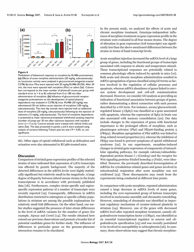
Modulation of behavioral effects of morphine by glucocorticoid receptor blockadeThe influence of GR blockade on locomotor stimulant effectsof morphine and development of physical dependence wasevaluated in the C57BL/6J strain. Acute morphine (20 mg/kg, subcutaneously) treatment induced typical hyper-loco-motion (Figure 6a). Effects of morphine in the GR antagonistRU486-treated (20 mg/kg) mice were significantly attenu-ated compared with those in the vehicle-treated animals (P <0.05). Control groups of animals (treated only with RU486,saline, or vehicle) did not exhibit locomotor stimulation dur-ing the experiment; there were also no statistically significantdifferences between these groups. RU486 treatment did notalter basal locomotor activity (Figure 6a).
A single dose of RU486 (20 mg/kg) was administered 30 minbefore an injection of morphine (100 mg/kg,subcutaneously). On the next day animals were injected withan additional dose of morphine (20 mg/kg, subcutaneously)followed by injection of naloxone (after 3 hours; 15 mg/kg).This treatment scheme resulted in robust physical depend-ence in morphine-treated mice, as revealed by the meannumber of jumps (71.5 ± 9.9 jumps per 15 min). RU486 pre-treatment significantly suppressed naloxone-precipitatedwithdrawal jumping response (21 ± 15.1 jumps per 15 min)compared with the morphine-treated group (P < 0.05; Figure
Genome Biology 2007, 8:R128

http://genomebiology.com/2007/8/6/R128 Genome Biology 2007, Volume 8, Issue 6, Article R128 Korostynski et al. R128.9
com
ment
reviews
reports
refereed researchdepo
sited researchinteractio
nsinfo
rmatio
n
Involvement of glucocorticoid receptor in transcriptional response to morphineFigure 5Involvement of glucocorticoid receptor in transcriptional response to morphine. Inhibition of morphine-induced transcription of selected genes by an antagonist of the glucocorticosteroid receptor, namely RU486. Five experimental groups were compared (see Materials and methods): vehicle and acute subcutaneous morphine 20 mg/kg (Veh/Mor); morphine preceded by injection of 20 mg/kg RU486 (RU20/Mor); morphine preceded by injection of 40 mg/kg RU486 (RU40/Mor); and control groups injected with saline and RU486 (RU40/Sal) or saline and vehicle (Veh/Sal). Gene expression in striatum of C57BL/6J mice was analyzed 4 hours after morphine treatment. The results of quantitative real-time reverse transcription polymerase chain reaction (qPCR) are presented as fold change of control group (Veh/Sal) with standard error. Differences between groups were analyzed by analysis of variance ANOVA following Bonferroni multiple comparison correction (n = 5 to 6; **P < 0.01, ***P < 0.001 versus control group; #P < 0.05, ##P < 0.01 versus morphine).
*****
####
##
Tsc22d3 Nfkbia
**
##
HprtZbtb16
# #
Fol
dch
ange
of c
ontr
olVe
h / S
al
Veh / M
or
RU40 /
Mor
RU40 /
Mor
RU40 /
Sal
Veh
/ Sal
Veh / M
or
RU40 /
Mor
RU40 /
Mor
RU40 /
Sal
Fol
dch
ange
of c
ontr
olF
old
chan
geof
con
trol
Fol
dch
ange
of c
ontr
ol
Veh
/ Sal
Veh / M
or
RU40 /
Mor
RU40 /
Mor
RU40 /
Sal
Veh
/ Sal
Veh / M
or
RU40 /
Mor
RU40 /
Mor
RU40 /
Sal
Genome Biology 2007, 8:R128
R128.10 Genome Biology 2007, Volume 8, Issue 6, Article R128 Korostynski et al. http://genomebiology.com/2007/8/6/R128
6b). Other signs of opioid withdrawal such as defecation andurination were also attenuated in RU486-treated mice.
DiscussionComparison of striatal gene expression profiles of the selectedstrains of mice indicated that expression of 2,870 transcriptswas affected by genetic background. The majority of thedetected differences in the mRNA levels were highly statisti-cally significant but relatively small in the magnitude. A largedegree of disparity between inbred mouse strains in the braintranscriptome is in accordance with previously publisheddata [18]. Furthermore, complex strain-specific and region-specific expression patterns of a number of transcripts wererecently reported [19]. Consequently, differences in mRNAdistribution restricted to subregions or specific cellular popu-lations in striatum are among the possible explanations forrelatively small fold differences. On the other hand, our ear-lier studies suggested the presence of inter-strain differencesin the level of mRNA variants for the number of genes (forexample, Atp1a2 and Comt) [14]. The results obtained hereextend our previous observations and present a broader list ofpotential candidate genes for further study. The influence ofdifferences in particular genes on the genome-phenomeinteraction remains to be elucidated.
In the present study, we analyzed the effects of acute andchronic morphine treatment. Genotype-independent influ-ences of morphine treatment on gene expression profile in thestriatum were evaluated in all four mouse strains. The degreeof alteration in gene expression (618 transcripts) was signifi-cantly less than the above-mentioned differences between thestrains in terms of basal transcript levels.
Acute morphine injection increased the mRNA level of a largegroup of genes, including the functional groups of transcriptsassociated with response to abiotic and temperature stimuli.These transcriptional responses are potentially related tocommon physiologic effects induced by opioids in mice [10].Both acute and chronic morphine administration resulted inmRNA upregulation of genes classified using GO terms as fac-tors involved in the regulation of cellular processes andapoptosis, whereas mRNA abundance of genes linked to nerv-ous system development and cell-cell communicationdecreased. However, we consider GO analysis as providing anindication of functional association between groups of genes,rather demonstrating a direct connection with each processdescribed by a GO term. For instance, serum/glucocorticoid-regulated kinase 3 (Sgk3) was classified as a gene associatedwith apoptosis, whereas the expression of Sgk3 in brain wasalso associated with memory consolidation [20]. Our datainclude changes in the expression of genes with functionalimportance to the prolonged effects of morphine, such asplasminogen activator (Plat) and FK506-binding protein 5(Fkbp5). Morphine upregulation of Plat mRNA was linked todrug-related reward properties [21], whereas the inhibition ofFkbp5 was found to prevent symptoms of opioid withdrawalsyndrome [22]. In our experiments, morphine-inducedchanges in striatal gene expression of components of intracel-lular signaling pathways, for example calcium/calmodulin-dependent protein kinase I γ (Camk1g) and the receptor forWnt signaling proteins frizzled homolog 2 (Fzd2), were iden-tified. However, the previously described downregulation ofmRNAs for cytoskeleton-related genes and genes involved inmitochondrial respiration after acute morphine was notconfirmed [23]. These discrepancies may result from theexperiments being conducted at different time points.
In comparison with acute morphine, repeated administrationcaused a large decrease in mRNA levels of many genes,including the over-represented group of histone transcriptsinvolved in nucleosome assembly and chromatin remodeling.However, remodeling of chromatin was identified as impor-tant regulatory mechanism of cocaine-induced plasticity instriatum [24]. Moreover, one of the genes with decreasedmRNA level after repeated morphine treatment, namely oli-godendrocyte transcription factor 2 (Olig2), was identified asan essential transcriptional regulator in neuron and oli-godendrocyte specification [25] and was recently postulatedto be involved in susceptibility to schizophrenia [26]. In sum-mary, these observations may suggest that chronic morphine-
Modulation of behavioral response to morphine by RU486 pretreatmentFigure 6Modulation of behavioral response to morphine by RU486 pretreatment. (a) Effects of acute morphine administration (20 mg/kg, subcutaneously) on locomotor activity were analyzed in glucocorticoid antagonist-treated C57BL/6J mice. Mice were injected with 20 mg/kg RU486 (RU20). After 30 min, the mice were injected with morphine (Mor) or saline (Sal). Column bars correspond to the mean number of photocell counts per group with standard error (n = 6 to 8) collected from 0 to 180 min after administration of morphine. (b) Influence of glucocorticoid receptor antagonist pretreatment on the development of opioid physical dependence was analyzed in C57BL/6J mice. RU486 (20 mg/kg) was administered 30 min before acute injection of morphine (100 mg/kg, subcutaneously). The next day animals were injected with an additional dose of morphine (20 mg/kg, subcutaneously) following by the injection of naloxone (15 mg/kg, subcutaneously). The level of morphine dependence is presented as mean naloxone-precipitated withdrawal jumping response (number of jumps per 30 min after naloxone injection) with standard error (n = 5 to 6). Control animals were treated with vehicle (Veh) and saline (Sal). The data presented in panels a and b were analyzed using analysis of variance following Tukey's post hoc test (*P < 0.05; ns, not significant).
**
*
(a) (b)RU20
/ Sal
Veh
/ Sal
Sal / S
al
RU20 /
Mor
Veh
/ Mor
Sal / M
or
RU20 /
SalVe
h / S
al
RU20 /
Mor
Veh
/ Mor
Loco
mot
or a
ctiv
ity
[are
a un
der
curv
e /1
80 m
in]
Jum
ping
freq
uenc
y [ju
mps
/15
min
]
ns
Genome Biology 2007, 8:R128

http://genomebiology.com/2007/8/6/R128 Genome Biology 2007, Volume 8, Issue 6, Article R128 Korostynski et al. R128.11
com
ment
reviews
reports
refereed researchdepo
sited researchinteractio
nsinfo
rmatio
n
induced alterations in the striatum are related to cell plastic-ity and structural changes.
One of the main aims of the present study was to investigategenotype-dependent differences in transcriptional responseinduced by morphine. Comparison of the number of genesregulated by morphine across the four strains has indicatedthat acute response is markedly greater in DBA/2J and SWR/J mice than in C57BL/6J and 129P3/J mice. DBA/2J andSWR/J strains exhibited over-representation of metabolism-related genes across the list of regulated transcripts. Bothstrains exhibited greater hypothermic response to single mor-phine administration [10]. Furthermore, acute morphineinduces increase in locomotor activation in C57BL/6J and129P3/J mice, but not in DBA/2J and SWR/J [10]. The find-ings suggest that strain differences in over-expression ofmetabolism-related genes may be associated with behavioralresponses to morphine. These changes in gene expressionmight be among the possible factors that determine develop-ment of morphine-related traits. For instance, DBA/2J andSWR/J strains exhibited low morphine preference, measuredas oral self-administration of morphine [12].
The obtained profiles of gene expression indicate that some ofthe alterations in transcription appear to be related to thephysiologic state of the whole body. Co-expression of threegenes that are typically associated with cellular stress andtemperature stimulus (Hspa1a, Haspa1b, and Dnajb1) wasdetected after morphine treatment. Further analysis of heatshock protein 1b gene expression revealed inter-strain differ-ences in response. The greatest induction of heat shock pro-tein (HSP) mRNAs after acute morphine administration wasobserved in DBA/2J, whereas after chronic morphine treat-ment it was observed in C57BL/6J mice. As mentioned above,acute morphine produces severe hypothermia in mice [10].However, the inter-strain differences in profile of HSP mRNAexpression after acute morphine were negatively related tochanges in body temperature. On the other hand, opioid-induced hypoxia caused by respiratory depression with differ-ent escalation in the strains might be involved in expressionof HSP genes. Increased expression of Hsp70 mRNA in ratbrain was suggested to be a protective mechanism against theharmful effects of opiates [27]. Interestingly, differentresponses in gene expression of Hsp70 were observedbetween rats after active and passive morphine administra-tion [28]. The presented results support previous data andimplicate regulation of HSP gene expression as a potentialmarker of physiologic alterations of homeostatic processesinduced by morphine in brain.
To identify transcriptional response associated with specificopioid-related traits, changes in mRNA level were correlatedwith morphine-induced behavioral traits (Additional data file5). Severe symptoms of opioid withdrawal observed in SWR/J mice were associated with strong transcriptional activationof cytotoxic T lymphocyte-associated protein 2 (Ctla2a) and
methionine adenosyltransferase II (Mat2a) genes after acuteadministration of morphine, whereas changes in gene expres-sion of adenosine A2a receptor (Adora2a) were negativelyrelated to the level of morphine physical dependence acrossthe four inbred mouse strains (Figure 7). A role of functionalactivation of the adenosine A2a receptor in opioid withdrawalwas previously reported [29].
Decreased expression of several genes linked to the transmis-sion of nerve impulse after chronic treatment may be con-nected with the development of tolerance to effects of opioidsobserved in C57BL/6J strain, including morphine-responsivegenes encoding NPY receptor (Npy5r) and GABAa receptorsubunit (Gabrg1). A moderate transcriptional response toacute treatment and a large decrease in the mRNA abundanceof many genes after chronic morphine in C57BL/6J occurtogether with an enhanced preference [30] for and intake ofmorphine in this strain [12]. Increased mRNA abundance ofTSC22 domain family 3 (Tsc22d3) and zinc finger and BTBdomain containing 16 (Zbtb16) transcripts was found to bepositively related to morphine preference (Figure 7).
Strain differences in response to acute morphine were alsodetected in the level of nuclear factor of kappa light chaingene enhancer in B-cells inhibitor α (Nfkbia) mRNA. Signifi-cant induction of Nfkbia gene expression after 4 hours wasdetected in C57BL/6J mice. A similar profile of genetranscription was observed for Tsc22d3. It is well establishedthat gene transcription of Nfkbia as well as Tsc22d3 can bemediated by GR and controlled by the level of glucocorticoids[17,31]. Nuclear receptor GR is a ligand-activated transcrip-tion factor that directly modulates transcription of down-stream genes [32]. Therefore, it has been suggested thatglucocorticoids are potentially factors that are responsible forthe detected increase in expression of these genes. Further-more, we have identified a relatively large group of genes withparallel expression profiles to that of Tsc22d3 (Sgk, Klf15,Fzd2, Gpt2, Rhpn2, Nt5e, and Nfkbia), which may indicatemorphine-induced co-regulation of these genes.
Therefore, the putative involvement of glucocorticoids in themodulation of transcription of these genes was further stud-ied using the GR antagonist RU486. Prior administration ofRU486 blocked morphine-produced induction of Tsc22d3,Zbtb16, and Nfkbia mRNAs. The functional consequences ofthis transcriptional process remain unknown. However, itwas previously shown that GR-dependent transmission influ-ences the behavioral response to morphine in rats [33,34].The present study demonstrated that GR blockade can inhibitan increase in locomotor activity following morphine admin-istration in C57BL/6J mice. Influence of the GR antagonist onthe relatively fast stimulatory effect of morphine on locomo-tion may suggest the involvement of a nongenomic mecha-nism. Nevertheless, several lines of evidence indicate the GRsalso play an important role in the development of opioid-related phenotype. Secretion of glucocorticoids is involved in
Genome Biology 2007, 8:R128

R128.12 Genome Biology 2007, Volume 8, Issue 6, Article R128 Korostynski et al. http://genomebiology.com/2007/8/6/R128
sensitization to activatory effects of morphine [35]. On theother hand, substantial attenuation of the morphine analgesictolerance after administration of RU486 has been reported inrats [36]. Furthermore, our findings indicated involvement ofGRs in the development of morphine physical dependence.Thus, the GR antagonist appears to modify both the behavio-ral and transcriptional responses to morphine. Therefore,
GR-mediated gene expression in the striatum may play a rolein the chronic effects of morphine and could be involved inthe formation of drug-associated behavior. This hypothesis isalso supported by the recent findings that GRs in the nucleusaccumbens are necessary for the development of conditionalplace preference for morphine [37]. The increase in glucocor-ticoid levels induced by acute stress facilitates the consolida-
Morphine-induced changes in gene expression correlated with the behavioral responseFigure 7Morphine-induced changes in gene expression correlated with the behavioral response. Transcriptional and behavioral responses to morphine were compared across the four inbred strains of mice with specific opioid-related phenotype (C57BL/6J, DBA/2J, 129P3/J, and SWR/J). Behavioral data were gathered as described in the Materials and methods section. The results were obtained by using Pearson correlation (Additional data file 5). Correlation between each opioid-related trait and transcriptional response to acute morphine is presented on the left panel along with chronic morphine on the right panel. Positive correlations between changes in gene expression and behavioral response are highlighted by red color (+), and negative ones are highlighted by the blue color (-).
Genome Biology 2007, 8:R128

http://genomebiology.com/2007/8/6/R128 Genome Biology 2007, Volume 8, Issue 6, Article R128 Korostynski et al. R128.13
com
ment
reviews
reports
refereed researchdepo
sited researchinteractio
nsinfo
rmatio
n
tion of memories associated with emotional stimuli [38]. Thegreatest induction of the genes putatively regulated by gluco-corticoids was detected in C57BL/6J mice, an inbred straincharacterized by high preference for morphine as well as alco-hol [12,39].
It is likely that induction of glucocorticoids and further GR-dependent gene expression might also enhance learning ofdrug-related stimuli. It was suggested that serum-glucocorti-coid-inducible kinase (Sgk) is involved in memory consolida-tion of hippocampus-dependent learning [40]. Also,implication of the Nfkbia regulatory protein of nuclear factor-κB in memory formation in mice has already been proposed[41]. Withdrawal from chronic morphine treatment inducedchanges in the mRNA abundance of Sgk and Nfkbia in ratprefrontal cortex [42] and mouse locus coeruleus [22]. Fur-thermore, alterations in expression of Sgk and Nfkbia werealso observed after acute administration of ethanol [43-45].However, shared transcriptional regulation of the genes men-tioned here was not emphasized or directly associated withthe action of glucocorticoids. The potential therapeutic use ofGR antagonists in treatment for drug abuse was previouslysuggested [46]. The present study identified a group of GR-dependent genes that are regulated in response to morphine.Moreover, our results provide new insights into themorphine-induced mechanism of action of glucocorticoids inthe brain.
ConclusionComparison of morphine-induced changes in striatal geneexpression across the four inbred mouse strains indicatednew biologic mechanisms that are potentially involved in theaction of morphine. The results describe strain differences inthe magnitude of transcriptional response to acute treatmentand in the degree of tolerance in gene expression observedafter chronic morphine administration. Further profiling ofgene expression and transcriptional activity are required tocharacterize fully the mechanisms of transcriptional regula-tion and the dynamics of changes in mRNA abundanceinduced by opioids in mice. As a final point, the obtainedresults indicated the participation of several novel molecularfactors in the effects of morphine and suggested a transcrip-tional basis for the well known association between glucocor-ticoids and opioid addiction.
Materials and methodsMiceAdult male (8 to 10 weeks old) 129P3/J (000690), DBA/2J(000671), C57BL/6J (000664), and SWR/J (000689) mice(Jackson Laboratory, Bar Harbor, ME, USA) were housed sixper cage, under a 12 hour dark/light cycle, with free access tofood and water. Animals weighing 20 to 30 g were usedthroughout the experiments. The animal protocols used inthe study were approved by the local Bioethics Commission at
the Institute of Pharmacology, Polish Academy of Sciences(Krakow, Poland).
Morphine treatmentMorphine (morphine hydrochloride; Polfa, Kutno, Poland)was administered subcutaneously. Experimental groups(control, and acute and chronic morphine) consisted of nineanimals from each strain. To obtain the most reliablecomparison, control and acute morphine groups receivedinjections of saline for 4 days at the same time schedule as thechronic group received morphine (Figure 1). On day 5, acutelytreated animals were injected with a single dose of morphineand killed by decapitation after 4 hours. Chronically treatedanimals were injected with increasing doses of morphine for5 days. Mice received morphine thrice daily (09:00 hours,13:00 hours, and 17:00 hours) for 4 days using a dosingschedule of 10, 20, 40, and 40 mg/kg morphine on days 1, 2,3, and 4, respectively. On the last day, a final morphine doseof 40 mg/kg was administered, and 4 hours after the lastinjection the animals were killed. Mice in control groups werekilled 4 hours after the last injection of saline. The dosescheme and time schedule were used in order to maximizestrain differences in response to morphine [10,13,47].
RU486 treatmentFive groups of six C57BL/6J mice were used in the geneexpression experiment with morphine and RU486 (Sigma-Aldrich, St. Louis, MO, USA) treatment. The control groupwas injected subcutaneously with saline and intraperitoneallywith 20% (2-Hydroxypryl)-β-cyclodextrine (vehicle forRU486; Sigma). RU486 (20 or 40 mg/kg in vehicle, intraperi-toneally) was administered 30 min before morphine adminis-tration (20 mg/kg, subcutaneously). The doses of RU486were selected based on a previous report that showed theeffects of peripheral administration on the central nervoussystem [48]. For the qPCR experiment, animals were killed 4hours after the morphine or saline injection.
Behavioral testingAn independent pool of C57BL/6J mice was used in thebehavioral experiments (five to eight animals per experimen-tal group). In the locomotor activity test mice wereindividually placed in the center of a test cage containing pho-tocells (20 cm × 10 cm × 12 cm). The photocells recorded thenumber of beam interruptions in the horizontal plane every15 min over a 4.5 hour period. After 1 hour, the mice wereinjected with RU486 (20 mg/kg in vehicle, subcutaneously).The control group was injected with saline and vehicle. Theinfluence of RU486 (20 mg/kg in vehicle, subcutaneously) onbasal locomotor activity was also evaluated. After 30 min,mice were injected with saline or morphine (20 mg/kg, sub-cutaneously). Measurement of locomotor activity was per-formed for the next 3 hours. The results were calculated as anarea under curve.
Genome Biology 2007, 8:R128

R128.14 Genome Biology 2007, Volume 8, Issue 6, Article R128 Korostynski et al. http://genomebiology.com/2007/8/6/R128
Physical dependence was induced by acute injections of mor-phine (100 mg/kg, subcutaneously). A single RU486 dose (20mg/kg in vehicle, subcutaneously) was administered 30 minbefore the injection of morphine. On the second day micereceived an additional subcutaneous dose of 20 mg/kg mor-phine, followed by a single naloxone dose (15 mg/kg, subcu-taneously) 3 hours later. Mice did not receive RU486 on theday of the test. For mice in control groups, saline was substi-tuted for morphine. Measurement of naloxone-precipitatedwithdrawal was performed as described in previous studies[13]. Mean jump frequency per 15 min was used as the meas-ure of dependence. Behavioral data was analyzed by two-wayanalysis of variance (ANOVA) followed by Bonferroni posthoc test, with RU486 treatment and morphine treatment asthe main factors.
Tissue collection and RNA isolationAfter decapitation, brains were removed from the skulls anddissected rapidly. Samples containing the rostral part of cau-date/putamen plus the nucleus accumbens (referred to as thestriatum) were collected. The samples were placed in individ-ual tubes with the tissue storage reagent RNA later (QiagenInc., Valencia, CA, USA), frozen on dry ice, and stored at -70°C until RNA isolation. Samples were thawed at room tem-perature and homogenized in 1 ml Trizol reagent (Invitrogen,Carlsbad, CA, USA). RNA isolation was performed in accord-ance with the manufacturer's protocol. Quality of the totalRNA was assessed by the intensity of 28S and 18S bands afterdenaturating agarose electrophoresis with SybrGold staining(Molecular Probes, Inc., Eugene, OR, USA) and by thespectrophotometric ratio A260/A280 (1.9 to 2.1). RNA con-centration was measured using the fluorescent reagentRiboGreen (Molecular Probes, Inc.).
Microarray hybridizationTotal RNA from three animals was pooled and further puri-fied using the RNeasy Mini Kit (Qiagen Inc.). The quality ofRNA was additionally determined by chip-based capillaryelectrophoresis using RNA 6000 Nano LabChip Kit andAgilent Bioanalyzer 2100 (Agilent, Palo Alto, CA, USA), andthere was little evidence of degradation products in any of thetotal RNA samples. For each array, independent pools of RNAfrom three animals were prepared. Preparation of cRNA wasperformed according to the protocol provided by Affymetrix(Santa Clara, CA, USA). Total RNA (5 μg) derived from eachpool was converted to double-stranded cDNA using theSuperScript System (Invitrogen) and an oligo(dT24) primercontaining a T7 RNA polymerase promoter site (Genset Oli-gos, La Jolla, CA, USA). Biotin-labeled cRNA was synthesizedfrom cDNA using a BioArray High Yield RNA Transcriptlabelling Kit (ENZO, Diagnostics, Farmingdale, NY, USA) andpurified using a GeneChip Cleanup Sample Module (QiagenInc.). The yield of the in vitro transcription reaction wasdetermined by product absorbance at 260 nm measuredusing NanoDrop ND-1000 (NanoDrop Technologies, Inc.,
Montchanin, DE, USA), and the size of cRNA probes was eval-uated by using the RNA 6000 Nano LabChip Kit (Agilent).
Following labeling, samples were hybridized to the GeneChipTest3 array (Affymetrix) for quality control. FragmentedcRNA (15 μg) was used for hybridization to the GeneChipMouse Genome 430 2.0 arrays (Affymetrix). Arrays werewashed and stained with streptavidin-phycoerythrin (Merck,Darmstadt, Germany) in Fluidic Station 400 (Affymetrix), inaccordance with the standard protocol of the manufacturer.The arrays were scanned using the GeneChip Scanner 3000(Affymetrix). Three biologic replicates of the microarrayswere prepared per experimental group of animals for a totalof 36 arrays.
Microarray quality control and normalizationThe expression data were processed using the GeneChipOperating Software (Affymetrix) to generate MAS5 CEL files.Chip quality was assessed using R 2.3.0 with the simpleaffypackage [49,50]. Quality control data for all 36 microarrayruns in the experiment were obtained using the MAS5 algo-rithm. The mean 3'/5' degradation ratio for control probe setsof housekeeping genes was measured: Gapdh (AFFX-Gapdh-Mur/M32599) -0.2 (-0.39> ... <0.57) and Actb (AFFX-b-Act-inMur/M12481) 0.57 (0.33> ... <1.24). Array normalizationresulted in a mean scaling factor of 0.87 (± 0.57). The meanpercentage of the present call was 51.34 (47.3> ... <54.63),and the mean intensity of the average background rangedfrom 27.9 to 76.9. Arrays determined to be acceptable werefurther analyzed to identify genes with altered expression pat-terns. Data was normalized using three different methodsusing RMAExpress 0.4.1 (RMA), dChip 2006 (MBEI), andPerfectMatch 2.3.3 (PDNN) software [51-53]. Quantile nor-malization was performed using RMA, PDNN, and MBEIalgorithms. A model-based expression index was calculatedusing perfect match/mismatch (PM/MM) method. Multipleanalysis strategies were used to identify the most robustchanges in gene expression.
Gene filtering and rankingTo remove genes that are regarded as not expressed in theanalyzed brain tissue, probe sets with hybridization signalsclose to the background level were filtered out. The followingcriteria were applied for probe set detection: present call atleast in 25% of PM/MM pairs and signal intensity greaterthan 6.64 (log2 data) in at least 25% of arrays, both measuredusing the MBEI algorithm. Statistical analysis was performedon the list of detected probe sets. Significance levels (P values)of differences between the groups were calculated for eachprobe set using a MANOVA for each of three pre-processingmethods. Correction for multiple testing was applied sepa-rately for each of the main factors from MANOVA by control-ling percentage FDR. The FDR was computed using thep.adjust function in R software [54]. Gene ranking was basedon FDR levels of three selected normalization methods. Aprobe set scored 1, 2, or 3 points if it achieved cut-off at P <
Genome Biology 2007, 8:R128

http://genomebiology.com/2007/8/6/R128 Genome Biology 2007, Volume 8, Issue 6, Article R128 Korostynski et al. R128.15
com
ment
reviews
reports
refereed researchdepo
sited researchinteractio
nsinfo
rmatio
n
0.01, P < 0.001, or P < 0.0001, respectively, for each of thethree methods. Two criteria for threshold point were used:the first produced a list of probe sets with rank greater than 3(P < 0.01 in all the three normalization methods) and the sec-ond list with rank greater than 7 (P < 0.0001 in two of themethods and at least 0.001 in the third). An applied approachthat takes into account the level of agreement between thethree methods of normalization was previously established[14].
To assess the contribution of strain and morphine treatmenteffects, we performed multiple regression analysis of expres-sion values, with strain and treatment as main effect predic-tors using the lm function in R. Contributions of the effectswere determined for each gene separately.
Hierarchical clustering was performed with dChip softwareusing Euclidean distance and average linkage method. Rela-tive expression levels and fold change measures were com-puted from MBEI PM/MM data. To simplify the descriptionof microarray data, results from each probe set were assumedto be the level of mRNA abundance of the transcript. Probesets on the Affymetrix microarray were designed to detectspecific transcripts and are adequately annotated. However,it must be noted that for some probe sets this assumption maynot be fulfilled. Therefore, every list of probe sets obtained inthe analyses is included in Additional data files. Direct probemapping against publicly available mRNAs/cDNA sequenceswas done by using mouse BLAT tool for the UCSC genomebrowser [55].
Gene Ontology analysisThe functional annotation analysis tool DAVID 2006 wasused to identify over-represented ontologic groups among thegene expression profiles and to group genes into functionalclassifications [56]. The list of 21,467 detected probe sets wasuploaded as a background list. Over-represented GO terms(GOTERM_ALL level) were defined as having at least threetranscripts and P = 0.01, under Fisher's exact test.
Correlation between genomic and behavioral response to morphineRelationships between the transcriptional response to mor-phine and opioid-related traits were studied using correlationanalysis. Gene expression results were compared with thepreviously published behavioral data for the four inbredmouse strains. The first line of evidence demonstrated openfield locomotor activity in response to acute morphine (16mg/kg), measured from 0 to 30 min after injection, and com-pared with a saline control group (C57BL/6J = 4,116, 129P3/J = 5,880, SWR/J = -1,500, and DBA/2J = -3,088); this wasreported by Belknap and coworkers [10]. Second, morphinetolerance has been demonstrated, calculated as a shift in mor-phine analgesic potency after chronic treatment (C57BL/6J =7.2, SWR/J = 5, DBA/2J = 2.9, and 129P3/J = 0.8); this wasreported by Kest and colleagues [47]. Third, physical depend-
ence on morphine has been demonstrated following chronictreatment, as indicated by a naloxone precipitated with-drawal jumping response (SWR/J = 204 jumps/15 min,C57BL/6J = 68 jumps/15 min, DBA/2J = 45 jumps/15 min,and 129P3/J = 7 jumps/15 min); these data were reported byKest and coworkers [13]. The fourth line of evidence demon-strated voluntary morphine consumption in two-bottlechoice paradigm (C57BL/6J = 134 mg/kg per day, 129P3/J =24 mg/kg per day, DBA/2J = 15 mg/kg per day, and SWR/J =6 mg/kg per day); these ata were reported by Belknap andcolleagues 1993 [12]. The expression level of genes altered byacute and/or chronic morphine (699 probe sets, obtained byMBEI PM/MM) and behavioral data were normalized usingz-score transformation. Associations were computed usingthe Pearson's correlation. Input data, annotations for probesets, and the obtained results are included in Additional datafile 5.
Identification of genes co-expressed in response to morphineGenes with related patterns of expression were selected fromthe list of probe sets with significant changes after morphinetreatment. To confirm associations between the transcripts,additional analysis was performed on an independent set ofdata. The WebQTL database was used to assemble transcriptsinto groups with potentially common regulatory mechanisms[16]. A hippocampus gene expression dataset with resultsfrom 86 BXD RI strains (Hippocampus Consortium M430v2Dec05 PDNN) was applied as an easily accessible tool withrelatively high ability to detect associations between genes.Selected probe sets for genes regulated by morphine wereanalyzed using the trait correlation WebQTL tool. Correla-tions were computed using Pearson's product-moment. Themost significant top 200 results were obtained. Only probesets with positive correlations (r > 0.6) were extracted andused in further analyses. A list of probe sets with significantchanges after morphine and positive correlation across theBXD RI panel was generated. Network graphs were obtainedwith WebQTL interface using Pearson's product-moment anddefault parameters.
Real-time PCRReverse transcription was performed using Omniscriptreverse transcriptase (Qiagen Inc.) at 37°C for 60 min. qPCRreactions were performed using Assay-On-Demand Taqmanprobes (Additional data file 4), in accordance with the manu-facturer's protocol (Applied Biosystems) and run on the iCy-cler device (BioRad) with the 3.0a software version. RTreactions were carried out in the presence of RNase inhibitor(rRNAsin; Promega, Madison, WI, USA) and oligo(dT16)primer (Qiagen Inc.). cDNAs were diluted 1:6 with H2O andfor each reaction about 50 ng of cDNA synthesized from totalRNA template from individual animals was used. To reducethe contribution of contaminating genomic DNA, primerswere designed to span exon junctions. In addition, for eachassay, control reactions without RT enzyme were performed.
Genome Biology 2007, 8:R128
R128.16 Genome Biology 2007, Volume 8, Issue 6, Article R128 Korostynski et al. http://genomebiology.com/2007/8/6/R128
Amplification efficiency for each assay was determined byrunning a standard dilution curve. Expression of hypoxan-thine guanine phosphoribosyl transferase 1 (Hprt1) tran-script with a stable level between the strains and after thetreatment was quantified to control for variation in cDNAamounts. The cycle threshold values were calculated auto-matically by iCycler IQ 3.0a software with default parameters.Abundance of RNA was calculated as 2-(thresholdcycle). Datawere analyzed by two-way ANOVA followed by Bonferronipost hoc test.
Additional data filesThe following additional data are available with the onlineversion of this manuscript. Additional data file 1 is a table list-ing the results of two-way ANOVA (FDR < 1%). Additionaldata file 2 contains lists of probe sets of genes with expressionaltered by acute and chronic morphine (ANOVA; FDR < 1%).Additional data file 3 is a table listing the results of the GOanalysis. Additional data file 4 contains the results of valida-tion of microarray data obtained using the qPCR method.Additional data file 5 lists the complete results of correlationanalysis between the transcriptional response to morphineand opioid-related traits (see Materials and methods).Additional data file 1Results of two-way ANOVA (FDR < 1%)Lists of probe sets and gene names altered by strain (3457) and morphine treatment (661) as well as with significant interaction (48) are available as separate sheets. List of transcripts with differ-ences in both the factors (178) was also included. In addition, genes reported in previous gene expression studies on morphine action in a brain are indicated.Click here for fileAdditional data file 2Probe sets of genes with expression altered by acute and chronic morphine (ANOVA, FDR < 1%)The results were obtained using fold change (>1.2) of gene expres-sion level compared with the saline control group. Effects of mor-phine treatment were analyzed in all of the strains as well as separately in each of them. Numbers of probe sets correspond to those in Table 2.Click here for fileAdditional data file 3Results of the Gene Ontology analysisPresented are significant functional categories (GO terms) enriched with genes regulated in response to acute and/or chronic morphine, along with lists of probe sets and gene names classified to each GO category.Click here for fileAdditional data file 4Results of validation of microarray data obtained using qPCR methodResults for selected genes are presented as mean (± standard error) compared with the saline control group. List of Taqman assays used in qPCR experiments with ID and exon boundaries is included.Click here for fileAdditional data file 5Complete results of correlation analysis between the transcrip-tional response to morphine and opioid-related traitsThe expression level (MBEI PM/MM algorithm) of genes altered by acute and/or chronic morphine (699 probe sets) and behavioral data were normalized using z-score transformation. Associations were computed using the Pearson's correlation.Click here for file
AcknowledgementsWe thank Barbara Ziolkowska and Agnieszka Gieryk for their help with thetissue preparation, and Jakub Kubik for his assistance during the behavioralstudies. We should like acknowledge the GeneNetwork project for creat-ing and maintaining the WebQTL database. This work was supported by EUgrant LSHM-CT-2004-005166, MNiSzW subsidiary grant 26/E-40/6.PR UE/DIE 305/2005-2008, and PBZ-MNiI-2/1/2005 grant.
References1. Nestler EJ, Aghajanian GK: Molecular and cellular basis of
addiction. Science 1997, 278:58-63.2. Koob GF, Sanna PP, Bloom FE: Neuroscience of addiction. Neu-
ron 1998, 21:467-476.3. Fadda P, Scherma M, Fresu A, Collu M, Fratta W: Dopamine and
serotonin release in dorsal striatum and nucleus accumbensis differentially modulated by morphine in DBA/2J andC57BL/6J mice. Synapse 2005, 56:29-38.
4. Wise RA: The role of reward pathways in the development ofdrug dependence. Pharmacol Ther 1987, 35:227-263.
5. Volkow ND, Wang GJ, Telang F, Fowler JS, Logan J, Childress AR,Jayne M, Ma Y, Wong C: Cocaine cues and dopamine in dorsalstriatum: mechanism of craving in cocaine addiction. JNeurosci 2006, 26:6583-6588.
6. Przewlocki R: Opioid abuse and brain gene expression. Eur JPharmacol 2004, 500:331-349.
7. Rhodes JS, Crabbe JC: Gene expression induced by drugs ofabuse. Curr Opin Pharmacol 2005, 5:26-33.
8. Kreek MJ, Bart G, Lilly C, LaForge KS, Nielsen DA: Pharmacoge-netics and human molecular genetics of opiate and cocaineaddictions and their treatments. Pharmacol Rev 2005, 57:1-26.
9. Belknap JK, Noordewier B, Lame M: Genetic dissociation of mul-tiple morphine effects among C57BL/6J, DBA/2J and C3H/HeJ inbred mouse strains. Physiol Behav 1989, 46:69-74.
10. Belknap JK, Riggan J, Cross S, Young ER, Gallaher EJ, Crabbe JC:Genetic determinants of morphine activity and thermalresponses in 15 inbred mouse strains. Pharmacol Biochem Behav1998, 59:353-360.
11. Semenova S, Kuzmin A, Zvartau E: Strain differences in the anal-gesic and reinforcing action of morphine in mice. Pharmacol
Biochem Behav 1995, 50:17-21.12. Belknap JK, Crabbe JC, Riggan J, O'Toole LA: Voluntary consump-
tion of morphine in 15 inbred mouse strains. Psychopharmacol-ogy (Berl) 1993, 112:352-358.
13. Kest B, Palmese CA, Hopkins E, Adler M, Juni A, Mogil JS: Naloxone-precipitated withdrawal jumping in 11 inbred mouse strains:evidence for common genetic mechanisms in acute andchronic morphine physical dependence. Neuroscience 2002,115:463-469.
14. Korostynski M, Kaminska-Chowaniec D, Piechota M, Przewlocki R:Gene expression profiling in the striatum of inbred mousestrains with distinct opioid-related phenotypes. BMC Genomics2006, 7:146.
15. Gene Expression Omnibus [http://www.ncbi.nlm.nih.gov/geo]16. Chesler EJ, Lu L, Shou S, Qu Y, Gu J, Wang J, Hsu HC, Mountz JD,
Baldwin NE, Langston MA, et al.: Complex trait analysis of geneexpression uncovers polygenic and pleiotropic networks thatmodulate nervous system function. Nat Genet 2005,37:233-242.
17. D'Adamio F, Zollo O, Moraca R, Ayroldi E, Bruscoli S, Bartoli A, Can-narile L, Migliorati G, Riccardi C: A new dexamethasone-inducedgene of the leucine zipper family protects T lymphocytesfrom TCR/CD3-activated cell death. Immunity 1997, 7:803-812.
18. Hovatta I, Zapala MA, Broide RS, Schadt EE, Libiger O, Schork NJ,Lockhart DJ, Barlow C: DNA variation and brain region-specificexpression profiles exhibit different relationships betweeninbred mouse strains: implications for eQTL mappingstudies. Genome Biol 2007, 8:R25.
19. Nadler JJ, Zou F, Huang H, Moy SS, Lauder J, Crawley JN, ThreadgillDW, Wright FA, Magnuson TR: Large scale gene expression dif-ferences across brain regions and inbred strain correlatewith behavioral phenotype. Genetics 2006, 174:1229-1236.
20. von Hertzen LS, Giese KP: Memory reconsolidation engagesonly a subset of immediate-early genes induced duringconsolidation. J Neurosci 2005, 25:1935-1942.
21. Nagai T, Yamada K, Yoshimura M, Ishikawa K, Miyamoto Y, Hashim-oto K, Noda Y, Nitta A, Nabeshima T: The tissue plasminogenactivator-plasmin system participates in the rewardingeffect of morphine by regulating dopamine release. Proc NatlAcad Sci USA 2004, 101:3650-3655.
22. McClung CA, Nestler EJ, Zachariou V: Regulation of gene expres-sion by chronic morphine and morphine withdrawal in thelocus ceruleus and ventral tegmental area. J Neurosci 2005,25:6005-6015.
23. Loguinov AV, Anderson LM, Crosby GJ, Yukhananov RY: Geneexpression following acute morphine administration. PhysiolGenomics 2001, 6:169-181.
24. Kumar A, Choi KH, Renthal W, Tsankova NM, Theobald DE, TruongHT, Russo SJ, Laplant Q, Sasaki TS, Whistler KN, et al.: Chromatinremodeling is a key mechanism underlying cocaine-inducedplasticity in striatum. Neuron 2005, 48:303-314.
25. Ligon KL, Fancy SP, Franklin RJ, Rowitch DH: Olig gene function inCNS development and disease. Glia 2006, 54:1-10.
26. Georgieva L, Moskvina V, Peirce T, Norton N, Bray NJ, Jones L, Hol-mans P, Macgregor S, Zammit S, Wilkinson J, et al.: Convergent evi-dence that oligodendrocyte lineage transcription factor 2(OLIG2) and interacting genes influence susceptibility toschizophrenia. Proc Natl Acad Sci USA 2006, 103:12469-12474.
27. Ammon-Treiber S, Grecksch G, Stumm R, Riechert U, Tischmeyer H,Reichenauer A, Hollt V: Rapid, transient, and dose-dependentexpression of hsp70 messenger RNA in the rat brain aftermorphine treatment. Cell Stress Chaperones 2004, 9:182-197.
28. Rodriguez Parkitna JM, Bilecki W, Mierzejewski P, Stefanski R, LigezaA, Bargiela A, Ziolkowska B, Kostowski W, Przewlocki R: Effects ofmorphine on gene expression in the rat amygdala. JNeurochem 2004, 91:38-48.
29. Bailey A, Davis L, Lesscher HM, Kelly MD, Ledent C, Hourani SM,Kitchen I: Enhanced morphine withdrawal and micro-opioidreceptor G-protein coupling in A2A adenosine receptorknockout mice. J Neurochem 2004, 88:827-834.
30. Orsini C, Bonito-Oliva A, Conversi D, Cabib S: Susceptibility toconditioned place preference induced by addictive drugs inmice of the C57BL/6 and DBA/2 inbred strains. Psychopharma-cology (Berl) 2005, 181:327-336.
31. Scheinman RI, Cogswell PC, Lofquist AK, Baldwin AS Jr: Role oftranscriptional activation of I kappa B alpha in mediation ofimmunosuppression by glucocorticoids. Science 1995,270:283-286.
Genome Biology 2007, 8:R128
http://genomebiology.com/2007/8/6/R128 Genome Biology 2007, Volume 8, Issue 6, Article R128 Korostynski et al. R128.17
com
ment
reviews
reports
refereed researchdepo
sited researchinteractio
nsinfo
rmatio
n
32. Phuc Le P, Friedman JR, Schug J, Brestelli JE, Parker JB, Bochkis IM,Kaestner KH: Glucocorticoid receptor-dependent gene regu-latory networks. PLoS Genet 2005, 1:e16.
33. Marinelli M, Aouizerate B, Barrot M, Le Moal M, Piazza PV:Dopamine-dependent responses to morphine depend onglucocorticoid receptors. Proc Natl Acad Sci USA 1998,95:7742-7747.
34. Stohr T, Almeida OF, Landgraf R, Shippenberg TS, Holsboer F, Span-agel R: Stress- and corticosteroid-induced modulation of thelocomotor response to morphine in rats. Behav Brain Res 1999,103:85-93.
35. Deroche V, Marinelli M, Maccari S, Le Moal M, Simon H, Piazza PV:Stress-induced sensitization and glucocorticoids. I. Sensiti-zation of dopamine-dependent locomotor effects of amphet-amine and morphine depends on stress-inducedcorticosterone secretion. J Neurosci 1995, 15:7181-7188.
36. Lim G, Wang S, Zeng Q, Sung B, Mao J: Spinal glucocorticoidreceptors contribute to the development of morphine toler-ance in rats. Anesthesiology 2005, 102:832-837.
37. Dong Z, Han H, Wang M, Xu L, Hao W, Cao J: Morphine condi-tioned place preference depends on glucocorticoid recep-tors in both hippocampus and nucleus accumbens.Hippocampus 2006, 16:809-813.
38. de Kloet ER, Oitzl MS, Joels M: Stress and cognition: are corti-costeroids good or bad guys? Trends Neurosci 1999, 22:422-426.
39. Meliska CJ, Bartke A, McGlacken G, Jensen RA: Ethanol, nicotine,amphetamine, and aspartame consumption and preferencesin C57BL/6 and DBA/2 mice. Pharmacol Biochem Behav 1995,50:619-626.
40. Tsai KJ, Chen SK, Ma YL, Hsu WL, Lee EH: sgk, a primary gluco-corticoid-induced gene, facilitates memory consolidation ofspatial learning in rats. Proc Natl Acad Sci USA 2002, 99:3990-3995.
41. Meffert MK, Chang JM, Wiltgen BJ, Fanselow MS, Baltimore D: NF-kappa B functions in synaptic signaling and behavior. NatNeurosci 2003, 6:1072-1078.
42. Ammon S, Mayer P, Riechert U, Tischmeyer H, Hollt V: Microarrayanalysis of genes expressed in the frontal cortex of ratschronically treated with morphine and after naloxone pre-cipitated withdrawal. Brain Res Mol Brain Res 2003, 112:113-125.
43. Kerns RT, Ravindranathan A, Hassan S, Cage MP, York T, Sikela JM,Williams RW, Miles MF: Ethanol-responsive brain regionexpression networks: implications for behavioral responsesto acute ethanol in DBA/2J versus C57BL/6J mice. J Neurosci2005, 25:2255-2266.
44. Rulten SL, Ripley TL, Hunt CL, Stephens DN, Mayne LV: Sp1 andNFkappaB pathways are regulated in brain in response toacute and chronic ethanol. Genes Brain Behav 2006, 5:257-273.
45. Treadwell JA, Singh SM: Microarray analysis of mouse braingene expression following acute ethanol treatment. Neuro-chem Res 2004, 29:357-369.
46. Deroche-Gamonet V, Sillaber I, Aouizerate B, Izawa R, Jaber M, Gho-zland S, Kellendonk C, Le Moal M, Spanagel R, Schutz G, et al.: Theglucocorticoid receptor as a potential target to reducecocaine abuse. J Neurosci 2003, 23:4785-4790.
47. Kest B, Hopkins E, Palmese CA, Adler M, Mogil JS: Genetic varia-tion in morphine analgesic tolerance: a survey of 11 inbredmouse strains. Pharmacol Biochem Behav 2002, 73:821-828.
48. Xu L, Holscher C, Anwyl R, Rowan MJ: Glucocorticoid receptorand protein/RNA synthesis-dependent mechanisms underliethe control of synaptic plasticity by stress. Proc Natl Acad SciUSA 1998, 95:3204-3208.
49. Wilson C, Miller CJ: Simpleaffy: a BioConductor package forAffymetrix quality control and data analysis. Bioinformatics2005, 21:3683-3685.
50. Gentleman RC, Carey VJ, Bates DM, Bolstad B, Dettling M, Dudoit S,Ellis B, Gautier L, Ge Y, Gentry J, et al.: Bioconductor: open soft-ware development for computational biology andbioinformatics. Genome Biol 2004, 5:R80.
51. Irizarry RA, Bolstad BM, Collin F, Cope LM, Hobbs B, Speed TP:Summaries of Affymetrix GeneChip probe level data. NucleicAcids Res 2003, 31:e15.
52. Li C, Wong WH: Model-based analysis of oligonucleotidearrays: expression index computation and outlier detection.Proc Natl Acad Sci USA 2001, 98:31-36.
53. Zhang L, Miles MF, Aldape KD: A model of molecularinteractions on short oligonucleotide microarrays. NatBiotechnol 2003, 21:818-821.
54. Benjamini Y, Hochberg Y: Controlling the false discovery rate: a
practical and powerful approach to multiple testing. J Roy StatSoc B 1995, 57:289-300.
55. Kent WJ: BLAT: the BLAST-like alignment tool. Genome Res2002, 12:656-664.
56. Dennis G Jr, Sherman BT, Hosack DA, Yang J, Gao W, Lane HC, Lem-picki RA: DAVID: Database for Annotation, Visualization, andIntegrated Discovery. Genome Biol 2003, 4:P3.
Genome Biology 2007, 8:R128