dynamic transcriptome profiling of mungbean genotypes
TRANSCRIPT
�����������������
Citation: Sudha, M.; Karthikeyan, A.;
Madhumitha, B.; Veera Ranjani, R.;
Kanimoli Mathivathana, M.;
Dhasarathan, M.; Murukarthick, J.;
Samu Shihabdeen, M.N.; Eraivan
Arutkani Aiyanathan, K.; Pandiyan,
M.; et al. Dynamic Transcriptome
Profiling of Mungbean Genotypes
Unveil the Genes Respond to the
Infection of Mungbean Yellow
Mosaic Virus. Pathogens 2022, 11, 190.
https://doi.org/10.3390/
pathogens11020190
Academic Editor:
Massimiliano Morelli
Received: 22 November 2021
Accepted: 21 January 2022
Published: 30 January 2022
Publisher’s Note: MDPI stays neutral
with regard to jurisdictional claims in
published maps and institutional affil-
iations.
Copyright: © 2022 by the authors.
Licensee MDPI, Basel, Switzerland.
This article is an open access article
distributed under the terms and
conditions of the Creative Commons
Attribution (CC BY) license (https://
creativecommons.org/licenses/by/
4.0/).
pathogens
Article
Dynamic Transcriptome Profiling of Mungbean GenotypesUnveil the Genes Respond to the Infection of MungbeanYellow Mosaic VirusManickam Sudha 1,*,†, Adhimoolam Karthikeyan 2,† , Balasubramaniam Madhumitha 3,Rajagopalan Veera Ranjani 1 , Mayalagu Kanimoli Mathivathana 4, Manickam Dhasarathan 5,Jayakodi Murukarthick 6 , Madiha Natchi Samu Shihabdeen 1 , Karuppiah Eraivan Arutkani Aiyanathan 7,Muthaiyan Pandiyan 8, Natesan Senthil 9 and Muthurajan Raveendran 1
1 Department of Plant Biotechnology, Centre for Plant Molecular Biology and Biotechnology, Tamil NaduAgricultural University, Coimbatore 641003, Tamil Nadu, India; [email protected] (R.V.R.);[email protected] (M.N.S.S.); [email protected] (M.R.)
2 Department of Biotechnology, Centre of Innovation, Agricultural College and Research Institute, Tamil NaduAgricultural University, Madurai 625104, Tamil Nadu, India; [email protected]
3 Department of Plant Pathology, Agricultural College and Research Institute, Tamil Nadu AgriculturalUniversity, Madurai 625104, Tamil Nadu, India; [email protected]
4 Department of Plant Breeding and Genetics, Agricultural College and Research Institute, Tamil NaduAgricultural University, Madurai 625104, Tamil Nadu, India; [email protected]
5 Agroclimate Research Centre, Directorate of Crop Management, Tamil Nadu Agricultural University,Coimbatore 641003, Tamil Nadu, India; [email protected]
6 Gene Bank, Leibniz Institute of Plant Genetics and Crop Plant Research (IPK) Stadt See land,06466 Seeland, OT Gatersleben, Germany; [email protected]
7 Agricultural College and Research Institute, Tamil Nadu Agricultural University,Killikulam 628252, Tamil Nadu, India; [email protected]
8 Regional Research Station, Tamil Nadu Agricultural University, Virudhachalam 606001, Tamil Nadu, India;[email protected]
9 Department of Plant Molecular Biology and Bioinformatics, Centre for Plant Molecular Biology andBiotechnology, Tamil Nadu Agricultural University, Coimbatore 641003, Tamil Nadu, India;[email protected]
* Correspondence: [email protected]† These authors contributed equally to this work.
Abstract: Yellow mosaic disease (YMD), incited by mungbean yellow mosaic virus (MYMV), is aprimary viral disease that reduces mungbean production in South Asia, especially in India. Thereis no detailed knowledge regarding the genes and molecular mechanisms conferring resistance ofmungbean to MYMV. Therefore, disclosing the genetic and molecular bases related to MYMV resis-tance helps to develop the mungbean genotypes with MYMV resistance. In this study, transcriptomesof mungbean genotypes, VGGRU-1 (resistant) and VRM (Gg) 1 (susceptible) infected with MYMVwere compared to those of uninfected controls. The number of differentially expressed genes (DEGs)in the resistant and susceptible genotypes was 896 and 506, respectively. Among them, 275 DEGswere common between the resistant and susceptible genotypes. Functional annotation of DEGsrevealed that the DEGs belonged to the following categories defense and pathogenesis, receptor-likekinases; serine/threonine protein kinases, hormone signaling, transcription factors, and chaperons,and secondary metabolites. Further, we have confirmed the expression pattern of several DEGs byquantitative real-time PCR (qRT-PCR) analysis. Collectively, the information obtained in this studyunveils the new insights into characterizing the MYMV resistance and paved the way for breedingMYMV resistant mungbean in the future.
Keywords: mungbean; RNA seq; whitefly; yellow mosaic disease
Pathogens 2022, 11, 190. https://doi.org/10.3390/pathogens11020190 https://www.mdpi.com/journal/pathogens
Pathogens 2022, 11, 190 2 of 15
1. Introduction
Yellow mosaic disease (YMD) is a major virus disease, and its incidence has becomesevere in the past three decades throughout most mungbean (Vigna radiata) producingregions in South Asia and particularly in India. Three different begomoviruses, i.e., mung-bean yellow mosaic virus (MYMV), mungbean yellow mosaic India virus (MYMIV), andhorse gram yellow mosaic virus (HgYMV), have been found to cause YMD in variousmungbean producing regions of Asia [1]. MYMV and MYMIV cause the YMD in India.It was reported that MYMV is primarily in India’s southern region, while MYMIV is inthe northern, central, and eastern regions of India [2,3]. The viruses are spread by whiteflyin a persistent and circulative manner but cannot be transmitted mechanically throughsap or seed. The effectiveness of whitefly transmission and behavior varies with that ofthe genotype and growth stage and virus strains [4]. The most common virus symptomsinclude yellowing or chlorosis of the leaves preceded by necrosis, fewer flowers and pods,and pods containing immature and abnormal seeds and stunting plants. Mungbean plantsinfected within 3 weeks of sowing may reduce yield up to 85%. So far, many investigationson the mungbean-MYMV have concentrated on the occurrence, characterization of isolates,symptoms, transmission and epidemiology, chemical and biological control of the virusreviewed by Karthikeyan et al. [5]. The use of mungbean cultivars resistant to MYMVhas long been considered an effective and economical way to control the virus [6]. Manystudies evaluated the mungbean germplasm for resistance to MYMV. However, only afew germplasm were found to be resistant [4,7–9]. Despite the advances in decipheringthe mungbean genome, limited information is known about the genes and mechanismsunderlying mungbean resistance to MYMV, which is essential for developing effectivecontrol methods. Therefore, comprehensive knowledge of mungbean responses to MYMVinfection is needed for developing methods for the management of virus.
Mungbean responses to the virus are complex and associated with the numerousbiological and physiological processes involving the up- or down-regulation of genes. Thediscovery of the differentially expressed genes (DEGs) regulating the mungbean defenseresponse to MYMV is vital in understanding genes and molecular mechanisms associatedwith resistance. This information is helpful for mungbean researchers to understand thecomplex interactions between mungbean and the virus. Taking advantage of second andthird-generation sequencing technologies, comparative transcriptome analysis throughRNA sequencing (RNA seq) is the most popular method for detecting DEGs betweentwo models [10–13]. Moreover, transcriptome analysis provides detailed informationto understand the dynamics of interaction between the host and pathogen [14,15]. Bycomparing the transcriptome data from the pathogen-infected and control, countless studieshave been attempted to elucidate the complete details of the genes and pathways involvedin the molecular mechanism of resistance to pathogens [16–18].
Recently, transcriptome comparisons among resistant and susceptible genotypes topathogens in various crops, including mungbean [19], urdbean [20,21], soybean [22,23],rice [24,25], and tomato [16] extended our knowledge on the genes and mechanisms under-lying the resistance to plant pathogens. In regard to the available literature information,RNA-Seq has yet to be used to investigate the transcriptome response of mungbean toMYMV. In the present investigation, the transcriptome response of two mungbean geno-types to MYMV was analysed using RNA seq. Our study identified specific and mutualDEGs and unveiled the different responses to MYMV infection in these two mungbeangenotypes. This information has given new insights into MYMV-resistant and paved theway for breeding MYMV resistance in the future.
2. Materials and Methods2.1. Genotype Panel and Pathogen Inoculation
Two mungbean genotypes were received from Agricultural Research Station, TamilNadu Agricultural University, Virinjupuram, India, i.e., MYMV-resistant genotype ‘VGGRU-1’ and the susceptible genotype ‘VRM (Gg) 1” [4]. Healthy seeds of both genotypes were
Pathogens 2022, 11, 190 3 of 15
agroinoculated with infectious virus construct VA 239 (KA30 DNA A + KA27 DNA B) [26]and raised in a plant growth chamber at ideal condition [Temperature (25 ◦C), relativehumidity level (60–70%) and light/dark photoperiod (16/8 h)]. Agroinoculation, assess-ment of MYMV symptoms, and virus detection was performed following the proceduredetailed by Karthikeyan et al. [6] and Sudha et al. [4,27]. The agroinoculation screening ofmungbean genotypes was completed thrice.
2.2. Library Preparation and Illumina Sequencing
Leaves were collected at mock and inoculated plants at 25- and 40-days post-inoculation(DPI) and finely ground using liquid nitrogen and stored at −70 ◦C. For the constructionof the cDNA libraries, the 25 and 40 DPI leaves were pooled together for RNA isolationand made as an infection library following the pooled cDNA library construction proce-dure [28], while leaves were uninoculated (Control) served as a non-infection library. TotalRNA was isolated by the RNeasy plant mini kit (Qiagen, Hilden, Germany) followingthe user guidelines and treated with RNase-free DNAseI (Promega, Madison, WI, USA).RNA quantity and quality were determined by the bio spectrometer (Eppendorf, Hamburg,Germany) based on the absorbance ratio at 260 nm and 280 nm. The RNA integrity value(RIN) of the samples was confirmed by the Agilent Bioanalyzer 2100 system (AgilentTechnologies, Santa Clara, CA, USA). Four cDNA libraries derived from VGGRU-1 control,VGGRU-1 infected, VRM (Gg) 1 control, and VRM (Gg) 1 infected were constructed byIllumina TruSeq RNA sample preparation kit following the user guidelines (Illumina Inc.,SanDiego, CA, USA). Four cDNA libraries were sequenced by IlluminaHiSeq2000 platformwith paired-end (PE) reads of 101 bp at Phyzen Genomics Institute, Seoul, South Korea.
2.3. Reads Filtering and Mapping
Before alignment, raw reads obtained from each library were filtered to get cleanhigh-quality reads by removing low-quality reads. The clean high-quality reads wereobtained by following the method with three steps: in the first step, bacterial contaminantswere removed from raw reads by mapping onto the available bacterial genomes through aburrows wheeler aligner (BWA) [29]. The second step included PCR copies, and ribosomal(rRNA) reads filtering by FastUniq [30] and SortMeRNA [31], respectively, and the uncom-promising quality control and taking out the adapter contamination by NGS QC Toolkit(v2.3.3) was followed in the third step [32]. The clean high-quality reads were mapped to themungbean reference genome (https://legumeinfo.org/genomes/gbrowse/Vr1.0; accessedon 6 February 2018) using burrows-wheeler aligner (BWA) [29] with default parameters.
2.4. Analysis of Differentially Expressed Genes
The expression levels of the gene transcripts were measured using fragments perKilobase per million (FPKM) values estimated by RNA-Seq by Expectation-Maximization(RSEM) [33]. DEGs in VGGRU-1 control vs VGGRU-1 infected, and VRM (Gg) 1 controlvs VRM (Gg) 1 infected were identified by Bioconductor package edgeR [34] using thethreshold of false discovery rate (FDR) of ≤0.001 and the absolute value of Log2 fold-change ≥2. DEGs were signified in Venn diagrams using VENNY v.2.1 (http://bioinfogp.cnb.csic.es/tools/venny/; accessed on 18 August 2018). To investigate the function of theDEG transcripts, gene ontology (GO) analysis was conducted by the BLAST2GO softwareprogram (http://www.blast2go.org; accessed on 11 December 2018) with the defaultparameter. The main GO categories to which the DEGs be appropriate were confirmednext, and then genes were subject to BLAST, mapping, and annotation.
2.5. Quantitative Real-Time PCR (qRT-PCR) Analysis
Based on the functional annotation importance, seven genes related to plant defenseresponse to pathogen infection were chosen for validation through quantitative real-timePCR (qRT-PCR). Primer pairs from the gene sequences were designed using Primer 5.0,and the primer specificity was verified by blasting the sequences at National Center for
Pathogens 2022, 11, 190 4 of 15
Biotechnology Information (NCBI) database. For normalizing the gene expression level,ubiquitin was used as a reference gene. Total RNA was isolated by RNeasy plant mini kit(Qiagen, Hilden, Germany). Consequently, the DNA-free RNA was used for first-strandcDNA synthesis by transcriptor First Strand cDNA Synthesis Kit (Roche Applied Science,Penzberg, Germany) following the manufacturer’s instructions. PCR amplification wasperformed using 2× SYBR Green PCR Master Mix (TaKaRa, Kusatsu, Shiga, Japan) andLight-Cycler® 480 (Roche Applied Science, Penzberg, Germany) following the standardprotocol. Three independent replicates were completed, and each gene’s relative expressionwas measured by the comparative 2-∆∆Ct method.
3. Results3.1. Mungbean Genotypes Reaction to MYMV
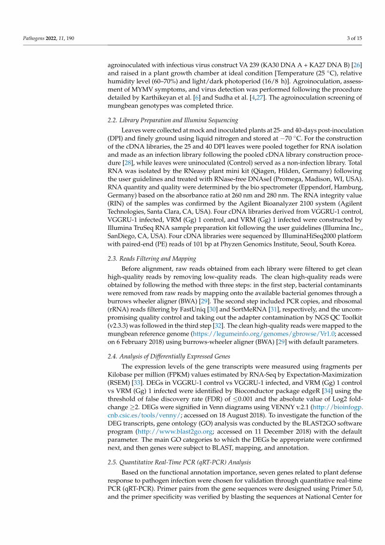
Two mungbean genotypes, VRM (Gg) 1 and VGGRU-1, were inoculated with MYMV,and symptoms were observed in uninfected control and infected plants. A typical mosaicsymptom was observed in susceptible mungbean genotype VRM (Gg) 1. On the contrary,there were no visible symptoms on the resistant genotype VGGRU-1 until 40 DPI (Figure 1).The viral DNA was detected in inoculated plants using PCR analysis of the coat proteingene of MYMV, while absent in uninfected control plants. The response of VRM (Gg) 1 andVGGRU-1 after infection with MYMV was as expected; thus, it is meaningful to use theirleaves for further transcriptome analysis.
Pathogens 2021, 10, x FOR PEER REVIEW 4 of 16
2.5. Quantitative Real-Time PCR (qRT-PCR) Analysis
Based on the functional annotation importance, seven genes related to plant defense
response to pathogen infection were chosen for validation through quantitative real-time
PCR (qRT-PCR). Primer pairs from the gene sequences were designed using Primer 5.0,
and the primer specificity was verified by blasting the sequences at National Center for
Biotechnology Information (NCBI) database. For normalizing the gene expression level,
ubiquitin was used as a reference gene. Total RNA was isolated by RNeasy plant mini kit
(Qiagen, Hilden, Germany). Consequently, the DNA-free RNA was used for first-strand
cDNA synthesis by transcriptor First Strand cDNA Synthesis Kit (Roche Applied Science,
Penzberg, Germany) following the manufacturer’s instructions. PCR amplification was
performed using 2× SYBR Green PCR Master Mix (TaKaRa, Kusatsu, Shiga, Japan) and
Light-Cycler® 480 (Roche Applied Science, Penzberg, Germany) following the standard
protocol. Three independent replicates were completed, and each gene's relative expres-
sion was measured by the comparative 2-ΔΔCt method.
3. Results
3.1. Mungbean Genotypes Reaction to MYMV
Two mungbean genotypes, VRM (Gg) 1 and VGGRU-1, were inoculated with
MYMV, and symptoms were observed in uninfected control and infected plants. A typical
mosaic symptom was observed in susceptible mungbean genotype VRM (Gg) 1. On the
contrary, there were no visible symptoms on the resistant genotype VGGRU-1 until 40
DPI (Figure 1). The viral DNA was detected in inoculated plants using PCR analysis of
the coat protein gene of MYMV, while absent in uninfected control plants. The response
of VRM (Gg) 1 and VGGRU-1 after infection with MYMV was as expected; thus, it is
meaningful to use their leaves for further transcriptome analysis.
Figure 1. Development of symptoms of MYMV in the agroinoculated plants (A) VGGRU-1 and (B)
VRM (Gg) 1.
3.2. Summary of Transcriptome Data Set
We sequenced four cDNA libraries (VGGRU-1 control, VGGRU-1 infected, VRM
(Gg) 1 control, and VRM (Gg) 1 infected) and generated a total of 95.8 million raw reads
ranging from 23.2 to 25.1 million raw reads per library. All the raw reads were submitted
to the NCBI database (Sequence Read Archive (SRA) with the accession number
PRJNA742191). The read length from each library was 101 bp. GC percentage of the se-
quence data was about 43% in four libraries. Further, we used stringent criteria to filter
Figure 1. Development of symptoms of MYMV in the agroinoculated plants (A) VGGRU-1 and(B) VRM (Gg) 1.
3.2. Summary of Transcriptome Data Set
We sequenced four cDNA libraries (VGGRU-1 control, VGGRU-1 infected, VRM (Gg)1 control, and VRM (Gg) 1 infected) and generated a total of 95.8 million raw reads rangingfrom 23.2 to 25.1 million raw reads per library. All the raw reads were submitted to theNCBI database (Sequence Read Archive (SRA) with the accession number PRJNA742191).The read length from each library was 101 bp. GC percentage of the sequence data wasabout 43% in four libraries. Further, we used stringent criteria to filter the clean high-quality reads and aligned more than 80% reads based on the reference genome and usedfor DEG analysis.
3.3. DEGs in Response to MYMV Infection
We used the criteria of false discovery rate (≤0.001) and fold change greater than orequal to identify DEGs in resistant and susceptible genotypes. The total number of DEGswas greater in VGGRU-1 than in VRM (Gg) 1. The Venn diagram shows the distributionof DEGs in both genotypes (Figure 2). There were 896 DEGs between the VGGRU-1control and infected samples. Among them, 583 genes were upregulated, and 313 geneswere down-regulated (Tables S1 and S2). Likewise, 506 DEGs were detected between the
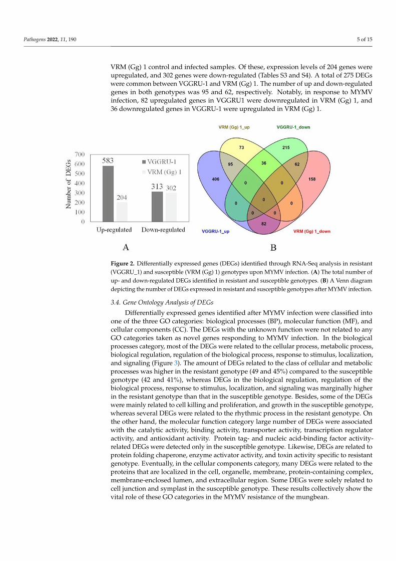
Pathogens 2022, 11, 190 5 of 15
VRM (Gg) 1 control and infected samples. Of these, expression levels of 204 genes wereupregulated, and 302 genes were down-regulated (Tables S3 and S4). A total of 275 DEGswere common between VGGRU-1 and VRM (Gg) 1. The number of up and down-regulatedgenes in both genotypes was 95 and 62, respectively. Notably, in response to MYMVinfection, 82 upregulated genes in VGGRU1 were downregulated in VRM (Gg) 1, and36 downregulated genes in VGGRU-1 were upregulated in VRM (Gg) 1.
1
Figure 2. Differentially expressed genes (DEGs) identified through RNA-Seq analysis in resistant(VGGRU_1) and susceptible (VRM (Gg) 1) genotypes upon MYMV infection. (A) The total number ofup- and down-regulated DEGs identified in resistant and susceptible genotypes. (B) A Venn diagramdepicting the number of DEGs expressed in resistant and susceptible genotypes after MYMV infection.
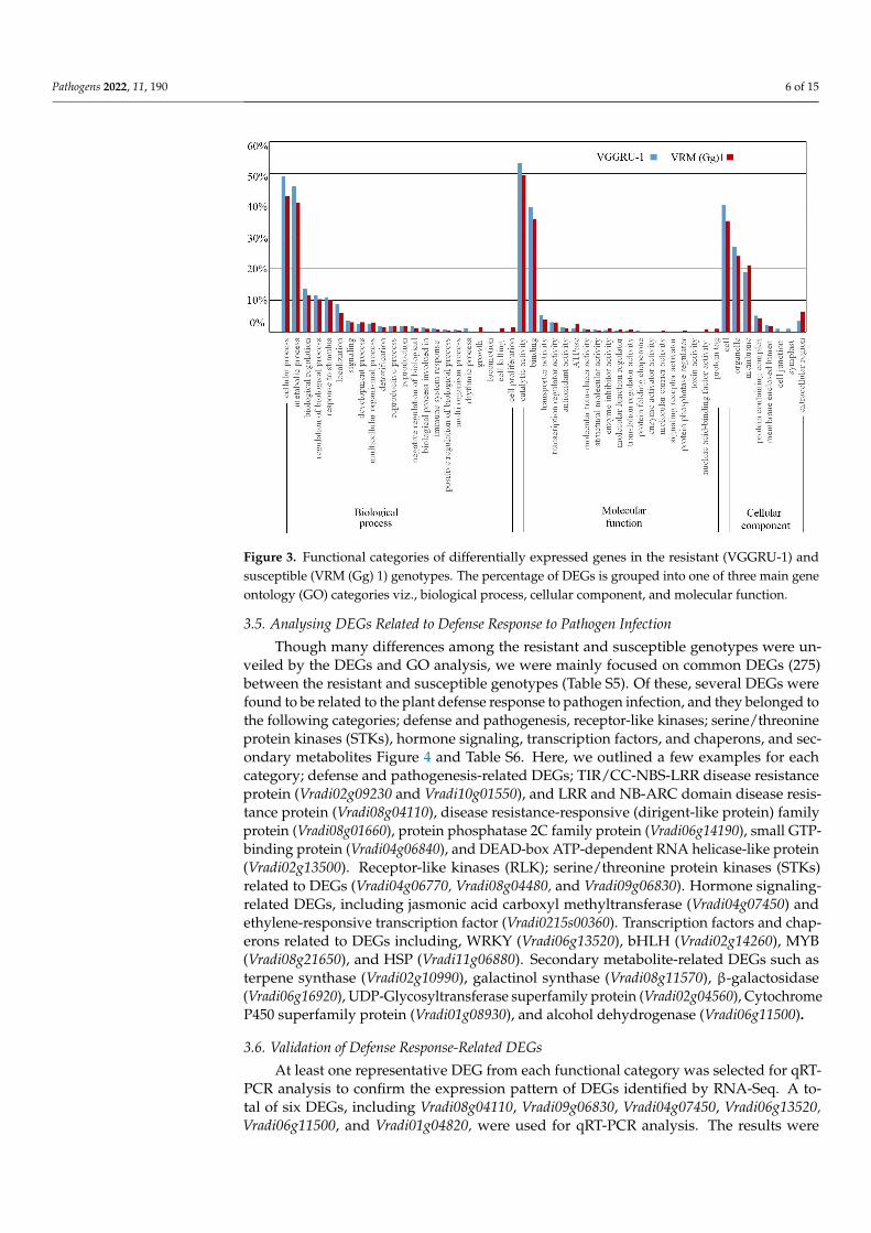
3.4. Gene Ontology Analysis of DEGs
Differentially expressed genes identified after MYMV infection were classified intoone of the three GO categories: biological processes (BP), molecular function (MF), andcellular components (CC). The DEGs with the unknown function were not related to anyGO categories taken as novel genes responding to MYMV infection. In the biologicalprocesses category, most of the DEGs were related to the cellular process, metabolic process,biological regulation, regulation of the biological process, response to stimulus, localization,and signaling (Figure 3). The amount of DEGs related to the class of cellular and metabolicprocesses was higher in the resistant genotype (49 and 45%) compared to the susceptiblegenotype (42 and 41%), whereas DEGs in the biological regulation, regulation of thebiological process, response to stimulus, localization, and signaling was marginally higherin the resistant genotype than that in the susceptible genotype. Besides, some of the DEGswere mainly related to cell killing and proliferation, and growth in the susceptible genotype,whereas several DEGs were related to the rhythmic process in the resistant genotype. Onthe other hand, the molecular function category large number of DEGs were associatedwith the catalytic activity, binding activity, transporter activity, transcription regulatoractivity, and antioxidant activity. Protein tag- and nucleic acid-binding factor activity-related DEGs were detected only in the susceptible genotype. Likewise, DEGs are related toprotein folding chaperone, enzyme activator activity, and toxin activity specific to resistantgenotype. Eventually, in the cellular components category, many DEGs were related to theproteins that are localized in the cell, organelle, membrane, protein-containing complex,membrane-enclosed lumen, and extracellular region. Some DEGs were solely related tocell junction and symplast in the susceptible genotype. These results collectively show thevital role of these GO categories in the MYMV resistance of the mungbean.
Pathogens 2022, 11, 190 6 of 15
Pathogens 2021, 10, x FOR PEER REVIEW 6 of 16
higher in the resistant genotype than that in the susceptible genotype. Besides, some of
the DEGs were mainly related to cell killing and proliferation, and growth in the suscep-
tible genotype, whereas several DEGs were related to the rhythmic process in the resistant
genotype. On the other hand, the molecular function category large number of DEGs were
associated with the catalytic activity, binding activity, transporter activity, transcription
regulator activity, and antioxidant activity. Protein tag- and nucleic acid-binding factor
activity-related DEGs were detected only in the susceptible genotype. Likewise, DEGs are
related to protein folding chaperone, enzyme activator activity, and toxin activity specific
to resistant genotype. Eventually, in the cellular components category, many DEGs were
related to the proteins that are localized in the cell, organelle, membrane, protein-contain-
ing complex, membrane-enclosed lumen, and extracellular region. Some DEGs were
solely related to cell junction and symplast in the susceptible genotype. These results col-
lectively show the vital role of these GO categories in the MYMV resistance of the mung-
bean.
Figure 3. Functional categories of differentially expressed genes in the resistant (VGGRU-1) and
susceptible (VRM (Gg) 1) genotypes. The percentage of DEGs is grouped into one of three main
gene ontology (GO) categories viz., biological process, cellular component, and molecular function.
3.5. Analysing DEGs Related to Defense Response to Pathogen Infection
Though many differences among the resistant and susceptible genotypes were un-
veiled by the DEGs and GO analysis, we were mainly focused on common DEGs (275)
between the resistant and susceptible genotypes (Table S5). Of these, several DEGs were
found to be related to the plant defense response to pathogen infection, and they belonged
Figure 3. Functional categories of differentially expressed genes in the resistant (VGGRU-1) andsusceptible (VRM (Gg) 1) genotypes. The percentage of DEGs is grouped into one of three main geneontology (GO) categories viz., biological process, cellular component, and molecular function.
3.5. Analysing DEGs Related to Defense Response to Pathogen Infection
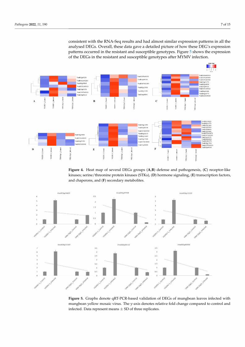
Though many differences among the resistant and susceptible genotypes were un-veiled by the DEGs and GO analysis, we were mainly focused on common DEGs (275)between the resistant and susceptible genotypes (Table S5). Of these, several DEGs werefound to be related to the plant defense response to pathogen infection, and they belonged tothe following categories; defense and pathogenesis, receptor-like kinases; serine/threonineprotein kinases (STKs), hormone signaling, transcription factors, and chaperons, and sec-ondary metabolites Figure 4 and Table S6. Here, we outlined a few examples for eachcategory; defense and pathogenesis-related DEGs; TIR/CC-NBS-LRR disease resistanceprotein (Vradi02g09230 and Vradi10g01550), and LRR and NB-ARC domain disease resis-tance protein (Vradi08g04110), disease resistance-responsive (dirigent-like protein) familyprotein (Vradi08g01660), protein phosphatase 2C family protein (Vradi06g14190), small GTP-binding protein (Vradi04g06840), and DEAD-box ATP-dependent RNA helicase-like protein(Vradi02g13500). Receptor-like kinases (RLK); serine/threonine protein kinases (STKs)related to DEGs (Vradi04g06770, Vradi08g04480, and Vradi09g06830). Hormone signaling-related DEGs, including jasmonic acid carboxyl methyltransferase (Vradi04g07450) andethylene-responsive transcription factor (Vradi0215s00360). Transcription factors and chap-erons related to DEGs including, WRKY (Vradi06g13520), bHLH (Vradi02g14260), MYB(Vradi08g21650), and HSP (Vradi11g06880). Secondary metabolite-related DEGs such asterpene synthase (Vradi02g10990), galactinol synthase (Vradi08g11570), β-galactosidase(Vradi06g16920), UDP-Glycosyltransferase superfamily protein (Vradi02g04560), CytochromeP450 superfamily protein (Vradi01g08930), and alcohol dehydrogenase (Vradi06g11500).
3.6. Validation of Defense Response-Related DEGs
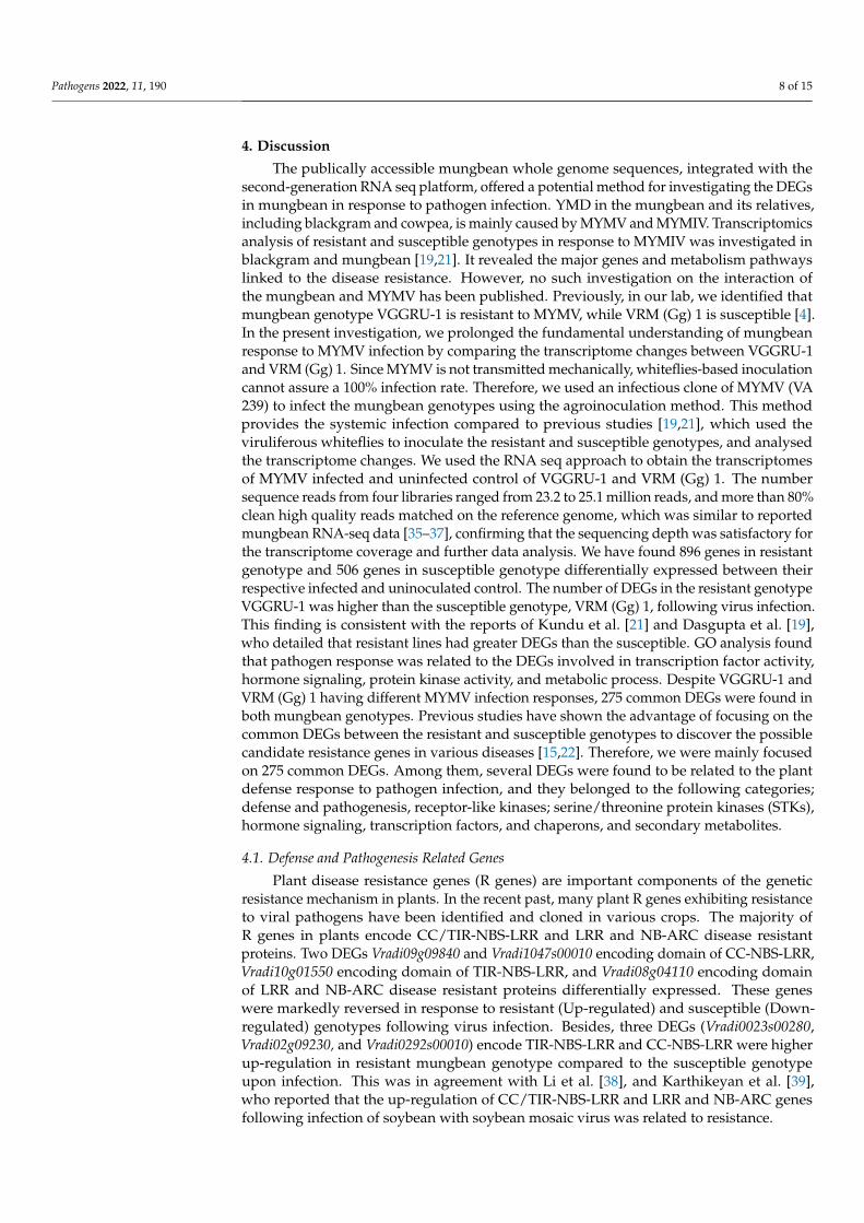
At least one representative DEG from each functional category was selected for qRT-PCR analysis to confirm the expression pattern of DEGs identified by RNA-Seq. A to-tal of six DEGs, including Vradi08g04110, Vradi09g06830, Vradi04g07450, Vradi06g13520,Vradi06g11500, and Vradi01g04820, were used for qRT-PCR analysis. The results were
Pathogens 2022, 11, 190 7 of 15
consistent with the RNA-Seq results and had almost similar expression patterns in all theanalysed DEGs. Overall, these data gave a detailed picture of how these DEG’s expressionpatterns occurred in the resistant and susceptible genotypes. Figure 5 shows the expressionof the DEGs in the resistant and susceptible genotypes after MYMV infection.
Pathogens 2021, 10, x FOR PEER REVIEW 7 of 16
to the following categories; defense and pathogenesis, receptor-like kinases; serine/threo-
nine protein kinases (STKs), hormone signaling, transcription factors, and chaperons, and
secondary metabolites Figure 4 and Table S6. Here, we outlined a few examples for each
category; defense and pathogenesis-related DEGs; TIR/CC-NBS-LRR disease resistance
protein (Vradi02g09230 and Vradi10g01550), and LRR and NB-ARC domain disease re-
sistance protein (Vradi08g04110), disease resistance-responsive (dirigent-like protein) fam-
ily protein (Vradi08g01660), protein phosphatase 2C family protein (Vradi06g14190), small
GTP-binding protein (Vradi04g06840), and DEAD-box ATP-dependent RNA helicase-like
protein (Vradi02g13500). Receptor-like kinases (RLK); serine/threonine protein kinases
(STKs) related to DEGs (Vradi04g06770, Vradi08g04480, and Vradi09g06830). Hormone sig-
naling-related DEGs, including jasmonic acid carboxyl methyltransferase (Vradi04g07450)
and ethylene-responsive transcription factor (Vradi0215s00360). Transcription factors and
chaperons related to DEGs including, WRKY (Vradi06g13520), bHLH (Vradi02g14260),
MYB (Vradi08g21650), and HSP (Vradi11g06880). Secondary metabolite-related DEGs such
as terpene synthase (Vradi02g10990), galactinol synthase (Vradi08g11570), β-galactosidase
(Vradi06g16920), UDP-Glycosyltransferase superfamily protein (Vradi02g04560), Cyto-
chrome P450 superfamily protein (Vradi01g08930), and alcohol dehydrogenase
(Vradi06g11500).
Figure 4. Heat map of several DEGs groups (A,B) defense and pathogenesis, (C) receptor-like ki-
nases; serine/threonine protein kinases (STKs), (D) hormone signaling, (E) transcription factors, and
chaperons, and (F)secondary metabolites.
3.6. Validation of Defense Response-Related DEGs
At least one representative DEG from each functional category was selected for qRT-
PCR analysis to confirm the expression pattern of DEGs identified by RNA-Seq. A total of
six DEGs, including Vradi08g04110, Vradi09g06830, Vradi04g07450, Vradi06g13520,
Vradi06g11500, and Vradi01g04820, were used for qRT-PCR analysis. The results were con-
sistent with the RNA-Seq results and had almost similar expression patterns in all the
analysed DEGs. Overall, these data gave a detailed picture of how these DEG's expression
Figure 4. Heat map of several DEGs groups (A,B) defense and pathogenesis, (C) receptor-likekinases; serine/threonine protein kinases (STKs), (D) hormone signaling, (E) transcription factors,and chaperons, and (F) secondary metabolites.
Pathogens 2021, 10, x FOR PEER REVIEW 8 of 16
patterns occurred in the resistant and susceptible genotypes. Figure 5 shows the expres-
sion of the DEGs in the resistant and susceptible genotypes after MYMV infection.
Figure 5. Graphs denote qRT-PCR-based validation of DEGs of mungbean leaves infected with
mungbean yellow mosaic virus. The y-axis denotes relative fold change compared to control and
infected. Data represent means ± SD of three replicates.
4. Discussion
The publically accessible mungbean whole genome sequences, integrated with the
second-generation RNA seq platform, offered a potential method for investigating the
DEGs in mungbean in response to pathogen infection. YMD in the mungbean and its rel-
atives, including blackgram and cowpea, is mainly caused by MYMV and MYMIV. Tran-
scriptomics analysis of resistant and susceptible genotypes in response to MYMIV was
investigated in blackgram and mungbean [19,21]. It revealed the major genes and metab-
olism pathways linked to the disease resistance. However, no such investigation on the
interaction of the mungbean and MYMV has been published. Previously, in our lab, we
identified that mungbean genotype VGGRU-1 is resistant to MYMV, while VRM (Gg) 1 is
susceptible [4]. In the present investigation, we prolonged the fundamental understand-
ing of mungbean response to MYMV infection by comparing the transcriptome changes
between VGGRU-1 and VRM (Gg) 1. Since MYMV is not transmitted mechanically, white-
flies-based inoculation cannot assure a 100% infection rate. Therefore, we used an infec-
tious clone of MYMV (VA 239) to infect the mungbean genotypes using the agroinocula-
tion method. This method provides the systemic infection compared to previous studies
[19,21], which used the viruliferous whiteflies to inoculate the resistant and susceptible
genotypes, and analysed the transcriptome changes. We used the RNA seq approach to
obtain the transcriptomes of MYMV infected and uninfected control of VGGRU-1 and
VRM (Gg) 1. The number sequence reads from four libraries ranged from 23.2 to 25.1 mil-
lion reads, and more than 80% clean high quality reads matched on the reference genome,
which was similar to reported mungbean RNA-seq data [35–37], confirming that the se-
quencing depth was satisfactory for the transcriptome coverage and further data analysis.
We have found 896 genes in resistant genotype and 506 genes in susceptible genotype
Figure 5. Graphs denote qRT-PCR-based validation of DEGs of mungbean leaves infected withmungbean yellow mosaic virus. The y-axis denotes relative fold change compared to control andinfected. Data represent means ± SD of three replicates.
Pathogens 2022, 11, 190 8 of 15
4. Discussion
The publically accessible mungbean whole genome sequences, integrated with thesecond-generation RNA seq platform, offered a potential method for investigating the DEGsin mungbean in response to pathogen infection. YMD in the mungbean and its relatives,including blackgram and cowpea, is mainly caused by MYMV and MYMIV. Transcriptomicsanalysis of resistant and susceptible genotypes in response to MYMIV was investigated inblackgram and mungbean [19,21]. It revealed the major genes and metabolism pathwayslinked to the disease resistance. However, no such investigation on the interaction ofthe mungbean and MYMV has been published. Previously, in our lab, we identified thatmungbean genotype VGGRU-1 is resistant to MYMV, while VRM (Gg) 1 is susceptible [4].In the present investigation, we prolonged the fundamental understanding of mungbeanresponse to MYMV infection by comparing the transcriptome changes between VGGRU-1and VRM (Gg) 1. Since MYMV is not transmitted mechanically, whiteflies-based inoculationcannot assure a 100% infection rate. Therefore, we used an infectious clone of MYMV (VA239) to infect the mungbean genotypes using the agroinoculation method. This methodprovides the systemic infection compared to previous studies [19,21], which used theviruliferous whiteflies to inoculate the resistant and susceptible genotypes, and analysedthe transcriptome changes. We used the RNA seq approach to obtain the transcriptomesof MYMV infected and uninfected control of VGGRU-1 and VRM (Gg) 1. The numbersequence reads from four libraries ranged from 23.2 to 25.1 million reads, and more than 80%clean high quality reads matched on the reference genome, which was similar to reportedmungbean RNA-seq data [35–37], confirming that the sequencing depth was satisfactory forthe transcriptome coverage and further data analysis. We have found 896 genes in resistantgenotype and 506 genes in susceptible genotype differentially expressed between theirrespective infected and uninoculated control. The number of DEGs in the resistant genotypeVGGRU-1 was higher than the susceptible genotype, VRM (Gg) 1, following virus infection.This finding is consistent with the reports of Kundu et al. [21] and Dasgupta et al. [19],who detailed that resistant lines had greater DEGs than the susceptible. GO analysis foundthat pathogen response was related to the DEGs involved in transcription factor activity,hormone signaling, protein kinase activity, and metabolic process. Despite VGGRU-1 andVRM (Gg) 1 having different MYMV infection responses, 275 common DEGs were found inboth mungbean genotypes. Previous studies have shown the advantage of focusing on thecommon DEGs between the resistant and susceptible genotypes to discover the possiblecandidate resistance genes in various diseases [15,22]. Therefore, we were mainly focusedon 275 common DEGs. Among them, several DEGs were found to be related to the plantdefense response to pathogen infection, and they belonged to the following categories;defense and pathogenesis, receptor-like kinases; serine/threonine protein kinases (STKs),hormone signaling, transcription factors, and chaperons, and secondary metabolites.
4.1. Defense and Pathogenesis Related Genes
Plant disease resistance genes (R genes) are important components of the geneticresistance mechanism in plants. In the recent past, many plant R genes exhibiting resistanceto viral pathogens have been identified and cloned in various crops. The majority ofR genes in plants encode CC/TIR-NBS-LRR and LRR and NB-ARC disease resistantproteins. Two DEGs Vradi09g09840 and Vradi1047s00010 encoding domain of CC-NBS-LRR,Vradi10g01550 encoding domain of TIR-NBS-LRR, and Vradi08g04110 encoding domainof LRR and NB-ARC disease resistant proteins differentially expressed. These geneswere markedly reversed in response to resistant (Up-regulated) and susceptible (Down-regulated) genotypes following virus infection. Besides, three DEGs (Vradi0023s00280,Vradi02g09230, and Vradi0292s00010) encode TIR-NBS-LRR and CC-NBS-LRR were higherup-regulation in resistant mungbean genotype compared to the susceptible genotypeupon infection. This was in agreement with Li et al. [38], and Karthikeyan et al. [39],who reported that the up-regulation of CC/TIR-NBS-LRR and LRR and NB-ARC genesfollowing infection of soybean with soybean mosaic virus was related to resistance.
Pathogens 2022, 11, 190 9 of 15
Disease resistance-responsive (dirigent-like protein), small GTP-binding proteins, andprotein phosphatase 2C family proteins are related to disease resistance in plants. DIRproteins and disease resistance response family proteins have a similar dirigent-conserveddomain. They play a major role in arbitrating the free radical coupling of monolignol plantphenols in plants to yield lignans and lignins; therefore, DIRs have been involved in diseaseresistance responses. Vradi08g01660 encoding disease resistance-responsive (dirigent-likeprotein) family protein up-regulated in resistant genotype. Similarly, the involvementof dirigent genes and their apparent upregulated expression in response to attacks bypathogens [40–42] is of particular interest. The small GTP-binding gene families are relatedto the signal transduction in plants. It involves GTPase activity and activates the proteinkinases and LRR related to disease resistance [43]. The up-regulation of DEG Vradi04g06840encoding Small GTP-binding protein in the resistant genotype compared to the susceptiblegenotype underlines the importance of this gene during plant disease resistance.
Protein phosphatase 2C family proteins have been important in regulating the abscisicacid signaling pathway and adaptation to environmental stresses. In our study, upregula-tion of two DEGs (Vradi02g08700 and Vradi06g14190) encoded protein phosphatase 2C wasobserved in resistant genotype; in contrast, lower-level expression compared to resistantgenotype or downregulation was seen in susceptible genotype. Protein phosphatase 2Cinvolves disease resistance by activating defense response in tobacco and soybean [44,45].The upregulation of protein phosphatase 2C showed ABA-induced functions as a keyregulator of Rsv3-mediated soybean mosaic virus resistance, limiting virus spread in soy-bean [45]. The involvement and significance of DEAD-box RNA helicases, which is themajor family of RNA helicases, are known for their role against pathogens in plants [46].Therefore, the expression levels of three DEGs belonging to the DEAD-box RNA helicasesfamily were examined in the present study. All three DEGs were upregulated in resis-tant genotype, suggesting their involvement resistance. Notably, Vradi0285s00030 andVradi02g13500 were upregulated in the resistant genotype, while they were down-regulatedin the susceptible genotypes. Vradi0007s01650 expression level was highly up-regulated inthe resistant genotype, whereas only a slight up-regulation was observed in the susceptiblegenotype after virus infection. Li et al. [47] showed that overexpression of OsBIRH1 encodeDEAD-box RNA helicases in transgenic Arabidopsis plants resulted in an increased expres-sion of defense-associated genes and improved the disease resistance as well as oxidativestress tolerance.
4.2. LRR-RLK/STK Genes
LRR-RLKs are important components in regulating hormone signaling, abiotic andbiotic stress responses in plants. STKs are receptor proteins that facilitate the signal trans-duction in plant defense responses [48,49]. It is mainly involved in identifying and trans-duction of pathogen-derived signals at the time of plant and microbe interactions. SeveralDEGs (Vradi0161s00420, Vradi0253s00140, Vradi0283s00060, Vradi04g06770, Vradi08g04480,and Vradi09g06830) from this family is upregulated in resistant genotype while downregu-lated in susceptible genotype. Although, several DEGs (Vradi0252s00080, Vradi02g11170,Vradi0366s00010, and Vradi05g08520) are highly up-regulated in the resistant genotype, butonly a slight up-regulation was detected in the susceptible genotype after virus infection.Previous studies showed that overexpression of this gene family is related to disease re-sistance and defense responses, emphasizing important amino acids in specific regions,which are closely associated with plant disease resistance signal transmission [50,51].
4.3. Genes Involved in SA, JA, and ET Pathway
Plants can develop refined defense systems to prevent themselves against pathogeninfection and cope with pathogen invasion by triggering numerous defense pathways.Pathogen infection induces the level of hormones such as salicylic acid (SA), jasmonate (JA),and ethylene (ET). These hormones are determined by the pathogen and play a major rolein developing a strong defense system [52,53]. The plant hormone JA and its derivatives
Pathogens 2022, 11, 190 10 of 15
have been identified as important regulators in a plant’s immune system, playing criticalroles in pathogen defense responses [54]. Vradi04g07450 encoded S-adenosyl-L-methionine:jasmonic acid carboxyl methyltransferase (JMT). JMT involves the conversion of JA toMeJA, and overexpression of the JMT gene in transgenic Arabidopsis and rice plantsinduces the constitutive expression of JA-responsive genes and regulates the plants todefend themselves against infection by pathogens and herbivores [55,56]. Besides, oneDEG Vradi0234s00020 encodes allene oxide synthase (AOS) upregulated in both genotypesduring virus infection, but up-regulation in resistant genotype was slightly high comparedto susceptible genotype. AOS is the first enzyme in the branch pathway leading to thebiosynthesis of JA, and studies reported that expression of AOS determines defense geneactivation in tomato [57]. Two DEGs (Vradi0215s00360 and Vradi01g10800) belong to theethylene-responsive transcription factor (ERF) family regulated during virus infection.ET has been shown to regulate the expression level of pathogenesis-related genes viaERFs in earlier studies, ERFs were likely to have a role in the control of plant defensemechanisms by acting as transcriptional activators or repressors of GCC-box mediatedgene expression [58–61].
4.4. Transcription Factors and Secondary Metabolites
Transcription factors WRKY, MYB, and bHLH families have been known for their rolein an elaborate regulation network by interacting with target genes in plants. Additionally,they are related to activation of defense gene expression and regulation of phytohormonescrosstalk [62–67]. In this study, three types of transcription factors, including three WRKYs,two bHLH, and two MYB, were differentially expressed. WRKY proteins are an importantfamily of transcriptional regulators identified solely in plants. They have been implicated inthe plant’s response to biotic stress in different ways, including as transcriptional activatorsor repressors [68]. We have identified three DEGs belonging to WRKYs, i.e., Vradi06g13520,Vradi0338s00060, and Vradi0158s00480, differentially expressed following virus inoculation.In particular, Vradi06g13520 and Vradi0338s00060 showed a 2–5-fold increase in resistantgenotype and a 2–3-fold decrease in susceptible genotype. Vradi0158s00480 is upregulatedin resistant genotype higher than susceptible genotype. So far, many researchers havediscussed the involvement of WRKYs in plant defense response against various pathogens,including MYMIV, and how WRKYs interact with their target genes or crosstalk with genesinvolved in plant hormone signaling such as SA, JA, and others [69]. The bHLH proteinsbelong to a class of superfamily transcription factors, which can bind to particular targetsites in DNA. Increasing evidence indicates that bHLHs regulate plant defense responses topathogens [66,70]. Two DEGs (Vradi02g14260 and Vradi09g06110) encoded bHLH showedup-regulation following virus infection in resistant genotype. In the susceptible genotype,Vradi02g14260 showed downregulation while Vradi09g06110 expressed a comparatively lowlevel than resistant genotype. MYB proteins are reportedly involved in several functions,including pathogen resistance [67]. Two DEG (Vradi02g08100 and Vradi08g21650) showedupregulation in both genotypes following virus infection. Ibraheem et al. [71] reportedthat expression of MYB induces 3-deoxyanthocyanidins and enhances resistance againstleaf blights in maize. The small heat shock proteins (sHSPs) and the related a-crystallinsare virtually ubiquitous proteins that are strongly induced by various other stresses inprokaryotic and eukaryotic cells. sHSPs were shown to be involved in the defense responseagainst Ralstoniasolanacearum in Nicotiana plants [72]. In this study, Vradi11g06880 encodedHSP21 showed differentially expressed. HSP21, a nuclear-encoded chloroplast-localizedsHSP, has been described for its role in stress tolerance in plants [73].
Many secondary metabolites found in plants have a role in defense against herbi-vores, pests, and pathogens. In this study, we found that DEGs encoding terpene synthase(Vradi02g10900, Vradi02g10990, and Vradi0399s00070) involved in terpene metabolism,galactinol synthase (Vradi08g11570) involved in raffinose metabolism, β-galactosidase(Vradi06g16920), and UDP-glycosyltransferase superfamily protein (Vradi0207s00030,Vradi0387s00030, and Vradi02g04560) involved in glycosylation, cytochrome P450 superfam-
Pathogens 2022, 11, 190 11 of 15
ily protein (Vradi01g08930, Vradi02g06800, Vradi03g01370, and Vradi11g02370), β-glucosidase(Vradi05g21890), and alcohol dehydrogenase (Vradi06g11500) were reported to involvedin plant defense mechanism against pathogens and insect pests infection [74–82]. Besides,we also found several DEGs belong to the family of formate dehydrogenase [83], chloro-phyllase 1 [84], F-box family protein [85], methionine sulfoxide reductase [86], glutathioneS-transferase family protein [87], and peroxidase superfamily [88] that are known to beinvolved indirectly or indirectly in plant defense mechanism against stress.
4.5. Comparison of the Major DEGs with the Previous Reports of YMD Resistance in Mungbean
The information related to genes and molecular mechanisms of YMD resistance inmungbean is very limited. Two studies, Mathivathana et al. [89] and Dasgupta et al. [19],discussed the possible candidate genes associated with YMD resistance in mungbean.The former used the QTL mapping approach, and the latter used the RNA seq approachlike our study. This study identified several DEGs linked to virus resistance, and theybelonged to the gene families that have been reported by Mathivathana et al. [89] andDasgupta et al. [19]. For instance, gene families including WRKY, bHLH, MYB S-adenosyl-L-methionine: jasmonic acid carboxyl methyltransferase, DEAD-box RNA helicases, smallGTP-binding, cytochrome P450, and protein kinase superfamily protein/serine-threoninekinase. Mathivathana et al. [89] reported the major QTL (qMYMV4_1) governing YMD re-sistance at nucleotide positions Vr04:14504302 and15788321 on chromosome 4. Three genesVradi04g06770 (Protein kinase superfamily protein/serine-threonine kinase), Vradi04g06840(small GTP-binding), and Vradi04g07450 (S-adenosyl-L-methionine: jasmonic acid carboxylmethyltransferase) differentially expressed in our study were existed in this genomic re-gion [89]. These genes are potential candidates to investigate MYMV resistance in thefuture. Although we have identified several DEGs from the gene families that are notpreviously reported in YMD resistance studies [19,21,89], for instance, DEGs belong to LRRand NB-ARC, and NBS-LRR gene families (Vradi02g09230, Vradi08g04110, Vradi09g09840,and Vradi10g01550), which are the largest group of plant R genes that play important rolesin plant defense responses to various pathogens. Plant defense response is a complexsystem that requires comprehensive investigation stage by stage. This study only pre-dicted the likely genes involved in MYMV resistance, and we do not have the functionalvalidation of the identified genes. Therefore, before utilizing these genes for breedingpurposes and understanding the specific roles of genes involved in the virus resistance, theidentified genes from this study could be verified using the overexpression, gene knockout,or CRISPR approaches.
In summary, our study postulates the putative genes linked with resistance to MYMV.It revealed how the mungbean genes interact and respond to MYMV infection and cause re-sistance and susceptibility. We used RNA sequencing technology to compare the transcriptschanges of control and infected resistant and susceptible genotypes in response to MYMV.Overall, the information generated from current research will be a valuable foundation fordeciphering the molecular mechanism governing MYMV resistance in mungbean that canprovide insights for future genetic breeding and facilitate the development of mungbeancultivars resistant against MYMV.
Supplementary Materials: The following supporting information can be downloaded at: https://www.mdpi.com/article/10.3390/pathogens11020190/s1, Table S1: List of DEGs up regulated inresistant genotype VGGRU-1; Table S2: List of DEGs down regulated in resistant genotype VGGRU-1;Table S3: List of DEGs up regulated in susceptible genotype VRM (Gg) 1; Table S4: List of DEGsdownregulated in susceptible genotype VRM (Gg) 1; Table S5: List of common DEGs betweenresistant and susceptible genotypes and their annotation details; Table S6: Few examples for DEGsfrom different categories related to defense response.
Author Contributions: Conceived and designed the experiments: M.S., A.K., N.S. and M.R., FundingAcquisition; A.K. and M.S., Performed the experiments: M.S., B.M., R.V.R. and M.N.S.S., Ana-lyzed the data: A.K., J.M. and M.D. Provided suggestions; N.S., K.E.A.A. and M.R., Contributed
Pathogens 2022, 11, 190 12 of 15
reagents/materials/analysis tools: M.S., M.P., A.K. and M.K.M., Draft the paper: A.K. and M.S. Allauthors have read and agreed to the published version of the manuscript.
Funding: The work was financially supported by the DST SERB 2016-2019 (YSS/2015/000321) to MSand DST-SERB NPDF fellowship program 2017–2019 (PDF/2016/003676) to AK from the Departmentof Science and Technology, Government of India, and, New Delhi, India. The funder had no role inthe work design, data collection, and analysis, or decision and preparation of the manuscript.
Institutional Review Board Statement: Not applicable.
Informed Consent Statement: Not applicable.
Data Availability Statement: All fastq files have been submitted to the NCBI Sequence Read Archivedatabase at https://www.ncbi.nlm.nih.gov/sra; accessed on 29 June 2021. NCBI accession for thisproject is PRJNA742191, and the SRA accession numbers: SRX11379693-SRX11379696.
Conflicts of Interest: The authors declare that they have no conflict of interest. The funder had norole in the work design, data collection, and analysis, or decision and preparation of the manuscript.
References1. Qazi, J.; Ilyas, M.; Mansoor, S.; Briddon, R.W. Legume yellow mosaic viruses: Genetically isolated begomoviruses. Mol. Plant
Pathol. 2007, 8, 343–348. [CrossRef] [PubMed]2. Karthikeyan, A.; Vanitharani, R.; Balaji, V.; Anuradha, S.; Thillaichidambaram, P.; Shivaprasad, P.; Parameswari, C.; Balamani, V.;
Saminathan, M.; Veluthambi, K. Analysis of an isolate of Mungbean yellow mosaic virus (MYMV) with a highly variable DNA Bcomponent. Arch. Virol. 2004, 149, 1643–1652. [CrossRef] [PubMed]
3. Usharani, K.; Surendranath, B.; Haq, Q.; Malathi, V. Yellow mosaic virus infecting soybean in northern India is distinct from thespecies infecting soybean in southern and western India. Curr. Sci. 2004, 845–850.
4. Sudha, M.; Karthikeyan, A.; Nagarajan, P.; Raveendran, M.; Senthil, N.; Pandiyan, M.; Angappan, K.; Ramalingam, J.;Bharathi, M.; Rabindran, R. Screening of mungbean (Vigna radiata) germplasm for resistance to Mungbean yellow mosaicvirus using agroinoculation. Can. J. Plant Pathol. 2013, 35, 424–430. [CrossRef]
5. Karthikeyan, A.; Shobhana, V.; Sudha, M.; Raveendran, M.; Senthil, N.; Pandiyan, M.; Nagarajan, P. Mungbean yellow mosaicvirus (MYMV): A threat to green gram (Vigna radiata) production in Asia. Int. J. Pest Manag. 2014, 60, 314–324. [CrossRef]
6. Karthikeyan, A.; Sudha, M.; Pandiyan, M.; Senthil, N.; Shobana, V.; Nagarajan, P. Screening of MYMV resistant mungbean (Vignaradiata L. Wilczek) progenies through agroinoculation. Int. J. Plant Pathol. 2011, 2, 115–125. [CrossRef]
7. Nair, R.; Götz, M.; Winter, S.; Giri, R.; Boddepalli, V.; Sirari, A.; Bains, T.; Taggar, G.; Dikshit, H.; Aski, M. Identification ofmungbean lines with tolerance or resistance to yellow mosaic in fields in India where different begomovirus species and differentBemisia tabaci cryptic species predominate. Eur. J. Plant Pathol. 2017, 149, 349–365. [CrossRef]
8. Parihar, A.K.; Basandrai, A.K.; Sirari, A.; Dinakaran, D.; Singh, D.; Kannan, K.; Kushawaha, K.P.; Adinarayan, M.; Akram, M.;Latha, T.K.S. Assessment of mungbean genotypes for durable resistance to Yellow Mosaic D isease: Genotype× Environmentinteractions. Plant Breed. 2017, 136, 94–100. [CrossRef]
9. Basavaraj, S.; Padmaja, A.; Nagaraju, N.; Ramesh, S. Identification of stable sources of resistance to mungbean yellow mosaicvirus (MYMV) disease in mungbean [Vigna radiata (L.) Wilczek]. Plant Genet. Resour. 2019, 17, 362–370.
10. Bai, T.-T.; Xie, W.-B.; Zhou, P.-P.; Wu, Z.-L.; Xiao, W.-C.; Zhou, L.; Sun, J.; Ruan, X.-L.; Li, H.-P. Transcriptome and expressionprofile analysis of highly resistant and susceptible banana roots challenged with Fusarium oxysporum f. sp. cubense tropical race 4.PLoS ONE 2013, 8, e73945. [CrossRef]
11. Jain, S.; Chittem, K.; Brueggeman, R.; Osorno, J.M.; Richards, J.; Nelson, B.D., Jr. Comparative transcriptome analysis ofresistant and susceptible common bean genotypes in response to soybean cyst nematode infection. PLoS ONE 2016, 11, e0159338.[CrossRef] [PubMed]
12. Ma, X.; Keller, B.; McDonald, B.A.; Palma-Guerrero, J.; Wicker, T. Comparative transcriptomics reveals how wheat responds toinfection by Zymoseptoria tritici. Mol. Plant-Microbe Interact. 2018, 31, 420–431. [CrossRef] [PubMed]
13. Tian, X.; Zhang, L.; Feng, S.; Zhao, Z.; Wang, X.; Gao, H. Transcriptome analysis of apple leaves in response to powdery mildew(Podosphaera leucotricha) infection. Int. J. Mol. Sci. 2019, 20, 2326. [CrossRef] [PubMed]
14. Du, H.; Wang, Y.; Yang, J.; Yang, W. Comparative Transcriptome Analysis of Resistant and Susceptible Tomato Lines in Responseto Infection by Xanthomonas perforans Race T3. Front. Plant Sci. 2015, 6, 1173. [CrossRef] [PubMed]
15. Lekota, M.; Muzhinji, N.; Van der Waals, J.E. Identification of differentially expressed genes in tolerant and susceptible potatocultivars in response to Spongospora subterranea f. sp. subterranea tuber infection. Plant Pathol. 2019, 68, 1196–1206. [CrossRef]
16. Chen, T.; Lv, Y.; Zhao, T.; Li, N.; Yang, Y.; Yu, W.; He, X.; Liu, T.; Zhang, B. Comparative transcriptome profiling of a resistantvs. susceptible tomato (Solanum lycopersicum) cultivar in response to infection by tomato yellow leaf curl virus. PLoS ONE 2013,8, e80816.
17. Wang, Y.; Zhou, L.; Yu, X.; Stover, E.; Luo, F.; Duan, Y. Transcriptome profiling of Huanglongbing (HLB) tolerant and susceptiblecitrus plants reveals the role of basal resistance in HLB tolerance. Front. Plant Sci. 2016, 7, 933. [CrossRef]
Pathogens 2022, 11, 190 13 of 15
18. Wang, Z.; Li, Y.; Li, C.; Song, X.; Lei, J.; Gao, Y.; Liang, Q. Comparative transcriptome profiling of resistant and susceptiblesugarcane genotypes in response to the airborne pathogen Fusarium verticillioides. Mol. Biol. Rep. 2019, 46, 3777–3789. [CrossRef]
19. Dasgupta, U.; Mishra, G.P.; Dikshit, H.K.; Mishra, D.C.; Bosamia, T.; Roy, A.; Bhati, J.; Aski, M.; Kumar, R.R.; Singh, A.K.Comparative RNA-Seq analysis unfolds a complex regulatory network imparting yellow mosaic disease resistance in mungbean[Vigna radiata (L.) R. Wilczek]. PLoS ONE 2021, 16, e0244593. [CrossRef]
20. Chakraborty, N.; Basak, J. Comparative transcriptome profiling of a resistant vs. susceptible Vigna mungo cultivar in responseto Mungbean yellow mosaic India virus infection reveals new insight into MYMIV resistance. Curr. Plant Biol. 2018, 15, 8–24.[CrossRef]
21. Kundu, A.; Singh, P.K.; Dey, A.; Ganguli, S.; Pal, A. Complex molecular mechanisms underlying MYMIV-resistance in Vignamungo revealed by comparative transcriptome profiling. Sci. Rep. 2019, 9, 1–13. [CrossRef] [PubMed]
22. Dong, H.; Shi, S.; Zhang, C.; Zhu, S.; Li, M.; Tan, J.; Yu, Y.; Lin, L.; Jia, S.; Wang, X. Transcriptomic analysis of genes in soybean inresponse to Peronospora manshurica infection. BMC Genom. 2018, 19, 1–13. [CrossRef] [PubMed]
23. Soria-Guerra, R.E.; Rosales-Mendoza, S.; Chang, S.; Haudenshield, J.S.; Padmanaban, A.; Rodriguez-Zas, S.; Hartman, G.L.;Ghabrial, S.A.; Korban, S.S. Transcriptome analysis of resistant and susceptible genotypes of Glycine tomentella during Phakop-sora pachyrhizi infection reveals novel rust resistance genes. Theor. Appl. Genet. 2010, 120, 1315–1333. [CrossRef] [PubMed]
24. Matic, S.; Bagnaresi, P.; Biselli, C.; Carneiro, G.A.; Siciliano, I.; Valé, G.; Gullino, M.L.; Spadaro, D. Comparative transcriptomeprofiling of resistant and susceptible rice genotypes in response to the seedborne pathogen Fusarium fujikuroi. BMC Genom. 2016,17, 1–17. [CrossRef] [PubMed]
25. Zheng, W.; Ma, L.; Zhao, J.; Li, Z.; Sun, F.; Lu, X. Comparative transcriptome analysis of two rice varieties in response to ricestripe virus and small brown planthoppers during early interaction. PLoS ONE 2013, 8, e82126. [CrossRef] [PubMed]
26. Balaji, V.; Vanitharani, R.; Karthikeyan, A.; Anbalagan, S.; Veluthambi, K. Infectivity analysis of two variable DNA B componentsof Mungbean yellow mosaic virus-Vigna in Vigna mungo and Vigna radiata. J. Biosci. 2004, 29, 297–308. [CrossRef]
27. Sudha, M.; Karthikeyan, A.; Shobhana, V.; Nagarajan, P.; Raveendran, M.; Senthil, N.; Pandiyan, M.; Angappan, K.;Balasubramanian, P.; Rabindran, R. Search for Vigna species conferring resistance to mungbean yellow mosaic virus in mungbean.Plant Genet. Resour. 2015, 13, 162–167. [CrossRef]
28. Eybishtz, A.; Peretz, Y.; Sade, D.; Akad, F.; Czosnek, H. Silencing of a single gene in tomato plants resistant to Tomato yellow leafcurl virus renders them susceptible to the virus. Plant Mol. Biol. 2009, 71, 157–171. [CrossRef]
29. Li, H.; Durbin, R. Fast and accurate short read alignment with Burrows–Wheeler transform. Bioinformatics 2009, 25, 1754–1760.[CrossRef]
30. Xu, H.; Luo, X.; Qian, J.; Pang, X.; Song, J.; Qian, G.; Chen, J.; Chen, S. FastUniq: A fast de novo duplicates removal tool for pairedshort reads. PLoS ONE 2012, 7, e52249. [CrossRef]
31. Kopylova, E.; Noé, L.; Touzet, H. SortMeRNA: Fast and accurate filtering of ribosomal RNAs in metatranscriptomic data.Bioinformatics 2012, 28, 3211–3217. [CrossRef] [PubMed]
32. Patel, R.K.; Jain, M. NGS QC Toolkit: A toolkit for quality control of next generation sequencing data. PLoS ONE 2012, 7, e30619.[CrossRef] [PubMed]
33. Li, B.; Dewey, C.N. RSEM: Accurate transcript quantification from RNA-Seq data with or without a reference genome. BMCBioinform. 2011, 12, 1–16. [CrossRef] [PubMed]
34. Robinson, M.D.; McCarthy, D.J.; Smyth, G.K. edgeR: A Bioconductor package for differential expression analysis of digital geneexpression data. Bioinformatics 2010, 26, 139–140. [CrossRef]
35. Chang, Y.; Sun, F.; Sun, S.; Wang, L.; Wu, J.; Zhu, Z. Transcriptome Analysis of Resistance to Fusarium Wilt in Mung Bean (Vignaradiata L.). Front. Plant Sci. 2021, 12, 1213. [CrossRef]
36. Ha, J.; Shim, S.; Lee, T.; Lee, E.; Yang, X.; Jeong, H.; Kim, M.Y.; Lee, S.-H. Transcriptomic and biochemical analyses ofthe accumulation of sucrose in mungbean (Vigna radiata (L.) Wilczek) leaves after pod removal. Theor. Appl. Genet. 2020,133, 2355–2362. [CrossRef]
37. Tian, X.; Li, S.; Liu, Y.; Liu, X. Transcriptomic profiling reveals metabolic and regulatory pathways in the desiccation tolerance ofMungbean (Vigna radiata [L.] R. Wilczek). Front. Plant Sci. 2016, 7, 1921. [CrossRef]
38. Li, C.; Adhimoolam, K.; Yuan, Y.; Yin, J.; Ren, R.; Yang, Y.; Zhi, H. Identification of candidate genes for resistance to Soybeanmosaic virus strain SC3 by using fine mapping and transcriptome analyses. Crop Pasture Sci. 2017, 68, 156–166. [CrossRef]
39. Karthikeyan, A.; Li, K.; Li, C.; Yin, J.; Li, N.; Yang, Y.; Song, Y.; Ren, R.; Zhi, H.; Gai, J. Fine-mapping and identifying candidategenes conferring resistance to Soybean mosaic virus strain SC20 in soybean. Theor. Appl. Genet. 2018, 131, 461–476. [CrossRef]
40. Li, N.; Zhao, M.; Liu, T.; Dong, L.; Cheng, Q.; Wu, J.; Wang, L.; Chen, X.; Zhang, C.; Lu, W. A novel soybean dirigent geneGmDIR22 contributes to promotion of lignan biosynthesis and enhances resistance to Phytophthora sojae. Front. Plant Sci. 2017,8, 1185. [CrossRef]
41. Reboledo, G.; Del Campo, R.; Alvarez, A.; Montesano, M.; Mara, H.; Ponce de León, I. Physcomitrella patens activates defenseresponses against the pathogen Colletotrichum gloeosporioides. Int. J. Mol. Sci. 2015, 16, 22280–22298. [CrossRef] [PubMed]
42. Zhu, L.; Zhang, X.; Tu, L.; Zeng, F.; Nie, Y.; Guo, X. Isolation and characterization of two novel dirigent-like genes highly inducedin cotton (Gossypium barbadense and G. hirsutum) after infection by Verticillium dahliae. J. Plant Pathol. 2007, 89, 41–45.
Pathogens 2022, 11, 190 14 of 15
43. Meyers, B.C.; Dickerman, A.W.; Michelmore, R.W.; Sivaramakrishnan, S.; Sobral, B.W.; Young, N.D. Plant disease resistance genesencode members of an ancient and diverse protein family within the nucleotide-binding superfamily. Plant J. 1999, 20, 317–332.[CrossRef] [PubMed]
44. Hu, X.; Song, F.; Zheng, Z. Molecular characterization and expression analysis of a rice protein phosphatase 2C gene, OsBIPP2C1,and overexpression in transgenic tobacco conferred enhanced disease resistance and abiotic tolerance. Physiol. Plant. 2006,127, 225–236. [CrossRef]
45. Seo, J.-K.; Kwon, S.-J.; Cho, W.K.; Choi, H.-S.; Kim, K.-H. Type 2C protein phosphatase is a key regulator of antiviral extremeresistance limiting virus spread. Sci. Rep. 2014, 4, 1–8. [CrossRef]
46. Wu, C.-Y.; Nagy, P.D. Blocking tombusvirus replication through the antiviral functions of DDX17-like RH30 DEAD-box helicase.PLoS Pathog. 2019, 15, e1007771. [CrossRef]
47. Li, D.; Liu, H.; Zhang, H.; Wang, X.; Song, F. OsBIRH1, a DEAD-box RNA helicase with functions in modulating defence responsesagainst pathogen infection and oxidative stress. J. Exp. Bot. 2008, 59, 2133–2146. [CrossRef]
48. Chae, L.; Sudat, S.; Dudoit, S.; Zhu, T.; Luan, S. Diverse transcriptional programs associated with environmental stress andhormones in the Arabidopsis receptor-like kinase gene family. Mol. Plant 2009, 2, 84–107. [CrossRef]
49. Gao, Y.; Wang, B.; Xu, Z.; Li, M.; Song, Z.; Li, W.; Li, Y. Tobacco serine/threonine protein kinase gene NrSTK enhances blackshank resistance. Genet. Mol. Res. 2015, 14, 16415–16424. [CrossRef]
50. Armijo, G.; Salinas, P.; Monteoliva, M.I.; Seguel, A.; García, C.; Villarroel-Candia, E.; Song, W.; Van Der Krol, A.R.; Álvarez, M.E.;Holuigue, L. A salicylic acid-induced lectin-like protein plays a positive role in the effector-triggered immunity response ofArabidopsis thaliana to Pseudomonas syringae Avr-Rpm1. Mol. Plant-Microbe Interact. 2013, 26, 1395–1406. [CrossRef]
51. Rui, R.; Liu, S.; Karthikeyan, A.; Wang, T.; Niu, H.; Yin, J.; Yang, Y.; Wang, L.; Yang, Q.; Zhi, H. Fine-mapping and identification ofa novel locus Rsc15 underlying soybean resistance to Soybean mosaic virus. Theor. Appl. Genet. 2017, 130, 2395–2410. [CrossRef][PubMed]
52. Dangl, J.L.; Jones, J.D. Plant pathogens and integrated defence responses to infection. Nature 2001, 411, 826–833. [CrossRef][PubMed]
53. Dong, X. SA, JA, ethylene, and disease resistance in plants. Curr. Opin. Plant Biol. 1998, 1, 316–323. [CrossRef]54. Pieterse, C.M.; Van der Does, D.; Zamioudis, C.; Leon-Reyes, A.; Van Wees, S.C. Hormonal modulation of plant immunity. Annu.
Rev. Cell Dev. Biol. 2012, 28, 489–521. [CrossRef] [PubMed]55. Qi, J.; Li, J.; Han, X.; Li, R.; Wu, J.; Yu, H.; Hu, L.; Xiao, Y.; Lu, J.; Lou, Y. Jasmonic acid carboxyl methyltransferase regulates
development and herbivory-induced defense response in rice. J. Integr. Plant Biol. 2016, 58, 564–576. [CrossRef] [PubMed]56. Seo, H.S.; Song, J.T.; Cheong, J.-J.; Lee, Y.-H.; Lee, Y.-W.; Hwang, I.; Lee, J.S.; Do Choi, Y. Jasmonic acid carboxyl methyltransferase:
A key enzyme for jasmonate-regulated plant responses. Proc. Natl. Acad. Sci. USA 2001, 98, 4788–4793. [CrossRef] [PubMed]57. Sivasankar, S.; Sheldrick, B.; Rothstein, S.J. Expression of allene oxide synthase determines defense gene activation in tomato.
Plant Physiol. 2000, 122, 1335–1342. [CrossRef]58. Alazem, M.; Lin, N.S. Roles of plant hormones in the regulation of host–virus interactions. Mol. Plant Pathol. 2015, 16, 529–540.
[CrossRef]59. Jin, J.-H.; Zhang, H.-X.; Tan, J.-Y.; Yan, M.-J.; Li, D.-W.; Khan, A.; Gong, Z.-H. A new ethylene-responsive factor CaPTI1 gene of
pepper (Capsicum annuum L.) involved in the regulation of defense response to Phytophthora capsici. Front. Plant Sci. 2016,6, 1217. [CrossRef]
60. Lai, Y.; Dang, F.; Lin, J.; Yu, L.; Shi, Y.; Xiao, Y.; Huang, M.; Lin, J.; Chen, C.; Qi, A. Overexpression of a Chinese cabbage BrERF11transcription factor enhances disease resistance to Ralstonia solanacearum in tobacco. Plant Physiol. Biochem. 2013, 62, 70–78.[CrossRef]
61. Tezuka, D.; Kawamata, A.; Kato, H.; Saburi, W.; Mori, H.; Imai, R. The rice ethylene response factor OsERF83 positively regulatesdisease resistance to Magnaporthe oryzae. Plant Physiol. Biochem. 2019, 135, 263–271. [CrossRef] [PubMed]
62. Akio Amorim, L.L.; da Fonseca dos Santos, R.; Pacifico Bezerra Neto, J.; Guida-Santos, M.; Crovella, S.; Maria Benko-Iseppon, A.Transcription factors involved in plant resistance to pathogens. Curr. Protein Pept. Sci. 2017, 18, 335–351. [CrossRef] [PubMed]
63. Pandey, S.P.; Somssich, I.E. The role of WRKY transcription factors in plant immunity. Plant Physiol. 2009, 150, 1648–1655.[CrossRef] [PubMed]
64. Seo, E.; Choi, D. Functional studies of transcription factors involved in plant defenses in the genomics era. Brief. Funct. Genom.2015, 14, 260–267. [CrossRef] [PubMed]
65. Singh, K.B.; Foley, R.C.; Oñate-Sánchez, L. Transcription factors in plant defense and stress responses. Curr. Opin. Plant Biol. 2002,5, 430–436. [CrossRef]
66. Yu, J.; Ai, G.; Shen, D.; Chai, C.; Jia, Y.; Liu, W.; Dou, D. Bioinformatical analysis and prediction of Nicotiana benthamiana bHLHtranscription factors in Phytophthora parasitica resistance. Genomics 2019, 111, 473–482. [CrossRef]
67. Zhang, Y.-L.; Zhang, C.-L.; Wang, G.-L.; Wang, Y.-X.; Qi, C.-H.; Zhao, Q.; You, C.-X.; Li, Y.-Y.; Hao, Y.-J. The R2R3 MYBtranscription factor MdMYB30 modulates plant resistance against pathogens by regulating cuticular wax biosynthesis. BMCPlant Biol. 2019, 19, 1–14. [CrossRef]
68. Bai, Y.; Sunarti, S.; Kissoudis, C.; Visser, R.G.; Van Der Linden, C. The role of tomato WRKY genes in plant responses to combinedabiotic and biotic stresses. Front. Plant Sci. 2018, 9, 801. [CrossRef]
Pathogens 2022, 11, 190 15 of 15
69. Bakshi, M.; Oelmüller, R. WRKY transcription factors: Jack of many trades in plants. Plant Signal. Behav. 2014, 9, e27700.[CrossRef]
70. Cheng, Q.; Dong, L.; Gao, T.; Liu, T.; Li, N.; Wang, L.; Chang, X.; Wu, J.; Xu, P.; Zhang, S. The bHLH transcription factor GmPIB1facilitates resistance to Phytophthora sojae in Glycine max. J. Exp. Bot. 2018, 69, 2527–2541. [CrossRef]
71. Ibraheem, F.; Gaffoor, I.; Tan, Q.; Shyu, C.-R.; Chopra, S. A sorghum MYB transcription factor induces 3-deoxyanthocyanidinsand enhances resistance against leaf blights in maize. Molecules 2015, 20, 2388–2404. [CrossRef] [PubMed]
72. Maimbo, M.; Ohnishi, K.; Hikichi, Y.; Yoshioka, H.; Kiba, A. Induction of a small heat shock protein and its functional rolesin Nicotiana plants in the defense response against Ralstonia solanacearum. Plant Physiol. 2007, 145, 1588–1599. [CrossRef][PubMed]
73. Sun, W.; Van Montagu, M.; Verbruggen, N. Small heat shock proteins and stress tolerance in plants. Biochim. Biophys. Acta(BBA)-Gene Struct. Expr. 2002, 1577, 1–9. [CrossRef]
74. Cao, T.; Lahiri, I.; Singh, V.; Louis, J.; Shah, J.; Ayre, B.G. Metabolic engineering of raffinose-family oligosaccharides in the phloemreveals alterations in carbon partitioning and enhances resistance to green peach aphid. Front. Plant Sci. 2013, 4, 263. [CrossRef]
75. Chen, X.; Chen, H.; Yuan, J.S.; Köllner, T.G.; Chen, Y.; Guo, Y.; Zhuang, X.; Chen, X.; Zhang, Y.J.; Fu, J. The rice terpene synthasegene Os TPS 19 functions as an (S)-limonene synthase in planta, and its overexpression leads to enhanced resistance to the blastfungus Magnaporthe oryzae. Plant Biotechnol. J. 2018, 16, 1778–1787. [CrossRef]
76. He, Y.; Ahmad, D.; Zhang, X.; Zhang, Y.; Wu, L.; Jiang, P.; Ma, H. Genome-wide analysis of family-1 UDP glycosyltransferases(UGT) and identification of UGT genes for FHB resistance in wheat (Triticum aestivum L.). BMC Plant Biol. 2018, 18, 1–20.[CrossRef]
77. He, Y.; Wu, L.; Liu, X.; Jiang, P.; Yu, L.; Qiu, J.; Wang, G.; Zhang, X.; Ma, H. TaUGT6, a novel UDP-Glycosyltransferase geneenhances the resistance to FHB and DON accumulation in wheat. Front. Plant Sci. 2020, 11, 1549. [CrossRef]
78. Li, R.; Yuan, S.; He, Y.; Fan, J.; Zhou, Y.; Qiu, T.; Lin, X.; Yao, Y.; Liu, J.; Fu, S. Genome-wide identification and expression profilinganalysis of the galactinol synthase gene family in cassava (Manihot esculenta Crantz). Agronomy 2018, 8, 250. [CrossRef]
79. Pandian, B.A.; Sathishraj, R.; Djanaguiraman, M.; Prasad, P.; Jugulam, M. Role of cytochrome P450 enzymes in plant stressresponse. Antioxidants 2020, 9, 454. [CrossRef]
80. Schmid, M.; Davison, T.S.; Henz, S.R.; Pape, U.J.; Demar, M.; Vingron, M.; Schölkopf, B.; Weigel, D.; Lohmann, J.U. A geneexpression map of Arabidopsis thaliana development. Nat. Genet. 2005, 37, 501–506. [CrossRef]
81. Shi, H.; Liu, W.; Yao, Y.; Wei, Y.; Chan, Z. Alcohol dehydrogenase 1 (ADH1) confers both abiotic and biotic stress resistance inArabidopsis. Plant Sci. 2017, 262, 24–31. [CrossRef] [PubMed]
82. Yao, J.; Huot, B.; Foune, C.; Doddapaneni, H.; Enyedi, A. Expression of a β-glucosidase gene results in increased accumulation ofsalicylic acid in transgenic Nicotiana tabacum cv. Xanthi-nc NN genotype. Plant Cell Rep. 2007, 26, 291–301. [CrossRef] [PubMed]
83. Choi, D.S.; Kim, N.H.; Hwang, B.K. Pepper mitochondrial FORMATE DEHYDROGENASE1 regulates cell death and defenseresponses against bacterial pathogens. Plant Physiol. 2014, 166, 1298–1311. [CrossRef] [PubMed]
84. Kariola, T.; Brader, G.; Li, J.; Palva, E.T. Chlorophyllase 1, a damage control enzyme, affects the balance between defense pathwaysin plants. Plant Cell 2005, 17, 282–294. [CrossRef]
85. Stefanowicz, K.; Lannoo, N.; Van Damme, E.J. Plant F-box proteins–Judges between life and death. Crit. Rev. Plant Sci. 2015,34, 523–552. [CrossRef]
86. Oh, S.-K.; Baek, K.-H.; Seong, E.S.; Joung, Y.H.; Choi, G.-J.; Park, J.M.; Cho, H.S.; Kim, E.A.; Lee, S.; Choi, D. CaMsrB2, peppermethionine sulfoxide reductase B2, is a novel defense regulator against oxidative stress and pathogen attack. Plant Physiol. 2010,154, 245–261. [CrossRef]
87. Gullner, G.; Komives, T.; Király, L.; Schröder, P. Glutathione S-transferase enzymes in plant-pathogen interactions. Front. Plant Sci.2018, 9, 1836. [CrossRef]
88. Chittoor, J.M.; Leach, J.E.; White, F.F. Induction of peroxidase during defense against pathogens. In Pathogenesis-Related Proteins inPlants; CRC Press LLC: Boca Raton, FL, USA, 1999; pp. 171–193.
89. Mathivathana, M.K.; Murukarthick, J.; Karthikeyan, A.; Jang, W.; Dhasarathan, M.; Jagadeeshselvam, N.; Sudha, M.;Vanniarajan, C.; Karthikeyan, G.; Yang, T.-J. Detection of QTLs associated with mungbean yellow mosaic virus (MYMV)resistance using the interspecific cross of Vigna radiata × Vigna umbellata. J. Appl. Genet. 2019, 60, 255–268. [CrossRef]