concurrent radiochemotherapy in advanced hypopharyngeal cancer
TRANSCRIPT
Krstevska et al. Radiation Oncology 2010, 5:39http://www.ro-journal.com/content/5/1/39
Open AccessR E S E A R C H
ResearchConcurrent radiochemotherapy in advanced hypopharyngeal cancerValentina Krstevska1, Igor Stojkovski*†1 and Dusko Lukarski†2
AbstractBackground: Concurrent platinum-based radiochemotherapy has been recommended as a standard of care in patients with locally advanced squamous cell head and neck carcinomas. Unfortunately, there is a lack of level one evidence on best treatment approach for advanced hypopharyngeal cancer. This report aims to summarize the results of our study on concurrent radiochemotherapy in patients with advanced hypopharyngeal cancer.
Methods: A retrospective analysis of 41 patients with stage III-IV hypopharyngeal cancer was performed. All patients were treated with three dimensional conformal radiotherapy and received 70 Gy in 35 fractions (2 Gy per fraction, 5 fractions per week). In dependence of the period when radiotherapy was realized, two different treatment techniques were used. Concurrent chemotherapy consisted of cisplatin 30 mg/m2 given on a weekly basis.
Results: The median age was 52 years (range 29-70). Stage IV disease was recognized in 73.2% of the patients. Complete response rates at the primary site and at the metastatic neck lymph nodes were 68.3% and 36.6%, respectively. A complete composite response was present in 27 patients (65.9%). Median follow-up was 13 months (range 7-36). Distant metastases as initial failure occurred in 7 patients (46.7%). The 2-year local relapse-free survival and regional relapse-free survival rates were 55.2% and 75.8%, respectively. The 2-year locoregional relapse-free survival rate was 51.3%. The 2-year disease-free survival and overall survival rates were 29.3% and 32.8%, respectively. Confluent mucositis was developed in 46.3% of patients. Leucopenia grade 1 was the most frequent hematological toxicity. The median weight loss at the end of treatment was 12% (range 5-21). The worst grade of late toxicity was most commonly pronounced in the skin and in the subcutaneous tissue.
Conclusions: Based on unsatisfactory results in our study we suggest that the use of sequential radiochemotherapy or chemotherapy given concomitantly with altered fractionation radiotherapy with the implementation of intensity-modulated radiotherapy as radiotherapy technique could represent treatment approaches able to improve outcome in patients with advanced hypopharyngeal cancer.
BackgroundHypopharyngeal cancer is a rare disease representingabout 0.5% of all human malignancies with an incidenceof less than 1 per 100 000 population and constitutingonly 3-5% of all head and neck cancers [1,3]. Hypopha-ryngeal cancers are often at an advanced stage at diagno-sis and are associated with a poor prognosis [4,6]. Thereasons for the unfavourable prognosis of hypopharyn-geal cancers are the strong tendency for extensive submu-cosal spread, the early occurrence of regional lymphatic
involvement, and the relatively high rate of distant spread[7,8].
In the 1970s and 1980s, surgery, followed by postopera-tive radiotherapy was the standard form of therapy foradvanced stage disease [9,10]. This radical approach oftreatment, lead to the loss of natural speech function andimpairment of swallowing ability with a consequent neg-ative impact on the quality of life, and low cure rates,reported 5-year survival between 20.0% and 50.0%[1,2,7,11,12].
The necessity for improvement of survival rates andpreserving organ function resulted in introduction ofchemotherapy as a third treatment modality for patientswith advanced hypoharyngeal cancer. The combinedmodality treatment was subject of analysis in two ran-
* Correspondence: [email protected] Department of Head and Neck Cancer, University Clinic of Radiotherapy and Oncology, Skopje, Macedonia† Contributed equallyFull list of author information is available at the end of the article
BioMed Central© 2010 Krstevska et al; licensee BioMed Central Ltd. This is an Open Access article distributed under the terms of the Creative CommonsAttribution License (http://creativecommons.org/licenses/by/2.0), which permits unrestricted use, distribution, and reproduction inany medium, provided the original work is properly cited.
Krstevska et al. Radiation Oncology 2010, 5:39http://www.ro-journal.com/content/5/1/39
Page 2 of 11
domized trials. In the study of Beauvillain et al. [13] com-paring chemotherapy plus radiotherapy withchemotherapy plus surgery plus radiotherapy, the out-come was better in the surgical arm. In the randomizedtrial conducted by the European Organization forResearch and Treatment of Cancer (EORTC) Head andNeck Cancer Cooperative group, comparing chemother-apy plus radiotherapy with surgery plus radiotherapy,surgical and non-surgical groups had similar 5-year sur-vival [14]. Both studies did not succeed to give evidenceto support radical surgery for curative treatment foradvanced hypoharynegal cancer. Also, the EORTC 24891trial [15] demonstrating that laryngeal preservation byinduction chemotherapy followed by definitive radiother-apy was a safe treatment alternative for patients with T2-T4 tumours, worked as a good basis for further investiga-tions of non-surgical management of hypopharyngealcancers [16].
Concurrent radiochemotherapy (CRCT) as definitivetreatment for advanced head and neck including cancersarising from the hypopharynx has been studied in thepast 15 years [17,23].
However, due to the low incidence, hypopharyngealcancers grouped with other head and neck cancers usu-ally represented only smaller subgroups with details oftheir treatment being rarely specifically reported [8,16].The rarity of this disease, and the time needed for datacollection could be accepted as an explanation for theabsence of multicenter randomized clinical trials under-taken to evaluate the role of CRCT in the treatment ofadvanced hypopharyngeal cancer. Despite the fact thatconcurrent platinum-based radiochemotherapy wasadopted as a standard of care in patients with locallyadvanced HNSCC [24], there is no level one evidence onbest treatment [16], or agreement on treatment foradvanced hypoharyngeal cancer [25].
In order to evaluate the results of non-surgical com-bined treatment approach we retrospectively analyzedpatients with advanced hypopharyngeal cancer treatedwith chemotherapy consisting of cisplatin given on aweekly basis administered concurrently with external-beam radiotherapy performed using three dimensionalconformal technique.
MethodsForty-one consecutive patients with newly diagnosedadvanced stage III-IV squamous cell carcinoma of thehypopharynx treated with definitive CRCT, from January2006 to October 2009 at the University Clinic of Radio-therapy and Oncology in Skopje were analyzed. Pre-treat-ment evaluations included history, physical examination,panendoscopy and biopsy, computed tomography (CT)and/or magnetic resonance imaging (MRI) of the hypo-pharyngeal and cervical region, chest x-ray, liver ultra-
sound and routine laboratory studies. Patients werestaged according to the 2002 criteria of the AmericanJoint Committee on Cancer [26]. Written informed con-sent was obtained from the patients for including in thestudy. A copy of the consent is available for review by theEditor-in-Chief of this journal.
RadiotherapyThe patients were immobilized in supine position with athermoplastic head and neck mask. They were treated byphotons with beam qualities of 6 MV and 15 MV andelectrons with energies 9-16 MeV. For the treatmentplanning, we used the Eclipse Version 7.3.10, a commer-cial 3D treatment planning system manufactured by Var-ian Medical Systems. The CT scanning was made foreach patient in the treatment position with slice thicknessof 0.5 cm.
The gross tumour volume of the primary tumour(GTVt70) and the metastatic lymph nodes (GTVn70)were defined as any visible tumour and the gross nodaldisease revealed on imaging studies and/or physicalexamination. Neck lymph nodes were considered meta-static when their smallest axis diameter was greater then1.0 cm. The clinical target volume (CTVt50) encom-passed the GTVt70 plus a margin of 1.0-2.0 cm for thepotential microscopic extension of the disease. Inpatients with negative neck lymph nodes the CTVn50included the nodal regions in the neck at levels II-IV. Inpatients with clinically involved neck lymph nodes,CTVn50 included GTVn70 with a margin of 0.5-1.0 cmand also encompassed retropharyngeal lymph nodes andnodal regions at levels I-V. Level VI was included inCTVn50 only in cases when primary tumour invadedoesophagus. CTV50 was created by integration ofCTVt50 and CTVn50. The planning target volumes werePTV50 and PTV70. The PTV50 provided a margin of 0.5cm around CTV50. If there were no positive lymph nodesin the neck, the PTV70 encompassed the GTVt70 plus a0.5 cm margin. In patients with nodal disease, the GTV70was union of GTVt70 and GTVn70, and by adding a mar-gin of 0.5 cm around it, we obtained PTV70.
The patients were treated by two different treatmenttechniques.
The first treatment technique used from January 2005until January 2008, consisted of three stages. In the firststage we used semi-fields, where the upper neck was irra-diated by two opposing lateral semi-fields, and the lowerneck was irradiated by anterior and posterior semi-fields.For the posterior semi-field we used 15 MV photons, andfor the other fields, 6 MV photons. This stage consisted of23 fractions, 2 Gy each. In the second stage, comprisingof 2 fractions, 2 Gy each, the lateral fields were reducedfrom the dorsal side in order to exclude the spinal cordfrom the fields. The dose to the shielded dorsal part of the
Krstevska et al. Radiation Oncology 2010, 5:39http://www.ro-journal.com/content/5/1/39
Page 3 of 11
PTV50 was delivered by two lateral electron fields, whichwere matched to the photon fields. In the third stage ofthe treatment, depending on the position and the volumeof the PTV70, we used arrangements with 2 to 4 photonfields with beam quality 6 MV in lateral or oblique direc-tions with occasional use of electron fields, delivering theremaining 20 Gy in 10 fractions. In this stage the spinalcord was completely out of field.
The second technique, started from February 2008,which we have named "oblique photon fields" technique,consisted of two stages. The idea was to eliminate the useof electron fields, because of the inconveniences thatoccur when matching photon and electron fields (thecold spots at the surface or the hot spots at greaterdepth). In the first stage we delivered 50 Gy in 25 frac-tions by 4 oblique isocentric photon fields of beam qual-ity 6 MV. Two of the fields, the anterior ones, werepositioned at gantry angles 300° and 60° and covered thewhole PTV50. The posterior oblique fields were at gantryangles between 210° and 220° from the right side of thepatient, and between 135° and 145° from the left side. Thespinal cord was shielded in these fields, so they coveredonly part of the PTV50. The weight of the posterior fieldswas approximately 4 times smaller than the weight of theanterior ones. The second stage was identical to the thirdstage of the first technique.
ChemotherapyChemotherapy consisted of cisplatin 30 mg/m2 alongwith standard hydration and antiemetic prophylaxisgiven to the patients concomitantly with radiation on aweekly basis. Patients commenced chemotherapy on thesame day as commencing radiotherapy. The full bloodcount and biochemistry were checked weekly before che-motherapy. The criteria of skipping or stopping chemo-therapy were leucopoenia or experienced severemucositis.
Response assessmentResponse evaluation was performed three months aftercompletion of radiochemotherapy by physical examina-tion, computed tomography CT and/or MRI, and endos-copy under general anaesthesia. For the primary tumour,a complete response was defined as complete disappear-ance of clinical and radiological evidence of disease with acomplete recovery of larynx mobility. Partial responsewas defined as a regression of a 50% or more of thetumour volume and at least a partial recovery of the lar-ynx mobility. A complete nodal response was defined as acomplete disappearance of the enlarged lymph nodes. Apartial response was defined as a 50% or more decrease ofthe sum of product of perpendicular diameters of allmeasurable nodes on imaging. No response (NR) wasdefined as stable or progressive disease. Complete com-
posite response was considered when both complete pri-mary and nodal response were achieved.
Follow-upAccording to the follow-up policy of our clinic, patientswere examined weekly during radiochemotherapy toassess treatment-induced toxicity. After the completionof treatment, all patients were followed up every monthover the first year, every other month in the second year,and at 3- to 6-month intervals thereafter. A physicalexamination and fiberoptic endoscopy were performedduring each follow-up examination. Baseline CT and/orMRI of the neck were done every 6 months over the first2 years. Additional investigations were performed when-ever necessary. Biopsy was performed if there was suspi-cion of residual or recurrent disease.
Treatment toxicity assessmentAcute reactions induced by radiotherapy were assessedaccording to the Acute Radiation Morbidity Scoring Cri-teria of the Radiation Therapy Oncology Group (RTOG)[27] and scored on a weekly basis during the course oftreatment and monthly during the first 3 months after theend of treatment. Chemotherapy-related toxicities wereassessed according to the World Health Organization(WHO) criteria [28] and were also recorded on a weeklybasis during radiochemotherapy. Late radiotherapy-related toxicities were evaluated according to the scales ofthe European Organization for Research and Treatmentof Cancer/Radiation Therapy Oncology Group (EORTC/RTOG) [27] and were recorded starting at 6 months aftertreatment completion.
Statistical analysisThe end points examined were local relapse-free survival(LRFS), regional relapse-free survival (RRFS), locore-gional relapse-free survival (LRRFS), distant metastases-free survival (DMFS), disease-free survival (DFS), andOS. LRFS was calculated from the first day of treatmentuntil the day when a recurrence at the primary site wasfirst reported, or until the day of the last follow-up.Patients who did not achieve complete primary responsewere assigned a LRFS of 0 months. RRFS was calculatedfrom the first day of treatment until the day of first occur-rence of nodal recurrence, or until the day of the last fol-low-up. Patients without complete nodal response wereassigned a RRFS of 0 months. LRRFS was also evaluatedand calculated from the first day of treatment until theday of first occurrence of primary and/or neck relapse, oruntil the day of the last follow-up. Patients who did notachieve complete composite response were assigned aLRRFS of 0 months. DMFS was measured from the startof treatment to the date of occurrence of clinicallydetected DM or to the date of the last follow-up. DFS wascalculated from the first day of treatment to the date
Krstevska et al. Radiation Oncology 2010, 5:39http://www.ro-journal.com/content/5/1/39
Page 4 of 11
when a relapse was first recorded or, in the case of persis-tent disease, to the date of first follow-up. The end pointof DFS was the occurrence of local, regional or distantrelapse. Patients without evidence of disease were cen-sored at the date of last follow-up. OS was calculatedfrom the start of treatment until death, or to the mostrecent follow-up date. The endpoint for OS was deathfrom all causes. LRFS, RRFS, LRFS, DMFS, DFS and OScurves were calculated using the Kaplan-Meier method[29].
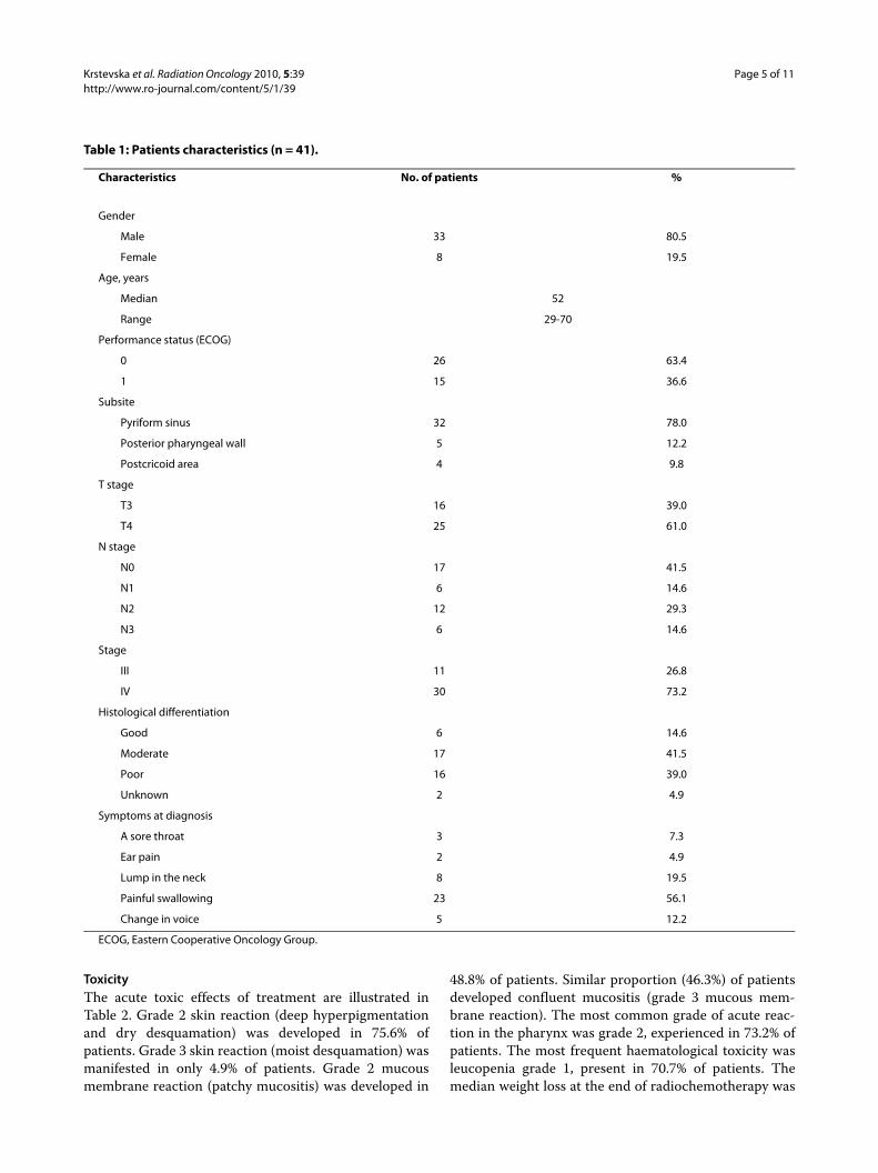
ResultsPatients characteristicsThe patient characteristics are described in Table 1. Themedian age was 52 years (range, 29-70), with a male pre-dominance (80.5%). The most common primary subsitewas pyriform sinus, present in 78.0% of patients. Locallyadvanced primary tumour (T4) was recognized in 25patients (61.0%). More than one-half of the patients werediagnosed with metastatic neck nodes. Clinically negativeneck (N0) was present in 17 patients (41.5%). In the wholenumber of patients more than two-thirds were seen withstage IV disease (73.2%). Moderate histological differenti-ation was present in 17 patients (41.5%). The most fre-quent symptom at diagnosis was painful swallowingpresent in 56.1% of patients.
Compliance of treatmentAll treated patients received the full planned dose ofradiotherapy (70 Gy). In 36 patients (87.8%), the overalltreatment time (OTT) for radiotherapy completion was ≤7 weeks. Photon-electron treatment was realized in 13patients (31.7%). The rest 28 patients (68.3%) were irradi-ated using the technique with oblique photon fields.Twenty-two patients completed all seven cycles of con-current chemotherapy. Six cycles of cisplatin was given in16 patients, while 3 patients had less than six cycles of cis-platin with patients' refusal being the only cause for con-current chemotherapy cessation. The mean total dose ofcisplatin given was 192 mg/m2 ± 23.2 SD.
Response to treatmentA complete response at the primary site occurred in 28patients (68.3%). In patients with positive neck theachieved complete response rate was 36.6%. A completecomposite response was present in 27 patients (65.9%). Apartial composite response was registered in 14 patients(34.1%). Of those, only one patient had a completeresponse of the primary tumor and a partial response ofthe nodal disease. In all other patients, there was a partialresponse at the primary site and at the neck region.
Patterns of failureMedian patient follow-up at the commencement of theanalysis was 13 months (range 7-36). Local recurrencewas developed in 3 patients, 1 patient developed regionalrecurrence, and 4 patients developed both. Distantmetastases were the predominant initial failure occurredin 7 patients and accounting for 46.7% of the cases whomanifested a relapse of the disease. Distant metastaseswere also developed in 3 patients who had not achievedcomplete composite response following treatment.Hence, the overall incidence of distant metastases was24.4% (10/41). Not one patient had an identification ofdistant metastatic disease preceded by the occurrence oflocal and/or regional recurrence. The most frequent siteof distant metastases (80.0%) was the lungs. The mediantime to development of local recurrence was 12 months(range 4-19). Regional recurrence developed as a singleevent occurred at 7 months after beginning of treatment.The median time to occurrence of locoregional recur-rence and distant metastases was 10.5 months (range 9-19) and 9.5 months (range 4-21), respectively.
SurvivalAt the time of analysis, 19 patients were alive. Amongthose, 1 patient had recurrence in the neck nodes, 1patient had recurrence of the primary tumour, 2 patientshad recurrence of the primary and of the nodal disease, 4patients had distant metastases, and 11 patients werealive free of disease. During the follow-up period, due totumor progression, a tracheotomy had been performed in8 patients, 3 patients required a placement of a feedingtube, and in other 3 patients a gastrostomy had been per-formed. Death was disease-related in vast majority ofpatients (21/22). Three patients had died of local orlocoregional recurrence, 1 patient had unknown cause ofdeath, 12 patients had died of the progression of theirpersistent disease, 3 patients had died of distant metasta-ses, and 3 patients had died due to both persistent diseaseand distant metastases.
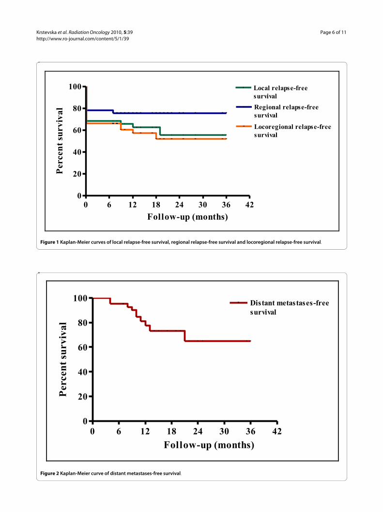
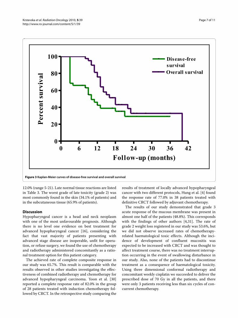
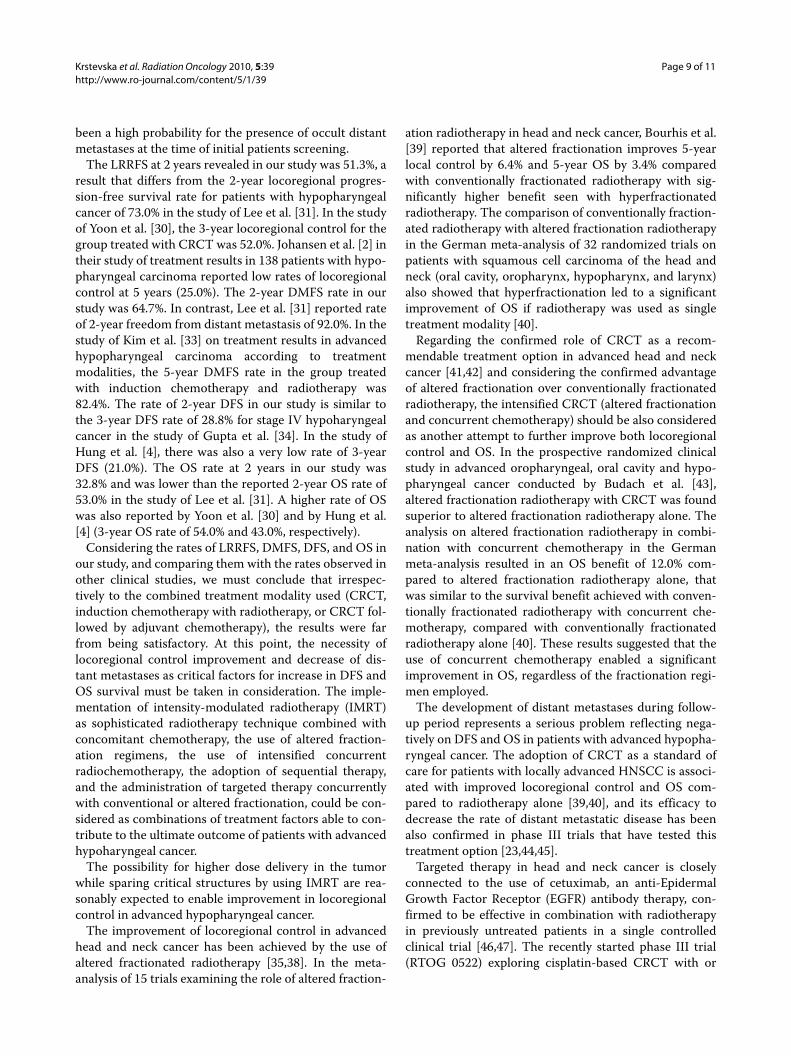
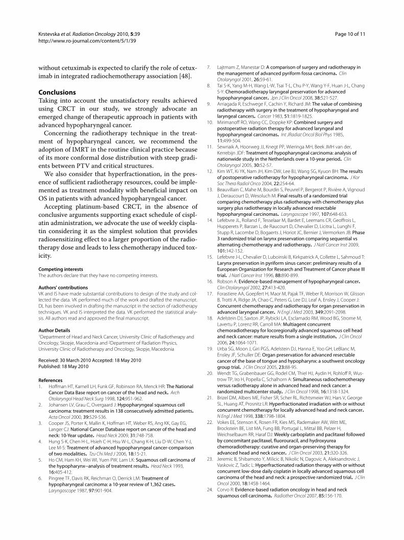
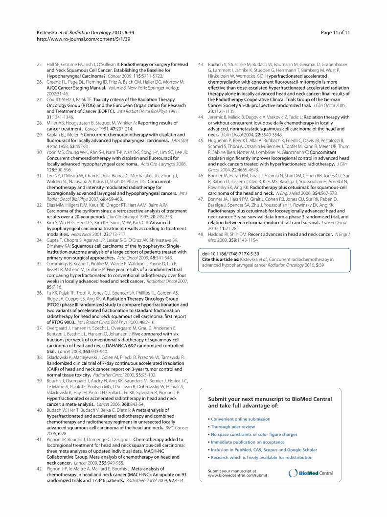
The 2-year LRFS and RRFS survival rates were 55.2%and 75.8%, respectively (Figure 1). The median durationof LRFS was 10 months (range 0-36) and the medianduration of RRFS was 12 months (range 0-36). TheLRRFS at 2 years was 51.3% (Figure 1). The median dura-tion of LRRFS was 10 months (range 0-36). The DMFS at2 years was 64.7% (Figure 2). The median duration ofDMFS was 12 months (range 4-36). The 2-year DFS andOS survival rates were 29.3% and 32.8%, respectively (Fig-ure 3). The median duration of DFS was 9 months (range3-36) and the median duration of OS was 14 months(range 7-36).
Krstevska et al. Radiation Oncology 2010, 5:39http://www.ro-journal.com/content/5/1/39
Page 5 of 11
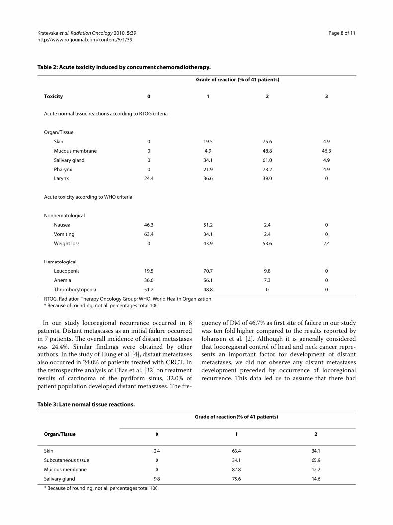
ToxicityThe acute toxic effects of treatment are illustrated inTable 2. Grade 2 skin reaction (deep hyperpigmentationand dry desquamation) was developed in 75.6% ofpatients. Grade 3 skin reaction (moist desquamation) wasmanifested in only 4.9% of patients. Grade 2 mucousmembrane reaction (patchy mucositis) was developed in
48.8% of patients. Similar proportion (46.3%) of patientsdeveloped confluent mucositis (grade 3 mucous mem-brane reaction). The most common grade of acute reac-tion in the pharynx was grade 2, experienced in 73.2% ofpatients. The most frequent haematological toxicity wasleucopenia grade 1, present in 70.7% of patients. Themedian weight loss at the end of radiochemotherapy was
Table 1: Patients characteristics (n = 41).
Characteristics No. of patients %
Gender
Male 33 80.5
Female 8 19.5
Age, years
Median 52
Range 29-70
Performance status (ECOG)
0 26 63.4
1 15 36.6
Subsite
Pyriform sinus 32 78.0
Posterior pharyngeal wall 5 12.2
Postcricoid area 4 9.8
T stage
T3 16 39.0
T4 25 61.0
N stage
N0 17 41.5
N1 6 14.6
N2 12 29.3
N3 6 14.6
Stage
III 11 26.8
IV 30 73.2
Histological differentiation
Good 6 14.6
Moderate 17 41.5
Poor 16 39.0
Unknown 2 4.9
Symptoms at diagnosis
A sore throat 3 7.3
Ear pain 2 4.9
Lump in the neck 8 19.5
Painful swallowing 23 56.1
Change in voice 5 12.2
ECOG, Eastern Cooperative Oncology Group.
Krstevska et al. Radiation Oncology 2010, 5:39http://www.ro-journal.com/content/5/1/39
Page 6 of 11
Figure 1 Kaplan-Meier curves of local relapse-free survival, regional relapse-free survival and locoregional relapse-free survival.
0 6 12 18 24 30 36 420
20
40
60
80
100 Local relapse-freesurvivalRegional relapse-freesurvival
Locoregional relapse-freesurvival
Follow-up (months)
Perc
ent s
urvi
val
Figure 2 Kaplan-Meier curve of distant metastases-free survival.
0 6 12 18 24 30 36 420
20
40
60
80
100 Distant metastases-freesurvival
Follow-up (months)
Perc
ent s
urvi
val
Krstevska et al. Radiation Oncology 2010, 5:39http://www.ro-journal.com/content/5/1/39
Page 7 of 11
12.0% (range 5-21). Late normal tissue reactions are listedin Table 3. The worst grade of late toxicity (grade 2) wasmost commonly found in the skin (34.1% of patients) andin the subcutaneous tissue (65.9% of patients).
DiscussionHypopharyngeal cancer is a head and neck neoplasmwith one of the most unfavourable prognosis. Althoughthere is no level one evidence on best treatment foradvanced hypopharyngeal cancer [16], considering thefact that vast majority of patients presenting withadvanced stage disease are inoperable, unfit for opera-tion, or refuse surgery, we found the use of chemotherapyand radiotherapy administered concomitantly as a ratio-nal treatment option for this patient category.
The achieved rate of complete composite response inour study was 65.7%. This result is comparable with theresults observed in other studies investigating the effec-tiveness of combined radiotherapy and chemotherapy foradvanced hypopharyngeal carcinoma. Yoon et al. [30]reported a complete response rate of 82.0% in the groupof 28 patients treated with induction chemotherapy fol-lowed by CRCT. In the retrospective study comparing the
results of treatment of locally advanced hypopharyngealcancer with two different protocols, Hung et al. [4] foundthe response rate of 77.0% in 38 patients treated withdefinitive CRCT followed by adjuvant chemotherapy.
The results of our study demonstrated that grade 3acute response of the mucous membrane was present inalmost one half of the patients (48.8%). This correspondswith the findings of other authors [4,31]. The rate ofgrade 2 weight loss registered in our study was 53.6%, butwe did not observe increased rates of chemotherapy-related haematological toxic effects. Although the inci-dence of development of confluent mucositis wasexpected to be increased with CRCT and was thought toaffect treatment course, there was no treatment interrup-tion occurring in the event of swallowing disturbance inour study. Also, none of the patients had to discontinuetreatment as a consequence of haematological toxicity.Using three dimensional conformal radiotherapy andconcomitant weekly cisplatin we succeeded to deliver theprescribed dose of 70 Gy in all the patients, and therewere only 3 patients receiving less than six cycles of con-current chemotherapy.
Figure 3 Kaplan-Meier curves of disease-free survival and overall survival.
0 6 12 18 24 30 36 420
20
40
60
80
100 Disease-freesurvivalOverall survival
Follow-up (months)
Perc
ent s
urvi
val
Krstevska et al. Radiation Oncology 2010, 5:39http://www.ro-journal.com/content/5/1/39
Page 8 of 11
In our study locoregional recurrence occurred in 8patients. Distant metastases as an initial failure occurredin 7 patients. The overall incidence of distant metastaseswas 24.4%. Similar findings were obtained by otherauthors. In the study of Hung et al. [4], distant metastasesalso occurred in 24.0% of patients treated with CRCT. Inthe retrospective analysis of Elias et al. [32] on treatmentresults of carcinoma of the pyriform sinus, 32.0% ofpatient population developed distant metastases. The fre-
quency of DM of 46.7% as first site of failure in our studywas ten fold higher compared to the results reported byJohansen et al. [2]. Although it is generally consideredthat locoregional control of head and neck cancer repre-sents an important factor for development of distantmetastases, we did not observe any distant metastasesdevelopment preceded by occurrence of locoregionalrecurrence. This data led us to assume that there had
Table 2: Acute toxicity induced by concurrent chemoradiotherapy.
Grade of reaction (% of 41 patients)
Toxicity 0 1 2 3
Acute normal tissue reactions according to RTOG criteria
Organ/Tissue
Skin 0 19.5 75.6 4.9
Mucous membrane 0 4.9 48.8 46.3
Salivary gland 0 34.1 61.0 4.9
Pharynx 0 21.9 73.2 4.9
Larynx 24.4 36.6 39.0 0
Acute toxicity according to WHO criteria
Nonhematological
Nausea 46.3 51.2 2.4 0
Vomiting 63.4 34.1 2.4 0
Weight loss 0 43.9 53.6 2.4
Hematological
Leucopenia 19.5 70.7 9.8 0
Anemia 36.6 56.1 7.3 0
Thrombocytopenia 51.2 48.8 0 0
RTOG, Radiation Therapy Oncology Group; WHO, World Health Organization.* Because of rounding, not all percentages total 100.
Table 3: Late normal tissue reactions.
Grade of reaction (% of 41 patients)
Organ/Tissue 0 1 2
Skin 2.4 63.4 34.1
Subcutaneous tissue 0 34.1 65.9
Mucous membrane 0 87.8 12.2
Salivary gland 9.8 75.6 14.6
* Because of rounding, not all percentages total 100.
Krstevska et al. Radiation Oncology 2010, 5:39http://www.ro-journal.com/content/5/1/39
Page 9 of 11
been a high probability for the presence of occult distantmetastases at the time of initial patients screening.
The LRRFS at 2 years revealed in our study was 51.3%, aresult that differs from the 2-year locoregional progres-sion-free survival rate for patients with hypopharyngealcancer of 73.0% in the study of Lee et al. [31]. In the studyof Yoon et al. [30], the 3-year locoregional control for thegroup treated with CRCT was 52.0%. Johansen et al. [2] intheir study of treatment results in 138 patients with hypo-pharyngeal carcinoma reported low rates of locoregionalcontrol at 5 years (25.0%). The 2-year DMFS rate in ourstudy was 64.7%. In contrast, Lee et al. [31] reported rateof 2-year freedom from distant metastasis of 92.0%. In thestudy of Kim et al. [33] on treatment results in advancedhypopharyngeal carcinoma according to treatmentmodalities, the 5-year DMFS rate in the group treatedwith induction chemotherapy and radiotherapy was82.4%. The rate of 2-year DFS in our study is similar tothe 3-year DFS rate of 28.8% for stage IV hypoharyngealcancer in the study of Gupta et al. [34]. In the study ofHung et al. [4], there was also a very low rate of 3-yearDFS (21.0%). The OS rate at 2 years in our study was32.8% and was lower than the reported 2-year OS rate of53.0% in the study of Lee et al. [31]. A higher rate of OSwas also reported by Yoon et al. [30] and by Hung et al.[4] (3-year OS rate of 54.0% and 43.0%, respectively).
Considering the rates of LRRFS, DMFS, DFS, and OS inour study, and comparing them with the rates observed inother clinical studies, we must conclude that irrespec-tively to the combined treatment modality used (CRCT,induction chemotherapy with radiotherapy, or CRCT fol-lowed by adjuvant chemotherapy), the results were farfrom being satisfactory. At this point, the necessity oflocoregional control improvement and decrease of dis-tant metastases as critical factors for increase in DFS andOS survival must be taken in consideration. The imple-mentation of intensity-modulated radiotherapy (IMRT)as sophisticated radiotherapy technique combined withconcomitant chemotherapy, the use of altered fraction-ation regimens, the use of intensified concurrentradiochemotherapy, the adoption of sequential therapy,and the administration of targeted therapy concurrentlywith conventional or altered fractionation, could be con-sidered as combinations of treatment factors able to con-tribute to the ultimate outcome of patients with advancedhypoharyngeal cancer.
The possibility for higher dose delivery in the tumorwhile sparing critical structures by using IMRT are rea-sonably expected to enable improvement in locoregionalcontrol in advanced hypopharyngeal cancer.
The improvement of locoregional control in advancedhead and neck cancer has been achieved by the use ofaltered fractionated radiotherapy [35,38]. In the meta-analysis of 15 trials examining the role of altered fraction-
ation radiotherapy in head and neck cancer, Bourhis et al.[39] reported that altered fractionation improves 5-yearlocal control by 6.4% and 5-year OS by 3.4% comparedwith conventionally fractionated radiotherapy with sig-nificantly higher benefit seen with hyperfractionatedradiotherapy. The comparison of conventionally fraction-ated radiotherapy with altered fractionation radiotherapyin the German meta-analysis of 32 randomized trials onpatients with squamous cell carcinoma of the head andneck (oral cavity, oropharynx, hypopharynx, and larynx)also showed that hyperfractionation led to a significantimprovement of OS if radiotherapy was used as singletreatment modality [40].
Regarding the confirmed role of CRCT as a recom-mendable treatment option in advanced head and neckcancer [41,42] and considering the confirmed advantageof altered fractionation over conventionally fractionatedradiotherapy, the intensified CRCT (altered fractionationand concurrent chemotherapy) should be also consideredas another attempt to further improve both locoregionalcontrol and OS. In the prospective randomized clinicalstudy in advanced oropharyngeal, oral cavity and hypo-pharyngeal cancer conducted by Budach et al. [43],altered fractionation radiotherapy with CRCT was foundsuperior to altered fractionation radiotherapy alone. Theanalysis on altered fractionation radiotherapy in combi-nation with concurrent chemotherapy in the Germanmeta-analysis resulted in an OS benefit of 12.0% com-pared to altered fractionation radiotherapy alone, thatwas similar to the survival benefit achieved with conven-tionally fractionated radiotherapy with concurrent che-motherapy, compared with conventionally fractionatedradiotherapy alone [40]. These results suggested that theuse of concurrent chemotherapy enabled a significantimprovement in OS, regardless of the fractionation regi-men employed.
The development of distant metastases during follow-up period represents a serious problem reflecting nega-tively on DFS and OS in patients with advanced hypopha-ryngeal cancer. The adoption of CRCT as a standard ofcare for patients with locally advanced HNSCC is associ-ated with improved locoregional control and OS com-pared to radiotherapy alone [39,40], and its efficacy todecrease the rate of distant metastatic disease has beenalso confirmed in phase III trials that have tested thistreatment option [23,44,45].
Targeted therapy in head and neck cancer is closelyconnected to the use of cetuximab, an anti-EpidermalGrowth Factor Receptor (EGFR) antibody therapy, con-firmed to be effective in combination with radiotherapyin previously untreated patients in a single controlledclinical trial [46,47]. The recently started phase III trial(RTOG 0522) exploring cisplatin-based CRCT with or
Krstevska et al. Radiation Oncology 2010, 5:39http://www.ro-journal.com/content/5/1/39
Page 10 of 11
without cetuximab is expected to clarify the role of cetux-imab in integrated radiochemotherapy association [48].
ConclusionsTaking into account the unsatisfactory results achievedusing CRCT in our study, we strongly advocate anemerged change of therapeutic approach in patients withadvanced hypopharyngeal cancer.
Concerning the radiotherapy technique in the treat-ment of hypopharyngeal cancer, we recommend theadoption of IMRT in the routine clinical practice becauseof its more conformal dose distribution with steep gradi-ents between PTV and critical structures.
We also consider that hyperfractionation, in the pres-ence of sufficient radiotherapy resources, could be imple-mented as treatment modality with beneficial impact onOS in patients with advanced hypopharyngeal cancer.
Accepting platinum-based CRCT, in the absence ofconclusive arguments supporting exact schedule of cispl-atin administration, we advocate the use of weekly cispla-tin considering it as the simplest solution that providesradiosensitizing effect to a larger proportion of the radio-therapy dose and leads to less chemotherapy induced tox-icity.
Competing interestsThe authors declare that they have no competing interests.
Authors' contributionsVK and IS have made substantial contributions to design of the study and col-lected the data. VK performed much of the work and drafted the manuscript.DL has been involved in drafting the manuscript in the section of radiotherapytechniques. VK and IS interpreted the data. VK performed the statistical analy-sis. All authors read and approved the final manuscript.
Author Details1Department of Head and Neck Cancer, University Clinic of Radiotherapy and Oncology, Skopje, Macedonia and 2Department of Radiation Physics, University Clinic of Radiotherapy and Oncology, Skopje, Macedonia
References1. Hoffman HT, Karnell LH, Funk GF, Robinson RA, Menck HR: The National
Cancer Data Base report on cancer of the head and neck. Arch Otolaryngol Head Neck Surg 1998, 124:951-962.
2. Johansen LV, Grau C, Overgaard J: Hypopharyngeal squamous cell carcinoma: treatment results in 138 consecutively admitted patients. Acta Oncol 2000, 39:529-536.
3. Cooper JS, Porter K, Mallin K, Hoffman HT, Weber RS, Ang KK, Gay EG, Langer CJ: National Cancer Database report on cancer of the head and neck: 10-Year update. Head Neck 2009, 31:748-758.
4. Hung S-K, Chen H-L, Hsieh C-H, Hsu W-L, Chang K-H, Liu D-W, Chen Y-J, Lee M-S: Treatment of advanced hypopharyngeal cancer-comparison of two modalities. Tzu Chi Med J 2006, 18:15-21.
5. Ho CM, Ham KH, Wei WI, Yuen PW, Lam LK: Squamous cell carcinoma of the hypopharynx--analysis of treatment results. Head Neck 1993, 16:405-412.
6. Pingree TF, Davis RK, Reichman O, Derrick LM: Treatment of hypopharyngeal carcinoma: a 10-year review of 1,362 cases. Laryngoscope 1987, 97:901-904.
7. Lajtmam Z, Manestar D: A comparison of surgery and radiotherapy in the management of advanced pyriform fossa carcinoma. Clin Otolaryngol 2001, 26:59-61.
8. Tai S-K, Yang M-H, Wang L-W, Tsai T-L, Chu P-Y, Wang Y-F, Huan J-L, Chang S-Y: Chemoradiotherapy laryngeal preservation for advanced hypopharyngeal cancer. Jpn J Clin Oncol 2008, 38:521-527.
9. Arriagada R, Eschwege F, Cachin Y, Richard JM: The value of combining radiotherapy with surgery in the treatment of hypopharyngeal and laryngeal cancers. Cancer 1983, 51:1819-1825.
10. Mirimanoff RO, Wang CC, Doppke KP: Combined surgery and postoperative radiation therapy for advanced laryngeal and hypopharyngeal carcinomas. Int JRadiat Oncol Biol Phys 1985, 11:499-504.
11. Sewnaik A, Hoorweg JJ, Knegt PP, Wieringa MH, Beek JMH van der, Kerrebijn JDF: Treatment of hypopharyngeal carcinoma: analysis of nationwide study in the Netherlands over a 10-year period. Clin Otolaryngol 2005, 30:52-57.
12. Kim WT, Ki YK, Nam JH, Kim DW, Lee BJ, Wang SG, Kyuon BH: The results of postoperative radiotherapy for hypopharyngeal carcinoma. J Kor Soc Thera Radiol Onco 2004, 22:254-64.
13. Beauvillain C, Mahe M, Bourdin S, Peuvrel P, Bergerot P, Rivière A, Vignoud J, Deraucourt D, Wesoluch M: Final results of a randomized trial comparing chemotherapy plus radiotherapy with chemotherapy plus surgery plus radiotherapy in locally advanced resectable hypopharyngeal carcinomas. Laryngoscope 1997, 107:648-653.
14. Lefebvre JL, Rolland F, Tesselaar M, Bardet E, Leemans CR, Geoffrois L, Hupperets P, Barzan L, de Raucourt D, Chevalier D, Licitra L, Lunghi F, Stupp R, Lacombe D, Bogaerts J, Horiot JC, Bernier J, Vermorken JB: Phase 3 randomized trial on larynx preservation comparing sequential vs alternating chemotherapy and radiotherapy. J Natl Cancer Inst 2009, 101:142-152.
15. Lefebvre J-L, Chevalier D, Luboinski B, Kirkpatrick A, Collette L, Sahmoud T: Larynx preservation in pyriform sinus cancer: preliminary results of a European Organization for Research and Treatment of Cancer phase III trial. J Natl Cancer Inst 1996, 88:890-899.
16. Robson A: Evidence-based management of hypopharyngeal cancer. Clin Otolaryngol 2002, 27:413-420.
17. Forastiere AA, Goepfert H, Maor M, Pajak TF, Weber R, Morrison W, Glisson B, Trotti A, Ridge JA, Chao C, Peters G, Lee DJ, Leaf A, Ensley J, Cooper J: Concurrent chemotherapy and radiotherapy for organ preservation in advanced laryngeal cancer. N Engl J Med 2003, 349:2091-2098.
18. Adelstein DJ, Saxton JP, Rybicki LA, Esclamado RM, Wood BG, Strome M, Lavertu P, Lorenz RR, Carroll MA: Multiagent concurrent chemoradiotherapy for locoregionally advanced squamous cell head and neck cancer: mature results from a single institution. J Clin Oncol 2006, 24:1064-1071.
19. Urba SG, Moon J, Giri PGS, Adelstein DJ, Hanna E, Yoo GH, LeBlanc M, Ensley JF, Schuller DE: Organ preservation for advanced resectable cancer of the base of tongue and hypopharynx: a southwest oncology group trial. J Clin Oncol 2005, 23:88-95.
20. Wendt TG, Grabenbauer GG, Rodel CM, Thiel HJ, Aydin H, Rohloff R, Wus-trow TP, Iro H, Popella C, Schalhorn A: Simultaneous radiochemotherapy versus radiotherapy alone in advanced head and neck cancer: a randomized multicenter study. J Clin Oncol 1998, 16:1318-1324.
21. Brizel DM, Albers ME, Fisher SR, Scher RL, Richtsmeier WJ, Hars V, George SL, Huang AT, Prosnitz LR: Hyperfractionated irradiation with or without concurrent chemotherapy for locally advanced head and neck cancer. N Engl J Med 1998, 338:1798-1804.
22. Vokes EE, Stenson K, Rosen FR, Kies MS, Rademaker AW, Witt ME, Brockstein BE, List MA, Fung BB, Portugal L, Mittal BB, Pelzer H, Weichselbaum RR, Haraf DJ: Weekly carboplatin and paclitaxel followed by concomitant paclitaxel, fluorouracil, and hydroxyurea chemoradiotherapy: curative and organ-preserving therapy for advanced head and neck cancer. J Clin Oncol 2003, 21:320-326.
23. Jeremic B, Shibamoto Y, Milicic B, Nikolic N, Dagovic A, Aleksandrovic J, Vaskovic Z, Tadic L: Hyperfractionated radiation therapy with or without concurrent low-dose daily cisplatin in locally advanced squamous cell carcinoma of the head and neck: a prospective randomized trial. J Clin Oncol 2000, 18:1458-1464.
24. Corvo R: Evidence-based radiation oncology in head and neck squamous cell carcinoma. Radiother Oncol 2007, 85:156-170.
Received: 30 March 2010 Accepted: 18 May 2010 Published: 18 May 2010This article is available from: http://www.ro-journal.com/content/5/1/39© 2010 Krstevska et al; licensee BioMed Central Ltd. This is an Open Access article distributed under the terms of the Creative Commons Attribution License (http://creativecommons.org/licenses/by/2.0), which permits unrestricted use, distribution, and reproduction in any medium, provided the original work is properly cited.Radiation Oncology 2010, 5:39
Krstevska et al. Radiation Oncology 2010, 5:39http://www.ro-journal.com/content/5/1/39
Page 11 of 11
25. Hall SF, Groome PA, Irish J, O'Sullivan B: Radiotherapy or Surgery for Head and Neck Squamous Cell Cancer. Establishing the Baseline for Hypopharyngeal Carcinoma? Cancer 2009, 115:5711-5722.
26. Greene FL, Page DL, Fleming ID, Fritz A, Balch CM, Haller DG, Morrow M: AJCC Cancer Staging Manual. Volume 6. New York: Springer-Verlag; 2002:31-46.
27. Cox JD, Stetz J, Pajak TF: Toxicity criteria of the Radiation Therapy Oncology Group (RTOG) and the European Organization for Research and Treatment of Cancer (EORTC). Int J Radiat Oncol Biol Phys 1995, 31:1341-1346.
28. Miller AB, Hoogstraten B, Staquet M, Winkler A: Reporting results of cancer treatment. Cancer 1981, 47:207-214.
29. Kaplan EL, Meier P: Concurrent chemoradiotherapy with cisplatin and fluorouracil for locally advanced hypopharyngeal carcinoma. J Am Stat Assoc 1958, 53:457-81.
30. Yoon MS, Chung W-K, Ahn S-J, Nam T-K, Nah B-S, Song J-Y, Lim SC, Lee JK: Concurrent chemoradiotherapy with cisplatin and fluorouracil for locally advanced hypopharyngeal carcinoma. Acta Oto-Laryngol 2008, 128:590-596.
31. Lee NY, O'Meara W, Chan K, Della-Bianca C, Mechalakos JG, Zhung J, Wolden SL, Narayana A, Kraus D, Shah JP, Pfister DG: Concurrent chemotherapy and intensity-modulated radiotherapy for locoregionally advanced laryngeal and hypopharyngeal cancers. Int J Radiat Oncol Biol Phys 2007, 69:459-468.
32. Elias MM, Hilgers FJM, Keus RB, Gregor RT, Hart AAM, Balm AJM: Carcinoma of the pyriform sinus: a retrospective analysis of treatment results over a 20-year period. Clin Otolaryngol 1995, 20:249-253.
33. Kim S, Wu H-G, Heo D-S, Kim KH, Sung M-W, Park C II: Advanced hypopharyngeal carcinoma treatment results according to treatment modalities. Head Neck 2001, 23:713-717.
34. Gupta T, Chopra S, Agarwal JP, Laskar S-G, D'Cruz AK, Shrivastava SK, Dinshaw KA: Squamous cell carcinoma of the hypopharynx: Single-institution outcome analysis of a large cohort of patients treated with primary non-surgical approaches. Acta Oncol 2009, 48:541-548.
35. Cummings B, Keane T, Pintilie M, Warde P, Waldron J, Payne D, Liu F, Bissett R, MsLean M, Gullane P: Five year results of a randomized trial comparing hyperfractionated to conventional radiotherapy over four weeks in locally advanced head and neck cancer. Radiother Oncol 2007, 85:7-16.
36. Fu KK, Pajak TF, Trotti A, Jones CU, Spencer SA, Phillips TL, Garden AS, Ridge JA, Cooper JS, Ang KK: A Radiation Therapy Oncology Group (RTOG) phase III randomized study to compare hyperfractionation and two variants of accelerated fractionation to standard fractionation radiotherapy for head and neck squamous cell carcinoma: first report of RTOG 9003. Int J Radiat Oncol Biol Phys 2000, 48:7-16.
37. Overgaard J, Hansen H, Specht L, Overgaard M, Grau C, Andersen E, Bentzen J, Bastholt L, Hansen O, Johansen J: Five compared with six fractions per week of conventional radiotherapy of squamous-cell carcinoma of head and neck: DAHANCA 6&7 randomized controlled trial. Lancet 2003, 363:933-940.
38. Skladowski K, Maciejewski J, Golen M, Pilecki B, Przeorek W, Tarnawski R: Randomized clinical trial of 7-day continuous accelerated irradiation (CAIR) of head and neck cancer: report on 3-year tumor control and normal tissue toxicity. Radiother Oncol 2000, 55:93-102.
39. Bourhis J, Overgaard J, Audry H, Ang KK, Saunders M, Bernier J, Horiot J-C, Le Maitre A, Pajak TF, Poulsen MG, O'Sullivan B, Dobrowsky W, Hliniak A, Skladowski K, Hay JH, Pinto LHJ, Fallai C, Fu KK, Sylvester R, Pignon J-P: Hyperfractionated or accelerated radiotherapy in head and neck cancer: a meta-analysis. Lancet 2006, 368:843-54.
40. Budach W, Her T, Budach V, Belka C, Dietz K: A meta-analysis of hyperfractionated and accelerated radiotherapy and combined chemotherapy and radiotherapy regimens in unresected locally advanced squamous cell carcinoma of the head and neck. BMC Cancer 2006, 6:28.
41. Pignon JP, Bourhis J, Domenge C, Designe L: Chemotherapy added to locoregional treatment for head and neck squamous-cell carcinoma: three meta analyses of updated individual data. MACH-NC Collaborative Group. Meta-analysis of chemotherapy on head and neck cancer. Lancet 2000, 355:949-955.
42. Pignon J-P, le Maitre A, Maillard E, Bourhis J: Meta-analysis of chemotherapy in head and neck cancer (MACH-NC): An update on 93 randomized trials and 17,346 patients. Radiother Oncol 2009, 92:4-14.
43. Budach V, Stuschke M, Budach W, Baumann M, Geismar D, Grabenbauer G, Lammert I, Jahnke K, Stueben G, Herrmann T, Bamberg M, Wust P, Hinkelbein W, Wernecke K-D: Hyperfractionated accelerated chemoradiation with concurrent fluorouracil-mitomycin is more effective than dose-escalated hyperfractionated accelerated radiation therapy alone in locally advanced head and neck cancer: final results of the Radiotherapy Cooperative Clinical Trials Group of the German Cancer Society 95-06 prospective randomized trial. J Clin Oncol 2005, 23:1125-1135.
44. Jeremic B, Milicic B, Dagovic A, Vaskovic Z, Tadic L: Radiation therapy with or without concurrent low-dose daily chemotherapy in locally advanced, nonmetastatic squamous cell carcinoma of the head and neck. J Clin Oncol 2004, 22:3540-3548.
45. Huguenin P, Beer KT, Allal A, Rufibach K, Friedli C, Davis JB, Pestalozzi B, Schmid S, Thöni A, Ozsahin M, Bernier J, Töpfer M, Kann R, Meier UR, Thum P, Sabine Bieri, Notter M, Lombriser N, Glanzmann C: Concomitant cisplatin significantly improves locoregional control in advanced head and neck cancers treated with hyperfractionated radiotherapy. J Clin Oncol 2004, 22:4665-4673.
46. Bonner JA, Harari PM, Giralt J, Azarnia N, Shin DM, Cohen RB, Jones CU, Sur R, Raben D, Jassem J, Ove R, Kies MS, Baselga J, Youssoufian H, Amellal N, Rowinsky EK, Ang KK: Radiotherapy plus cetuximab for squamous-cell carcinoma of the head and neck. N Engl J Med 2006, 354:567-578.
47. Bonner JA, Harari PM, Giralt J, Cohen RB, Jones CU, Sur RK, Raben D, Baselga J, Spencer SA, Zhu J, Youssoufian H, Rowinsky EK, Ang KK: Radiotherapy plus cetuximab for locoregionally advanced head and neck cancer: 5-year survival data from a phase 3 randomised trial, and relation between cetuximab-induced rash and survival. Lancet Oncol 2010, 11:21-28.
48. Haddad RI, Shin DM: Recent advances in head and neck cancer. N Engl J Med 2008, 359:1143-1154.
doi: 10.1186/1748-717X-5-39Cite this article as: Krstevska et al., Concurrent radiochemotherapy in advanced hypopharyngeal cancer Radiation Oncology 2010, 5:39