molecular imaging of inflammation in atherosclerosis with targeted ultrasound detection of vascular...
TRANSCRIPT

Molecular Imaging of Inflammation in Atherosclerosis WithTargeted Ultrasound Detection of Vascular Cell
Adhesion Molecule-1Beat A. Kaufmann, MD; John M. Sanders, BS; Christopher Davis, MD; Aris Xie, MS;
Patrick Aldred, MS; Ian J. Sarembock, MB, ChB; Jonathan R. Lindner, MD
Background—The ability to image vascular inflammatory responses may allow early diagnosis and treatment ofatherosclerosis. We hypothesized that molecular imaging of vascular cell adhesion molecule-1 (VCAM-1) expressionwith contrast-enhanced ultrasound (CEU) could be used for this purpose.
Methods and Results—Attachment of VCAM-1–targeted and control microbubbles to cultured endothelial cells wasassessed in a flow chamber at variable shear stress (0.5 to 12.0 dynes/cm2). Microbubble attachment to aortic plaque wasdetermined by en face microscopy of the thoracic aorta 10 minutes after intravenous injection in wild-type orapolipoprotein E–deficient mice on either chow or hypercholesterolemic diet. CEU molecular imaging of the thoracicaorta 10 minutes after intravenous microbubble injection was performed for the same animal groups. VCAM-1–targetedbut not control microbubbles attached to cultured endothelial cells, although firm attachment at the highest shear rates(8 to 12 dynes/cm2) occurred only in pulsatile flow conditions. Aortic attachment of microbubbles and targeted CEUsignal was very low in control wild-type mice on chow diet. Aortic attachment of microbubbles and CEU signal forVCAM-1–targeted microbubbles differed between treatment groups according to extent of VCAM-1–positive plaqueformation (median CEU videointensity, 1.8 [95% CI, 1.2 to 1.7], 3.7 [95% CI, 2.9 to 7.3], 6.8 [95% CI, 3.9 to 7.6], and11.2 [95% CI, 8.5 to 16.0] for wild-type mice on chow and hypercholesterolemic diet and for apolipoprotein E–deficientmice on chow and hypercholesterolemic diet, respectively; P�0.001).
Conclusions—CEU molecular imaging of VCAM-1 is capable of rapidly quantifying vascular inflammatory changes thatoccur in different stages of atherosclerosis. This method may be potentially useful for early risk stratification accordingto inflammatory phenotype. (Circulation. 2007;116:276-284.)
Key Words: atherosclerosis � echocardiography � imaging � inflammation
Atherosclerosis is a chronic inflammatory disorder thatoften progresses silently for decades before becoming
clinically evident.1 In current clinical practice, C-reactivepeptide is the only inflammatory marker routinely used forrisk assessment in patients. Noninvasive imaging of vascularchanges such as coronary calcification, carotid intima-mediathickening, and plaque morphology has recently been used toassess patient risk.2–6 However, these methods detect changesthat occur relatively late in the disease process and do notdirectly assess inflammatory status. Because inflammationparticipates in plaque initiation and progression, a methodcapable of imaging the extent of vascular inflammation couldpotentially provide powerful predictive information on bothearly disease presence and future risk for disease progression.At latter stages of disease, it also could provide informationon plaque vulnerability to erosion and rupture.7 It also isimportant to recognize that new therapies aimed at inhibiting
Clinical Perspective p 284
vascular inflammatory responses are being developed andwill likely be most effective when used in conjunction withquantitative methods that can detect early inflammatorychanges.
Vascular cell adhesion molecule-1 (VCAM-1), ex-pressed by activated endothelial cells, participates inleukocyte rolling and adhesion primarily by interactingwith its counterligand VLA-4 (�4�1) on monocytes andlymphocytes.8,9 VCAM-1 expression on the vessel endo-thelial surface or the underlying vasa vasorum plays animportant role in atherosclerotic plaque development bymonocyte and T-lymphocyte recruitment.10 It is an idealtarget for molecular imaging because there is little consti-tutive expression and its upregulation occurs at the earlieststages of atherogenesis.11,12 We hypothesized that molec-
Received December 15, 2006; accepted May 7, 2007.From the Cardiovascular Divisions of Oregon Health and Sciences University, Portland (B.A.K., A.X., P.A., J.R.L.); and the University of Virginia,
Charlottesville (J.M.S., C.D., I.J.S.).Correspondence to Jonathan R. Lindner, MD, Cardiovascular Division, UHN62, Oregon Health and Sciences University, 3181 SW Sam Jackson Park
Rd, Portland, OR 97239. E-mail [email protected]© 2007 American Heart Association, Inc.
Circulation is available at http://www.circulationaha.org DOI: 10.1161/CIRCULATIONAHA.106.684738
276
Imaging
by guest on June 21, 2015http://circ.ahajournals.org/Downloaded from

ular imaging of VCAM-1 with targeted contrast-enhancedultrasound (CEU) could be used to evaluate the degree ofvascular inflammation in atherosclerosis. CEU is wellsuited for such screening purposes because of practicalconsiderations such as cost, short duration of imagingprotocols (10 minutes), and balance between spatial reso-lution and sensitivity for targeted contrast agent detection.To test our hypothesis, attachment of VCAM-1–targetedmicrobubbles to endothelial cells was evaluated undervariable shear conditions. Microbubble attachment in vivoand signal enhancement of the aorta were assessed inanimal models of various degrees of atherosclerosis pro-duced by dietary intervention in wild-type and apolipopro-tein E– deficient (ApoE�/�) mice.
MethodsMicrobubble PreparationBiotinylated, lipid-shelled decafluorobutane microbubbles were pre-pared by sonication of a gas-saturated aqueous suspension ofdistearoylphosphatidylcholine, polyoxyethylene-40-stearate, anddistearoylphosphatidylethanolamine-PEG(2000)biotin. Rat anti-mouse monoclonal IgG1 against VCAM-1 (MK 2.7) or isotypecontrol antibody (R3–34, Pharmingen Inc, San Diego, Calif) wasconjugated to the surface of microbubbles as previously described toproduce VCAM-1–targeted (MBV) or control (MBc) microbubbles.13
For flow-chamber and in vivo attachment studies, microbubbleswere fluorescently labeled by the addition of either dioctadecyltet-ramethylindocarbocyanine (DiI) or dioctadecyloxacarbocyanine(DiO) perchlorate (Molecular Probes Inc, Carlsbad, Calif) to theaqueous suspension. Microbubble concentration was measured byelectrozone sensing (Multisizer III, Beckman Coulter, Fullerton,Calif).
Flow-Chamber Adhesion StudiesMurine endothelial cells (SVEC4–10, ATCC, Manassas, Va) thatexpress VCAM-1 were grown to confluence in Dulbecco’s modifiedEagle’s medium supplemented with 10% fetal bovine serum onfibronectin-coated culture dishes.14 For activation, cells were pre-treated with tumor necrosis factor-� (20 ng/mL) for 4 hours. Culturedishes were mounted on a parallel plate flow chamber (Glycotech,Gaithersburg, Md) with controlled gasket thickness and a channelwidth of 2.5 mm. The flow chamber was placed in an invertedposition on a microscope (Axioskop2-FS, Carl Zeiss Inc, Thornburg,NY) with a �40 objective and high-resolution charge-coupleddevice camera (C2400, Hamamatsu Photonics, Hamamatsu City,Japan) for video recording. A suspension of control or VCAM-1–targeted microbubbles (3�106 mL�1) in cell culture medium wasdrawn through the flow chamber with an adjustable withdrawalpump. The number of microbubbles attached to cells was determinedfor 20 optical fields (total area, 0.5 mm2) after 5 minutes ofcontinuous flow at rates to generate shear rates of 0.5 to 12.0dynes/cm2. Experiments were performed in triplicate as a minimum.Because aortic flow is pulsatile, adhesion at the highest shear rates (8and 12 dynes/cm2; n�6 for each) also was assessed after transient(5-second) reductions of shear to �0.5 dyne/cm2. This duration wasthe minimum required for significant flow reduction as a result of thecapacitance of the flow chamber system. Three sequential flowreductions were performed after 5 minutes of continuous flow, andmicrobubble attachment after each was determined once shear hadreturned to prepause levels.
Animal Models and PreparationThe study protocol was approved by the institutional AnimalResearch Committee. Twenty-six male wild-type C57Bl/6 and 23ApoE�/� mice (The Jackson Laboratory, Bar Harbor, Maine) werestudied at 22 to 24 weeks of age. Mice were fed either chow diet or,from 14 weeks of age on, a hypercholesterolemic diet (HCD)
containing 21% fat by weight, 0.15% cholesterol, and 19.5% caseinwithout sodium cholate. Anesthesia was induced with an intraperi-toneal injection (12.5 �L · g�1) of a solution containing ketaminehydrochloride (10 mg · mL�1), xylazine (1 mg · mL�1), and atropine(0.02 mg · mL�1). A jugular vein was cannulated for administrationof microbubbles.
Assessment of Microbubble Attachmentto the AortaIn anesthetized mice, VCAM-1–targeted and control microbubbles(1�106 for each) labeled with DiI and DiO, respectively, wereinjected simultaneously by intravenous route. After 10 minutes, aright atriotomy incision was made through an anterior thoracotomy.The blood volume was removed with 10 mL of 5% bovine serumalbumin containing heparin at 35°C to 37°C infused via a leftventricular puncture at an infusion pressure �100 mm Hg. The aortawas removed, a longitudinal incision was made, and the aorta waspinned flat on a microscopy platform. En face microscopy of theascending arch and descending portions of the thoracic aorta wasmade with a �20 objective. A minimum of 10 optical fields wereobserved under fluorescent epi-illumination at excitation wave-lengths of both 490 and 530 nm.
CEU ImagingUltrasound imaging (Sequoia, Siemens Medical Systems, MountainView, Calif) was performed with a high-frequency linear-array probeheld in place by a railed gantry system. The aortic arch and proximaldescending aorta arch were imaged from a left parasternal windowwith fundamental imaging at 14 MHz to optimize the imaging planein the longitudinal axis. CEU was performed with Contrast PulseSequencing (Siemens), which detects the nonlinear fundamentalsignal component for microbubbles. Imaging was performed at acenterline frequency of 7 MHz and a mechanical index of 0.2. Thegain was set just below visible speckle at baseline and held constant.Real-time imaging was performed 10 minutes after intravenousinjection of 1�106 MBC or MBV performed in random order. Afterseveral seconds of continuous imaging at a mechanical index of 0.2,microbubbles in the sector were destroyed by increasing the mechan-ical index to 1.0 for 1 second. Subsequent postdestruction imageswere acquired at a mechanical index of 0.2. To determine signal fromretained microbubbles alone, several postdestruction contrast framesrepresenting freely circulating microbubbles were averaged anddigitally subtracted from several averaged predestruction frames.13
Background-subtracted intensity was measured from a region ofinterest placed over the aorta with the 14-MHz image as a guide.
Because microbubble attachment is dependent on contact with theaortic wall, the axial distribution of microbubbles immediately afterinjection was assessed in 3 wild-type mice. Imaging was performedwith an ultra–high-frequency (30 MHz) mechanical sector imagingsystem (Vevo 770, VisualSonics Inc, Toronto, Ontario, Canada)during an intravenous injection of MBC (1�106). Ultrasound wastransmitted with 1-cycle pulses with an axial resolution of 55 �m.Images were aligned and displayed as a maximum-intensity proj-ection for 3 seconds after microbubble appearance.
Measurement of Ultrasound Pressure ProfileThe 2-dimensional display of ultrasound reflects information that isreceived from a 3-dimensional beam that has an out-of-planeelevational dimension. Therefore, it was necessary to determine theportion of the mouse aorta (imaged in long axis) that fit within thedetection profile of the elevational plane to determine whethercontrast material from the front and back walls would be detected.Acoustic pressures within the imaging sector were measured in awater bath with a needle hydrophone (PVDF-Z44, Specialty Engi-neering Associates, Sunnyvale, Calif) coupled with an oscilloscope(TDS-3012, Tektronix Inc, Beaverton, Ore). Peak negative acousticpressure measurements were made at the focal depth using thesystem settings for targeted imaging. A 2-dimensional pressureprofile was obtained by making 0.5-mm adjustments in the in-plane
Kaufmann et al Molecular Imaging of VCAM-1 in Atherosclerosis 277
by guest on June 21, 2015http://circ.ahajournals.org/Downloaded from
lateral dimension (beam width) and elevational dimension (beamthickness).
EchocardiographyThe peak flow velocity at the midarch was measured by pulsed-waveDoppler with a gate size at the minimum setting. Left ventricularsystolic function was assessed by imaging in the short-axis plane atthe midpapillary muscle level with fundamental imaging at 14 MHz.Fractional shortening in the anterior-posterior and septal-lateraldimensions was measured by video calipers and averaged.
ImmunohistologyImmunostaining for VCAM-1 was performed on paraffin-embeddedsections of the proximal and distal aortic arch after microwavetreatment with Antigen Unmasking Solution (Vector Laboratories,Burlingame, Calif) for several animals in each group. Goat poly-clonal antibody to human VCAM-1 with cross-reactivity for mouseVCAM-1 (sc1504, Santa Cruz Biotechnology Inc, Santa Cruz, Calif)was used as a primary antibody with a biotinylated secondaryanti-goat antibody (Vector Laboratories). Staining was performedwith a peroxidase kit (ABC Vectastain Elite, Vector Laboratories)and 3,3�-diaminobenzidine chromagen (Dako, Glostrup, Denmark).Slides were counterstained with hematoxylin.
Statistical AnalysisUnless otherwise specified, parametric data are expressed asmean�SD. Comparisons between microbubble agents for flowchamber studies were made by the Mann-Whitney rank sum test.Flow chamber data with sequential flow interruptions and targetedimaging data were analyzed with mixed-effect repeated-measuresmodels. For the latter, microbubble preparation and animal groupwere included as fixed factors, as well as the interaction between the2. Follow-up comparisons were made between bubble types for eachanimal group using a Bonferroni multiple-comparison adjustment.For microscopy data, individual comparisons were performed with1-way ANOVA and a Tukey post hoc test or, when appropriate, witha Kruskal-Wallis test with Dunn post-hoc test. Differences wereconsidered significant at P�0.05 (2 sided).
The authors had full access to and take full responsibility for theintegrity of the data. All authors have read and agree to themanuscript as written.
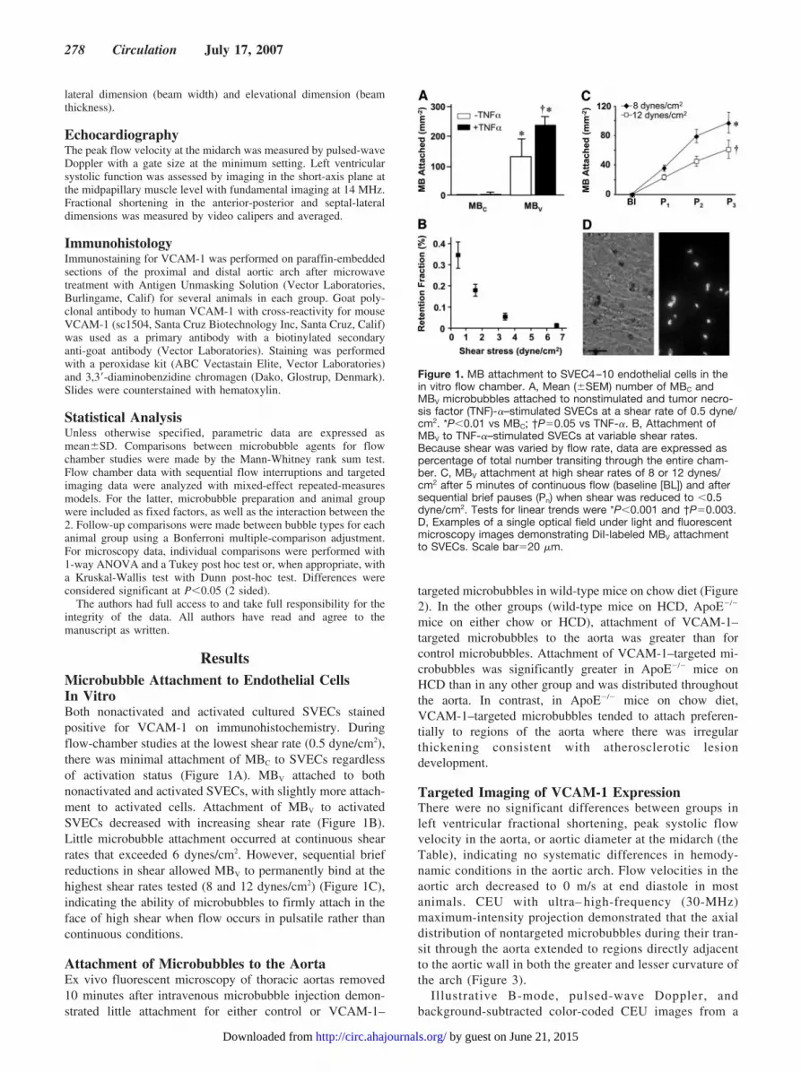
ResultsMicrobubble Attachment to Endothelial CellsIn VitroBoth nonactivated and activated cultured SVECs stainedpositive for VCAM-1 on immunohistochemistry. Duringflow-chamber studies at the lowest shear rate (0.5 dyne/cm2),there was minimal attachment of MBC to SVECs regardlessof activation status (Figure 1A). MBV attached to bothnonactivated and activated SVECs, with slightly more attach-ment to activated cells. Attachment of MBV to activatedSVECs decreased with increasing shear rate (Figure 1B).Little microbubble attachment occurred at continuous shearrates that exceeded 6 dynes/cm2. However, sequential briefreductions in shear allowed MBV to permanently bind at thehighest shear rates tested (8 and 12 dynes/cm2) (Figure 1C),indicating the ability of microbubbles to firmly attach in theface of high shear when flow occurs in pulsatile rather thancontinuous conditions.
Attachment of Microbubbles to the AortaEx vivo fluorescent microscopy of thoracic aortas removed10 minutes after intravenous microbubble injection demon-strated little attachment for either control or VCAM-1–
targeted microbubbles in wild-type mice on chow diet (Figure2). In the other groups (wild-type mice on HCD, ApoE�/�
mice on either chow or HCD), attachment of VCAM-1–targeted microbubbles to the aorta was greater than forcontrol microbubbles. Attachment of VCAM-1–targeted mi-crobubbles was significantly greater in ApoE�/� mice onHCD than in any other group and was distributed throughoutthe aorta. In contrast, in ApoE�/� mice on chow diet,VCAM-1–targeted microbubbles tended to attach preferen-tially to regions of the aorta where there was irregularthickening consistent with atherosclerotic lesiondevelopment.
Targeted Imaging of VCAM-1 ExpressionThere were no significant differences between groups inleft ventricular fractional shortening, peak systolic flowvelocity in the aorta, or aortic diameter at the midarch (theTable), indicating no systematic differences in hemody-namic conditions in the aortic arch. Flow velocities in theaortic arch decreased to 0 m/s at end diastole in mostanimals. CEU with ultra– high-frequency (30-MHz)maximum-intensity projection demonstrated that the axialdistribution of nontargeted microbubbles during their tran-sit through the aorta extended to regions directly adjacentto the aortic wall in both the greater and lesser curvature ofthe arch (Figure 3).
Illustrative B-mode, pulsed-wave Doppler, andbackground-subtracted color-coded CEU images from a
Figure 1. MB attachment to SVEC4–10 endothelial cells in thein vitro flow chamber. A, Mean (�SEM) number of MBC andMBV microbubbles attached to nonstimulated and tumor necro-sis factor (TNF)-�–stimulated SVECs at a shear rate of 0.5 dyne/cm2. *P�0.01 vs MBC; †P�0.05 vs TNF-�. B, Attachment ofMBV to TNF-�–stimulated SVECs at variable shear rates.Because shear was varied by flow rate, data are expressed aspercentage of total number transiting through the entire cham-ber. C, MBV attachment at high shear rates of 8 or 12 dynes/cm2 after 5 minutes of continuous flow (baseline [BL]) and aftersequential brief pauses (Pn) when shear was reduced to �0.5dyne/cm2. Tests for linear trends were *P�0.001 and †P�0.003.D, Examples of a single optical field under light and fluorescentmicroscopy images demonstrating DiI-labeled MBV attachmentto SVECs. Scale bar�20 �m.
278 Circulation July 17, 2007
by guest on June 21, 2015http://circ.ahajournals.org/Downloaded from

single ApoE�/� mouse on HCD are shown in Figure 4.Strong signal enhancement was observed for VCAM-1–targeted but not control microbubbles. The profile of thepeak negative acoustic pressures at the acoustic focus forthe transducer and settings used for targeted CEU areillustrated in Figure 5. According to the dimensions of theelevational plane, the entire volume of the aortic archwould be exposed to a peak negative acoustic pressure of�120 kPa before accounting for attenuation and �96 kPaafter correcting for attenuation assuming a coefficient of1.1 dB · mm�1 · MHz�1 (Figure 5).15 These data indicatethat the entire circumference of the aorta (circle in Figure
5B) would fit in the effective detection profile of eleva-tional plane. Hence, elevation plane averaging wouldpermit detection of microbubbles attached to the front orback wall that were seemingly “out of plane” and explainsthe appearance of targeted stationary microbubble signal inthe center of the apparent “lumen” in Figure 4. Figure 6summarizes CEU data for all groups. Signal enhancementfor control microbubbles was low and similar betweengroups. In wild-type mice on chow diet, signal for VCAM-1–targeted microbubbles was low and similar to that forcontrol microbubbles. In contrast, in all other groups, therewas greater signal enhancement for VCAM-1–targetedcompared with control microbubbles. Signal enhancementfor VCAM-1–targeted microbubbles increased incremen-tally from wild-type mice on HCD to ApoE�/� mice onchow to ApoE�/� mice on HCD. The interaction betweenmicrobubble agent and animal group was highly signifi-cant (P�0.0001), suggesting that the difference in signalfor control and VCAM-1–targeted microbubbles dependedon group.
0
40
80
120
160
mm(
dere
hd
Ael
bb
ub
orciM
2-)
†*
MBC
MBV
Apo E-/-HCD(n=3)
Apo E-/-Chow Diet
(n=4)
Wild typeHCD(n=4)
Wild typeChow Diet
(n=4)
†*
‡*
ApoE-/- chow (+thickening)ApoE-/- chow (-thickening)
WT chow ApoE-/- HCD
A
B
Figure 2. Microbubble attachment to the thoracic aorta 10 min-utes after intravenous injection assessed by ex vivo fluorescentmicroscopy. A, Mean (�SEM) attachment of MBC and MBV
microbubbles. *P�0.05 vs MBC; †P�0.05 vs MBV in wild-typemice on chow diet; ‡P�0.05 vs MBV in all other groups. B,Examples of en face fluorescent microscopy of the thoracicaorta after injection of DiI-labeled MBV. Examples of theApoE�/� mouse on chow diet are shown for regions with andwithout evidence for irregular wall thickening on transillumina-tion. Scale bar�25 �m.
Echocardiographic and Vascular Ultrasound Characteristics
Wild-Type Mice on Chow Diet(n�9)
Wild-Type Mice on HCD(n�10)
ApoE�/� Mice on Chow Diet(n�10)
ApoE�/� Mice on HCD(n�8)
Aortic peak velocity, m/s 0.53�0.10 0.52�0.7 0.46�0.13 0.50�0.14
Fractional shortening, % 0.35�0.04 0.35�0.05 0.34�0.05 0.38�0.04
Aortic diameter, mm 1.2�0.2 1.4�0.1 1.3�0.2* 1.4�0.00.3
Figure 3. Distribution of nontargeted microbubbles in transitthrough the aortic lumen assessed by high-frequency (30-MHz)CEU acquired at a frame rate of 20 Hz. A, Illustrations ofregions of interest spanning from position 1 (adjacent to thegreater curvature) to position 5 (adjacent to the lesser curva-ture). B through H, Maximum intensity projection images 400ms apart during appearance phase of microbubbles in the aortademonstrating diffuse distribution of microbubbles throughoutthe lumen. The graph at the bottom depicts CEU maximumintensity projection data for the different regions of interest. Datawere not significantly altered by analysis of frames only fromend systole or end diastole.
Kaufmann et al Molecular Imaging of VCAM-1 in Atherosclerosis 279
by guest on June 21, 2015http://circ.ahajournals.org/Downloaded from
ImmunohistochemistryOn histology, there was no evidence for plaque develop-ment in wild-type mice regardless of diet. On immuno-staining, however, VCAM-1 expression was detected onthe luminal endothelial surface of the aorta in wild-typemice on HCD (Figure 7). In ApoE�/� mice, there wasintimal thickening and large atherosclerotic plaques pro-truding into the lumen, particularly in animals on HCD.Immunohistochemistry in ApoE�/� mice demonstrateddense VCAM-1 expression on the endothelium, particu-larly overlying regions of plaque development. There alsowas VCAM-1 staining of neointimal monocytes. Thedegree of VCAM-1 staining on cells in the neointimaqualitatively correlated with the degree of endothelialstaining and was more robust in ApoE�/� mice when fed anHCD.
DiscussionThe critical role that inflammation plays in atherosclerosishas produced a swell of interest in better methods to evaluateit. Ideally, such a technique should be specific for inflamma-tory responses that occur in the vasculature, sufficientlysensitive to detect early events, able to provide spatialinformation, and practical in terms of cost, speed, and ease ofuse for application as a rapid screening tool. To that end, weinvestigated whether CEU molecular imaging could be usedto evaluate expression of the endothelial cell adhesion mol-ecule VCAM-1 in murine models of atherosclerosis. VCAM-1–targeted signal enhancement in the different animal groupsin this study varied according to the severity of atheroscle-rotic plaque development.
Molecular Imaging in AtherosclerosisA method for imaging vascular inflammation may have amajor impact in both the clinical and research laboratorysettings. Strategies currently used to evaluate risk ofcardiovascular disease or major adverse cardiac eventsmay not necessarily meet the clinical needs of the future,given the trend toward earlier and more aggressive ther-apy. The Framingham risk score and modifications thereoftake into account multiple different clinical variables.However, �40% of the adult US population fall into anintermediate-risk category16 with a 6% to 20% risk ofdeveloping symptomatic coronary heart disease within theensuing 10 years. Further refinement in risk stratificationfor this intermediate-risk category is desirable to makebetter use of long-term preventive therapies. There is alsothe notion that atherosclerosis, like many other diseases, ismost amenable to treatment at an early stage. Efforts areunderway to create novel therapies aimed at interruptingthe inflammatory events that initiate plaque formation andtrigger secondary growth responses. If treatment is to beinitiated years to decades before atherosclerosis wouldotherwise become clinically evident, then a method foraccurately detecting vascular inflammation would seem tobe a critical factor.
Methods currently used to evaluate those who havedeveloped symptoms of cardiovascular disease are de-signed to measure either the anatomic severity of diseaseor the physiological consequences of increased circuit
A
C
B
D
Ao
A
C
B
D
Ao
Figure 4. Representative images from an ApoE�/� mouse onHCD. A, The aortic arch by 2-dimensional ultrasound imaging(Ao). B, Pulsed-wave Doppler imaging of the arch. C and D,CEU images of the aortic arch 10 minutes after intravenousinjection of either MBV (C) or MBC (D). Color scale for the CEUimages is at the bottom of each frame, and each targeted imag-ing example is shown after correction for signal from freely cir-culating microbubbles.
Figure 5. Nonattenuated peak negative acousticpressure measurements at the focal depth for thelinear-array transducer used for targeted CEUimaging. A, Peak negative acoustic pressureaccording to in-plane lateral position and eleva-tional position. B, Elevational dimension powerprofile averaged from all lateral positions. Theaverage cross-sectional internal dimension of theaorta (1.3 mm) is superimposed on the elevationplane power profile, reflecting the orientation dur-ing long-axis imaging. All portions of the aortaare exposed to pressures �100 kPa, implyingthat microbubble contrast information from thefront and back walls of the aorta would bereceived and displayed.
280 Circulation July 17, 2007
by guest on June 21, 2015http://circ.ahajournals.org/Downloaded from

resistance such as ischemia or reduced flow reserve.Imaging the inflammatory phenotype in those patients willlikely add unique information because inflammation is akey factor in the progression to unstable disease. Therecruitment of inflammatory cells to the neointima results
in release of prothrombotic, promitogenic, proangiogenic,and detrimental vasoactive molecules; release of oxygen-derived free radicals; and production of proteases thatcontribute to adverse remodeling and erosion of the plaqueprotective barrier. It is necessary that new methods forevaluating inflammation occur in parallel with new thera-peutic strategies. Likewise, the use of molecular imagingin the preclinical development of therapies would providea means to assess the pathogenic pathways being targeted.For this application, a technique should be quantitative,have high-throughput capacity, and possess sufficientlyhigh-resolution for small animal model testing.
Microbubbles as a Targeting AgentIn this study, we chose to target microbubble contrast agentsto VCAM-1. Microbubble contrast agents are pure intravas-cular agents and accordingly do not have access to extravas-cular events or epitopes that have been proposed for targetingsuch as resident inflammatory cells (macrophages, T lympho-cytes), proteases, or oxidation byproducts.17–20 Instead, wetargeted an endothelial cell adhesion molecule that is acritical participant in inflammatory cell recruitment in ath-erosclerosis. VCAM-1 is present on endothelial cells earlyduring the development of atherosclerosis and is otherwiseexpressed only in very low levels.11,12 VCAM-1 has beeninvestigated as a potential target for molecular imaging inmice with other imaging techniques such as targeted infrared
0
5
10
15
20
Apo E-/-HCDn=8
Apo E-/-Chow Diet
n=10
Wild typeHCDn=11
Wild typeChow Diet
n=9
p=0.01 p=0.002
p<0.0001
ytisnet
nioe
diV
MBC
MBV
*
†
Figure 6. Background-subtracted CEU signal intensity from theaortic arch 10 minutes after intravenous injection of MBC andMBV microbubbles in the different animal groups. Data depictmedian value (horizontal line), 25% to 75% percentiles (box),and range of values (whiskers). The interaction between micro-bubble agent and animal group by mixed-effect repeated-measures analysis was significant (P�0.0001). *P�0.05 vs MBV
in wild-type mice on chow diet; †P�0.001 vs MBV in other ani-mal groups.
C D
FE
A B
Figure 7. Representative images of VCAM-1staining by immunohistochemistry of the tho-racic aorta. A, Wild-type mouse on chow dietdemonstrating minimal endothelial VCAM-1staining. B, Wild-type mouse on HCD demon-strating VCAM-1 expression localized to theluminal endothelial surface. C and D, ApoE�/�
mouse on chow diet demonstrating VCAM-1staining particularly on the endothelial surfaceoverlying regions of neointimal thickening. Eand F, ApoE�/� mouse on HCD demonstratingrobust VCAM-1 staining throughout the aortabut especially on the endothelial surface over-lying severe plaque formation and on cellswithin the neointima.
Kaufmann et al Molecular Imaging of VCAM-1 in Atherosclerosis 281
by guest on June 21, 2015http://circ.ahajournals.org/Downloaded from

and magnetic resonance probes.21,22 In these studies,VCAM-1 signal in advanced stages of disease decreased withstatin therapy, suggesting that the effects of therapy could bemonitored with molecular imaging.22 Information from mi-crobubble targeting is different from these diffusible tracersin that only endothelial VCAM-1 expression will be detected.
For targeting purposes, monoclonal antibodies againstthe extracellular domain of VCAM-1 were conjugated tothe surface of the microbubbles. This construct is charac-terized by an average of �50 000 antibodies per micro-bubble and a surface density of several thousand per 1�m2. Although an antibody-based strategy for targetinghas several advantages, including excellent bond dissoci-ation constants, it also has potential problems. One majorconcern is the relatively low bond formation rates forantibodies that could potentially preclude adequate attach-ment of rapidly transiting particles in high-shear vesselswhere contact time is short. Most of the previous studiesusing targeted microbubbles for molecular imaging haveconcentrated on molecular targets present in venules oftissues where wall shear stresses and flow velocities arerelatively low. In the mouse aorta, peak wall shear stresscan reach up to 80 to 90 dynes/cm2,23,24 and the pulsatilevariations in flow and thus wall shear stress are high.Despite this problem, there has been successful targetingof smaller echogenic liposomes to vascular surfaceepitopes in large animal models of atherosclerosis.25,26
These studies demonstrated conclusively that endothelialcell adhesion molecules could be targeted with acousti-cally active compounds, albeit with high-dose upstreamadministration.
In the present study, flow-chamber experiments demon-strated that VCAM-1–targeted microbubble attachmentefficiency was very low during continuous high shear.However, a marked increase in attachment occurred whenvery high shear was interrupted briefly. Resumption offlow at high shear stress did not dislodge these micro-bubbles even at the maximum shear rate (12 dynes/cm2)that could be withstood without detachment of the SVECsfrom fibronectin-coated plates. Flow-chamber experimentswith precipitated Fc-VCAM-1 chimera have demonstratedthe ability of VCAM-1–targeted microbubbles to firmlyadhere even at shear rates of 50 and 90 dynes/cm2
(unpublished data). However, our en face microscopystudies of the aortic arch 10 minutes after intravenousinjection of fluorescent microbubbles provided the bestevidence that microbubbles could attach in high density tothe aortic arch in vivo despite high peak shear stressesduring systole.
Evaluation of Disease SeverityMolecular imaging of VCAM-1 has the potential to diag-nose inflammatory processes that initiate atherosclerosislong before symptoms arise. Although temporal character-ization was not performed in this study, our data showingVCAM-1–targeted microbubble attachment and signal en-hancement in wild-type mice on HCD without evidence ofplaque development suggest that early inflammatorychanges can be detected. The finding that targeted micro-
bubble attachment and signal enhancement was muchgreater in ApoE�/� mice on HCD indicates that variousdegrees of inflammatory response can be discerned. Thesemice had not only the greatest extent of endothelialVCAM-1 expression but also the most severe form ofdisease in terms of plaque burden and the number ofVCAM-1– expressing cells (macrophages) within theplaque. In these mice, both CEU and en face microscopywere consistent with a diffuse and widespread attachmentof VCAM-1–targeted microbubbles, the density of whichwas within the dynamic range for detection of micro-bubbles attached to a 2-dimensional surface.27 The diffusenature of attachment suggests that a surrogate large vesselmay be used for evaluation when vascular inflammatorystatus is severe, although this was not directly tested. InApoE�/� mice on chow diet, attachment of microbubblestargeted to VCAM-1 was more pronounced in regions ofatherosclerotic plaque, consistent with reports on upregu-lation of VCAM-1 predominantly in regions prone toplaque development.11 In the control wild-type mice onchow diet, attachment of VCAM-1–targeted microbubbleswas not different from control microbubbles, reflectinglow or absent expression of VCAM-1.11,12 This latterfinding is important when considering the need for diseasespecificity (low false-positive rate) required for a screen-ing test.
Study LimitationsAlthough the spatial resolution of the CEU methods of thisstudy was adequate to localize signal enhancement to theaorta, it was not sufficient to determine whether VCAM-1–targeted microbubbles colocalized with plaque forma-tion in the ApoE�/� models. Likewise, on CEU imaging,we could not determine how much of the signal originatedfrom vasa vasorum or plaque neovascular endothelium.However, the ApoE�/� model does not necessarily recapit-ulate the degree of vasa vasoral proliferation in humans,and most of the adhesion on en face microscopy appearedto be on the luminal surface. In a related matter, theacoustic pressure profile of the beam elevational planeindicated that microbubbles attached anywhere in thecircumference of the aortic wall would be exposed to aminimum pressure of �96 kPa and should be detectablewithin the beam profile. Hence, microbubbles retained inareas other than the greater or lesser curvature appearedstationary in the center of the lumen. All of these limita-tions are related to scale, and it is anticipated that imagingin larger animal models or in humans will provide suchspatial information.
To the best of our knowledge, this study is the first touse low-power continuous imaging for in vivo targetedultrasound. Greater signal enhancement could be expectedfor high-power imaging. When microbubbles are ligated tocells, there can be physical impairment of microbubbleoscillation in the acoustic field and subsequent dampeningof their signal that appears to be less for high versus lowpower imaging.27 However, the potential for motion arti-fact in the mouse aorta from respiration on the initial
282 Circulation July 17, 2007
by guest on June 21, 2015http://circ.ahajournals.org/Downloaded from

imaging frame precluded the use of high-power imagingtechniques. In terms of shear forces, imaging was per-formed in anesthetized mice, and shear stress may havebeen lower than in conscious animals because of theeffects of anesthesia. On the basis of our Doppler andB-mode imaging data, we do not believe that there wereany significant differences in shear between the animalgroups. It is important to realize that one would expect aneven greater attachment efficiency in humans. The large-vessel shear stresses in humans are more than an order ofmagnitude lower than in mice,24 and low-shear vasavasoral vessels are more abundant.
ConclusionsThe results of this study indicate that contrast ultrasound withtargeted microbubbles can detect inflammatory processes inatherosclerosis and discriminate the severity of inflammatoryburden. Consequently, molecular imaging with targeted mi-crobubbles and ultrasound can potentially be useful in theearly diagnosis of atherosclerosis and in monitoring theefficacy of therapeutic interventions. Future studies areneeded to determine whether the inciting inflammatoryevents can be imaged and whether signal enhancement can bemodified with therapies aimed at quelling the inflammatoryresponse.
AcknowledgmentsWe are grateful to Lisa Tesch for her assistance with cell culture andto Dawn Peters for her assistance with statistical analysis.
Sources of FundingThis work was supported by grants (R01-HL-074443, R01-HL-078610 and R01-DK-063508) to Dr Lindner from the NationalInstitutes of Health, Bethesda, Md. Dr Kaufmann is supported byresearch grants from the Novartis Foundation and the LichtensteinFoundation.
DisclosuresDr Lindner is a member of the scientific advisory board forVisualSonics Inc. The other authors report no conflicts.
References1. Ross R. Atherosclerosis: an inflammatory disease. N Engl J Med. 1999;
340:115–126.2. Arad Y, Spadaro LA, Goodman K, Newstein D, Guerci AD. Pre-
diction of coronary events with electron beam computed tomography.J Am Coll Cardiol. 2000;36:1253–1260.
3. Greenland P, LaBree L, Azen SP, Doherty TM, Detrano RC. Coronaryartery calcium score combined with Framingham score for risk pre-diction in asymptomatic individuals. JAMA. 2004;291:210 –215.
4. Chambless LE, Folsom AR, Clegg LX, Sharrett AR, Shahar E, NietoFJ, Rosamond WD, Evans G. Carotid wall thickness is predictive ofincident clinical stroke: the Atherosclerosis Risk in Communities(ARIC) study. Am J Epidemiol. 2000;151:478 – 487.
5. O’Leary DH, Polak JF, Kronmal RA, Manolio TA, Burke GL,Wolfson SK Jr. Carotid-artery intima and media thickness as a riskfactor for myocardial infarction and stroke in older adults: Cardio-vascular Health Study Collaborative Research Group. N Engl J Med.1999;340:14 –22.
6. Leber AW, Becker A, Knez A, von Ziegler F, Sirol M, Nikolaou K,Ohnesorge B, Fayad ZA, Becker CR, Reiser M, Steinbeck G, Boek-stegers P. Accuracy of 64-slice computed tomography to classify andquantify plaque volumes in the proximal coronary system: a com-
parative study using intravascular ultrasound. J Am Coll Cardiol.2006;47:672– 677.
7. Virmani R, Burke AP, Farb A, Kolodgie FD. Pathology of the vul-nerable plaque. J Am Coll Cardiol. 2006;47(suppl):C13–C18.
8. Carlos T, Kovach N, Schwartz B, Rosa M, Newman B, Wayner E,Benjamin C, Osborn L, Lobb R, Harlan J. Human monocytes bind totwo cytokine-induced adhesive ligands on cultured human endothelialcells: endothelial-leukocyte adhesion molecule-1 and vascular celladhesion molecule-1. Blood. 1991;77:2266 –2271.
9. Huo Y, Hafezi-Moghadam A, Ley K. Role of vascular cell adhesionmolecule-1 and fibronectin connecting segment-1 in monocyte rollingand adhesion on early atherosclerotic lesions. Circ Res. 2000;87:153–159.
10. O’Brien KD, McDonald TO, Chait A, Allen MD, Alpers CE. Neo-vascular expression of E-selectin, intercellular adhesion molecule-1,and vascular cell adhesion molecule-1 in human atherosclerosis andtheir relation to intimal leukocyte content. Circulation. 1996 15;93:672– 682.
11. Nakashima Y, Raines EW, Plump AS, Breslow JL, Ross R. Upregu-lation of VCAM-1 and ICAM-1 at atherosclerosis-prone sites on theendothelium in the ApoE-deficient mouse. Arterioscler Thromb VascBiol. 1998;18:842– 851.
12. Iiyama K, Hajra L, Iiyama M, Li H, DiChiara M, Medoff BD,Cybulsky MI. Patterns of vascular cell adhesion molecule-1 andintercellular adhesion molecule-1 expression in rabbit and mouseatherosclerotic lesions and at sites predisposed to lesion formation.Circ Res. 1999;85:199 –207.
13. Lindner JR, Song J, Christiansen J, Klibanov AL, Xu F, Ley K.Ultrasound assessment of inflammation and renal tissue injury withmicrobubbles targeted to P-selectin. Circulation. 2001;104:2107–2112.
14. Sasaki M, Ostanin D, Elrod JW, Oshima T, Jordan P, Itoh M, Joh T,Minagar A, Alexander JS. TNF-alpha -induced endothelial celladhesion molecule expression is cytochrome P-450 monooxygenasedependent. Am J Physiol Cell Physiol. 2003;284:C422–C428.
15. Teotico GA, Miller RJ, Frizzell LA, Zachary JF, O’Brien W. Atten-uation coefficient estimates of mouse and rat chest wall. IEEE TransUltrason Ferroelectr Freq Control. 2001;48:593– 601.
16. Jacobson TA, Griffiths GG, Varas C, Gause D, Sung JC, BallantyneCM. Impact of evidence-based “clinical judgment” on the number ofAmerican adults requiring lipid-lowering therapy based on updatedNHANES III data: National Health and Nutrition ExaminationSurvey. Arch Intern Med. 2000;160:1361–1369.
17. Schafers M, Riemann B, Kopka K, Breyholz HJ, Wagner S, SchafersKP, Law MP, Schober O, Levkau B. Scintigraphic imaging of matrixmetalloproteinase activity in the arterial wall in vivo. Circulation.2004;109:2554 –2559.
18. Deguchi JO, Aikawa M, Tung CH, Aikawa E, Kim DE, NtziachristosV, Weissleder R, Libby P. Inflammation in atherosclerosis: visu-alizing matrix metalloproteinase action in macrophages in vivo. Cir-culation. 2006;114:55– 62.
19. Tsimikas S, Palinski W, Halpern SE, Yeung DW, Curtiss LK,Witztum JL. Radiolabeled MDA2, an oxidation-specific, monoclonalantibody, identifies native atherosclerotic lesions in vivo. J NuclCardiol. 1999;6:41–53.
20. Ruehm SG, Corot C, Vogt P, Kolb S, Debatin JF. Magnetic resonanceimaging of atherosclerotic plaque with ultrasmall superparamagneticparticles of iron oxide in hyperlipidemic rabbits. Circulation. 2001;103:415– 422.
21. Kelly KA, Allport JR, Tsourkas A, Shinde-Patil VR, Josephson L,Weissleder R. Detection of vascular adhesion molecule-1 expressionusing a novel multimodal nanoparticle. Circ Res. 2005;96:327–336.
22. Nahrendorf M, Jaffer FA, Kelly KA, Sosnovik DE, Aikawa E, LibbyP, Weissleder R. Noninvasive vascular cell adhesion molecule-1imaging identifies inflammatory activation of cells in atherosclerosis.Circulation. 2006;114:1504 –1511.
23. Eriksson EE, Werr J, Guo Y, Thoren P, Lindbom L. Direct obser-vations in vivo on the role of endothelial selectins and alpha(4)integrin in cytokine-induced leukocyte-endothelium interactions inthe mouse aorta. Circ Res. 2000;86:526 –533.
24. Greve JM, Les AS, Tang BT, Draney Blomme MT, Wilson NM,Dalman RL, Pelc NJ, Taylor CA. Allometric scaling of wall shearstress from mice to humans: quantification using cine phase-contrastMRI and computational fluid dynamics. Am J Physiol Heart CircPhysiol. 2006;291:H1700 –H1708.
Kaufmann et al Molecular Imaging of VCAM-1 in Atherosclerosis 283
by guest on June 21, 2015http://circ.ahajournals.org/Downloaded from

25. Hamilton AJ, Huang SL, Warnick D, Rabbat M, Kane B, Nagaraj A,Klegerman M, McPherson DD. Intravascular ultrasound molecularimaging of atheroma components in vivo. J Am Coll Cardiol. 2004;43:453– 460.
26. Demos SM, kan-Onyuksel H, Kane BJ, Ramani K, Nagaraj A, GreeneR, Klegerman M, McPherson DD. In vivo targeting of acoustically
reflective liposomes for intravascular and transvascular ultrasonicenhancement. J Am Coll Cardiol. 1999;33:867– 875.
27. Lankford M, Behm CZ, Yeh J, Klibanov AL, Robinson P, Lindner JR.Effect of microbubble ligation to cells on ultrasound signalenhancement: implications for targeted imaging. Invest Radiol. 2006;41:721–728.
CLINICAL PERSPECTIVEAtherosclerosis often progresses silently for decades before becoming clinically evident. Accordingly, there is growinginterest in the ability to detect the vascular inflammatory responses that play a critical role in plaque development andinstability. Specifically, methods are being developed to image molecular processes that participate in the pathogenesis ofcoronary and peripheral artery disease to detect disease early, to improve risk stratification, and to evaluate responses tonovel inflammation-targeted therapies. With contrast-enhanced ultrasound, our targeted probes are not able to reachcomponents within the plaque matrix. Hence, to evaluate disease activity in this study, we targeted microbubbles tovascular cell adhesion molecule-1, an endothelial cell adhesion molecule involved in leukocyte trafficking to atheroscle-rotic lesions. We studied 4 different mouse groups with different degrees of atherosclerotic lesion severity and vascular celladhesion molecule-1 expression. Microbubble attachment and signal enhancement were different between these groups andvaried according to disease severity. These findings indicate that contrast ultrasound molecular imaging of vascular celladhesion molecule-1 is capable of quantifying vascular inflammatory changes that occur in different stages ofatherosclerosis. This method may be potentially useful for early risk stratification according to inflammatory phenotype inpatients because it is both quantitative and specific for disease activity. Practical advantages of this approach are that it israpid, taking only minutes to perform, and relatively inexpensive.
284 Circulation July 17, 2007
by guest on June 21, 2015http://circ.ahajournals.org/Downloaded from
Sarembock and Jonathan R. LindnerBeat A. Kaufmann, John M. Sanders, Christopher Davis, Aris Xie, Patrick Aldred, Ian J.
Detection of Vascular Cell Adhesion Molecule-1Molecular Imaging of Inflammation in Atherosclerosis With Targeted Ultrasound
Print ISSN: 0009-7322. Online ISSN: 1524-4539 Copyright © 2007 American Heart Association, Inc. All rights reserved.
is published by the American Heart Association, 7272 Greenville Avenue, Dallas, TX 75231Circulation doi: 10.1161/CIRCULATIONAHA.106.6847382007;116:276-284; originally published online June 25, 2007;Circulation.
http://circ.ahajournals.org/content/116/3/276World Wide Web at:
The online version of this article, along with updated information and services, is located on the
http://circ.ahajournals.org//subscriptions/
is online at: Circulation Information about subscribing to Subscriptions:
http://www.lww.com/reprints Information about reprints can be found online at: Reprints:
document. Permissions and Rights Question and Answer this process is available in the
click Request Permissions in the middle column of the Web page under Services. Further information aboutOffice. Once the online version of the published article for which permission is being requested is located,
can be obtained via RightsLink, a service of the Copyright Clearance Center, not the EditorialCirculationin Requests for permissions to reproduce figures, tables, or portions of articles originally publishedPermissions:
by guest on June 21, 2015http://circ.ahajournals.org/Downloaded from